Embed Size (px)
Citation preview

A Model for the Functioning of the Striatum
Christopher I. Connolly
Computer Science Department
University of Massachusetts
Amherst, Massachusetts, 01003 USA
Tel. (413) 545-3492, FAX (413) 545-1249
J. Brian Burns
Teleos Research
576 Middle�eld Rd
Palo Alto, California, 94301
�
June 11, 1993
Abstract
A model is presented for the operation of the striatum. The model posits that the
basal ganglia are responsible for driving smooth transitions of state for an organism. We
propose that this is accomplished through the computation of a potential function within
the striatum on which a gradient descent is performed toward the goal state. The model
suggests that various somatotopic regions of the striatum correspond to state spaces, each
of which pertains to a di�erent aspect of the organism. This paper discusses this model
only in the context of motor control, i.e., egomotion and limb movement. The model
appears to account for a variety of experimental results, and for some unusual properties
of the striatum.
1 Introduction
The basal ganglia have been one of the more enigmatic structures in the brain. While they
have been extensively studied (DeLong and Georgopoulos 1981; Wilson, et al. 1990; Kandel,
et al. 1991), and have been very clearly implicated in motor function (Yahr 1976; Alexander,
et al. 1990), their exact function remains unclear.
�
Biological Cybernetics: 68(6):535-544.
1

In this paper, a theory is put forth on the mechanisms of the planning of goal-oriented and
obstacle-avoiding behavior in biological systems. It is argued here that the basal ganglia may
be an important locus for aspects of these activities. Furthermore, the mechanisms for basal
ganglia function may be characterized in part as the computation and storage of harmonic
functions. These functions are computed in parameter spaces associated with the positions
and movements of various body parts, and possibly additional representations of action and
intention.
1.1 Goals and obstacles: a class of behaviors and control problems
The problem considered in this paper is that of planning the purposeful state changes of a
mechanical system or organism. In other words, how does the system make its way from its
current state to some goal state, avoiding undesirable, or even dangerous states? This is a
general question which describes a variety of problems faced by di�erent organisms and the
designers of robot systems, e.g., in reaching and egomotion.
Research in cognitive psychology (see, for example, Neisser 1976) has suggested that, at
least for certain tasks, the brain is capable of spontaneously generating trajectories for limb-
and egomotion. Such path generation is not learned (Menzel 1973), but is dependent on the
environment. What part of the brain, then, is computing this information, and how is it
performing this computation?
1.2 Problems in Robotics
A related problem in robotics concerns how to build systems which are capable of deriving
collision-free paths from one point to another. We would like to further motivate our model by
considering what is known in robotics as the path-planning problem. There are many di�erent
types of robots, ranging from mobile robots which roam in a two-dimensional environment to
complex robot arms with several degrees of freedom. For each of these robot types, one can
describe robot position and motion in terms of the robot's con�guration space (Lozano-P�erez
1981, Udupa 1977). Con�guration space is the space of parameters which can be used to
describe the pose of the robot. For a small mobile robot, these parameters are the x and
y positions of the robot on the oor (with respect to some �xed coordinate frame). For a
jointed robot arm, the parameters are the angles of each joint. Note that any conventional
robot (e.g., �xed-base jointed robots) can be represented as a single point in its con�guration
space. Changing con�guration is represented by the movement of a point in con�guration
2

space. Put Figure 1
near here.
Several approaches have been taken for solving the path planning problem in robotics
(Schwartz and Sharir 1983; Khatib 1985; Arkin 1989; Canny and Lin 1990). Recent work
in robot path planning (Connolly, et al. 1990; Connolly and Grupen 1992) revolves around
the use of harmonic functions for complete robot path planning. Harmonic functions are
solutions to Laplace's Equation:
n
X
i=0
@
2
f
@x
2
i
= 0 (1)
which describes several physical phenomena including uid ow and the distribution of elec-
trical charge in a medium. In Equation 1, f is a harmonic function, n refers to the number of
variables for the function, and each x
i
is a function variable. This equation, then, says that
all second derivatives of the harmonic function f sum to zero. Analog computers have been
constructed to compute solutions to this equation, and consist of resistive grids (McCann and
Wilts 1949). Since Kirchho�'s Laws
1
represent a discrete form of Laplace's Equation, such
grids are able to compute discrete solutions to Laplace's Equation very rapidly as voltages.
Recently, Tarassenko and Blake (Tarassenko and Blake 1991) employed a resistive grid to
solve the path-planning problem for a mobile robot. The path-planning problem is primarily
concerned with qualitative solutions. Hence, resistances within the grid need not be precise,
nor does the grid need to be connected in a regular fashion. The former property is exploited
by Stan and Burleson (Stan and Burleson 1992) in their path-planning grid.
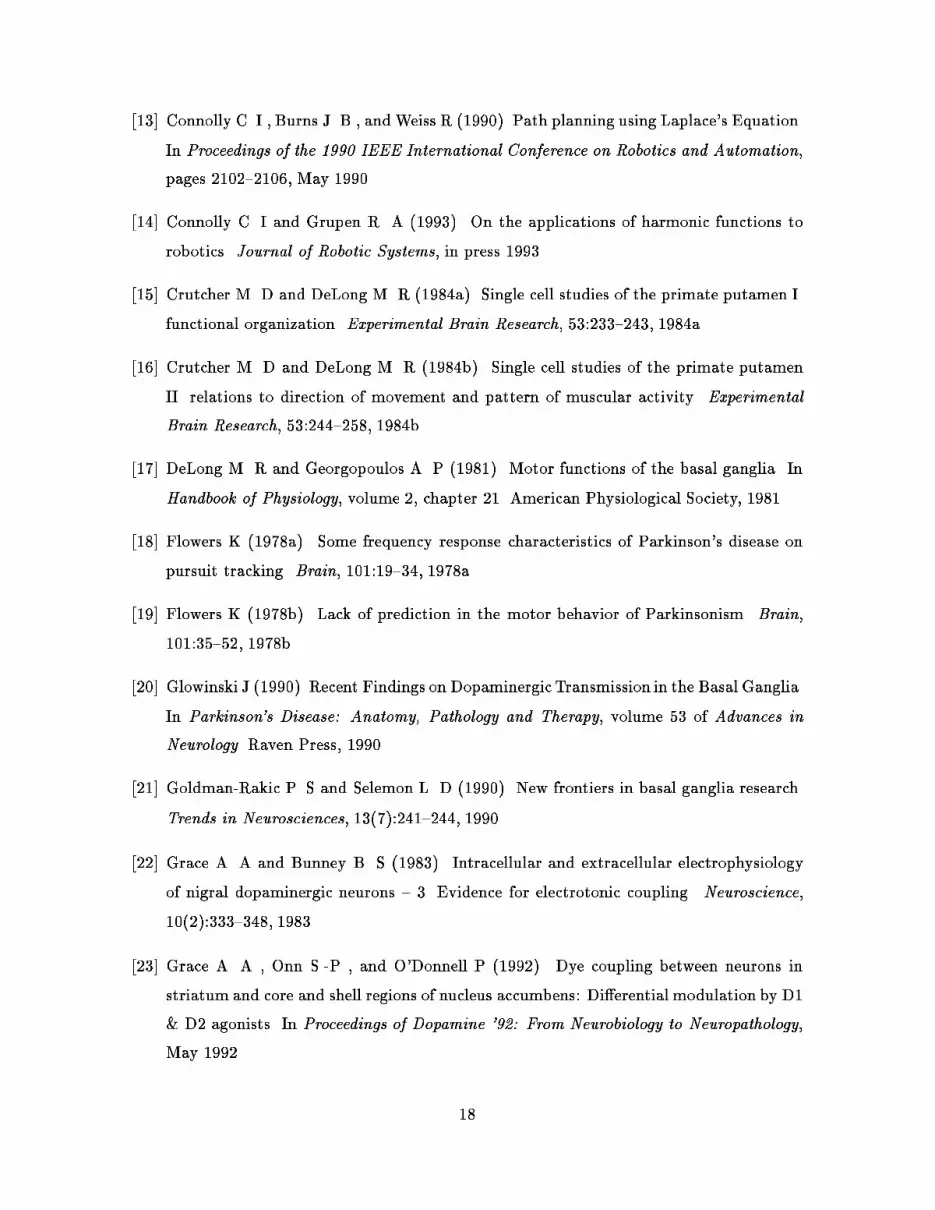
Figure 1 shows an example of a two-joint robot, its con�guration space, and a path
generated by a harmonic function. Shaded areas represent obstacles, while solid black areas
are goal regions. The left-hand side of the �gure shows the robot in its environment. Several
robot positions along the path have been superimposed so that the reader can see how the path
progresses. On the right-hand side is the con�guration space for the robot. The horizontal
axis represents the �rst joint angle, while the vertical axis is the second joint angle. In this
space, the robot is represented as a point. The shaded areas on the right-hand side are the
result of mapping obstacles from Cartesian space into the robot's con�guration space. Now
the problem of moving the robot between two con�gurations (left side) has been reduced to
determining a suitable path for a point (right side).
In the case shown in Figure 1, a harmonic function is computed over the free (white)
1
The governing laws for resistances in a circuit: Kirchho�'s current law states that current owing into a
junction must equal the current owing out of a junction.
3

regions of con�guration space. One can compute a harmonic function on a digital computer
simply by assigning high potentials to obstacles, low potentials to goals, and repeatedly
averaging points in the interior. If every free point's potential is replaced with the average of
its neighbors at each iteration (leaving goals and obstacles �xed at their initial potentials),
then after several iterations, the values over the whole free area will converge to a harmonic
function. Similarly, in a resistive network, the voltage at any free node after settling will be Put Figure 2
near here.
the average of the voltages at neighboring nodes. In a resistive network, obstacles may be
represented either by �xed high potentials (resulting in repulsion) or by increased resistances
(resulting in the absence of ow across obstacles). Mathematical literature refers to the
former case as the Dirichlet boundary condition, while the latter is known as the Neumann
boundary condition. In either case, the obstacle is avoided. For our model, it su�ces to think
of the majority of striatum cells as behaving in a manner similar to the resistive elements
described here. Figures 2 and 3 show a sample problem for the Dirichlet and Neumann
obstacle boundary conditions, respectively. Put Figure 3
near here.
Using a grid-based representation, the mapping from Cartesian to con�guration space
(e.g. joint angles) can easily be accomplished. Such mappings take into account the possible
collisions of the e�ector with other obstacles and with itself. Once this mapping is obtained,
it is permanent for a given e�ector, and can be used to set up obstacles and goals in state
space, given information about their three-dimensional form in Cartesian space. Figure 4
illustrates the nature of the con�guration space mapping. Grid points in Cartesian space are
marked as either occupied or empty. Each point in state space corresponds to an arm state,
which consists of several Cartesian space points. If any one of these Cartesian space points
is occupied, the corresponding point in state space is also marked as occupied, otherwise it
is marked empty. A harmonic function may then be computed over the free (unoccupied)
points of the con�guration space grid.
2 The Basal Ganglia
The basal ganglia constitute a large subcortical system that is densely connected to the
cerebral cortex, thalamus and other brain centers. There is considerable evidence, including
degenerative diseases which are largely restricted to the basal ganglia, that they are essential
to motor control (Yahr 1976; Kandel, et al. 1991). Though their exact role is still not clear,
there is growing neurological, anatomical and physiological support for the view that the
basal ganglia may be involved in the planning aspects of behavior (Alexander, et al. 1992). Put Figure 4
near here.
4

The basal ganglia seem to be organized into separate functional circuits; the two major
circuits are involved in motor and cognitive functions, respectively. Though these circuits
seem to operate in a separate, parallel fashion (Alexander and Crutcher 1990), they share
much the same physiology and disease pathology, including their degeneration in Parkinson's
disease (PD), which principally a�ects the basal ganglia.
An important pattern emerges in the cognitive de�cits studied in PD patients (Taylor, et
al. 1986; Brown and Marsden 1990). In a recent survey and analysis of these studies, Brown
and Marsden conclude that the de�cits are best characterized as a general dysfunction in
planning; i.e., the ability to generate novel responses to novel situations and organize the
correct sequence of actions. Across a wide variety of cognitive tasks, including visual-spatial
and memory recall, it is the break-down of the planning aspect of the response that seems to
best explain the behavior of the PD subjects. Taylor, et al. also report di�culties in forming
cognitive strategies in subjects with Huntington's chorea, a degenerative disease restricted to
the basal ganglia.
There is growing evidence that the motor circuit of the basal ganglia may also be involved
in planning, at the motor level. Anatomically, it is connected to areas of the cortex associated
with motor planning. An important input area to the basal ganglia motor circuit is the
premotor cortex, which appears to be crucial for such functions as obstacle avoidance during
reaching in monkeys (Moll and Kuypers 1977). A major output target of the circuit is
the supplementary motor area, which appears important in programming motor sequences
and coordinating bilateral movements (Kandel, et al. 1991). In general, the anatomical
connections of the motor circuit of the basal ganglia indicate an emphasis on preparing
motor actions rather than simply executing them (Kandel, et al. 1991).
The physiological evidence is also consistent with this interpretation. Experimental stud-
ies have yet to pinpoint the details of basal ganglia function during sensory-motor behavior;
however, given recent cellular recording studies (Mink and Thach 1991a; Kimura 1990), it is
unlikely that the basal ganglia are simply involved in the initiation of actions or their guidance
through sensory feedback while movement is taking place. Mink and Thach recorded basal
ganglia �ring patterns during �ve di�erent types of motor tasks, which included an open-
loop visually guided task, as well as two classi�ed as memory-guided and two which were
described as closed-loop visual tracking. Given the cell activity, these researchers conclude
that basal ganglia activity is strongest during the visually guided open-loop task. In this
task, the monkey had to move to the correct location as indicated by a randomly positioned
5

visual cue. As described in (Mink and Thach 1991a), this task involves a \pre-programming"
of the motor system based on sensory cues { rather than the use of these cues as feedback
control during movement.
In general, the nature of basal ganglia input and output, and the cellular response timing
during sensory-motor tasks seems appropriate for a planning role. This is re ected by the
�ring activity of basal ganglia cells studied by Kimura (Kimura 1990), and also by the work
of Crutcher and DeLong (Crutcher and DeLong 1984a, 1984b). Two important classes of
cells reported by Kimura were referred to as IIa and IIb. The IIa cell activity is correlated
with external sensory input relevant to the trained task. It occurs during the sensory phase of
the task: soon after the sensory stimuli and not during the actual movement of the monkey.
The IIb cell activity is much more directly related to the movement of limbs. It appears too
late to be signaling the initiation of the motor phase of the task, but could be involved in
the ongoing control. Given the behavior of these two cell types, it is not unreasonable to
assume that the basal ganglia could be generating limb control instructions based, in part,
on sensory processing occurring much earlier than the motion { too early to be used strictly
as sensory feedback during motion.
Finally, experiments studying motor behavior in subjects with PD also indicate an open-
loop aspect to the motor control de�cit associated with the disease. In Flowers (Flowers
1978a, 1978b), both normal controls and PD subjects were asked to actively track a moving
visual target. The tracking trials varied in the quality of the information available to make
predictions concerning the target motion: there were regular and irregular paths, and trials
where the target disappeared for intervals. As expected, the tracking errors in the normal
control group were much lower when the movement was regular than when it was irregu-
lar. This degree of improvement was not found in the PD group, in spite of the fact that
individuals in this group recognized the regularity and could accurately describe it verbally.
Flowers attributes this to an inability to perform open loop or predictive control. In this type
of control, the subject is expected to generate a path based on advance knowledge, rather
than simply follow the target in a closed-loop fashion. This interpretation is reinforced by
experiments in which the target disappeared for periods of time. Subjects in the normal
control group were quite competent at tracking in spite of the gaps in visibility. They seemed
to be able to e�ectively utilize the information about the position and shape of the trajec-
tory before the target disappeared to generate the appropriate motion in anticipation of its
reappearance. The subjects in the PD group had little or no ability to do this.
6

2.1 Anatomy
The model is primarily concerned with the putamen and to a lesser extent, the caudate nucleus
(collectively termed the neostriatum). This represents the part of the striatum involved in
cortical and basal ganglia motor circuits. The intent of this paper is to provide a theory of the
functioning of these nuclei in a motor context. It is beyond the scope of this paper to explore
implications of the model for other functions of the striatum (e.g. possible involvement in
speech, mood, and control of autonomic systems).
Corticostriatal projections arise throughout the cortex, arising mainly from cortex layer
V. The putamen receives projections speci�cally from the primary motor cortex, the sup-
plementary motor area, the premotor cortex, and the somatosensory cortex (Kandel, et al.
1991). These projections are glutamatergic, and are consistent with experiments showing the
involvement of the putamen in motor function (Yahr 1976; DeLong and Georgopoulos 1981).
Moreover, these projections are somatotopic, and divide the putamen into contiguous regions
corresponding (in primates) to the arms, legs, face, and hands.
The striatal primary neurons project to the globus pallidus, and thence to the thalamus.
The striatopallidal projection is convergent, (perhaps by a ratio of as much as 200:1; see
Alexander, et al. 1992; Shepherd 1990) and inhibitory. Globus pallidus cells are themselves
inhibitory and tonically active. Thus, they continually inhibit their target thalamic cells
(DeLong and Georgopoulos 1981). There is also evidence that these thalamic cells \close the
loop", in the sense that striatal neurons receive a�erents from the same cortical areas which
are their ultimate targets (via the globus pallidus and thalamus; see, for example, Holsapple,
et al. 1991).
In summary, the putamen receives dense, somatotopic input from much of the sensory-
motor cortex, and indirectly provides disinhibitory output to the thalamus. Speci�cally, it
projects to those parts of the thalamus that project in turn to the sensory-motor cortex.
2.2 Cytology
The cells in the striatum have been divided into three groups (Shepherd 1990): giant aspiny,
medium spiny, and medium aspiny. The giant aspiny neurons are generally regarded as being
tonically active and cholinergic. These cells receive cortical a�erents, and are interneurons,
i.e., their e�ect is local. Kimura and colleagues (Kimura, et al. 1984) have shown that these
cholinergic cells cease �ring for a short interval immediately before the onset of voluntary
7

motion. The medium spiny cells are phasically active and GABAergic,
2
having e�erents to
the globus pallidus and the substantia nigra. These cells �re in relation to movement. Many
studies have been performed on these cells, and several researchers have shown clearly that
these neurons �re in direct relation to passive and active movement (Crutcher and DeLong
1984a, 1984b; Kimura 1990; Kimura, et al. 1990). Finally, the medium aspiny neurons
are also GABAergic, but are interneurons. They �re in short bursts which have not been
correlated with movement (Shepherd 1990).
Table 1 summarizes the characteristics of each of these cells and gives their population
percentages (from Shepherd 1990 ). Though the percentages vary with species, the medium
spiny cells generally make up an overwhelming majority. Given this and the synaptic con-
nections of this cell type, Goldman-Rakic and Selemon (Goldman-Rakic and Selemon 1990)
conclude:
In order to discuss [basal ganglia] circuitry, the striatum may be approximated
by a simple, 3D conglomeration of medium-sized spiny neurons, each receiving
cortical input and sending it outward to the pallidum and [substantia nigra];
interneurons comprise such a small proportion of the neostriatal population that
their numbers may be considered negligible.
Thus even though the interneurons are undoubtedly important for its normal functioning,
accurate connectionist models of the striatum cannot involve cascades of interneurons or
\hidden units".
Put Table 1
near here.
2.3 Neurotransmitters
The cortex provides input to the putamen and caudate nuclei via glutamate, an excitatory
amino acid. Herrling, et al. and Heinonen et al. have shown that glutamate elevates mem-
brane potentials in medium spiny cells (Herrling, et al. 1983; Heinonen, et al. 1990). Shimizu
et al. and Krebs et al. provide evidence that glutamate is at least partly responsible for fa-
cilitating dopamine release (using microdialysis; Shimizu, et al. 1990; Krebs, et al. 1991; see
also Roberts and Sharif 1978 ). The results of Leviel, et al. show that at low concentrations
(10
�8
M), glutamate can induce dopamine release, whereas higher concentrations (10
�4
M)
inhibit dopamine release (Leviel, et al. 1990; in vivo, under anesthesia). Pin and Bockaert
also show that glutamate also induces Ca
++
-independent GABA release (Pin and Bockaert
2
Neurotransmitter abbreviations: GABA is -aminobutyric acid, ACh is acetylcholine, AChE is acetyl-
cholinesterase, GLU is glutamate, and DA is dopamine.
8

1989; in culture). This release is apparently dendritic, rather than via the conventional action
potential/exocytosis mechanism. Thus, glutamate appears to be performing at least three
main functions:
� medium spiny cell depolarization
� DA release, resulting in medium spiny cell desensitization
� dendritic GABA release from medium spiny cells
Since the main glutamatergic pathway in the striatum is corticostriatal, these functions all
appear to be under cortical control.
Dopamine is provided to the striatum by the substantia nigra. There is evidence for
both D
1
and D
2
dopamine receptors in the striatum (Calabresi, et al. 1987). Calabresi et
al. have also shown that D
2
receptors are primarily responsible for presynaptic inhibition
of glutamate release. D
1
receptors are responsible for desensitizing medium spiny cells (in
vitro). Bernath and Zigmond (Bernath and Zigmond 1990) show that dopamine may also in-
uence GABA release. Williams and Millar (Williams and Millar 1990) show that dopamine
at low concentrations (roughly 100nM) apparently induces cell �ring, while higher DA con-
centrations (1-10�M) inhibited the cells. Herrling and Hull (Herrling and Hull 1980) report
a DA-induced slow depolarization without an increase in �ring rate. At high concentrations,
DA does not seem to a�ect membrane potentials. It seems possible, then, that the main
purpose of dopamine is to act as a thresholding mechanism.
Acetylcholine is produced by the giant aspiny cells. Glowinski (Glowinski 1990) reports
that acetylcholine presynaptically regulates dopamine release in the striatum. The giant
aspiny cells �re tonically, hence these cells must be responsible for providing a continual
release of DA from nigrostriatal terminals.
The medium spiny cells project to the substantia nigra and the globus pallidus. These
e�erents are GABAergic. As mentioned previously, GABA is released both through exocy-
tosis and by reversal of the dendritic GABA uptake system. Many studies have suggested an
apparently contradictory role for GABA. Some studies report hyperpolarization (Lighthall,
et al. 1981; Herrling, et al. 1984) while others report depolarization (Misgeld, et al. 1982).
Nonetheless, the consensus appears to be that GABA is an inhibitory, hyperpolarizing neu-
rotransmitter. GABA
A
receptors are associated with Cl
�
channels, and the equilibrium
potential for Cl
�
is �75mV (Shepherd, 1990). GABA is probably acting in the striatum as
a sort of voltage clamp somewhere near this potential.
9

2.4 Neuronal Response Characteristics
Researchers have uncovered some unusual characteristics of the �ring patterns of neurons
within the striatum. Relatively few cells within the striatum �re spontaneously. The giant
aspiny cells appear to �re tonically, but at somewhat random intervals. The bulk of the cells
are medium spiny cells, which rarely �re. When these cells do �re, at least in the putamen,
it is related to voluntary movement. Kimura has shown that some of these cells will also �re
in response to passive limb movement. Wilson (in Shepherd 1990) notes that the medium
aspiny cells �re in bursts.
Crutcher and DeLong (Crutcher and DeLong 1984a, 1984b) and Kimura and his colleagues
(Kimura, et al. 1984; Kimura 1990) have performed studies of the primate putamen which
strongly indicate that medium spiny cells are sensitive to joint motion, often in a directionally-
dependent manner. These cells usually correspond to joints rather than muscles. Crutcher
and DeLong have found that neurons related to joints are grouped in clusters (100-500 �)
which are widely dispersed through the corresponding somatotopic region of the putamen.
They further determined that many of these neurons were sensitive to the direction of joint
movement, whether active or passive.
2.5 Electrotonic Coupling
Recent dye-coupling experiments (Cepeda, et al. 1989; Walsh, et al. 1989; Cepeda, et
al. 1991; Walsh, et al. 1991; O'Donnell and Grace 1991; Onn, et al. 1991; Grace, et
al. 1992) suggest the possibility that electrotonic coupling via gap junctions exists between
striatal medium spiny cells. A survey by Pasik, et al. (Pasik, et al. 1990), also cites
possible micrographic evidence for gap junctions between GABAergic cells in striatum. Gap
junctions allow a cytoplasmic (and hence electrical) connection between cells. It is known
that dopamine modulates electrotonic coupling in a variety of other brain structures (Grace
and Bunney 1983). If electrotonic communication is indeed occurring among the medium
spiny cells of the striatum, then these cells are e�ectively behaving as (nonlinear) resistors.
Cell groups which do respond to dye-coupling are a�ected by dopamine agonists (Grace, et
al. 1992) and thus intercellular coupling appears to be regulated by dopamine.
10

3 The Model
Given the apparent cytological, chemical, and electrophysiological features of the striatum,
we wish to suggest the following model. We present the model �rst in terms of an operating
principle, and then suggest possibilities for the physiological realization of this principle.
3.1 Harmonic Functions
The notion that certain aspects of biological systems can be described in terms of control
surfaces has been proposed by Arbib (Arbib 1987). Similar schemes have been proposed in
robotics for generating trajectories (see for example Arkin 1989). As discussed in Section 1.2,
Laplace's equation provides a useful constraint on the potential function (control surface) to
be used in generating trajectories. Laplace's equation describes a variety of natural phenom-
ena. Such phenomena (e.g., resistive networks) can be used to compute harmonic functions
very quickly. We propose that the resistive network has a biological analogue in the striatum.
In Section 1.2, the mapping from Cartesian space to con�guration space was described.
The striatum has several interesting properties that suggest that it may well be the repository
for such a mapping:
� In primates, the putamen is divided into separate somatotopic regions corresponding
to the limbs and face, each re ecting a separate motor circuit (Alexander 1990).
� Each region receives projections from corresponding somatotopic areas in the cortex.
� Substantial arborizations of the a�erents from the cortex to the striatum have been
observed (Shepherd 1990).
� Cell recordings in the putamen often correspond to speci�c joint movements.
We suggest that the somatotopic regions of the putamen could be direct representations of
state spaces for motor control. The mapping of Cartesian to state space can very easily be
accomplished via the corticostriatal arborizations, which could be implementing a many-to-
one boolean \or" function of the Cartesian inputs.
If this is the case, then the striatum is in a prime position to be generating a control
surface or potential function for guiding state changes. Such a model of neostriatal operation
is consistent with recent objections to the \motor programming" concept (Alexander, et al.
1992). The output of the potential function scheme described here is simply a gradient or
state-change signal, not a complete path. A planning system based on such a scheme would
11

be su�ciently plastic to adapt to obstacle and goal con�gurations that change with time. No
reprogramming need occur, since the striatum simply responds to changing input by shifting
the potential function into a new shape constrained by the new obstacle/goal con�guration.
Hence, the only e�ect on the output of the striatal system is a shift in the path gradient.
In summary, we propose that the striatum is divided into contiguous regions which are
cellular representations of state spaces. These state spaces correspond to various aspects of
organism function. Each region continually computes (deforms) a harmonic function based
on cortical input consisting of obstacle and goal information. The word \obstacle" is used
here in a broad sense as \a set of states to be avoided". In the putamen, the somatotopic
regions are proposed to be joint spaces, and the output (via the globus pallidus) is proposed
to be an encoding of a trajectory vector, de�ning the next state change for the motor system.
3.2 Biological Realization
Medium spiny neurons may form an electrotonically-coupled resistive network which is pro-
grammable via cortical input. Cortical input sets up the potential function by directly de-
polarizing certain medium spiny cells using glutamate EPSPs, and indirectly clamping other
medium spiny cells via GABA. The remainder of medium spiny cells receive no membrane
potential regulation except through electrical coupling. The di�erence in potentials thus pro-
duced is propagated electrically among these unregulated striatal neurons, whose membrane
potentials are allowed to oat.
We propose that electrotonic propagation may be responsible for computing a potential
function. The membrane potential of each medium spiny cell corresponds to the function
value. Higher potentials represent repelling states, while low potentials indicate goal states
(or vice-versa). The spines on medium spiny neurons can act as a low-pass �lter (Wilson
1984; Shepherd 1990). This greatly diminishes the e�ect of an action potential on membrane
potentials at the dendrites. Such a �ltering e�ect would tend to protect the integrity of
the potential function being computed. Local, high-frequency, temporary changes to a cell's
potential would not a�ect the overall potential function. Consistent with observed behavior,
such a system would result in very few neurons actually �ring at any given time, since the
vast majority of neurons would be computing the harmonic function passively. Electrotonic
coupling provided by gap junctions would further enhance the low-pass �ltering e�ect, since
gap junctions and cell membranes form an RC circuit.
Once computed, the function can be used to drive state changes within the organism.
12

Research suggests (Crutcher 1984b; Kimura 1990) that medium spiny cells �re both to induce
joint torque (and hence motion), and in response to joint torques (e.g., passive movement).
We propose that at any one time during the execution of movement, only a small cluster
3
of medium spiny cells will �re within the striatum. One can make an analogy of this cluster
to a ball rolling down the potential surface. The cluster of �ring cells changes as movement
progresses, but tends to migrate in a continuous fashion toward those cells which represent
the organism's goal state (as expressed by the GABA-clamped cells in the network). Once
the organism has reached the desired state, the process is shut o�.
As clusters of medium spiny cells �re, they inhibit globus pallidus cells, which in turn
disinhibit certain thalamic cells. This process induces motion of appropriate joints. Exper-
iments show (Crutcher and DeLong 1984b; Kimura 1984) that such movements can cause
certain other medium spiny cells to �re. Most of these cells respond to movement, many to
movement only in a particular direction, and some respond to varying load (i.e. joint torque).
In addition, it is known that glutamate can presynaptically induce dopamine release from
nigrostriatal a�erents (Leviel, et al. 1990; Krebs, et al. 1991).
We suggest that the aforementioned characteristics may in fact indicate a feedback loop
which sustains and moves the current-state cluster. We propose that the same cortical af-
ferents which cause striatal directional cells to �re also presynaptically facilitate dopamine
release at the opposite end of a current-state cluster, with respect to the directional cell (see
�gure 5). This serves to quench the old current-state cluster. It also seems plausible that
Put Figure 5
near here.
cortical input indirectly inhibits dopamine release in the area of the new current-state cluster.
This in turn presents a plausible mechanism for the \ball-rolling" e�ect. We wish to empha-
size that the following discussion is quite speculative, and is intended mainly as an example
of how the harmonic function computed by the striatum could be utilized. It is also possible,
for example, that thalamostriatal axons (Shepherd 1990) contribute to this feedback loop.
Migration of the current-state cluster is accomplished when a joint is moved in a particular
direction. This causes the cortex to stimulate those striatal cells which are torque-sensitive.
It is possible that such cells are o�set from the cluster in a particular way, so as to bias the
migration of the cluster. This leads to a sort of chain reaction, wherein a �ring cluster leads to
joint motion, causing certain other striatal cells to �re, and this process in turn quenches the
old cluster, perhaps through cortically-evoked dopamine secretion. This process is repeated
until the cluster (and hence the joints) reaches a goal state. It should be noted that some
3
We denote this as the \current-state cluster".
13

researchers have observed the network-like arrangement of groups of cells in the striatum
(Walsh, et al. 1989; Walsh, et al. 1991; Paskevich, et al. 1991).
The giant aspiny cells appear to have a damping e�ect. Their tonic activity tends to
prevent spontaneous �ring of medium spiny neurons. Glowinski (Glowinski 1990) suggests
that these cells can presynaptically regulate dopamine release. Giant aspiny cells appear to
cease �ring near the onset of movement (Shepherd 1990), further suggesting that they act as
dampers to prevent inappropriate striatal activity.
4 Consequences of the Model
Given the speci�c model proposed in the previous section, some general predictions can be
made of the behavior of the basal ganglia in normal and diseased conditions.
In normal, healthy animals, the following experiments may be useful to determine the
soundness of the model:
(1) Depolarize a cell in the putamen and record the level of polarization in nearby cells. If
the modulated neuron is a medium spiny cell, there should tend to be a depolarization in the
nearby medium spiny cells, correlated with this arti�cial depolarization. For the theory to
be correct, the recorded neurons need not be immediate neighbors, just medium spiny cells
in the same functional section of the putamen.
(2) Depolarize or hyperpolarize a medium spiny cell in the putamen and observe the
e�ect on limb movements. A sustained and substantial change in the potential of a cell
should distort the potential surface in the extended neighborhood of the cell. The model
predicts that this would move a limb associated with the a�ected section consistently away
from or towards some region in joint space and hence a corresponding arm con�guration in
Cartesian space. It is important to note that our model as it stands does not predict speci�c
mappings between brain regions and space regions, just that some such mapping exists.
In the diseases associated with the basal ganglia, there are some known relationships
between the pathology and the symptoms that can be predicted. Huntington's chorea is
characterized by the progressive death of populations of medium spiny cells (Barbeau, et al.
1972). Given the model, this means that the potential surfaces { and thus motor control
surfaces { are being represented by fewer and fewer cells. The coarser, cruder representation
of the movement space should lead to coarser, jerkier movements in the subjects, which may
explain the wild errors in intentional movement actually found in Huntington's disease. The
cell deaths would also produce holes in the network of cells computing the surface. This
14

would have the e�ect of producing permanent, non-existent \obstacles" that the subject
would appear to be \avoiding". This suggests that the irregularities in the movement of
the patients may be repeatable; i.e., the same type of movement should happen if the arm
approaches the same point in joint space from the same direction and speed. We do not know
if this has been observed, although Bruyn (Bruyn 1972) notes a \conspicuous stereotypy" in
the movements of Huntington's disease victims.
In Parkinson's disease, the level of dopamine in the basal ganglia is progressively reduced.
Unfortunately, this transmitter has many e�ects and interactions in the basal ganglia, as well
as in the model, which makes it di�cult to form predictions. One prediction is that if the
level is reduced somewhat, but not completely, the background �ring rate of the medium
spiny neuron should go up (as seen, for example, in Williams and Millar 1990). This may
explain the tremors associated with the disease. When the level of dopamine drops even
lower, the �ring rate of the medium spiny neurons should decrease again, and �nally stop
�ring altogether. This may explain the severe akinesia present in patients with advanced
Parkinsonism.
Another e�ect of dopamine predicted by the model is to alter the resistance in gap junc-
tions and hence the resistance in the network throughout a region of the putamen. With
pathologically reduced levels of dopamine, the resistance may go down and the current (ion
ow) between primary neurons would increase. The potentials are being maintained by a
�xed number of ion pumps, and at some current ow, these membrane mechanisms may
not be su�cient to maintain the potentials. This would have the e�ect of producing a at
potential or control surface, which may lead to the akinesia observed in patients. In addition,
the loss of dopamine may make it di�cult to represent obstacles in the putamen, and thus
the resulting potential sufaces would not prevent the subject from hitting them. We do not
know whether this has been observed in subjects with PD.
5 Summary
We have described very roughly a model for the low-level functioning of the striatum within
the basal ganglia. We have also suggested certain roles for the globus pallidus and the
substantia nigra. In short, the model proposes that the basal ganglia are responsible for
generating motor or state-change programs which drive the organism in an organized fashion
from its current state to some desired state, while avoiding undesirable states. In contrast
to models which suggest selection of a motor program, we propose that the basal ganglia
15

actually create the program by performing a gradient descent on a potential function, thereby
generating a trajectory which describes the successive state changes needed to reach the
desired state. The potential function is produced by a relaxation process on the membrane
potentials within cells in the striatum. In the speci�c biological realization proposed here,
this relaxation process results from two factors: a) cortically \programmed" high and low
membrane potentials at di�erent regions within the striatum and b) electrical coupling of the
cells via gap junctions.
Given that the structure of the caudate nucleus is similar to the putamen, it seems
plausible to apply the same model to the caudate. Here, the states to be maintained appear
to be related to egomotion, speech, mood, and even certain autonomic systems. It has
already been suggested (Potegal 1972) that the caudate nucleus is involved in localization
and egomotion, and the harmonic function approach very easily applies to mobile robots,
for which egomotion is the only path-planning problem involved. Certainly, then, the model
applies directly to some aspects of the operation of the caudate nucleus. Indeed, one can
think of the scheme described here as a constrained interpolation, where the interpolant is a
harmonic function. Its purpose then is not necessarily limited to the determination of state
changes, and may involve \�lling in" missing information. Further study is required before
examining this model in the context of the more esoteric functions of the caudate nucleus.
6 Acknowledgements
The authors are indebted to Dr. Mary Andrianopoulos of the University of Massachusetts,
Dr. Michael Levine and his colleagues at UCLA and Dr. Charles Wilson of the University of
Tennessee for their advice, criticism, and encouragement. We thank Dr. Anthony Grace of the
University of Pittsburgh for his suggestions and news on recent experiments in striatal cell-
coupling. We would also like to acknowledge Dr. Roderic Grupen, Dr. Neil Berthier and Dr.
John Moore, all of the University of Massachusetts, for their help and suggestions. Thanks
also go to Dr. Rachel Clifton of the University of Massachusetts Psychology Department,
and her seminar on Infant Reaching and Grasping.
References
[1] Alexander G. E and Crutcher M. D (1990). Functional architecture of basal ganglia
circuits: neural substrates of parallel processing. Trends in Neurosciences, 13(7):266{
16

271, 1990.
[2] Alexander G. E , DeLong M. R , and Crutcher M. D (1992). Do cortical and basal
ganglia motor areas use `motor programs' to control movement? Behavioral and Brain
Sciences, to appear, 1992.
[3] Arbib M. A (1987). Levels of modeling of mechanisms of visually guided behavior.
Behavioral and Brain Sciences, 10(3):407{465, 1987.
[4] Arkin R. C (1989). Motor schema-based mobile robot navigation. International Journal
of Robotics Research, 8(4):92{112, 1989.
[5] Barbeau A , Chase T. N , and Paulson G. W , editors. Huntington's Chorea 1872-1972,
volume 1 of Advances in Neurology. Raven Press, New York, 1972.
[6] Bernath S and Zigmond M. J (1990). Calcium-independent GABA release from striatal
slices: the role of calcium channels. Neuroscience, 36(3):677{682, 1990.
[7] Brown R. G and Marsden C. D (1990). Cognitive function in Parkinson's disease: from
description to theory. Trends in Neurosciences, 13:21, 1990.
[8] Bruyn G. W (1972). Clinical variants and di�erential diagnosis. In Barbeau A , Chase
T. N , and Paulson G.W , editors, Huntington's Chorea 1872-1972, volume 1 of Advances
in Neurology, pages 51{56. Raven Press, New York, 1972.
[9] Calabresi P , Mercuri N , Stanzione P , Stefani A , and Bernardi G (1987). Intracellular
studies on the dopamine-induced �ring inhibition of neostriatal neurons in vitro: evidence
for D1 receptor involvement. Neuroscience, 20(3):757{771, 1987.
[10] Canny J. F and Lin M. C (1990). An opportunistic global path planner. In Proceedings of
the 1990 IEEE International Conference on Robotics and Automation, pages 1554{1559,
May 1990.
[11] Cepeda C , Walsh J. P , Buchwald N. A , and Levine M. S (1991). Neurophysiological
maturation of cat caudate neurons: Evidence from in vitro studies. Synapse, 7:278{290,
1991.
[12] Cepeda C , Walsh J. P , Hull C. D , Howard S. G , Buchwald N. A , and Levine M. S
(1989). Dye-coupling in the neostriatum of the rat: I. modulation by dopamine-depleting
lesions. Synapse, 4:229{237, 1989.
17
[13] Connolly C. I , Burns J. B , and Weiss R (1990). Path planning using Laplace's Equation.
In Proceedings of the 1990 IEEE International Conference on Robotics and Automation,
pages 2102{2106, May 1990.
[14] Connolly C. I and Grupen R. A (1993). On the applications of harmonic functions to
robotics. Journal of Robotic Systems, in press 1993.
[15] Crutcher M. D and DeLong M. R (1984a). Single cell studies of the primate putamen I.
functional organization. Experimental Brain Research, 53:233{243, 1984a.
[16] Crutcher M. D and DeLong M. R (1984b). Single cell studies of the primate putamen
II. relations to direction of movement and pattern of muscular activity. Experimental
Brain Research, 53:244{258, 1984b.
[17] DeLong M. R and Georgopoulos A. P (1981). Motor functions of the basal ganglia. In
Handbook of Physiology, volume 2, chapter 21. American Physiological Society, 1981.
[18] Flowers K (1978a). Some frequency response characteristics of Parkinson's disease on
pursuit tracking. Brain, 101:19{34, 1978a.
[19] Flowers K (1978b). Lack of prediction in the motor behavior of Parkinsonism. Brain,
101:35{52, 1978b.
[20] Glowinski J (1990). Recent Findings on DopaminergicTransmission in the Basal Ganglia.
In Parkinson's Disease: Anatomy, Pathology and Therapy, volume 53 of Advances in
Neurology. Raven Press, 1990.
[21] Goldman-Rakic P. S and Selemon L. D (1990). New frontiers in basal ganglia research.
Trends in Neurosciences, 13(7):241{244, 1990.
[22] Grace A. A and Bunney B. S (1983). Intracellular and extracellular electrophysiology
of nigral dopaminergic neurons { 3. Evidence for electrotonic coupling. Neuroscience,
10(2):333{348, 1983.
[23] Grace A. A , Onn S.-P , and O'Donnell P (1992). Dye coupling between neurons in
striatum and core and shell regions of nucleus accumbens: Di�erential modulation by D1
& D2 agonists. In Proceedings of Dopamine '92: From Neurobiology to Neuropathology,
May 1992.
18

[24] Heinonen E , Leino M , and
�
Akerman K. E. O (1990). Low a�nity binding to glu-
tamate receptor sites correlates with depolarizing responses induced by glutamate and
quisqualate in striatal synaptoneurosomes. Neuroscience, 37(2):295{299, 1990.
[25] Herrling P. L (1984). Evidence for GABA as the transmitter for early cortically evoked
inhibition of cat caudate neurons. Experimental Brain Research, 55:528{534, 1984.
[26] Herrling P. L and Hull C. D (1980). Iontophoretically applied dopamine depolarizes
and hyperpolarizes the membrane of cat caudate neurons. Brain Research, 192:441{462,
1980.
[27] Herrling P. L , Morris R , and Salt T. E (1983). E�ects of excitatory amino acids and
their antagonists on membrane and action potentials of cat caudate neurones. Journal
of Physiology, 339:207{222, 1983.
[28] Holsapple J. W , Preston J. B , and Strick P. L (1991). The origin of thalamic inputs
to the \Hand" representation in the primary motor cortex. Journal of Neuroscience,
11(9):2644{2654, September 1991.
[29] Kandel E. R , Schwartz J. H , and Jessell T. M , editors. Principles of Neural Science.
Elsevier Science Publishing Co., 1991.
[30] Khatib O (1985). Real-time obstacle avoidance for manipulators and mobile robots. In
Proceedings of the 1985 IEEE International Conference on Robotics and Automation,
pages 500{505. IEEE, March 1985.
[31] Kimura M (1990). Behaviorally contingent property of movement-related activity of the
primate putamen. Journal of Neurophysiology, 63(6):1277{1296, June 1990.
[32] Kimura M , Kato M , and Shimazaki H (1990). Physiological properties of projection
neurons in the monkey striatum to the globus pallidus. Experimental Brain Research,
82:672{676, 1990.
[33] Kimura M , Rajkowski J , and Evarts E (1984). Tonically discharging putamen neurons
exhibit set-dependent responses. Proceedings of the National Academy of Sciences, USA,
81:4998{5001, August 1984.
[34] Krebs M.-O , Trovero F , Desban M , Gauchy C , Glowinski J , and Kemel M.-L
(1991). Distinct presynaptic regulation of dopamine release through NMDA receptors
19

in striosome- and matrix-enriched areas of the rat striatum. Journal of Neuroscience,
11(5):1256{1262, May 1991.
[35] Leviel V , Gobert A , and Guibert B (1990). The glutamate-mediated release of dopamine
in the rat striatum: further characterization of the dual excitatory-inhibitory function.
Neuroscience, 39(2):305{312, 1990.
[36] Lighthall J. W , Park M. R , and Kitai S. T (1981). Inhibition in slices of rat neostriatum.
Brain Research, 212:182{187, 1981.
[37] Lozano-P�erez T (1981). Automatic planning of manipulator transfer movements. IEEE
Transactions on Systems, Man, and Cybernetics, SMC-11(10):681{698, October 1981.
[38] McCann G. D and Wilts C. H (1949). Application of electric-analog computers to
heat-transfer and uid- ow problems. Journal of Applied Mechanics, 16(3):247{258,
September 1949.
[39] Menzel E. W (1973). Chimpanzee spatial memory organization. Science, 182:943{945,
1973.
[40] Mink J. W and Thach W. T (1991a). Basal ganglia motor control. I. Nonexclusive
relation of pallidal discharge to �ve movement modes. Journal of Neurophysiology,
65(2):273{300, February 1991a.
[41] Misgeld U , Wagner A , and Ohno T (1982). Depolarizing IPSPs and depolarization by
GABA of rat neostriatum cells in vitro. Experimental Brain Research, 45:108{114, 1982.
[42] Moll L and Kuypers H. G. J. M (1977). Premotor cortical ablations in monkeys: con-
tralateral changes in visually guided reaching behavior. Science, 198:317{319, October
1977.
[43] Neisser U (1976). Cognition and Reality. W. H. Freeman and Company, San Francisco,
1976.
[44] O'Donnell P and Grace A. A (1991). Dye-coupling is di�erentially a�ected by apomor-
phine in accumbens core and shell neurons. In Proceedings of the 1991 Annual Meeting
of the Society for Neuroscience, November 1991.
20

[45] Onn S.-P , Berger T. W , and Grace A. A (1991). Dye-coupling in type I and type
II striatal neurons: Alteration by apomorphine and localization to the patch/matrix.
In Proceedings of the 1991 Annual Meeting of the Society for Neuroscience, November
1991.
[46] Pasik P , Pasik T , and Holstein G. R (1990). The ultrastructural chemoanatomy of the
basal ganglia: 1984-1989 I. The neostriatum. In Bernardi G , Carpenter M. B , Chiana
G. D , Morelli M , and Stanzione P , editors, The Basal Ganglia III. Plenum Press, New
York, 1990.
[47] Paskevich P. A , Evans H. K , and Domesick V. B (1991). Morphological assessment of
neuronal aggregates in the striatum of the rat. The Journal of Comparative Neurology,
305:361{369, 1991.
[48] Pin J.-P and Bockaert J (1989). Two distinct mechanisms, di�erentially a�ected by ex-
citatory amino acids, trigger GABA release from fetal mouse striatal neurons in primary
culture. Journal of Neuroscience, 9(2):648{656, 1989.
[49] Potegal M (1972). The caudate nucleus egocentric localization system. Acta Neurobiol.
Exp., 32:479{494, 1972.
[50] Roberts P. J and Sharif N. A (1978). E�ects of L-glutamate and related amino acids
upon the release of [
3
H]dopamine from rat striatal slices. Brain Research, 157:391{395,
1978.
[51] Schwartz J. T and Sharir M (1983). On the Piano Movers' Problem I: the case of a two-
dimensional rigid polygonal body moving amidst polygonal barriers. Communications
on Pure and Applied Mathematics, 36:345{398, 1983.
[52] Shepherd G. M , editor. The Synaptic Organization of the Brain. Oxford University
Press, New York, 1990.
[53] Shimizu N , Duan S , Hori T , and Oomura Y (1990). Glutamate modulates dopamine
release in the striatum as measured by brain microdialysis. Brain Research Bulletin,
25:99{102, 1990.
[54] Stan M and Burleson W (1992). Analog vlsi for robot path planning. In Proceedings of
the Asilomar Conference on Signals, Systems and Computers, 1992. Submitted.
21

[55] Tarassenko L and Blake A (1991). Analogue computation of collision-free paths. In
Proceedings of the 1991 IEEE International Conference on Robotics and Automation,
pages 540{545. IEEE, April 1991.
[56] Taylor. A. E , Saint-Cyr J. A , and Lang A. E (1986). Frontal lobe dysfunction in
Parkinson's disease. Brain, 109:845{883, 1986.
[57] Udupa S. M (1977). Collision detection and avoidance in computer controlled manipula-
tors. In Proceedings of the Fifth International Joint Conference on Arti�cial Intelligence,
pages 737{748, 1977.
[58] Walsh J. P , Cepeda C , Buchwald N. A , and Levine M. S (1991). Neurophysiological
maturation of cat substantia nigra neurons: Evidence from in vitro studies. Synapse,
7:291{300, 1991.
[59] Walsh J. P , Cepeda C , Hull C. D , Fisher R. S , Levine M. S , and Buchwald N. A
(1989). Dye-coupling in the neostriatum of the rat: II. decreased coupling between
neurons during development. Synapse, 4:238{247, 1989.
[60] Williams G. V and Millar J (1990). Concentration-dependent actions of stimulated
dopamine release on neuronal activity in rat striatum. Neuroscience, 39(1):1{16, 1990.
[61] Wilson C. J (1984). Passive cable properties of dendritic spines and spiny neurons.
Journal of Neuroscience, 4(1):281{297, January 1984.
[62] Wilson C. J , Chang H. T , and Kitai S. T (1990). Firing patterns and synaptic potentials
of identi�ed giant aspiny interneurons in the rat neostriatum. Journal of Neuroscience,
10(2):508{519, February 1990.
[63] Yahr M. D , editor. The Basal Ganglia. Raven Press, New York, 1976.
22
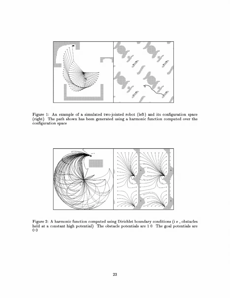
Figure 1: An example of a simulated two-jointed robot (left) and its con�guration space
(right). The path shown has been generated using a harmonic function computed over the
con�guration space.
Figure 2: A harmonic function computed using Dirichlet boundary conditions (i.e., obstacles
held at a constant high potential). The obstacle potentials are 1.0. The goal potentials are
0.0.
23

type �ring neurotransmitter a�erent e�erent population
medium spiny phasic GABA cortical GP and SN 77-95%
medium aspiny burst GABA cortical striatal 3-25%
giant tonic ACh cortical striatal 1-2%
Table 1: Types and characteristics of striatal neurons. Note that population percentages are
species-dependent.
Figure 3: A harmonic function computed using Neumann boundary conditions, where the
function gradient is constrained to be parallel to obstacle surfaces.
Cartesian bitmap c-space bitmap
x
y
t1
t2
OR
Effector
Figure 4: Schematic diagram of the mapping from Cartesian world coordinates into con�gu-
ration space.
24

Cortical afferent
Current-state Cluster
To directional cell
To nigral axons
Direction of Cluster Migration
(GLU)
Figure 5: Schematic of the migration of a current-state cluster within the striatum.
Directionally-dependent release of dopamine results in cluster movement.
25

Contents
1 Introduction 1
1.1 Goals and obstacles: a class of behaviors and control problems : : : : : : : : 2
1.2 Problems in Robotics : : : : : : : : : : : : : : : : : : : : : : : : : : : : : : : : 2
2 The Basal Ganglia 4
2.1 Anatomy : : : : : : : : : : : : : : : : : : : : : : : : : : : : : : : : : : : : : : 7
2.2 Cytology : : : : : : : : : : : : : : : : : : : : : : : : : : : : : : : : : : : : : : : 7
2.3 Neurotransmitters : : : : : : : : : : : : : : : : : : : : : : : : : : : : : : : : : 8
2.4 Neuronal Response Characteristics : : : : : : : : : : : : : : : : : : : : : : : : 10
2.5 Electrotonic Coupling : : : : : : : : : : : : : : : : : : : : : : : : : : : : : : : 10
3 The Model 11
3.1 Harmonic Functions : : : : : : : : : : : : : : : : : : : : : : : : : : : : : : : : 11
3.2 Biological Realization : : : : : : : : : : : : : : : : : : : : : : : : : : : : : : : 12
4 Consequences of the Model 14
5 Summary 15
6 Acknowledgements 16
26

List of Figures
1 An example of a simulated two-jointed robot (left) and its con�guration space
(right). The path shown has been generated using a harmonic function com-
puted over the con�guration space. : : : : : : : : : : : : : : : : : : : : : : : : 23
2 A harmonic function computed using Dirichlet boundary conditions (i.e., ob-
stacles held at a constant high potential). The obstacle potentials are 1.0. The
goal potentials are 0.0. : : : : : : : : : : : : : : : : : : : : : : : : : : : : : : : 23
3 A harmonic function computed using Neumann boundary conditions, where
the function gradient is constrained to be parallel to obstacle surfaces. : : : : 24
4 Schematic diagram of the mapping from Cartesian world coordinates into con-
�guration space. : : : : : : : : : : : : : : : : : : : : : : : : : : : : : : : : : : 24
5 Schematic of the migration of a current-state cluster within the striatum.
Directionally-dependent release of dopamine results in cluster movement. : : 25
27

List of Tables
1 Types and characteristics of striatal neurons. Note that population percent-
ages are species-dependent. : : : : : : : : : : : : : : : : : : : : : : : : : : : : 24
28


