Embed Size (px)
Citation preview
A multi-sample denaturation temperature tester for collagenous biomaterials
J.M. Lee, C.A. Pereira, D. Abdulla, W.A. Naimark and I. Crawford
Centre for Biomaterials, University of Toronto, 1’70 College St., Toronto, Ontario. Canada, M5S 1Al
ABSTRACT
The tcmprat we at which collagen d~~ature.s j?om a tri$e h&u io a wisdom roil .str-uc~ure is a ~c.seful measure II/
thp degrw oJcros.tlinking. A np(1, multi-sample denatut-a/ion temperature trster (1Yl”l’) has been c~onstnrrted fin- rajC1
drt~mination of the cnllagen dena~uration temperalure 01 natural tissues and c~ol1agenou.s biomateria1.s. To -oalidatr
thr sy.stem. the dena&ration temjwratures mmsuredju thr D?‘?‘ arc compared with re.sults from di&erential rcannin,q
mlwimetq (1).X:). Data are ,bresented.fbr bovine ppn’cardirom in three slates with denaturation tempera~urc:s ranging
,Jrom 68 to 85” C: /i-es/l. or crosslinked with ~glutaraldehydr or &he @oxide r-rage& IInlarol KY-512 po(y ($yidyl
rthvr 1. Ilenalu mlion /empat urns measured by D7’7‘ wre no/ ,si,qniJlcn71ll~ d~fJwvr~1 Jinm thov measured /I? di//ien-
tial .sc~anning calorimetv (DSC); however, I1.K orl.set ,sl;slrmaticall~ or~uwd at a slightlhl]y /ower temperature than
that measured 0~ 117‘7: 7‘hi.s result, swp11 only ,Ji,rJr& t&w is in agreemrnt with earlier expm’mrnts using h\‘tlrother
ma1 ~wmetrir &ion (HIT) tf,stin<g, By rontrnst, 1YlY‘ and IXC onset 7~7~~ identical for the e~o,genou.sl~~ rr~~~lirtkrtl
matrrial,r Since thr meas urrd tr-ansition tPmperat u w 7m~ in d+v&nt of initial load, ihis variable may De c-hocell
to ~~irld .I tm$wr fi,n.f-lf mpfr-&uf 1ransitiori.c rclilh (I ginfn 5amnplf gfromrtry. 7% is inslnl mf n 1 allow,s m-u rcl tr a s.sf.5 \
mfn t II/ rollqw dfnntlrrution tfmpvroturf.~ Jo, multi@ .samplrc in n fr-ac?ion of thr time wpir-ed 0~ other nrrthnd.s.
Keywords: Collagen. &maturation, shrinkage temperatllre. glutaraldehyde, crosslinking, bioproschesih.
epositlt, hydrothermal isometric tension. pericardium
\4d. Eng. Ph!;s., 199.5. Vol. 17. 115-121. March
INTRODUCTION
/n v&o, long-term survival of biomaterials derived from collagenous tissues has demanded develop- ment of means to stabilize the collagen against breakdown and resorption by enzymatic chain scission. The production of exogenous crosslinks through either chemical or physical treatment has been the preferred means to this end’-;‘. In each case, some method must be employed to evaluate the quality of the stabilization produced.
Collagen stabilization has typically been meas- ured by one of: (1) thermal stability, (2) resistance to enzymatic or chemical solubilization, or (3) in z~ivo persistence. These techniques probably meas- ure different characteristics of the treated material. Thermal stability tests measure the tem- perature at which the collagen denatures from its hydrogen-bonded triple helical structure to a ran- dom coil”. This denaturation temperature will be increased primarily by crosslinks between the indi- vidual collagen molecules in the triple helix (intrahelical crosslinking) and secondarily by intermolecular crosslinks. Solubilization tests
using enzymes (pronase, collagenase, pepsin, etc.) or chemical agents (cyanogen bromide, etc.) mea- sure the amount of material unsolubilized after a predetermined incubation with the agent”,“. These results are sensitive to both intrahelical and intermolecular crosslinks which prevent solution of scission fragments. IFS uiz~ persistence is ulti- mately the most important solubilization test since it simulates actual service for the material, but it is both expensive and sensitive to the details of the animal surgical model. As a result, in 71itr0 test- ing for denaturation temperature and resistance to solubilization remain first line screening tools for processing of bioprosthetic materials. In the study of natural materials, too, denaturation tem- perature and solubilization characteristics of col- lagen have been important tools in the study of maturation and of molecular defects linked with connective tissue diseases7-14.
Protocols for measurement of collagen denatur- ation temperature can be divided into three broad classes: (1) hydrothermal isometric tension tests (HIT tests), (2) shrinkage temperature tests, and
Multi-samfh denaturation temperature tester J.M. Lee et al.
(3) differential scanning calorimetry. The first two tests are unique to study of collagenous materials. In HIT tests, a sample is held at its unloaded length (isometric constraint) while the surround- ing solution is heated toward 100°C. When the collagen denatures, the force exerted by the sam- ple rises; the collagen seeks to coil up but the sam- ple is constrained to fixed length. Both the tem- perature of the transition and the developed tension are recorded8,i5*‘6. Alternatively, the sam- ple may be held at constant thickness and the pressure exerted due to swelling measured”.“. In a shrinkage temperature test, the sample is either unconstrained or held under a fixed tensile force (isotonic constraint). The length of the sample may be monitored with an LVDT, an indicator on a knife edge balance, or simply viewed against a scale7*t8 or with a modified spectrophotometer ce1114. The temperature at which denaturation - and hence shrinkage under constant load - begins is termed the ‘shrinkage temperature’: a term often applied to the collagen denaturation temperature, even if measured by other means. Differential scanning calorimetry (DSC), by con- trast, is a well developed analytic tool used for measurement of transitions and associated enthalpies in polymers and other chemical sys- tems. For collagenous materials, one pan contains a small sample of tissue in a bathing solution while the other pan contains-as a blank- an equival- ent volume of solution only. The two pans are increased in temperature, and a difference in heat flow to the two pans indicates an enthalpy depen- dent transition in the tissue. At the denaturation temperature, at least one peak is observed and the heat flow/temperature curve is recorded as well as the enthalpy of the transition calculated from the area under the peak’2,1g-23.
The HIT, shrinkage, and DSC protocols out- lined above are typically carried out on one sam- ple at a time in a custom built or commercial sys- tem. Since heating rates are of the order of l”C/min to avoid superheating effects, and a tem- perature range of 20 to 90°C is required, each test takes at least one hour and accumulation of stat- istically valid data is very time consuming. The objective of the present study was to develop a relatively low cost system which could test many samples in the time normally required to test one. The developed system operates under isometric constraints (as per the HIT tests described above) but allows application of an arbitrary initial force. To ensure that the measured denaturation tem- peratures were accurate, we have also performed DSC me~uremen~ on each of three sample materials: one natural (fresh bovine pericardium) and two of interest for cardiovascular devices (bovine pericardium crosslinked with glutaral- dehyde or Denacol EX-512: a poly (glycidyl ether) epoxide reagent being examined as an alternative to glutaraldehyde) ‘.
METHODS
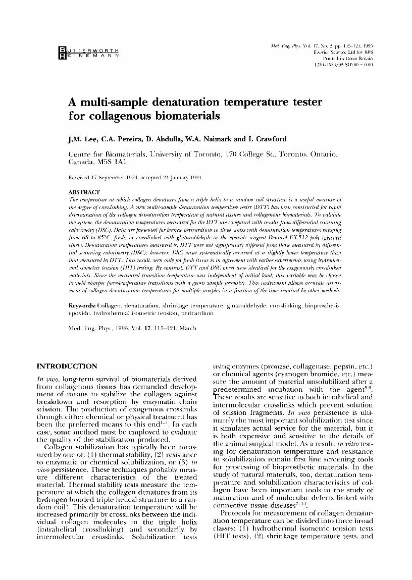
The denaturation temperature tester (DTT) and test protocol A schematic diagram of the DTT is shown in Figure 1. Each of six test samples (strips of tissue, arterial
segments, tendons, etc.) is held between two self- tightening inner grips machined from polycar- bonate which slide into stainless steel jackets. The polycarbonate was chosen since it is resistant to softening in boiling water (glass transition = 200°C). Each end of the sample is placed between the serrated ends of an inner grip and then pressed firmly into the narrowing jacket which squeezes the grip ends together, holding the sam- ple very securely. With the sample holder out of its beaker, each grip/sample/grip assembly is hung from an upper hook attached to a force trans- ducer, and the lower grip is slipped over the lower hook which is attached to a fixed rod. Adjustment of the knurled knob on the upper hook extends the sample for imposition of a chosen initial force.
The force transducers are simple spring steel cantilevers which are strain gauged in a half bridge configuration on their upper (tensile) and lower (compressive) surfaces. The strain gauge amplifiers [Vishay Model 212OA] provide DC volt- age outputs of O-10 V for a force range of O-200 g. The DC outputs are input to an Applied Engineer- ing S-bit analog to digital (A/D) converter card in an Apple IIGS computer. Force measurement resolution is therefore 200 g/256 or approxi- mately 0.8 g. Each transducer is calibrated with a series of test weights to provide a voltage/force regression line which the computer uses to con- vert voltage to force during the experiment.
The temperature transducer is an RTD probe [Cole-Parmer] which is centrally located between the six samples, the height of its end coinciding with the centre of the samples. It is attached to another strain gauge amplifier and the resulting DC output is again input to the A/D converter for a resolution of (100 - 20°C) / 256 or approxi- mately 0.3%. This transducer is calibrated with heated water and a mercury thermometer stan- dard, again producing a regression relation. The temperature transducer requires more frequent calibration than do the strain transducers.
Once all the samples have been attaohed to the calibrated holder, it is lowered into the four litre Pyrex beaker of test solution at room tempera- ture. In the experiments reported here., we have used distilled water; however, stainless steel fix- tures are essential to avoid corrosion of the holder during testing at temperatures approaching loO°C in saline solutions. The samples initially hang under a tension corresponding to the dead weight of the lower grip. The force amplifiers are balanced to display this force, and the knurled knobs on each upper sample hook are adjusted to impose a chosen initial force: usually 200 g. The data acquisition system is then activated, dis- playing six load/temperature plots onscreen. The heater below the beaker is turned on and adjusted to a level corresponding to the desired rate of temperature change: l”C/min for the exper- iments reported here (adjustable to 2”C/min).
Once the samples are extended to produce the desired initial force, the load immediately begins to decline due to stress relaxation in the viscoelas- tic tissue. As the temperature increases, the com- puter monitors the temperature and, at preset temperature increments (typically 1 “C) , repeat-
Strain Gauge
Polycarbonate Stainless Steel Self-lightening ----
Design of Two-Part Self-~~htentng
fhaker containing distilled water
To Excitation Amplifiers
l!TIzzd Pvrex 4000 ml
C-- Spring Steel
_ Fixture with Tension Adjustment
- Self-Tightening Grips
- Tissue Sample
- Rigid Fixture
Tissue Gilps
Figure 1 Schematic diagram of the sample holder, transducers, and heater from the drnaturation temperature tester. Only one sample of six is shown. The six samples are located at 60” angles around the central tempcrattrre prtrhe. The six strain gdttged force transducers and the thermistor probe arc connected to conditioning amplifiers, the outputs of which are read hv computer.
edly scans the six force transducers, prints the resulting averaged force/temperature values for each point and extends the six curves onscreen. At the denaturation temperature, the declining force/temperature curve suddenly begins to rise. The temperature at the beginning of a consistent rise in load is termed the denaturation tempera- ture. The test is continued until a preset tempera- ture is reached: usually 100°C.
Test samples
Intact bovine pericardia and enclosed hearts were obtained fresh from slaughter of 6-8 months old calves and transported to the laboratory in Hanks physiological saline (pH 7.4, 310 mOs). The per- icardia were cleaned of adherent fat and samples of ventral pericardium approximately 10 cm square were labeled in the base-to-apex direction with sutures and harvested. Each sample was div- ided in the base-to-apex direction into three sec- tions which were randomly assigned to one of the three treatment groups: (1) Fresh: tested within 8 h of harvesting; (2) Glutaraldehyde: treated for 48 h in 0.5% glutaraldehyde at pH 7 in 0.1 M phosphate buffer: (3) Polyepoxy: treated for 48 h in 0.6% Denacol EX-512 poly (glycidyl ether) at pH 11 in 0.1 M sodium hydroxide buffer.
Differenti~ scanning calorimetry
Small samples (5-10 mg) of each tissue tested on the DTT were also examined for denaturation temperature on a DuPont Model 900 DTA differ- ential scanning calorimeter. Crimpable alu- minium pans were scrupulously cleaned for 1 hour with 2% Contrad 70 (Canlab) in distilled deionized water using an ultrasonic cleaner, and
similarly rinsed with distilled deionized water. The pans were then rinsed with deionized water three times and then 100% ethanol three times before being placed in a drying oven. Tttis cleaning was essential to eliminate thermal artifacts due to vari- able wetting of the pan interior. For each test, the tissue sample was placed in one pan and distilled water added for an internal weight of 5-10 g. A second pan was filled with the same volume of distilled water alone. The pans were crimped to ~lermetica~ly seal the interior and placed in the USC sample holder. Thermal analysis was perfor- med at heating rates of 1 or 2’C/minute from room temperature to a maximum temperature of 95°C. After testing, the tissue sample was freeze- dried and the dly weight recorded.
The temperature corresponding to the highest point on the denaturation peak and to the onset of the peak were recorded. Onset was defined by extrapolation of the rising edge of the peak to intercept the baseline’“. The area under the peak was integrated using the attached microprocessor to yield an enthalpy corresponding to the denat- uration transition. The enthalpy was calculated as” 1
where it/l is the mass of the sample (g), A is the area under the thermogram (cm‘-‘), X and Y are sensitivity settings on the DSC, c( is the heating rate (“Q’min) and Eis the calibration coefficient (J/cm’). The DSC system was calibrated with an Indium standard.
117
Multi-sample aknuturation tempera&w tester;J.M. Lee et al
statistical analysis
From each pericardial sample, two base-to-apex strips were cut for DlT analysis, and two small tissue samples were cut for DSC analysis: for a minimum of 10 data points per technique per treatment group. The denaturation temperature data were analyzed by a S-way analysis of variance (ANOVA) with variables of treatment and test method. The initial F-test was followed with Fish- er’s Least Significant Difference tests for multiple comparisons with significance accepted at a ~~0.05 level (StatView II, Abacus Concepts). Lin- ear fits to data were obtained by least squares regression and examined by F-testing for signifi- cance (StatView 4, Abacus Concepts). All data are expressed as the mean rt: the standard error of the mean (SE).
RESULTS
Denatwation temperature tester (DTIY) data
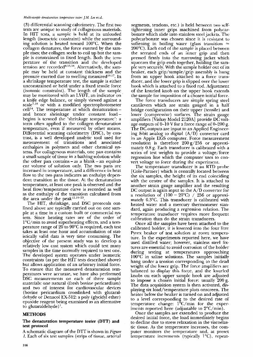
Sample force-temperature data for all three test groups are shown in Figure 2 for tests begun with initial loads of 200 g. In each case, stress relax- ation was clearly observable during the heating of the samples. The relaxation was increased by the elevated temperatures over that seen in our pre- viously published experiments with similar materials at 37°C’*25. Careful compa~son of the curves from the repeated experiments, and reheating of samples heated to 60°C and then cooled, failed to show any repeatability of the small variations in load data, suggesting that they represented noise rather than thermally-induced changes in the material. The definition of the denaturation temperature as the temperature at which a steady climb in force began was adequate for unequivocal identification of the transition.
The denaturation temperatures measured were independent of the applied initial force in tests on all three sample groups. An example of the force-temperature data from these tests is shown
“5i 160 E i?
9 140
! 2 120
Fresh I
30 40 so 60 70 8-O dcr
Temperature (“C)
Figure 2 Sample foree-remperaazre curves for fresh, @tan&d- ehyde-ueated, and ~~y~~~1 ether)-tneased bovine pericardium. Initial load (L,J = 2’00 g. Hea&sg rate = l*C/min. The denaturation temperatcne is taken as the temperarure of the last point before a steady rise in force.
150
‘;ii E CE 9 100
3 ii
u 50
0 30 40 50 60 70 60 90 1
Temperature (“C)
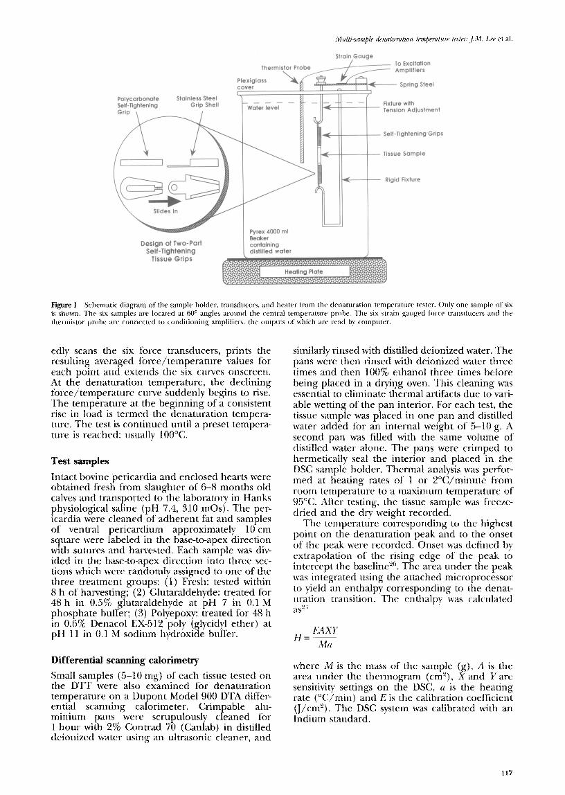
Figure3 Force-temperature curves for gI~l~alde~yde treated tis sues with initial load values (I,,) of 5-200 g. Heating rate = l”C/min. The denaturation temperatures measured showed no dependence on the initial force for any of the materials tested.
in Fig-w-e 3 for glutaraldehyde-treated material. The lower initial forces produced somewhat sharper transitions and may be preferred for rou- tine testing.
The denaturation temperatures measured with the DTT for the three treatment groups were sig- nificantly different (Table I). Glutaraldehyde treatment increased the denaturation tempera- ture from 67.720.3”C to 85.2%0.2”C, while the poly (glycidyl ether) treatment only increased the value to 75.8-tO.3”C.
Correlation with differential scanning calorimetry (DSC) data
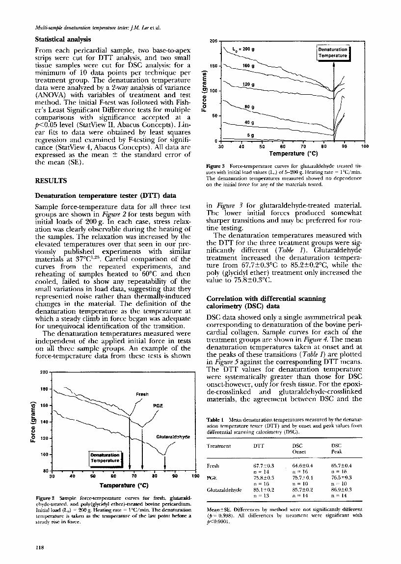
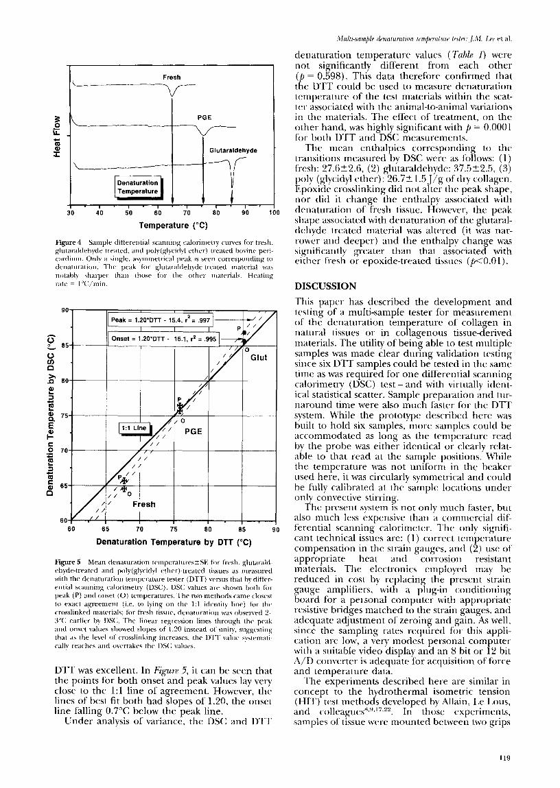
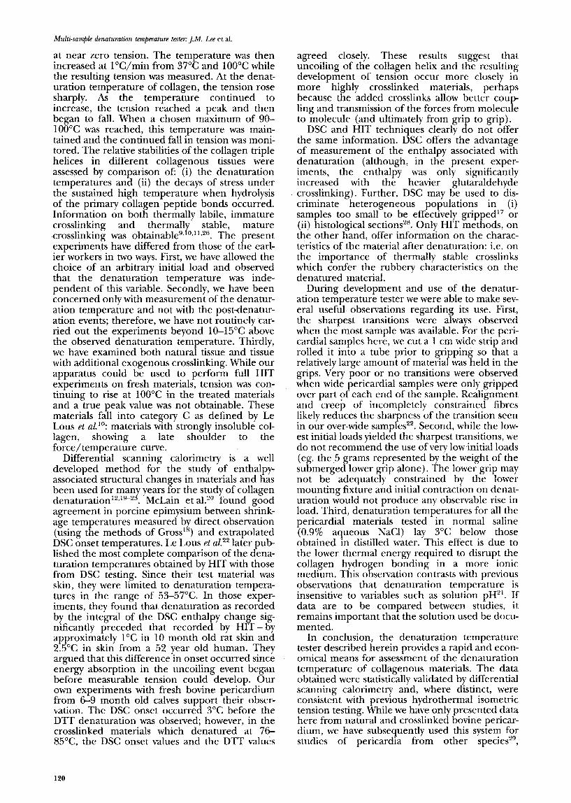
DSC data showed only a single asymmetrical peak corresponding to denaturation of the bovine peri- car-dial collagen. Sample curves for each of the treatment groups are shown in Figure 4. The mean denaturation temperatures taken at onset and at the peaks of these transitions (T&e 1) are plotted in Figure 5 against the corresponding DTT means. The DTT values for denaturation temperature were systematically greater than those for DSC onset-however, only for fresh tissue. For the epoxi- de-crosslinked and glutaraldehyde-crosslinked materials, the agreement between DSC and the
Tuble 1 Mean denaturation temperatures measured by the denatur- ation temperature tester (DTT) and by onset and peak values from differential scanning calorimetry (DSC).
Treatment DTT DSC DSC Onset Peak
Fresh 67.720.3 64.620.4 65.720.4 n = I4 n = 16 n = 16
PGE 75.8r0.3 75750.4 76.5203 n= 16 n = 10 n = 10
Clutaraldehyde 85.1 t-O.2 85.7?0.2 86.920.3 n = 13 n= 14 n = 14
,Mean%SE Differences by method were not significantly different (p = 0.598). All differences by treatment were significant with p~O.iM?o1.
118
Fresh
I ’ 1 ’ I m I ’ I ’ I ’ 40 50 60 70 60 90 1
Temperature (“C)
Figure 4 Sample differential scanning calorimetq curves li)r trrsh, gluraraldehyde treated. and poly(glycidyl ether) treated bovine prri- cardium. Only a single. asymmetrical peak is seen corresponding to drnaturation. The peak for Rllltaraldehyde-tl-eated material wa\ notably sharper than Ihow for the other materials. Heating rate = I”(:/rriin
c 60
Onset = l.SO’DTT - 16.1, r2 = ,995 -
I1:l Line L //O
/ / /
I - I - I ’ I ’ I .
65 70 75 60 65 90
Denaturation Temperature by DTT (“C)
Figure 5 Mean denaturation temprratures?SE for fresh. giutarald- ehyde-treated and poly(glycidy1 ether)-treated tissues as measuwd with the denaturation temperaturr tester (DTT) versus that by diffrr- vntial scanning calorimetry (DSC). DSC values are shown both for peak (P) and onset (0) temperatures. The two methods came closest to exact agreement (i.e. to lying on the 1:l identity line) for the crosslinked materials; for fresh tissue, denaturation was ohservcd L’- KY: earlier by IN:. The linear regression lines through the pea!, and onset values showed slopes of 1.20 instead of unity. suggestin! that as the level of crosslinking increases, the DTT \;alur systrmatl- callv reacheq and overtalcs the DX; values.
DTT was excellent. In I;igure 5, it can be seen that the points for both onset and peak values lay very close to the 1:l line of agreement. However, the lines of best fit both had slopes of 1.20, the onset line falling 0.7”C below the peak line.
Under analysis of variance, the DSC and DTT
denaturation temperature values (7hhb I) were not significantly different from each other (p = 0.598). This data therefore confirmed that the DTT could be used to measure denaturation temperature of the test materials within the scat- ter associated with the animal-to-animal variations in the materials. The effect of treatment, on the other hand, was highly significant with p = 0.0001 for both DTT and DSC measurements.
The mean enthalpies corresponding to the transitions measured by DSC were as follows: (1) fresh: 27.622.6, (2) glutaraldehyde: 37.5k2.5, (3) poly (glycidyl ether): 26.721.5 J/g of dry collagen. Epoxide crosslinking did not alter the peak shape, nor did it change the enthalpy associated with denaturdtion of fresh tissue. However, the peak shape associated with denaturation of the glutaral- dehyde treated material was altered (it was nar- rower and deeper) and the enthalpy change was significantly greater than that associated with either fresh or epoxide-treated tissues (pcO.01).
DISCUSSION
This paper has described the development and testing of a multi-sample tester for measurement of the denaturation temperature of collagen in natural tissues or in collagenous tissue-derived materials. The utility of being able to test multiple samples was made clear during validation testing since six DTT samples could be tested in the same time as was required for one differential scanning calorimetry (DSC) test - and with virtually ident- ical statistical scatter. Sample preparation and tur- naround time were also much faster for the DTT system. While the prototype described here was built to hold six samples, more samples could be accommodated as long as the temperature read by the probe was either identical or clearly relat- able to that read at the sample positions. While the temperature was not uniform in the beaker used here, it was circularly symmetrical and could be fully calibrated at the sample locations under only convective stirring.
The present system 1s not only much faster, but also much less expensive than a commercial dif- ferential scanning calorimeter. The only signifi- cant technical issues are: (1) correct temperature compensation in the strain gauges, and (2) use of appropriate heat and corrosion resistant materials. The electronics employed may be reduced in cost by replacing the present strain gauge amplifiers, with a plug-in conditioning board for a per,sonal computer with appropriate resistive bridges matched to the strain gauges, and adequate acljustment of zeroing and gain. As well, since the sampling rates required for this appli- cation are low, a very modest personal computer with a suitable video display and an 8 bit or 12 bit A/D converter is adequate for acquisition of force and temperature data.
The experiments described here are similar in concept to the hydrothermal isometric tension (HIT) test methods developed by Allain, Le Lous, and colleaguesx~“.‘7~z. In those experiments, samples of tissue were mounted between two grips
119
Multi-sample denaturation temperature tester: JM. Lee et al.
at near zero tension. The temperature was then increased at l”C/min from 37°C and 1OOOC while the resulting tension was measured. At the denat- uration temperature of collagen, the tension rose sharply. As the temperature continued to increase, the tension reached a peak and then began to fall. When a chosen maximum of 90- 100°C was reached, this temperature was main- tained and the continued fall in tension was moni- tored. The relative stabilities of the collagen triple helices in different collagenous tissues were assessed by comparison of: (i) the denaturation temperatures and (ii) the decays of stress under the sustained high temperature when hydrolysis of the primary collagen peptide bonds occurred. Information on both thermally labile, immature crosslinking and thermal1
Y stable, mature
crosslinking was obtainableg* 0*1’*26. The present experiments have differed from those of the earl- ier workers in two ways. First, we have allowed the choice of an arbitrary initial load and observed that the denaturation temperature was inde- pendent of this variable. Secondly, we have been concerned only with measurement of the denatur- ation temperature and not with the post-denatur- ation events; therefore, we have not routinely car- ried out the experiments beyond lO-15OC above the observed denaturation temperature. Thirdly, we have examined both natural tissue and tissue with additional exogenous crosslinking. While our apparatus could be used to perform full HIT experiments on fresh materials, tension was con- tinuing to rise at 100°C in the treated materials and a true peak value was not obtainable. These materials fall into category C as defined by Le Lous et aZ.‘? materials with strongly insoluble col- lagen, showing a late shoulder to the force/temperature curve.
Differential scanning calorimetry is a well developed method for the study of enthalpy- associated structural changes in materials and has been used for many years for the study of collagen denaturation’“,‘“-** . McLain et alzo found good agreement in porcine epimysium between shrink- age temperatures measured b (using the methods of Gross’ ;I’
direct observation ) and extrapolated
DSC onset temperatures. Le Lous et ai.22 later pub lished the most complete comparison of the dena- turation temperatures obtained by HIT with those from DSC testing. Since their test material was skin, they were limited to denaturation tempera- tures in the range of 53-57*C. In those exper- iments, they found that denaturation as recorded by the integral of the DSC enthalpy change sig- nificantly preceded that recorded by HIT- by approximately 1°C in 10 month old rat skin and 25°C in skin from a 52 year old human. They argued that this difference in onset occurred since energy absorption in the uncoiling event began before measurable tension could develop. Our own experiments with fresh bovine pericardium from 6-9 month old calves support their obser- vation. The DSC onset occurred 3°C before the DTT denaturation was observed; however, in the crosslinked materials which denatured at 76- 85X, the DSC onset values and the DTT values
agreed closely. These results suggest that uncoiling of the collagen helix and the resulting development of tension occur more closely in more highly crosslinked materials, perhaps because the added crosslinks allow better coup- ling and transmission of the forces from molecule to molecule (and ultimately from grip to grip).
DSC and HIT techniques clearly do not offer the same information. DSC offers the advantage of measurement of the enthalpy associated with denaturation (although, in the present exper- iments, the enthalpy was only significantly increased with the heavier glutaraldehyde crosslinking). Further, DSC may be used to dis- criminate heterogeneous populations in (i) samples too small to be effectively gripped” or (ii) histological sections 28. Only HIT methods, on the other hand, offer information on the charac- teristics of the material after denaturation: i.e. on the importance of thermally stable crosslinks which confer the rubbery characteristics on the denatured material.
During development and use of the denatur- ation temperature tester we were able to make sev- eral useful, observations regarding its use. First, the sharpest transitions were always observed when the most sample was available. For the peri- cardial samples here, we cut a 1 cm wide strip and rolled it into a tube prior to gripping so that a relatively large amount of material was held in the grips. Very poor or no transitions were observed when wide pericardial samples were only gripped over part of each end of the sample. Realignment and creep of incompletely constrained fibres likely reduces the sharpness of the transition seen in our over-wide samples22. Second, while the low- est initial loads yielded the sharpest transitions, we do not recommend the use of very low initial loads (eg. the 5 grams represented by t.he weight of the submerged lower grip alone). The lower grip may not be adequately constrained by the lower mounting fixture and initial contraction on denat- uration would not produce any observable rise in load. Third, denaturation temperatures for all the pericardial materials tested in normal saline (0.9% aqueous NaCl) lay 3OC below those obtained in distilled water. This effect is due to the lower thermal energy required to disrupt the collagen hydrogen bonding in a more ionic medium. This observation contrasts with previous observations that denaturation temperature is insensitive to variables such as solution pII*‘. If data are to be compared between studies, it remains important that the solution used be docu- mented.
In conclusion, the denaturation temperature tester described herein provides a rapid and econ- omical means for assessment of the denaturation temperature of collagenous materials. The data obtained were statistically validated by differential scanning calorimetry and, where distinct, were consistent with previous hydrothermal isometric tension testing. While we have onfy presented data here from natural and crosslinked bovine pericar- dium, we have subsequently used this system for studies of pericardia from other species’“,
120
materials from bovine arterial tissues’” and por- cine aortic valve leaflets with similar success. With appropriate grips, this system could easily be used for studies of skin, tendons, and with other col- lagenous materials.
ACKNOWLEDGEMENTS
The authors wish to express their thanks to Pro- fessor Steve Balke, Department of Chemical Engineering, for the use of the differential scan- ning calorimeter and to Professor Robert Pilliar, Centre for Biomaterials, for the use of the strain gauge amplifiers. Thanks are also extended to the staff, Canada Department of Agriculture and Grober Farms, Cambridge, Ontario, for assistance in obtaining intact hearts and pericardia.
This work was supported by grants to Dr. Lee from the Canadian Heart and Stroke Foundation and the Natural Sciences and Engineering Research Council of Canada (NSERC).
REFERENCES
1. Pereira CA, Lee JM, Haberer SA. Effect of alternative crosslinking methods on the low strain rate viscoelastic properties of bovine pericardial bioprosthetic material. ,I Hiomd Mntul- I&,\ 1990; 24: 345-61.
2. Weadock K, Olsen RM, Silver FH. Evaluation of collagen crosslinking techniques. Rjom&~ n@Pn I>ucl Ar/i/ 0rgan.s
1983--l; 11: 293-18. 3. Petite H. Rault I, Hut A, Hemasche Ph., Herbage D. Llsc
of the acyl azide method for cross-linking collagen-rich tis- sues sttch as pericardium. ,/ Hiomed Mtkr- Rcs 1990: 24:
179-87. 4. Olde Datnink I,HH. Dijkstra F’J, van Luyn MJA, \-an
Wachem PB. Nieuwenhuis P, Feijen J. In vitro comparison of collagen crosslinking methods. ‘I’mns 4th World Congwc
Biomnlfv 1992; pp. 45. 5. Nimni ME, Harkness RD. Molecular structures and func-
tions of collagett. In: C~ollqen, Volumr 1: Hiorhrmistq, CRC: Press, Boca Raton (1988) pp. 1-78.
6. Haut RC, Lancaster RI,, DeCatnp CE. Mechanical proper- ties of the canine patellar tendon: some correlations with age atid the content of collagen. J Riomrrh 199”: 25: 163-73.
7. Ch\apil M,.Jensovsky 1,. The shrinkage temperature of co- lagen fibres isolated from the tail tendons of rats of vari- ous ages and frotn different places of the same tendon. C:rrontr,lo<gic~ 1963; 1: 18-29.
8. Allain J-C:, Bazin S, I.c 1.0~s M, Delaunay A. \‘ariations de la co&action hydrothermique de la peau du Rat en fonc- tion de I’agc drs animaux. CRH &YI~ Sci F’cuG 1977: 284
(D): 1131-4. 9. 1.c I,ous M, Allaitt JC, Cohen-Solal I,, Maroteaux P. The
rate of collagen maturation in rat and human skin. COU)/ ‘l’k Rcr 198?; 9: 253-62.
10. 1.e 1.0~s M. Allain J-C, <lohen-Solal L, Maroteaux P. Hydrothermal isometric tension curves from different ccjnnective tissues. Role of collagen genetic types and notlcollagctto~ts components. (:onrl 7‘i.r.~ Ku5 1983: 11: 199406.
11. 1.e I.ous M, (:ohett-Solal I., Allain .J-(:, Bonavrnture J, Maroteaux P. Age related evolution of stable collagen reticulation in human skin. Call,! 7i’s.s Kp,r 1985; 13: l~l:i-:;.~.
l?.
I :<.
11.
15.
16.
17.
IX.
19.
“0.
21.
“2.
L’3.
24.
‘1.5.
%.
27.
“8.
Flandin F, Buffevant C, Herbage D. A differential scan- ning calorimetry analysis of the age-related changes in the thermal stability of rat skin collagen. Biochimirn r/
Nio~hysicn Ada 1984; 791: 205-l 1. Danielsen CC. Thermal stability of reconstituted collagen fibrils. Shrinkage characterisiics upon in vitro matu- ration. Mrrhanisms Aging Ikvel 1981; 15: 269-78. Danielsen CC. Age-related thermal stability and suscepti- bility to proteolysis of rat bone collagen. Riocl~m ,/ 1990; 272: 697-701. Elden HR. Hydration of connective tissue and tendon elasticity. Riorhim Biophys Akta 1964; 79: 592-9. \?idik A. An apparatus for measurement of’ small ten- sions. ~xprimn~tin 1968; 24: 861-2. ;Ulain J(:, l.c Lous M, Cohen-Solal I., Razin S, Maroteaux P. Isometric tensions developed during the hvdrothermal swelling of ~a: skin. (;oro)1~ Tits RP\ 1980: 7: 197-133 (iross .J. Thermal denaturation of collagen in the dis- perscd and solid state. .Sciww 1964; 143: 960-l. Dichson IR, Ilappey F, Pearson CH. Naylor A, Turner RI,. Decrease in hydrothermal stahilitv of collagen on ageing in rhr human intenertrhral disk. :%t~rv 1967; 215: .X-2. McClain PE, Pearson AM, Miller ER, Ljugan 1,R Jr. Appli- cation of differential thermal analysis to the study of ltvdt-othermal shrinkage in epimysial and coriutn col- lagen. Riorhim Biobhys Acta 1968: 168: 14%!). Finch A, I,cd\rard DA. Shrinkage of collagen fibrcs: A differential scanning calorimetric studs,. B&him Rio/~hyc
.~CYO lcj72. 278: 433-9. L , 1,~ Lous M. Flandin F, Herbage D, Allain ,J-(Z, Influence of’ collagen dertaturation on tltc- chcmorheological pt-oprt-tic’5 of skin , assessed by differcnrial scanning calor- imcln- and hvdrothertnal isometric lrttsion mrasure- tnetit. Nioc,itim I’+~z~s Acf0 1982; 717: 29.5300. Horgan I?], King i%, Kurth I,B, Kuyperh R. (:ollagen crosslinks attd their relatiottship to the rhrt-mal proper- ties of calf tendons. ,-lrch Biochem Wiophl;.s 1990; 15: 21-6. Mc(:lain PE. Wiley ER. Differential scanning calorimeter Etudirs of the thermal transitions of collagen. Itnpli- cations 011 structure and stabilitv. ,I Ijiol (,‘h~l 1972: 247: 692-7. Lee .JM, Haberer SA. Boughner DR. The bovine pericar- dial xenograft: 1. Effect of fixation itt aldchvdes without constraint on the tensile viscoelastic prop&es of bovine pericardimn. ,/ Hiomrd Mntrr Krs 1989: 23: 457-7.5. Flory I’J, Spurr OK. Melting eqktiliht-ium fi,r collagen fibers under stress. Elasticitv in anrorphott? state. ,/ ,tnt UIPNI SW 196 1; 83: 1308-6 1, Kronick P, Maleeff B, (Carroll R. ‘r‘hc 1oc:ltion.s of col- laget~s with different thermal stabilities in fihrils of’ hov- ittr reticular dertttis. (:OIOI~V/~TW ?‘ic%crr, /+c 1988: 18: 1 ‘V-‘54. - ~ Fisher J. Got-ham SD, Howic AM. Whratley I>J. Examin- ,nion ot fixative penerrarion in glitlaraldchydc-treated bovine pericardium by stratigraphic analysis of shrinkage temperature mcasuremertts using differential scanning calorinret~. I,@ Suppot-/ Syst 1987; 5: 189-93.
L’Y. Naitnark WA. Lee JM, I.it&back H. (heung DT. Corre- lation of structure ‘and viscoelastic propet-ties in the per- icardia of four mammalian species. .4rn ,/ f/t~sio/ 1992: 263: H109.5-106.
Z30. Lee JM, Thyagamjan K, Pereira (:,\, Mcltttyre J, Tu R. Crosslinking of a prototype bovine artet-\ xenogrdft: Mechaniral and biochemical cotnpat-isons of. treatment in glutdr-dldehyde and fout- polv (glycidyl pounds (Ahsrract). Prowedirp o/ th
ether) cotn- Y 9h 1: f[ top 0 II (lonJ&
fnff on Niomczlfvicds 1991 ; pp. I 12.
121


