Embed Size (px)
Citation preview

Thomas Jefferson UniversityJefferson Digital Commons
Division of Gastroenterology and HepatologyFaculty Papers Division of Gastroenterology and Hepatology
8-1-2013
A multicenter, prospective, randomizedcomparison of a novel signal transmission capsuleendoscope to an existing capsule endoscope.Eric H ChoiRiverside Medical Clinic, Riverside, California, [email protected]
Klaus MergenerDigestive Health Specialist, Tacoma, Washington
Carol SemradUniversity of Chicago, Chicago, Illinois
Laurel FisherUniversity of Michigan, Ann Arbor, Michigan
David R CaveUniversity of Massachusetts, Worchester, Massachusetts
See next page for additional authors
Let us know how access to this document benefits youFollow this and additional works at: http://jdc.jefferson.edu/gastro_hepfp
Part of the Gastroenterology Commons, and the Hepatology Commons
This Article is brought to you for free and open access by the Jefferson Digital Commons. The Jefferson Digital Commons is a service of ThomasJefferson University's Center for Teaching and Learning (CTL). The Commons is a showcase for Jefferson books and journals, peer-reviewed scholarlypublications, unique historical collections from the University archives, and teaching tools. The Jefferson Digital Commons allows researchers and
Recommended CitationChoi, Eric H; Mergener, Klaus; Semrad, Carol; Fisher, Laurel; Cave, David R; Dodig, Milan; Burke,Carol; Leighton, Jonathan A; Kastenberg, David; Simpson, Peter; Sul, James; Bhattacharya,Kanishka; Charles, Roger; Gerson, Lauren; Weber, Luke; Eisen, Glenn; Reidel, Warren; Vargo, JohnJ; Wakim-Fleming, Jamile; and Lo, Simon K, "A multicenter, prospective, randomized comparison ofa novel signal transmission capsule endoscope to an existing capsule endoscope." (2013). Division ofGastroenterology and Hepatology Faculty Papers. Paper 19.http://jdc.jefferson.edu/gastro_hepfp/19

AuthorsEric H Choi, Klaus Mergener, Carol Semrad, Laurel Fisher, David R Cave, Milan Dodig, Carol Burke,Jonathan A Leighton, David Kastenberg, Peter Simpson, James Sul, Kanishka Bhattacharya, Roger Charles,Lauren Gerson, Luke Weber, Glenn Eisen, Warren Reidel, John J Vargo, Jamile Wakim-Fleming, and Simon KLo
This article is available at Jefferson Digital Commons: http://jdc.jefferson.edu/gastro_hepfp/19

As submitted to:
Gastrointestinal endoscopy.
And later published as:
A multicenter, prospective, randomized comparison of a
novel signal transmission capsule endoscope to an
existing capsule endoscope.
August 2013, Volume 78, Issue 2, pp. 325-332.
DOI: 10.1016/j.gie.2013.02.039.
Abstract
Background: MiroCam®, a capsule endoscope, utilizes a novel transmission technology, electric-
field propagation that uses the human body as a conductive medium for data transmission.
Objective: To compare the ability of the MiroCam® (MC) and PillCam® (PC) to identify sources of
obscure GI bleeding (OGIB)
Design: Prospective, multicenter, comparative study.
Setting: Six academic hospitals.
Patients: 105 patients with OGIB.
Interventions: Subjects ingested both the MC and PC capsules sequentially in a randomized
fashion.
Main outcome measurements: Concordance of rates in identifying a source of OGIB, operational
times and rates of complete small bowel examination.

1
Results: Data analysis resulted in 43 (48%) "abnormal" cases identifying a source of OGIB by
either capsule. Twenty-four cases (55.8%) were positive by both capsules. There was negative
agreement in 46/58 cases (79.3%). The κ-index was 0.547 (χ2=1.32, p=0.36). In 12 cases, MC
positively identified a source that was not seen on PC; whereas in 7 cases, PC positively identified
a source that was not seen on MC. MC had a 5.6% higher rate of detecting small bowel lesions
(p=0.54). MC captured images at 3 frames/sec for 11.1 hours and PC captured images at 2
frames/sec for 7.8 hours (p< 0.0001). Complete small bowel examination was achieved in 93.3%
for MC and 84.3% for PC (p=0.10).
Limitations: Readers could not blinded to the particular capsule they were reading.
Conclusions: A positive diagnostic finding for OGIB was identified by either capsule in 48% of
cases. The concordance rate between the 2 capsules was comparable to prior studies in
identifying sources of small bowel bleeding. The longer operational time of the MC may result in
higher rates of complete small bowel examination which may, in turn, translate into a higher rate
of detecting small bowel lesions.
2
INTRODUCTION
Since its first report over a decade ago, capsule endoscopy (CE) has revolutionized the evaluation
of patients with suspected small bowel disease and is now considered the test of choice in the
evaluation of obscure gastrointestinal bleeding (OGIB) 1. Two small bowel CE systems (Pillcam
SB2®, Given Imaging, Yoqneam, Israel, and Endocapsule®, Olympus America, Allentown, PA) are
currently being used in the United States 2. Both systems use radiofrequency to transmit images
from the device to an external receiver.
In 2009, a Korean group reported on the safety and efficacy of a new type of capsule endoscope,
the MiroCam® (MC), which utilizes a novel transmission technology known as electric-field
propagation 3. With this technology, the human body serves as the medium for data
transmission, propagating low voltage signals through a pair of gold plates on the surface of the
capsule device. The low voltage requirement results in lower power consumption, allowing for a
longer operation time, and a device, which captures a greater number of images compared to
capsules that utilize radiofrequency transmission.
We report the results of a multicenter study comparing PillCam SB2® (PC) and MC capsules
swallowed sequentially in randomized order by patients with OGIB. The primary study endpoint
was the identification of a bleeding source.
METHODS
3
Patient enrollment
Between January 2009 and July 2010, 105 patients with suspected small bowel bleeding were
competitively enrolled into the study at six US academic sites.
All patients underwent a prior esophagogastroduodenoscopy (EGD) and colonoscopy within the
previous 12 months or clinically demonstrated continued bleeding, requiring blood transfusions
since their initial evaluation. Enrolled patients needed to demonstrate a hemoglobin value of <
11g/dL (or < 13g/dL if documented iron deficient anemia) in males or < 10g/dL (or < 12g/dL if
documented iron deficient anemia) in females within 30 days of capsule endoscopy or prior to
transfusions if transfusions were clinically necessary. Patients needed to be referred for a capsule
endoscopy as part of their diagnostic evaluation and needed to be competent and willing to sign
a written informed consent.
Patients with a diagnosis or suspicion of a gastrointestinal obstruction, stricture, perforation,
swallowing disorder, significant loss of gastrointestinal motility, three or more known small
bowel diverticula, radiation or chemotherapy-induced enteritis, history of skin allergy or
sensitivity to adhesives were excluded from the study. Pregnant patients, patients who were not
candidates for surgical intervention, and patients with a cardiac pacemaker, defibrillator or
another type of implanted electro-mechanical device, as well as patients who were scheduled for
an MRI and those who had participated in another research study within the last 30 days were
also excluded.

4
Capsule endoscope preparation and administration
At the time of a study initiation visit, all patients underwent a complete physical examination,
review of their medical and medication history, and a urine pregnancy test as indicated. Starting
38 hours prior to capsule ingestion, all study participants were instructed to consume a liquid
diet for 24 hours, consume only water for the following 12 hours, and then fast for at least 2
hours prior to the procedure.
On the day of the procedure, patients were fitted with receivers and sensors as recommended by
both manufacturers and ingested both capsules in a randomized order 90 minutes apart. They
were accordingly placed in an alternating fashion into a "MC-first" or "PC-first" cohort by
competitive enrollment. Each subject was given a “subject number” to conceal the identity of the
patient. Only the study coordinators were aware of the subject numbers, which corresponded to
the patient identity. All subjects wore the receivers for 11 hours after ingestion of the second
capsule. They were allowed to ingest up to 4 ounces of water to facilitate ingestion of each
capsule. Patients were provided an anti-foaming agent (simethicone or an equivalent alternative)
to swallow within five minutes prior to capsule ingestion. Subjects remained at the recruitment
site for 30 minutes after swallowing the second capsule for observation and to document any
complaints.
Receivers were returned the day of the procedure or the following business day. Patients were
subsequently interviewed and instructed to call the office to report the date and time of capsule
passage. Patients who were unable to verify capsule passage were followed with serial
abdominal radiographs until passage of the device was confirmed.

5
Image interpretation
All images were collected from each capsule and were interpreted by two independent
experienced readers, who were blinded to the particular subject and the interpretation of the
other reader. They were not blinded to the source of the images (MC versus PC) on account of
the differing image-viewing software programs and respective image qualities. Readers were
given the freedom to use their own preferred viewing mode (single /dual / quad) and viewing
speed, but were required to use the same reading preferences for evaluating the MC and PC
images. A pair of readers was randomly selected from a pool of five reader pairs and these two
reviewers read both sets of images (MC and PC) for each individual subject. The reader pairs
were presented the image sets for any given subject with all subject-identifying information
masked at separated time points to prevent bias. The reader pairs remained fixed for the
duration of the trial. In situations in which the two readers reviewing the same capsule images
for a given subject did not agree in their diagnostic findings, an adjudication committee,
consisting of three members, who were not part of the reader pool, was asked to review the
images for the respective capsule to determine a diagnosis, which was then considered the final
diagnosis for the respective capsule. The adjudication committee was blinded to the findings of
the opposing capsule for all cases.
Image interpretation bias was minimized by using independent readers rather than site
investigators to interpret the imaging files and by blinding each reader to the findings of the
other readers. The interpretation of an abnormal finding was chosen from a predetermined list of
possible diagnoses including: ulcerative lesions, tumor/polyp, vascular lesions (AVM), blood

6
(without ability to identify the lesion), and "other". To be considered concordant, it was
necessary for the responses provided by the readers to agree on the specific finding.
Statistical analysis
A sample size of 85 subjects was determined to allow for the detection of a concordance rate of
at least 85% with a two-sided 95% confidence interval of 12% between MC and PC. A total of 105
subjects were enrolled in the study to allow for subject dropout or for not meeting criteria for the
effectiveness evaluation at a rate of 20-25%. All study subjects, who ingested at least one
capsule, were included in the safety analyses as an intention to treat analysis. A chance-adjusted
kappa statistic was calculated to evaluate the strength of the agreement between MC and PC
with respect to the detection and identification of sources of small bowel bleeding. The
concordance rate and corresponding 95% confidence interval was estimated. An 85%
concordance rate was predetermined to be evidence of equivalence between the MC and PC.
The mean total transit times through specific anatomic landmarks (esophagus, stomach, small
bowel and colon), mean total transit times from ingestion to capsule expulsion, and mean total
operation times of the MC and PC were compared using the paired t-test at the 5% significance
level. An exploratory analysis of variance, including the order of ingestion of the two capsules,
was performed to assess for the potential impact of the order in which the two capsules were
ingested.
The percent of MC capsules reaching the cecum during the operation time of the MC capsule
compared to the PC was tested using the McNemar test at the 5% significance level. The
concordance between MC and PC with respect to detection of sources of small bowel bleeding

7
was assessed using the chance-adjusted kappa statistic, and the estimated concordance rate with
the corresponding 95% confidence interval was evaluated.
IRB approval
The study was approved by the Institutional Review Board of each participating center prior to
patient enrollment. Each patient signed an informed consent form prior to enrollment.
RESULTS
A total of 105 patients were prospectively recruited into the study for obscure GI bleeding among
the six institutions. The clinical features of the study subjects are listed in Table 1. Fifty-two
patients were randomized to ingest the MC capsule first and fifty-three patients ingested the PC
first. Forty-two of the patients in the MC first cohort had evaluable data, and 47 of the patients in
the PC first cohort had evaluable data (Figure 1). As a result, 16 patients were excluded from this
study for various reasons, including 10 cases of <2 hours of evaluable data on either capsule, one
case of being lost to follow-up, four cases of lack of imaging data and one case of voluntary
patient withdrawal from the study. Of the four cases of lack of imaging data, two cases were due
to gastric retention of the PC (the MC passed into the small bowel and obtained evaluable data),
and one case of both the MC and PC being retained in the stomach. There was one case of
inability to properly upload the MC data and the subsequent loss of data. This issue was rectified
by re-training the staff on equipment use. A total of 89 patients were therefore included in the
final analysis.
8
Capsule concordance by either “normal” or “abnormal” results
Data analysis of the 2 capsules for a dichotomized result of "normal" or "abnormal"
demonstrated 43 (48.3%) positive cases in which there was an identifiable source of OGIB by
either capsule. Paired responses of the 89 cases are shown in Table 3. Of the 89 cases, both
devices reported normal findings in 46 cases, and abnormal findings in 24 cases. Of the remaining
19 cases, the devices disagreed in their findings, with MiroCam reporting a normal finding to
PillCam’s abnormal finding in 7 cases, and a MiroCam finding of abnormal to PillCam’s normal
finding in 12 cases. These data represent an overall concordance of 78.7% (95% CI, 68.7-86.6%), a
positive agreement of 77.4% (95% CI, 58.9-90.4%), and a negative agreement of 79.3% (95% CI,
66.7-88.8%) (Table 4). McNemar’s test was conducted to determine whether the disagreements
between the two capsules occurred randomly as opposed to being biased in one direction or the
other. The total of 19 disagreements, which split into 12 and 7 as indicated above, gives rise to a
p-value of 0.36 (chi-square=1.32 on one degree of freedom) and indicates that there is no
evidence of a statistically significant difference between the two capsules in detecting small
bowel lesions. The overall k coefficient was 0.55 (95% CI 0.37-0.72).
Capsule concordance by type of lesion
The most frequent positive findings at capsule endoscopy were vascular lesions (Table 5): MC
detected vascular lesions in 15 of 89 patients (16.9%), while PC detected 14 cases of vascular
lesions (15.7%). The second most common finding was that of blood without an identifiable
lesion: MC detected 10 such cases (11.2%), while the PC detected 11 cases (12.4%).

9
PC was read as "normal" in twelve patients (13.48 %) in which MC detected a lesion: 6 vascular
lesions, 3 cases of blood without an identifiable source, 2 cases of ulcers, and 1 tumor/polyp. MC
was read as "normal" in 7 patients (7.9%) in which PC detected a lesion: 3 vascular lesions, 3
cases of blood without an identifiable source, and 1 tumor/polyp.
Transit and operational times for MiroCam vs. PillCam
The transit times of the respective capsules were evaluated by their transit through the
esophagus, stomach, small bowel and to complete expulsion per rectum (Table 6). An equal
number of capsules (n=74) could be evaluated for total gastrointestinal tract transit time, and
there was no statistical difference by capsule type. However, a greater percentage of MC
capsules reached the cecum (93.3%) as opposed to PC (84.3%), although this was not statistically
significant (p= 0.10).
There were 15 subjects in which at least one capsule did not remain operational until the cecum.
In 5 cases, both the MC and PC capsules endoscopes did not record data to the cecum. In 9 cases,
the PC capsules were not operational until the cecum, when the paired MC remained
operational. There was one case in which the MC was not operational to the cecum, when the
paired PC remained operational.
Safety

10
There were a total of 7 adverse events during this study (Table 7). Capsule retention occurred in 3
patients (2.9%) and all three cases occurred in patients who ingested the PC first. One of these
cases involved retention of both capsules in the esophagus. Upper endoscopy was performed
and both capsules were successfully removed using a net. No obvious stricture or obstructing
lesion was seen in the esophagus. A manometric study was not performed in this patient. The
remaining two cases of capsule retention were secondary to enteric strictures and required
surgical removal of both capsules. There was one case of cardiac arrest one day after capsule
ingestion, in which the patient was found to be in atrial fibrillation. The patient had an underlying
history of cardiac disease, and after transfer and appropriate treatment in the ICU, the patient
incurred no lasting sequelae. There was one case of epigastric abdominal pain, however an
abdominal x-ray demonstrated the MC to be in the rectum, while the PC appeared to have
already been expelled. There was a mild case of contact dermatitis that resolved with removal of
the adhesive sensors after completion of the capsule endoscope study. There was one instance
of a skin blister at the waist line away from any adhesives used for the study, which could not be
definitively attributed to either MC or PC. There were no deaths reported in the course of this
study. There was no evidence that either capsule hindered in the diagnostic accuracy of the other
capsule due to capsule collision or interference in the operational quality of the other capsule.
DISCUSSION:
This study is the first US study to evaluate the MiroCam®, a novel capsule endoscope which
utilizes electric-field propagation for signal transmission, and to directly compare it to the existing
PC capsule endoscopic technology. This study demonstrates that MC is safe, has a significantly
longer operational life than PC, and is concordant with PC for the diagnosis of OGIB.

11
The study was designed for an estimated concordance rate of 85% between the two capsules.
However, the actual rate of agreement found in this trial was 78.7% (k value of 0.55), a result that
is in line with the concordance rate found in a study that compared the Olympus Endocapsule to
PillCam SB1. In that study, the overall agreement rate was 74.5% (k value of 0.48) 2. Similar to the
previous study, we demonstrated both intra-capsule and inter-capsule disagreement in a number
of patients. These types of issues are likely to be unavoidable in studies of this nature.
Furthermore, the interpretation of still images from a capsule endoscope is likely to involve a
significant subjective opinion by the readers in the study.
In evaluating the transit times of the two capsules through various anatomic landmarks, there
were no significant differences in transit times to the small bowel (first duodenal image) or to
capsule expulsion per rectum (Table 6). However, there was a significant difference in transit
time to first cecal image (6.3 hours for MC and 5.2 hours for PC, p <
0.0001). An explanation for this 1.1 hour longer transit time in the small bowel may again be due
to the smaller dimensions of the MC (Table 2). The smaller MC may be sufficiently large to
ultimately be propelled through the small bowel; however, there may be some “slippage” with
each peristalsis that results in the MC to linger in the small bowel for greater time periods.
Nevertheless, despite this longer transit time, the MC did not result in a statistically significant
fewer number of MC capsules with complete small bowel examinations (p = 0.10). Rather, there
was a 9% higher rate of MC’s complete examination of the small bowel: 93.3% of MC capsules
reached the cecum in contrast to 84.3% of PC capsules. The diagnostic yield of the MC at 40.4%

12
was also similar to PC, which detected an abnormality in 34.8% of cases (difference not
statistically significant).
If the yield for abnormal cases is assessed for at least one capsule being positive for a lesion,
there were 43 abnormal cases or 48.3% of the study patients. The detection of abnormalities was
somewhat lower in this study compared to previous rates seen in the literature which range from
50-65% 2,4-6. The reasons for this lower rate of yield for abnormal cases is unclear, although it is
likely influenced by the case mix and referral patterns for capsule studies at these centers.
One possible benefit that the MC offers is the larger number of images generated coupled with a
significant increase of operational time. Although the exact number of images generated by each
capsule was not collected, some simple calculations can be made to extrapolate this difference.
Since the total operation time of the MC was 3.3 hours longer than the PC and with the MC
generating 3 images per second versus 2 images per second, this roughly correlates with greater
than 63,000 more images generated by MC. Additionally, the small intestinal transit time of the
MC was longer than PC by 1.1 hours. This alone would roughly correlate with more than 30,000
small bowel images generated by MC. The MC also has a higher resolution image in comparison
to PC (Table 2). Overall, there were 12 missed lesions by PC that MC identified, whereas there
were 7 cases that MC missed that were identified by PC. These aforementioned factors may, in
part, explain this 42% reduction in “missed” lesions with the MC. This argument, however, can be
countered by whether there is a substantial and statistically significantly increased reading time
and whether the increased efforts of reading more images is worth this incremental increase in

13
detected lesions. In this study, the time to read each capsule study was not recorded and further
studies to examine this difference need to be performed.
Prior studies have demonstrated upwards of 16-22% rates of incomplete small bowel
examinations 7-10. The PC was in line with these prior studies as it demonstrated a 15.7% rate of
failure to achieve complete small bowel examination. On the contrary, the incompletion rate for
MC was only 7% (6/89). This difference was felt to be a result of the lower battery consumption,
based on the electric-field propagation technology used by MC to transmit data, allowing for a
significantly longer operational time (Figure 2) and a trend toward a higher rate of complete
small bowel examinations.
The safety profile is in line with what is expected of capsule endoscopy done with PC in the
clinical setting, with the key adverse event being capsule retention. Interestingly, all three
episodes of capsule retention (2.9% of all test subjects) occurred in the cohort that ingested the
PC first. In each case, both capsules were retained and thus, it is uncertain if the MC had been the
lead capsule in these cases whether the MC would have been retained as well. Two prior
published studies involving MC demonstrated no cases of MC capsule retention 3,11. Although
some of this may be as a direct result of fortuitous patient selection or small sample sizes in
these prior studies, one putative explanation may be the smaller external dimensions of the MC
capsule compared to the PC (Table 2).
14
The main limitation to this study was the inability to blind readers to the particular capsule they
were reading. This was an expected limitation and one that could not be overcome, due to
differences in the computer interface, shape of the capsule image generated, and image
resolutions. Nevertheless, the readers were blinded to the patient’s identity, and the reading of
the second capsule for any given patient was separated in time in a randomized fashion by study
design. Importantly, none of the readers had any financial or professional affiliation with MC or
its manufacturer, reducing any positive bias towards MC. A second limitation to this study is that
it was not sufficiently powered to determine whether there was a difference in complete small
bowel examination.
In conclusion, the two capsules demonstrated comparable diagnostic yield and utility. The MC
with its electric field propagation technology was safe in clinical use. The lower energy system
technology allowed for a statistically significantly longer operational time, and demonstrated
comparable rates of detecting small bowel lesions and complete small bowel examinations.

15
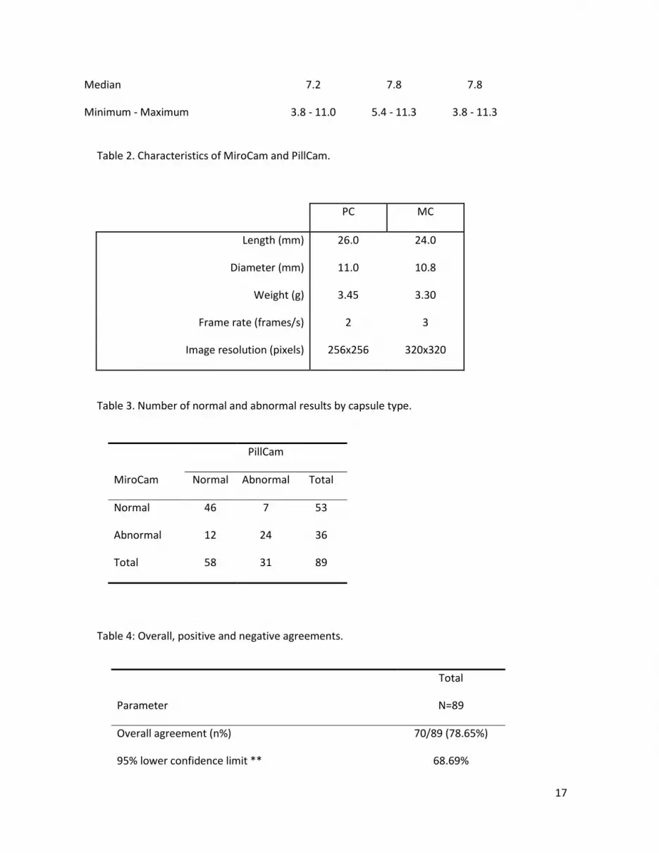
Table 1. Patient demographics and characteristics.
Parameter MiroCam First PillCam First Total p-value
Age (years)
0.6536
Number of subjects (n) 52 53 105
Mean (STD) 60.3 (15.89) 62.8 (12.85) 61.5 (14.42)
Median 63.0 63.0 63.0
Minimum - Maximum 22.0 - 87.0 28.0 - 87.0 22.0 - 87.0
Gender (n%)
1.0000
Male 28 (53.8) 29 (54.7) 57 (54.3)
Female 24 (46.2) 24 (45.3) 48 (45.7)
Ethinicity (n%)
0.5247
White 37 (71.2) 38 (71.7) 75 (71.4)
Native Hawaiian / Other Pacific
Islander 0 (0.0) 0 (0.0) 0 (0.0)
Black 7 (13.5) 3 (5.7) 10 (9.5)
Hispanic 6 (11.5) 7 (13.2) 13 (12.4)
Asian 2 (3.8) 2 (3.8) 4 (3.8)
American Indian / Alaskan Native
American 0 (0.0) 2 (3.8) 2 (1.9)
Other 0 (0.0) 1 (1.9) 1 (1.0)
Height (inches)
0.4815
n 52 53 105
Mean (STD) 66.1 (6.33) 65.9 (4.26) 66.0 (5.37)
Median 66.5 66.0 66.0

16
Minimum - Maximum 34.0 - 74.5 56.0 - 77.0 34.0 - 77.0
Weight (lbs)
0.4193
n 52 53 105
Mean (STD) 173.5 (46.34) 187.8 (54.73) 180.7 (51.01)
Median 170.0 170.0 170.0
Minimum - Maximum 52.0 - 313.9 110.0 - 332.0 52.0 - 332.0
Pregnancy (n%)
Negative 5 (9.6) 3 (5.7) 8 (7.6)
Positive 0 (0.0) 0 (0.0) 0 (0.0)
Pregnancy test not performed
(reason) 47 (90.4) 50 (94.3) 97 (92.4)
Male 28 (53.8) 29 (54.7) 57 (54.3)
Post-menopausal 17 (32.7) 18 (34.0) 35 (33.3)
Surgically sterile 2 (3.8) 3 (5.7) 5 (4.8)
Other 0 (0.0) 0 (0.0) 0 (0.0)
Occult bleeding (n%) 17 (32.7) 18 (34.0) 35 (33.3) 1.0000
Overt bleeding (n%) 20 (38.5) 23 (43.4) 43 (41.0) 0.6926
Iron deficiency anemia (n%) 46 (88.5) 49 (92.5) 95 (90.5) 0.5260
Blood transfusion within last 3 months (n%) 29 (55.8) 28 (52.8) 57 (54.3) 0.8454
Hemoglobin prior to last transfusion (g/dL)
0.1777
n 28 27 55
Mean (STD) 7.5 (1.80) 8.2 (1.49) 7.9 (1.68)
17
Median 7.2 7.8 7.8
Minimum - Maximum 3.8 - 11.0 5.4 - 11.3 3.8 - 11.3
Table 2. Characteristics of MiroCam and PillCam.
PC MC
Length (mm) 26.0 24.0
Diameter (mm) 11.0 10.8
Weight (g) 3.45 3.30
Frame rate (frames/s) 2 3
Image resolution (pixels) 256x256 320x320
Table 3. Number of normal and abnormal results by capsule type.
PillCam
MiroCam Normal Abnormal Total
Normal 46 7 53
Abnormal 12 24 36
Total 58 31 89
Table 4: Overall, positive and negative agreements.
Total
Parameter N=89
Overall agreement (n%) 70/89 (78.65%)
95% lower confidence limit ** 68.69%

18
95% upper confidence limit ** 86.63%
Positive agreement (n%)* 24/31 (77.42%)
95% lower confidence limit ** 58.90%
95% upper confidence limit ** 90.41%
Negative agreement (n%)* 46/58 (79.31%)
95% lower confidence limit ** 66.65%
95% upper confidence limit ** 88.83%
* Agreement was assessed by comparing the two independent readers' responses for each capsule to the question: 'Were you able
to identify the primary cause of small bowel bleeding? (Yes/No) ' .
**CI computed using Exact Binomial Test.
Table 5. Types of lesions identified by capsule type.
PillCam
MiroCam Normal
Ulcerative
lesions Tumor/Polyp
Vascular
lesions
Blood - lesion
unidentified Other Total
Normal 46 0 1 3 3 0 53
Ulcerative lesions 2 4 0 0 0 0 6
Tumor/Polyp 1 0 1 1 0 0 3
Vascular lesions 6 0 0 9 0 0 15
Blood - lesion unidentified 3 0 0 1 6 0 10
Other 0 0 0 0 2 0 2
Total 58 4 2 14 11 0 89

19
Table 6. Transit times.
MiroCam PillCam p-value**** ICR*****
(N=89) (N=89) 25th
/75th
percentiles
Esophageal transit time (minutes)
N 88* 87* NS 0.20 / 0.40
Mean (SD) 1.4 (6.6) 1.1 (2.0) 0.0183
Median 0.3 0.6
Minimum-Maximum 0.0-61.2 0.1-18.0
Gastric transit time (minutes)
N 89 88** NS 164.5 / 95.6
Mean (SD) 58.9 (66.4) 44.0 (56.0) NS
Median 30.2 21.5
Minimum-Maximum 1.1-366.3 1.3-304.6
Small bowel transit time (hours) 3.0 / 2.1
N 83 75 NS
Mean (SD) 6.6 (2.2) 5.2 (1.4) <0.0001
Median 6.3 5.1
Minimum-Maximum 2.3-11.8 2.4-7.9
Total transit time (hours) *** 70.7 / 70.5
N 74 74 NS
Mean (SD) 115.3
108.5
(147.1) NS

20
Median 53.5 45.8
Minimum-Maximum 5.2-742.6 3.1-652.7
Total operation time
N 89 89 NS 10.6 / 0.10
Mean (SD) 11.1 (1.5) 7.8 (0.8) <0.0001
Median 11.4 7.9
Minimum-Maximum 5.1-12.0 3.7-10.0
* A MiroCam capsule was retained in the esophagus for 139 minutes and a PillCam capsule was retained in
the esophagus for 261 minutes
** In one subject, the first image recorded by Pillcam was a duodenal image. As a result, no esophageal or
gastric transit time could be recorded in which an esophageal time could not be recorded
*** Time to capsule expulsion from the body
**** The p-value refers to the log-rank test comparing the two distributions with respect to the time to
event listed.
***** IQR: interquartile range with the 25th
and 75th
percentiles of the distribution of times to the events
Table 7. Adverse events.
Attribution
Adverse Event* (n %) Total
MiroCam (MC),
PillCam (PC),
(n=105)
Unknown (U), Both
(B)
Subjects with any events 7 (6.7%)

21
Retention of Capsule 3 (2.9%) B
Cardiac arrest 1 (1.0%) U
Skin blister at waist line 1 (1.0%) U
Contact dermatitis 1 (1.0%) M
Nausea 0 (0.0%)
Vomiting 0 (0.0%)
Abdominal Pain 1 (1.0%) U
Dizziness 0 (0.0%)
Syncope 0 (0.0%)
Intestinal Perforation 0 (0.0%)
Aspiration into Airway 0 (0.0%)
Figure 1. Randomization of patients.
1One subject was allowed to enroll twice; therefore 2 subject identification numbers were assigned. Subject data only
reported once. 2One subject terminated the study early but had evaluable reader data (subject did not return for
abdominal x-ray to confirm capsule passage and was lost-to-follow-up)3 One subject terminated the study early and did
not have evaluable reader data (subject could not swallow first capsule and withdrew) 4One subject’s imaging files
would not upload to the receiver due to technical issues and two subjects had one or more capsules that did not reach the
small bowel while operational.
Figure 2. Product Limit Survival Estimates

22

23
REFERENCES
1. Iddan G, Meron G, Glukhovsky A, et al. Wireless capsule endoscopy. Nature. 2000 May
25;405(6785):417.
2. Cave DR, Fleischer DE, Leighton JA, et al. A multicenter randomized comparison of the
Endocapsule and the Pillcam SB. Gastrointest Endosc. 2008 Sep;68(3):487-94.
3. Bang S, Park JY, Jeong S, et al. First clinical trial of the "MiRo" capsule endoscope by using a
novel transmission technology: electric-field propagation. Gastrointest Endosc. 2009
Feb;69(2):253-9.
4. Hartmann D, Schmidt H, Bolz G, et al. A prospective two-center study comparing wireless
capsule endoscopy with intraoperative enteroscopy in patients with obscure GI bleeding.
Gastrointest Endosc. 2005 Jun;61(7):826-32.
5. Triester SL, Leighton JA, Leontiadis GI et al. A meta-analysis of the yield of capsule
endoscopy compared to other diagnostic modalities in patients with obscure gastrointestinal
bleeding. Am J Gastroenterol. 2005 Nov;100(11):2407-18.
6. Mishkin DS, Chuttani R, Croffie J, et al. ASGE Technology Status Evaluation Report:
wireless capsule endoscopy. Gastrointestinal Endoscopy 2006:63:539-545.
7. Pennazio M, Santucci R, Rondonotti E et al. Outcome of patients with obscure gastrointestinal
bleeding after capsule endoscopy: report of 100 consecutive cases. Gastroenterology 2004;
126: 643–53.
8. Endo H, Kondo Y, Inamori M et al. Ingesting 500 mL of polyethylene glycol solution during
capsule endoscopy improves the image quality and completion rate to the cecum. Dig. Dis.
Sci. 2008; 53: 3201–5.
9. Höög CM, Bark LÅ, Arkani J, et al. Capsule retentions and incomplete capsule endoscopy
examinations: an analysis of 2300 examinations. Gastroenterol Res Pract. 2012;2012:518718.
10. Gao YJ, Ge ZZ, Chen HY et al. Endoscopic capsule placement improves the completion rate
of small-bowel capsule endoscopy and increases diagnostic yield. Gastrointest. Endosc. 2010;
72: 103–8.
11. Kim HM, Kim YJ, Kim HJ, et al. A Pilot Study of Sequential Capsule Endoscopy Using
MiroCam and PillCam SB Devices with Different Transmission Technologies. Gut Liver.
2010 Jun;4(2):192-200.