Embed Size (px)
Citation preview
A New Neoceratopsian Dinosaur Linking North American and Asian TaxaAuthor(s): Brenda J. Chinnery and John R. HornerSource: Journal of Vertebrate Paleontology, Vol. 27, No. 3 (Sep. 12, 2007), pp. 625-641Published by: Taylor & Francis, Ltd. on behalf of The Society of Vertebrate PaleontologyStable URL: http://www.jstor.org/stable/30126363 .Accessed: 04/03/2011 19:27
Your use of the JSTOR archive indicates your acceptance of JSTOR's Terms and Conditions of Use, available at .http://www.jstor.org/page/info/about/policies/terms.jsp. JSTOR's Terms and Conditions of Use provides, in part, that unlessyou have obtained prior permission, you may not download an entire issue of a journal or multiple copies of articles, and youmay use content in the JSTOR archive only for your personal, non-commercial use.
Please contact the publisher regarding any further use of this work. Publisher contact information may be obtained at .http://www.jstor.org/action/showPublisher?publisherCode=vertpaleo. .
Each copy of any part of a JSTOR transmission must contain the same copyright notice that appears on the screen or printedpage of such transmission.
JSTOR is a not-for-profit service that helps scholars, researchers, and students discover, use, and build upon a wide range ofcontent in a trusted digital archive. We use information technology and tools to increase productivity and facilitate new formsof scholarship. For more information about JSTOR, please contact [email protected].
The Society of Vertebrate Paleontology and Taylor & Francis, Ltd. are collaborating with JSTOR to digitize,preserve and extend access to Journal of Vertebrate Paleontology.
http://www.jstor.org

Journal of Vertebrate Paleontology 27(3):625-641, September 2007 c 2007 by the Society of Vertebrate Paleontology
ARTICLE
A NEW NEOCERATOPSIAN DINOSAUR LINKING NORTH AMERICAN AND ASIAN TAXA
BRENDA J. CHINNERY*,' and JOHN R. HORNER2 1School of Biological Sciences, University of Texas in Austin, 1 University Station, Austin, Texas 78712, U.S.A.,
[email protected] 2Museum of the Rockies, Montana State University, 600 W. Kagy Boulevard, Bozeman, Montana 59717, U.S.A.,
ABSTRACT--Basal (cladistically) neoceratopsians are relatively small, gracile members of Ceratopsia ('horned' dino- saurs), which also includes larger forms such as Triceratops and Centrosaurus. The Asian basal neoceratopsians share some very important traits not found in any North American group until now, including a fenestrated frill and premax- illary teeth. Likewise, the North American basal taxa have some traits not found in the Asian forms, the most important of which is a very specialized tooth wear pattern. Cerasinops hodgskissi, a new basal neoceratopsian from the Lower Two Medicine River Formation of Montana, exhibits all of the above characters along with others previously found on only one of the two continents. The new species is a sister group to Leptoceratopsidae in a cladistic analysis, and is a link between the taxa on the two continents. Cerasinops also exhibits extremely interesting anatomical and histological features that indicate the possibility of bipedality in this taxon, a locomotor pattern not found previously in basal neoceratopsians (it has been suggested in some, but with little evidence).
INTRODUCTION
Until recently only two genera and species have been de- scribed and are still recognized of basal (cladistically) neocera- topsian taxa (members of Ceratopsia ['horned' dinosaurs]). These are Leptoceratops gracilis (Brown, 1914) and Mon- tanoceratops cerorhynchus (Brown and Schlaikjer, 1942) as well as isolated elements and teeth identified as Neoceratopsia indet. (i. e., Ryan and Currie, 1998; Chinnery et al., 1998). One addi- tional genus has recently been described, Prenoceratops pie- ganensis (Chinnery, 2004a), and the current description is of an- other new genus and species, Cerasinops hodgskissi, thus dou- bling the number of North American genera and species in this taxonomic group. The new species described here reveals a mix- ture of traits known from taxa found in North America along with traits known only in the Asian forms (Fig. 1). Of the nu- merous mixed traits exhibited by Cerasinops, perhaps the most interesting are the unique tooth wear pattern shared by the North American taxa and the fenestrated frill and premaxillary teeth known only in Asian species until now.
Due to the differences between the Asian and North Ameri- can basal neoceratopsians, the taxa have not formed a monophy- letic clade in any of the more recent cladistic analyses. The North American species fairly consistently fall into a monophyletic group, usually termed Leptoceratopsidae (You and Dodson, 2004; Makovicky and Norell, in press). The Asian taxa are vari- ously grouped together into small clades within Ceratopsia, de- pending on the cladistic analysis used (Xu et al., 2002; Chinnery, 2004a; You and Dodson, 2004; Makovicky and Norell, in press). The inclusion of Cerasinops in a cladistic analysis shows an in- teresting removal from the Leptoceratopsidae clade, making it a sister group to the family and maybe linking the Asian and North American taxa closer together.
Cerasinops hodgskissi also has the unique feature of having very short and gracile forelimbs. Forelimb to hindlimb length ratio in basal neoceratopsians is of interest as a possible indicator of stance and locomotion. The sister group of Neoceratopsia, Psittacosauridae, is composed of taxa that have been determined to be bipedal based on limb length and anatomical features (Sereno, 1990; Chinnery, 2004b; You and Dodson, 2004). Al- though the larger ceratopsids do not exhibit any features of pos- sible bipedality, the smaller, more gracile basal neoceratopsians are closer in body form to psittacosaurids. However, limb length ratios and lack of manus size reduction has ruled out bipedality in most basal neoceratopsians. Cerasinops, however, has very short forelimbs, shorter than in some psittacosaurids, and has additional anatomical and histologic characters possibly associ- ated with a different use of the forelimb than the hindlimb.
-.l : ll ~-;l I ~Lr -r 'r`lF11~l l
''
l, '' 'r
.Y
'' ,, r I
~R~H~r :
l ; l l; .. .l i :illl~l '' "
.~ ...
r. r
FIGURE 1. Reconstruction of the skull of Cerasinops hodgskissi, MOR 300. Reconstruction is based on original art by Meredith Wolfe. Scale bar equals 5 cm.
625
*Corresponding author.

626 JOURNAL OF VERTEBRATE PALEONTOLOGY, VOL. 27, NO. 3, 2007
SYSTEMATIC PALEONTOLOGY
CERATOPSIA Marsh, 1888 NEOCERATOPSIA Sereno, 1986
LEPTOCERATOPSIDAE Makovicky, 2001 CERASINOPS HODGSKISSI, gen. et sp. nov.
Etymology-The genus name is from cerasinus (L., of cherry) + -ops (L., face), or 'face of cherry' referring to the red beds of the type locality and the dark red tinge of color on the specimen. This name also incorporates the nickname Cera, by which the specimen has been referred for over two decades. The species name refers to Wilson Hodgskiss, on whose property the speci- men was discovered.
Diagnosis-Member of Neoceratopsia with a unique charac- ter combination including: basioccipital tubera intermediate in shape between those of Protoceratops and Montanoceratops, a prootic that extends further caudally than in either Protoceratops or Montanoceratops, straight nasal, a short but high frill based on shape of the squamosal, deep posttemporal depression relative to that of other basal neoceratopsians, horizontal jugal crest in addition to the typical vertical one, a uniquely-shaped quadra- tojugal, extremely robust surangular relative to those of other basal forms, a narrow coronoid process of the mandible, and pronounced medial bend of the distal ulna. The specimen also has a low forelimb to hindlimb ratio (0.60, lower than in any other basal neoceratopsian), as well as unique bone histology of the forelimb elements (the forelimb elements have a longitudinal canal structure, relative to the circumferential structure found in the hind limbs and in all limbs of the comparison taxa).
Holotype Specimen--MOR (Museum of the Rockies) 300, associated skull and skeleton, about 80% complete. Included with the specimen are the basioccipital and basisphenoid fused together, the left paroccipital process with the exoccipital, pro- otic, and caudal portion of the laterosphenoid fused, the right exoccipital, and the unfused supraoccipital; both partial maxillae; both palpebrals; nearly complete left and partial right jugals; left and partial right quadratojugals; fused left and right frontals and partial left postorbital; right and left squamosals and partial pa- rietal; most of the right quadrate and part of the left; both man- dibles; most of the cervical, dorsal, and sacral vertebrae, several of the caudal vertebrae, the majority of the ribs, both scapulae, the left coracoid, both humeri, right and partial left ulnae, both radial shafts, fragmentary manus elements, very fragmentary pel- vic elements, right and partial left femora and tibiae, right fibula, astragalus, calcaneum, and left pes are preserved.
Referred Specimens-USNM (United States National Mu- seum) 13863 and 13864, both partial skeletons previously re- ferred to Leptoceratops sp. (Gilmore, 1939). The specimens are referred based on similarity of skull elements including the ba- sisphenoid, parietal, postorbital, maxilla, pterygoid, surangular, and quadrate. The surangular is especially diagnostic in Cerasi- nops, as it is extremely robust and has a thick buttress that con- tacts the coronoid process. These unique characters are found on both MOR 300 and USNM 13864 surangulars.
Horizon and Locality-The locality of the holotype specimen is Museum of the Rockies TM-012, Red Rocks Site, Teton County, Montana. Both referred specimens were excavated from a site designated as Locality TM-067, Pondera Co., Montana. All specimens are from the lower Two Medicine Formation (Cam- panian Stage), between ash beds dated at 76.5 and 80.0 mya (Rogers et al., 1993; Horner et al., 2001).
Figure Abbreviations-an, angular; aof, antorbital fossa; bo, basioccipital; bpt, basipterygoid processes; bt, basioccipital tu- bera; cnf, cranial nerve foramina; cp, coronoid process; eoc, ex- occipital; fen, parietal fenestra; fm, foramen magnum; hr, hori- zontal ridge; ice, internal carotid canal; lac, lacrimal attachment site; orb, orbit; oc, occipital condyle; p, parietal; pf, attachment
site of prefrontal; po, postorbital; sur, surangular; vr, vertical ridge.
DESCRIPTION
The specimen includes an articulated partial skull and skeleton of an adult individual. Skull material includes the caudal portion of the braincase, facial and frill elements, primarily unfused. The rostral portion of the face, including the rostrum, nasals, and dorsal facial elements, is not preserved. The postcranial skeleton is represented by the majority of vertebrae, ribs, and at least one of each appendicular element. The pelvic girdle is not preserved apart from the ischia and the sacral vertebrae. Some elements are better preserved than others due to the caliche surrounding the specimen, which tends to cause the ends of the elements, especially limb bones, to be more damaged than the midshafts. Measurements are listed in Table 1.
Skull
Basioccipital-As in other basal neoceratopsians, the Cerasi- nops occipital condyle is composed of two-thirds basioccipital and one-third exoccipitals. In the holotype the exoccipitals are not fused to the basioccipital, indicating possible immaturity of the specimen, but this unfused condition is not seen in Mon- tanoceratops and Prenoceratops specimens also determined to be immature (MOR 542 and TCM 2003.1.1). However, the condyle
TABLE 1. Skull and postcranial measurements of MOR 300, Cerasi- nops hodgeskissi (in mm).
Skull Occipital condyole mediolateral width
.............................................27.6 Nasal (USNM 13864) length..........................................................101.0e thickness at midline suture................................8.3 width at frontal contact.....................................17.3 Maxilla height--tooth row to antorbital fossa............73.2
Frontal length ................................. ........59.9e skull width at posterior orbit .........................158
Postorbital length--dorsal surface.....................................95.6e Squamosal length...................... .......................126.7e
height ...................... .......................121.5 Jugal length-dorsal..................... .....................120.2 height-orbit rim caudally .............................141.0 Quadrate height ................................ ....... 1790e
length condyles .......................................36.9 width condyles .............................................18.7
Quadratojugal length.........................................36.1 height.............................................................. .
e Dentary length-rostral end to coronoid process...194.5e
max. height with tooth row horizontal........139.2 shelf width at rostral coronoid process..........47.8 max. width across coronoid process...............62.3
Surangular length ................. .....................73.1 height .............. .......................92.3 rostral width
....................... ........................41.6 caudal width...................... ........................47.9 Articular length ................................ ..................... 0
width ..................................... .........20.1 Coronoid length ............................... .. ......42.1e
height .............................. ......................54.1 Appendicular skeleton Scapula length...................... .......................274 Coracoid length ................. .......................99 Humerus length .....................................................260.3 FemUlna length ....................... .................352 TibiaFemur length...................... ......................344.6 Tibia length .....................................................34 . Fibula length...................... .................................333 M etatarsal I length............................................................... 111.7 M etatarsal III length............................................................170.7 Metatarsal IV length .................... ... .....................143.4
Those measurements followed by e are of incomplete elements. Postcra- nial measurements were obtained from the one most complete element.

CHINNERY AND HORNER-NEW NEOCERATOPSIAN 627
of Cerasinops is spherical in shape, unlike the sub rounded con- dition seen in the immature specimens of Montanoceratops and Prenoceratops, but similar to that of a more mature specimen of Montanoceratops (Makovicky, 2001). The basioccipital tubera extend laterocaudally and lateroventrally around the occipital condyle, forming a shelf around the ventral portion of the con- dyle as in Montanoceratops (Fig. 2A, B). However, the shelf in Cerasinops is much shallower, and less concave, than in Mon- tanoceratops (MOR 542). It is intermediate between the flat tu- bera of Protoceratops and the highly curved ones of Mon- tanoceratops (Brown and Schlaikjer, 1940). The basal neocera- topsian tubera are formed from the fusion of basioccipital and basisphenoid, the latter providing the majority of the mass to the tubera (confirmed by the unfused components included with Prenoceratops; Chinnery, 2004a). On the rostral aspect of the midline union of the two components in Cerasinops is a promi- nent ovate fossa or pit as in Bagaceratops, some individuals of Leptoceratops, Protoceratops and some ceratopsids (Makovicky, 2001).
Basisphenoid-The basisphenoid of MOR 300 is relatively complete apart from the fragmentary and obscured rostral and superior borders. Just rostral to the basioccipital tubera is the internal carotid artery canal, which is much longer in Cerasinops than in Montanoceratops, Prenoceratops, and Leptoceratops (Chinnery, pers. obs.). The canal is also straight and relatively vertical, much more like that of Protoceratops than those of the North American genera. The canal is bordered primarily by the basisphenoid, with the dorsal portion enclosed laterally by the crista prootica. The basipterygoid processes extend down from the main body of the basisphenoid just rostral to the internal carotid artery canals. These processes in Cerasinops are similar to those in Asian forms over those of North American taxa. They extend ventrally almost vertically as in Protoceratops and Ba- gaceratops, with no caudal curvature as in Montanoceratops and Prenoceratops. Also, the basipterygoid processes in Cerasinops are not separated from the basioccipital tubera by a deep groove as in the other three North American genera, but are positioned closer together as in Protoceratops and Bagaceratops (Fig. 2B). The dorsal and rostral extents of the basisphenoid appear to be similar in Cerasinops to those of other basal forms, but the ros- tral border is not preserved and the dorsal border is obscured by deformation.
In USNM 13864, the basioccipital and basisphenoid complex, as seen from a ventral view, shows the midline ventral portion of the basioccipital tubera and the basisphenoid base. The basi- pterygoid processes are broken off. The midline of the tubera on both the paratype and the holotype includes one central crest with a shallow groove on either side. Just rostral to the midline of the tubera is an ovoid fossa. This fossa is located between and dorsal to the basipterygoid processes. The base of the processes are grooved for the internal carotid arteries which rise up be- tween the processes, wrap laterally around them, and then course nearly vertically up to the internal carotid foramina. The USNM specimen shows clear demarcation of the basioccipital and basisphenoid, showing that the basisphenoid contributes a large portion of the basioccipital tubera as in Prenoceratops (Chinnery, 2004a). The ventral border of the basioccipital ex- tends relatively further ventrally than on MOR 300, but the specimen shows lateral deformation and could be deformed dor- soventrally as well.
Prootic-As in other basal neoceratopsians, the Cerasinops prootic extends from the lateral surface of the paroccipital pro- cess ventrally to the basisphenoid. In Cerasinops this element extends further caudodorsally on the paroccipital process than in Montanoceratops and Protoceratops. The caudal border of the prootic forms the lateral wall of the middle ear region and the foramen ovale. Rostral to the middle ear region is the small foramen for cranial nerve (CN) VII, and rostral to this is the CN
V2 foramen. The articulation for the laterosphenoid is a heavy buttress dorsally. Medial to this is the articular surface for the supraoccipital - a transverse ridge from the buttress that curves caudally as it continues on the paroccipital process. The ventral aspect of the prootic is not preserved. The Cerasinops prootic exhibits a curving ridge from the paroccipital process to the but- tress for articulation with the laterosphenoid. This ridge is not nearly as prevalent in the immature specimen of Montanocera- tops (MOR 542) but is seen in Protoceratops (Brown and Schlaikjer, 1940).
Laterosphenoid-Only a fragment of the laterosphenoid is preserved with the Cerasinops braincase material. The fragment forms the rostral border of the foramen for CN V2. Articulations with the supraoccipital, basisphenoid and pterygoid are not pre- served, and those with the frontal and postorbital are only pre- served as facets on the latter elements.
Exoccipital-Neither of the Cerasinops holotype exoccipitals are complete distally. As discussed before, the left one is fused to its corresponding prootic, while the right one exhibits the articu- lar surface for this element. No sign of the opisthotic is present. Length and rostrocaudal dimensions of the Cerasinops exoccipi- tal do not appear to vary significantly from those of other basal forms (Figs. 2A, B). The pterygoid facet, located at midlength of the exoccipital, is small on Cerasinops. It is similar in relative size to the one in the immature Montanoceratops specimen (MOR 542). At the caudoventral corner of the exoccipital are three foramina for cranial nerves X through XII as in other basal forms.
Supraoccipital-The Cerasinops supraoccipital indicates a definite contribution of this element to the foramen magnum, also found in Protoceratops, Leptoceratops, Bagaceratops, and most likely Montanoceratops (contrary to Chinnery and Weishampel, 1998; Makovicky, 2001). Dorsal to the foramen magnum a single prominent midline ridge extends up to the parietal (Fig. 2A). Lateral to the midline ridge is a thick trian- gular articular surface for contact with the paroccipital process as it widens proximally. The lateral extent of the supraoccipital is not preserved. The contact with the parietal is present but ob- scured by deformation. The supraoccipital of Cerasinops is ori- ented in the same plane as the basioccipital tubera as in other North American basal neoceratopsians (Makovicky, 2001). This condition is not found in the Asian Archaeoceratops, Protocera- tops, or Bagaceratops.
Facial Elements
Premaxilla-The only known portion of the Cerasinops pre- maxilla is a fragment preserved with paratype USNM 13863 (Fig. 3E). This specimen includes the roots of two premaxillary teeth. Premaxillary teeth are absent in all of the other three North American basal taxa, but are known to occur in the Asian taxa Protoceratops, Liaoceratops, Archaeoceratops, and Chaoyang- saurus (but not in all Asian taxa). The preserved fragment is from the left side of the face and exhibits the entire tongue-in- groove sutural surface for contact with the rostral process of the maxilla (discussed for Prenoceratops-Chinnery, 2004a) and is therefore probably a portion of the caudal half of the element. The Cerasinops fragment does not extend far enough rostrally to include the articular contact for the opposing premaxilla or the rostral bone, and the caudal border is also not preserved. The premaxillary tooth roots are both broken off at the level of the alveoli, but can be seen to be cylindrical and small. The more rostral of the two tooth roots is only one half the diameter of the more caudal one, a condition similar to that found in Protocera- tops (Brown and Schlaikjer, 1940). The ventral border of the fragment narrows rostrally, and the conclusion of the articular groove for contact with the rostral process of the maxilla signifies that the caudal end of the element is close by.

628 JOURNAL OF VERTEBRATE PALEONTOLOGY, VOL. 27, NO. 3, 2007
SO
oc
eoc
Fm cnf
bt bt
A bpt
B icc
bpt
fen fen
C D
po
\E -
il ,
FIGURE 2. Skull elements of Cerasinops: A, caudal view of braincase of MOR 300; B, left side of braincase and caudal portion of skull of MOR 300; C, parietal of MOR 300 in dorsal view; D, portion of parietal bar of USNM 13864, again in dorsal view; E, dorsal view of frontal and left postorbital of MOR 300; F, frontal of USNM 13864 in dorsal view. Scale bar equals 3 cm.

CHINNERY AND HORNER-NEW NEOCERATOPSIAN 629
Maxilla-The maxillae of the holotype are partially preserved, from the caudal border forward to around half the length of the tooth battery, and the dorsal borders are unknown (Fig. 3C). Paratype USNM 13863 also includes a partial right maxilla. The known portion of the Cerasinops maxilla is similar to that of Leptoceratops, except that the tooth battery is straight in the former and ventrally convex in the latter. Leptoceratops is the only currently known North American taxon to have a convexly- curving tooth row, and interestingly shares this trait with Uda- noceratops from Asia. The preserved portion of the rostral ex- tent of the Cerasinops maxilla is tall, indicating that the antor- bital fossa is located further up on the face than in Montanocera- tops, Leptoceratops, and maybe Prenoceratops. Shape of the ant- orbital fossa can not be determined. The left maxilla included in MOR 300 is fused with the ectopterygoid, and the caudal border of the right maxilla clearly shows the articulation with the right ectopterygoid.
Nasal-The left nasal of paratype USNM 13864 is mostly com- plete, and a fragment of the right nasal of the same specimen is also available for study (Fig. 3D). The Cerasinops nasal is shorter and stouter than that of Prenoceratops, but is even straighter than in that genus, with no flattening of the element. The nasal of Montanoceratops is currently unknown, and the nasal of Lep- toceratops flattens and dips ventrally toward the top of the skull, in consequence exhibiting a concave area at the midline. Also, caudally the Leptoceratops nasal widens greatly in front of the orbits. The Cerasinops nasal has a constricted caudal end, almost but not quite as constricted as in Prenoceratops (Chinnery, 2004a). The articular area for the prefrontal is relatively much larger in Cerasinops than in Prenoceratops. The articular area for the caudal portion of the Cerasinops premaxilla is also present, but the nasal is broken above the contribution to the external naris. The internal surface of the nasal is smooth with a lightly defined channel for the nasal passage. Caudal to this is the over- lapping and grooved articular area for the frontal. The midline suture is of the tongue-in-groove variety discussed in Prenocera- tops (Chinnery, 2004a).
Prefrontal-One partial left prefrontal is preserved with para- type USNM 13864. The rostrodorsal corner of the orbit is a right angle, although the internal orbit border has a smoother curve. Extent of the contribution to the dorsal and rostral orbit borders is unknown. The fragment shows a well defined groove and ar- ticular area for articulation with the palpebral, unlike in Lepto- ceratops (Sternberg, 1951). A portion of the internal groove for articulation with the frontal is present.
Palpebral-The Cerasinops palpebral is shaped similarly to an obtuse triangle. The palpebral is very large relative to the pre- served orbit fragments and in comparison to that of Leptocera- tops (Plate XLIX, Sternberg, 1951).
Jugal-The left jugal of MOR 300 is fused to the quadratoju- gal. The jugal crest is well-developed, and the orbit rim curves out laterally from the main body. The rostral border curves ros- troventrally and the ventral tip is positioned more directly below the dorsal tip, unlike the caudally-oriented jugals of Leptocera- tops and Protoceratops. Prenoceratops is currently the only other basal neoceratopsian with this unique curve in the rostral border. The Cerasinops jugal is unique in the possession of a crest ori- ented at ninety degrees from the main dorsoventral jugal crest. This secondary crest is not as prominent as the vertical one but is distinct. It runs from the tip of the jugal horn rostrally to the rostral border, just opposite the articular notch for the maxilla. A notch just caudal to the orbit rim shows where the postorbital articulates with the jugal, but the dorsal extent of this portion is not preserved. The jugal horn is located at the caudal border of the element. The jugal extends inward from this caudal border at a right angle as in Leptoceratops, forming a slightly concave caudal surface. The extent of the element medially is greater than in Leptoceratops or Prenoceratops. The ventral tip of the
Cerasinops jugal curves rostrally as in Prenoceratops. No epijugal is present, and no clear suture marks on the jugal or quadrato- jugal for attachment of the epijugal are present.
Only a fragment of the left jugal is present with the USNM 13864 material, showing part of the rostral portion of the element with a portion of the ventral border. The fragment is significant as on the external surface the horizontal ridge characteristic of the Cerasinops holotype is preserved. Position of the fragment is confirmed by the notch for articulation with the maxilla on the internal surface and the ventral border segment.
Quadratojugal-The quadratojugal of Cerasinops is triangu- lar, robust, and has a straight caudal border for articulation with the quadrate (Fig. 3B). The element thickens markedly in the midsection, unlike in Leptoceratops where the thickest part is located further ventrally (Sternberg, 1951). The Cerasinops quadratojugal extends three centimeters below the jugal, and wraps around the internal, rostral, and caudal borders of the nutrient foramen located here. The thickened area of the bone is buttressed against the ventral tip of the jugal, above which the quadratojugal narrows. At this point the quadratojugal is articu- lated on the internal caudal surface of the jugal. The Cerasinops epijugal is much thicker than that of Prenoceratops, and differs in shape from those of Protoceratops and Leptoceratops.
Frontal-The holotype frontals are incomplete and fused to- gether at the midline (Fig. 2E). The Cerasinops frontals are more concave than in Leptoceratops or Prenoceratops. A low ridge, curving caudolaterally, delineates the dorsal surface from the frontal depression. The frontal depression is deep as in Preno- ceratops. A low midline ridge extends from the rostral end of the depression and joins up with the rostral end of the parietal bar. In contrast, in Leptoceratops a tall midline ridge is present on the dorsal surface of the frontals, but it does not extend through the (shallower) frontal depression (Sternberg, 1951). Rostral por- tions of the element are not preserved apart from a partial ar- ticular surface for the prefrontal orbit rim contribution. The su- tural contact with the postorbital is almost horizontal, but is slightly angled caudally towards the midline. This contact is angled more acutely in Leptoceratops. The holotype frontal pair is fused to the left postorbital, and the suture line is visible. Caudally the frontals are fused to fragments of the parietal and supraoccipital. The internal aspect of the dorsal portion of the braincase is smooth but distorted. However, it appears that the caudodorsal braincase was roofed primarily by the supraoccipital and to a lesser extent by the parietal. The internal orbit roof is also smooth, with varying nutrient foramina. The external sur- face above the orbits is rugose, with radiating foramina, and the frontal depression surface is smooth.
Portions of both left and right frontals are included with USNM 13864 (Fig. 2F). The fragments are associated with each other, but appear to be glued together rather than fused. The most prominent comparable feature of the frontals is the deep frontal depression, delineated from the rest of the element by a prominent ridge that curves laterally and caudally. The depres- sion is also characterized by a midline ridge that meets the pa- rietal bar. The rostral portion of the frontals is concave in caudal view. The USNM frontals both include the frontal portions of the orbit rims, with sutures for the prefrontals on both sides and the postorbital on the left. This area between the orbits is rugose and has a set of nutrient foramina opening out of the dorsal aspect in radiating lines lateral to the midline. The same foramina and rugose texture are also visible on MOR 300, but the orbit rim areas are not preserved on this specimen. No apparent differ- ences exist in the dorsal view of the elements. In ventral view, grooves for the orbitosphenoid and ethmoid are similarly curved caudolaterally in the paratype and holotype.
Postorbital-The left postorbital is partially preserved, fused to the frontals in MOR 300 (Fig. 2E). The element is robust, with a wide sutural union to the frontal (about three cm.). Only a

630 JOURNAL OF VERTEBRATE PALEONTOLOGY, VOL. 27, NO. 3, 2007
small portion of the dorsal orbit border is formed by the post- orbital before it curves ventrally forming the caudal orbit rim. As with the prefrontal, the outer orbit rim has a ninety degree bend at the dorsocaudal corner, only slightly less sharp than on the prefrontal. The inner orbit rim has a gentler slope in this area. The caudal and ventral extent of the element is unknown. As the postorbital extends laterally and caudally from the frontal con- tact, the element turns caudally abruptly creating a corner or ridge. This ridge, extending from the dorsocaudal orbit rim, can also be seen in Protoceratops, Leptoceratops, and Montanocera- tops, but in these latter taxa the ridge is much more prominent than in Cerasinops. The ridge doesn't exist at all in Prenocera- tops. The internal surface of the Cerasinops postorbital is smooth, with a shallow oval fossa for articulation with the lat- erosphenoid. The dorsal end of the jugal articulation is pre- served, showing that this part of the jugal has a horizontal squared-off ascending process.
A fragment of the left postorbital is preserved with USNM 13864. The fragment includes the dorsocaudal orbit border, showing the outer ninety-degree angle and the more gradually curved inner border. Internally the USNM specimen and MOR 300 have the groove for articulation with the laterosphenoid just caudal to the orbit border. The paratype has a groove traversing the dorsal orbit rim for blood vessel and nerve transmission. This groove is obscured on MOR 300 by a break, and may or may not be there. Otherwise, both specimens are similar in shape, but the MOR 300 one is larger overall.
Squamosal--The Cerasinops squamosal is similar in shape to those of other basal taxa, but does exhibit some unique charac- teristics (Fig. 3A). The rostral portion of the element is not pre- served, but it was relatively very tall above the infratemporal fenestra, evidenced by the position of the preserved dorsocaudal corner of the fenestra rim. The dorsocaudal tip of the squamosal extends slightly medially and quite far dorsally, more so than in Prenoceratops. In Montanoceratops and especially in Protocera- tops this part of the element extends further caudally, while in Cerasinops it abruptly turns dorsally suggesting a short but high frill. From a caudal view the Cerasinops squamosal is very wide like those of Leptoceratops and Protoceratops, but unlike the slim squamosal of Prenoceratops. As in other basal neoceratop- sians, the squamosal forms a u-shape in dorsal view between the external main body and the internal wing that extends up to meet the parietal. Although not complete, the internal wing of the Cerasinops squamosal extends far up at the rostral end but then shortens distally as the parietal extends further laterally. The articulation for the parietal consists of longitudinal grooves. The articulation for the head of the quadrate is deep but otherwise similar to that of other basal taxa.
Parietal-Much of the caudal border of the parietal is pre- served, along with a fragment that articulates with the left squa- mosal and a fragment of the rostral end of the parietal bar fused to the frontal complex (Figs. 2C, D). The Cerasinops parietal includes distinct fenestrae, a feature not previously described for any North American basal genus. Leptoceratops has a solid frill, and currently the frill of Montanoceratops and Prenoceratops are unknown. The Cerasinops parietal flares laterally very briefly at the most rostral end, but narrows immediately to form the nar- row, high midline parietal bar. Most of the bar is missing, but a segment remains at the caudal end where it joins with the caudal fenestra borders. The lateral border portions are smooth and gently curved laterally as in some younger Protoceratops skulls (Brown and Schlaikjer, 1940). In the latter, however, a slight dip occurs at the midline in dorsal view, while in Cerasinops this area has a straight border. There are no indications of epoccipitals, but faint striations on both dorsal and ventral surfaces indicate attachment of soft tissues. The parietal fenestrae in Cerasinops are located closer to the midline than in Protoceratops, with the medial borders located ventral to the midline bar, which is el-
evated relative to the fenestrae. The fenestrae extend laterally almost to the squamosal contact, evidenced by the preserved parietal fragment that articulates with the left squamosal. This lateral extension of the fenestrae is also found in Protoceratops, but unlike in Protoceratops the caudal corners of the Cerasinops fenestrae curve much more gently, suggesting deeper, more rounded fenestrae in Cerasinops. The lateral contact with the squamosal indicates that the parietal extends further caudally than the squamosal, as the caudal parietal border is angled up at 45 degrees from the horizontal when articulated. The union of the parietal and squamosal is smooth, the curve dorsally of the squamosal continuing on with the parietal.
The parietal fragment preserved with USNM 13864 includes the caudal portion of the parietal bar and corresponding caudal frill border. Also included is a portion of the left fenestra border. On the dorsal surface the parietal bar has three uneven ridges running lengthwise. These ridges end where the bar merges with the lateral processes, at which point the element becomes smooth. The three uneven ridges are also on MOR300. The USNM specimen is different in that the merge of the midline bar and the lateral processes is smoother-on MOR 300 the dorsal aspect of the midline bar is raised relative to the lateral pro- cesses, and a bump is created on the lateral processes where the bar joins them. This raising of the midline bar on MOR 300 becomes more apparent just rostral to the lateral processes. As the caudal borders of the fenestrae curve inward to become the medial borders, they converge under the dorsal parietal bar, forming a depression or fossa on either side of the bar. This structure makes the midline bar tall overall. On USNM 13864 the fenestra borders do not converge as close to each other, but stay lateral to the midline bar, which is thus much shorter between fenestra borders. The ventral aspect of both specimens includes an off-center ridge running axially along the parietal bar.
Quadrate-The element is long and straight, with no caudal arch as in Montanoceratops and Prenoceratops. The Cerasinops quadrate is narrow rostrally and robust caudally at the contact with the quadratojugal. The articular area for contact with the quadratojugal is wider than the rest of the element, with a strong protrusion caudally about one third the distance up from the ventral end. The condyles are unequal in shape, the lateral one larger and the medial one smaller, and when articulated with the mandible the medial condyle is situated further rostrally than the lateral one. These conditions are also found in Prenoceratops.
On the USNM 13864 specimens, the articular area for the quadratojugal is taller and narrower than on MOR 300, and has a smoother surface. These differences could be due to a differ- ence in maturity, as the USNM 13864 skeleton is smaller overall than that of MOR 300. Otherwise the quadrates are similar.
Mandible
Both mandibles of the holotype are preserved, with dentaries fused to the caudal elements, but the left one is distorted and both are missing the rostral end. The mandible of Cerasinops is deep as in Leptoceratops, but has a narrower rostral end and more robust caudal end. The tooth row is straight compared to the ventrally curved ones of Leptoceratops and Udanoceratops, and the coronoid process is angled further caudally in Cerasinops than in Leptoceratops. From a lateral view, the caudal end of the Cerasinops mandible is oriented at a steeper angle, relative to the tooth row, than in Leptoceratops.
Dentary-The Cerasinops dentary is deep, the coronoid pro- cess is narrower from a lateral view, and the rostral end is nar- rower than in Leptoceratops (Fig. 3F). The tooth row is posi- tioned dorsal to the lateral shelf as in older specimens of Proto- ceratops, as well as in Prenoceratops and Montanoceratops. The ventral aspect of the rostral end of the Cerasinops dentary curves inward more than in Leptoceratops. Otherwise, the dentaries of

CHINNERY AND HORNER-NEW NEOCERATOPSIAN 631
P ----D l"
po
vr
orb
iL~ po
hr
B A
, pf aof
D lac
cp SUr
sur l H
G a n
FIGURE 3. Skull elements of Cerasinops: A, lateral view of left squamosal of MOR 300; B, lateral view of right jugal and quadratojugal of MOR 300; C, lateral view of left maxilla of MOR 300; D, lateral view of left nasal of USNM 13864; E, ventral view of left premaxilla of USNM 13864; F, lateral view of right mandible of MOR 300; G-I, surangular of USNM 13864 in dorsal, lateral, and rostral views, respectively. The arrows in E, G, and H point in the rostral direction. Scale bar equals 3 cm.

632 JOURNAL OF VERTEBRATE PALEONTOLOGY, VOL. 27, NO. 3, 2007
the two genera appear very similar in dorsal view. However, the caudal border of the Cerasinops dentary differs markedly as the coronoid process is much narrower than in Leptoceratops and has a straight caudal border down to the articulation with the angular. Below this point the sutures are unknown due to miss- ing and poorly preserved areas, but the surangular and angular extend much further rostrally in Cerasinops, well beyond the rostral border of the coronoid process. The coronoid process does not extend as far inward as in Leptoceratops, but further than in Prenoceratops and Montanoceratops (in dorsal view). A portion of the ventral predentary contact is preserved, and is very wide. Since the rostrodorsal tip of the dentary is missing, the rostral predentary contact and pit are not known. The isolated dentary described by Ryan and Currie in 1998 is not referable to Cerasinops, as it exhibits a much taller rostral end than in Cera- sinops. In addition, the contacts for the surangular and articular below the coronoid process are located further caudally on the Leptoceratops sp. specimen than on the Cerasinops one.
Predentary-Only a midline portion of the predentary is pre- served with the paratype (USNM 13864). This fragment provides no information on the shape or extent of the element or its sutural contacts.
Surangular-Unique in its shape, the Cerasinops surangular is extremely robust, with a thick buttress of bone at the contact with the coronoid process of the dentary (Fig. 3F-I). The holo- type surangulars are both preserved, and fused with surrounding mandible elements (Fig. 3F). Partial right and left surangulars are also included with USNM 13864. The unique shape of the surangular is one of the primary reasons for a lack of identifica- tion of USNM 13864 and 13863 until now (Gilmore, 1939; Chin- nery, 2004a). The Cerasinops surangular includes a robust ridge running from the coronoid process of the dentary back to the caudal end of the glenoid. This ridge does not form a deep pocket on the lateral side of the mandible, but instead is located at more of a dorsal angle. In lateral view the surangular is con- vexly curved, another feature not seen in other North American basal neoceratopsian forms, although some Protoceratops indi- viduals do exhibit this (Brown and Schlaikjer, 1940). As dis- cussed above, the Cerasinops surangular extends much further rostrally than in other basal forms. In dorsal view the Cerasinops surangular is wider than in Leptoceratops and other taxa due again to the heavy ridge. The element extends caudally from the coronoid process, has a medial flange that encloses the rostral end of the articular, forms a long, shallow cotyle for articulation with the lateral condyle of the quadrate, and again extends me- dially and slightly caudally forming a squared caudal end of the mandible. The lateral ridge enclosing the cotyle is low, unlike the tall ridge found in Leptoceratops.
USNM 13864 includes a mostly complete right surangular with the articular area for the quadrate condyle. The preserved por- tion includes the extension caudal to the glenoid and cranial to the articular surface for the dentary. Also included is a fragment of the left one including the rostral end of the element at the dentary contact, showing the dorsal and ventral extent of the contact. This fragment shows that the element does narrow down to a short flange that fits inside the divided caudal border of the coronoid process of the dentary. The USNM surangular is smaller than that of MOR 300 and the paratype has a slightly deeper cotyle of the glenoid, but otherwise no differences are apparent.
Articular-The Cerasinops articular is oval, wide but not long, sitting at a 30 degree angle from the transverse. The rostrolateral end of the element is surrounded by the surangular. The shape is more oblong than in Leptoceratops, in which it is more circular. In Prenoceratops the articular is also oblong but is oriented al- most directly craniocaudally. In Montanoceratops the element is also oriented craniocaudally and is wider than in Prenoceratops and Cerasinops. The glenoid for the quadrate is angled back very
steeply in Cerasinops, suggesting that the quadrate extended fur- ther caudally than in Leptoceratops, similar to the condition in Montanoceratops and Prenoceratops. The glenoid indicates that the quadrate condyles sit at an angle in Cerasinops, with the medial condyle positioned rostral to the lateral condyle as in Prenoceratops and Montanoceratops. This feature has not been discussed for the Asian taxa.
Angular-Much of the preserved right angular is missing, and the sutures with the dentary and surangular are obscured by fusion and poor preservation. Generally the angular appears to resemble those of other basal forms, but in Cerasinops it appears to extend further rostrally as does the surangular.
Coronoid-The Cerasinops coronoid is much larger than the corresponding coronoid process of the dentary, extending fur- ther rostrally, caudally, and dorsally. The lateral surface has a strong ridge running dorsoventrally that sits between the dentary and the surangular. The ventral extension is not preserved.
Teeth-Preserved teeth of Cerasinops are nearly all worn and in place in the mandibles and maxillae. Tooth morphology con- forms to that seen in Leptoceratops and Prenoceratops, but Cera- sinops does not have a primary ridge on the buccal side of the dentary teeth as described for Montanoceratops (Chinnery and Weishampel, 1998). The tooth wear pattern of Cerasinops is the same as that found in all known North American genera as well as Udanoceratops from Asia. The teeth are used to shear against each other in a vertical manner, creating a vertical wear pattern on both the maxillary and mandibular teeth. The mandibular teeth of these genera have a bulbous shape on the shearing side (the buccal side), which becomes a horizontal shelf through wear against the maxillary teeth.
Postcrania
Axial Skeleton-The first three cervical vertebrae are not fused in Cerasinops. Only the axis is preserved, and is similar in shape to that of Leptoceratops (NMC 8889). The neural spine is not as axially long as in Protoceratops. Four other cervical ver- tebrae are preserved, three of them anterior cervicals and the other one from the middle of the series. The most cranial one does not exhibit the ventral orientation of the transverse pro- cesses as C4 does in Montanoceratops, but the three more cranial of the Cerasinops vertebrae are axially short as in the proximal cervicals of Montanoceratops. Ventral crests are visible on all cervical vertebrae of MOR 300.
All but the very first dorsal vertebrae are preserved with the Cerasinops holotype (Figs. 4A-F). The first three have transverse processes at a very high angle, higher than in Montanoceratops, and from the fifth one caudally the angle decreases until by D11 the processes are horizontal. The centra are oval in cranial view, taller than mediolaterally wide, but the width does increase to- ward the sacrum. The capitular facets do not migrate out on to the transverse processes, but are already positioned out on the processes in D2 and only move laterally a small amount through- out the series. Nearly all of the neural spines are broken, so trends in height change can not be determined. The pre- and postzygopophyses of Cerasinops exhibit a tongue-in-groove ar- ticulation but not to the extent seen in Montanoceratops. The dorsal vertebra zygopophyses of Graciliceratops, Protoceratops, and Archaeoceratops also exhibit the tongue-in-groove condi- tion, while those of Leptoceratops and Udanoceratops are smooth (Makovicky, 2001).
Unlike in Montanoceratops, fusion of neural arches to centra does not progress in a caudal direction in Cerasinops. The axial neural spine is not fused to the centrum. The three more cranial of the free cervical vertebrae are fused, but all dorsal vertebrae back to D10 are unfused. Dorsal vertebrae 10 to 12 do exhibit fusion of neural arches to centra, but almost no fusion is found in sacral vertebrae and the caudals are completely mixed.

CHINNERY AND HORNER-NEW NEOCERATOPSIAN 633
A C E
I
B DF
I
L N
I
K
J K M
,
FIGURE 4. Postcranial elements of Cerasinops, MOR 300: A-F, dorsal vertebrae in cranial (A, C, E) and right lateral (B, D, F) views; G, lateral view of left coracoid; H, lateral view of left scapula; I, dorsal view of left scapula; J, dorsal (lateral) view of right humerus; K, caudal view of right humerus; L, lateral view of right ulna; M, cranial view of right ulna; N, cranial view of right femur; O, cranial view of right tibia and fibula; P, dorsal view of left pes. Scale bar equals 3 cm.
Fragments of six or possibly seven sacral centra are preserved, along with miscellaneous fragments of the neural arches. Only two of the more caudal sacral centra appear to be fused. A ventral crest is seen on the more caudal of the centra, but not on
the others. The more cranial sacral centra are wider than they are tall, but they narrow caudally, until the most caudal of the pre- served centra is taller than it is wide. The neural spines are too fragmentary to find any distinguishing characteristics.
634 JOURNAL OF VERTEBRATE PALEONTOLOGY, VOL. 27, NO. 3, 2007
The Cerasinops caudal vertebrae have tall and straight neural spines and rod-like distal chevrons. Sixteen caudal centra are preserved from the proximal end of the tail, some completely fused to the neural spines, some half fused, and some unfused. Location in the series does not seem to be a factor. The tallest, but broken, neural spine is approximately seven cm tall. No ven- tral crests are present.
Both series of ribs of MOR 300 are fairly complete. The rib cage of Cerasinops is laterally compressed as in other basal neo- ceratopsians (Chinnery and Weishampel, 1998), and the ribs are tall and axially narrow. No distinguishing characteristics are present.
Pectoral Girdle-The Cerasinops pectoral girdle is very simi- lar to those of other basal neoceratopsian taxa. The element with the most distinguishing characteristics, the clavicle, is unfortu- nately not currently known for Cerasinops (Chinnery and Weishampel, 1998).
Both scapulae of the holotype are partially preserved, as well as fragments included with USNM 13864 (Figs. 4H, I). The ele- ment is long but not relatively tall. The proximal end is much taller than the distal end. The dorsal border of the scapula is strongly ventrally concave, while the ventral border is straight. The glenoid fossa is formed from equal contributions of the scapula and coracoid. The distal end of the Cerasinops scapular blade is very narrow, with almost no transverse flaring. The shape of the scapular blade resembles that of adult Protoceratops over those of other taxa (Brown and Schlaikjer, 1940). The Cera- sinops scapula is less laterally curved than in Montanoceratops or Leptoceratops.
The left Cerasinops coracoid is mostly preserved along with fragments of the right one (Fig. 4G). The element is long as in Protoceratops (Brown and Schlaikjer, 1940), but appears to have a rounded sternal edge as in Montanoceratops (MOR 542). An articular surface for the clavicle is not visible. The coracoid of Cerasinops forms one half of the glenoid fossa.
One partial sternal plate is preserved with MOR 300. The cranial end of the reniform element is narrower than in Mon- tanoceratops, and the position of the medial border suggests that the element is narrower overall than those of Montanoceratops and Leptoceratops. Also, the lateral border exhibits less of a curve than seen in other basal forms.
Forelimb-The articulated skeleton of the Cerasinops holo- type includes a forelimb that is very much shorter than the hind- limb. In fact, the ratio of forelimb to hindlimb (humerus + radius / femur + tibia) is as low in Cerasinops at .60 as in Psittacosaurus at .62 (Maryaiska and Osm6lska, 1975). This ratio in Leptocera- tops ranges from .64 to .72 (the smallest ratio is found in the youngest individual, NMC 8887). Forelimb to hindlimb ratios in Protoceratops, Prenoceratops, and Microceratops are also higher than in Cerasinops and Psittacosaurus (see Table 2). The short length of the Cerasinops forelimb suggests the possibility of a different use of the forelimb in this genus. The ratio of forelimb to hindlimb has been used as an argument for bipedalism in Psittacosaurus (Sereno, 1990; You and Dodson, 2004). Psittaco- saurus has been found to have additional anatomical correlates that strengthen the argument of bipedalism in this group, includ- ing pelvic elements that support weight caudally in comparison to those of neoceratopsians (Chinnery, 2004b) and a reduced manus (Sereno, 1990). The manus and pelvic elements of Cera- sinops are not preserved well enough to provide any functional information, but histological examination of the limb bones shows additional differences between forelimb and hindlimb el- ements in this specimen (see below).
The humerus of Cerasinops is very gracile compared to those of other basal forms, especially at the proximal end (Figs. 4J, K). Similar to other elements in this skeleton, the proximal and distal ends are not well preserved in the right humerus of MOR 300 and not preserved at all in the left one. However, it is likely that the distal end of the element is actually nearly as wide, in caudal view, as the proximal end. The proximal portion of the element, from the deltopectoral crest proximally, is oriented at an angle relative to the shaft and distal end (not unusual for neoceratop- sians). The deltopectoral crest is only well developed at the distal end, and is oriented 45 degrees caudal from the head. The lateral condyle does not extend as far distally as the medial condyle, a character found among many basal neoceratopsian taxa. The shape of the Cerasinops humerus is most similar to that of Preno- ceratops over the more robust humeri of Leptoceratops and Pro- toceratops. The relatively gracile Cerasinops humerus may not have been used for primary weight support and locomotion, as discussed below.
The right ulna of MOR 300 is fairly complete except for the
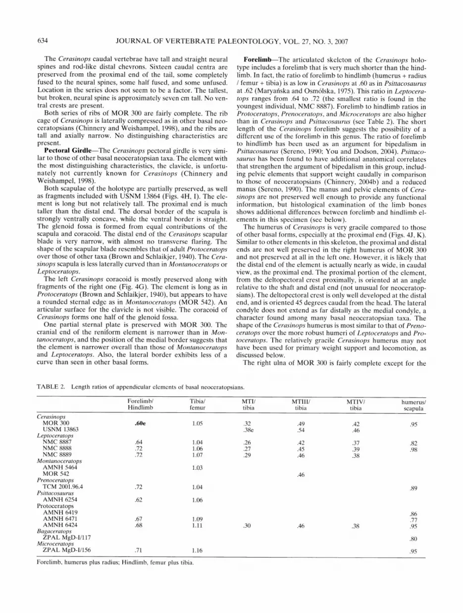
TABLE 2. Length ratios of appendicular elements of basal neoceratopsians.
Forelimb/ Tibia/ MTI/ MTIII/ MTIV/ humerus/ Hindlimb femur tibia tibia tibia scapula
Cerasinops MOR 300 .60e 1.05 .32 .49 .42 .95 USNM 13863 .38e .54 .46
Leptoceratops NMC 8887 .64 1.04 .26 .42 .37 .82 NMC 8888 .72 1.06 .27 .45 .39 .98 NMC 8889 .72 1.07 .29 .46 .38
Montanoceratops AMNH 5464 1.03 MOR 542 .46
Prenoceratops TCM 2001.96.4 .72 1.04 .89
Psittacosaurus AMNH 6254 .62 1.06
Protoceratops AMNH 6419 .86 AMNH 6471 .67 1.09 .77 AMNH 6424 .68 1.11 .30 .46 .38 .95
Bagaceratops ZPAL MgD-I/117 .80
Microceratops ZPAL MgD-I/156 .71 1.16 .95
Forelimb, humerus plus radius; Hindlimb, femur plus tibia.

CHINNERY AND HORNER-NEW NEOCERATOPSIAN 635
distal end, but the left one is more fragmentary (Figs. 4L, M). The Cerasinops ulna is the most interesting of the postcranial elements, as the shaft has a distinct medial bend at the distal end. Most basal neoceratopsian ulnae have a slight medial bend of the caudal portion of the distal end, but the only taxon that exhibits this trait to the level seen in Cerasinops is Udanoceratops, in which the bend is even more pronounced. This trait may be linked to mode of locomotion, as both Cerasinops and Uda- noceratops exhibit other characters that may indicate bipedality in these taxa (see below). The olecranon process of the Cerasinops ulna is smaller than in other closely related taxa, again similar in this regard to the ulna of Udanoceratops, but differing from the latter taxon in being much more gracile. Mediolateral widths throughout the Cerasinops ulna are much less than in Uda- noceratops and more similar to those of Prenoceratops and im- mature specimens of Protoceratops.
The shafts of both Cerasinops holotype radii are preserved, but none of the ends are preserved. No distinguishing character- istics can be found in these fragments.
Five partial metacarpals are preserved with MOR 300, and they are tentatively assigned as Metatarsal I, II, III, and IV of the right manus and an unknown one from the left manus. Only two phalanges are represented, again by fragmentary specimens. The preserved fragments appear to be of similar shape and propor- tions to those of Montanoceratops (MOR 542), Protoceratops (Brown and Schlaikjer, 1940), and Leptoceratops (Brown, 1914).
Pelvis-The Cerasinops ilium and pubis are only represented by small fragments. The ilium fragments indicate that the iliac blade was vertical as in other basal forms, and that the ischiac peduncle is similar in shape to those of Protoceratops and Lep- toceratops.
Both ischia are preserved with the holotype of Cerasinops, as well as with USNM 13864, but the articular areas are not com- plete on any of the specimens. The ischial shaft of Cerasinops differs from those of other basal taxa in shape. The proximal end of the element is taller than mediolaterally wide as in other forms, but a ridge extends from the dorsomedial corner of the iliac peduncle and extends down the medial surface of the ele- ment to one quarter of its total length. Another ridge is devel- oped several centimeters distal to this, this time on the lateral side of the ischial shaft. This lateral ridge extends distally and becomes very prominent; the widest part at two-thirds the length of the shaft. In this area the height of the ischium decreases, and another ridge is developed on the internal aspect of the shaft, thus increasing the mediolateral dimensions to greater than the height. The dimensions shift toward the distal end back to taller and mediolaterally narrower. At the distal end the ventral aspect is expanded ventrally while the dorsal aspect is not (as in Pro- toceratops). The shape of the ischial shaft of Cerasinops there- fore does not have a gentle curve throughout, but instead has more of a bend at two-thirds the length.
Hindlimb-The right femur is preserved with the Cerasinops holotype (Fig. 4N). It is almost complete, with slight mediolateral crushing at the proximal and distal ends. The left femur is par- tially preserved, but is lacking the trochanters and the distal end. The Cerasinops femur is the same length as the tibia, as in Mon- tanoceratops. In Leptoceratops and Protoceratops the tibia is longer than the femur, but in ceratopsids the femur is longer. The Cerasinops femur is similar in shape to that of Montanoceratops, with the femoral head oriented caudally as well as medially. The shaft is sub-triangular, with the sharpest angle on the lateral aspect of the element. The Cerasinops femoral head extends as far caudally as the greater trochanter, a condition not described for other basal forms. Deformation does not appear to be a factor in this condition. The greater trochanter ends ventral to the head, and is axially long. The lesser trochanter is slightly shorter than the greater trochanter and is well delineated on
both external and internal surfaces. The fourth trochanter is large and rugose. It extends distally for almost six cm. along the shaft and then the pendent portion extends almost two cm. fur- ther. The fourth trochanter faces caudally and has a slight lateral bend. The medial condyle is smaller than the lateral one. It is axially shorter, narrower, and does not extend as far distally as the lateral condyle. When articulated, the femur sits at a very slight medial angle if the tibia is held horizontally, not nearly as much as in ceratopsids, however (Chinnery, 2004b). The resting position of the femur also seems to be angled caudally from the tibia at approximately 10-15 degrees, not as much of an angle as in mounted specimens of Protoceratops and Montanoceratops, but the range of motion in the Cerasinops knee joint does reach the angles seen in these other reconstructions. USNM 13863 includes the distal end of the right femur, which agrees com- pletely in shape with that of the holotype although it is smaller.
The tibiae of Cerasinops are again imperfectly preserved, and even the relatively complete right tibia of the paratype (USNM 13863) has a distorted proximal end. Of the holotype tibias, the proximal end of the left one is not preserved, but the distal end is quite undistorted. Both the proximal and distal ends of the right one are imperfectly preserved (Fig. 40). The right astraga- lus is fused to the right tibia. The Cerasinops tibia is similar to those of Protoceratops and Leptoceratops, all of which have tibias slightly less robust than in Montanoceratops. The proximal end is expanded craniocaudally, with a deep groove between the well-developed head and caudal process for articulation with the femoral condyles. The proximal tibia also exhibits a deep groove for the fibula, partially surrounding it on three sides. The shaft is cylindrical as in other basal forms. A ridge is developed about three fifths down from the proximal end for articulation with the fibula. This ridge is found on the smaller paratype as well, and in Cerasinops is much more pronounced than in Montanoceratops, Leptoceratops, and Protoceratops. As in other basal neoceratop- sians, the distal end of the Cerasinops tibia is transversely wide, and the facet extends further distally than the medial portion. In Cerasinops the distal end of the tibia angles medially slightly, a trait seen in Protoceratops but not in Leptoceratops or Mon- tanoceratops.
The right fibula, apart from the distal end, is preserved with the Cerasinops holotype (Fig. 40). The proximal end is large, extending out to the level of the tibial head. Below the proximal end the element curves out from the tibial shaft, creating a space between them about one cm. wide. It then curves in again to meet the tibia at about three-fifths of the distance down, where it abuts the ridge discussed above on the tibia. From this point distally the elements are closely positioned, with a thin flange extending medially over the surface of the tibia. The distal end of the fibula of USNM 13863 is distinctly triangular, with one point facing cranially and positioned adjacent to the calcanear ridge.
The Cerasinops astragalus does not exhibit the proximal ex- pansion found in Montanoceratops (Chinnery and Weishampel, 1998) nor the prominent ridge found in the Protoceratops ele- ment. It is similar in shape to the astragalus of Leptoceratops (Sternberg, 1951).
The calcaneum of Cerasinops has a prominent trochlear ridge extending cranially. Lateral to this the element is quite concave. These features are not described for any other basal neoceratop- sian.
Pes Elements-The left pes of MOR 300 is preserved (Fig. 4P), apart from the tarsal elements and the distal end of meta- tarsal (MT) II. All Cerasinops metatarsals are positioned closely together, with only a slight divergence of MTIV. This is a con- dition found in Protoceratops, but is different from the more divergent metatarsals of Montanoceratops. Metatarsals I and IV are longer compared to MTII and III in Cerasinops. All meta- tarsals are more similar in length in this taxon than those of Protoceratops. Another difference is the size of the first phalanx

636 JOURNAL OF VERTEBRATE PALEONTOLOGY, VOL. 27, NO. 3, 2007
on Digit 1. This phalanx is longer in both Montanoceratops and Cerasinops than it is in Protoceratops. The ungual phalanges are very long and pointed as in Montanoceratops, Leptoceratops, and Prenoceratops.
HISTOLOGY
A histologic examination was conducted on several of the skel- etal elements of MOR 300, for two purposes. MOR 300 consists of elements that are robust and have a rugose surface texture, which may indicate maturity of the specimen (Sampson et al., 1997). However, the majority of cranial and postcranial elements are disarticulated, a condition sometimes associated with imma- turity (but see Chinnery, 2004a). Histology can provide age in- dicators within the cortical bone structure of a specimen, such as presence or absence of secondary bone growth, orientation of canal structure, lines of arrested growth (LAGs), and the pres- ence or absence of an external fundamental system (EFS of Cor- mack, 1987). The second reason for studying the histology of the Cerasinops elements was to look for any indicators of function or difference in function in the Cerasinops forelimb and hindlimb. As discussed previously, the Cerasinops forelimb is much shorter than the hindlimb, suggesting the possibility of a bipedal stance for this animal.
Histological specimens were taken from the humerus, radius, femur, and tibia of MOR 300. The diaphyses of the latter three elements were thin-sectioned transversely through the mid-shaft (narrowest part of the humeral shaft). The elements were drawn, measured, photographed, and molded prior to the acquisition of the sections. The sections were embedded in polyester resin, cut, and then mounted on glass slides using two-ton epoxy (e.g. Ric- qlks and Bolt, 1983; Wilson, 1994). The mounted sections were then ground on a lap wheel with silicon carbide paper to a thick- ness that provided the desired optical contrast.
The limb bones of Cerasinops have experienced a great deal of bacterial invasion that has masked much of the histological detail in several areas of each slide (Fig. 5A-C). Results based on examination of bacterial-free zones indicate that this was a rela- tively mature individual that was still slowly growing. Seven LAGs were counted in the tibia, femur, and humerus, and the thickness between them was decreasing toward the periosteal surface. The innermost space between LAGs, as recorded in the tibia, has an average thickness of 1.84 mm, and the last complete one averages 0.66 mm in thickness. Although there is no evi- dence of an avascular external fundamental system (Cormack, 1987) in any of the elements, the humerus possesses a very thin (0.05 mm thick) external zone, bounded internally by a LAG, and containing a single row of longitudinal vascular canals. This zone might indicate a major slow-down in growth.
LAG count plus retrocalculation of missing LAGs (see Hor- ner and Padian, 2004) reveal that the maximum age of the animal was 11 years.
Vascularization patterns of the humerus and radius of the Cerasinops specimen is mixed, with longitudinal canals being the most prevalent (Fig. 5A, B). The hindlimb elements exhibit a different vascular pattern that is much more circumferential in nature (Fig. 5C). This is an important observation, as discussed below.
The Cerasinops histological specimens were compared to simi- lar specimens of a ceratopsid (Einiosaurus) and a hadrosaur (Prosaurolophus). Ceratopsids are obligatory quadrupeds (Dod- son et al., 2004), and hadrosaurs are most likely facultative bi- peds (Horner et al., 2004). Forelimb specimens of all taxa were compared with hindlimb elements of the same individual, if pos- sible.
Adult ceratopsids and hadrosaurs are extremely similar in forelimb and hindlimb histology. In all specimens the cortical bone tissue is fibrolamellar, and the canal systems are either
circumferential or trending toward circumferential, with all more clearly circumferential near the periosteal surface (Fig. 5D-F). Erosion rooms and secondary osteons are few and are randomly scattered throughout the specimens. In the ceratopsid and had- rosaur analyzed for this project, the cortical bone tissue of the forelimb (humerus and radius) is identical to that of the hindlimb (femur and tibia) of individual animals.
Interestingly, this is not the case with the Cerasinops elements. The MOR 300 hindlimb elements have cortical tissue very simi- lar to that of Einiosaurus and Prosaurolophus that is fibrolamel- lar tissue with a circumferential vascular system. Forelimb ele- ments of Cerasinops, however, have a different histology from both the Cerasinops hindlimb elements and the comparison specimens, with a fibrolamellar tissue exhibiting a longitudinal canal structure oriented in circular rows. What this may indicate about the stance and locomotor patterns of Cerasinops is un- clear, but it does not refute the possibility of more bipedality in Cerasinops. Interestingly, the fore and hind limb elements of Psittacosaurus may have different histology as well, as shown in Figure 3 of Erickson and Tumanova, 2000, but this is not dis- cussed by the authors. Obviously, much more histological work needs to be done before drawing any conclusions.
CLADISTIC ANALYSIS
The cladistic analysis was conducted using PAUP*, version Beta 10 (Swofford, 2002). A representative of Pachycephalosau- ria was included as the outgroup, and Triceratops and Centro- saurus were included to represent the two subfamilies of Cera- topsidae. Bainoceratops (Tereschenko and Alifanov, 2003), La- maceratops, Platyceratops (Alifanov, 2003), and Magnisrostris (You and Dodson, 2003) are all taxa that have been determined to be either variants or immature specimens of other genera (Makovicky and Norell, in press), and are not included in the current analysis. All other currently known basal neoceratopsian taxa were included except for Kulceratops and Asiaceratops (in- complete material) and Breviceratops (synonymous with Baga- ceratops-Sereno, 2000). The 122 characters are equally weighted and unordered, and the Branch and Bound option was chosen to find the most parsimonious trees (Appendices 1, 2).
The analysis resulted in 2 shortest trees. The only difference between the two trees is the position of Bagaceratops, which is the reason for the polytomy of Graciliceratops, Bagaceratops, and Protoceratops in the concensus tree in Figure 6. This con- sensus tree has a length of 222 steps, a consistency index of 0.65, and a retention index of 0.73. Bootstrapping, which is rerunning the analysis over and over to see what trees form, was then applied to the data. Bootstrap values are the percentage of times that the resulting trees have the particular clade in questions. The bootstrap values in Figure 6 show that nearly all inclusive clades are fairly strong. Cerasinops is the sister group to Lepto- ceratopsidae, which has some interesting biogeographic implica- tions, discussed below.
In this analysis, Chaoyangsaurus is not a member of Ceratop- sia (as in Xu et al., 2002 but not Chinnery, 2004). The tree is also similar to those of recent analyses in the relative positions of Liaoceratops and Archaeoceratops. As in other analyses, the Asian basal neoceratopsians Graciliceratops, Bagaceratops, and Protoceratops are more closely related to Ceratopsoidea than is Leptoceratopsidae. The North American basal neoceratopsians (and Udanoceratops from Asia) all cluster together as in other analyses (Xu et al., 2002; Chinnery, 2004), but in the current analysis Montanoceratops and Prenoceratops are sister groups, unlike in Chinnery, 2004. As in other analyses, Zuniceratops and the ceratopsids form a strongly supported Ceratopsoidea, and Ceratopsidae is very well supported.

CHINNERY AND HORNER NEW NEOCERATOPSIAN 637
AiriP D
,,l.=,;w [J
-1 l.l l l1, -, -
FIGURE 5. Histological sections of limb elements. A, Cerasinops humerus (MOR300-H1-2) showing high density of longitudinal vascular canals (small white spots). Black areas are locations of bacterial invasion; B, Cerasinops radius (MOR300-R1-4) showing two LAGs, and external areas with very low vascularity, indicative of slowed growth; C, Cerasinops tibia (MOR300-T1-1) showing circumferentially oriented vascular canals. Black areas are locations of bacterial invasion; D, Einiosaurus humerus (MOR456-H2-2) showing circumferentially oriented tissues; E, Prosaurolophus humerus (MOR553B-H1-2) showing circumferentially oriented vascular canals; F, Einiosaurus tibia (MOR456-T9-2) showing circumferentially oriented vascular canals. Scale bars for A, C, D E, and F equal one mm, and for B equals one p.m.

638 JOURNAL OF VERTEBRATE PALEONTOLOGY, VOL. 27, NO. 3, 2007
C o Co
~~89
FIGURE 6. Cladogram of consensus tree resulting from cladistic analysis of taxa and characters listed in Appendices 1 and 2. The numbers are bootstrap support values-the percentage of bootstrap replicates in which that particular clade was recovered.
DISCUSSION
Cerasinops exhibits features that are found either in Asian taxa only or in North American taxa only, as well as features found in taxa from both continents. The ceratopsian frill is a very diagnostic element of the skull, especially in the larger ceratop- sids, in which it is variably ornamented with spikes and bumps (Dodson et al., 2004). All ceratopsian frills are either solid, as in Triceratops, or fenestrated, as in Styracosaurus. The frills of the smaller basal neoceratopsians do not have any ornamentation, but are still either solid as in Leptoceratops of North America (Sternberg, 1951), or fenestrated as in the Asian Protoceratops (Brown and Schlaikjer, 1940). A fenestrated frill has not been described for any North American basal neoceratopsian taxon until now.
The ceratopsian rostral bone opposes a beak-like predentary. Caudal to the rostral bone is the premaxilla, and then the maxilla with the majority of the chewing teeth. The evolution of the premaxilla shows a decline in the number of premaxillary teeth, from more than three in the ancestors of Neoceratopsia to two in Protoceratops and other Asian forms, to none in the North American neoceratopsians (Makovicky, 2001). However, Cera- sinops has been determined to have two premaxillary teeth, an- other important feature that connects this genus to those that lived in Asia. The internal carotid artery canal of Cerasinops is similar in length and shape to those of the Asian taxa, as well, and the basipterygoid processes are vertical, not curved caudally as in the North American taxa. The Cerasinops quadrate is straight, unlike the curved quadrates of Montanoceratops and Leptoceratops. The Cerasinops scapula resembles that of Proto- ceratops in shape, and does not curve laterally nearly as much as the condition found in Montanoceratops and Leptoceratops.
The teeth of nearly all Asian basal forms grind together in a manner that wears the teeth down at an oblique angle and all members of Ceratopsoidea have a vertical-shear wear pattern (Ostrom, 1966). All four of the North American basal neocera- topsian genera (as well as Udanoceratops) have a vertical-notch tooth wear pattern, which includes vertical shearing as well as a horizontal shelf, or notch, for grinding vegetation (Brown, 1914; Chinnery, 2004a; Dodson et al., 2004). This is an important fea- ture shared by Cerasinops and the other North American taxa. In addition, the orientation of the supraoccipital is similar to the
condition found in the North American taxa, and the ventral tip of the jugal curves rostrally, a condition found only in Preno- ceratops. The Cerasinops axial neural spine resembles that of Leptoceratops versus Protoceratops.
Autapomorphies and characteristics of Cerasinops include ba- sioccipital tubera intermediate in shape between those of Proto- ceratops and Montanoceratops, a prootic that extends further caudally than in either Protoceratops or Montanoceratops, a horizontal ridge on the jugal, a uniquely-shaped quadratojugal, a short but high frill based on shape of the squamosal, a narrow coronoid process of the mandible, and a uniquely shaped and extremely robust surangular.
Cerasinops is also unique postcranially. The ratio of forelimb to hind limb is 0.60 in the new genus, less even than the 0.62 ratio seen in some members of Psittacosauridae, the sister group to Neoceratopsia (Maryaiska and Osm6lska, 1975 (see Table 2). Psittacosaurids have been determined to be bipedal based on the limb length ratios and the reduction of digits in the manus, and more recently by other characters found in the forelimb and pelvic girdle (Sereno, 1990; Chinnery, 2004b; Dodson et al., 2004). The only member of Neoceratopsia thought to be possibly bipedal is Udanoceratops, which shares characters of the hu- merus, radius, and ilium with Psittacosaurus, and also has a distal ulna with a pronounced medial bend (Chinnery, 2004b). The Cerasinops manus and pelvis are poorly preserved, but the distal ulna exhibits a medial bend that, although not as pronounced as in Udanoceratops, is definitely more marked than in all other basal forms known. The Cerasinops humerus is very gracile as well as short, the olecranon process of the ulna is small, and the ischium is robust and has a different shape than found in other basal neoceratopsians. These characteristics, along with the his- tology discussed above, indicate that this animal was not using its forelimbs in the same manner as it was using its hindlimbs, and it is quite possible that Cerasinops was a facultatively bipedal dinosaur.
The age of the species, early Campanian, allows for a hypoth- esis of Cerasinops as a link between the Asian forms and the later-evolving North American forms-Prenoceratops from the late Campanian, Montanoceratops from the early Maastrichtian, and Leptoceratops from the late Maastrichtian. Monophyly of the clade including Cerasinops and Leptoceratopsidae, along with the known ages of Cerasinops and Prenoceratops, suggest at least three dispersal events of basal neoceratopsians from and to Asia. The first dispersal event from Asia includes the common ancestor of the clade including Cerasinops and Leptoceratopsi- dae, and the second dispersal event is of the ancestor of Cera- topsoidea. As Udanoceratops (an Asian taxon) is nested in Lep- toceratopsidae in this analysis as well as all other recent analyses (You and Dodson, 2004, Makovicky and Norell, in press), there must have been a dispersal event from North America to Asia at some point. The biogeography of Ceratopsia is extremely inter- esting and is currently being studied further (Chinnery, in prep.).
Cerasinops hodgskissi is truly a unique dinosaur, exhibiting a mixture of characters found in taxa from Asia and taxa from North America. Cerasinops also shows a possible bipedal stance and gait, based on anatomical and histological structures, that has not been described until now for any member of Neocera- topsia.
ACKNOWLEDGMENTS
The authors would like, first of all, to thank Mr. and Mrs. Wilson Hodgskiss, for permission for the Museum of the Rockies to collect dinosaur specimens on their land. Collection of MOR 300 was done by Jill Peterson and Bob Makela, and excellent preparation was done by Carrie Ancel. Ellen Lamm provided the histological specimens as well as histological instruction. We would also like to thank Jeff and Diana Hooker, Mike Brett-

CHINNERY AND HORNER-NEW NEOCERATOPSIAN 639
Surman for helping with the loan of specimens from the Smith- sonian Institution, and Meredith Wolff for providing photo- graphs of the specimen and a reconstruction of the skull. Funding for the fieldwork was provided by NSF grant number EAR8305173, and we are especially grateful to Paul and Su- zanne Del Rossi for providing funding for a postdoctoral posi- tion for Brenda.
LITERATURE CITED
Alifanov, V. R. 2003. Two new dinosaurs of the infraorder Neoceratopsia (Ornithischia) from the Upper Cretaceous of the Nemegt depres- sion, Mongolian People's Republic. Paleontological Journal 37: 524-534.
Brown, B. 1914. Leptoceratops, a new genus of Ceratopsia from the Edmonton Cretaceous of Alberta. Bulletin of the American Mu- seum of Natural History 33:567-580.
Brown, B. and E. M. Schlaikjer. 1940. The structure and relationship of Protoceratops. Annals Of the New York Academy of Sciences 40: 135-266.
Chinnery, B. J. 2004a. Description of Prenoceratops pieganensis gen. et sp. nov. (Dinosauria: Neoceratopsia) from the Two Medicine For- mation of Montana. Journal of Vertebrate Paleontology 24:572-590.
Chinnery, B. J. 2004b. Morphometric analysis of evolutionary trends in the ceratopsian postcranial skeleton. Journal of Vertebrate Paleon- tology 24:591-609.
Chinnery, B. J. and D. B. Weishampel. 1998. Montanoceratops cerorhyn- chus (Dinosauria:Ceratopsia) and relationships among basal neo- ceratopsians. Journal of Vertebrate Paleontology 18:569-585.
Cormack, D. 1987. Ham's histology. Lippincott, New York. Dodson, P. and P. J. Currie, 1990. Neoccratopsia; pp. 593-618 in
Weishampel, D. B., P. Dodson, and H. Osm61ska (eds.), The Dino- sauria. University of California Press, Berkeley.
Erickson, G. M. and T. A. Tumanova, 2000. Growth curve of Psittaco- saurus mongoliensis Osborn (Ceratopsia: Psittacosauridae) inferred from long bone histology. Zoological Journal of the Linnean Society 130:551-566.
Gilmore, C. W. 1939. Ceratopsian dinosaurs from the Two Medicine Formation, Upper Cretaceous of Montana. Proceedings of the United States National Museum 87:1-18.
Granger, W. K., and W. K. Gregory. 1923. Protoceratops andrewsi, a pre-ceratopsian dinosaur from Mongolia. American Museum Novi- tates 72:1-9.
Horner, J. R., J. Schmitt, F. Jackson, and R. Hannah, 2001. SVP Field Guidebook; pp. 3-13 in Hill, C. L., (ed.), Museum of the Rockies Occasional Paper #3.
Makovicky, P. J. 2001. A Montanoceratops cerorhynchus (Dinosauria: Ceratopsia) braincase from the Horseshoe Canyon Formation of Alberta; pp. 243-262 in Tanke, D. H. and K. Carpenter (eds.), Me- sozoic Vertebrate Life. Indiana University Press, Bloomington.
Makovicky, P. J. and M. A. Norell. In press. Yamaceratops dorngobien- sis, a new primitive Ceratopsian (Dinosauria: Ornithischia) from the Cretaceous of Mongolia. American Museum Novitates.
Marsh, O. C. 1888. A new family of horned dinosaurs from the Creta- ceous. American Journal of Science 36:477-478.
Maryaiska, T., and H. Osm6lska. 1975. Protoceratopsidae (Dinosauria) of Asia. Palaeontologica Polonica 33:133-182.
Ostrom, J. H. 1966. Functional morphology and evolution of the cera- topsian dinosaurs. Evolution 20:290-308.
Ricqlks, A. de, and J. Bolt. 1983. Jaw growth and tooth replacement in Captorhinus aguti (Reptilia, Captorhinomorpha): a morphological and histological analysis. Journal of Vertebrate Paleontology 3:7-24.
Rogers, R. R., C. C. Swisher, and J. R. Horner, 1993. Ar4)/Ar3" age and correlation of the nonmarine Two Medicine Formation (Upper Cre- taceous), northwestern Montana, USA. Canadian Journal of Earth Sciences 30:1066-1075.
Ryan, M. J., and P. J. Currie, 1998. First report of protoceratopsians (Neoceratopsia) from the Late Cretaceous Judith River Group, Al- berta, Canada. Canadian Journal of Earth Sciences 35:820-826.
Sampson, S. D., M. J. Ryan, and D. H. Tanke. 1997. Craniofacial ontog- eny in centrosaurine dinosaurs (Ornithischia; Ceratopsidae): taxo- nomic and behavioral implications. Zoological Journal of Linnean Society 121:293-337.
Sereno, P. C. 1986. Phylogeny of the bird-hipped dinosaurs (Order Or- nithischia). National Geographic Research 2:234-256.
Sereno, P. C. 2000 Psittacosauridae; pp. 579-592 in Weishampel, D. B., P. Dodson, H. Osm6lska (eds.), The Dinosauria. University of Califor- nia Press, Berkeley, 1990.
Sereno, P. C. 2000. The fossil record, systematics and evolution of pacy- cephalosaurs and ceratopsians from Asia; pp. 480-516 in Benton, M. J., M. A. Shishkin, D. M. Unwin, and E. N. Kurochkin (eds.), The Age of Dinosaurs in Russia and Mongolia. Cambridge University Press.
Sternberg, C. M. 1951. Complete skeleton of Leptoceratops gracilis Brown from the Upper Edmonton member on the Red Deer River, Alberta. Bulletin of the National Museum of Canada 123:225-255.
Swofford, D. L. 2002. PAUP*. Phylogenetic Analysis Using Parsimony (*and Other Methods). Version 4 (updated to 10 beta). Sinauer Associates, Sunderland, Massachusetts.
Tereschenko, V. S. and V. R. Alifanov. 2003. Bainoceratops efremovi, a new protoceratopsid dinosaur (Protoceratopsidae, Neoceratopsia) from the Bain-Dzak locality (south Mongolia). Paleontological Journal 37:293-302.
Wilson, J. W. 1994. Histological techniques; pp. 205-234 in Leiggi, P. and P. May (eds.), Vertebrate Paleontological Techniques, Volume 1. Cambridge University Press, New York.
Wolfe, D. G. and J. I. Kirkland. 1998. Zuniceratops christopheri n. gen. and n. sp., a ceratopsian dinosaur from the Moreno Hill Formation (Cretaceous, Turonian) of west-central New Mexico; pp. 303-317 in Lucas, S. G., J. I. Kirkland, and J. W. Estep (eds.), Lower and Middle Cretaceous Terrestrial Ecosystems. New Mexico Museum of Natural History and Science, Bulletin.
Xu, X., P. J. Makovicky, X. L. Wang, M. A. Norell, and H. L. You. 2002. A ceratopsian dinosaur from China and the early evolution of Cera- topsia. Nature 416:314-317.
You, H. L. and P. Dodson. 2003. A new protoceratopsid (Dinosauria: Neoceratopsia) from the Late Cretaceous of Inner Mongolia, China. Acta Geologica Sinica-English Edition 77:299-303.
You, H. L. and P. Dodson. 2004. Basal Ceratopsia; pp. 478-493 in Weishampel, D., P. Dodson, H. Osm6lska (eds.), The Dinosauria, 2nd edition. Princeton University Press, Princeton, N. J.
Submitted November 2, 2006; accepted February 11, 2007.
APPENDIX 1. Description of characters used in phylogenetic analy- ses. Pachycephalosauria is the outgroup, and all characters are unor- dered.
1. Presence of rostral ventral process: absent (0), present (1). 2. Shape of rostral keel: rounded (0), sharp (1). 3. Relative position of rostral: rostral tip at same level as maxillary
tooth row (0), located dorsal to tooth row (1). 4. Nasal septum formed by nasals and premaxillaries: absent (0),
present (1). 5. Nasal horn: absent (0), present (1). 6. External nare size: width less than 10% basal skull length (0), width
more than 10 %(1). 7. Position of ventral border of external nare relative to that of the
infratemporal fenestra: nare border significantly below fenestra (0), same level (1), significantly above (2).
8. Choanae shape, or extent of internal nares: positioned at the ante- rior end of the tooth row (0), or caudally and more elongated (1).
9. Shape of premaxillary palatal region: flat in ventral view (0), vaulted dorsally (1).
10. Presence of "eustachian canal" groove on ventral aspect of ptery- goid: absent (0), present (1).
11. Height of premaxilla relative to orbital region of face: low (0), deep (1).
12. Shape of ventral border of premaxilla: ventrally convex (0), straight (1), concave (2).
13. Position of premaxilla-maxilla suture: caudal to convex buccal pro- cess in lateral view (0), extends through process (1).
14. Presence of depression rostroventral to the external nare: absent (0), present (1).
15. Ventral contact of maxillae at rostral border of internal nares: not present, separated by vomers (0), present (1).
16. Pterygoid-maxilla contact: absent (0), at caudal end of tooth row (1), rostral to caudal end of tooth row (2).

640 JOURNAL OF VERTEBRATE PALEONTOLOGY, VOL. 27, NO. 3, 2007
17. Shape of tooth bearing margin of maxilla: straight (0), ventrally convex (1).
18. Antorbital fossa size: less than 10% basal skull length (0), greater than 10% (1).
19. Antorbital fossa shape: round (0), ovate (1). 20. Palatal exposure of the ectopterygoid: exposed (0), not exposed (1). 21. Orbit size: large, width 20% or more of the basal skull length (0),
less than 20% (1). 22. Lacrimal contribution to orbit: over /2 of the anterior orbit rim (0),
less than 1/2 (1). 23. Palpebral attachment: free articulation of palpebral to prefrontal
(0), fused to prefrontal (1). 24. Shape of rostral orbit border: curved (0), straight (1). 25. Orbital horns: absent (0), present (1). 26. Laterotemporal fenestrae: width greater than 10% basal skull
length (0), width less than 10% (1). 27. Midline prefrontal suture: absent (0), present (1). 28. Shape of postorbital: inverted and L-shaped (0), triangular (1). 29. Angle of frontal-postorbital suture: craniocaudal (0), angled (1). 30. Contribution of postorbital to infratemporal fenestra: postorbital is
excluded from border (0), postorbital forms part of border (1). 31. Parietal frill: absent (0), less than 70% of basal skull length (1),
greater than 70% (2). 32. Shape of squamosal in lateral view: sub triangular (0), T-shaped (1),
expanded both rostrocaudally and dorsoventrally as part of the frill (2).
33. Shape of rostral aspect of squamosal: solid (0), bifurcate around caudal process of postorbital (1).
34. Orientation of squamosals: parasagital (0), caudally divergent (1). 35. Parietal fenestrae: absent (0), present (1). 36. Basioccipital: contributes to foramen magnum (0), forms 2/3 or
more of the occipital condyle (1), forms less than 2/3 (2). 37. Basioccipital tubera formation: basisphenoid only (0), basioccipital
and basisphenoid (1). 38. Basal tubera shape: flat (0), everted caudally (1). 39. Orientation of basipterygoid: rostral (0), ventral (1), caudoventral
(2). 40. Depth of groove caudal to basipterygoid processes: deep (0), shal-
low (1). 41. Supraoccipital position: contributes to the foramen magnum (0),
does not contribute (1). 42. Supraoccipital shape: tall and triangular (0), trapezoid (1), square
(2). 43. Presence of secondary skull roof: absent (0), present (1). 44. Presence of epoccipital bones: absent (0), present (1). 45. Cranial foramina number in exoccipitals: three (0), two (1). 46. Quadrate-exoccipital contact: contacting (0), not contacting (1). 47. Squamosal position: extends to the caudal border of the parietals
(0), shortened (1). 48. Paroccipital process length: less than 40% basal skull length (0),
more than 40% (1). 49. Jugal-lacrimal contact: reduced (0), expanded (1). 50. Squamosal-jugal contact above or beside the infratemporal fenes-
tra: absent (0), narrow (1), wide (2). 51. Presence of ectopterygoid-jugal contact: present (0), absent (1). 52. Epijugal ossification: absent (0), present (1). 53. Shape of epijugal: crest-shaped (0), conical (1). 54. Shape of quadratojugal: mediolaterally constricted (0), triangular in
coronal section (1), triangular with a rostral flange (2). 55. Quadrate shape in lateral view: caudally arching (0), straight (1). 56. Angle of quadrate condyles: transverse (0), angled rostromedially
(1). 57. Shape of mandible ventral border: not curved or slightly curved (0),
greatly curved (1). 58. Predentary length: below 67% of the dentary length (0), close to or
above 67% of the length of the dentary (1). 59. Predentary shape: caudal border straight (0), slightly divided (1),
very divided (2). 60. Predentary buccal margin: sharp (0), rounded and beveled (1),
grooved and triturating (2). 61. Presence of diastema between predentary and first dentary tooth:
absent (0), present (1). 62. Dentary depth relative to length: depth less than 40% length (0),
greater than 40% (1). 63. Large pit at rostral end of dentary: absent (0), present (1).
64. Coronoid process shape: gently curved, convex summit (0), cranial hook present (1).
65. Coronoid process position: close to the tooth row, and caudal to it (0), lateral, with cranial process level with end of tooth row (1), lateral, with posterior process level with end of tooth row (2).
66. Dentary-prearticular contact: absent (0), present (1). 67. Jaw articulation position relative to tooth row: below the tooth row
level (0), at or above the tooth row (1). 68. Contact point of angular, surangular, and dentary on lateral surface:
triradiate contact point (0), dentary-surangular suture at acute angle to angular-surangular suture (1).
69. Lateral ridge on surangular: absent or non-distinct (0), well- developed (1).
70. Articular proportions: one half or less of quadrate articulation (0), more than V2 (1).
71. Presence of premaxillary teeth: three or more (0), two (1), absent (2).
72. Cheek tooth root number: one (0), two (1). 73. Cheek tooth spacing: uneven and wide (0), closely packed and in-
terlocked (1). 74. Presence of median primary ridge on teeth: absent (0), weak and
present only on maxillary teeth (1), distinct and on both maxillary and dentary teeth (2).
75. Tooth occlusion direction: oblique sheer (0), vertical sheer (1), ver- tical-notch sheer (2).
76. Number of teeth in battery: two or three (0), more than three (1). 77. Tooth enamel placement: both sides of teeth (0), lateral maxillary
and medial mandibular (1). 78. Grooves on cheek tooth roots: absent (0), present (1). 79. Shape of dentary tooth crowns: crown with smooth, continuous
transition to root (0), bulbous labial expansion of crown at transi- tion (1).
80. Dentary alveoli number in adults: less than 20 (0), more than 20 (1). 81. Fusion of first three cervical vertebrae in adult: no (0), centra only
(1), complete (2). 82. Atlas intercentrum shape: semicircular (0), ring-shaped (1). 83. Shape of axial neural spine: low (0), tall and hatchet-shaped (1),
elongate and caudally inclined (2). 84. Number of sacral vertebrae: eight or less (0), more than eight (1). 85. Midcaudal vertebral spine length: similar in length to dorsal verte-
brae spines (0), significantly longer than dorsals (1). 86. Presence of clavicles: present (0), absent (1). 87. Lateral curvature of scapula: significant lateral curvature (0),
straighter (1). 88. Angle of scapular blade relative to glenoid fossa: acute (0), almost
perpendicular (1). 89. Glenoid fossa formation: equal contributions from scapula and
coracoid (0), greater contribution from coracoid (1), greater con- tribution from scapula (2).
90. Olecranon process development compared to total ulna length: not well developed (0), well developed (1).
91. Shape of distal ulna: relatively straight shaft (0), pronounced medial bend of distal shaft (1).
92. Pubis shape: prepubis rod-like and short, pubic body size relatively large (0), prepubis expanded and pubic body relatively reduced (1).
93. Shape of ischial shaft: straight (0), curved and more robust (1). 94. Fourth femoral trochanter size: large and pendant (0), reduced (1). 95. Ratio of femur versus tibia length: less than 1.0 (0), greater than 1.0
(1). 96. Ungual phalange shape: narrow (claw-shaped) (0), hoof-like (1). 97. Pes shape: gracile with constricted metatarsals (0), shorter, more
robust, with unconstricted metatarsals (1). 98. Head shape in dorsal view: elongate (0), triangular (1). 99. Rostral forming beak: absent (0), present (1).
100. Preorbital region size: greater than 40% the length of the skull (0), less than 40% the length of the skull (1).
101. Premaxilla-maxilla buccal margin shape: straight (0), sinuous with premaxillary palatal region flaring laterally (1).
102. Presence of jugal horns: absent (0), present and flaring laterally (1), present and flaring ventrally (2).
103. Parietal-frontal contact: flat (0), depressed (1), invaginated by fon- tanelle (2).
104. Position of supraoccipital: rostrally inclined relative to the basioc- cipital (0), in the same plane as the basioccipital (1).
105. Shape of predentary tip: shallow (0), scoop-like (1).

CHINNERY AND HORNER-NEW NEOCERATOPSIAN 641
106. Mandible symphysis size: small (0), large (1). 107. Lateral surface of the surangular: flat or weakly convex (0), with
pronounced convexity (1). 108. Presence of lateral tab on surangular: not present (0), present (1). 109. Maxillary tooth cingulum position: confluent with the primary ridge
(0), continuous, with the primary ridge set back (1). 110. Dorsal vertebrae zygapophysis articular surface: smooth (0),
tongue-in-groove articulation (1). 111. Nare position: close to buccal margin (0), dorsal, away from the
buccal margin (1), very far, level with the orbit (2). 112. Presence of large depression on premaxilla rostroventral to the
naris: absent (0), present (1). 113. Presence of elongate parasagittal palatine process: absent (0),
present (1). 114. Length of pterygoid mandibular process: long, extending below
maxillary tooth row (0), short (1). 115. Pterygoid mandibular process formation: formed by pterygoid and
ectopterygoid (0), formed only by pterygoid (1). 116. Shape of caudal splenial: simple or with shallow dent (0), with bifid
overlap of the angular (1). 117. Shape of lateral surface of angular: flat or slightly convex (0), an-
gular has a ridge along the ventral margin and the lateral surface is concave (1).
118. Fusion of atlas intercentrum: not fused to odontoid (0), fused to odontoid (1).
119. Fusion of atlas neuropophyses: not fused (0), fused to intercentrum (1).
120. Shape of sacrum in dorsal view: rectangle or hourglass shape (0), oval (1).
121. Number of distal carpals: more than two (0), two or less (1). 122. Manus size: much smaller than pes (0), close to pes size (1).
APPENDIX 2. Character-taxon matrix used for phylogenetic analysis.
Pachycephalosauria-- ??000100000-00000- 00010010-0-0000-000000 00000001000-?0000??0000000??0000000000? ???0?100?0?0010?00000000?000010000000?0 ??0
Psittacosauridae 000000121001- 01100 -10100/100001000000010 00010001000000-00000100100000?002000000 00000000011000000000101110001000020000? 000000
Chaoyangsaurus 0000???2?00?00?00???0???000????????010? ????????????0--00 2000?20?00?100???0?10000000 0?0000????????????010100??11000?10?0??? 00???
Archaeoceratops 110000?01?1?10??01??0?0000010?1000?1101 1010??10????1011?20?10??0??1???1?10010??? ??00?0???????0??00010011?011110110?0??0 000??
Graciliceratops ??????0?????????????0????0?????1011?1?? ????0??1??????11?2????1?01?011??00200?0 0????01???2??000000110?1???11101???????
Bagaceratops 11?0100?1111101101000000000100110111101 1010001110101121021011000110?1120020011
0????0???1????0???0110?1?011110?1010?11 ?????
Protoceratops 111010021112101101000001000100210111101 101000111000112101111110011101110020011 00101010111000001001100110111101101??11 11000
Udanoceratops 1 110000?1?11-0??1????0?0??0????? ??????? ????????????101111?1??1101?100020?2201 1 1????01011?01??????1?0?1???1111010???0? 99999
Leptoceratops 111000021110001111000000000101101001112 00000010100?101101111011011110020022011 10101010111000000001101111111110100?001 10000
Montanoceratops ??????0?1?1??0??011??00????1?1?01??1112 000??0?01?1?101002??????011011?20022011 1010101011200000000110?101 ?1 11111????0? 11000
Prenoceratops ???000??1?1200?2011?000100011??011?1112 0??????0????100011 1 1001001?0000200220?1 10???0101?20000?000?101111?1111?10????? 99999
Centrosaurus 111111111110011100- 111101111-0210112101 112111010121111102022100121011021121111 012121011121011121111012211111000111100 11111
Triceratops 111111111110011100-111101111-0210102101 112111010121111102022100121011021121111 012121011121011121111012211111000111100 11111
Zuniceratops ?1?101??1????1?101??1??011????2??11???? ???10????????- ???202?1000/12?0??02012111 10?????0111??0??????????1???1??0??????? 999999
Cerasinops ????0?????1110?201??0?0?00?1??101111111 10 ? 0 0 0 ? 0 ? ? ? ? 1 0 1 1 1 ? ? ? ? 10 0 1/2 ? 0 0 1110 0 2 2 0 ? 1100???1?11201?0000011??11??11101?????? ?????0
Liaoceratops 11?0000?101?10?001000?000001?110?010101 101000?010100- 00?2?110?0000001?00010000 ?0??????????????????00110011100?10?0000 ?????
IGM 100/1315101???0??0????100100?101000111101 0?1011011010?0?0?0101011?0?1101001?1? ? 110 010000??????1?????????001?01100111100? ?10000?????


