Embed Size (px)
Citation preview

676
Journal of Vertebrate Paleontology 23(3):676–687, September 2003q 2003 by the Society of Vertebrate Paleontology
A NEW PLIOCENE XERINE SCIURID (RODENTIA) FROM KOSSOM BOUGOUDI, CHAD
CHRISTIANE DENYS1, LAURENT VIRIOT2, REMMERT DAAMS3,5, PABLO PELAEZ-CAMPOMANES3,PATRICK VIGNAUD2, LIKIUS ANDOSSA4, and MICHEL BRUNET2
1Laboratoire Mammiferes et Oiseaux, Museum National d’Histoire Naturelle, 55 rue Buffon, 75005 Paris, France,[email protected];
2Laboratoire de Geobiologie Biochronologie et Paleontologie Humaine, CNRS UMR 6046, Universite de Poitiers,40 avenue du Recteur Pineau, 86022 Poitiers Cedex, France;
3Departamento de Paleobiologıa, Museo Nacional de Ciencias Naturales (C.S.I.C.), Jose Gutierrez Abascal 2,Madrid 28006, Espana;
4Universite de N’Djamena, BP 1117, N’Djamena, Tchad;5Deceased
ABSTRACT—A very well preserved, incomplete, articulated skeleton with nearly complete skull and mandible of aterrestrial squirrel belonging to the genus Xerus was unearthed at the Pliocene site of Kossom Bougoudi in Chad. Xerusdaamsi, sp. nov. is characterized by a narrow nasal associated with medium size. The phylogenetic position of the newspecies among African Sciuridae was determined using cladistic analysis of craniodental characters. It is most similarto extant Xerus rutilus and Xerus erythropus, currently found in Ethiopia and Chad, respectively. Cladistic analysisalso supports the monophyly of the African members of the tribe Xerini, and a sister group relationship between X.daamsi and X. rutilus whose position within the Xerini is poorly supported. The North African genus Atlantoxerus isvalid and distinct from Xerus, emphasizing the faunal differences between North African and sub-Saharan regions ofthe continent. The close affinities of Xerus daamsi with xerines which are currently living in northern savannas, moreprecisely in the Horn of Africa, suggests the presence of the Somali-Masai vegetation type in Chad by 5 Ma.
INTRODUCTION
A number of Pliocene sites in Chad in Central Africa arewell known because they have produced the first australopith-ecines west of the Rift Valley (Brunet et al., 1995, 1996, 1997).In the area south of Djourab Erg, the MPFT (Mission Paleoan-thropologique Franco-Tchadienne) has discovered many siteswith abundant, well preserved vertebrate fossils (Brunet et al.,1998). Kossom Bougoudi (KB) produces one of the richest fau-nas. It is dated by biochronology close to the Miocene-Plioceneboundary, and has yielded a well preserved skeleton and skullof a sciurid (Brunet and MPFT, 2000). This specimen is de-scribed here for the first time and compared with East Africanfossil forms and extant relatives. Taphonomic and phylogeneticanalyses are made to test several biogeographic and paleoeco-logical hypotheses.
Ethiopian ground squirrels of the tribe Xerini are not uncom-mon in East African Plio-Pleistocene deposits. Although thesesciurids are known from the early Miocene in East and NorthAfrica, this is the oldest find in Central Africa. The new recordfrom Chad raises interesting questions about their biogeograph-ic distribution around 5 to 4 Ma and about within-tribe phylo-genetic relationships especially between modern and fossil spe-cies.
REVIEW OF THE AFRICAN XERINI RECORD
Moore (1959) described the tribe Xerini as being composedof three genera: Xerus, Atlantoxerus, and Spermophilopsis. Thelast is the curious ‘‘prairie dog’’ of Eastern European deserts,whose phylogenetic relationships are still unclear. Xerus isfound today only in tropical Africa, while Atlantoxerus is amonotypic genus from North Africa.
Sciurids are well known from Miocene deposits of Europe,the Siwalik Range (Pakistan), and North Africa. They areknown in East Africa only by Vulcanisciurus (Lavocat andMein, 1973) found at Songhor, Koru, Napak, and Fort Ternan
(Kenya). Senut et al. (1992) also recorded Vulcanisciurus fromNamibia (Berg Aukas sites 47 and 63 dated at 14 Ma and Har-asib 3a dated at 10.5 Ma). No sciurids are known from lateMiocene or Pliocene sites in Namibia, nor from Langebaanweg(South Africa) at about 5 Ma (Hendey, 1981). Munthe (1987)reported Atlantoxerus cf. getulus from the Pliocene of Sahabi,Libya. Currently, the oldest record of Xerus is from the 3.7 MaLaetolil Beds (Denys, 1987). Xerus cf. janenschi and Xerus sp.were both reported from the Laetolil and Upper Ndolanya beds(Denys, 1987). Wesselman (1984) attributed teeth from Omosites members B and C (3 to 2.5 Ma) to Xerus erythropus andothers from Omo member F to Xerus sp. For Olduvai Bed I,Denys (1990) reported the oldest occurrence of Xerus cf. in-auris. Xerine sciurids are absent from South African Plio-Pleis-tocene cave sites (Denys, 1990) and the Humpata deposits inAngola (Pickford et al., 1992). Cuenca Bescos (1988) describedthe sciurids from the Catalayud Basin (early Miocene of Spain)and included two more genera in the tribe Xerini (Aragoxerusand Heteroxerus) on the basis of dental characters and by com-parison with Atlantoxerus.
Xerus is a diurnal, terrestrial sciurid found in the Sahelo-Sudanian, Guinean, Somali-Masai, and Zambezian savannas oftropical Africa and in primary forest regions, but is absent fromthe mountain forest zone (above 1,500 m). It is also present inthe temperate subtropical grassland of the highveld in SouthAfrica and in the Kalahari-SW arid vegetation zone, but is ab-sent from the Namib and the Cape regions (Denys, 1999). Fourextant species are currently recognized (Hoffmann et al., 1993):Xerus rutilus Cretzschmar, 1826; Xerus erythropus Geoffroy,1803; Xerus inauris Zimmermann, 1780; and Xerus princepsThomas, 1929.
MATERIALS AND METHODS
Terminology for Xerus molars follows Cuenca (1986). Mea-surements of the skulls were made with calipers with a preci-
677DENYS ET AL.—NEW XERUS FROM CHAD
sion of 0.01 mm. Images were digitized using a CCD cameraand MTV 1.3. Phylogenetic analysis were performed withPAUP 3.1 and tree branch support was estimated using the de-cay index of Bremer (1994).
SYSTEMATIC PALEONTOLOGY
Order RODENTIA Bowdich, 1821Family SCIURIDAE Fischer de Waldheim, 1817
Subfamily SCIURINAE Fischer de Waldheim, 1817Tribe XERINI Murray, 1866
Genus XERUS Hemprich and Ehrenberg, 1832XERUS DAAMSI, sp. nov.
Holotype Specimen KB03-97-162, an incomplete, articu-lated skeleton consisting of a skull with mandible, two distalhumeri, proximal radii and ulnae, a single caudal vertebra, apartial pelvis, and two nearly complete posterior limbs withoutmetapodials.
Type Locality Site 3 of Kossom Bougoudi, south of Djour-ab Erg, northern Chad (168 199 N, 188 429 E).
Age and Horizon Early Pliocene, close to the Miocene-Pliocene boundary (Brunet and MPFT, 2000); collected fromgreen sandstone.
Repository The specimen is housed in the Centre Nationald’Appui a la Recherche, N’Djamena, Chad.
Etymology Named in honor of Remmert Daams (who diedsuddenly in 1999), former eminent specialist on fossil rodentsat the Museo Nacional de Ciencias Naturales, Madrid. Remmertfound the holotype at the height of a sandstorm during the 1997field trip.
Diagnosis Xerus of medium size with bunodont teeth lack-ing visible roots; anterior cingulum linked to M2 protocone; noanterolophid on lower molars. Xerus daamsi differs from X.rutilus in having a P3, a distinct hypocone on M3, more alter-nating and unfused protoconids and metaconids, and a moreprominent mastoid. The talonid of m3 is clearly separate fromthe rest of the tooth and has three distinct cusps, unlike X.rutilus. X. daamsi differs from Xerus erythropus in smaller size,and by having an anterior cingulum that is not connected to theprotocone on M1, a stout mesostyle, a hypocone and protoconenot connected by a loph on M3, and a longitudinal entoconidon m3. The lack of any connection between the anterior cin-gulum and protocone on M1, a loph connecting protocone andhypocone on M3, an anterolophid, and an ectolophid on m3,the separation of the protoconid and metaconid, and the pres-ence of a hypoconulid on m3 are among the features that dif-ferentiate X. daamsi from Atlantoxerus getulus. Xerus daamsidiffers from Xerus inauris and Xerus princeps in smaller size,lack of interparietal and loph connecting protocone and hypo-cone on M3, and in having a P3, a bunodont hypocone, and ahypoconulid. X. daamsi differs from Xerus janenschi by thepresence of a P3, a hypoconid on m3, and a crest-shaped en-toconid on m2–m3, and the absence of a protocone-anterolophconnection on the upper molars and a metaloph.
DESCRIPTION OF THE KOSSOMBOUGOUDI SPECIMEN
Skeletal Preservation and Taphonomy
The Kossom Bougoudi deposits form a regular pattern ofsand-clay sequences sometimes with discontinuous layers of di-atomite. The skeleton was found in a weakly consolidated bedof fine-to-medium grit. The sequences very probably reflect ep-isodic climatic variations during which ephemeral stream andlake environments alternated.
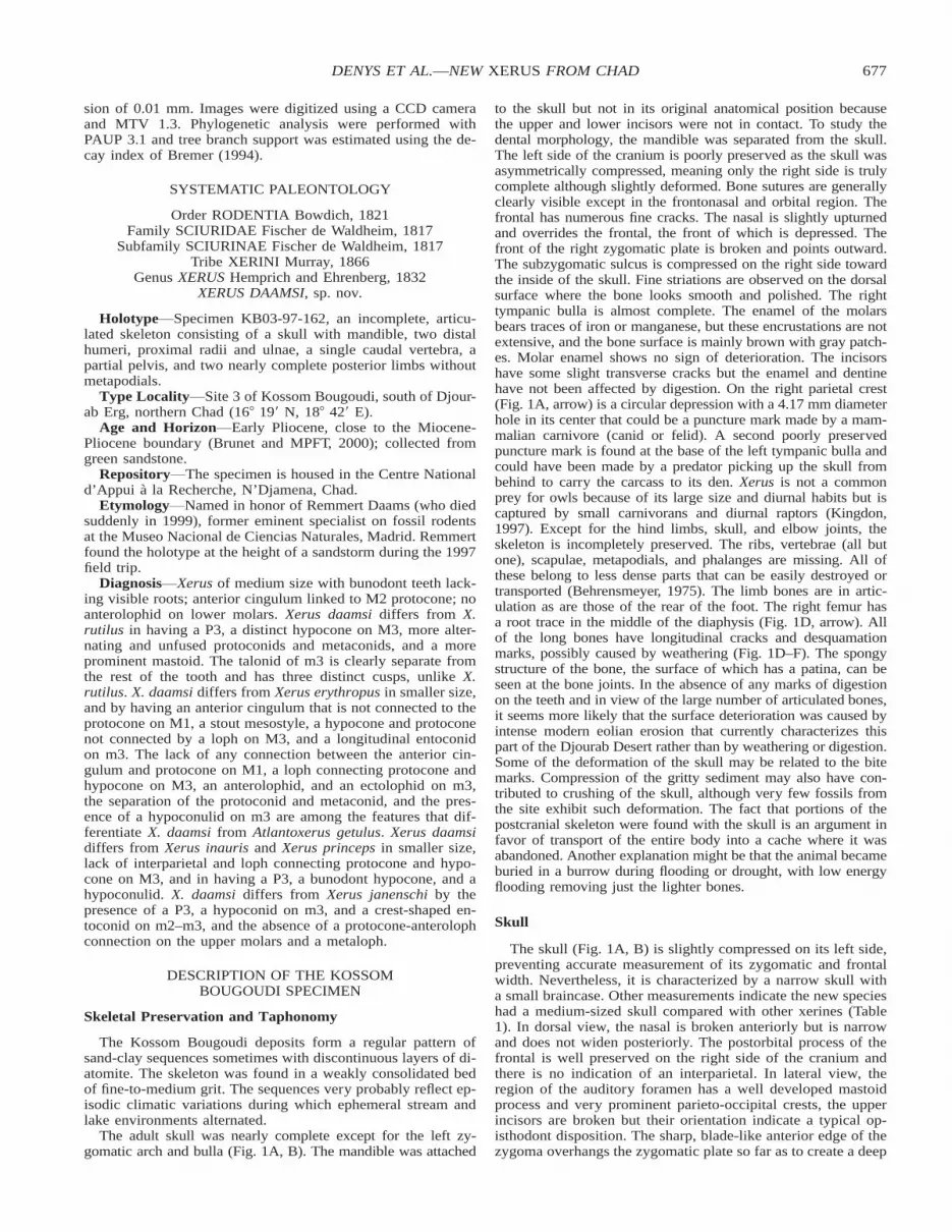
The adult skull was nearly complete except for the left zy-gomatic arch and bulla (Fig. 1A, B). The mandible was attached
to the skull but not in its original anatomical position becausethe upper and lower incisors were not in contact. To study thedental morphology, the mandible was separated from the skull.The left side of the cranium is poorly preserved as the skull wasasymmetrically compressed, meaning only the right side is trulycomplete although slightly deformed. Bone sutures are generallyclearly visible except in the frontonasal and orbital region. Thefrontal has numerous fine cracks. The nasal is slightly upturnedand overrides the frontal, the front of which is depressed. Thefront of the right zygomatic plate is broken and points outward.The subzygomatic sulcus is compressed on the right side towardthe inside of the skull. Fine striations are observed on the dorsalsurface where the bone looks smooth and polished. The righttympanic bulla is almost complete. The enamel of the molarsbears traces of iron or manganese, but these encrustations are notextensive, and the bone surface is mainly brown with gray patch-es. Molar enamel shows no sign of deterioration. The incisorshave some slight transverse cracks but the enamel and dentinehave not been affected by digestion. On the right parietal crest(Fig. 1A, arrow) is a circular depression with a 4.17 mm diameterhole in its center that could be a puncture mark made by a mam-malian carnivore (canid or felid). A second poorly preservedpuncture mark is found at the base of the left tympanic bulla andcould have been made by a predator picking up the skull frombehind to carry the carcass to its den. Xerus is not a commonprey for owls because of its large size and diurnal habits but iscaptured by small carnivorans and diurnal raptors (Kingdon,1997). Except for the hind limbs, skull, and elbow joints, theskeleton is incompletely preserved. The ribs, vertebrae (all butone), scapulae, metapodials, and phalanges are missing. All ofthese belong to less dense parts that can be easily destroyed ortransported (Behrensmeyer, 1975). The limb bones are in artic-ulation as are those of the rear of the foot. The right femur hasa root trace in the middle of the diaphysis (Fig. 1D, arrow). Allof the long bones have longitudinal cracks and desquamationmarks, possibly caused by weathering (Fig. 1D–F). The spongystructure of the bone, the surface of which has a patina, can beseen at the bone joints. In the absence of any marks of digestionon the teeth and in view of the large number of articulated bones,it seems more likely that the surface deterioration was caused byintense modern eolian erosion that currently characterizes thispart of the Djourab Desert rather than by weathering or digestion.Some of the deformation of the skull may be related to the bitemarks. Compression of the gritty sediment may also have con-tributed to crushing of the skull, although very few fossils fromthe site exhibit such deformation. The fact that portions of thepostcranial skeleton were found with the skull is an argument infavor of transport of the entire body into a cache where it wasabandoned. Another explanation might be that the animal becameburied in a burrow during flooding or drought, with low energyflooding removing just the lighter bones.
Skull
The skull (Fig. 1A, B) is slightly compressed on its left side,preventing accurate measurement of its zygomatic and frontalwidth. Nevertheless, it is characterized by a narrow skull witha small braincase. Other measurements indicate the new specieshad a medium-sized skull compared with other xerines (Table1). In dorsal view, the nasal is broken anteriorly but is narrowand does not widen posteriorly. The postorbital process of thefrontal is well preserved on the right side of the cranium andthere is no indication of an interparietal. In lateral view, theregion of the auditory foramen has a well developed mastoidprocess and very prominent parieto-occipital crests, the upperincisors are broken but their orientation indicate a typical op-isthodont disposition. The sharp, blade-like anterior edge of thezygoma overhangs the zygomatic plate so far as to create a deep

678 JOURNAL OF VERTEBRATE PALEONTOLOGY, VOL. 23, NO. 3, 2003
FIGURE 1. Xerus daamsi, sp. nov., holotype, KB03-97-162. A, dorsal view of the skull (black arrow indicates a possible puncture mark madeby a mammalian carnivore). B, ventral view of the skull. C, vestibular view of the left dentary. D, medial view of the right hind limb showinga broken pelvis and a complete femur in articulation with the tibia (the black arrow indicates a root trace). E, medial view of the broken distalpart of the femur (black arrow) in connection with a complete tibia and the proximal fragment of the fibula. F, anterior view of the left proximalfemur. Scale bars equal 1 cm.
679DENYS ET AL.—NEW XERUS FROM CHAD
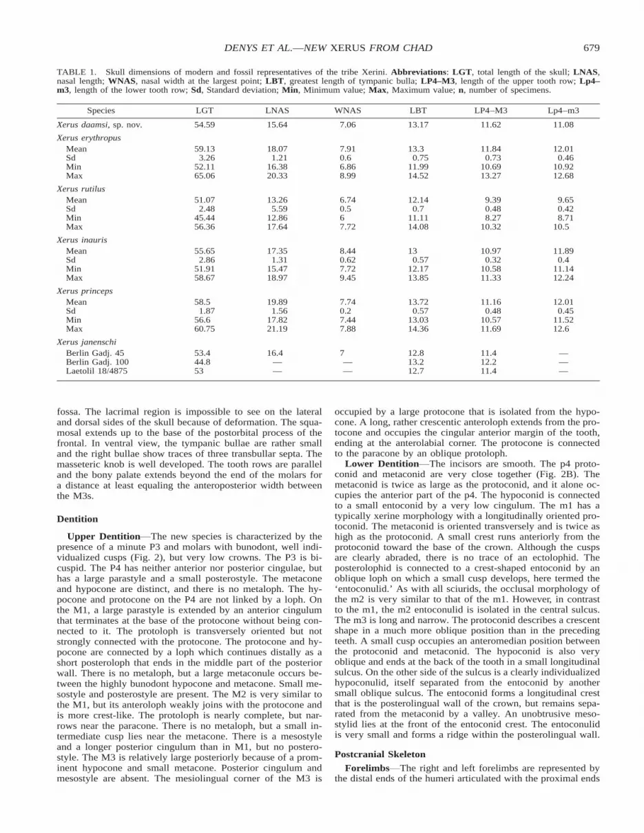
TABLE 1. Skull dimensions of modern and fossil representatives of the tribe Xerini. Abbreviations: LGT, total length of the skull; LNAS,nasal length; WNAS, nasal width at the largest point; LBT, greatest length of tympanic bulla; LP4–M3, length of the upper tooth row; Lp4–m3, length of the lower tooth row; Sd, Standard deviation; Min, Minimum value; Max, Maximum value; n, number of specimens.
Species LGT LNAS WNAS LBT LP4–M3 Lp4–m3
Xerus daamsi, sp. nov. 54.59 15.64 7.06 13.17 11.62 11.08
Xerus erythropusMeanSdMinMax
59.133.26
52.1165.06
18.071.21
16.3820.33
7.910.66.868.99
13.30.75
11.9914.52
11.840.73
10.6913.27
12.010.46
10.9212.68
Xerus rutilusMeanSdMinMax
51.072.48
45.4456.36
13.265.59
12.8617.64
6.740.567.72
12.140.7
11.1114.08
9.390.488.27
10.32
9.650.428.71
10.5
Xerus inaurisMeanSdMinMax
55.652.86
51.9158.67
17.351.31
15.4718.97
8.440.627.729.45
130.57
12.1713.85
10.970.32
10.5811.33
11.890.4
11.1412.24
Xerus princepsMeanSdMinMax
58.51.87
56.660.75
19.891.56
17.8221.19
7.740.27.447.88
13.720.57
13.0314.36
11.160.48
10.5711.69
12.010.45
11.5212.6
Xerus janenschiBerlin Gadj. 45Berlin Gadj. 100Laetolil 18/4875
53.444.853
16.4——
7——
12.813.212.7
11.412.211.4
———
fossa. The lacrimal region is impossible to see on the lateraland dorsal sides of the skull because of deformation. The squa-mosal extends up to the base of the postorbital process of thefrontal. In ventral view, the tympanic bullae are rather smalland the right bullae show traces of three transbullar septa. Themasseteric knob is well developed. The tooth rows are paralleland the bony palate extends beyond the end of the molars fora distance at least equaling the anteroposterior width betweenthe M3s.
Dentition
Upper Dentition The new species is characterized by thepresence of a minute P3 and molars with bunodont, well indi-vidualized cusps (Fig. 2), but very low crowns. The P3 is bi-cuspid. The P4 has neither anterior nor posterior cingulae, buthas a large parastyle and a small posterostyle. The metaconeand hypocone are distinct, and there is no metaloph. The hy-pocone and protocone on the P4 are not linked by a loph. Onthe M1, a large parastyle is extended by an anterior cingulumthat terminates at the base of the protocone without being con-nected to it. The protoloph is transversely oriented but notstrongly connected with the protocone. The protocone and hy-pocone are connected by a loph which continues distally as ashort posteroloph that ends in the middle part of the posteriorwall. There is no metaloph, but a large metaconule occurs be-tween the highly bunodont hypocone and metacone. Small me-sostyle and posterostyle are present. The M2 is very similar tothe M1, but its anteroloph weakly joins with the protocone andis more crest-like. The protoloph is nearly complete, but nar-rows near the paracone. There is no metaloph, but a small in-termediate cusp lies near the metacone. There is a mesostyleand a longer posterior cingulum than in M1, but no postero-style. The M3 is relatively large posteriorly because of a prom-inent hypocone and small metacone. Posterior cingulum andmesostyle are absent. The mesiolingual corner of the M3 is
occupied by a large protocone that is isolated from the hypo-cone. A long, rather crescentic anteroloph extends from the pro-tocone and occupies the cingular anterior margin of the tooth,ending at the anterolabial corner. The protocone is connectedto the paracone by an oblique protoloph.
Lower Dentition The incisors are smooth. The p4 proto-conid and metaconid are very close together (Fig. 2B). Themetaconid is twice as large as the protoconid, and it alone oc-cupies the anterior part of the p4. The hypoconid is connectedto a small entoconid by a very low cingulum. The m1 has atypically xerine morphology with a longitudinally oriented pro-toconid. The metaconid is oriented transversely and is twice ashigh as the protoconid. A small crest runs anteriorly from theprotoconid toward the base of the crown. Although the cuspsare clearly abraded, there is no trace of an ectolophid. Theposterolophid is connected to a crest-shaped entoconid by anoblique loph on which a small cusp develops, here termed the‘entoconulid.’ As with all sciurids, the occlusal morphology ofthe m2 is very similar to that of the m1. However, in contrastto the m1, the m2 entoconulid is isolated in the central sulcus.The m3 is long and narrow. The protoconid describes a crescentshape in a much more oblique position than in the precedingteeth. A small cusp occupies an anteromedian position betweenthe protoconid and metaconid. The hypoconid is also veryoblique and ends at the back of the tooth in a small longitudinalsulcus. On the other side of the sulcus is a clearly individualizedhypoconulid, itself separated from the entoconid by anothersmall oblique sulcus. The entoconid forms a longitudinal crestthat is the posterolingual wall of the crown, but remains sepa-rated from the metaconid by a valley. An unobtrusive meso-stylid lies at the front of the entoconid crest. The entoconulidis very small and forms a ridge within the posterolingual wall.
Postcranial Skeleton
Forelimbs The right and left forelimbs are represented bythe distal ends of the humeri articulated with the proximal ends

680 JOURNAL OF VERTEBRATE PALEONTOLOGY, VOL. 23, NO. 3, 2003
FIGURE 2. Left upper (A) and lower (B) cheek tooth rows of Xerusdaamsi, sp. nov., holotype, KB03-97-162. Scale bar equals 2 mm; an-terior to top.
FIGURE 3. Scatterplots showing differences in size proportions amongthe compared species of Xerini (Xerus rutilus: filled diamonds, Xeruserythropus: filled circles, Xerus daamsi KB03-97-162: filled triangle, Xe-rus inauris: open squares, and Xerus princeps: stars). In A (total skulllength versus nasal length), Xerus daamsi fits with the largest individualsof Xerus rutilus. In B (length of the tympanic bullae versus length ofupper tooth row), Xerus daamsi plots among Xerus erythropus.
of the radii and ulnae. The connection appears to be natural,corresponding to anatomical position.
Left Hind Limb The proximal part of the femur is wellpreserved (Fig. 1F). The broken distal part of this femur is inconnection with a complete tibia and the proximal fragment ofthe fibula (Fig. 1E). Parts of the pes are connected to the distalend of the tibia (calcaneum, astragalus, cuboid, navicular, andthe three cuneiforms). The end of the tibia forms an angle of1288 with the long axis of the calcaneum, which appears to bein a normal position. The distal end of the tibia has an extendedinternal malleolus beside a deep longitudinal sulcus.
Right Hind Limb The femur is continuous with a brokenpelvis but they are not fully articulated (Fig. 1D). Relative tothe femoral head, which fits into the acetabulum of the leftpelvis, the pubic symphysis is found facing posteriorly, as ifthe pelvis had pivoted through 1808. The femur is complete andarticulated with the tibia and probably with the fibula, which isin matrix. The calcaneum, astragalus and navicular are foundin continuity with the distal end of the tibia.
COMPARISON WITH MODERN AND FOSSIL XERINI
Xerus daamsi exhibits some of the skull characters that definethe Xerini sensu Moore (1959): the squamosal extends to thebase of the postorbital process of the frontal; three transbullartympanic septae in each auditory bulla; a bony palate extendingposteriorly beyond the ends of maxillary tooth rows; opistho-dont upper incisors; and a thick, prominent masseteric tubercle.However, Xerus daamsi is characterized by a short nasal that isnot broadened anteriorly and by relatively large size (Table 1).
Although the amount of intraspecific variation is great, on av-erage modern Xerus erythropus skulls are larger than those ofXerus rutilus. X. daamsi is of intermediate size between thosetwo species, but towards the upper end of the range for modernX. rutilus (Fig. 3). Xerus inauris and Xerus princeps are alsointermediate in size, but distinguished by a very wide nasal.
X. daamsi has bunodont teeth and a hypocone and entolophidon m1, which are dental characters of the Xerini noted by Cuen-ca Bescos (1988). But X. daamsi does not have a metaloph,while Cuenca Bescos (1988) noted the presence of a completeto incomplete metaloph on M1 or M2 in the definition of theXerini.
Comparison with Atlantoxerus getulus
Although Moore (1959) voiced doubts about the position ofAtlantoxerus within the Xerini and it is impossible to verify onour fossil the key character of the jugolacrimal suture differ-

681DENYS ET AL.—NEW XERUS FROM CHAD
FIGURE 4. Upper (top) and lower (bottom) cheek tooth rows of At-lantoxerus getulus specimens at different wear stages (A, NHM21.5.3015, slight wear; B, MNHN CG 1974-259, moderate wear; C,MNHN CG 1959-145, heavy wear). Scale bar equals 2 mm.
FIGURE 5. Upper (top) and lower (bottom) cheek tooth rows of Xerusrutilus specimens at different wear stages (A, MNHN CG 1978-254; B,MNHN CG 1974-2; C, NHM 9.6.1.16). Scale bar equals 2 mm.
entiating Atlantoxerus from Xerus, we prefer to include thisgenus in the comparison. Atlantoxerus getulus of Morocco hasa smaller cranium, and smaller teeth and limbs than X. daamsi.An interparietal can be seen clearly at the back of the skull ofA. getulus. The braincase is wide and the occipital region notinflated. Above the auditory foramen, the ectotympanic-parietalsuture forms a right angle and then rises to follow the outlineof the tympanic bulla. The mastoid process is posterior to theauditory foramen, but less developed than in X. daamsi, as isthe masseteric tubercle. The number of transbullar septa variesfrom three to four. The molar crowns are lower in A. getulusat the same stage of wear. P4 has no hypocone (Fig. 4). Themetaconid and protoconid of p4 are of the same size and in thesame alignment at the front of the tooth. The lower molars haveno entoconulid, but the trace of an ectolophid can be detectedon m2 and m3 even at very slight wear stages. There is also aclearly individualized mesostylid. The limbs are more robustand longer. There is no trace of the longitudinal sulcus at thedistal end of the tibia as observed in X. daamsi. Resemblancesbetween the two species include the presence of a P3, a me-
sostyle on the upper molars, and the absence of a metaloph onthe upper molars even at stages of advanced wear.
Comparison with Modern Xerus rutilus
The skull of Xerus rutilus is shorter than that of X. daamsi(Table 1). Some specimens of X. rutilus exhibit no traces of aninterparietal. There is no swelling of the mastoid process in thezone lateral to the auditory foramen. The teeth are smaller andthe P3 is absent (Fig. 5). The crowns of Xerus daamsi are tallerthan in X. rutilus. The molars are very variable in X. rutilus. Ina tooth row where P4 is erupting, the lophs and cusps are ar-ranged much as in X. daamsi, with, in particular, a small pos-teroloph and no metaloph. There is a significant difference inthe M3, which in X. rutilus has a crest-shaped lateral wall end-ing at the back of the tooth with no trace of posteroloph norseparate hypocone, whereas in X. daamsi this is not such alateral continuous wall. An anteroloph and a protoloph extendfrom the protocone in both species, but the protoloph is morecrest-shaped in X. rutilus than in X. daamsi. On the other upper

682 JOURNAL OF VERTEBRATE PALEONTOLOGY, VOL. 23, NO. 3, 2003
FIGURE 6. Upper (A–D) and lower (E–H) cheek tooth rows of Xeruserythropus specimens at different wear stages (A and E, MNHN CG1973-170; B and F, MNHN CG 1904-1985; C and G, MNHN CG1986-582; D and H, MNHN CG 1904-1985). Scale bar equals 2 mm.
FIGURE 7. Upper (A–B) and lower (C–D) cheek tooth rows of Xerusinauris specimens at different wear stages (A, TM 5395; C, TM 25631;B and D, TM 25632). Scale bar equals 5 mm.
molars of X. rutilus, the cusps become broader and bunodontat more advanced stages, and in modern specimens a postero-loph occupies the entire posterior wall of the molar, in contrastto X. daamsi where it is very short (Fig. 5). The anterior cuspsof the lower molars seem more closely spaced in X. daamsi. Avery slight transverse connection can be seen as the metaconidis much taller in X. rutilus, there being no sign of this connec-tion in X. daamsi. The m3 of X. daamsi has a crest-shapedentoconid oriented longitudinally, a feature found only in veryyoung specimens of X. rutilus. As with the upper molars, theback of the m3 of X. rutilus forms an unbroken high crest withpoorly individualized cusps whereas in X. daamsi, the cusps aremore distinct although they are joined. No ectolophid is foundin X. rutilus or X. daamsi. X. daamsi has longer and wider limbbones than X. rutilus.
Comparison with Xerus erythropus
The skull has the same general aspect in X. erythropus andX. daamsi, in particular its narrow, elongate shape and the ab-sence of an interparietal with the parietal crests meeting in themiddle of the occipital suture. The tympanic bullae are larger
in X. erythropus. At the back of the auditory foramen, X. ery-thropus and X. daamsi have a small swelling at the end of theoccipital crest, however it is clearly more swollen in the latter(Fig. 3). The molars are larger and more hypsodont in X. ery-thropus (Fig. 6, Table 1). P3 is found in most specimens of X.erythropus, but is monocuspid. X. daamsi has a similar molararrangement to that of a X. erythropus from Niger (MNHN CG1986-582, Fig. 6), although the latter has a much more markedposterior cingulum and a nearly formed metaloph on the M2.However, the metaloph is not developed in other specimens ofX. erythropus. The M3 of X. erythropus from Niger, which ap-pears a little less worn than that of X. daamsi, has a doublehypocone with a posterior cingulum and a large metacone;whereas the metacone is very small and there is no posteriorcingulum in X. daamsi. On the lower molars, the size of thehypoconid in X. daamsi is striking, occupying as it does therear half of the m3. It is much smaller in X. erythropus wherethe three cusps of the talonid are of similar size and are inter-connected. However, they never form a high wall bounding adeep basin and connecting the talonid to the front of the toothas in X. rutilus. X. erythropus also tends to have many acces-sory tubercles. In X. erythropus, the entoloph is clearly visible,and there is a well developed mesostyle not found in X. daamsi.
For the m3, a longitudinal, crest-shaped entoconid, a proto-conid, and a hypoconulid are clearly visible in Xerus daamsias in X. erythropus. There is no ectolophid, nor any transverseconnection between the anterior cusps of any of the molars inX. erythropus. On the upper molars, however, there are cleartraces of lophs with a complete metaloph.
Comparisons with Xerus inauris and Xerus princeps
There are very few differences between extant Xerus inaurisand Xerus princeps (Denys, 1987). The skull of X. inauris isslightly smaller or of similar size to that of Xerus daamsi, butnarrower. At least four septae are found in the tympanic bulla.There is also an interparietal and a slight mastoid process. Incontrast, X. princeps is larger than X. daamsi with no interpa-rietal, a slight mastoid process, and a tympanic bulla with threeseptae (Fig. 7, Table 1). However, the molar crowns are tallerin X. princeps than in X. daamsi. No ectolophid is found in X.inauris and X. daamsi, whereas this feature is reported in twospecimens of X. princeps. The m3 of both X. princeps and X.inauris is like that of X. daamsi in having a prominent hypo-

683DENYS ET AL.—NEW XERUS FROM CHAD
FIGURE 8. Upper (A–B) and Lower (C–D) cheek tooth rows of Xerusprinceps specimens at different wear stages (A and C, TM 38117; Band D, TM 11527). Scale bar equals 5 mm.
TA
BL
E2.
Den
tal
mea
sure
men
tsof
som
efo
ssil
Xer
ini.
Sam
eab
brev
iati
ons
asin
Tab
le1.
Spe
cies
P4
LW
M1
LW
M2
LW
M3
LW
p4
LW
m1
LW
m2
LW
m3
LW
Xer
usda
amsi
,no
v.sp
.2.
502.
802.
862.
862.
763.
052.
763.
052.
412.
312.
622.
92.
763.
172.
972.
66X
erus
jane
nsch
iU
NB
-Lae
toli
lM
ean
Min
Max
Sd
3.13
2.49
3.77
0.40
2.65
1.66
3.64
0.62
3.08
2.39
3.78
0.28
3.72
3.08
4.37
0.26
3.08 — — —
3.55 — — —
2.83 — — —
3.10 — — —
2.76
2.38
3.14
0.36
2.68
2.3
3.06
0.36
3.13
2.99
3.26
0.16
3.21
2.96
3.46
0.30
3.32
2.92
3.72
0.16
3.47
3.17
3.77
0.12
3.78 — — —
3.30 — — —
Xer
user
ythr
opus
Om
o18
mem
bers
B&
CX
erus
sp.
Om
o33
— 2.55
— 2.98
— —— —
— —— —
— —— —
2.50
2.60
2.76
2.88
3.12
3.46
3.60
3.70
— —— —
— —— —
Xer
uscf
.in
auri
sO
lduv
ai3.
502.
77M
ean
Min
Max
Sd
— 2.75 — —
— 2.80 — —
3.49
3.40
3.60
0.08
3.36 — — —
— — — —
— — — —
— — — —
— — — —
3.13
2.30
3.40
0.42
3.07
2.05
3.45
0.51
3.48
3.30
3.65
0.14
3.51
3.30
3.80
0.19
3.63
3.45
3.80
0.15
3.57
3.35
3.70
0.13
3.55
3.45
3.60
0.09
3.00
3.50
3.40
3.70
Lae
t15
62X
erus
sp.
——
——
——
——
——
2.50
2.50
2.65
2.60
—0.
17
conid and a crestiform but oblique entoconid with an entocon-ulid. In X. daamsi, the entoconid is more longitudinal and thereis a strong hypoconulid while only slightly worn specimens ofX. inauris still have a hypoconulid.
Comparison with Xerus janenschi from Upper NdolanyaBeds at Laetolil
Xerus daamsi has smaller molars than X. janenschi (Table 2).The skulls are about the same size, but the braincase and tym-panic bullae are wider in X. daamsi (Table 1). One of the twoskulls found at Laetolil (Denys, 1987:figs. 6–7) exhibits notrace of an interparietal and the two parietal crests join in themiddle of the occipital crest. There is no P3. As in X. daamsi,the molars attributed to X. janenschi have no ectolophid andthe crown height of M1 is similar. The entoconid of X. janen-schi is less crestiform, but both have similar orientations. Onthe upper molars, the anteroloph and a prominent metaloph arewell developed in X. janenschi, and the M3 has a trace of anectoloph.
Comparison with Xerus cf. inauris from Olduvai
Xerus cf. inauris from Olduvai Bed I is characterized by tallcrowns and a high loph connects the poorly differentiated pro-tocone and hypocone (Denys, 1990). The posteroloph is longerthan that of X. daamsi. Talonid cusps of the m3 are arrangeddifferently, with a well developed entoconid oriented trans-versely to the longest axis of the molar, whereas in X. daamsiit is less developed and oriented longitudinally along the lingualwall of the tooth. This character clearly aligns the Olduvai fos-sils with Xerus inauris of South Africa. The hypoconid is veryprominent and (except on m1) the trace of the hypoconulid,observed only on the left m3 of X. daamsi, is no longer visible.A trace of an ectolophid is found on the worn molars fromOlduvai Bed I.
Comparison with Xerus erythropus from Omo B and C
On drawings of X. erythropus from Omo, Wesselman (1984:fig. 55) depicted lower molars with an anterolophid joining theprotoconid (not present in Xerus daamsi). The entoconid of theOmo specimens is transverse and not oblique. The posterolo-phid is long, but the trace of the hypoconulid is visible, whichis not true for X. daamsi. X. erythropus from Omo is different

684 JOURNAL OF VERTEBRATE PALEONTOLOGY, VOL. 23, NO. 3, 2003
from X. daamsi in having an anterolophid and a transverse en-toconid. The modern specimens of X. erythropus we examinedhave no anterolophid on the lower molars, unlike X. rutilus,and their entoconid is clearly connected to the hypoconid, andis slightly oblique.
Comparison with Vulcanisciurus
Vulcanisciurus was described from lower Miocene depositsof Rusinga (Lavocat and Mein, 1973:239) and the middle Mio-cene site of Fort Ternan (Denys and Jaeger, 1992). The lowermolars have a well developed, longitudinal, crestiform ento-conid and a clearly distinct posterolophid. The ectolophid iswell developed on all the molars. An anterolophid is found onthe m3 but not a hypoconulid. For the upper cheek teeth, Lav-ocat and Mein (1973) reported a bicuspid P3 and a P4 with avery prominent metaconule and an incipient hypocone. A pro-toloph and metaloph join with the protocone. Only one M1 ispreserved. Vulcanisciurus teeth bear a marked resemblance tothose of Xerus rutilus and Atlantoxerus getulus, but nothing isknown of their variability. X. daamsi appears to be clearly morebunodont than Vulcanisciurus. With its bunodonty, small hy-pocone, and cusp arrangement of its lower molars, Vulcani-sciurus shares some characters of tribe Xerini sensu CuencaBescos (1988). But Vulcanisciurus does not have an entolophidon the m1 or m2, nor a metaloph on the single M1 figured byLavocat and Mein (1973).
PHYLOGENETIC ANALYSIS
This analysis includes all extant species of African Xerini.Moore (1959) suggested that African arboreal squirrels evolvedfrom ‘‘ground squirrel stock’’ on the basis of long orbits andshort interorbital breadths of the Protoxerini and Funisciurini.Accordingly, this analysis includes representatives of the Pro-toxerini (Heliosciurus, Epixerus, Protoxerus), Funambulini,Myosciurini, and Funisciurus of the tribe Funisciurini. Sciurusis used as an outgroup. Fossil species represented only by poor-ly preserved dental material have not been incorporated in thedata matrix.
Jaeger (1977) reported that Atlantoxerus from the Plio-Pleis-tocene of Morocco has a smaller anteroconid, wider lower mo-lars, and a larger metaconule than early Heteroxerus. Lavocatand Mein (1973) proposed affinities between Heteroxerus andAtlantoxerus as did Cuenca (1986, 1988) who defined differenttrends in the evolution of Heteroxerus and Aragoxerus lineagesfrom Spain including enlarged cheek teeth, increased bunodon-ty, and a slight increase in hypsodonty.
Trees and Phylogeographic Hypotheses
The first analysis includes only the African Xerini specieswith Sciurus as an outgroup and using only characters C1 toC17 (Appendices 1 and 2). All 17 characters were consideredunordered and of equal weight. A heuristic search based on thecriterion of maximum parsimony obtained a single tree withlength of 32 steps, consistency index of 0.625, and retentionindex of 0.52 (Fig. 9A). All the species of the Xerus genusform a monophyletic clade defined by three synapomorphies(C7 bunodonty; C14 nasal not extended; C17 mastoid processmarked, but in X. rutilus its absence is interpreted as a reversal).Xerus daamsi is in the same clade as X. rutilus and the twoshare three apomorphies concerning bunodonty (C4, C6) andm3 hypoconulid differentiation (C13). X. rutilus has four au-tapomorphies (C1, C12, C16, C17). X. erythropus, X. inauris,and X. princeps form a clade united by two synapomorphies,absence of a mesostyle (C5) and arrangement of the entoconid(C10). The position of Atlantoxerus getulus is not resolved inthis analysis. Most of the clades obtained in the most parsi-
monious tree are not well supported and have a decay index ofonly 1 except the X. inauris–X. princeps clade which has adecay index of 4.
When analyzing all the African Sciuridae using the same 17characters plus four more skull characters adapted from Moore(1959) and taking Sciurus as outgroup, we obtained 6 mostparsimonious trees of length 71, with consistency index of0.394, and retention index of 0.566. The positions of Funam-bulus and Myosciurus are ambiguous (Fig. 9B). The strict con-sensus yields a Rohlf consistency index of 0.808 (Fig. 9B), andXerus daamsi is grouped in 100% of the trees with X. rutilus,while X. erythropus is basal to the monophyletic Xerus clade.The 50% majority rule consensus indicates that only in 33% ofthe cases is the daamsi-rutilus clade grouped with the inauris-princeps clade due to relative instability of the position of thedaamsi-rutilus clade and a low decay index of 1 for those twonodes. This tree shows a probable monophyly of the Xerini(sensu Moore, 1959) with 5 synapomorphies and a decay indexof 2. The African tree squirrels forming the tribe Funambulinisensu Simpson (1945) is paraphyletic as is the Protoxerini.Myosciurus is not close to Funisciurus, contrary to the resultsof Moore (1959). It is the only African squirrel with two trans-bullar septae. This may indicate that terrestriality in Africansquirrels occurred only once and that African ground squirrelscould be derived from arboreal squirrels contrary to Moore’s(1959) hypothesis.
SYSTEMATICS AND EVOLUTION OF AFRICANPLIO-PLEISTOCENE XERINI
The systematics and the evolution of the African membersof the Xerini remains very unclear without further analyses ofother anatomical and molecular characters. The clade consistingof all Xerus species shows a similar general trend toward thedevelopment of bunodonty as reported by Cuenca (1986) forspecies of Heteroxerus in Europe. The North African terrestrialsquirrel Atlantoxerus is basalmost among the Xerini (Fig. 9)and may have evolved independently of those of tropical Af-rica. Its divergence would date to about 7 to 8 Ma in NorthAfrica according to the fossil record and this may correlate witha major modification of climate with the onset of the Asianmonsoon and the dominance of C4 plants (grass). Then, byvicariance or by dispersal, the isolation of tropical Africa ledto speciation among Xerus, first in West Africa (X. erythropus)and later in East and South Africa. The development of buno-donty as well as the crestiform arrangement of the back of theM3 may be an adaptation to a more granivorous diet. AlthoughX. daamsi shares morphological features with both X. rutilusand X. erythropus, both cladistic analyses determined that X.daamsi has more synapomorphies with X. rutilus.
PALEOECOLOGICAL ANDPALEOBIOLOGICAL IMPLICATIONS
Hubert (1978) reported modern Xerus rutilus in the OmoValley area of Ethiopia on plateau covered by open shrub orAcacia bush savanna well away from the river. Similarly, Coe(1972) reported Xerus in the transition zone between thicketand grassland. Generally, Xerus is found in dry savannas (300to 900 mm annual rainfall; Kingdon, 1974), and is essentiallya bushland, burrowing animal. It is omnivorous and diurnal. X.erythropus has been recorded in Bandia (Senegal) in the Su-danian zone in a woodland with 570 mm isohyet (Hubert, 1977)and in southeastern Chad in bushes in a Sudanian savanna with700 mm rainfall (pers. obs.). Despite a few similarities betweenthe fauna of KB and those from North (Sahabi), East (Loth-agham, Aramis), and South (Langebaanweg) Africa, this sitedisplays rather marked provincialism (Brunet and MPFT, 2000),and Xerus daamsi could be a new indicator of its endemism.

685DENYS ET AL.—NEW XERUS FROM CHAD
FIGURE 9. Phylogenetic affinities of Xerus daamsi, sp. nov. with other African sciurids. A. Xerini phylogeny, showing X. daamsi close to X.rutilus. Most parsimonious tree for the partial matrix including African Xerini, with Sciurus as outgroup (32 steps, CI 0.625, RI 0.520). Onlysynapomorphies are shown here together with the state of the character in parentheses on the cladogram (1: C6(1), 2: C13(0), 4: C7(1), 5: C14(1),6: C17(0), 7: C2(2), 8: C12(2), 9: C15(2), 11: C5(1), 12: C10(1)). X. rutilus and X. princeps are respectively characterized by one autapomorphy(3: C17(1), 10: C10(0)) but not X. daamsi. B, African sciurid phylogeny (total matrix) with Sciurus as outgroup. Strict consensus of 6 mostparsimonious trees (71 steps, CI 0.394, RI 0.566, Rohlf index 0.808). Note that Xerus daamsi and X. rutilus belong to the same clade but itsposition is not well defined among Xerus.
Today, Xerus belongs to the domain of the Sudano-Guinean,Somali Masai, and Zambezian savannas (Denys, 1999). X.daamsi is clearly different from the Tanzanian lineage of X.inauris, and seems closer to Xerus rutilus presently found onlytoday in the Horn of Africa in the Somali-Masai zone. However,X. rutilus is currently not found in Chad, instead X. erythropusis present. It is primarily an animal of open woodland and su-danic savannas, living in more arid regions than X. rutilus.However, the two species are sympatric over parts of Uganda,Sudan, and Kenya. Their natural foods are roots, grass seeds,storage leaf bases of some grass species, green leaves, fallenfruits, nuts, Acacia pods, and some insects, especially termites(Kingdon, 1997).
CONCLUSIONS
The close phylogenetic affinities of Xerus daamsi, sp. nov.and extant Xerus rutilus suggests that Somali Masai elementsof vegetation were present at the KB site about 5 Ma. Moreover,differences between Xerus daamsi and the two modern speciesfrom South Africa supports the hypothesis that this area ofChad was biogeographically separated from southern savannasby 5 Ma. But connections with Ethiopia are confirmed and morecomparisons need to be made with rodent fossils from Hadarand Aramis. Unfortunately, Xerus is currently not yet knownfor these sites.
The African modern Xerini, which form a monophyleticclade, but with low support, may have evolved by allopatric
speciation in the different savannas of Africa from a commonancestor in North Africa in late Miocene times. Xerus daamsishares some dental and cranial characters with X. janenschifrom Laetolil and X. erythropus from Omo, indicating somerelationships from a common ancestor with an early divergence.However, little is known of the variability of fossil species be-cause of the paucity of the fossil record. Due to the absence offossils of this genus at other sites of the same age, there are nopossibilities of biochronologic correlations and thus no new in-formation about the age of the KB site. Further discoveries arerequired to improve our understanding of the evolution of thisgroup of rodents in the Pliocene of Africa.
ACKNOWLEDGMENTS
We are indebted to the Chad authorities (Ministere del’Education Nationale, de l’Enseignement Superieur et de laRecherche), to the French Ministere de l’Enseignement Super-ieur, de la Recherche et de la Technologie, to the French Min-istere des Affaires Etrangeres (Cooperation: MCACN’Djamena), to Region Poitou-Charente, Departement de laVienne, Groupe Elf, Association pour le Prix scientifique PhilipMorris, to the French armed forces, and to all the troops ofMAM and Epervier, who, through their logistical support, con-tributed along with members of MPFT to the success of fieldprograms. We are extremely grateful to Paula Jenckins (NaturalHistory Museum, London) and Ducan MacFadyan (TransvaalMuseum, Pretoria) for access to comparative material of mod-

686 JOURNAL OF VERTEBRATE PALEONTOLOGY, VOL. 23, NO. 3, 2003
ern Xerini. We would also like to thank Alain Beauvillain forfield help, Guy Mouchelin for skeleton preparation, Sabine Rif-faut for drawings and photographic plate mounting, GhislaineFlorent for administrative guidance of MPFT, and ChristopherSutcliffe for critical reading of this manuscript.
LITERATURE CITED
Behrensmeyer, A. K. 1975. The taphonomy and paleoecology of Pio-Pleistocene vertebrate assemblages east of Lake Rudolf, Kenya.Bulletin of the Museum of Comparative Zoology 146:473–578.
Black, C. C. 1965. New species of Heteroxerus (Rodentia, Sciuridae)in the French Tertiary. Verhandlungen der Naturforschenden Ge-sellschaft, Basel 76:185–196.
Bremer, K. 1994. Branch support and tree stability. Cladistics 10:295–304.
Brunet, M., A. Beauvilain, Y. Coppens, E. Heintz, A. H. E. Moutaye,and D. Pilbeam. 1995. The first australopithecine 2,500 kilometerswest of the Rift Valley (Chad). Nature 378:273–275.
———, ———, ———, ———, ———, and ——— 1996. Austral-opithecus bahrelghazali, une nouvelle espece d’Hominide anciende la region de Koro Toro (Tchad). Comptes Rendus de l’Academiedes Sciences, Paris 322:907–913.
———, ———, D. Geraads, F. Guy, M. Kasser, H. T. Mackaye, L.MacLatchy, G. Mouchelin, J. Sudre, and P. Vignaud. 1997. Tchad:un nouveau site a Hominides Pliocene. Comptes Rendus del’Academie des Sciences, Paris 324:341–345.
———, ———, ———, ———, ———, ———, ———, ———,———, and ——— 1998. Tchad: decouverte d’une faune de Mam-miferes du Pliocene inferieur. Comptes Rendus de l’Academie desSciences, Paris 326:153–158.
———, and MPFT (Mission Paleoanthropologique Franco-Tchadi-enne). 2000. Chad: discovery of a vertebrate fauna close to theMio-Pliocene boundary. Journal of Vertebrate Paleontology 20:205–209.
Coe, M. J. 1972. The South Turkana expedition IX. Ecological studiesof the small mammals of South Turkana. Geographical Journal 138:316–338.
Cuenca, G. 1986. Heteroxerus insignis, n. sp. (Sciuridae, Rodentia,Mammalia) from the lower Miocene of Spain. Casopis pro Miner-alogii a Geologii 31:131–142.
Cuenca Bescos, G. 1988. Revision de los Sciuridae del Aragoniense ydel Rambliense en la fosa de Catalayud-Montalban. Scripta Geo-logica 87:1–116.
Denys, C. 1987. Fossil rodents (other than Pedetidae) from Laetolil; pp.118–170 in M. D. Leakey and J. M. Harris (eds.), Laetolil, a Pli-ocene Site in Tanzania. Oxford University Press, London.
——— 1990. First occurrence of Xerus cf. inauris (Rodentia, Sciuridae)at Olduvai Bed I (lower Pleistocene, Tanzania). PalaontologischeZeitschrift 64:359–365.
——— 1999. Of mice and men. Evolution in East and South Africaduring Plio-Pleistocene times; pp. 226–252 in T. Bromage and F.Schrenk (eds.), African Biogeography, Climate Change and HumanEvolution. The Human Evolution series, Oxford University Press.
———, and J. J. Jaeger. 1992. Rodents of the Miocene site of FortTernan (Kenya) first part: phiomyids, bathyergids, sciurids and an-omalurids. Neues Jahrbuch fur Geologie und Palaontologie Abhan-dlungen 185:63–84.
Hendey, Q. B. 1981. Paleoecology of the late Tertiary fossil occurrencesin ‘E’ Quarry, Langebaanweg, and a reinterpretation of their geo-logical context. Annals of the South African Museum 1:1–104.
Hoffmann, R. S., C. G. Anderson, R. W. Thorington, and L. R. Heaney.1993. Family Sciuridae; pp. 419–465 in D. E. Wilson and D. A.Reeder (eds.), Mammal Species of the World. A Taxonomic andGeographic Reference. Smithsonian Institution Press, Washingtonand London.
Hubert, B. 1977. Ecologie des populations de rongeurs de Bandia (Se-negal), en zone sahelo-soudanienne. La Terre et la Vie 31:33–100.
——— 1978. Modern rodent fauna of the Lower Omo Valley, Ethiopia.Bulletin of the Carnegie Museum of Natural History 6:109–112.
Jaeger, J. J. 1977. Les rongeurs du Miocene moyen et superieur duMaghreb. Paleovertebrata 8:1–166.
Kingdon, J. 1974. East African Mammals. An Atlas of Evolution inAfrica, Vol. II, part B: Hares and Rodents. Academic Press, Londonand New York, pp. 343–703.
——— 1997. The Kingdon Field Guide to African Mammals. Academ-ic Press, London and New York, 464 pp.
Lavocat, R., and P. Mein. 1973. Sous-Ordre Sciuromorpha; pp. 239–243 in Les Rongeurs du Miocene d’Afrique Orientale. I. Mioceneinferieur. Memoires et Travaux de l’Ecole Pratique des HautesEtudes, Institut de Montpellier 1:1–284.
Moore, J. C. 1959. Relationships among the living squirrels of the Sci-urinae. Bulletin of the American Museum of Natural History 118:153–206.
Munthe, J. 1987. Small-mammal fossils from the Pliocene Sahabi for-mation of Libya; pp. 135–144 in N. T. Boaz, A. El-Arnauti, A. W.Gaziry, J. de Heinzelin, and D. Dechant Boaz (eds.), Neogene Pa-leontology and Geology of Sahabi. A. R. Liss, Inc., New York.
Pickford, M., P. Mein, and B. Senut. 1992. Primate bearing Plio-Pleis-tocene cave deposits of Humpata, Southern Angola. Human Evo-lution 7:17–33.
Senut, B., M. Pickford, P. Mein, G. Conroy, and J. Van Couvering.1992. Discovery of 12 new Late Cenozoic fossiliferous sites inpalaeokarsts of the Otavi Mountains, Namibia. Comptes Rendus del’ Academie des Sciences, Paris 314:727–733.
Simpson, G. G. 1945. The principles of classification and a classificationof mammals. Bulletin of the American Museum of Natural History85:1–350.
Wesselman, H. B. 1984. The Omo micromammals; pp. 1–219 in M. K.Hecht and F. S. Szalay (eds.), Contributions to Vertebrate Evolu-tion. S. Karger, Basel.
Received 7 September 2000; accepted 24 June 2002.
APPENDIX 1
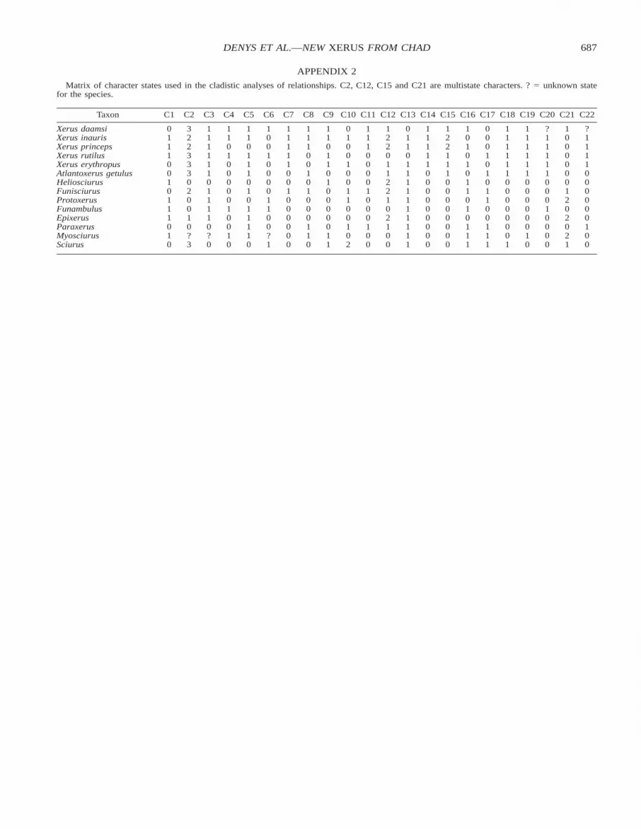
List of dental (C1–13) and skull (C14–22) characters used in thisanalysis:
C1. P3: present (0), absent (1).C2. Metaloph connection and development on M1–M2 (after morpho-
types defined by Black, 1965). Long metaloph faintly connectedto the protocone (0), connected to the hypocone (1), short or re-duced metaloph connected with the posteroloph (2), short unat-tached metaloph (3).
C3. Metaconule on M1–M2 absent (0), or well individualized (1).C4. Anterior cingulum connected to the protocone on M1 (0), or end-
ing very low to the cusp and separated from it by a transversevalley (1).
C5. Mesostyle on M1–M2 present (0), absent (1), or poorly differen-tiated (2).
C6. Hypocone and protocone connected on the M3 (0), or not con-nected (1).
C7. Roots visible on upper molars in lateral view (0), or not visible(1).
C8. Anterolophid on m1 present (0), or absent (1).C9. Ectolophid on m1–m2 present (0), or absent (1).
C10. Entoconid on m1–m2 oblique to longitudinal (0), transverse (1),or bunodont and median on labial wall (2).
C11. Entolophid on m1–m2 not visible (entoconid included in the pos-terolophid) (0), or visible (1).
C12. Protoconid and metaconid on m1–m2 at same level unfused (0),fused by the anterior side (1), or fused by posterior part of cusps(protolophid) (2).
C13. Hypoconulid differentiated on m3 (0), or barely visible or absent(1).
C14. Nasal extended at front of the skull (0), or not extended (1).C15. Tympanic bullae septa: 0 to 2 (0), or 3 (1). (Moore, 1959 reported
that most Sciurinae have 1 or 2 tympanic septae, so this is takento be the primitive condition).
C16. Interparietal present (0), or absent (1).C17. Mastoid process marked (0), or absent (1).C18. Bony palate extended beyond the end of maxillary tooth rows (1),
not extended (0).C19. Squamosal extended to the base of postorbital process of the fron-
tal (1), not extended (0).C20. Length of lacrimomaxillary contact: lower or identical to lacri-
mojugal one (0), larger (1).C21. Masseteric tubercle of the maxilla: prominent (0), normal (1), ab-
sent (2).C22. In the pterygoid fossa close to the tympanic bullae: large round
opening of alisphenoid canal (0), small round opening (1).
687DENYS ET AL.—NEW XERUS FROM CHAD
APPENDIX 2
Matrix of character states used in the cladistic analyses of relationships. C2, C12, C15 and C21 are multistate characters. ? 5 unknown statefor the species.
Taxon C1 C2 C3 C4 C5 C6 C7 C8 C9 C10 C11 C12 C13 C14 C15 C16 C17 C18 C19 C20 C21 C22
Xerus daamsiXerus inaurisXerus princepsXerus rutilusXerus erythropus
01110
32233
11111
11010
11011
10010
11111
11100
11011
01001
11100
12201
01101
11111
12211
10101
00010
11111
11111
?1111
10000
?1111
Atlantoxerus getulusHeliosciurusFunisciurusProtoxerusFunambulus
01011
30200
10111
00001
10101
00011
00100
10100
01000
00110
00100
12210
11111
00000
10000
01101
10110
10000
10000
10001
00120
00000
EpixerusParaxerusMyosciurusSciurus
1010
10?3
10?0
0010
1110
00?1
0000
0110
0011
0102
0100
2100
1111
0000
0000
0111
0111
0001
0010
0000
2021
0100


