Embed Size (px)
Citation preview

A-RAF Kinase Functions in ARF6 Regulated EndocyticMembrane TrafficElena Nekhoroshkova, Stefan Albert, Matthias Becker, Ulf R. Rapp*
Institut fur Medizinische Strahlenkunde und Zellforschung (MSZ), University of Wurzburg, Wurzburg, Germany
Abstract
Background: RAF kinases direct ERK MAPK signaling to distinct subcellular compartments in response to growth factorstimulation.
Methodology/Principal Findings: Of the three mammalian isoforms A-RAF is special in that one of its two lipid bindingdomains mediates a unique pattern of membrane localization. Specific membrane binding is retained by an N-terminalfragment (AR149) that corresponds to a naturally occurring splice variant termed DA-RAF2. AR149 colocalizes with ARF6 ontubular endosomes and has a dominant negative effect on endocytic trafficking. Moreover actin polymerization of yeast andmammalian cells is abolished. AR149/DA-RAF2 does not affect the internalization step of endocytosis, but trafficking to therecycling compartment.
Conclusions/Significance: A-RAF induced ERK activation is required for this step by activating ARF6, as A-RAF depletion orinhibition of the A-RAF controlled MEK-ERK cascade blocks recycling. These data led to a new model for A-RAF function inendocytic trafficking.
Citation: Nekhoroshkova E, Albert S, Becker M, Rapp UR (2009) A-RAF Kinase Functions in ARF6 Regulated Endocytic Membrane Traffic. PLoS ONE 4(2): e4647.doi:10.1371/journal.pone.0004647
Editor: Howard Riezman, University of Geneva, Switzerland
Received July 15, 2008; Accepted January 13, 2009; Published February 27, 2009
Copyright: � 2009 Nekhoroshkova et al. This is an open-access article distributed under the terms of the Creative Commons Attribution License, which permitsunrestricted use, distribution, and reproduction in any medium, provided the original author and source are credited.
Funding: EN was supported by DFG grant RA-642/11-2, Deutsch-Franzosiches Graduierten kolleg GRK1141/1, SFB 581 Project B5 and SFB 487 Project C3. MB wassupported by SFB TR 17 of the DFG. The funders had no role in the study design, data collection and analysis, decision to publish, or preparation of themanuscript.
Competing Interests: The authors have declared that no competing interests exist.
* E-mail: [email protected]
Introduction
RAF protein kinases were originally identified as viral
oncogenes [1,2] found in murine and avian retroviruses. RAF
genes encode protein serine/threonine kinases [3,4] that mediate
transduction of extracellular mitogenic signals from activated Ras
GTPases at the plasma membrane to a MAP kinase module (RAF-
MEK-ERK), the mitogenic cascade (reviewed in [5]). As a result,
complex physiological responses to growth factor stimulation take
place at multiple cellular levels.
Insect genomes contain only a single RAF gene whereas
vertebrates have refined RAF signaling and employ three isoforms
that target ERK signalling to different subcellular compartments
[6]. Specialized functions are reflected in differential regulation of
RAF kinase activity [7] and varying phenotypes of RAF knockout
mice [8–10]. The three RAF isoforms and their splice variants
share common structural features comprising three conserved
regions, CR1, CR2, CR3. The N-terminal CR1 encompasses the
Ras binding domain (RBD) and the cysteine-rich domain (CRD),
CR2 contains a conserved 14-3-3 binding motif and the C-
terminally located CR3 encodes the kinase domain [11].
Whereas B- and C-RAF were studied extensively, little is known
about A-RAF function. A-RAF is marked by a low basal kinase
activity, which has been attributed to substitutions in its N-region,
where tyrosine 296 plays a central role [12]. In contrast to animals
with a genomic deletion of B- or C-RAF, A-RAF 2/2 mice are viable,
but die perinatally, depending on the genetic background [8].
Budding yeast Saccharomyces cerevisiae is an established eukaryotic
model organism that has played a key role in the elucidation of the
MAP kinase signaling pathway. Despite the presence of two
redundant RAS genes and at least 6 MAPK cascades [13], no
RAF kinases are present in S. cerevisiae. Nevertheless, yeast was
important for defining RAF function as an activator of a
prototypical MAP kinase cascade in experiments that involved
ectopic expression of C-RAF [14]. RAF isoforms are known to
function as homo and heterodimers in mammalian cells [6],
complicating assignment of individual function. Therefore yeast is
an attractive system for investigation of a single RAF isoform.
Endocytosis is a process essential for many aspects of cellular life,
including receptor internalization and recycling. ARF6 GTPase was
shown to regulate endocytosis at several levels [15]. The activation
state of ARF6 is determined by the bound nucleotide, GTP or GDP,
which also affects intracellular localization. Nucleotide loading is
regulated by specialized guanine nucleotide exchange factors
(GEFs) and GTPase activating proteins (GAPs) that catalyze
hydrolysis of bound GTP [16]. Endocytosis and signal transduction
are known to be functionally linked and regulating each other (Von
Zastrow and Sorkin, 2007, Polo and Di Fiore, 2006). ARF6 as a
central regulator of endocytic trafficking was shown to activate ERK
[17,18]. Based on changes in endocytosis upon inhibition of ERK
signaling Robertson et al. (2006) suggested a role of ERK signaling
in the regulation of clathrin-independent endocytosis.
Here we describe the role of A-RAF in membrane trafficking
and identify its function at a specific step of endocytosis. This work
PLoS ONE | www.plosone.org 1 February 2009 | Volume 4 | Issue 2 | e4647

RAF Kinases and Endocytosis
PLoS ONE | www.plosone.org 2 February 2009 | Volume 4 | Issue 2 | e4647

led to the discovery of a C-terminally truncated version of A-RAF,
AR149 that strongly interfered with cell growth and polarization
in yeast and with endocytosis and actin polymerization in
mammalian cells. As this work was in progress two splicing
isoforms of A-RAF, termed DA-RAF1,2 were described that act as
natural inhibitors of RAS-ERK signaling during myogenic
differentiation [19]. DA-RAF2 contains the first 153 aar of A-
RAF and thus is nearly identical with AR149. AR149 localized
specifically to the recycling endosomal compartments as confirmed
by colocalization and coprecipitation with ARF6. Expression of
AR149 interferes with recycling of endocytosed transferrin (Tfn)
and with actin polymerization. siRNA-mediated depletion of
endogenous A-RAF or inhibition of MEK by U0126 mimic
AR149 function, supporting a role of A-RAF regulated ERK
signaling at endosomes that is controlled by AR149/DA-RAF2
and targets ARF6.
Materials and Methods
If not otherwise stated reagents were of p.a. purity (Sigma,
USA), restriction enzymes were from New England Biolabs (USA)
and Fermentas (Lithuania).
Plasmids used in this studyDeletion mutants of A-RAF were generated by insertion of PCR
products into the pUG36 plasmid. In the case of N-terminal
mutants of A-RAF, BamHI and HindIII recognition sequences
have been attached to the sequence of forward and reverse primers
respectively. Primers for C-terminal mutants of A-RAF contained
SmaI and HindIII respectively. Doubly truncated mutants
contained BamHI and XhoI restriction sites. PCR products were
generated using primers listed in Table S1.
For expression of the GFP fusion proteins in mammalian cells
we used pEGFP-C-1 and pDS-Red2 (Invitrogen, USA). ARF6
constructs tagged with hemagglutinin (HA) in pLNCX, FLAG-
EFA6 and GST-GGA3 were generous gift from Margaret Chou,
University of Pennsylvania. GFP-tagged ARF6 was kindly
provided by Antoine Galmiche. GFP-ARF6(Q67L) and GFP-
ARF6(T27N) were generated by site directed mutagenesis
(QuikChange, Stratagene) with the primers listed in Table S1.
GFP-A-RAF in pEGFP-C1 was kindly donated by Angela Baljuls.
Cell culture and cell fractionationMedia and reagents were purchased from Invitrogen (USA).
HeLa, NIH 3T3 and COS7 cells were maintained in DMEM
supplemented with 10% fetal bovine serum (FBS), penicillin and
streptomycin. For starvation, DMEM was supplemented with
0,03% FBS. Cell transfections utilized jetPEI (Biomol).
Subcellular fractionation of HeLa cell lysates employed
‘‘ProteoExtract Subcellular Proteome Extraction Kit ‘‘(Calbio-
chem) according to manufacturer’s instructions.
Fluorescence MicroscopyFluorescence microscopy was done with an Openlab software
(Improvision, UK) controlled inverted DMIRBE microscope
(Leica, Germany) with Leica oil immersion objective. All images
were captured and stored as Openlab LIF files. Images were
subsequently processed using Photoshop software.
Yeast live cell imaging. Yeast cells were transferred into a
self-made chamber slide for imaging.
Fixed yeast cells imaging. Cells were fixed in 3.7%
paraformaldehyde in PBS, washed and subsequently digested for
1 hour with lyticase (Sigma). After washing and mounting, samples
were either stored at 4uC or processed for imaging.
Mammalian cells imaging. Cells were grown on cover slips,
treated with growth factors or serum as indicated and subsequently
fixed in 3.7% paraformaldehyde, permeabilized with 0.1% Triton
X-100. Stainings were performed with specific antibodies and
fluorescently labeled secondary antibodies.
Indirect immunofluorescence after cytosol depletionHeLa cells were grown on coverslips overnight. After two washes
with PBS cells were treated with 0,05% digitonin in isotonic sucrose
buffer for 4 min on ice [20]. After digitonin treatment, cells were
fixed with 3.7% paraformaldehyde in microtubule-stabilizing buffer
(MSB; 0.1 M PIPES, pH 6.9, 2 mM MgCl2, 2 mM EGTA),
washed and subsequently permeabilized with 0,1% w/v Saponin in
MSB with 0.5% BSA for 10 min. To stain non-cytosolic A-RAF,
cells were incubated with anti-A-RAF antibodies (Santa Cruz, USA)
in combination with anti b-Tubulin antibodies (Chemicon
International) at concentration of 20 mg/ml in MSB buffer with
0,5% BSA and 0,1% Saponin at room temperature for 2 h.
Unbound antibodies were removed by 3 washes with the same
buffer. The coverslips were incubated with appropriate secondary
antibody (conjugated to TRITC or CY5) diluted 1:200 for 1 h.
After three washes with MSB and brief wash with deionized water
the coverslips were mounted using MOWIOL (Calbiochem, USA).
siRNA-mediated depletion of human A-RAFFor generation of A-RAF specific siRNA we used ‘‘X-tremeGENE
siRNA Dicer Kit’’ (Roche). Prepared siRNA mix containied about 15
different siRNAs. The target sequence for human A-RAF located at
the 39end of A-RAF coding region was from ‘‘Human esiRNA
resource’’ (German Resource Center for Genome). Scrambled
siRNA was from QIAGEN. Transfection was carried out using
2 mg of siRNA mixture and 10 ml of ‘‘X-tremeGENE siRNA
Transfection Reagent’’ (Roche) for 6-well culture plates, according to
the instructions provided by the manufacturer.
Transferrin internalizationHeLa cells were grown on coverslips overnight, transfected with
either GFP or GFP-fused AR149, ARF6(Q67L), ARF6(T27N)
respectively for 48 h.
Figure 1. Differential localization of wild type and mutant forms of RAF in yeast. A. N- and C-terminal deletions or point mutations wereexpressed as GFP fusions in yeast. Staining patterns of mutants are shown on the right. The largest C- and N-terminal deletions showing the ‘‘wildtype’’ localisation are 1–388 and 88–606. Lipid binding domains (see also Figure S2), are indicated by yellow boxes. B. Localization of RAF isoforms inyeast. Only GFP-A-RAF localizes to small dots in the cell cortex, which accumulate to tips of small buds and to necks of larger buds (upper row).Induced polarization of yeast by a-factor leads to relocation of GFP-A-RAF to the tip of mating projections called shmoos (lowermost row). C.Localization patterns of C- and N-terminal deletion mutants. The smallest N-terminal deletion (88–606) retains wild type distribution. C-terminaldeletions that lost the presumptive PA binding motif, while retaining the PtdIns(4,5)P2 binding motif in CRD (see the scheme above) arehomogenously distributed to plasma membrane. The same magnification was used throughout.doi:10.1371/journal.pone.0004647.g001
RAF Kinases and Endocytosis
PLoS ONE | www.plosone.org 3 February 2009 | Volume 4 | Issue 2 | e4647
RAF Kinases and Endocytosis
PLoS ONE | www.plosone.org 4 February 2009 | Volume 4 | Issue 2 | e4647

The cells were pre-incubated in serum-free medium for 1 h at
37uC. For continuous Tfn uptake, the cells were incubated in
internalization medium (HBSS medium plus 1% BSA) containing
5 mg/ml Alexa Fluor 546-conjugated human Tfn (Invitrogen) at
37uC for indicated time. After Tfn internalization the cells were
extensively washed three times with ice-cold PBS and fixed with
3.7% PFA.
Yeast strains and techniquesProtease deficient strains cI3 ABYS 86, BJ 5459 (Yeast Genetic
Stock Center, University of California, Berkeley) were used in
order to prevent degradation of expressed proteins.
Standard protocols for yeast growth, transformation and
manipulations were employed [21]. Yeast transformation was
performed by a modified litihum acetate method. The following
plasmids were used in this study: pUG36 and pUG36 for detection
of GFP-fusion proteins; pEG-KT for galactose-inducible expres-
sion of GST-fusion proteins. Membrane fractionation on 20–35%
sucrose gradients and indirect immunofluorescence microscopy
were done as described previously [22].
Immunoprecipitation of GFP-AR149 and ARF6COS7 cells were transfected with GFP-AR149 and HA-
ARF6wt, HA-ARF6(Q67L) or HA-ARF6(T27N) respectively with
jetPEI (Biomol). After 24 h, cells were lysed in ARF6 lysis buffer
(50 mM Tris-HCl pH 7.0, 2 mM MgCl2, 100 mM NaCl, 10%
Glycerol, 0.75% NP-40) containing protease inhibitors. To avoid
high signal from heavy and light chains of antibodies on the
Western blot we used Mouse IgG TruhBlot Set (NatuTec)
including beads and secondary HRP-conjugated antibody. The
clarified lysates were divided into two equilibrated parts, each of
them was incubated for 2 h at 4uC with anti-GFP or anti-HA
antibody respectively (Santa Cruz Biotechnology, USA). Next, the
probes were precipitated with mouse-TruhBlot agarose (NatuTec)
for 1 h at 4uC. Beads were washed three times in the lysis buffer
with 0.2% NP-40 and protease inhibitors.
GGA3 pulldown assayActivated ARF6NGTP was monitored by binding to its effector
GGA3 as described previously [23]. Briefly, COS7 cells were co-
transfected with HA-ARF6 plus indicated plasmids using jetPEI
and grown for 24 h. EGF stimulation was performed after 24 h
starvation in 0.03% serum with 100 ng/ml EGF (Cell System
Biotechnologie Vertrieb) for 10 min at 37uC. Clarified lysates were
incubated with 25 mg of GST-GGA3 immobilized on glutathione-
Sepharose beads for 2 h at 4uC. The beads were washed three
times with PBS, resuspended in SDS PAGE loading buffer and
boiled. Bound proteins were size-fractionated by SDS-PAGE and
detected by immunoblotting.
Results
Isotype specific distribution of RAF proteins in yeastAll three human RAF genes were fused with the C-terminus of
green fluorescent protein (GFP) and expressed under the control of
a moderately inducible MET25 promoter in S. cerevisiae. GFP-B-
RAF and GFP-C-RAF fluorescence was evenly distributed in the
cytosol of induced cells. In contrast, A-RAF fluorescence localized
to distinct punctate cortical structures, which were polarized
toward the tips of small buds and bud necks of larger buds
(Figure 1B). Treatment with a-factor, concentrated GFP-A-RAF
at the tips of mating projections termed shmoos (Figure 1B).
Sucrose density gradient centrifugation was employed to test the
observed differences in localization of GFP-RAF proteins.
Consistent with the flourescence data, both, C- and -B-RAF
segregated with cytosolic proteins in a sucrose density gradient,
whereas A-RAF was predominantly found in the heavy membrane
fraction (Figure S1A). Taken together, of the three RAF isoforms
only A-RAF interacts with yeast membrane components or
proteins that are polarized during cell division and mating.
Multiple lipid binding motifs of A-RAF are required for itsunique localization
To test, which A-RAF domain was responsibe for its subcellular
distribution in yeast we generated a set of amino- (N-) and
carboxy-terminal (C-) A-RAF deletion mutants fused to GFP. The
localization of each construct was controlled by fluorescence
microscopy. A schematic overview of their subcellular distribution
is presented in Figure 1A, right panel. Representative micrographs
are shown in Figure 1C. Expression and correct size of the deletion
mutants was ascertained by Western blot analysis (Figure S1B).
Three types of staining patterns were observed: (i) homogeneous
cytosolic as seen with full length B- and C-RAF (see 142–606
construct in Figure 1C), (ii) a punctate pattern (see 88–606
construct in Figure 1C), and (iii) an intermediate pattern, with
intensive homogeneous labeling of plasma membranes (see
Figure 1C, 1–149 construct as an example).
Two different lipid-binding domains have previously been
identified in the structure of C-RAF. A phosphatidic acid (PA)
binding domain, located in the N-terminal part of CR3 [24,25],
and a phosphatidyl serine (PS) binding domain located in the
cysteine-rich region of CR1 [25,26]. Moreover, deletion of CR1 of
A-RAF (which encompasses RBD+CRD) led to loss of A-RAF-
specific binding to PtdIns(4,5)P2 suggesting a third lipid binding
site [27]. As these sites are well conserved among RAF isoforms,
we mutagenized their key residues, which have previously been
shown to affect the interaction of C-RAF with PA and PS (Figure
S2) [28]. Substitution of basic residues in the PS site led to
localization of mutant protein in the cytosol. This pattern strongly
resembled that of N-terminally truncated deletion mutants. In
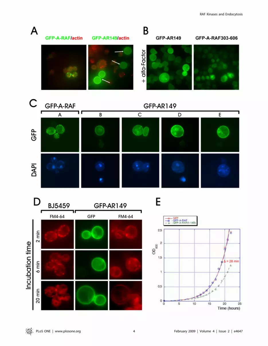
Figure 2. Physiological effects of AR149 overexpression in yeast. A. Cells expressing high amounts of GFP-AR149 fail to form cortical actinpatches. Cells expressing GFP-A-RAF or GFP-AR149 were grown, fixed as described in ‘‘Experimental procedures’’. Polymerized actin was visualizedwith Alexa Fluor 546-conjugated Phalloidin. Note that cells expressing higher levels of GFP-AR149 but not of full length A-RAF (bright green cellsmarked with arrows) lost polymerized actin. B. GFP-AR149 expression inhibits /-factor induced shmoo formation. Cells transformed with pUG36-AR149 (right) or A-RAF(303–606) (left) were treated with /-factor for 2 h. Note increased overall size and absence of shmoos in cells expressing GFP-AR149. C. Changes in nuclear morphology of GFP-AR149 expressing cells. DNA of the nucleus and mitochondria was stained with DAPI. Morphologyof nuclei of AR149 expressors varied from fragmented (left) to completely dispersed (right). D. FM-4-64 uptake is affected in AR149-expressing yeast.Yeast transformed with pUG36-AR149 and untransformed control were incubated with lipophilic styryl dye FM 4–64 for indicated time, washed andobserved by fluorescence microscopy. Transition of the red fluorescence from periplasmic endocytic sites to vacuoles is clearly visible at 20 minutesin control cells, but not in GFP-AR149 expressing cells. E. Growth inhibition of AR149 expressing cells. Cells transformed with pUG36, pUG36-A-RAF orpUG36-AR149 were grown under selective conditions in fresh selective medium. Cell proliferation was monitored by spectrophotometry.doi:10.1371/journal.pone.0004647.g002
RAF Kinases and Endocytosis
PLoS ONE | www.plosone.org 5 February 2009 | Volume 4 | Issue 2 | e4647

RAF Kinases and Endocytosis
PLoS ONE | www.plosone.org 6 February 2009 | Volume 4 | Issue 2 | e4647

contrast, mutant protein with substitution of basic residues in the
PA binding site led to homogeneous distribution in the plasma
membrane, the typical pattern for C-terminally truncated mutants
(Figure S2). We conclude that the A-RAF specific punctate pattern
requires both, the PS/PIP2 and PA lipid binding sites.
Expression of AR149 in yeast causes growth inhibition,defects in cell polarization, in nuclear morphology and inmembrane trafficking
In the course of deletion analysis, we noted that cells expressing
several C-terminally truncated mutants showed homogenous
membrane localization, different from that of full-length protein.
In addition, a significant fraction of cells expressing the C-terminal
deletion mutants was larger and apolar (round in shape instead of
oval) as compared to GFP or GFP-A-RAF expressing cells (see
Figure 2A, B). Polarization during cell growth by directed
membrane trafficking is essential for cell division, therefore we
hypothesized that GFP-AR149 expressing cells may exhibit
retarded cell cycle progression and as a consequence loss of
viability. To test this, we grew GFP, GFP-A-RAF and GFP-
AR149 in liquid culture, under promoter de-repressing conditions.
Cells expressing GFP-AR149 showed sustained growth inhibition.
The difference of 28 minutes in duplication time confirms that
expression of GFP-AR149, but not that of GFP or full-length A-
RAF exhibits an inhibitory effect on expressing cells (Figure 2E).
We stained actin and DNA in GFP-AR149 and GFP-A-RAF
expressing cells. In agreement with the apolar phenotype, no
polymerized actin could be detected in the large round cells
expressing higher levels of GFP-AR149 (Figure 2A arrows).
Intriguingly, in the same cells (large, apolar) no intact nuclei
could be detected by DAPI staining (Figure 2C). Viability tests
showed about 20% decrease in survival rate of GFP-A-RAF149
expressing cells, which is in good agreement with the percentage of
apolar cells found in the culture. In a FM4-64 uptake assay we
found that yeasts expressing the AR-149 were affected in
endocytosis (Figure 2D) conversely endocytosis was unaffected
when GFP-A-RAF was expressed (Figure S1D).
To test the effect of the fusion partner and of high level expression
of A-RAF and of AR149 on yeasts, these proteins were expressed as
GST-fusions under the strongly inducible GAL10 promoter. Cells
were transformed with GST-A-RAF or GST-AR149, streaked on
glucose- (‘‘promoter off’’) or galactose (‘‘promoter on’’) media and
grown at 30uC. Figure S1C shows that whereas all three strains
grew well in the repressive medium, in the inductive medium the
GST-AR149 expressing strain did not grow.
Taken together, AR149, but not full-length A-RAF, localizes
homogeneously to the plasma membrane, and blocks yeast cell
polarization, actin polymerization, and endocytosis, resulting in
cell growth inhibition. Moreover cells overexpressing AR149 from
a strong GAL10 promoter were not viable (Figure S1C).
Localization of A-RAF and AR149 in human cell linesThe dramatic differences in localization between B- and C-RAF
versus A-RAF and its N-terminal fragment, AR149, observed in
yeast, prompted us to analyze the latter proteins in mammalian
cell lines. AR149 expression had a strong negative effect on actin
polymerization in yeast (Figure 2A). To test this effect in
mammalian cells, we chose NIH 3T3 cell line known for extended
fibers of polymerized actin called stress fibers. Similar to yeast,
stress fibers in NIH 3T3 cells were diminished by GFP-AR149
expression (Figure S3). This effect was also described for DA-
RAF1, 2 by Yokoyama et al. (2007).
We next examined the subcellular localization of A-RAF and
AR149 in HeLa cells. Cells were transfected with GFP-tagged A-
RAF and AR149 respectively. For control, an N-terminal
fragment of C-RAF (aar 1–256) described originally as a dominant
inhibitory RAF mutant [29] that corresponds to AR149 (named
C-RAF-C4) was included in the analysis. As observed in yeast,
AR149 was unique in that it labeled the plasma membrane.
Additionally in HeLa cells ‘‘beads on a string’’ structures
(Figure 3A, top panel) were seen, demonstrating isotype specificity
of the lipid binding site in CR1 as B- and C-RAF were reported to
localize instead to ER/Golgi complex or mitochondria respec-
tively [30,20]. Disruption of microtubules by Nocodazole
treatment separated beads from the strings (Figure 3A, top panel).
The stained vesicular tubular structures most likely represent
recycling endosomes. To test this hypothesis, we studied the
localization of RFP-AR149 and that of an established endosomal
marker, ARF6. As documented in Figure 3A, middle panel, there
is a high degree of colocalization between RFP-AR149 and GFP-
ARF6. ARF6 was reported to regulate central steps of endocytosis
and recycling of endocytosed material by early and recycling
endosomes (see [16] for review). Like nearly all GTPases, ARF6
functions as a molecular switch alternating between GTP-bound
(active) and GDP bound (inactive) states. Consequently, overex-
pressed GTP-, GDP-locked or nucleotide-free mutants of ARF6
(Q67L, T27N and N121I respectively) have distinct dominant
effects on endosomal morphology and endocytosis [31].
In co-transfected cells we found colocalization of RFP-AR149
(Figure 3A, middle panel) or wild type A-RAF (Figure 3A, bottom
panel) with GFP-ARF6 in tubular vesicular structures.
In summary, AR149 and wild type A-RAF, but not C-RAF-C4
localizes to tubular endosomes, as proven by specific colocalization
with ARF6.
Figure 3. Localization of GFP-A-RAF, GFP-AR149 and endogenous A-RAF in mammalian cells. A. Top row: HeLa cells were transientlytransfected with pEGFP bearing the indicated genes for 2 days. GFP fusion proteins were detected by fluorescence microscopy. GFP-A-RAF is presentthroughout the cytoplasm and accumulates around the nucleus. In contrast, GFP-AR149 labels punctate structures often aligned on strings. Stringswere disassembled by treatment with Nocodazole. A C-RAF fragment orthologous to AR149 is GFP-C-RAF(C4). Representative images are shown.Scale bar = 10 mm. Middle row: HeLa cells were cotransfected with RFP-AR149 and GFP-ARF6 as described above. RFP and GFP fluorescences wererecorded separately. Bottom row: HeLa cells were cotransfected with Myc-A-RAF and GFP-ARF6. After two days transfected cells were treated withdigitonin to extract cytosol and processed for detection of A-RAF and ARF6 as described in Figure 3C. Boxed areas are shown at higher magnification.Arrows indicate vesicles with colocalization. Representative images are shown. Scale bar = 10 mm. B. Cells were fractionated using ‘‘ProteoExtractSubcellular Proteome Extraction Kit’’ (Calbiochem) and analyzed by immunoblotting. antibodies against following proteins were used ascompartmental markers: vimentin as cytoskeletal marker, PARP as nuclear marker, 2MPK as cytosolic marker. Both A-RAF and AR149 are co-fractionating predominantly with cytoskeleton and cytosol. Small portion of AR149 was also found in plasma membrane fraction. C. HeLa cells weretreated with digitonin to extract cytosol. After fixation and washing, immunofluorescence microscopy with antibodies against A-RAF and against b-tubulin was carried out. The boxed area is shown at higher magnification. Small A-RAF positive vesicles are at the periphery and line microtubules(arrows). Representative images are shown. Scale bar = 10 mm.doi:10.1371/journal.pone.0004647.g003
RAF Kinases and Endocytosis
PLoS ONE | www.plosone.org 7 February 2009 | Volume 4 | Issue 2 | e4647

RAF Kinases and Endocytosis
PLoS ONE | www.plosone.org 8 February 2009 | Volume 4 | Issue 2 | e4647

A fraction of endogenous A-RAF localizes to vesicleslocated along microtubuli
Considering the dominant negative effect of AR149 on
endocytosis in yeast, we set out to determine whether endogenous
A-RAF in HeLa cells was localized on endocytic vesicles.
After cytosol depletion, fixation and immunostaining with
specific antibodies the remaining endogenous full-length A-RAF
was found associated with vesicular structures. A-RAF positive
vesicles were localized in the periplasmic area and at nearly each
plus end of microtubules (Figure 3C). A significant fraction of A-
RAF-positive punctate structures is lining the microtubular
network that extends to the perinuclear area. A-RAF siRNA
removed the punctate labeling, but not the diffuse staining over
the nucleus that was also seen with normal rabbit IgG, (Fig S4C)
and is therefore considered non-specific background. Consistently
in cell fractionation experiments a significant part of A-RAF
fractionates with cytoskeletal, but not with the nuclear fraction
(Figure 3B).
Overexpressed AR149 exhibits a dominant inhibitoryeffect on transport and traps internalized transferrin inARF6 and RAB11 positive endosomes
We asked whether overexpression of AR149 affects endocytic
trafficking in mammalian cells. First, uptake and recycling of Tfn
was investigated. Tfn is internalized upon binding to its receptor via
a clathrin-dependent pathway and, after pH-regulated release,
recycles back from the recycling/late endosomes. These endosomes
are characterized by their accumulation in the pericentriolar space.
GFP or GFP-AR149 transfected HeLa cells were incubated with
Alexa-546 labelled Tfn for the indicated time (Figure 4). In control
cells, after 30 to 40 minutes, endocytosed Tfn accumulated in the
pericentriolar compartment (Figure 4, GFP panel). In GFP-AR149
expressing cells, relocation of endocytosed Tfn to pericentriolar
compartments was significantly inhibited (Figure 4, GFP-AR149
panel). The difference is clearly visible at the 40 min timepoint,
where transfected and non-transfected cells are next to each other.
Therefore expression of AR149 does not influence the endocytic
uptake of Tfn, but strongly inhibits its relocation to recycling
endosomes. To pinpoint the cellular endosome compartment,
which Tfn is trapped in, EEA1, RAB11 and ARF6 were used as
markers for early and recycling endosomes. The data clearly show
significant overlap between AR149, internalized Tfn and ARF6- as
well as between AR149, Tfn and RAB11-positive vesicles (Figure 5).
Consistently, EEA1 positive vesicles did not accumulate Tfn (Figure
S5). Thus we conclude that the block in endocytic trafficking is at
the level of tubular recycling endosomes.
siRNA silencing of A-RAF and MEK inhibition mimicks theAR149 overexpression phenotype
The splice variant DA-RAF was shown to function as an
effective inhibitor of RAF-ERK signaling [19]. As AR149 is
expected to share this function, AR149 overexpression or A-RAF
depletion should have the same effect on Tfn trafficking. For
depletion we prepared a set of A-RAF specific siRNAs and
transfected them into HeLa cells. Degree of depletion and
specificity of A-RAF siRNAs were determined by immunoblotting.
As documented in Figure S4A significant depletion of A-RAF but
not of the other two RAF isoforms was achieved. Semi-
quantitative RT PCR showed that siRNA down-regulates A-
RAF mRNA selectively without affecting the mRNA levels of DA-
RAF (Figure S4B), which was expressed at a lower level than full
length A-RAF in this cell line.
To test the effect of A-RAF depletion on Tfn trafficking, A-RAF
siRNA-transfected, and control (scrambled siRNA-transfected)
cells were subjected to a Tfn uptake assay. Similar to GFP-AR149
expressing cells, defective or significantly delayed accumulation of
endocytosed Tfn in the pericentriolar space was observed
(Figure 6A) indicating that A-RAF kinase activity and thus the
activation of the mitogenic cascade is required for normal Tfn
trafficking. Consistent with a requirement of localized ERK
activation, chemical inhibition of the mitogenic cascade by U0126,
a specific inhibitor of the RAF effector MEK, prevented the
aggregation of Tfn positive endosomes in a similar way as A-RAF
depletion (Figure 6A).
As the phenotypes of A-RAF depletion and mitogenic cascade
inhibition overlap, we conclude that localized ERK signalling is
required for endosomal maturation and AR149/DA-RAF func-
tions as a dominant negative inhibitor of A-RAF on endosomes.
Expression of GDP-locked ARF6(T27N) also blocksaccumulation of Tfn in the pericentriolar endosomalcompartment
Dominant negative ARF6 has been shown previously to
interfere with internalisation of Tfn [15]. To test whether the
pattern of Tfn accumulation by ARF6(T27N) and AR149 are
comparable transfected HeLa cells were examined for co-
localization of internalized Tfn and ARF6(T27N) (Figure 6B).
Inspection of Figure 4 and Figure 6A reveals similar re-distribution
of Tfn in HeLa cells in which A-RAF has been knocked down or
inhibited (AR149 or U0126) and HeLa cells that express
ARF6(T27N). This data suggests that ARF6 may be a target for
regulation by A-RAF on endosomes.
Tfn accumulates in ARF6 positive and RAB11 positivevesicles after A-RAF knock down
To characterize the endosome compartment in which Tfn
accumulates after A-RAF knock down ARF6 and RAB11 were
used as markers (Figure 7). Additionally we included EEA1 as
marker of early endosomes (Figure S6). Co-localisation experi-
ments in HeLa cells identify vesicles with accumulated Tfn as
RAB11- and ARF6- positive. No EEA1 Tfn double positive
vesicles were found. We conclude that the block in Tfn trafficking
in the absence of A-RAF lies between tubular- and TGN-
assotiated recycling endosome compartments.
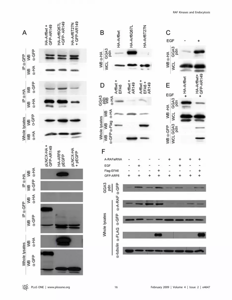
AR149 Interacts with ARF6 independent of nucleotidestatus
We have shown that AR149 and both (active and inactive)
ARF6 mutants partially colocalize on endocytic vesicles and the
plasma membrane (Figure S7). We were interested whether
interaction of these two proteins could be shown at the
biochemical level. HeLa cells were co-transfected with GFP,
Figure 4. Expression of GFP-AR149 inhibits maturation of endosomes. GFP-AR149-transfected and control (GFP-transfected) cells wereincubated with fluorescent Tfn for indicated times. Tfn containing vesicles accumulate in the pericentriolar area of control cells, but remain in theGFP-AR149 positive vesicles scattered throughout the cytosol of GFP-AR149 transfected cells. Representative images are shown.doi:10.1371/journal.pone.0004647.g004
RAF Kinases and Endocytosis
PLoS ONE | www.plosone.org 9 February 2009 | Volume 4 | Issue 2 | e4647

RAF Kinases and Endocytosis
PLoS ONE | www.plosone.org 10 February 2009 | Volume 4 | Issue 2 | e4647

GFP-AR149 and HA-tagged ARF6 wild type, GTP-locked (Q67L)
or GDP-locked (T27N) mutants. After immunoprecipitation with
anti-GFP antibodies, the precipitates were separated by SDS
PAGE and tested for bound ARF6 by immunoblotting against
HA. In a reverse experiment ARF6 mutants were first precipitated
and the presence of GFP-A-RAF in the precipitate was
demonstrated by immunoblotting with anti-GFP antibodies.
Figure 8A documents that similar amounts of ARF6,
ARF6(Q67L) and ARF6(T27N) co-precipitated with GFP-
AR149, irrespective of which protein was precipitated first. Thus
interaction of A-RAF with ARF6 is independent of the ARF6
nucleotide status.
A-RAF and AR149 regulate ARF6 activationNext we addressed whether overexpression of dominant
negative AR149 has an effect on activation of ARF6. ARF6
interacts specifically with its effector GGA3 in a nucleotide
dependent manner. Therefore binding of GGA3 can be used as
readout of the activation status of ARF6 [23]. In a control
experiment, cell lysates from COS7 cells transfected with wild
type, dominantly activated (Q67L) and dominant negative (T27N)
mutants of HA-ARF6 were incubated with immobilized GST-
GGA3 and washed. As shown in Figure 8B, GGA3 pulls effectively
ARF6(Q67L), and less effectively ARF6wt. In contrast, no binding
of ARF6(T27N) to GGA3 could be detected.
In subsequent experiments, COS7 cells were starved and
induced with EGF, a strong elicitor of mitogenic signalling.
Activated ARF6 was pulled-down by immobilized GGA3 as
described above. Large amounts of ARF6 were pulled-down from
EGF stimulated cells compared to non-stimulated cells (Figure 8C).
Thus, ARF6 is activated by the mitogenic cascade. This activation
is significantly diminished by the coexpression of AR149
(Figure 8E) or A-RAF knock down (Figure 8F).
Of note pulled-down ARF6 contains coexpressed AR149, which
precludes competition of AR149 and GGA3 for binding to
activated ARF6.
To pinpoint the position of AR149 action in a signalling
pathway, COS7 cells were co-transfected with HA-ARF6 and
either AR149, EFA6 or both. It has been previously described that
overexpression of the ARF6 exchange factor, EFA6, increases the
amount of ARF6 bound to GST-GGA3 beads. As depicted in
Figure 8D, less ARF6 was pulled down in the presence of AR149.
Similarly, A-RAF knock down reduced the amount of ARF6 that
could be pulled-down with GST-GGA3. EFA6 partially rescued
this effect (Figure 8F).
We conclude that AR149 co-expression or A-RAF knock down
negatively affects the activation of ARF6 by EFA6, i.e. A-RAF
functions upstream of ARF6.
Discussion
A-RAF is a distinct member of the RAF kinase familyIn this work we unveil an unexpected link between A-RAF and
regulation of ARF6 activity.
A-RAF possesses several features that set it apart from the other
RAF kinases. A-RAF maps to the X chromosome and is the only
steroid hormone-regulated RAF isoform, [32]. A-RAF protein has
substitutions in a negatively charged region immediately upstream
of the kinase domain (N-region), which is at least partially
responsible for its low basal activity [12]. In contrast to C-RAF
[33,34], feedback phosphorylation of A-RAF by its downstream
effector, ERK, has an activating effect on its kinase activity [35].
Interestingly, out of 590 kinases tested, the three RAF isoforms
were among 208 kinases affecting clathrin- or caveolae-dependent
endocytosis [36]. si-RNA mediated silencing of A-RAF inhibits,
whereas that of C- and B-RAF activate SV40 uptake.
Role of lipid binding domains, identification andproperties of inhibitory A-RAF, AR149
Our initial studies on RAF distribution in a heterologous yeast
system showed that of the three RAF isoforms, only A-RAF
located to the cell cortex of yeast cells (Figure 1). This pattern
resembles that of components of the endocytic machinery, such as
Sla1p, Sla2p, clathrin light chain protein Clc1p [37]. Differences
in localization of A-RAF and AR149 can be explained by the
existence of two lipid-binding domains in the structure of A-RAF,
each domain binding to specific sets of lipids.
The unique distribution pattern of A-RAF in yeast resembles
the distribution of a PtdIns(4,5)P2 sensor, AP180 N-terminal
homology (ANTH) domain [37]. Consistently, A-RAF is the only
RAF isoform, which binds to immobilized PtdIns(4,5)P2 [27].
Using a set of point mutations, Johnson et al. (2005) located
PtdIns(4,5)P2 binding site in the RAS binding domain of A-RAF.
In contrast, we mapped PtdIns(4,5)P2 binding site to the cysteine
rich domain of A-RAF because a RAS binding domain deleted
mutant (88–606) still shows wild type distribution.
In agreement with Johnson et al (2005), replacement of arginine
52 in the RBD with leucine, which prevents binding of C-RAF
RBD to RAS [38], disturbs the membrane localization of both full-
length A-RAF and AR149 in yeast (Figure 1). We interpret this
discrepancy by an interaction between RBD and CRD. It is
known that RBD and CRD bind to RAS cooperatively [39]. The
R52L mutation in the RBD may cause structural rearrangements
disabling the interaction of the PtdIns(4,5)P2 binding site in the
CRD with membranes.
CRDs are conserved among RAF isoforms, but only the A-RAF
CRD possesses the unique property of PtdIns(4,5)P2 binding [27].
However as we show here the PtdIns(4,5)P2 binding site per se is not
sufficient to specify the A-RAF wild type localization pattern. The
wild type pattern of membrane binding requires in addition the
PA binding site in the catalytic domain. In regard to the
colocalization of A-RAF with ARF6 in mammalian cells, it is
noteworthy that ARF6 is known to regulate the PtdIns(4,5)P2
content of membrane microdomains to which it binds.
In yeast Arf3p is the functional homolog of ARF6. Arf3p
regulates PtdIns(4,5)P2 levels, endocytosis and actin polymeriza-
tion [40,41]. Therefore it is likely that the dominant lethal
phenotype of AR149 in yeast is mediated by interference with
Arf3p.
Localization of A-RAF and AR149 in mammalian cellsThe differences between RAF isoforms were most pronounced
in yeast, where evidence for membrane attachment was restricted
to A-RAF. The punctate cortical structures that are sites of A-RAF
Figure 5. AR149 traps inernalized transferrin in ARF6 and RAB11 positive endosomes. HeLa cells were transfected with RFP-AR149 andeither GFP-ARF6 or RAB11 as indicated. Uptake of fluorescent Tfn was examined after 30 and 60 minutes. Representative images show that Tfn istrapped by AR149 in GFP-ARF6 positive and in GFP-RAB11 positive vesicles. Enlarged areas are marked by boxes. Arrows indicate co-localization.doi:10.1371/journal.pone.0004647.g005
RAF Kinases and Endocytosis
PLoS ONE | www.plosone.org 11 February 2009 | Volume 4 | Issue 2 | e4647

RAF Kinases and Endocytosis
PLoS ONE | www.plosone.org 12 February 2009 | Volume 4 | Issue 2 | e4647

accumulation in yeast are known to contain proteins associated
with early steps of endocytosis such as AP180 as well as Pan1p,
that is associated with cortical actin patches. It is tempting to
speculate that in mammalian cells the localization of A-RAF not
only brings A-RAF into the neighbourhood of ARF6 on tubular
endosomes, but additionally involves interaction with mammalian
homologs of AP180/Pan1p that function at the interface of
clathrin coated vesicles and an actin cytoskeleton regulatory
complex. Such complexes are essential for endocytosis and also
linked with the microtubule network [42]. Consistently GFP-
AR149 decorated predominantly tubulo-vesicular endosomes, as
confirmed by co-localization with ARF6 (Figure 3, middle panel).
These endosomes lost their beads on the strings appearance upon
Nocodazole treatment (Figure 3, top panel).
Role of A-RAF and AR149 in endocytosisDefinitions of endocytic compartments are poorly standardized
[43]. Here we used definitions given in [44], where endocytosed
Tfn is first enclosed in small vesicles. These vesicles fuse to form
early endosomes. Further maturation of early endosomes through
tubular recycling endosomes leads to the pericentriolar TGN-
associated recycling compartment. AR149 is targeted to tubular
endosomes where it colocalizes with ARF6. Exogenous expression
of AR149 or dominant negative ARF6(T27N) mutant causes
trapping of endocytosed Tfn in tubular endosomes, which prevents
transfer to pericentriolar recycling compartment and subsequent
return to the plasma membrane. AR149 and ARF6 occur in the
same complex as demonstrated both by fluorescence microscopy
and coprecipitation experiments (Figure 8A).
A-RAF kinase was previously reported to participate in
regulation of caveolae/raft-mediated endocytosis by stabilizing
the caveolar coat [36,45]. Our discovery of a requirement for A-
RAF activity at a step subsequent to endocytosis, in the transfer to
the recycling compartment, point to a broader role of A-RAF in
membrane trafficking that additionally involves feedback regula-
tion by the inhibitory splice variants DA-RAF 1,2 [19]. Our data
suggest that DA-RAF does not necessarily work as a general
inhibitor of mitogenic signalling as initially described [19]. More
likely, due to its unique intracellular localization, AR149/DA-
RAF primarily inhibits just a specific endosome-associated branch
of mitogenic signalling, which is responsible for regulation of
receptor recycling and/or restoration of signalling molecules.
The classical mechanism of cytoskeleton dependent endocytosis
is subdivided in two parts: first, a short-distance step in the cortical
area depends on actin and second long-distance microtubule
dependent vesicular transport [42]. Our micrographs do not
provide time resolution, but considering our functional analyses, it
is conceivable that A-RAF functionally associates with the
endosomes shuttling along microtubules between plasma mem-
brane and the perinuclear recycling compartment.
From the functional analysis of Tfn endocytosis (Figure 6A), we
conclude that not only localization to endosomes but also activity
of A-RAF kinase in the mitogenic cascade on endosomes are a
prerequisite for the translocation of Tfn-positive endosomes to the
pericentriolar region.
ARF6 regulation by A-RAF and AR149Our data suggest that A-RAF functions upstream of ARF6.
There has been an earlier report by Robertson [31], who
suggested, based on epistasis experiments with ERK and ARF6
that ERK functions upstream and downstream of ARF6.
Several other reports also suggested dependence of ERK
activation on ARF6 [17,46], which is in direct contrast with the
results of our epistasis analysis. This contradiction may be
explained by a pleiotropic effect of active ARF6, which is known
to block internalization of activated receptors [15], leading to
sustained signalling through the mitogenic cascade. Our data
additionally suggest EFA6 as a cooperation partner of A-RAF in
the activation of ARF6. Of note inspection of the primary
structure of EFA6 revealed multiple potential ERK/MAP kinase
phosphorylation sites that suggest that EFA6 is a substrate of ERK
downstream of A-RAF.
There are several examples for crosstalk between endocytosis
and signal transduction [47,48]. In addition, there is a growing
body of evidence that at least part of mitogenic signalling takes
place on endosomes: for example, the MEK partner MP1 was
isolated as a component of endosomal vesicles and associates with
MEK and participates in signal transduction [49]. Another
scaffold of the mitogenic cascade, KSR1, was also shown to
localize and mediate signalling to endosomes [31].
Model of A-RAF function in regulation of endocytosisThe novel findings on the A-RAF localization and the
interaction with ARF6 have led to a new model of A-RAF
function shown in Figure 9. Stimulation of growth factor receptors
is followed by RAS activation. RASNGTP recruits RAFs and leads
to assembly of the RAF-MEK-ERK module at membranes. The
three RAF isozymes become activated consecutively and mediate
diversification of the signal to different subcellular compartments.
A-RAF activation is delayed [50] because it has a corequirement
for ERK phosphorylation [35], which uncouples A-RAF from B-
and C-RAF that in turn are inhibited by feedback phosphorylation
[33,34]. Due to the unique localization of A-RAF to endosomes,
this delay is optimal for regulation of later endocytic events such as
recycling of receptors [51]. A-RAF function at endosomes also
involves the mitogenic cascade and triggers ARF6 activation
possibly via EFA6.
A-RAF involvement in ARF6 dependent endocytic recycling
provides a new perspective for explaining the phenotype of A-RAF
knock out mice, which exhibit severe neurological defects such as
ataxia, rigidity of the musculature and continuous tremor [8].
Intriguingly, A-RAF, ARF6 and EFA6 are strongly expressed in
Purkinje cells of mouse cerebellum [52,53]. Along this line, DA-
RAF expression is particularly strong in brain [19]. Endocytosis
and rapid recycling of synaptic vesicles is critically important for
the physiological function of neurons, which may further stress the
role of A-RAF in the nervous system.
Figure 6. siRNA mediated A-RAF depletion, MEK inhibition and expression of ARF6(T27N) also block accumulation of Tfn in thepericentriolar endosome compartment. A. HeLa cells transfected with A-RAF siRNA, scrambled siRNA or treated with MEK inhibitor UO126 weresubjected to Tfn uptake assay. Lack of accumulation of endocytosed Tfn in the pericentriolar compartment is similar to AR149-expressing cells(compare with Figure 4). Representative images are shown. Scale bar = 10 mm. B. HeLa cells were transfected with ARF6(T27N) before assay forfluorescent Tfn uptake. ARF6(T27N) positive vesicles accumulate internalised Tfn. The pattern of distribution is similar to that in A-RAF siRNA and toU0126 treated cells. Representative images are shown. Scale bar = 10 mm.doi:10.1371/journal.pone.0004647.g006
RAF Kinases and Endocytosis
PLoS ONE | www.plosone.org 13 February 2009 | Volume 4 | Issue 2 | e4647

RAF Kinases and Endocytosis
PLoS ONE | www.plosone.org 14 February 2009 | Volume 4 | Issue 2 | e4647
Supporting Information
Table S1 List of primers used in this study.
Found at: doi:10.1371/journal.pone.0004647.s001 (0.03 MB
DOC)
Figure S1 A. Fractionation of yeast lysates by sucrose gradient
centrifugation. Cell lysates were loaded on the top of sucrose
gradient and centrifuged at 100.0006g. Fractionated lysates were
loaded on SDS PAGE and immunoblotted. Proteins were
visualized with specific antibodies. A-RAF is the only RAF
protein, which segregates into heavy membrane/particle fractions.
GFP alone fused with B- and C-RAF segregated into cytosolic/
vacuolar fractions. Distribution of yeast membrane markers is
shown in the lower rows. B. Immunoblot analysis of expressed
GFP-A-RAF deletion mutants Yeast cell lysates expressing
indicated GFP-A-RAF constructs were loaded on SDS PAGE
and analyzed by Western blotting with antibodies against GFP. C.
Lethality of GST-AR149. S.cerevisiae strain BJ 5459 was
transformed with pEG-KT vehicle, pEG-A-RAF and pEG-
AR149. Obtained colonies were streaked on uracildropout
medium with glucose or galactose. Induction of protein production
by galactose was lethal for GST-AR149 expressing cells, but not
for those expressing either empty vehicle or full-length A-RAF. D.
Part of GFP-A-RAF colocalizes with sites of endocytosis Yeast
transformed with pUG36-AR149 and non-transformed control
were incubated with lipophilic styryl dye FM 4–64 at 30uC for
indicated time, washed and observed by flourescece microscopy.
Some of the GFP-A-RAF positive spots overlap with sites of FM
4–64 uptake.
Found at: doi:10.1371/journal.pone.0004647.s002 (5.70 MB TIF)
Figure S2 Mutational analysis of A-RAF with respect to its lipid
binding properties. Basic residues in two presumptive lipid binding
domains and in the RAS binding domain were replaced with
leucine (R52) or alanine and subcellular distribution of mutant
GFP fusion proteins was inspected by microscopy. Mutation of
R359 and K360 in the Cterminal lipid binding domain
(corresponding to phosphatidic acid binding domain of CRAF)
gives the same distribution as deletion mutants which lost this
domain. Mutation of R103 and K104 in CRD fully dislocated the
protein into cytosol. R52L mutation, which is known to disturb the
interaction of RAF with RAS had the same effect.
Found at: doi:10.1371/journal.pone.0004647.s003 (3.38 MB TIF)
Figure S3 Effect of AR149 expression on the cytoskeleton of
NIH 3T3 cells. NIH 3T3 cells were transfected with GFP-AR149
for 24 hours. After fixation, the polymerized actin was visualized
with Alexa-546 conjugated phalloidin. Note the remarkable
regression of actin stress fibers in the transfected cell. Scale
bar = 10 mm.
Found at: doi:10.1371/journal.pone.0004647.s004 (4.52 MB TIF)
Figure S4 Specific depletion of A-RAF protein and mRNA by
siRNA A. HeLa cells were treated with A-RAF specific or
scrambled siRNA and subjected to Western blot analysis with
antibodies against actin (loading control) and three RAF isoforms.
A-RAF is the only RAF isoform that decreased after siRNA
treatment. B. HeLa cells were first treated with two different
batches of A-RAF-specific or scrambled siRNA. Afterwards, the
RNA was reverse transcribed and used as a template for
quantitative PCR with primers specific for A-RAF, DA-RAF2
and Actin mRNAs. The ratio between the tested mRNA and actin
mRNA was calculated from the qPCR data. From the diagram it
can be concluded that A-RAF mRNA amount is decreasing
significantly. DA-RAF2 mRNA was poorly expressed in these cells
and its expression level did not change upon siRNA treatment. C.
Controls of indirect immunofluorecent staining of endogenous A-
RAF. HeLa cells were incubated with normal rabbit serum (left
panel) or with A-RAF specific antibodies after A-RAF knock-down
with siRNA (right panel). In both cases periplasmic punctate
structures (see Fig. 3) disappeared, whereas nuclear staining
remained. Scale bar = 10 mm.
Found at: doi:10.1371/journal.pone.0004647.s005 (5.98 MB TIF)
Figure S5 Tfn does not accumulate in EEA1 positive early
endosomes in AR149 expressing cells. HeLa cells were transfected
as indicated and used for Tfn uptake assays. Note that fluorescence
of Tfn and EEA1 do not mark identical vesicles. Enlarged areas
are marked by boxes. Arrows indicate co-localization. Scale
bar = 10 mm.
Found at: doi:10.1371/journal.pone.0004647.s006 (9.23 MB TIF)
Figure S6 A-RAF knock down phenocopies the AR149 effect
sparing EEA1 endosomes from Tfn accumulation. HeLa cells were
transfected as indicated and used for Tfn uptake assays. Note that
fluorescence of Tfn and EEA1 do not mark identical vesicles.
Enlarged areas are marked by boxes. Arrows indicate co-
localization. Scale bar = 10 mm.
Found at: doi:10.1371/journal.pone.0004647.s007 (8.47 MB TIF)
Figure S7 AR149 colocalizes with dominant active and
dominant negative ARF6 mutants in HeLa cells. RFP-AR149
was cotransfected with dominant active GFP-ARF6(Q67L) or
dominant negative GFP-ARF6(T27N) and inspected by fluores-
cent microscopy. High degree of colocalization with both ARF6
mutants is documented in overlay figures. Scale bar = 10 mm.
Found at: doi:10.1371/journal.pone.0004647.s008 (9.78 MB TIF)
Acknowledgments
We thank Margaret M. Chou and Xiang Dong Gao (University of
Pennsylvania) for plasmids and helpful comments on this work, Angela
Baljuls and Antoine Galmiche (this laboratory) for donation of plasmids
and sharing protocols and results prior to publication, Eugen Kerkhoff
(University of Augsburg) for kindly provided pEGFP-Rab11 plasmid.
Author Contributions
Conceived and designed the experiments: EN SA MB URR. Performed
the experiments: EN SA MB. Analyzed the data: EN SA MB URR.
Contributed reagents/materials/analysis tools: URR. Wrote the paper: EN
SA MB URR.
Figure 7. Tfn accumulates in ARF6 positives, RAB11 positives vesicles after A-RAF knock down. HeLa cells were cotransfected with eitherGFP-ARF6 or GFP-RAB11 and A-RAF siRNA. Tfn uptake was assayed as before. Tfn was found to co-localize with ARF6 or RAB11 as was observed inAR149 expressing cells. Enlarged areas are marked by boxes. Arrows indicate co-localization. Representative images are shown. Scale bar = 10 mm.doi:10.1371/journal.pone.0004647.g007
RAF Kinases and Endocytosis
PLoS ONE | www.plosone.org 15 February 2009 | Volume 4 | Issue 2 | e4647
RAF Kinases and Endocytosis
PLoS ONE | www.plosone.org 16 February 2009 | Volume 4 | Issue 2 | e4647

References
1. Rapp UR, Goldsborough MD, Mark GE, Bonner TI, Groffen J, et al. (1983)
Structure and biological activity of v-raf, a unique oncogene transduced by a
retrovirus. Proc Natl Acad Sci U S A 80: 4218–4222.
2. Jansen HW, Lurz R, Bister K, Bonner TI, Mark GE, et al. (1984) Homologous
cell-derived oncogenes in avian carcinoma virus MH2 and murine sarcoma virus
3611. Nature 307: 281–284.
Figure 8. Down regulation of A-RAF by either AR149 or A-RAF siRNA interferes with activation of ARF6. A. Interaction of AR149 andARF6. COS7 cells were co-transfected with GFP-AR149 and either HA-tagged ARF6wt, GTP-locked [ARF6(Q67L)] or GDP-locked [ARF6(T27N)]. Afterimmunoprecipitation with a-GFP antibodies, co-precipitated ARF proteins were detected with a-HA antibodies. In the second experiment,precipitation and detection antibodies were exchanged. Expression levels in whole cell lysates (WCL) is shown in two bottom panels. Empty vectorswere used for control (bottom panel). B. GGA3 interacts with ARF6NGTP. COS7 cells were transfected with wild type, GTP-locked (Q67L) or GDP-locked(T27N) HA-ARF6. Proteins pulled-down by incubation with GST-GGA3-Sepharose were detected with a-HA antibodies. C.,E. AR149 suppresses EGF-stimulated ARF6 activation. COS7 cells, transfected with either ARF6 alone or ARF6+GFP-AR149, were treated with EGF for 10 min and subjected toGGA3 pull-down. Bound protein was analysed by immunoblotting. Note decrease in the amount of ARFNGTP by AR149 coexpression. AR149 remainsin the pulled-down ARF6 complex confirming the immunoprecipitation data. D. AR149 inhibits ARF6 activation by EFA6. COS7 cells were transfectedwith HA-ARF6 and either AR149, EFA6, or both. ARF6NGTP was pulled-down by GST-GGA3. AR149 decreases EFA6-stimulated ARF6 activation. F. ARF6activation by EGF or EFA6 requires A-RAF. ARF6 activation was assessed by GGA3 pull-down. The amount of A-RAF and ARF6 protein were determedby Western blotting (WB). Treatment conditions are as indicated.doi:10.1371/journal.pone.0004647.g008
Figure 9. Model of A-RAF and AR149/DA-RAF function in regulation of endocytosis. Activation of receptor tyrosine kinase (here EGFreceptor) leads to RAS-mediated activation of RAF kinases. RAF isoforms sort into different membrane microdomains, such as A-RAF into PtdIns(4,5)P2
rich domains. Activated ERK has opposing effects on A-RAF and C-RAF. Whereas A-RAF is activated, C-RAF becomes inactivated by feedbackphosphorylation. A-RAF bound to PtdIns(4,5)P2 rich membranes continues to signal on endosomes leading to ARF6 activation. AR149/DA-RAF locatesto recycling endosomes and blocks ERK activation in this compartment. See the main text for details. EE - early endosome, RC – recyclingcompartment, PM – plasma membrane. Red arrows indicate positive regulation of the process. Pale brown color indicates PtdIns(4,5)P2 richmembrane microdomains.doi:10.1371/journal.pone.0004647.g009
RAF Kinases and Endocytosis
PLoS ONE | www.plosone.org 17 February 2009 | Volume 4 | Issue 2 | e4647

3. Mark GE, Rapp UR (1984) Primary structure of v-raf: relatedness to the src
family of oncogenes. Science 224: 285–289.4. Moelling K, Heimann B, Beimling P, Rapp UR, Sander T (1984) Serine- and
threonine-specific protein kinase activities of purified gag-mil and gag-raf
proteins. Nature 312: 558–561.5. Daum G, Eisenmann-Tappe I, Fries HW, Troppmair J, Rapp UR (1994) The
ins and outs of Raf kinases. Trends Biochem Sci 19: 474–480.6. Rapp UR, Gotz R, Albert S (2006) BuCy RAFs drive cells into MEK addiction.
Cancer Cell 9: 9–12.
7. Wellbrock C, Karasarides M, Marais R (2004) The RAF proteins take centrestage. Nat Rev Mol Cell Biol 5: 875–885.
8. Pritchard CA, Bolin L, Slattery R, Murray R, McMahon M (1996) Post-natallethality and neurological and gastrointestinal defects in mice with targeted
disruption of the A-Raf protein kinase gene. Curr Biol 6: 614–617.9. Wojnowski L, Zimmer AM, Beck TW, Hahn H, Bernal R, et al. (1997)
Endothelial apoptosis in Braf-deficient mice. Nat Genet 16: 293–297.
10. Wojnowski L, Stancato LF, Larner AC, Rapp UR, Zimmer A (2000)Overlapping and specific functions of Braf and Craf-1 proto-oncogenes during
mouse embryogenesis. Mech Dev 91: 97–104.11. Rapp UR, Heidecker G, Huleihel M, Cleveland JL, Choi WC, et al. (1988) raf
family serine/threonine protein kinases in mitogen signal transduction. Cold
Spring Harb Symp Quant Biol 53 Pt 1: 173–184.12. Baljuls A, Mueller T, Drexler HC, Hekman M, Rapp UR (2007) Unique N-
region determines low basal activity and limited inducibility of A-RAF kinase:the role of N-region in the evolutionary divergence of RAF kinase function in
vertebrates. J Biol Chem 282: 26575–26590.13. van Drogen F, Peter M (2001) MAP kinase dynamics in yeast. Biol Cell 93:
63–70.
14. Irie K, Gotoh Y, Yashar BM, Errede B, Nishida E, et al. (1994) Stimulatoryeffects of yeast and mammalian 14-3-3 proteins on the Raf protein kinase.
Science 265: 1716–1719.15. D’Souza-Schorey C, Li G, Colombo MI, Stahl PD (1995) A regulatory role for
ARF6 in receptor-mediated endocytosis. Science 267: 1175–1178.
16. D’Souza-Schorey C, Chavrier P (2006) ARF proteins: roles in membrane trafficand beyond. Nat Rev Mol Cell Biol 7: 347–358.
17. Li M, Ng SS, Wang J, Lai L, Leung SY, et al. (2006) EFA6A enhances gliomacell invasion through ADP ribosylation factor 6/extracellular signal-regulated
kinase signaling. Cancer Res 66: 1583–1590.18. Tague SE, Muralidharan V, D’Souza-Schorey C (2004) ADP-ribosylation factor
6 regulates tumor cell invasion through the activation of the MEK/ERK
signaling pathway. Proc Natl Acad Sci U S A 101: 9671–9676.19. Yokoyama T, Takano K, Yoshida A, Katada F, Sun P, et al. (2007) DA-Raf1, a
competent intrinsic dominant-negative antagonist of the Ras-ERK pathway, isrequired for myogenic differentiation. J Cell Biol 177: 781–793.
20. Galmiche A, Fueller J, Santel A, Krohne G, Wittig I, et al. (2008) Isoform-
specific Interaction of C-RAF with Mitochondria. J Biol Chem 283:14857–14866.
21. Guthrie C, Fink GR (1991) Guide to Yeast Genetics and Molecular Biology. SanDiego: Academic Press. 933 p.
22. Schroder S, Schimmoller F, Singer-Kruger B, Riezman H (1995) The Golgi-localization of yeast Emp47p depends on its di-lysine motif but is not affected by
the ret1-1 mutation in alpha-COP. J Cell Biol 131: 895–912.
23. Martinu L, Masuda-Robens JM, Robertson SE, Santy LC, Casanova JE, et al.(2004) The TBC (Tre-2/Bub2/Cdc16) domain protein TRE17 regulates plasma
membrane-endosomal trafficking through activation of Arf6. Mol Cell Biol 24:9752–9762.
24. Rizzo MA, Shome K, Watkins SC, Romero G (2000) The recruitment of Raf-1
to membranes is mediated by direct interaction with phosphatidic acid and isindependent of association with Ras. J Biol Chem 275: 23911–23918.
25. Hekman M, Hamm H, Villar AV, Bader B, Kuhlmann J, et al. (2002)Associations of B- and C-Raf with cholesterol, phosphatidylserine, and lipid
second messengers: preferential binding of Raf to artificial lipid rafts. J Biol
Chem 277: 24090–24102.26. Improta-Brears T, Ghosh S, Bell RM (1999) Mutational analysis of Raf-1
cysteine rich domain: requirement for a cluster of basic aminoacids forinteraction with phosphatidylserine. Mol Cell Biochem 198: 171–178.
27. Johnson LM, James KM, Chamberlain MD, Anderson DH (2005) Identificationof key residues in the A-Raf kinase important for phosphoinositide lipid binding
specificity. Biochemistry 44: 3432–3440.
28. Ghosh S, Strum JC, Sciorra VA, Daniel L, Bell RM (1996) Raf-1 kinasepossesses distinct binding domains for phosphatidylserine and phosphatidic acid.
Phosphatidic acid regulates the translocation of Raf-1 in 12-O-tetradecanoyl-
phorbol-13-acetate-stimulated Madin-Darby canine kidney cells. J Biol Chem
271: 8472–8480.
29. Bruder JT, Heidecker G, Rapp UR (1992) Serum-, TPA-, and Ras-induced
expression from Ap-1/Ets-driven promoters requires Raf-1 kinase. Genes Dev 6:
545–556.
30. Fischer A, Hekman M, Kuhlmann J, Rubio I, Wiese S, et al. (2007) B- and C-
RAF display essential differences in their binding to Ras: the isotype-specific N
terminus of B-RAF facilitates Ras binding. J Biol Chem 282: 26503–26516.
31. Robertson SE, Setty SR, Sitaram A, Marks MS, Lewis RE, et al. (2006)
Extracellular signal-regulated kinase regulates clathrin-independent endosomal
trafficking. Mol Biol Cell 17: 645–657.
32. Lee JE, Beck TW, Wojnowski L, Rapp UR (1996) Regulation of A-raf
expression. Oncogene 12: 1669–1677.
33. Dougherty MK, Muller J, Ritt DA, Zhou M, Zhou XZ, et al. (2005) Regulation
of Raf-1 by direct feedback phosphorylation. Mol Cell 17: 215–224.
34. Hekman M, Fischer A, Wennogle LP, Wang YK, Campbell SL, et al. (2005)
Novel C-Raf phosphorylation sites: serine 296 and 301 participate in Raf
regulation. FEBS Lett 579: 464–468.
35. Baljuls A, Schmitz W, Mueller T, Zahedi RP, Sickmann A, et al. (2008) Positive
regulation of A-RAF by phosphorylation of isoform-specific hinge segment and
identification of novel phosphorylation sites. J Biol Chem 283: 27239–27254.
36. Pelkmans L, Fava E, Grabner H, Hannus M, Habermann B, et al. (2005)
Genome-wide analysis of human kinases in clathrin- and caveolae/raft-mediated
endocytosis. Nature 436: 78–86.
37. Sun Y, Carroll S, Kaksonen M, Toshima JY, Drubin DG (2007) PtdIns(4,5)P2
turnover is required for multiple stages during clathrin- and actin-dependent
endocytic internalization. J Cell Biol 177: 355–367.
38. Fabian JR, Vojtek AB, Cooper JA, Morrison DK (1994) A single amino acid
change in Raf-1 inhibits Ras binding and alters Raf-1 function. Proc Natl Acad
Sci U S A 91: 5982–5986.
39. Drugan JK, Khosravi-Far R, White MA, Der CJ, Sung YJ, et al. (1996) Ras
interaction with two distinct binding domains in Raf-1 may be required for Ras
transformation. J Biol Chem 271: 233–237.
40. Smaczynska-de Rooij I, Costa R, Ayscough KR (2008) Yeast Arf3p modulates
plasma membrane PtdIns(4,5)P2 levels to facilitate endocytosis. Traffic 9:
559–573.
41. Lambert AA, Perron MP, Lavoie E, Pallotta D (2007) The Saccharomyces
cerevisiae Arf3 protein is involved in actin cable and cortical patch formation.
FEMS Yeast Res 7: 782–795.
42. Murray JW, Wolkoff AW (2003) Roles of the cytoskeleton and motor proteins in
endocytic sorting. Adv Drug Deliv Rev 55: 1385–1403.
43. Maxfield FR, McGraw TE (2004) Endocytic recycling. Nat Rev Mol Cell Biol 5:
121–132.
44. D’Souza-Schorey C, van Donselaar E, Hsu VW, Yang C, Stahl PD, et al. (1998)
ARF6 targets recycling vesicles to the plasma membrane: insights from an
ultrastructural investigation. J Cell Biol 140: 603–616.
45. Pelkmans L, Zerial M (2005) Kinase-regulated quantal assemblies and kiss-and-
run recycling of caveolae. Nature 436: 128–133.
46. Tushir JS, D’Souza-Schorey C (2007) ARF6-dependent activation of ERK and
Rac1 modulates epithelial tubule development. Embo J 26: 1806–1819.
47. Polo S, Di Fiore PP (2006) Endocytosis conducts the cell signaling orchestra. Cell
124: 897–900.
48. Rapp UR, Fensterle J, Albert S, Gotz R (2003) Raf kinases in lung tumor
development. Adv Enzyme Regul 43: 183–195.
49. Teis D, Taub N, Kurzbauer R, Hilber D, de Araujo ME, et al. (2006) p14-MP1-
MEK1 signaling regulates endosomal traffic and cellular proliferation during
tissue homeostasis. J Cell Biol 175: 861–868.
50. Wixler V, Smola U, Schuler M, Rapp U (1996) Differential regulation of Raf
isozymes by growth versus differentiation inducing factors in PC12 pheochro-
mocytoma cells. FEBS Lett 385: 131–137.
51. von Zastrow M, Sorkin A (2007) Signaling on the endocytic pathway. Curr Opin
Cell Biol 19: 436–445.
52. Luckett JC, Huser MB, Giagtzoglou N, Brown JE, Pritchard CA (2000)
Expression of the A-raf proto-oncogene in the normal adult and embryonic
mouse. Cell Growth Differ 11: 163–171.
53. Matsuya S, Sakagami H, Tohgo A, Owada Y, Shin HW, et al. (2005) Cellular
and subcellular localization of EFA6C, a third member of the EFA6 family, in
adult mouse Purkinje cells. J Neurochem 93: 674–685.
RAF Kinases and Endocytosis
PLoS ONE | www.plosone.org 18 February 2009 | Volume 4 | Issue 2 | e4647


