Embed Size (px)
Citation preview
A Role for the Eph Ligand Ephrin-A3 in Entorhino-HippocampalAxon Targeting
Eckart Stein,1 Nicolai E. Savaskan,1 Olaf Ninnemann,1 Robert Nitsch,1 Renping Zhou,2 and Thomas Skutella1
1Institute of Anatomy, Department of Cell and Neurobiology, Humboldt University Hospital (Charite), 10098 Berlin,Germany, and 2Laboratory for Cancer Research, Department of Chemical Biology, College of Pharmacy, RutgersUniversity, Piscataway, New Jersey 08855
Neurons of layers II and III of the entorhinal cortex constitute themajor afferent connection of the hippocampus. The molecularmechanisms that target the entorhinal axons to specific layersin the hippocampus are not known. EphA5, a member of theEph receptor family, which has been shown to play critical rolesin axon guidance, is expressed in the entorhinal cortex, theorigin of the perforant pathway. In addition, ligands that interactwith EphA5 are expressed in distinct hippocampal regionsduring development of the entorhino-hippocampal projection.Of these ligands, ephrin-A3 mRNA is localized both in thegranular cell layer of the dentate gyrus and in the pyramidal celllayer of the cornu ammonis, whereas ephrin-A5 mRNA is onlyexpressed in the pyramidal cell layer of the cornu ammonis. Inthe dentate gyrus, the ligand protein is not present in thetermination zone of the entorhinal efferents (the outer molecular
layer of the dentate gyrus) but is concentrated in the innermolecular layer into which entorhinal efferents do not grow. Weused outgrowth and stripe assays to test the effects ofephrin-A3 and ephrin-A5 on the outgrowth behavior of entorhi-nal axons. This functional analysis revealed that entorhinalneurites were repelled by ephrin-A3 but not by ephrin-A5.These observations suggest that ephrin-A3 plays an importantrole in the layer-specific termination of the perforant pathwayand that this ligand may interact with the EphA5 receptor torestrict entorhinal axon terminals in the outer molecular layer ofthe dentate gyrus.
Key words: axonal targeting; entorhino-hippocampal system;dentate gyrus; development; Eph receptors; ephrins; axon out-growth; stripe assay
The entorhinal cortex (EC) is the major integration zone of theneocortex, distributing its efferents to the hippocampus via theperforant and alvear pathways in a highly organized manner(Steward and Scoville, 1976; Tamamaki and Nojyo, 1993; Witter,1993). Entorhinal fibers originating from stellate neurons in layerII and pyramidal cells in layer III terminate on distal dendriticsegments in the outer molecular layer of the fascia dentata andstratum lacunosum moleculare of the hippocampus proper, onboth sides of the hippocampal fissure. The entorhinal efferentsthereby form a highly demarcated lamina at a certain distanceparallel to the granular cell layer at a right angle to the apicaldendrites (Fig. 1). In contrast, hippocampal commissural andassociational fibers occupy the inner third of the molecular layer.This leads to the assumption that specific guidance molecules areexpressed in the inner and outer molecular layers determining theingrowth of these fiber systems.
A number of diffusible and membrane-associated attractive orrepulsive axon guidance molecules have been identified. Theseinclude netrins, semaphorins, and Eph tyrosine kinase receptorsand their ligands (for review, see Culotti and Kolodkin, 1996;Friedman and O‘Leary, 1996; Tessier-Lavigne and Goodman,1996; Zhou, 1998). At present, it is not known whether theseguidance molecules function in the lamina-specific axon targeting
of the entorhino-hippocampal pathway. Recent studies haveshown that the Eph family of tyrosine kinases and their ligandsare involved in the specification of topographic maps in the brain(for review, see Friedman and O‘Leary, 1996; Flanagan andVanderhaeghen, 1998; Holt and Harris, 1998; Zhou, 1998) andmay also play a role in the development of hippocampal efferentand afferent pathways. It has been shown that several Eph recep-tors and ephrins are expressed during development of the hip-pocampal system and its efferent and afferent connections (Tayloret al., 1994; Zhou et al., 1994; Mori et al., 1995; Gao et al., 1996;Zhang et al., 1996, 1997; Kozlosky et al., 1997; Martone et al.,1997; Zhou, 1997). For example, Mori et al. (1995) and Martoneet al. (1997) have shown by in situ hybridization or immunohis-tochemistry that EphA4 is localized in the entorhinal cortex,subiculum, and hippocampus during development. EphA5 ishighly expressed in all limbic structures, including the entorhinalcortex and the pyramidal cell layer of the cornu ammonis of thehippocampus during development (Taylor et al., 1994; Zhou etal., 1994; Zhang et al., 1996; Zhou, 1997). In contrast to mostmembers of the EphA subfamily, EphA6 shows only a very weakexpression during development of the brain but is highly ex-pressed in adult hippocampus (Maisonpierre et al., 1993). Also,EphA7 has been localized in the hippocampus but not in theentorhinal cortex (Ciossek et al., 1995). In addition, several EphAsubfamily ligands, including ephrin-A1, ephrin-A3, ephrin-A4,and ephrin-A5, are expressed during hippocampal development(Carpenter et al., 1995; Zhang et al., 1996; Kozlosky et al., 1997;Zhou, 1998).
In this paper, we examine the roles of EphA5, ephrin-A3, andephrin-A5 in the development of the entorhino-hippocampal
Received Jan. 27, 1999; revised July 23, 1999; accepted July 30, 1999.Drs. Stein and Savaskan contributed equally to this work.This work was supported by the Deutsche Forschungsgemeinschaft (SK 49/3–1)
and SFB 515.Correspondence should be addressed to Dr. Thomas Skutella, Institute of Anat-
omy, Department of Cell and Neurobiology, Humboldt University Hospital Charite,Philippstrasse 12, 10098 Berlin, Germany.Copyright © 1999 Society for Neuroscience 0270-6474/99/198885-09$05.00/0
The Journal of Neuroscience, October 15, 1999, 19(20):8885–8893
system. In situ hybridization and binding studies with a fusionprotein of the EphA5 receptor domain with alkaline phosphatase(AP) show that the two ligands are expressed in the hippocampusand the receptor EphA5 in the entorhinal cortex. Outgrowth andstripe assays show that entorhinal neurites are specifically re-pelled by ephrin-A3. These results reveal the potential role of theinteraction of EphA5 and ephrin-A3 in the developmental con-struction of the entorhino-hippocampal connection.
MATERIALS AND METHODSTracing of the perforant pathwayMini Ruby (Molecular Probes, Eugene, OR) was applied stereotacticallyinto the entorhinal cortex of postnatal day 15 (P15) rats, using a conven-tional spinal needle consisting of an inner needle sheathed in a metalenvelope. The most distal tip of the envelope was cut in such a way toallow the inner needle to contact only the entorhinal region but not brainregions above the entorhinal cortex and meninges. By doing this, labelingof meningeal macrophages and brain regions other than the target areacould be minimized. The tracer was applied with the inner needlebrought into contact with only a few crystals of Mini Ruby and guidedwithin the envelope into the entorhinal cortex (Molecular Probes). This
gives better results in myelinated axons than with Dye I tracing, asdescribed previously (Bechmann and Nitsch, 1997). One day after tracerapplication, animals were transcardially perfused, and horizontal sec-tions (100 mm) of the brain were cut on a vibratome.
In situ hybridization. For in situ hybridization experiments, NMRI micewere decapitated, and brains were dissected from embryonic day 17(E17)–P10 and adult animals and frozen in liquid nitrogen. Horizontalcryostat sections (20 mm) were fixed in 4% formaldehyde and washed in0.1 M phosphate buffer, pH 7.4.
Antisense oligonucleotides 59GCGCTGTAACGCTGGAACTTCTCGGAGAACTTGATGG GGCTG39 complementary to bases 355–397of the mouse ephrin-A3 cDNA (Zhang et al., 1996), 59GCTTGATTGG-GATCTTCATAAGTGTGCGGATCAATATAGGTT39 complementaryto bases 1878–1920 of the mouse EphA5 cDNA (Zhou et al., 1994), andan antisense riboprobe transcribed from a 0.7 kb human ephrin-A5cDNA cloned in pBluescript (Zhang et al., 1996) were used for in situhybridization. Sense probes served as controls and revealed no specifichybridization.
Oligonucleotides were end-labeled using terminal deoxynucleotidetransferase (Boehringer Mannheim, Indianapolis, IN) and [a- 35S]dATP(12.5 mCi/ml; DuPont NEN, Boston, MA) according to the manufactur-er’s protocol. Hybridization was performed according to Zhang et al.(1996) for 16 hr at 42°C in a humidified chamber, after which the slideswere washed two times for 30 min in 13 SSC at 56°C and one time for5 min in 0.53 SSC at room temperature. Finally, the sections were rinsedin H2O at room temperature and dehydrated.
For riboprobe preparation, the vector carrying the correspondingcDNA was digested with appropriate restriction enzymes, and the lin-earized vector was incubated for 2 hr at 37°C in 30 ml of a reaction mix(containing: 13 reaction buffer; 10 mM ATP, GTP, and CTP each; 0.4mM P 32 UTP; 13 BSA; 100 mM DTT; 32 U RNaseA-inhibitor (Boehr-inger Mannheim); and 50 U T7 RNA polymerase (Boehringer Mann-heim) for antisense probes or 50 U T3 RNA polymerase (BoehringerMannheim) for sense riboprobes). The reactions were then treated withDNaseI (7.5 U for 30 min at 37°C; Boehringer Mannheim), and theriboprobes were partially degraded with 1⁄10 vol of NaOH for 10 min at4°C and neutralized with 2:10 vol of acetate. After hybridization for 16hr at 52°C in a humidified chamber, the slides were washed one time inH2O at room temperature and then incubated with 20 mg/ml RNase-A ina buffer containing 10 mM Tris-HCl, pH 8.0, 1 mM EDTA, and 500 mMNaCl for 15 min at 37°C. The slides were washed one time in H2O, twotimes in 13 SSC at 56°C for 30 min each, and two times in 0.13 SSC at56°C for 30 min each. Slides were exposed for 1 week to Kodak (EastmanKodak, Rochester, NY) X-OMAT AR x-ray films.
EphA5-AP fusion protein bindingEphA5-AP binding to ligand molecules was assayed as described previ-ously (Gao et al., 1996). Briefly, to study whether the ligands are ex-pressed in the target tissues, we investigated the expression of EphA5ligands using a ligand-affinity probe, EphA5-AP, which consisted of theextracellular domain of Eph-A5 fused in frame with a heat-stable humanplacental alkaline phosphatase. To detect expression of EphA5 ligandproteins, rat embryo sections (20 mm thickness) were prepared on acryostat and fixed with methanol at 280°C for 5 min. After rehydratingthe sections in PBS, they were equilibrated in HBSS without Ca 21/Mg 21
for 5 min and incubated in HBSS supplemented with 20% fetal calfserum for 2 hr. The sections were then overlaid with concentratedconditioned medium containing the recombinant protein diluted inHBSS plus 20% FCS for 90 min. After one wash with HBSS and threewashes with TBS (20 mM Tris-HCl and 135 mM NaCl, pH 7.5) for 5 mineach, the sections were equilibrated with PBS for 5 min and fixed in 3.7%formaldehyde in PBS for 5 min. After one wash with PBS, endogenousphosphatases were heat-inactivated at 65°C for 50 min. After equilibrat-ing with AP buffer (100 mM Tris, 100 mM NaCl, and 5 mM MgCl2, pH9.5), bound EphA5-AP fusion proteins were visualized with a stainingsolution containing 34 mg/ml nitro-blue-tetrazolium and 18 mg/ml5-bromo-4-chloro-3-indolylphosphate (Boehringer Mannheim) in APbuffer. The specificity of the EphA5-AP binding was determined bycompetition through excess of unlabeled EphA5, which abolished thebinding.
Neurite outgrowth assayFor assaying growth inhibition of axons from primary culture, entorhinalcortex, hippocampus, and cerebellum were dissected, and cells weredissociated with 0.25% trypsin. Neurons (4 3 10 5 cells per well) were
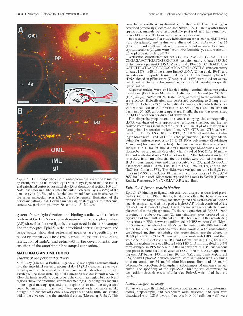
Figure 1. Lamina-specific entorhino-hippocampal projection visualizedby tracing with the fluorescent dye (Mini Ruby) injected into the ipsilat-eral entorhinal cortex of postnatal day 15 rat (horizontal section, 100 mm).Note that entorhinal fibers enter the outer molecular layer (OML) of thedentate gyrus (A, B), and no labeled entorhinal fibers can be observed inthe inner molecular layer (IML). Inset, Schematic illustration of theperforant pathway. CA, Cornu ammonis; dg, dentate gyrus; ec, entorhinalcortex.; pp, perforant pathway. Scale bar: A, B, 250 mm.
8886 J. Neurosci., October 15, 1999, 19(20):8885–8893 Stein et al. • Ephrins and Entorhino-Hippocampal Pathfinding
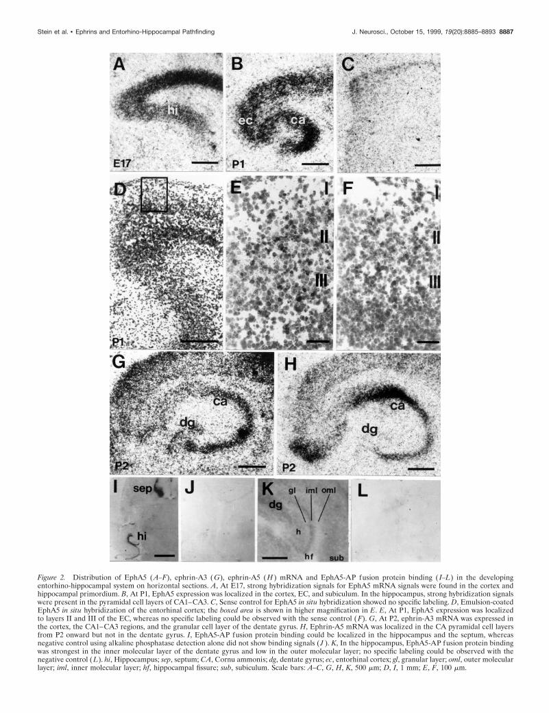
Figure 2. Distribution of EphA5 (A–F), ephrin-A3 (G), ephrin-A5 (H ) mRNA and EphA5-AP fusion protein binding ( I–L) in the developingentorhino-hippocampal system on horizontal sections. A, At E17, strong hybridization signals for EphA5 mRNA signals were found in the cortex andhippocampal primordium. B, At P1, EphA5 expression was localized in the cortex, EC, and subiculum. In the hippocampus, strong hybridization signalswere present in the pyramidal cell layers of CA1–CA3. C, Sense control for EphA5 in situ hybridization showed no specific labeling. D, Emulsion-coatedEphA5 in situ hybridization of the entorhinal cortex; the boxed area is shown in higher magnification in E. E, At P1, EphA5 expression was localizedto layers II and III of the EC, whereas no specific labeling could be observed with the sense control (F). G, At P2, ephrin-A3 mRNA was expressed inthe cortex, the CA1–CA3 regions, and the granular cell layer of the dentate gyrus. H, Ephrin-A5 mRNA was localized in the CA pyramidal cell layersfrom P2 onward but not in the dentate gyrus. I, EphA5-AP fusion protein binding could be localized in the hippocampus and the septum, whereasnegative control using alkaline phosphatase detection alone did not show binding signals ( J ). K, In the hippocampus, EphA5-AP fusion protein bindingwas strongest in the inner molecular layer of the dentate gyrus and low in the outer molecular layer; no specific labeling could be observed with thenegative control (L). hi, Hippocampus; sep, septum; CA, Cornu ammonis; dg, dentate gyrus; ec, entorhinal cortex; gl, granular layer; oml, outer molecularlayer; iml, inner molecular layer; hf, hippocampal fissure; sub, subiculum. Scale bars: A–C, G, H, K, 500 mm; D, I, 1 mm; E, F, 100 mm.
Stein et al. • Ephrins and Entorhino-Hippocampal Pathfinding J. Neurosci., October 15, 1999, 19(20):8885–8893 8887
plated in 24-well dishes onto confluent monolayers of ephrin-A3- orephrin-A5-expressing NIH 3T3 cells or control NIH 3T3 cells trans-fected with the vector in DMEM supplemented with fetal bovine serum(10%), penicillin (50 ng/ml), and streptomycin (50 mg/ml), cultivated for3 d and fixed in 4% paraformaldehyde in PBS. After fixation, neuronalprocesses were incubated with an anti-neurofilament 160 kDa antibodyobtained from Boehringer Mannheim (catalog #814334) in a concentra-tion of 5 mg/ml, followed by an subsequent incubation in avidin-biotinperoxidase complex (Vectastain ABC kit; Vector Laboratories, Burlin-game, CA) and visualized with 39-39diaminobenzidine as a chromogen.Random fields were selected, and all anti-neurofilament antibody-stainedneurites growing out from aggregates and single cells in these fields werescored. At least 200 fields were surveyed for each sample assayed, and theexperiments were repeated a minimum of three times.
Explant outgrowth and stripe assayExplant preparation. Fetuses and offspring of timed pregnant E17–E18Wistar rats were used. To collect embryonic tissue, pregnant rats wereanesthetized with Nembutal (5 mg/100 mg), and embryos were removedfrom the uterus. Embryos were placed in cold, oxygenated L15 medium(Life Technologies, Gaithersburg, MD) supplemented with 0.6% glu-cose. Animals were killed by decapitation, the brains were dissected out,and the meninges were removed. Horizontal sections were cut at 400 mmin cold, oxygenated PBS with a vibratome, and entorhinal explants fromthe superficial layers were dissected out with tungsten needles underbinocular optics with 403 magnification. After dissection, the explantswere placed in suspension culture in a 5.5% CO2 humidified incubator inDMEM–F-12 supplemented with 2 mM glutamine, 0.6% glucose, 100U/ml penicillin, 100 mg/ml streptomycin, 5% heat-inactivated rat serum,10% heat-inactivated fetal bovine serum, and 10 mM cytosine arabinoside
to control proliferation of non-neuronal cells. Entorhinal explants wereused for the assays on the same day.
Preparation of membranes. Membrane suspensions were prepared fromNIH 3T3 cells expressing ephrin-A3 or ephrin-A5 or from microdis-sected hippocampal regions of the pig (6 months old) from which it ispossible to dissect the outer molecular layer and the inner molecular–granular layer of the dentate gyrus. The membranes were preparedaccording to a protocol of Walter et al. (1987a,b). All solutions used weresterile, 4°C, pH 7.4, and supplemented with protease inhibitors as de-scribed previously (Simon and O’Leary, 1992). Cells were homogenizedin homogenization buffer (10 mM Tris-HCI, pH 7.4, 1.5 mM CaCl2, and1 mM spermidine; Serva Feinbiochemica, Heidelberg, Germany) bypressing the tissue first through a narrow pipette and then two to threetimes through syringe needles. The homogenate was layered on top of astep gradient of 50 and 5% sucrose in homogenization buffer andcentrifuged for 10 min at 50,000 3 g at 4°C in an SW 50 L rotor(Beckman Instruments, Fullerton, CA). Cytoplasmic and mitochondrialmembrane fragments formed a turbid layer at the boundary between 5and 50% sucrose, while nuclei were pelleted. The membrane fragmentswere collected with a syringe and washed with PBS. The concentration ofthe membrane suspension was adjusted to a concentration of 100–200mg/ml protein as determined by a modified Bradford method with bovineserum albumin as standard. For phosphatidylinositol (PI)-specific phos-pholipase C (PLC) treatment, membrane suspensions were adjusted to anoptical density of 0.5 (measured at 220 nm) in Tris buffer containing 1.5mM CaCl2 and protease inhibitors. The membrane suspensions werethen treated with PI-PLC (1 U/ml; ICN Biochemicals, Montreal, Quebec,Canada) for 1 hr, washed in cold PBS containing protease inhibitors, andresuspended in the same PBS solution.
Explant outgrowth assay. Entorhinal cortex explants were plated onmembranes prepared from the ephrin-A3, ephrin-A5, or the mock-transfected NIH 3T3 cells. Uniform membrane carpets for outgrowthmeasurements were made by pipetting 150 ml of membrane suspension(100–200 mg/ml) onto a filter placed over a uniform mesh and applyingsuction for up to 3 min. The total outgrowth length of all fluorescence-stained processes from the entorhinal explants was scored, and data fromeach experimental group were pooled and analyzed by ANOVA andMann–Whitney U test.
Stripe assay. Membrane stripes were prepared with membranes ob-tained either from ephrin-A3, ephrin-A5, or mock-transfected NIH 3T3cells on polycarbonate filters precoated with laminin. Membrane extractsfrom the outer molecular layer and inner molecular–granular layer wereprepared using the same protocol. The membrane carpets were thenplaced on sterile, porous (0.4 mm) membranes (Millicell-CM; Millipore,Eschborn, Germany) and transferred into a 35 mm tissue culture dishwith 1.5 ml culture medium. Explants were positioned on the membranecarpets using forceps. Cultures were maintained in a 5.5% CO2 humid-ified incubator for up to 5 d.
Analysis of outgrowth preference. Neurites growing out from the ex-plants were visualized with a fluorescent vital dye, 5 and6-carboxyfluorescein diacetate, succinimidyl ester (Molecular Probes),which labels all living cells and their processes, or by immunolabelingwith a neurofilament antibody (Boehringer Mannheim). A 6.15 mg/mlstock solution of dye was diluted 1:300 in PBS. Culture medium wasremoved from the dishes 5 d after the explants were placed on thecarpets, and 1–2 ml of the dye solution was added for 2 min. To inhibitphotobleaching, the dye solution was then replaced with a solution of 5mM p-phenylenediamine (Eastman Kodak) in PBS. Neurite growth from
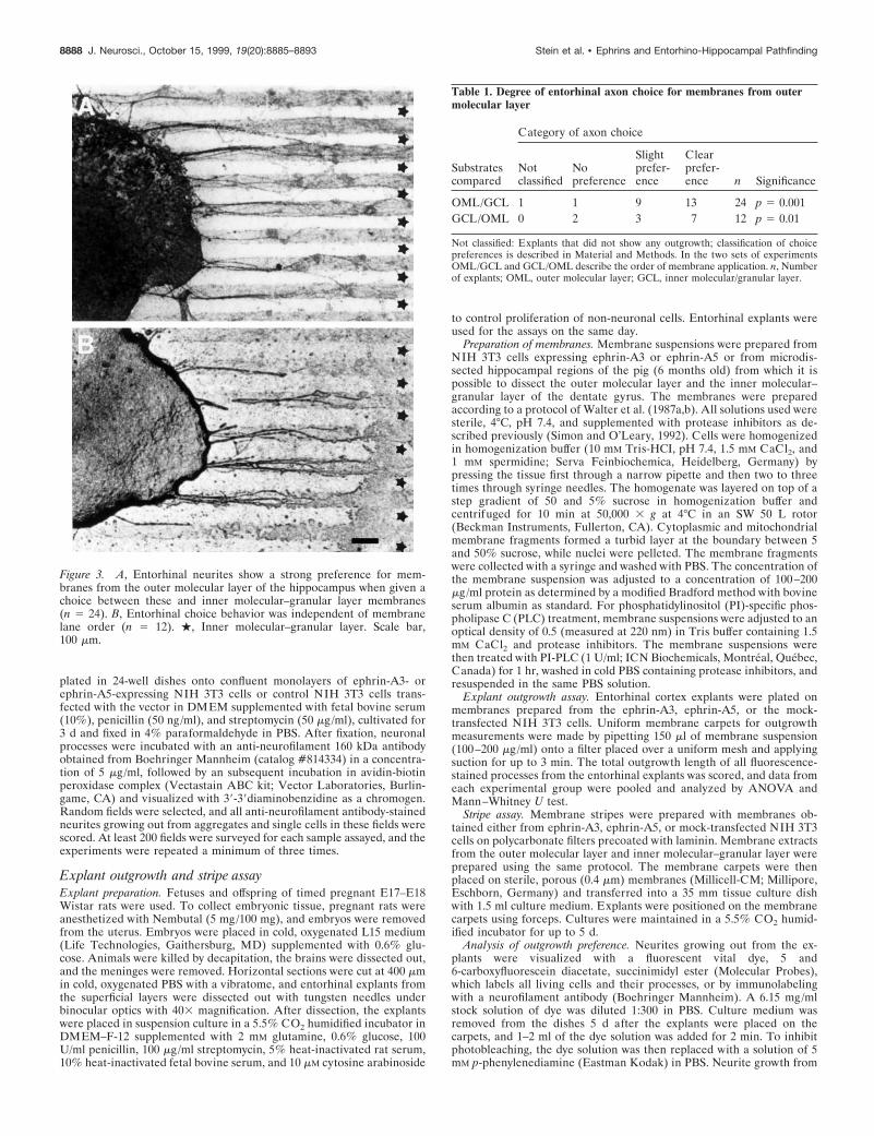
Figure 3. A, Entorhinal neurites show a strong preference for mem-branes from the outer molecular layer of the hippocampus when given achoice between these and inner molecular–granular layer membranes(n 5 24). B, Entorhinal choice behavior was independent of membranelane order (n 5 12). ., Inner molecular–granular layer. Scale bar,100 mm.
Table 1. Degree of entorhinal axon choice for membranes from outermolecular layer
Substratescompared
Category of axon choice
Notclassified
Nopreference
Slightprefer-ence
Clearprefer-ence n Significance
OML/GCL 1 1 9 13 24 p 5 0.001GCL/OML 0 2 3 7 12 p 5 0.01
Not classified: Explants that did not show any outgrowth; classification of choicepreferences is described in Material and Methods. In the two sets of experimentsOML/GCL and GCL/OML describe the order of membrane application. n, Numberof explants; OML, outer molecular layer; GCL, inner molecular/granular layer.
8888 J. Neurosci., October 15, 1999, 19(20):8885–8893 Stein et al. • Ephrins and Entorhino-Hippocampal Pathfinding
the explant was examined and photographed with FITC optics on anepifluorescence microscope.
Growth preferences for one or the other set of membrane stripes wereevaluated using a three class system: (A) clear cut preference, withalmost all of the fibers growing on one of the membrane lanes, (B) slightor moderate preference, with most fibers growing preferentially on onemembrane lane, although others cross randomly, and (C) no choice or
random outgrowth. The analysis of axonal choice behavior was per-formed as a double-blind experiment by three independent observers.Each observer scored the axon density on the different membrane stripesprepared in each experiment. In general, it was impossible to countindividual entorhinal axons because of the variability in axon outgrowthdensity and fasciculation in some experiments. Rating classes A and Bwere scored 1, and rating class C was scored 0. The scores from eachexperimental condition were totaled, data from individual experimentswere pooled, and x 2 tests were performed with an inter-rater reliabilityof .90%, which did not vary significantly in the different experimentsperformed.
RESULTSEphA5 is present in the EC, and ephrin-A3 and ephrin-A5 are expressed in hippocampus during developmentof the perforant pathwayTo obtain clues about the function of EphA5 and its ligandsephrin-A3 and ephrin-A5 in the developing entorhino-hippocampal formation, we used radioactive in situ hybridizationto investigate the spatiotemporal distribution of their mRNA. AtE17, an EphA5-specific probe detected strong signals in thecerebral cortex and in the hippocampal anlage (Fig. 2A). At P1,EphA5 expression was localized in the cortex, EC, subiculum, andCA1–CA3 (Fig. 2B). In the EC, layers II and III showed thestrongest signal (Fig. 2E). Between P2 and P10, EphA5 expres-sion was similar to that at P1 but with a stronger signal in thepyramidal layer of CA3 and weaker signals in the pyramidal layerof CA1, the granular layer of the dentate gyrus, and in the
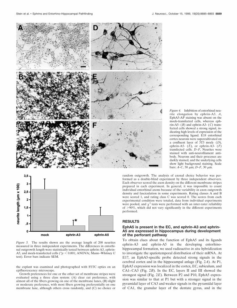
Figure 4. Inhibition of entorhinal neu-rite elongation by ephrin-A3. A,EphA5-AP staining was absent on themock-transfected cells, whereas eph-rin-A5- (B) and ephrin-A3- (C) trans-fected cells showed a strong signal, in-dicating high levels of expression of thecorresponding ligand. E18 entorhinalcortex neurons were supercultivated ona confluent layer of 3T3 mock- (D),ephrin-A5- ( E), or ephrin-A3- ( F)transfected cells. D–F, Neurites werestained with anti-neurofilament anti-body. Neurons and their processes aredarkly stained, and the underlying cellsshow light background staining. Scalebars: A–C, 50 mm; D–F, 30 mm.
Figure 5. The results shown are the average length of 200 neuritesmeasured in three independent experiments. The differences in entorhi-nal outgrowth length were statistically tested between ephrin-A3, ephrin-A5, and mock-transfected cells (*p , 0.001; ANOVA; Mann–Whitney Utest). Error bars indicate SEM.
Stein et al. • Ephrins and Entorhino-Hippocampal Pathfinding J. Neurosci., October 15, 1999, 19(20):8885–8893 8889
superficial layers of the EC (data not shown), consistent withprevious observations (Zhang et al., 1997).
We further examined the expression of the EphA subfamilyligands ephrin-A3 and ephrin-A5 in the developing entorhino-hippocampal system. At E17, ephrin-A3 was detected in theentire cortex and in the hippocampus (data not shown). At P2,ephrin-A3 mRNA was strongly expressed in the CA1–CA3 re-gion and in the granular cell layer of the dentate gyrus (Fig. 2G).Ephrin-A5 mRNA was not detected during embryonic stages butcould be localized in all CA pyramidal cell layers from P2 onward(Fig. 2H).
Detection of the EphA5 ligand proteins inthe hippocampusBecause ephrin-A3 and ephrin-A5 mRNA were expressed in thehippocampus and EphA5 in the EC, we were interested in study-ing the localization of the EphA5 ligand proteins in the hip-pocampus in more detail. To detect EphA5 binding in the hip-pocampus, we stained embryonic and postnatal mouse brainsections with a ligand-affinity probe, which consisted of the ex-tracellular domain of EphA5 fused in frame with a human pla-cental AP, which binds to both ephrin-A3 and ephrin-A5. Theseptum, hypothalamus, and olfactory bulbus showed the strongestbinding during embryonic stages, as shown previously by Zhanget al. (1996). Binding in the hippocampus was most prominent in
the dendritic fields of the CA1 and dentate gyrus during postnatalstages (Fig. 2K). No staining was observed on the granular celllayer and in the outer molecular layers in the dentate gyrus. Thestrongest EphA5-AP binding was localized in the inner molecularlayer. In CA1, the strongest EphA5-binding was detected in thestratum radiatum, whereas in the stratum lacunosum moleculare,EphA5-AP binding was weak.
Entorhinal axons prefer membranes from the outermolecular layers of the hippocampus to membranesobtained from the inner molecular–granular layerWhen cell membranes of the outer molecular layer and the innermolecular–granular cell layer of the dentate gyrus were arrangedin alternating stripes and explants of embryonic EC (n 5 36) wereplaced on the stripe carpet, the entorhinal axons showed a selec-tive growth preference for the outer molecular layer membranesand did not grow on inner molecular–granular layer membranes(Fig. 3, Table 1). This preference was independent of the order ofmembrane application on the filter and thus not attributable todifferences in the protein content of the lanes (Fig. 3B). Out-growth of entorhinal neurites on uniform membrane carpets ofthe inner molecular–granular layer compared with the outermolecular layers of the dentate gyrus (stripes offered alone andnot in alternating stripes) was also reduced (data not shown).
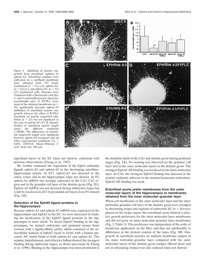
Figure 6. Inhibition of neurite out-growth from entorhinal explants byephrin-A3. Entorhinal explants werecultivated on a confluent membranelayer obtained from 3T3 mock-transfected (n 5 41) (A), ephrin-A3-(n 5 62) ( C), and ephrin-A5- (n 5 31)(E) transfected cells. Neurites werevisualized with a fluorescent vital dye,5- and 6-carboxyfluorescein diacetate,succinimidyl ester. D, PI-PLC treat-ment of the obtained membranes (n 536) significantly prevents ephrin-A3inhibition of entorhinal neurite out-growth, whereas the effect of PI-PLCtreatment on neurite outgrowth inhi-bition (n 5 25) was not significant inthe case of ephrin-A5 ( F). B, Quanti-fication of entorhinal neurite lengthunder the different conditions(6SEM). The differences of entorhi-nal outgrowth length were significantbetween ephrin-A3 treatment and allother experimental conditions (*p ,0.001; ANOVA; Mann–Whitney Utest). Scale bar, 100 mm.
8890 J. Neurosci., October 15, 1999, 19(20):8885–8893 Stein et al. • Ephrins and Entorhino-Hippocampal Pathfinding
Outgrowth of entorhinal neurites is inhibitedby ephrin-A3Entorhinal neurons from E18 rat brain were plated on a confluentmonolayer of NIH 3T3 cells expressing ephrin-A3 or ephrin-A5,or transfected with the expression vector alone (Fig. 4). The celllines were constructed previously by Gao et al. (1996), and theexpression of the ligands was further confirmed by EphA5-APbinding (Fig. 4A–C). Neurite extension from EC neurons wasstrongly inhibited by 3T3 cells expressing ephrin-A3 (Figs. 4F, 5)and to a lesser extent by those expressing ephrin-A5 (Fig. 4E)compared with the length of entorhinal neurons on mock-transfected NIH 3T3 cells (Fig. 4D). In contrast, neitherephrin-A3 nor ephrin-A5 inhibited neurite outgrowth from cer-ebellar neurons (data not shown). In accordance with the findingsof Gao et al. (1996), the outgrowth of hippocampal neurons was
significantly inhibited by both ephrin-A3 and ephrin-A5 (data notshown).
Similar effects were observed with entorhinal explants culti-vated on uniform membranes obtained from cells expressingephrin-A3 (n 5 62) or ephrin-A5 (n 5 31), or transfected with theexpression vector alone (n 5 41) (Fig. 6). In particular, theoutgrowth of entorhinal neurites was strongly inhibited byephrin-A3 membranes (Fig. 6C), whereas membranes obtainedfrom cells expressing ephrin-A5 (Fig. 6E) and control cells (Fig.6A) had significantly less effect on entorhinal fiber extension.Because ephrin-A3 and ephrin-A5 can be removed from themembrane preparation by PI-PLC treatment, we analyzed theeffect of PI-PLC treatment on ephrin-A3 inhibition of entorhinalneurite outgrowth (n 5 36). Removal of ephrin-A3 from themembranes restored neurite elongation from entorhinal explants(Fig. 6D). Entorhinal outgrowth was not significantly affected onmembranes obtained from ephrin-A5-transfected NIH 3T3 cellsthat had been treated with PI-PLC (Fig. 6F). This observationsuggests that ephrin-A3 in particular selectively and specificallyinhibits entorhinal axon outgrowth.
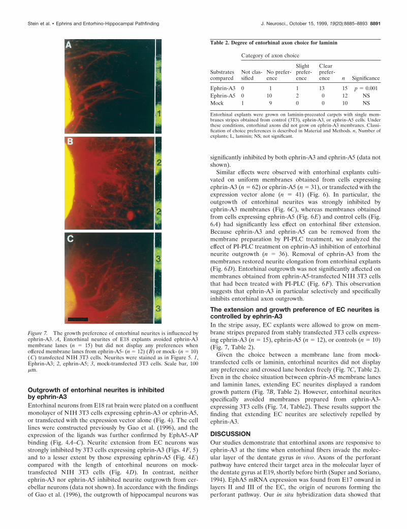
The extension and growth preference of EC neurites iscontrolled by ephrin-A3In the stripe assay, EC explants were allowed to grow on mem-brane stripes prepared from stably transfected 3T3 cells express-ing ephrin-A3 (n 5 15), ephrin-A5 (n 5 12), or controls (n 5 10)(Fig. 7, Table 2).
Given the choice between a membrane lane from mock-transfected cells or laminin, entorhinal neurites did not displayany preference and crossed lane borders freely (Fig. 7C, Table 2).Even in the choice situation between ephrin-A5 membrane lanesand laminin lanes, extending EC neurites displayed a randomgrowth pattern (Fig. 7B, Table 2). However, entorhinal neuritesspecifically avoided membranes prepared from ephrin-A3-expressing 3T3 cells (Fig. 7A, Table2). These results support thefinding that extending EC neurites are selectively repelled byephrin-A3.
DISCUSSIONOur studies demonstrate that entorhinal axons are responsive toephrin-A3 at the time when entorhinal fibers invade the molec-ular layer of the dentate gyrus in vivo. Axons of the perforantpathway have entered their target area in the molecular layer ofthe dentate gyrus at E19, shortly before birth (Super and Soriano,1994). EphA5 mRNA expression was found from E17 onward inlayers II and III of the EC, the origin of neurons forming theperforant pathway. Our in situ hybridization data showed that
Figure 7. The growth preference of entorhinal neurites is influenced byephrin-A3. A, Entorhinal neurites of E18 explants avoided ephrin-A3membrane lanes (n 5 15) but did not display any preferences whenoffered membrane lanes from ephrin-A5- (n 5 12) (B) or mock- (n 5 10)(C) transfected NIH 3T3 cells. Neurites were stained as in Figure 5. 1,Ephrin-A3; 2, ephrin-A5; 3, mock-transfected 3T3 cells. Scale bar, 100mm.
Table 2. Degree of entorhinal axon choice for laminin
Substratescompared
Category of axon choice
Not clas-sified
No prefer-ence
Slightprefer-ence
Clearprefer-ence n Significance
Ephrin-A3 0 1 1 13 15 p 5 0.001Ephrin-A5 0 10 2 0 12 NSMock 1 9 0 0 10 NS
Entorhinal explants were grown on laminin-precoated carpets with single mem-branes stripes obtained from control (3T3), ephrin-A3, or ephrin-A5 cells. Underthese conditions, entorhinal axons did not grow on ephrin-A3 membranes. Classi-fication of choice preferences is described in Material and Methods. n, Number ofexplants; L, laminin; NS, not significant.
Stein et al. • Ephrins and Entorhino-Hippocampal Pathfinding J. Neurosci., October 15, 1999, 19(20):8885–8893 8891
ephrin-A3 mRNA is expressed in the granular cell layer of thedentate gyrus. In addition, the ligand protein was detected usingEphA5-AP binding in the inner molecular layer of the dentategyrus, a region not invaded by EC neurites from layer II in vivo.Because EphA5 interacts with multiple ligands in vitro but onlyephrin-A3 is present in the dentate gyrus, the EphA5-AP bindingdata probably reflects the distribution of this guidance cue in thedentate gyrus. Several observations indicate that ephrin-A3 has aspecific effect in entorhino-hippocampal axon targeting. First, theneurite and outgrowth explant assay only displayed a significantinhibitory effect by ephrin-A3 but not by ephrin-A5 or controls.Second, digestion of the GPI-anchor of ephrin-A3 by PI-PLCtreatment abolished this effect. Third, the EphA5 receptor issynthesized in EC neurons, and soluble AP-tagged EphA5 bindsstrongly to the inner molecular layer. Thus, entorhinal fiberoutgrowth to the inner molecular layer of the dentate gyrus wouldbe repelled, whereas the outer molecular layer would be permis-sible for these ingrowing axons.
The afferent connections of the hippocampus and its entorhinalinput, the perforant and alvear pathways, represent a well estab-lished model for the analysis of target-specific pathfinding, termi-nation, and reinnervation (Frotscher and Heimrich, 1993; Li etal., 1993, 1994, 1995; Woodhams 1993; Woodhams and Atkinson,1996; Frotscher et al., 1997). It was shown that the molecularsignals necessary for the formation of an entorhino-dentate pro-jection are present in hippocampal tissue maintained in organo-typic coculture with explants of the EC (Frotscher and Heimrich,1993; Li et al., 1993; Woodhams and Atkinson, 1996). Hetero-chronic slice culture experiments and transplantation of embry-onic entorhinal cells to adult hippocampus have also outlined thepresence of positional guidance cues in the adult hippocampus(Zhou et al., 1989; Li et al., 1995). These experiments indicatedthat lamina-specific molecules in the hippocampal fields playimportant roles in the formation of layer-specific afferent projec-tions. But none of these studies provided information about themechanisms entorhinal axons use to select their original pathwaysand target fields.
Del Rıo et al. (1997) showed that ablation of Cajal-Retzius
cells influenced the ingrowth of entorhinal fibers into the molec-ular layer of the dentate gyrus. This finding suggests that, inaddition to repulsive guidance factors such as ephrin-A3, which isexpressed in the granular cell layer of the dentate gyrus, Cajal-Retzius cells localized in the molecular layer of the dentate gyrusproduce an attractive factor that guides entorhinal neurons to-ward the molecular layer. Superculturing of prelabeled entorhinalneurons or membranes on hippocampal slice preparations led tothe observation of lamina-specific adhesion in the terminationzones of the entorhino-hippocampal formation (Forster et al.,1998). These adhesion experiments documented the presence ofmembrane-bound adhesive properties in the outer molecularlayer of the dentate gyrus and the absence of these activities inthe granular cell layer, a zone that is not targeted by mostentorhinal axons in vivo. Our data and the observations of DelRıo et al. (1997) and Forster et al. (1998) indicate the presence ofattractive adhesive and repulsive properties in the developingdentate gyrus.
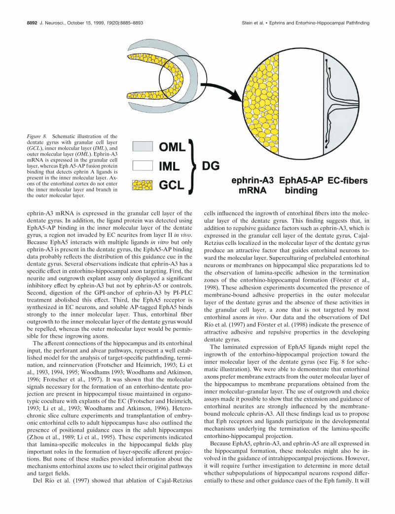
The laminated expression of EphA5 ligands might repel theingrowth of the entorhino-hippocampal projection toward theinner molecular layer of the dentate gyrus (see Fig. 8 for sche-matic illustration). We were able to demonstrate that entorhinalaxons prefer membrane extracts from the outer molecular layer ofthe hippocampus to membrane preparations obtained from theinner molecular–granular layer. The use of outgrowth and choiceassays made it possible to show that the extension and guidance ofentorhinal neurites are strongly influenced by the membrane-bound molecule ephrin-A3. All these findings lead us to proposethat Eph receptors and ligands participate in the developmentalmechanisms underlying the termination of the lamina-specificentorhino-hippocampal projection.
Because EphA5, ephrin-A3, and ephrin-A5 are all expressed inthe hippocampal formation, these molecules might also be in-volved in the guidance of intrahippocampal projections. However,it will require further investigation to determine in more detailwhether subpopulations of hippocampal neurons respond differ-entially to these and other guidance cues of the Eph family. It will
Figure 8. Schematic illustration of thedentate gyrus with granular cell layer(GCL), inner molecular layer (IML), andouter molecular layer (OML). Ephrin-A3mRNA is expressed in the granular celllayer, whereas Eph A5-AP fusion proteinbinding that detects ephrin A ligands ispresent in the inner molecular layer. Ax-ons of the entorhinal cortex do not enterthe inner molecular layer and branch inthe outer molecular layer.
8892 J. Neurosci., October 15, 1999, 19(20):8885–8893 Stein et al. • Ephrins and Entorhino-Hippocampal Pathfinding
also be interesting to analyze the phenotype of hippocampalprojections in ephrin-A3 and ephrin-A5 knock-out mice.
It should also be emphasized that the hippocampus is wellknown for structural remodeling and for lamina-specific sprout-ing after lesioning its different afferent inputs (for review, seeFrotscher et al., 1997). The continuous expression of EphA5,ephrin-A3, and ephrin-A5 in the hippocampal system duringadulthood suggests that these molecules might also play a role insynaptic plasticity.
In summary, our observations indicate that ephrin-A3 acts as arepulsive signal that helps to pattern the perforant pathway bylimiting the growth of entorhinal fibers to the outer molecularlayer of the dentate gyrus. It may also perform a similar functionfor intrahippocampal connections. However, the laminated dis-tribution of hippocampal afferents is probably the result of acooperation between both attractive and repulsive guidance fac-tors. Further studies have to address the question of how thetemporal and spatial expression patterns of these factors manageto lead the ingrowing afferents to their appropriate target area.
REFERENCESBechmann I, Nitsch R (1997) Astrocytes and microglial cells incorporate
degenerating fibers following entorhinal lesion: a light, confocal, andelectron microscopical study using a phagocytosis-dependent labelingtechnique. Glia 20:145–154.
Carpenter MK, Shilling H, VandenBos T, Beckmann MP, Cerretti DP,Kott JN, Westrum LE, Davison BL, Fletcher FA (1995) Ligands forEPH-related tyrosine kinase receptors are developmentally regulatedin the CNS. J Neurosci Res 42:199–206.
Ciossek T, Lerch MM, Ullrich A (1995) Cloning, characterization, anddifferential expression of MDK2 and MDK5, two novel receptor ty-rosine kinases of the eck/eph family. Oncogene 11:2085–2095.
Culotti JG, Kolodkin AL (1996) Functions of netrins and semaphorinsin axon guidance. Curr Opin Neurobiol 6:81–88.
Del Rıo JA, Heimrich B, Borrell V, Forster E, Drakew A, Alcantara S,Nakajima K, Miyata T, Ogawa M, Mikoshiba K, Derer P, Frotscher M,Soriano E (1997) A role for Cajal-Retzius cells and reelin in thedevelopment of hippocampal connections. Nature 385:70–74.
Flanagan JG, Vanderhaeghen P (1998) The ephrins and Eph receptorsin neural development. Annu Rev Neurosci 21:309–345.
Forster E, Kaltschmidt C, Deng J, Cremer H, Deller T, Frotscher M(1998) Lamina-specific cell adhesion on living slices of hippocampus.Development 125:3399–3410.
Friedman GC, O’Leary DD (1996) Eph receptor tyrosine kinases andtheir ligands in neural development. Curr Opin Neurobiol 6:127–133.
Frotscher M, Heimrich B (1993) Formation of layer-specific fiber pro-jections to the hippocampus in vitro. Proc Natl Acad Sci USA90:10400–10403.
Frotscher M, Heimrich B, Deller T (1997) Sprouting in the hippocampusis layer-specific. Trends Neurosci 20:218–223.
Gao PP, Zhang JH, Yokoyama M, Racey B, Dreyfus CF, Black IB, ZhouR (1996) Regulation of topographic projection in the brain: Elf-1in the hippocamposeptal system. Proc Natl Acad Sci USA 93:11161–11166.
Holt CE, Harris WA (1998) Target selection: invasion, mapping and cellchoice. Curr Opin Neurobiol 8:98–105.
Kozlosky CJ, VandenBos T, Park L, Cerretti DP, Carpenter MK (1997)LERK-7: a ligand of the Eph-related kinases is developmentally regu-lated in the brain. Cytokine 9:540–549.
Li D, Field PM, Starega U, Li Y, Raisman G (1993) Entorhinal axonsproject to dentate gyrus in organotypic slice co-culture. Neuroscience52:799–813.
Li D, Field PM, Yoshioka N, Raisman G (1994) Axons regenerate withcorrect specificity in horizontal slice culture of the postnatal ratentorhino-hippocampal system. Eur J Neurosci 6:1026–1037.
Li D, Field PM, Raisman G (1995) Failure of axon regeneration inpostnatal rat entorhinohippocampal slice coculture is due to maturationof the axon, not that of the pathway or target. Eur J Neurosci7:1164–1171.
Maisonpierre PC, Barrezueta NX, Yancopoulos GD (1993) Ehk-1 andEhk-2: two novel members of the Eph receptor-like tyrosine kinasefamily with distinctive structures and neuronal expression. Oncogene8:3277–3288.
Martone ME, Holash JA, Bayardo A, Pasquale EB, Ellisman MH (1997)Immunolocalization of the receptor tyrosine kinase EphA4 in the adultrat central nervous system. Brain Res 771:238–250.
Mori T, Wanaka A, Taguchi A, Matsumoto K, Tohyama M (1995)Differential expressions of the eph family of receptor tyrosine kinasegenes (sek, elk, eck) in the developing nervous system of the mouse.Brain Res Mol Brain Res 29:325–335.
Simon DK, O’Leary DD (1992) Responses of retinal axons in vivo and invitro to position-encoding molecules in the embryonic superior collicu-lus. Neuron 9:977–989.
Steward O, Scoville SA (1976) Cells of origin of entorhinal corticalafferents to the hippocampus and fascia dentata of the rat. J CompNeurol 169:347–370.
Super H, Soriano E (1994) The organization of the embryonic and earlypostnatal murine hippocampus. II. Development of entorhinal, com-missural, and septal connections studied with the lipophilic tracer DiI.J Comp Neurol 344:101–120.
Tamamaki N, Nojyo Y (1993) Projection of the entorhinal layer II neu-rons in the rat as revealed by intracellular pressure-injection of neuro-biotin. Hippocampus 3:471–480.
Taylor V, Miescher GC, Pfarr S, Honegger P, Breitschopf H, LassmannH, Steck AJ (1994) Expression and developmental regulation ofEhk-1, a neuronal Elk-like receptor tyrosine kinase in brain. Neuro-science 63:163–178.
Tessier-Lavigne M, Goodman CS (1996) The molecular biology of axonguidance. Science 274:1123–1133.
Walter J, Henke Fahle S, Bonhoeffer F (1987a) Avoidance of posteriortectal membranes by temporal retinal axons. Development101:909–913.
Walter J, Kern Veits B, Huf J, Stolze B, Bonhoeffer F (1987b) Recog-nition of position-specific properties of tectal cell membranes by retinalaxons in vitro. Development 101:685–696.
Witter MP (1993) Organization of the entorhinal-hippocampal system: areview of current anatomical data. Hippocampus 3:33–44.
Woodhams PL (1993) Laminar and region-specific cell surface markersin the entorhinal cortex and hippocampus. Hippocampus 3:183–189.
Woodhams PL, Atkinson DJ (1996) Entorhinal axons perforate hip-pocampal field CA3 in organotypic slice culture. Brain Res Dev BrainRes 95:144–147.
Zhang JH, Cerretti DP, Yu T, Flanagan JG, Zhou R (1996) Detectionof ligands in regions anatomically connected to neurons expressing theEph receptor Bsk: potential roles in neuron-target interaction. J Neu-rosci 16:7182–7192.
Zhang JH, Pimenta AF, Levitt P, Zhou R (1997) Dynamic expressionsuggests multiple roles of the eph family receptor brain-specific kinase(Bsk) during mouse neurogenesis. Brain Res Mol Brain Res47:202–214.
Zhou CF, Li Y, Raisman G (1989) Embryonic entorhinal transplantsproject selectively to the deafferented entorhinal zone of adult mousehippocampi, as demonstrated by the use of Thy-1 allelic immunohisto-chemistry. Effect of timing of transplantation in relation to deafferen-tation. Neuroscience 32:349–362.
Zhou R (1997) Regulation of topographic projection by the Eph familyreceptor Bsk (EphA5) and its ligands. Cell Tissue Res 290:251–259.
Zhou R (1998) The Eph family receptors and ligands. Pharmacol Ther77:151–181.
Zhou R, Copeland TD, Kromer LF, Schulz NT (1994) Isolation andcharacterization of Bsk, a growth factor receptor-like tyrosine kinaseassociated with the limbic system. J Neurosci Res 37:129–143.
Stein et al. • Ephrins and Entorhino-Hippocampal Pathfinding J. Neurosci., October 15, 1999, 19(20):8885–8893 8893


