Embed Size (px)
Citation preview
A specific isoform of poly(ADP-ribose) glycohydrolase is targetedto the mitochondrial matrix by a N-terminal mitochondrialtargeting sequence
Clifford J. Whatcott*, Mirella L. Meyer-Ficca#, Ralph G. Meyer#, and Myron K. Jacobson*,¶*Department of Pharmacology and Toxicology, College of Pharmacy, Arizona Cancer Center,University of Arizona, Tucson, AZ 85728, USA#Department of Animal Biology and Mari Lowe Center for Comparative Oncology, School ofVeterinary Medicine, NBC Center for Animal Transgenesis and Germ Cell Research, University ofPennsylvania, Kennett Square, PA 19348, USA
AbstractPoly(ADP-ribose) polymerases (PARPs) convert NAD to polymers of ADP-ribose that are convertedto free ADP-ribose by poly(ADP-ribose) glycohydrolase (PARG). The activation of the nuclearenzyme PARP-1 following genotoxic stress has been linked to release of apoptosis inducing factorfrom the mitochondria, but the mechanisms by which signals are transmitted between nuclear andmitochondrial compartments are not well understood. The study reported here has examined therelationship between PARG and mitochondria in HeLa cells. Endogenous PARG associated with themitochondrial fraction migrated in the range of 60 kDa. Transient transfection of cells with PARGexpression constructs with amino acids encoded by exon 4 at the N-terminus were targeted to themitochondria as demonstrated by subcellular fractionation and immunofluorescence microscopy ofwhole cells. Deletion and missense mutants allowed identification of a canonical N-terminalmitochondrial targeting sequence consisting of the first 16 amino acids encoded by PARG exon 4.Sub-mitochondrial localization experiments indicate that this mitochondrial PARG isoform istargeted to the mitochondrial matrix. The identification of a PARG isoform as a component of themitochondrial matrix raises several interesting possibilities concerning mechanisms of nuclear-mitochondrial cross talk involved in regulation of cell death pathways.
KeywordsPoly(ADP-ribose) glycohydrolase; PARG; mitochondrial targeting sequence; mitochondrial matrix;ADPR polymers
IntroductionPolymers of ADP-ribose (ADPR) are synthesized by a family of poly(ADP-ribose)polymerases (PARPs) encoded by a number of different genes [1,2]. The best understood are
¶To whom correspondence should be addressed: College of Pharmacy, University of Arizona, Room 3985 Arizona Cancer Center, 1515North Campbell Avenue, Tucson, AZ 85724, USA, Tel: 520-626-5957, Fax: 520-626-8657, [email protected]'s Disclaimer: This is a PDF file of an unedited manuscript that has been accepted for publication. As a service to our customerswe are providing this early version of the manuscript. The manuscript will undergo copyediting, typesetting, and review of the resultingproof before it is published in its final citable form. Please note that during the production process errors may be discovered which couldaffect the content, and all legal disclaimers that apply to the journal pertain.
NIH Public AccessAuthor ManuscriptExp Cell Res. Author manuscript; available in PMC 2010 December 10.
Published in final edited form as:Exp Cell Res. 2009 December 10; 315(20): 3477–3485. doi:10.1016/j.yexcr.2009.04.005.
NIH
-PA Author Manuscript
NIH
-PA Author Manuscript
NIH
-PA Author Manuscript
the nuclear PARPs 1 and 2 that play a role in the maintenance of genomic integrity viapromotion of DNA repair and cell recovery at low levels of genotoxic stress and promotion ofcell death at higher levels of damage [3,4]. The central role of ADPR polymer metabolism inmodulating cell recovery or cell death has potentially important implications for the therapeutictargeting of this metabolism [5,6].
Activation of PARP-1 has been specifically linked to the release of apoptosis inducing factor(AIF) from mitochondria, resulting in cell death [7,8]. A number of possible mechanismswhereby PARP-1 is involved in nuclear-mitochondrial cross talk leading to AIF release havebeen proposed that include nuclear/cytoplasmic NAD depletion resulting in glycolysis blocksthat deplete substrates for mitochondrial metabolism [9,10], direct effects of ADPR polymerson mitochondria [11,12], involvement of receptor-interacting protein-1, tumor necrosis factorreceptor-associated factor and c-Jun N-terminal kinase [13] and involvement of calpains andBax [14].
Poly(ADP-ribose) glycohydrolase (PARG) catalyzes the opposing arm of ADPR polymercycles initiated by PARPs [15]. In contrast to multiple genes that encode proteins with PARPactivity, only a single gene that encodes PARG activity clearly involved in ADPR polymermetabolism has been described [16]. A second enzyme with PARG activity has been described[17], but its functional significance is not yet clear. However, alternative splicing leads tomultiple PARG gene transcripts, resulting in generation of a number of different PARGisoforms targeted to nuclear and extranuclear cell compartments [18]. A PARG isoform ofapproximately 111 kDa facilitates DNA repair via regulation of ADPR polymer levelsfollowing DNA damage [19,20]. A number of studies suggest an association of PARG (andthus ADPR polymer metabolism) with mitochondria [16,21-24]. The PARG gene shares apromoter with a gene encoding TIM23, a protein involved in import of proteins intomitochondria [16]. A hypomorphic mouse mutant derived from disruption of the PARG genethat contains only small PARG isoforms including an isoform with an N-terminus that beginswith amino acids encoded by PARG exon 4 shows high levels of PARG associated with themitochondria [21]. This same PARG isoform has been subsequently detected in wild type cellsand shown to be associated with the mitochondrial fraction [22]. Activities capable ofdegrading ADPR polymers in vivo have been detected in the mitochondrial matrix [23]. PARGshows a strong association with the mitochondrial fraction in brain and other tissues fromrodents [24]. In the present work, we have examined the relation between PARG andmitochondria in more detail in HeLa cells and we present here evidence that a specific PARGisoform is a valid and legitimate component of the mitochondrial matrix.
Methods and MaterialsCell culture and transfection methods
HeLa cells were cultured (37°C, 5% CO2) in Dulbecco's modified Eagle's Medium (DMEM,Sigma) supplemented with 10% bovine calf serum (BCS, Hyclone). For the overexpression ofconstructs encoding wild type and mutant PARG, cells were seeded in 150 mm diameter cellculture dishes or six-well plates (Sarstedt), and transfected using Lipofectamine 2000transfection reagent (Invitrogen) according to the manufacturer's protocol. Alternatively, cellswere transfected using a calcium phosphate transfection method [25].
Western blotting methodsSubcellular fractions and other protein samples were applied to 10% polyacrylamide gels, andseparated by SDS-PAGE [26]. Samples were then transferred to PVDF membranes (Millipore)for analysis. Membranes were analyzed with anti-V5 (Invitrogen), anti-SMAC/Diablo(Abcam), anti-Hsp60 (Stressgen), anti-MnSOD (Stressgen), anti-Histones (Millipore), or anti-
Whatcott et al. Page 2
Exp Cell Res. Author manuscript; available in PMC 2010 December 10.
NIH
-PA Author Manuscript
NIH
-PA Author Manuscript
NIH
-PA Author Manuscript
Lactate Dehydrogenase (Abcam) antibodies. Antibodies for the detection of endogenousPARG in total lysates and mitochondrial fractions were described previously [22]. Membraneswere subsequently detected using horseradish peroxidase-conjugated goat anti-mouse or goatanti-rabbit secondary antibodies (Jackson ImmunoResearch Laboratories) and visualized withan enhanced chemiluminescent (ECL) reaction. Densitometric analysis of western blots wasperformed using Scion Image for Windows (Scion Corporation).
Deletion and site-directed mutagenesispΔE-C1hPARG59, a pEGFP-C1 (Clontech) plasmid containing the hPARG59 isoform [22]was created by deleting EGFP using the Nhe1 and Kpn1 restriction sites and primers shownin Table 1. Site-directed mutagenesis (Fig. 3) was performed using the Quickchange II-Emutagenesis kit (Stratagene), according to the manufacturer's protocol, using primers shownin Table 1. For generation of deletion mutants (Fig. 2), the entire plasmid was amplified bypolymerase chain reaction using the Phusion high-fidelity DNA polymerase (Finnzymes) anddeletion primers shown in Table 1, and then self-circularized with T4 DNA ligase (Fermentas).
Fusion of putative MTS to EGFPThe pΔE-C1hPARG59 plasmid and the PARG mutant vectors were used as templates for theconstruction of vectors expressing PARG MTS-EGFP fusion proteins. Using the primersshown in Table 1, the MTS of hPARG59 and PARG mutants was amplified by polymerasechain reaction (PCR). The primers were designed to introduce NheI restriction sites into thePCR product. PCR products were subsequently subcloned into the pEGFP-C1 vector, givingrise to a vector expressing a fusion protein in which the first 94 amino acids of PARG werefused N-terminally to the EGFP. Cells were subsequently transfected for visualization byimmunofluorescence microscopy.
Immunofluorescence microscopyAt 24 hours following transfection, cells seeded on coverslips were washed with phosphatebuffered physiological saline (PBS) and 5% formaldehyde (Sigma). Cells were then fixed with5% formaldehyde in PBS for 30 min at room temperature, protected from light with a foilcovering. Fixed cells were washed three times in PBS, deactivated in 100 mM glycine for 1min, washed three more times in PBS, and permeabilized for 4 min with 0.4% Triton X-100in PBS. Following three more washes, coverslips were blocked in 3% bovine serum albumin(BSA, Sigma) in PBS for 30 min at room temperature. Coverslips were subsequently washedthree more times with PBS, and incubated with anti-V5 (PARG) and anti-MnSOD (Stressgen)antibodies for 2 hrs at 37°C in a humid environment. Cells were then washed and incubatedwith FITC or TRITC-labeled secondary antibodies (Jackson ImmunoResearch). Alternatively,cells transfected with PARG-MTS-EGFP constructs were pretreated with Mitotracker(Invitrogen) prior to fixation, according to the manufacturer's protocol. Subsequently, cellswere processed as described above. Following final washing, coverslips were mounted andDNA counterstained with Vectashield (Vector Laboratories) supplemented with 1μg/mL DAPI(Sigma). Images were captured by confocal laser microscopy (Zeiss LSM 510 META NLOsystem) and extracted with Zeiss LSM Image Browser software (Zeiss).
Subcellular and Submitochondrial fractionationMitochondria were isolated from cells 24 hours post-transfection using a mitochondrialisolation kit (Sigma) and a Potter-Elvehjem homogenizer (Fisher Scientific) for cell disruption.Briefly, following trypsinization, cells were washed in PBS and then in extraction buffer (50mM HEPES, pH 7.5, containing 1 M mannitol, 350 mM sucrose, and 5 mM EGTA). Cellswere incubated for 30 min on ice in extraction buffer supplemented with 2mg/mL BSA and acomplete protease inhibitor cocktail (Roche). Cells were homogenized with 100 strokes in a
Whatcott et al. Page 3
Exp Cell Res. Author manuscript; available in PMC 2010 December 10.
NIH
-PA Author Manuscript
NIH
-PA Author Manuscript
NIH
-PA Author Manuscript
Potter-Elvehjem homogenizer fitted to an overhead stirrer (IKA) set at 650 rpm. Lysates weresubjected to differential centrifugation at 27g, 1,000g and 11,000g to obtain purified cellularfractions. For submitochondrial analysis, mitochondrial fractions were resuspended in astorage buffer (50 mM HEPES, pH 7.5, containing 1.25 M sucrose, 5 mM ATP, 0.4 mM ADP,25 mM sodium succinate, 10 mM K2HPO4, and 5 mM DTT) and treated with 5μg/mLProteinase K (Roche), and 0.1 - 0.4 mg/mL digitonin (Sigma) for 30 min at 37°C. Followingheat-inactivation of Proteinase K (95°C for 10 min), samples were subsequently analyzed bySDS-PAGE and western blotting techniques.
ResultsMitochondrial fractions are enriched in smaller size PARG isoforms
The routine detection of PARG in HeLa cell extracts is limited by the low abundance of theprotein. However, it was possible to detect PARG isoforms in total cell extracts andmitochondrial fractions using a polyclonal anti-peptide antibody directed against a C-terminalPARG peptide sequence [22]. This antibody has been used previously to detect endogenousisoforms of PARG in the range of approximately 100 to 110 kDa and 55 to 60 kDa [18], buttheir subcellular localization was not determined. Consistent with the previous study, detectionof PARG in total HeLa cell extracts yielded bands in the same molecular weight ranges reportedpreviously as well as immunoreactive material in the range of 30 kDa (Fig. 1A). Mitochondrialfractions showed PARG signals primarily in the range of approximately 55 to 60 kDa,indicating that endogenous mitochondrial PARG is comprised of the smaller PARG isoforms.This result is in agreement with previous studies involving transient transfection of cells withcDNAs expressing different PARG isoforms [22].
PARG expressed from a plasmid containing a putative N-terminal mitochondrial targetingsequence is targeted to the mitochondria
To study the mechanisms by which PARG is targeted to mitochondria, a vector expressinghPARG59 [22] under the control of a CMV promoter was constructed. This vector expressesa PARG containing a putative N-terminal mitochondrial targeting sequence (MTS) that isencoded by exon 4 of the PARG gene. A V5 tag flanking the C-terminus has been added tothe construct to facilitate PARG detection. HeLa cells were transfected with the hPARG59vector and equal amounts of protein from the nuclear, cytosolic and mitochondrial fractionswere analyzed, utilizing the V5 tag on PARG to assess the relative protein concentration ofPARG in the different fractions. The results show that the expressed hPARG59 was targetedto the mitochondrial fraction (Fig. 1B). The purity of the mitochondrial fraction was confirmedby the absence of histones as an indicator of possible contamination with nuclei. Mitochondriallocalization of hPARG59 was further confirmed microscopically in transfected cells by co-localization of the punctate cellular distribution the V5 tag with the Mitotracker dye (Fig. 1C).
Deletion mutagenesis indicates that PARG exon 4 encodes a mitochondrial targetingsequence
Deletion mutagenesis was used to examine the amino acid residues encoded by exon 4, to betterunderstand their effect on mitochondrial targeting (Fig. 2). The amino acid sequence encodedby exon 4, the putative MTS, and the predicted site of cleavage are shown at the top of the leftpanel. Also shown are the deletion mutants constructed, the relative PARG concentration incytosolic and mitochondrial fractions and the quantification of the relative content in cytosolicand mitochondrial fractions by densitometry. Results shown are a representative of multipleexperiments performed. Immunoblotting of cytosolic and mitochondrial fractions for markerproteins revealed no detectable histones, indicating very low cross contamination with nuclei.Blots of the cytosolic marker lactate dehydrogenase and the mitochondrial marker MnSODindicated very low levels of cross contamination of the cytosolic and mitochondrial fractions.
Whatcott et al. Page 4
Exp Cell Res. Author manuscript; available in PMC 2010 December 10.
NIH
-PA Author Manuscript
NIH
-PA Author Manuscript
NIH
-PA Author Manuscript
Deletion of Arg2 and Arg3 (mutant 2) had little effect on mitochondrial targeting. However,deletion of the remaining portion of the predicted MTS up to the predicted cleavage point ofthe MTS (mutant 3) and further to the end of the region coded for by exon 4 (mutant 4) resultedin substantial loss of mitochondrial targeting. However, deletion of the predicted cleavage siteup to the end of residues encoded by exon 4 (mutant 5) did not result in a substantial loss ofmitochondrial targeting. The results indicate that the N-terminal 16 amino acids encoded byexon 4 play an important role in the mitochondrial PARG targeting.
Site directed mutagenesis indicates that both positively charged and hydrophobic aminoacid residues are involved in the PARG mitochondrial targeting
A common feature of many MTS is an amphipathic alpha helix containing positively chargedresides on one face of the helix and hydrophobic residues on the other face [22,27]. In orderto further evaluate the involvement of the PARG residues identified by deletion mutagenesisas pivotal in mitochondrial targeting, and to assess the contribution of the positively chargedand hydrophobic residues to the mitochondrial localization of PARG, site-directed mutagenesisof Arg2, Arg3, Arg6, Arg10, as well as Leu11, Leu13, and Leu14 was completed. Figure 3shows the mutants constructed, their relative concentration in cytosolic and mitochondrialfractions and quantification of the targeting. To examine the role of the positively chargedresidues, arginine to alanine mutants were made. In agreement with the deletion mutagenesisexperiments, the R2A and R3A mutations alone (mutants 6 and 7) had little effect on themitochondrial localization of the PARG protein. However, the R6A (mutant 8) and R10A(mutant 9) mutations each resulted in a larger decrease in mitochondrial targeting. The doubleR2A, R3A mutant (mutant 10) showed less mitochondrial targeting, suggesting additivecontributions by Arg2 and Arg3 to mitochondrial targeting. Mutagenesis of Arg2, Arg3, Arg6,and Arg10 (mutant 11) showed a substantial loss of mitochondrial localization, similar thatseen with the deletion of the sequence containing these amino acids (mutant 3, Fig. 2). In orderto examine the hydrophobic contribution to targeting, a number of Leu to Asp mutations weregenerated to retain similar side-chain size but to convert the hydrophobic residue to ahydrophilic residue. Mutation of Leu 11 alone (mutant 12) substantially decreasedmitochondrial localization and further mutagenesis of Leu 11 and Leu13 (mutant 13) and Leu11, Leu 13, and Leu14 (mutant 14) almost completely abolished the mitochondrial targetingof the PARG protein. In some, but not all cases, the expressed PARG present in the cytosolicfractions appeared as a doublet, which may represent some proteolysis in that compartment.
Immunofluorescence microscopy of whole cells supports a role of exon 4 encoded aminoacids in mitochondrial targeting
In order to further examine the role of the putative MTS in mitochondrial targeting, confocalmicroscopy was used. For these experiments, amino acids 1 to 94 of wild type hPARG59 andthis segment containing the site directed mutants described in Fig. 3 were fused to an enhancedgreen fluorescent protein (EGFP). Confirming what we have reported previously [22], thehPARG59 N-terminal sequence localized to the mitochondria as evidenced by the punctatestaining pattern and its co-localization with the mitotracker dye (Fig. 4, top panel). In supportof the analyses with isolated mitochondria, substantial mitochondrial EGFP localization wasobserved in the R2A (mutant 6E) and R2A/R3A (mutant 10E) mutants. However, the R2A/R3A/R6A/R10A mutant (mutant 11E) showed diffuse cytoplasmic staining, similar to anEGFP control that did not contain the PARG sequence (result not shown). As was also seen inthe Western blot analyses of isolated mitochondria, the single and multiple L to D mutations(mutants 12E, 13E, 14E) abrogated mitochondrial localization. The results of theseexperiments support the conclusion that amino acid residues encoded by exon 4 of the PARGgene are responsible for PARG mitochondrial targeting.
Whatcott et al. Page 5
Exp Cell Res. Author manuscript; available in PMC 2010 December 10.
NIH
-PA Author Manuscript
NIH
-PA Author Manuscript
NIH
-PA Author Manuscript

PARG is targeted to the mitochondrial matrixIn order to understand the potential role of PARG in mitochondria, experiments were completedto assess the location of PARG within the mitochondria. Using varying concentrations ofdigitonin in combination with protease treatment, the localization of PARG was compared withproteins of known mitochondrial location (Fig. 5). Smac/Diablo was used as an inter membranespace (IMS) marker protein and Hsp 60 was used as a matrix protein marker. Treatment with0.1 mg/ml digitonin achieved removal of the outer mitochondrial membrane allowing proteaseaccess to Smac/Diablo but not Hsp 60. Treatment with 0.3 to 0.4 mg/ml digitonin achievedremoval of the inner membrane for protease access to Hsp 60. For these experiments, bothhPARG59 that contains amino acids encoded by exon 5 and hPARG55 that does not containthe residues encoded by exon 5 [22] were examined. In both cases, the results show that thePARG signal mirrored that of the matrix protein marker, although slight differences betweenthe experiments shown in digitonin concentrations needed for complete protease sensitivitywere observed. These results indicate that mitochondrial PARG localizes to the mitochondrialmatrix.
DiscussionThe ability of cells exposed to genotoxic stress to recover or engage in programmed cell deathdepending upon the degree of damage is fundamentally important to the maintenance ofgenomic integrity of multi-cellular organisms. Mitochondrial metabolism is central to cellularresponses to genotoxic stress as the release of mitochondrial proteins play important roles aseffectors of programmed cell death [28]. The existence of cross talk between PARP-1 andmitochondrial metabolism in this regard was first shown by the studies of Yu et al. thatestablished that activation of PARP-1 following high levels of damage is required formitochondrial AIF release [11]. The involvement of ADPR polymer metabolism in modulationof cell recovery or cell death has potentially important therapeutic consequences as PARPinhibitors show promise for conditions such as cancer that evade programmed cell death [5,6] and for conditions such as ischemia-reperfusion injury where excessive cell death leads tosevere impairment or death [8].
Previous studies have provided evidence that PARG activity is an integral component ofPARP-1 dependent cell death that can either enhance or protect against cell death. Partialsilencing of PARG does not affect PARP-1 dependent cell death induced by MNNG [29] butprotects against H2O2 induced cell death [30]. PARG inhibitors have been shown to providepartial protection against MNNG induced cell death [31]. PARG gene disruption that resultsin the loss of the normal nuclear isoform of PARG confers protection against renal ischemia/reperfusion injury [32] but increases sensitivity to alkylating agents, ionizing radiation, andendotoxic stress [21].
The nuclear-mitochondrial cross talk involving ADPR metabolism has raised the possibilitythat ADPR polymer metabolism in the mitochondrial compartment may be a component ofPARP-1 dependent cell death. There have been numerous reports of association of PARPactivity, and thus presumably ADPR polymer metabolism, with mitochondria (reviewed in[33]), but this question is still unresolved as it has been difficult to rule out cross contaminationof mitochondrial fractions with nuclei. Previous studies have reported the association of PARGactivity with mitochondrial fractions [21,24]. A PARG isoform of approximately 60 kDa wasfirst identified in PARG gene disrupted animals [21], but this led to the discovery that thisisoform also is also present in wild type cells [22]. The deletion and site-directed studiespresented here (Figs. 3-5) provide compelling evidence that this small PARG isoform,containing amino acids encoded by exon 4 at the N-terminus of the protein, is a legitimatemitochondrial protein targeted to the mitochondria by the presence of a N-terminal MTS withproperties similar to MTS sequences of other mitochondrial proteins [27].
Whatcott et al. Page 6
Exp Cell Res. Author manuscript; available in PMC 2010 December 10.
NIH
-PA Author Manuscript
NIH
-PA Author Manuscript
NIH
-PA Author Manuscript
The studies reported here demonstrate that PARG is primarily a component of themitochondrial matrix (Fig. 5), although our studies could not rule out the possibility that aminor fraction of PARG is associated with the outer mitochondrial membrane or themitochondrial intermembrane space. A previous study has described PARG activity in themitochondrial matrix [23] and our studies provide a mechanism by which PARG is targetedto this compartment. The predominant mitochondrial matrix location of PARG differs fromAIF and other cell death proteins that are located in or facing the mitochondrial intermembranespace [28].
While most human and mouse PARG isoforms share sequence homology, there is a speciesdifference in the short mitochondrial PARG isoforms as the amino acids encoded by exon 5of the PARG gene are present in mouse cells but absent in human cells [18]. Indeed, many ofthe experiments shown in Figs. 2 and 3 were completed before the discovery that mitochondrialisoform of PARG does not contain amino acids encoded by exon 5. However, multiple piecesof evidence indicate that the presence or absence of amino acids encoded by exon 5 does notaffect the function of the MTS that results in targeting this PARG isoform to the mitochondrialmatrix compartment. In a previous study [22], we have shown that both hPARG59 constructscontaining exon 5 encoded amino acids and hPARG55 constructs that do not contain theseamino acids are both targeted to the mitochondria. The results with mutant 5 in Fig. 2 showthat a sizable sequence of amino acids on the carboxy terminal side of the MTS can be deletedwithout affecting MTS function. Finally, our results in Fig. 5 show that the presence or absenceof exon 5 encoded amino acids does not affect mitochondrial targeting or location within themitochondrion. While this species difference in mitochondrial PARG does not affectmitochondrial targeting, further study will be needed to determine if this difference has otherfunctional significance.
The transcript for the mitochondrial PARG isoform previously detected [22] contains twoalternative sites of protein translation initiation, one that would contain a number of aminoacids N-terminal to the MTS and a second that would place the MTS at the N-terminus of theprotein. The constructs used for the studies shown in Figs. 2 to 5 have used a translationinitiation site that places the MTS at the N-terminus of the protein. The MTS is present at theN-terminus of almost all proteins targeted to mitochondria [27]. In a previous study, bothconstructs were expressed and the construct with both potential initiation sites yielded twoprotein bands while the construct with the initiation site placing the MTS at the N-terminuswas also efficiently expressed as a single protein band [22]. These data indicate that theinitiation site resulting in the MTS of PARG at the N-terminus can be used in cells. Whetherthe expression of the isoform that places the MTS internally is targeted to the mitochondriawill require further study.
A prior study that has reported association of PARG with the mitochondrial fraction in rodenttissues [24] shows several differences from those reported here. First, PARG associated withthe mitochondrial fraction in brain tissue was in the range of 100 kDa, which contrasts withthe smaller isoform described here. Typically MTS sequences are N-terminal [27]. A PARGisoform in the molecular weight range of 100 kDa would not likely contain the MTS encodedby exon 4 at the N-terminus. Second, most of the PARG associated with the mitochondrialfraction was extracted with salt under conditions where mitochondrial marker proteins wereresistant to extraction, although some PARG was resistant to extraction. These differencessuggest the possibility of multiple associations of PARG with mitochondria as large isoformsmay be associated with the outer mitochondrial membrane in some tissues while smallerisoforms containing an N-terminal MTS are targeted to the mitochondrial matrix.
The presence of PARG in the mitochondrial matrix raises interesting questions concerning itsfunction(s) in mitochondrial metabolism that will require further study to answer. Polymers of
Whatcott et al. Page 7
Exp Cell Res. Author manuscript; available in PMC 2010 December 10.
NIH
-PA Author Manuscript
NIH
-PA Author Manuscript
NIH
-PA Author Manuscript
ADPR are the physiological substrate for PARG and it is possible that the function ofmitochondrial PARG is hydrolysis of ADPR polymers generated by mitochondrial PARPs.Nuclear PARPs 1 and 2 function in DNA repair [2] and it has been previously reported thatPARP inhibitors also inhibit repair of mitochondrial DNA [34], which supports the possibilitythat mitochondria contain functional cycles of ADPR polymer synthesis catalyzed bymitochondrial PARPs and PARG. There have been previous reports of PARP activityassociated with mitochondrial fractions [33] and modification of mitochondrial proteins byADPR polymers has been described [35]. The presence of a mitochondrial PARG isoformdictates additional searches for mitochondrial PARPs and studies that can rule out thepossibility of nuclear contamination accounting for the PARP activity.
A second possibility for the function of a mitochondrial matrix PARG is that it catalyzeshydrolysis of ADPR polymers generated by nuclear PARPs that are exported from the nucleusto the mitochondria following high levels of genotoxic stress. Evidence has been presentedindicating that ADPR polymers can exit the nucleus and cause AIF release [12]. In this setting,it is possible that mitochondrial PARG may play a protective role in preventing inappropriaterelease of AIF or could promote AIF release by generating free ADPR that has been shown toactivate membrane calcium channels [36,37] and thus alter mitochondrial calciumhomeostasis. Finally, it cannot be ruled out at present that mitochondrial matrix PARG mayplay a role in mitochondrial metabolism that does not involve ADPR polymer hydrolysis.Nevertheless, the definitive identification of PARG as a legitimate component of mitochondriapresented here dictates a closer examination of the possibility that ADPR polymer cycles playsa role in mitochondrial metabolism.
AcknowledgmentsThis research was supported in part by NIH Grants CA043894 (to MKJ), HD048837 (to RGM), CA106677, CA27502,and ES06694, and by a pre-doctoral fellowship from the American Foundation for Pharmaceutical Education (to CJW).DNA sequence analyses were performed by the University of Arizona Genetic Analysis and Technology Core ServiceFacility. Confocal microscopy was completed in the Microscopy Facility of the University of Arizona SouthwestEnvironmental Health Sciences Center.
References1. Ame JC, Spenlehauer C, de Murcia G. The PARP superfamily. Bioessays 2004;26:882–893. [PubMed:
15273990]2. Hassa PO, Hottiger MO. The diverse biological roles of mammalian PARPS, a small but powerful
family of poly-ADP-ribose polymerases. Front Biosci 2008;13:3046–3082. [PubMed: 17981777]3. Oei SL, Keil C, Ziegler M. Poly(ADP-ribosylation) and genomic stability. Biochemistry and cell
biology = Biochimie et biologie cellulaire 2005;83:263–269. [PubMed: 15959554]4. Malanga M, Althaus FR. The role of poly(ADP-ribose) in the DNA damage signaling network.
Biochemistry and cell biology = Biochimie et biologie cellulaire 2005;83:354–364. [PubMed:15959561]
5. Curtin NJ. PARP inhibitors for cancer therapy. Expert Rev Mol Med 2005;7:1–20. [PubMed:15836799]
6. Zaremba T, Curtin NJ. PARP inhibitor development for systemic cancer targeting. Anti-cancer agentsin medicinal chemistry 2007;7:515–523. [PubMed: 17896912]
7. Yu SW, Wang H, Poitras MF, Coombs C, Bowers WJ, Federoff HJ, Poirier GG, Dawson TM, DawsonVL. Mediation of poly(ADP-ribose) polymerase-1-dependent cell death by apoptosis-inducing factor.Science 2002;297:259–263. [PubMed: 12114629]
8. Andrabi SA, Dawson TM, Dawson VL. Mitochondrial and nuclear cross talk in cell death: parthanatos.Annals of the New York Academy of Sciences 2008;1147:233–241. [PubMed: 19076445]
Whatcott et al. Page 8
Exp Cell Res. Author manuscript; available in PMC 2010 December 10.
NIH
-PA Author Manuscript
NIH
-PA Author Manuscript
NIH
-PA Author Manuscript
9. Ying W, Garnier P, Swanson RA. NAD+ repletion prevents PARP-1-induced glycolytic blockade andcell death in cultured mouse astrocytes. Biochem Biophys Res Commun 2003;308:809–813. [PubMed:12927790]
10. Ying W, Alano CC, Garnier P, Swanson RA. NAD+ as a metabolic link between DNA damage andcell death. J Neurosci Res 2005;79:216–223. [PubMed: 15562437]
11. Yu SW, Andrabi SA, Wang H, Kim NS, Poirier GG, Dawson TM, Dawson VL. Apoptosis-inducingfactor mediates poly(ADP-ribose) (PAR) polymer-induced cell death. Proc Natl Acad Sci U S A2006;103:18314–18319. [PubMed: 17116881]
12. Andrabi SA, Kim NS, Yu SW, Wang H, Koh DW, Sasaki M, Klaus JA, Otsuka T, Zhang Z, KoehlerRC, Hurn PD, Poirier GG, Dawson VL, Dawson TM. Poly(ADP-ribose) (PAR) polymer is a deathsignal. Proc Natl Acad Sci U S A 2006;103:18308–18313. [PubMed: 17116882]
13. Xu Y, Huang S, Liu ZG, Han J. Poly(ADP-ribose) polymerase-1 signaling to mitochondria in necroticcell death requires RIP1/TRAF2-mediated JNK1 activation. J Biol Chem 2006;281:8788–8795.[PubMed: 16446354]
14. Moubarak RS, Yuste VJ, Artus C, Bouharrour A, Greer PA, Menissier-de Murcia J, Susin SA.Sequential activation of poly(ADP-ribose) polymerase 1, calpains, and Bax is essential in apoptosis-inducing factor-mediated programmed necrosis. Mol Cell Biol 2007;27:4844–4862. [PubMed:17470554]
15. Meyer, RG.; Meyer-Ficca, ML.; Jacobson, EL.; Jacobson, MK. Enzymes in Poly(ADP-Ribose)Metabolism. In: Bürkle, A., editor. In Poly(ADP-Ribosyl)ation. Landes Bioscience/Eurekah.com;Georgetown, TX: 2004. p. 1-12.
16. Meyer RG, Meyer-Ficca ML, Jacobson EL, Jacobson MK. Human poly(ADP-ribose) glycohydrolase(PARG) gene and the common promoter sequence it shares with inner mitochondrial membranetranslocase 23 (TIM23). Gene 2003;314:181–190. [PubMed: 14527731]
17. Oka S, Kato J, Moss J. Identification and characterization of a mammalian 39-kDa poly(ADP-ribose)glycohydrolase. J Biol Chem 2006;281:705–713. [PubMed: 16278211]
18. Meyer-Ficca ML, Meyer RG, Coyle DL, Jacobson EL, Jacobson MK. Human poly(ADP-ribose)glycohydrolase is expressed in alternative splice variants yielding isoforms that localize to differentcell compartments. Exp Cell Res 2004;297:521–532. [PubMed: 15212953]
19. Gao H, Coyle DL, Meyer-Ficca ML, Meyer RG, Jacobson EL, Wang ZQ, Jacobson MK. Alteredpoly(ADP-ribose) metabolism impairs cellular responses to genotoxic stress in a hypomorphicmutant of poly(ADP-ribose) glycohydrolase. Exp Cell Res 2007;313:984–996. [PubMed: 17276427]
20. Fisher AE, Hochegger H, Takeda S, Caldecott KW. Poly(ADP-ribose) polymerase 1 acceleratessingle-strand break repair in concert with poly(ADP-ribose) glycohydrolase. Mol Cell Biol2007;27:5597–5605. [PubMed: 17548475]
21. Cortes U, Tong WM, Coyle DL, Meyer-Ficca ML, Meyer RG, Petrilli V, Herceg Z, Jacobson EL,Jacobson MK, Wang ZQ. Depletion of the 110-kilodalton isoform of poly(ADP-ribose)glycohydrolase increases sensitivity to genotoxic and endotoxic stress in mice. Mol Cell Biol2004;24:7163–7178. [PubMed: 15282315]
22. Meyer RG, Meyer-Ficca ML, Whatcott CJ, Jacobson EL, Jacobson MK. Two small enzyme isoformsmediate mammalian mitochondrial poly(ADP-ribose) glycohydrolase (PARG) activity. Exp Cell Res2007;313:2920–2936. [PubMed: 17509564]
23. Niere M, Kernstock S, Koch-Nolte F, Ziegler M. Functional localization of two poly(ADP-ribose)-degrading enzymes to the mitochondrial matrix. Mol Cell Biol 2008;28:814–824. [PubMed:17991898]
24. Poitras MF, Koh DW, Yu SW, Andrabi SA, Mandir AS, Poirier GG, Dawson VL, Dawson TM.Spatial and functional relationship between poly(ADP-ribose) polymerase-1 and poly(ADP-ribose)glycohydrolase in the brain. Neuroscience 2007;148:198–211. [PubMed: 17640816]
25. Sambrook, JaR; David, W. Molecular Cloning: A Laboratory Manual. Cold Spring Harbor LaboratoryPress; Cold Spring Harbor: 2001.
26. Gallagher, SR., editor. One-Dimensional SDS Gel Electrophoresis of Proteins. John Wiley & Sons,Inc.; Hoboken: 2002.
27. Neupert W, Herrmann JM. Translocation of proteins into mitochondria. Annual review ofbiochemistry 2007;76:723–749.
Whatcott et al. Page 9
Exp Cell Res. Author manuscript; available in PMC 2010 December 10.
NIH
-PA Author Manuscript
NIH
-PA Author Manuscript
NIH
-PA Author Manuscript
28. Smith DJ, Ng H, Kluck RM, Nagley P. The mitochondrial gateway to cell death. IUBMB life2008;60:383–389. [PubMed: 18425780]
29. Cohausz O, Blenn C, Malanga M, Althaus FR. The roles of poly(ADP-ribose)-metabolizing enzymesin alkylation-induced cell death. Cell Mol Life Sci 2008;65:644–655. [PubMed: 18202825]
30. Blenn C, Althaus FR, Malanga M. Poly(ADP-ribose) glycohydrolase silencing protects againstH2O2-induced cell death. Biochem J 2006;396:419–429. [PubMed: 16526943]
31. Formentini L, Arapistas P, Pittelli M, Jacomelli M, Pitozzi V, Menichetti S, Romani A, GiovannelliL, Moroni F, Chiarugi A. Mono-galloyl glucose derivatives are potent poly(ADP-ribose)glycohydrolase (PARG) inhibitors and partially reduce PARP-1-dependent cell death. British journalof pharmacology 2008;155:1235–1249. [PubMed: 18806807]
32. Patel NS, Cortes U, Di Poala R, Mazzon E, Mota-Filipe H, Cuzzocrea S, Wang ZQ, ThiemermannC. Mice lacking the 110-kD isoform of poly(ADP-ribose) glycohydrolase are protected against renalischemia/reperfusion injury. J Am Soc Nephrol 2005;16:712–719. [PubMed: 15677308]
33. Scovassi AI. The poly(ADP-ribosylation) story: a long route from Cinderella to Princess. Rivista dibiologia 2007;100:351–360. [PubMed: 18278737]
34. Druzhyna N, Smulson ME, LeDoux SP, Wilson GL. Poly(ADP-ribose) polymerase facilitates therepair of N-methylpurines in mitochondrial DNA. Diabetes 2000;49:1849–1855. [PubMed:11078451]
35. Lai Y, Chen Y, Watkins SC, Nathaniel PD, Guo F, Kochanek PM, Jenkins LW, Szabo C, Clark RS.Identification of poly-ADP-ribosylated mitochondrial proteins after traumatic brain injury. Journalof neurochemistry 2008;104:1700–1711. [PubMed: 17996029]
36. Buelow B, Song Y, Scharenberg AM. The Poly(ADP-ribose) polymerase PARP-1 is required foroxidative stress-induced TRPM2 activation in lymphocytes. J Biol Chem 2008;283:24571–24583.[PubMed: 18599483]
37. Perraud AL, Takanishi CL, Shen B, Kang S, Smith MK, Schmitz C, Knowles HM, Ferraris D, Li W,Zhang J, Stoddard BL, Scharenberg AM. Accumulation of free ADP-ribose from mitochondriamediates oxidative stress-induced gating of TRPM2 cation channels. J Biol Chem 2005;280:6138–6148. [PubMed: 15561722]
Whatcott et al. Page 10
Exp Cell Res. Author manuscript; available in PMC 2010 December 10.
NIH
-PA Author Manuscript
NIH
-PA Author Manuscript
NIH
-PA Author Manuscript
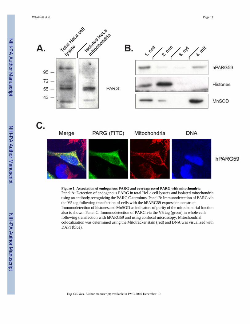
Figure 1. Association of endogenous PARG and overexpressed PARG with mitochondriaPanel A: Detection of endogenous PARG in total HeLa cell lysates and isolated mitochondriausing an antibody recognizing the PARG C-terminus. Panel B: Immunodetection of PARG viathe V5 tag following transfection of cells with the hPARG59 expression construct.Immunodetection of histones and MnSOD as indicators of purity of the mitochondrial fractionalso is shown. Panel C: Immunodetection of PARG via the V5 tag (green) in whole cellsfollowing transfection with hPARG59 and using confocal microscopy. Mitochondrialcolocalization was determined using the Mitotracker stain (red) and DNA was visualized withDAPI (blue).
Whatcott et al. Page 11
Exp Cell Res. Author manuscript; available in PMC 2010 December 10.
NIH
-PA Author Manuscript
NIH
-PA Author Manuscript
NIH
-PA Author Manuscript
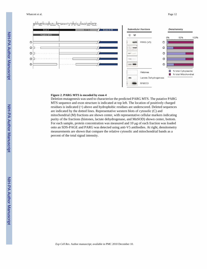
Figure 2. PARG MTS is encoded by exon 4Deletion mutagenesis was used to characterize the predicted PARG MTS. The putative PARGMTS sequence and exon structure is indicated at top left. The location of positively chargedresidues is indicated (+) above and hydrophobic residues are underscored. Deleted sequencesare indicated by the dotted lines. Representative western blots of cytosolic (C) andmitochondrial (M) fractions are shown center, with representative cellular markers indicatingpurity of the fractions (histones, lactate dehydrogenase, and MnSOD) shown center, bottom.For each sample, protein concentration was measured and 10 μg of each fraction was loadedonto an SDS-PAGE and PARG was detected using anti-V5 antibodies. At right, densitometrymeasurements are shown that compare the relative cytosolic and mitochondrial bands as apercent of the total signal intensity.
Whatcott et al. Page 12
Exp Cell Res. Author manuscript; available in PMC 2010 December 10.
NIH
-PA Author Manuscript
NIH
-PA Author Manuscript
NIH
-PA Author Manuscript
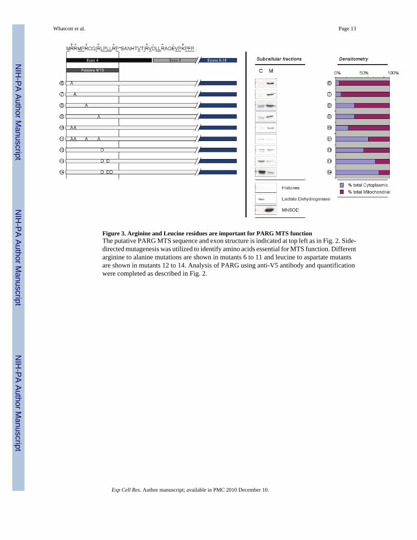
Figure 3. Arginine and Leucine residues are important for PARG MTS functionThe putative PARG MTS sequence and exon structure is indicated at top left as in Fig. 2. Side-directed mutagenesis was utilized to identify amino acids essential for MTS function. Differentarginine to alanine mutations are shown in mutants 6 to 11 and leucine to aspartate mutantsare shown in mutants 12 to 14. Analysis of PARG using anti-V5 antibody and quantificationwere completed as described in Fig. 2.
Whatcott et al. Page 13
Exp Cell Res. Author manuscript; available in PMC 2010 December 10.
NIH
-PA Author Manuscript
NIH
-PA Author Manuscript
NIH
-PA Author Manuscript
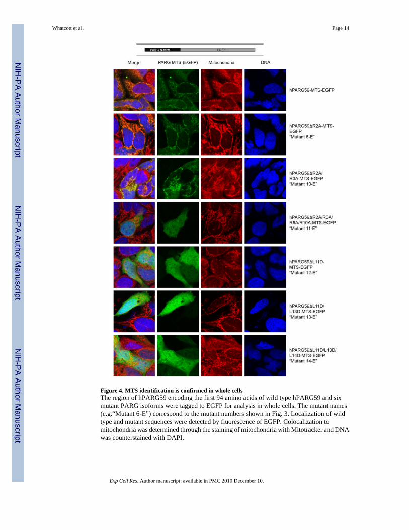
Figure 4. MTS identification is confirmed in whole cellsThe region of hPARG59 encoding the first 94 amino acids of wild type hPARG59 and sixmutant PARG isoforms were tagged to EGFP for analysis in whole cells. The mutant names(e.g.“Mutant 6-E”) correspond to the mutant numbers shown in Fig. 3. Localization of wildtype and mutant sequences were detected by fluorescence of EGFP. Colocalization tomitochondria was determined through the staining of mitochondria with Mitotracker and DNAwas counterstained with DAPI.
Whatcott et al. Page 14
Exp Cell Res. Author manuscript; available in PMC 2010 December 10.
NIH
-PA Author Manuscript
NIH
-PA Author Manuscript
NIH
-PA Author Manuscript
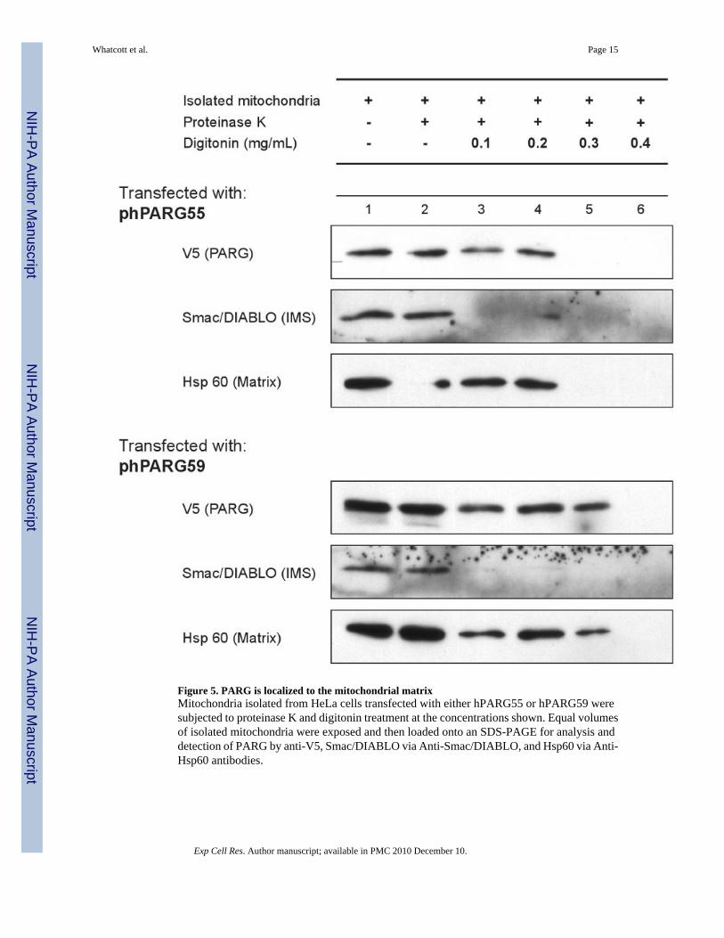
Figure 5. PARG is localized to the mitochondrial matrixMitochondria isolated from HeLa cells transfected with either hPARG55 or hPARG59 weresubjected to proteinase K and digitonin treatment at the concentrations shown. Equal volumesof isolated mitochondria were exposed and then loaded onto an SDS-PAGE for analysis anddetection of PARG by anti-V5, Smac/DIABLO via Anti-Smac/DIABLO, and Hsp60 via Anti-Hsp60 antibodies.
Whatcott et al. Page 15
Exp Cell Res. Author manuscript; available in PMC 2010 December 10.
NIH
-PA Author Manuscript
NIH
-PA Author Manuscript
NIH
-PA Author Manuscript
NIH
-PA Author Manuscript
NIH
-PA Author Manuscript
NIH
-PA Author Manuscript
Whatcott et al. Page 16
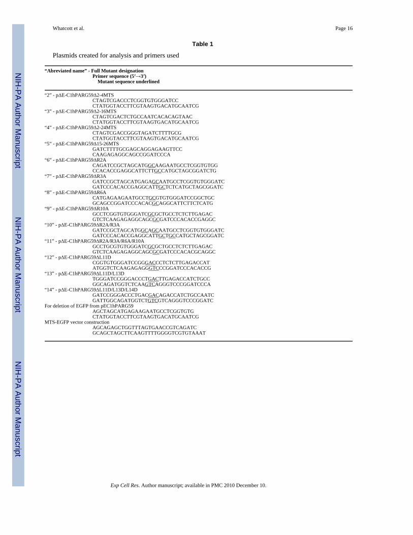
Table 1
Plasmids created for analysis and primers used
“Abreviated name” - Full Mutant designationPrimer sequence (5′→3′) Mutant sequence underlined
“2” - pΔE-C1hPARG59Δ2-4MTSCTAGTCGACCCTCGGTGTGGGATCCCTATGGTACCTTCGTAAGTGACATGCAATCG
“3” - pΔE-C1hPARG59Δ2-16MTSCTAGTCGACTCTGCCAATCACACAGTAACCTATGGTACCTTCGTAAGTGACATGCAATCG
“4” - pΔE-C1hPARG59Δ2-24MTSCTAGTCGACCGGGTAGATCTTTTGCGCTATGGTACCTTCGTAAGTGACATGCAATCG
“5” - pΔE-C1hPARG59Δ15-26MTSGATCTTTTGCGAGCAGGAGAAGTTCCCAAGAGAGGCAGCCGGATCCCA
“6” - pΔE-C1hPARG59ΔR2ACAGATCCGCTAGCATGGCAAGAATGCCTCGGTGTGGCCACACCGAGGCATTCTTGCCATGCTAGCGGATCTG
“7” - pΔE-C1hPARG59ΔR3AGATCCGCTAGCATGAGAGCAATGCCTCGGTGTGGGATCGATCCCACACCGAGGCATTGCTCTCATGCTAGCGGATC
“8” - pΔE-C1hPARG59ΔR6ACATGAGAAGAATGCCTGCGTGTGGGATCCGGCTGCGCAGCCGGATCCCACACGCAGGCATTCTTCTCATG
“9” - pΔE-C1hPARG59ΔR10AGCCTCGGTGTGGGATCGCGCTGCCTCTCTTGAGACGTCTCAAGAGAGGCAGCGCGATCCCACACCGAGGC
“10” - pΔE-C1hPARG59ΔR2A/R3AGATCCGCTAGCATGGCAGCAATGCCTCGGTGTGGGATCGATCCCACACCGAGGCATTGCTGCCATGCTAGCGGATC
“11” - pΔE-C1hPARG59ΔR2A/R3A/R6A/R10AGCCTGCGTGTGGGATCGCGCTGCCTCTCTTGAGACGTCTCAAGAGAGGCAGCGCGATCCCACACGCAGGC
“12” - pΔE-C1hPARG59ΔL11DCGGTGTGGGATCCGGGACCCTCTCTTGAGACCATATGGTCTCAAGAGAGGGTCCCGGATCCCACACCG
“13” - pΔE-C1hPARG59ΔL11D/L13DTGGGATCCGGGACCCTGACTTGAGACCATCTGCCGGCAGATGGTCTCAAGTCAGGGTCCCGGATCCCA
“14” - pΔE-C1hPARG59ΔL11D/L13D/L14DGATCCGGGACCCTGACGACAGACCATCTGCCAATCGATTGGCAGATGGTCTGTCGTCAGGGTCCCGGATC
For deletion of EGFP from pEC1hPARG59AGCTAGCATGAGAAGAATGCCTCGGTGTGCTATGGTACCTTCGTAAGTGACATGCAATCG
MTS-EGFP vector constructionAGCAGAGCTGGTTTAGTGAACCGTCAGATCGCAGCTAGCTTCAAGTTTTGGGGTCGTGTAAAT
Exp Cell Res. Author manuscript; available in PMC 2010 December 10.


