Embed Size (px)
Citation preview

BASIC PHYSIOPATHOLOGY OFGENERAL HEMATOLOGY
Version 12.0, 2010 SERVICE OF HEMATOLOGY CHUV - Lausanne
A SYNOPSIS OF HEMATOLOGY
Pierre-Michel SchmidtPierre CornuAnne Angelillo-Scherrerwith the collaboration of :
Stéphane QuarrozPieter Canham van Dijken

CONTENTS (1)
Part 1 : Red Blood Cell (RBC) pathology PAGESDifferentiation of blood cells 10Normal ranges in hematology 11Erythropoiesis 12Evaluation of anemia 13 - 16Reticulocytes 16Mechanisms of anemia 17 - 19Pathophysiological classification of anemias 20Hyporegenerative normocytic normochromic anemia 21
Anemia of renal failure 22Pure red cell aplasia 23Bone marrow aplasia 24Aplastic anemia 25 - 27
Microcytic hypochromic anemia 28 - 46Iron cycle 29Physiological iron losses 30Iron bioavailability 30Iron metabolism 31Transferrin cycle 32Regulation of ferritin, transferrin receptor and DMT-1 32Iron deficiency anemia 33 - 35
Stages of iron deficiency development 33Serum iron, transferrin and ferritin 33Etiology of iron deficiency 34Treatment of iron deficiency 35
Anemia of chronic disease / Inflammatory anemia 36 - 37Heme synthesis / Porphyrias 38Hemoglobin catabolism 39Globin structure 40Hemoglobins / Interaction O2 and 2,3 DPG 41Hemoglobin dissociation curve 42Anemia with iron utilization disorder 43 - 46
Sideroblastic anemia 43Thalassemias 44 - 46
α-thalassemia 45β-thalassemia 46
2

CONTENTS (2)
3
PAGESMacrocytic normochromic hyporegenerative anemia 47 - 60
Pathophysiology of macrocytic megaloblastic anemia 48Chemical structure of vitamin B12 49Vitamin B12 and folates / General data 50Absorption of vitamin B12 51LDH and anemia 52DNA synthesis anomaly 53Schilling test 53Normal and megaloblastic erythropoiesis 54Causes of vitamin B12 deficiency 55Pernicious anemia 56 - 58Causes of folate deficiency 59Workup of macrocytic anemia 60
Normocytic normochromic regenerative anemia 61 - 87Acute blood loss 61 - 62Hemolytic anemia / Basic data 63 - 64
Measure of RBC half life 65Hemolytic anemia due to corpuscular defect 66 - 81
RBC glycolysis 67 - 68Structure of red blood cell membrane 68RBC enzymopathies 69 - 72
Glucose-6-phosphate dehydrogenase deficiency 70 - 72Anomaly of RBC membrane 73 - 78
Hereditary spherocytosis autosomal dominant 74 - 75Paroxysmal Nocturnal Hemoglobinuria 76 - 78
Hemoglobinopathies 79 - 81Sickle cell disease 80 - 81
Hemolytic anemia due to extracorpuscular defect 82 - 87Immune hemolytic anemia 82Toxic hemolytic anemias 83 - 84Hemolytic anemia of infectious origin 85Hemolytic anemia due to mechanic RBC fragmentation 86 - 87
Thrombotic thrombocytopenic purpura (TTP) / Hemolytic uremic syndrome (HUS) 86Thrombotic microangiopathy / Diagnostic algorithm 87
3

CONTENTS (3)Part 2 : White Blood Cell (WBC) pathology PAGESDifferential leukocyte count 89Neutrophil granulocytes kinetics 90Etiology of neutrophilic leukocytosis 91Toxic changes of neutrophils 92Erythroblastosis and myelocytosis 93Neutropenia 94 - 96Hereditary morphological neutrophil anomalies 97Eosinophils 98Basophils / Mastocytes 99Monocytes / Macrophages 100 - 101Lymphocytes / Lymphoid organs 102 - 113B-lymphocytes 103Steps of B-lymphocyte maturation in secondary lymphoid organs 104T-lymphocytes / Thymic selection 105B- and T-lymphocyte differentiation markers 106NK-lymphocytes 107Lymphocytes / Immune response 108 - 111Lymphocytosis / Lymphopenia 112Plasmacytosis / Mononucleosis syndrome 113Tumors of hematopoietic and lymphoid tissues 114 - 192
WHO classification 2008 114 - 116Myeloid neoplasms 117 - 156
Myeloproliferative neoplasms 118 - 133Polycythemia Vera 119 - 120
Differential diagnosis of erythrocytosis 121 - 123Chronic myelogenous leukemia 124 - 126Essential thrombocythemia 127 - 128
Differential diagnosis of thrombocytosis 129Primary myelofibrosis 130 - 131Chronic neutrophilic leukemia 132Chronic eosinophilic leukemia, N OS 132
Myeloid and lymphoid neoplasms with eosinophilia and anomalies of PDGFRA, PDGFRB or FGFR1 133Myelodysplastic syndromes (MDS) 134 - 141
General features 134Myelodysplasia 135Morphological signs of myelodysplasia 136Classification of MDS / Peripheral blood and bone marrow features 137Differential diagnosis of MDS and acute myeloid leukemia (AML) 138 4
5
CONTENTS (4)PAGES
Anomalies related to MDS 138International prognostic score of MDS 139Other adverse prognostic factors in MDS 140Complications / Evolution / Survival 140Treatment of MDS 141
Myelodysplastic / Myeloproliferative neoplasms 142Chronic myelomonocytic leukemia 142
Acute myeloid leukemia (AML) 143 - 156Epidemiology 143Clinical features of AML 144 - 145Bone marrow and peripheral blood features 146WHO classification 2008 147 - 150Prognostic factors 151Karnofsky performance status 152Therapeutical principles 153Chemotherapy of AML 154Kinetics of leukemic cells in relation with treatment 155Allogeneic transplantation 156
Lymphoid neoplasms 157 - 192General data 157 - 162Simplified classification (WHO 2008) 157Proof of monoclonality 158ECOG clinical performance status 158Prognostic factors / Clinical behavior 158Staging (Ann Arbor) 159Initial assessment 160Treatment of lymphoid neoplasms 161B-cell differentiation / Relationship to major B-cell neoplasms 162Lymphoid leukemias 163 - 177
B, T and NK proliferations 163B-cell lymphoid leukemias 164 - 172
Chronic lymphocytic leukemia (CLL) 164 - 168Definition / Symptoms / Clinical features / Blood picture 164Staging (Rai and Binet) 165Course / Complications / Differential diagnosis 166Prognostic factors 167Treatment of CLL 168 5

6
CONTENTS (5)
6
CONTENTS (5)PAGES
Other B-cell lymphoid leukemias 169 - 172B-cell prolymphocytic leukemia 169Hairy cell leukemia 169Splenic B-cell marginal zone lymphoma (SMZL) 170Splenic B-cell marginal zone lymphoma unclassifiable 170
Splenic diffuse red pulp small B-cell lymphoma (SMZL-diffuse variant) 170Hairy cell leukemia-variant 170
Lymphoplasmacytic lymphoma / Waldenström macroglobulinemia 171Immunological markers, cytogenetics and molecular biology in B-cell lymphoid leukemias 172
T-cell and NK-cell lymphoid leukemias 173 - 177T-cell prolymphocytic leukemia (T-PLL) 173T-cell large granular lymphocyte leukemia (T-LGL) 173Chronic lymphoproliferative disorders of NK-cells (CLPD-NK) 174Aggressive NK-cell leukemia 174Adult T-cell leukemia / lymphoma 175Sézary syndrome 176Immunological markers, cytogenetics and molecular biology in T- and NK-cell lymphoid leukemias 177
Lymphoblastic leukemia / lymphoma 178 - 183Classification WHO 2008 178B lymphoblastic leukemia / lymphoma - Clinical features 179B lymphoblastic leukemia / lymphoma with recurrent genetic anomalies 180T lymphoblastic leukemia / lymphoma - Mature B-cell Burkitt leukemia variant / Clinical features 181Immunological markers of B-ALL and T-ALL 182Treatment principles 183
Plasma cell myeloma 184 - 188Definition / Clinical features / Blood picture / Biology / Clinical variants 184Diagnostic criteria of symptomatic plasma cell myeloma 185International staging system 185Paraproteins / Complications / Prognosis / Survival (ISS) 186Differential diagnosis (MGUS / Smoldering myeloma / Primary amyloidosis / Heavy chain diseases) 187Treatment of plasma cell myeloma 188
Hodgkin lymphoma 189 - 192Symptoms / Clinical features / Histology 189Staging / Cotswolds revision of Ann Arbor classification 190Differential diagnosis / Prognostic factors / Complications 191Treatment / Prognosis and response predictive factors 192
6

77
CONTENTS (6)
7
CONTENTS (6)
Part 3 : Hemostasis PAGESExploration methods 194Thrombus and embolus 195Actors of hemostasis 196Steps of hemostasis 197Primary hemostasis 198Von Willebrand factor 199Production of platelet by the megakaryocyte 200Secondary hemostasis / Coagulation 201Coagulation factors 202 - 203
Vitamin K dependent coagulation factors 203Coagulation cascade 204 - 206
Classical scheme 204Modified concept 205 - 206
Factor XIII and fibrin stabilization 207Natural anticoagulants 208Tertiary hemostasis / Fibrinolysis 207Hemorrhagic syndrome / Primary hemostasis 210 - 217
Vascular purpura 210Prolongation of occlusion time (PFA-100TM) 211Thrombopathy 212Thrombocytopenia 213 - 217
Definition / Hemorrhagic risk / Recommendations 213Thrombocytopenia in the setting of bi- or pancytopenia 214Solitary thrombocytopenia 214Solitary central thrombocytopenia 214Non-immunological solitary peripheral thrombocytopenia 215Immunological solitary peripheral thrombocytopenia 216Investigation of thrombocytopenia 217
Hemorrhagic syndrome / Coagulation 218 - 221Constitutional and acquired coagulation anomalies 218Hemophilia 219 - 220Von Willebrand disease 221
7

CONTENTS (7)
PAGES
Thromboembolic disease 222 - 225Virchow's triad / Risk factors 222Treatment and prophylaxis 223 - 225
Antiplatelet drugs 223Heparin / thrombin and factor Xa inhibitors 223Vitamin K antagonists 224
INR 224Fibrinolytic agents 224Anticoagulation guidelines 225
Part 4 : Algorithms
Anemia 227Normocytic normochromic hyporegenerative anemia 228Microcytic hypochromic anemia 229Macrocytic anemia 230Regenerative anemia 231Polycythemia 232Absolute neutropenia 233Absolute neutrophilia 234Absolute lymphocytosis 235Absolute eosinophilia 236Absolute monocytosis 237Thrombocytopenia 238Thrombocytosis 239Prolonged prothrombin time (PT / Quick) 240Prolonged activated partial thromboplastin time (aPTT) 241
Conclusion 242
8

Part 1
RED BLOOD CELL PATHOLOGY
9

DIFFERENTIATION OF BLOOD CELLS
Pluripotentstem cell
Myeloidstem cell
CFU-GEMM
Lymphoidstem cell
BFU-E CFU-MEG
CFU-GM
CFU-G CFU-M CFU-Baso
CFU-E
SFTPOG-CSFIL-6IL-1IL-11IL-3
SFIL-3GM-CSF
SFIL-3GM-CSF
SFIL-3GM-CSF
SFIL-3GM-CSF
TPOIL-11
IL-3GM-CSFEPO
EPO
IL-3GM-CSFIL-11IL-6IL-7TPO
GM-CSFG-CSF
IL-3GM-CSF
IL-3GM-CSFG-CSF
IL-8
GM-CSFM-CSF
ERYTHROCYTEPLATELETS
NEUTROPHILGRANULOCYTE
MONOCYTE BASOPHIL (Blood)
EOSINOPHILTISSUE MACROPHAGEAlveolarKupffer cellLangerhans cellOsteoclastMicroglial cell
MAST CELL (Tissues)1
SFIL-4 IL-9
SFIL-3IL-10
IL-4IL-9 GM-CSF
IL-5
IL-3GM-CSF
SFIL-3
GM-CSF
Other Interleukins
IL-1 / 4 / 7 / 8 / 9 / 10
Early-acting hematopoietic growth factors
SF : Stem cell factorIL-3 : Interleukin 3IL-6 : Interleukin 6IL-11 : Interleukin 11GM-CSF : Granulocyte-Monocyte Colony-Stimulating
FactorG-CSF : Granulocyte Colony-Stimulating FactorTPO : Thrombopoietin
Lineage specific hematopoietic growth factors
EPO : ErythropoietinTPO : ThrombopoietinG-CSFM-CSF
CFU : Colony Forming Units
CFU-Eo
1 Exact development still unclear 10

NORMAL RANGES IN HEMATOLOGY
UNIT MAN WOMANHEMOGLOBIN g / L 133 – 177 117 – 157HEMATOCRIT % 40 – 52 35 – 47RED BLOOD CELLS T / L 4.4 – 5.8 3.8 – 5.2MCV fL 81 – 99MCH pg 27 – 34MCHC g / L 310 – 360
RDW1 (anisocytosis index) < 15
RETICULOCYTES (Relative count) ‰ 5 – 15
RETICULOCYTES (Absolute count) G / L 20 – 120WHITE BLOOD CELLS G / L 4 – 10PLATELETS G / L 150 – 350
T / L : Tera / L = 1012 / LG / L : Giga / L = 109 / LfL : Femtoliter = L-15
pg : Picogram = g-12
1 RDW : Red cell distribution width
LCH-CHUV, 2009 11
ERYTHROPOIESIS
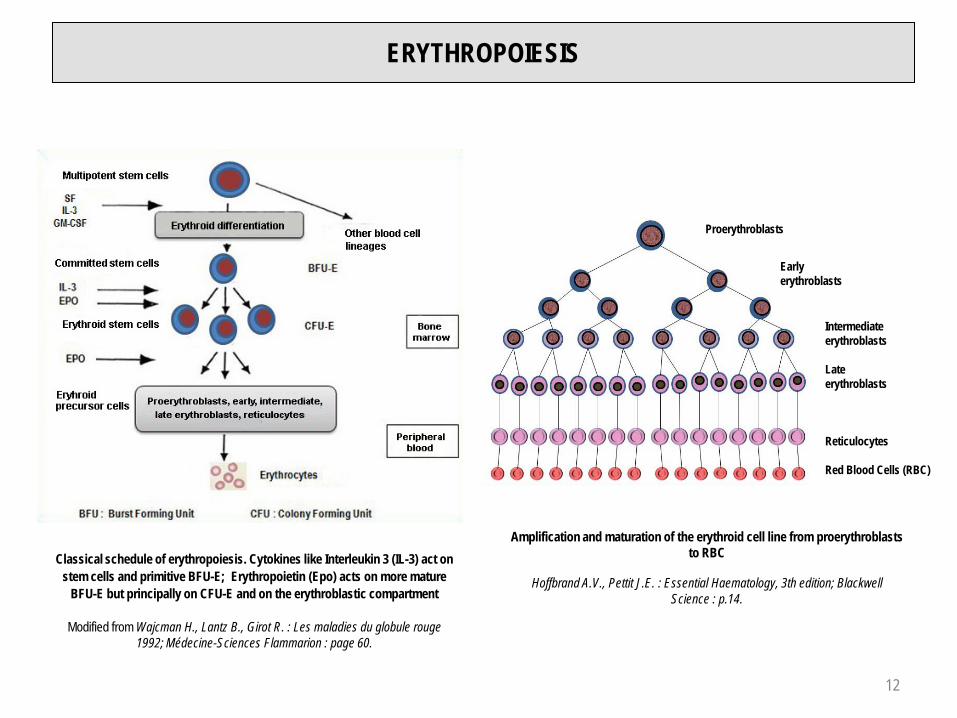
Classical schedule of erythropoiesis. Cytokines like Interleukin 3 (IL-3) act on stem cells and primitive BFU-E; Erythropoietin (Epo) acts on more mature
BFU-E but principally on CFU-E and on the erythroblastic compartment
Modified from Wajcman H., Lantz B., Girot R. : Les maladies du globule rouge 1992; Médecine-Sciences Flammarion : page 60.
Intermediateerythroblasts
Lateerythroblasts
Reticulocytes
Red Blood Cells (RBC)
Amplification and maturation of the erythroid cell line from proerythroblasts to RBC
Hoffbrand A.V., Pettit J.E. : Essential Haematology, 3th edition; Blackwell Science : p.14.
Proerythroblasts
Earlyerythroblasts
12

EVALUATION OF ANEMIA (1)
3 PARAMETERS
3 INDICES
RETICULOCYTE COUNT
13

EVALUATION OF ANEMIA (2)
PARAMETERS
HEMOGLOBIN (g / L)
RED BLOOD CELL COUNT (T / L = 1012 / L)
HEMATOCRIT (%)
ANEMIA = DIMINUTION OF HEMOGLOBIN(At sea level, WHO 1968)
Child (6 months-6 years) < 110 g / L
Child (6 years-14 years) < 120 g / L
Adult man < 130 g / L
Adult woman < 120 g / L
Pregnant woman < 110 g / L
Influence of altitude : + 4% / 1'000 m
14

EVALUATION OF ANEMIA (3)
RED BLOOD CELL INDICES
MCV : Mean Corpuscular Volume (Hct / RBC) x 10 (fL)MCH : Mean Corpuscular Hemoglobin Hb / RBC (pg)MCHC : Mean Corpuscular Hemoglobin Concentration :
(Hb / Hct) x 100 or (MCH / MCV) x 1'000 (g / L)
MORPHOLOGICAL CLASSIFICATION OF ANEMIAS
MCV MCH MCHC
Normocytic normochromic anemia no no no
Microcytic hypochromic anemia
Macrocytic normochromic anemia no
15

EVALUATION OF ANEMIA (4)RETICULOCYTES
Absolute reticulocyte count :< 120 G / L : Hyporegenerative anemia> 120 G / L : Regenerative anemia
Reticulocyte production index (RPI)
Normal : 1.0 - 2.0Hyporegenerative anemia : < 2.0Regenerative anemia : > 2.0
1 Reticulocyte have a total maturation time of 4.5 days :- Normally 3.5 days in bone marrow and 1 day in peripheral blood - In case of hematocrit / hemoglobin reduction reticulocytes leave the bone marrow
earlier at a less mature stage, maturation > 1,0 day in peripheral blood (where thereticulocyte count is performed)
Reticulocyte maturation related to anemia severity1
Reticulocytes distribution related to RNA2 content : HFR (High-Fluorescence Reticulocytes) : high Immature reticulocytes (IRF : Immature Reticulocyte Fraction3)MFR (Medium-Fluorescence Reticulocytes : mediumLFR (Low-Fluorescence Reticulocytes : low Mature reticulocytes
2 By flow cytometry3 Increase of this fraction may precede the reticulocyte increase in peripheral blood. Therefore it can be an early sign of recovery or stimulation of erythropoiesis.
e.g. : a) after bone marrow / stem cell transplantation; b) monitoring of EPO treatment16

MECHANISMS OF ANEMIA (1)
P
P
P
P
H
H
H
H
P
H
RED BLOOD CELLS (RBC)
RBC
RBC
RBC
Blood loss: PRODUCTION
: HEMOLYSIS / RBC SENESCENCE
17

MECHANISMS OF ANEMIA (2)
P HANEMIA
Blood loss
or
HYPOXIA
ERYTHROPOIETIN
(+)
P
H
: PRODUCTION
: HEMOLYSIS / RBC SENESCENCE
18

MECHANISMS OF ANEMIA (3)WHOLE BLOOD, RED CELL, PLASMA VOLUME
RCV30 mL / kg
PV45 mL / kg
RCV
PV
RCV30 mL / kg
PV
Normal True anemia Falseanemia
PV : Plasma Volume
RCV : Red Cell Volume
Increased plasma volume
PregnancyParaproteinSplenomegalyLiver cirrhosis
19

ANEMIAPATHOPHYSIOLOGICAL CLASSIFICATION
HYPOREGENERATIVE ANEMIA(Reticulocyte count < 120 G / L / RPI < 2.0)
NORMOCYTIC NORMOCHROMICRenal failurePure red cell aplasiaBone marrow aplasiaBone marrow infiltrationAnemia of chronic disease / Inflammatory anemiaHypothyroidism
MICROCYTIC HYPOCHROMICIron deficiencyAnemia of chronic disease / Inflammatory anemiaIron utilization disorder (sideroblastic anemia, thalassemia)
MACROCYTIC NORMOCHROMICVitamin B12 and / or folate deficiencyCytotoxic drugsAlcoholism, liver diseases hypothyroidismMyelodysplastic syndromeBone marrow aplasia
REGENERATIVE ANEMIA(Reticulocyte count > 20 G / L / RPI > 2.0 / IRF )
NORMOCYTIC NORMOCHROMICAcute blood lossHemolytic anemia
20

SOLITARY ANEMIARENAL FAILUREPURE RED CELL APLASIAHYPOTHYROIDISM1
PANCYTOPENIA ("CENTRAL" ORIGIN)BONE MARROW APLASIA1
BONE MARROW INFILTRATION (Acute leukemia, lymphoid neoplasm, metastatic cancer)MYELOFIBROSISHEMOPHAGOCYTOSIS
1 Normocytic or slightly macrocytic anemia
HYPOREGENERATIVE NORMOCYTIC NORMOCHROMIC ANEMIA
MCV : normal 81 – 99 fLMCH : normal 27 – 34 pgMCHC : normal 310 – 360 g / LReticulocyte count : < 120 G / L
CLASSIFICATION
21
ANEMIA OF RENAL FAILURE
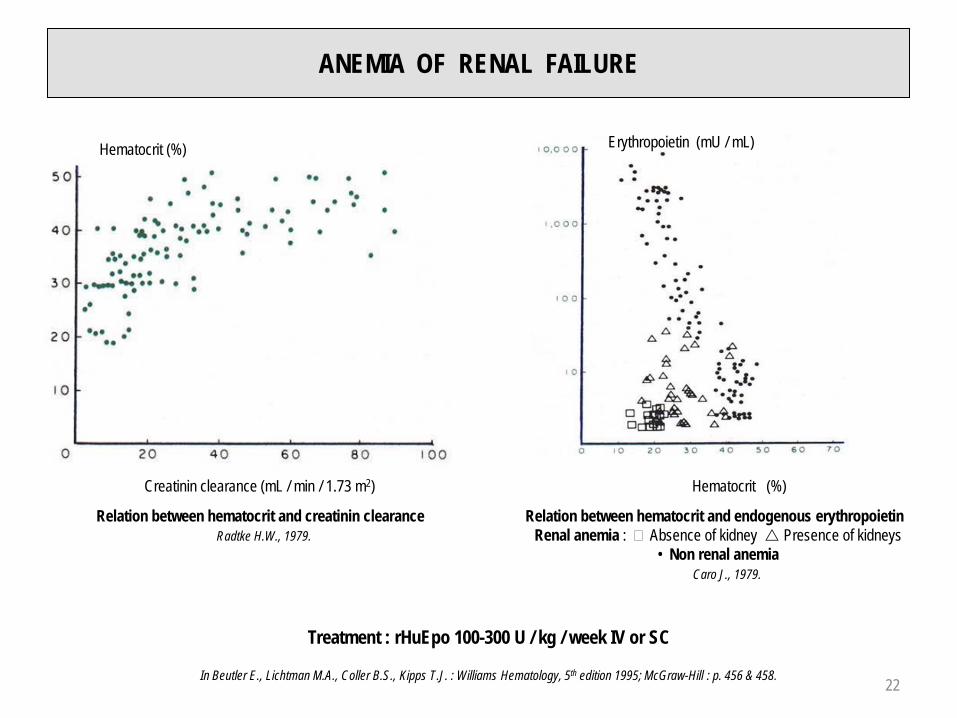
Hematocrit (%)
Creatinin clearance (mL / min / 1.73 m2)
Relation between hematocrit and creatinin clearanceRadtke H.W., 1979.
Erythropoietin (mU / mL)
Hematocrit (%)
Relation between hematocrit and endogenous erythropoietinRenal anemia : � Absence of kidney Presence of kidneys
• Non renal anemiaCaro J., 1979.
Treatment : rHuEpo 100-300 U / kg / week IV or SC
In Beutler E., Lichtman M.A., Coller B.S., Kipps T.J. : Williams Hematology, 5th edition 1995; McGraw-Hill : p. 456 & 458. 22

ERYTHROBLASTOPENIA - PURE RED CELL APLASIA
HEREDITARYBLACKFAN-DIAMOND ANEMIA
ACQUIRED
PRIMARY
SECONDARY
THYMOMA (~ 5% of patients with thymoma have pure red cell aplasia)LYMPHOID NEOPLASMCANCER (lung, breast, stomach, thyroid, biliary tract, skin)COLLAGEN VASCULAR DISEASEPARVOVIRUS B19 INFECTIONPREGNANCYDRUG INDUCED : Anticonvulsants
AzathioprineChloramphenicolSulfonamidesIsoniazidProcainamide
23

BONE MARROW APLASIA ETIOLOGY
HEREDITARY BONE MARROW APLASIA
FANCONI ANEMIA
ACQUIRED BONE MARROW APLASIA
IDIOPATHIC
SECONDARY
IrradiationChemicals (benzene…)Drugs
Obligatory bone marrow aplasiaCytotoxic drugs (alkylating agents)
Occasional or uncommon bone marrow aplasia ChoramphenicolPhenylbutazoneGold salts
Viral infection (EBV, Hepatitis, Parvovirus B19, CMV, HIV)Immune disorder (thymoma)Paroxysmal Nocturnal Hemoglobinuria (PNH)Hypoplastic myelodysplastic syndromePregnancy
24

APLASTIC ANEMIA (1)
DRUG INDUCED BONE MARROW TOXICITY
OBLIGATORY : dosis related Alkylating agents
OPTIONAL : dosis related Chloramphenicol dosis unrelated Chloramphenicol
CHLORAMPHENICOL INDUCED APLASTIC ANEMIA
TOXICITY DOSE RELATED DOSE UNRELATED
INCIDENCE FREQUENT UNCOMMON
BEGIN IMMEDIATE DELAYED(months)
SYMPTOMS LIGHT SEVERE(infection, bleeding)
COURSE SPONTANEOUSLY FAVORABLE FREQUENTLY FATAL
25

APLASTIC ANEMIA (2)IDIOSYNCRASY1 OVER 4 DECADES2
1950 - 1959 1960 - 1969 1970 - 1979 1980 - 1989Drugs3 427 (56%) 203 (60%) 523 (40%) 163 (20%)Benzene and other solvants4 24 (3%) 14 (4%) 37 (3%) 21 (3%)
Insecticides 9 (1%) 29 (9%) 15 (1%) 11 (1%)Idiopathic5 / others6 296 (40%) 93 (27%) 717 (56%) 616 (76%)Total 756 339 1292 811
1 Idiosyncrasy : occasional or uncommon bone marrow depression2 Patients collective recruited in USA, Europe and Asia3 Chloramphenicol, Phenylbutazone, anticonvulsants, gold salts, others4 Benzene : obligatory toxicity or idiosyncrasy5 On the basis of some studies, 40-70% of idiosyncratic bone marrow aplasia are considered idiopathic6 Viral infection (EBV, hepatitis non-A, non-B, non-C, non-G, parvovirus, HIV), immune disease (eosinophilic fasciitis, thymoma,hypogammaglobulinemia, GvH : graft versus host disease in the context of immunodeficiency, pregnancy), PNH (Paroxysmal NocturnalHemoglobinuria)
Modified from data quoted by Young N.S. in Handin R.I., Lux S.E., Stossel T.P. : Blood, Principles & Practice of Hematology 1995; J.B. Lippincott : p. 303.
26

APLASTIC ANEMIA (3)TREATMENT
a) Comparison between allogeneic BMT and Immunosuppressive Treatment (IS). b) Neutrophils < 0.2 G / L, (p < 0.01). c) Neutrophils < 0.2 G / L + infections (EBMT 1987). d) IS + high dose steroids ± cyclosporine (Frickhofen et al.,1992).
Probability to find an HLA-compatible sibling as bone marrow / hematopoietic stem cells donor : 20-30%Adapted from Hoffbrand A.V., Pettit J.E. : Essential Haematology, 3th edition 1993; Blackwell Science p. 127.
(IS = anti-thymocyte globulin)
27

MICROCYTIC HYPOCHROMIC ANEMIA DECREASED MCV, MCH AND MCHC
IRONDEFICIENCY
ANEMIA OFCHRONIC DISEASE
IRON UTILIZATIONDISORDER
Chronic blood lossIncreased demandMalabsorptionPoor diet
Acute and chronic infection Inflammatory disorderCancerRheumatoid arthritis
HEMOGLOBINOPATHYSIDEROBLASTIC
ANEMIA
β-Thalassemiaα-ThalassemiaHemoglobinopathies E, C
HereditaryAcquired : Primary
SecondaryLead poisoningDrugsAlcohol
28

IRON CYCLE
RED BLOOD CELLS IRON
1.5 – 3.0 g
ERYTHROPOIESISIron incorporation in the erythroblasts
15 – 30 mg / d
HEMOLYSISIron release from destroyed RBC
15 – 30 mg / d
PLASMA IRON10.7 – 25.1 µmol / L fixed on
transferrin
IRON STORES(0.6 – 1.2 g)
Ferritin ⇔ Hemosiderin
MINIMAL INTAKE1 mg / day (Male)
2 – 3 mg / day (Female)
AVERAGE NORMAL LOSSES 1 mg / day (Male)
2 – 3 mg / day (Female)
Normal range1 : Iron (serum) 12.5 – 25.1 µmol / L (M ) 10.7 – 21.4 µmol / L (F)Transferrin 24.7 – 44.4 µmol / LFerritin (serum) 10 – 300 µg / L
1 LCC-CHUV, 2009
29

PHYSIOLOGICAL IRON LOSSES
MAN : 14 µg / kg / day (Green, 1968)(0.9 – 1.0 mg / day)
WOMAN : 0.8 mg / day + menstruations : 1.4 – 2.2 mg / day – 50% if oral contraception
+ 100% if intrauterine device
IRON BIOAVAILABILITY
ABSORPTION :
Heme iron 25 – 30%Non heme iron 1 – 7%
Ascorbates, citrates, tartrates, lactates Tannates, wheat, calcium, phosphates, oxalates, soya proteins
30

IRON METABOLISM
1 HCP 1 : Heme Carrier Protein 1 2 Dcytb : Duodenal cytochrome b reductase3 DMT 1 : Divalent Metal Transporter 1 4 TfR : Transferrin Receptor5 Hp : Hephaestine 6 HO 1 : Heme Oxygenase 1
HFE : Human hemochromatosis protein
MACROPHAGE BONE MARROW
IRON ABSORPTION :- Heme iron : by a special pathway, probably HCP 11, followed by
heme degradation through Heme-Oxygenase (HO 16) with ironrecycling
- Non-heme iron : reduction of Fe+++ to Fe++ by Dcytb2 withfollowing absorption by DMT 13 to the intracellular labile iron poolthen to ferritin
IRON CIRCULATIONFe++ leaves the intestinal cell through the Ferroportin pathway, negatively regulated by HepcidinIron is reoxidated to Fe+++ through Hephaestin (Hp5) in presence of Cu++
Iron then binds to Transferrin (Tf) a specific bivalent transporter protein.By binding of Tf to theTransferrin Receptors (TfR4) iron can be delivered to the cells, in particular to the erythroblasts for heme synthesis
Iron is also stored in the macrophages.They also "recycle" the senescent RBC with recuperation and storage of their Heme iron Release of iron from the stores proceeds by the Ferroportin pathway, also negatively regulated by Hepcidin
Hepcidin : blocks Ferroportin by cellular internalization of the formed complex, stopping the process of iron release. This may lead to iron oveload in the cells with functional iron deficiency (e.g. anemia of chronic disorders / inflammatory anemia)
Hepcidin : favours iron transfer and supply to the cells (e.g. iron deficiency)
BLOOD VESSEL
INTESTINAL CELL
31

TRANSFERRIN CYCLE
SYNTHESIS OF FERRITIN, TRANSFERRIN RECEPTOR AND DMT-1
Andrews N.C. : Disorders of Iron Metabolism. NEJM 1999; 341 : 1986-1995.
IRP 1 / IRP 2 : Iron Regulatory Proteins (sensors of intracellular labile iron)IRE and IREs(5) : Iron Responsive Elements (ARNm motives)
Interactions between IRE(s) and IRP lead to regulation of ferritin, DMT 1 and transferrin receptor synthesis related to the iron load of the labile intracellular pool
By high intracellular iron pool, IRP 1 and IRP 2 have low or absent activity leading to facilitated Ferritin mARN transcription with ferritin synthesis. Transcription of TfR and DMT-1 mARN cannot proceed, leading to of TfR and DMT-1, with reduction of iron absorption and transport capacity
By low intracellular iron pool, IRP-IRE binding leads to inhibition of initiation complex of Ferritin mARN transcription in 5’ : of ferritin synthesisStabilization of mARN in 3' by absence of endonuclease cleavage leads to of TfR and DMT-1 synthesis
TfR : Transferrin Receptor. Binds 2 molecules of bivalent transferrinDMT 1 : Divalent Metal Transporter 1. Transport in the cell of non-heme ironAPO-Tf : Apotransferrin
ORF : Open Reading Frame 32

STAGES OF IRON DEFICIENCY DEVELOPMENTSERUM IRON - TRANSFERRIN - FERRITIN
STAGE 1 STAGE 2 STAGE 3FERRITIN
IRON (Bone marrow) Absent Absent
TRANSFERRIN (Serum) Normal
IRON (Serum) Normal
HEMOGLOBIN Normal Normal
MCV Normal Normal
MCHC Normal Normal
SERUM IRON TRANSFERRIN FERRITIN
IRON DEFICIENCY
INFLAMMATORY ANEMIA
IRON UTILIZATION DISORDER no /
SOLUBLE TRANSFERRIN RECEPTORS :Increased in isolated iron deficiency and in this associatedwith inflammatory processesNormal in isolated inflammatory anemia
RING SIDEROBLASTS :Increased in sideroblastic anemia (indication to bone marrowexamination), cf. page 43
33

ETIOLOGY OF IRON DEFICIENCY
Chronic blood lossIncreased iron demandMalabsorptionPoor diet
CAUSES OF CHRONIC IRON LOSSUterine (menorrhagia, metrorrhagia), digestive bleeding (hematemesis, melaena), parasites (hookworm), hematuriaChronic intravascular hemolysis (Paroxysmal Nocturnal Hemoglobinuria)Frequent blood donations, phlebotomies, provoked bleedings (Lasthénie de Ferjol syndrome)Chronic bleeding (microcytic hypochromic hyporegenerative anemia) must imperatively be distinguished from acute blood loss (normocytic normochromic regenerative anemia). Remember that 1 L of blood = 500 mg of iron
INCREASED IRON DEMANDPregnancyBreast feeding (maternal milk = 0.3 – 0.5 mg / L)Growth
IRON DEMAND IN PREGNANCYIncreased maternal total red cell volume 500 mgFetal needs 290 mgPlacenta 25 mgBasal iron loss (0.8 mg / d for 9 months) 220 mgTOTAL : 1'035 mg
FUNCTIONAL IRON DEFICIENCYAbsence of adequate erythropoietin response in case of anemia secondary to renal failure or to an inflammatory process with ferritin level in normal or high range (cf. following page)
34

CAUSAL TREATMENT IRON SUBSTITUTION (anemia correction and iron stores reconstitution)Oral substitution :
Basic data :1 L of blood = 500 mg of iron and 160 g of hemoglobin.1 g of hemoglobin : 500 / 160 = ± 3 mg of iron Blood volume : 75 mL / kg. Iron reserves : 1'000 mg
Example : Woman, 56 years old, BW 50 kg, hemoglobin 80 g / L Iron needs for anemia correction and iron stores reconstitution :[ Blood volume (L) x (160 - Hb patient) x 3] + 1'000 mg → [ 3.75 x (160 – 80) x 3] + 1'000 mg = 1'900 mg of ironPatient receives 100 mg elementary iron q.d. with a mean resorption of 15 mg q.d. Duration of substitution : 1'900 / 15 = 126 days ( ± 4 months)Anemia correction within ± 1 month. Iron deficiency corrected when serum ferritin in normal range
Parenteral substitution : 100-200 mg IV 1-3 x weekly or perfusion of 1'000 mg (15 mg / kg) offerric carboxymaltose once or twice
Indications : Functional iron deficiency (Hb content in reticulocytes (CHr) < 28 pg ; percentage of hypochromic RBC (HYPO) : > 5%)Malabsorption syndromeDigestive oral iron intolerancePoor patient complianceImportant chronic, persisting hemorrhage
TREATMENT OF IRON DEFICIENCY ANEMIA
35

ANEMIA OF CHRONIC DISORDERS / INFLAMMATORY ANEMIA (1)INFECTION
INFLAMMATIONAUTOIMMUNE DISEASE
MALIGNANCYRENAL FAILURE
Fe+++ / Transferrin
Functional iron deficiency
T-lymphocyte activation
Interleukin-6
Hepcidin
Inhibition of iron release from
macrophages
Interleukin-1TNF-α
Erythropoietin
Interleukin-1TNF-αInterferon -γ
Hepcidin
TNF-α
Monocyte activation
MONOCYTET-LYMPHOCYTE LIVER
KIDNEY
MACROPHAGE
BONE MARROW
Intestinal ironresorption
Erythropoiesis
Hemophagocytosis of senescent RBC
36

MACROPHAGE ACTIVATION
IL-1 TNF-α
γ-IFN β-IFN
EPO SECRETION IRON RELEASE FROM MACROPHAGES
ERYTHROBLASTSPROLIFERATION
HEPCIDIN
INTESTINAL IRON ABSORPTION
STIMULATION
INHIBITION
INFLAMMATORY ANEMIA
CRP FIBRINOGEN SEDIMENTATION RATE
Modified from Means R.T.& Krantz S.B., Blood 1992; 80 :1639-1647.
ANEMIA OF CHRONIC DISEASE / INFLAMMATORY ANEMIA (2)ROLE OF MACROPHAGE RELEASED CYTOKINES IN ITS PATHOPHYSIOLOGY
3737

The heme molecule
Porphyric nucleus + iron
Succinic acid+ Glycin
δ-aminolevulinic acid
δ-aminolevulinic acidsynthetase
δ-aminolevulinic aciddehydratase
Porphobilinogen
Hydroxymethylbilane Uroporphyrinogen III
Coproporphyrinogen III
Uroporphyrinogendecarboxylase
Protoporphyrinogen
Coproporphyrinogenoxydase
Protoporphyrinogenoxydase
Protoporphyrin
Uroporphyrinogencosynthetase
CYTOSOL
Porphobilinogen deaminase
HEME
IRON
Ferrochelatase
HEPATIC (H) AND ERYTHROPOIETIC (E) PORPHYRIAS
DISEASE TYPE ENZYME DEFICIENCY
Doss porphyria H ALA dehydratase
Acute intermittent porphyria H Porphobilinogen deaminase
Congenital erythropoietic porphyria E Uroporphyrinogen cosynthetase
Cutaneous porphyria H Uroporphyrinogen decarboxylase
Hereditary coproporphyria H Coproporphyrinogen oxydase
Porphyria variegata H Protoporphyrinogen oxydase
Protoporphyria E Ferrochelatase
Wajcman H., Lantz B., Girot R. : Les maladies du globule rouge 1992; Médecine-Sciences. Flammarion : p. 418 & 420.
MITOCHONDRION
HEME SYNTHESIS
Methyl Vinyl
Vinyl Methyl
Methyl Methyl
PropionatePropionate
38

HEMOGLOBIN DEGRADATION
39

HEMOGLOBIN STRUCTURE
Central cavity : Binding site of 2,3-DPG
α2β2 dimer α1β1 dimer
α1β2 contact area
α1β1 contact area
Heme with iron atom
Hemoglobine tetramer with contact areas
40

HEMOGLOBIN / INTERACTION O2 AND 2,3-DPG
oOXYHEMOGLOBIN DEOXYHEMOGLOBINCompetition between oxygen and 2,3-diphosphoglycerate (2,3-DPG)
GLOBINSTRUCTURE HEMOGLOBIN
Embryonic hemoglobins
ξ2 ε2 Gower 1
ξ2 γ2 Portland
α2 ε2 Gower 2
Adult hemoglobins
α2 β2 A
α2 δ2 A2 (1.5 – 3.0%)
α2 γ2 F (< 1%)
GENES CODING FOR THE DIFFERENT GLOBIN CHAINS AGE OF PREGNANCY (WEEKS)
BIRTHPercentage of synthesis
AGE AFTER BIRTH(MONTHS)
Synthesis of the different globin chains during ontogenesis
Wajcman H., Lantz B., Girot R. : les maladies du globule rouge 1992; Médecine-Sciences Flammarion : p. 12.
* Heme
41

HEMOGLOBIN DISSOCIATION CURVE
Right shift of the hemoglobin dissociation curve through of 2,3-DPG : of oxygen affinity of hemoglobinIn this situation : 12% increase of O2 tissues delivery
Left shift of the hemoglobin dissociation curve through of 2,3-DPG : of oxygen affinity of hemoglobin
in this situation : 20% diminution of O2 tissues delivery
42

PATHOPHYSIOLOGY
Anomaly of porphyric nucleus synthesisPresence of ring sideroblasts (bone marrow) Role of vitamin B6 (Pyridoxin)
CLASSIFICATION
Acquired sideroblastic anemia : PrimarySecondary
LeadIsoniazidChloramphenicolPyrazinamideAlcohol
Hereditary sideroblastic anemia : X - linkedAutosomalMitochondrial
ANEMIA WITH IRON UTILIZATION DISORDER (1)SIDEROBLASTIC ANEMIA
43

ANEMIA WITH IRON UTILIZATION DISORDER (2)THALASSEMIA
PATHOPHYSIOLOGY
GLOBIN SYNTHESIS DEFECT
Great genetic heterogeneity at molecular level (DNA lesions, i.e. more or less important deletions, point mutations)
α-Thalassemia : or absence of α-chain synthesis of globin
β-Thalassemia : or absence of β-chain synthesis of globin
CENTRAL (BONE MARROW) AND PERIPHERAL HEMOLYSIS THROUGH TETRAMERS INSTABILITY
α4 for β-Thalassemia
β4 for α-Thalassemia (Hemoglobin H)
44

α-THALASSEMIA
CLINICAL VARIETIESNormal α α / α αAsymptomatic carrier – α / α αα-Thalassemia minor – – / α α or – α / – αHemoglobin H disease – – / – α
Moderate, sometimes severe chronic anemiaSplenomegalyInclusion bodies
Hemoglobin Bart – – / – –Hydrops fetalisHb Bart = γ4
DIAGNOSISSearch for inclusion bodiesElectrophoresis of a fresh1 hemolysate at alkaline or neutral pH. Isoelectric focusing (Hb H)DNA analysis
1 Hb H is unstable !
CHROMOSOME 16
45

β-THALASSEMIA
β-THALASSEMIA MINOR
β / β +-thal (heterozygocity)
"Micropolyglobulia" : e.g. RBC : 6.2 T / LHb : 105 g / LMCV : 62 fL
Target cells, coarse basophilic stippling. Hb electrophoresis : Hb A2 and FGenetic counseling
β-THALASSEMIA MAJOR
β 0-thal / β 0-thal (homozygocity) orβ 0-thal / β +-thal (double heterozygocity)
Severe anemia, hemolytic icterus, erythroblasts on blood smearSplenomegaly, hepatomegalyGrowth retardationHb electrophoresis : or absence of Hb A
Hb F 20-80 %Treatment : Transfusions, iron chelation, allogeneic stem cell / bone marrow transplantation
46

MACROCYTIC NORMOCHROMIC HYPOREGENERATIVE ANEMIA
MCV : > 99 fLMCH : > 34 pgMCHC : normal 310 – 360 g / LReticulocyte count : < 120 G / L
CLASSIFICATION
MEGALOBLASTIC MACROCYTIC ANEMIAVitamin B12 deficiencyFolate deficiencyCytotoxic drugs
6-mercaptopurin5-fluorouracilCytosine arabinosideHydroxyureaMethotrexateZidovudin (AZT)
NON MEGALOBLASTIC MACROCYTIC ANEMIAAlcoholismLiver diseaseMyxedemaMyelodysplastic syndrome
47

MEGALOBLASTIC MACROCYTIC ANEMIAPATHOPHYSIOLOGY
Hoffbrand A.V., Moss P.A.H., Pettit J.E. : Essential Haematology, 5th edition 2006; Blackwell Publishing : p. 47.
Role of vitamin B12 (cobalamin) and folates in DNA metabolism
Methyl THF : methyltetrahydrofolate A : adenineTHF : tetrahydrofolate G : guanineDHF : dihydrofolate C : cytosineMP : monophosphate T : thymidineDP : diphosphate U : uridineTP : triphosphate d : deoxyribose
Methionine deficiency might be the cause of myelin synthesis anomaly, leading to the neurological signs and symptoms found in vitamin B12 deficiency
Methotrexate
48

VITAMIN B12 AND FOLATESCHEMICAL STRUCTURE
Structure of methylcobalamin (plasma)Other compounds : deoxyadenosylcobalamin (tissues),
hydroxocobalamin and cyanocobalamin (used in treatment of vitamin B12 deficiency)
Structure of folic acid (pteroylglutamic acid) : pteridinenucleus + para-aminobenzoic acid + glutamate(s)
Hoffbrand A.V., Pettit J.E. : Essential Haematology, 3th edition 1993; Blackwell Science : p. 54 & 57.49

VITAMIN B12 AND FOLATESGENERAL DATA
VITAMIN B12 FOLATESBalanced diet ( / day) 7 – 30 µg 200 – 250 µg
Daily needs 1 – 2 µg 100 – 150 µg
Origin Animal Vegetables, liver, yeast
Cooking (heat) Few effect Thermolabile
Reserves 2 – 3 mg 10 – 12 mg
Exhaustion of stores 2 – 4 years 3 – 4 months
Absorption
Site Ileum Jejunum
Mechanism Intrinsic factor1 Methyltetrahydrofolate conversion
Plasmatic transport
Transcobalamins (TC)TC II : transport and intracellular transfer
of cobalaminsTC I 2 : transports the major part of
circulating cobalamins TC III : isoprotein of TC I
Albumin
Active physiological forms Methyl- and deoxyadenosylcobalamins Polyglutamates
Compounds used for therapeutic substitution
HydroxocobalaminCyanocobalamin Folic acid (pteroylglutamic acid)
Serum levels (physiological) 133 – 675 pmol / L3 > 5.3 nmol / L3
1 Cobalamins of dietary origin are unspecifically bound to proteins. In the stomach, peptic digestion at low (acid) pH separates dietary proteins from cobalamins which then bind to Protein R (or haptocorrin) of salivary origin. In the duodenum, degradation of Protein R by pancreatic proteases allows binding of the cobalamins to intrinsic factor of gastric origin
2 TC I and TC III are abundant in secondary granules of neutrophils
3 LCC-CHUV, 2009 50

ABSORPTION OF VITAMIN B12
PHYSIOPATHOLOGICAL MECHANISMSOF VITAMIN B12 (COBALAMINE)
DEFICIENCY
1 Cobalamin dietary deficiency
2 Anomaly of cobalamine - fooddissociation
3Quantitative or qualitative defect of Intrinsic Factor (IF)
4Deficiency of pancreatic proteaseAbnormal utilization of vitamin B12 by bacterias (blind loop syndrome), fish worm (diphyllobothrium latum)
5Anomaly of ileal mucosa and / or of the IF receptors and / or transfer in the enterocyte
51

LDH AND ANEMIA
Modified from Emerson P.M., Wilkinson J.H., Br J Haematol 1966; 12 : 678-688.
Iron Deficiency
B12 Deficiency
HemolyticAnemia
LDH activity in iron deficiency, megaloblastic and hemolytic
anemiasDotted line : upper limit of the reference
interval
52

MEGALOBLASTIC ANEMIA WITH DNA SYNTHESIS ANOMALYNuclear maturation slowdown Optimal hemoglobin concentration reached before the usual 4 mitosisReduction of the number of mitosisIncreased size of the cells
Bone marrow : megaloblastsPeripheral blood : megalocytes ("macroovalocytes")
Intramedullary and peripheral hemolysisBone marrow with megaloblastic hyperplasia by erythroid stem cell recruitment through erythropoietin
SCHILLING TEST
Saturation of transcobalamins by IM injection of 1 mg vitamin B12Oral administration of 0.5 -1 µg radiolabeled vitamin B1248 hours urine collection and measure of excreted radioactivityIn case of pathological result repeat the test with concomitant oral intrinsic factor administration (IF)
Urinary excretion of radiolabeled vitamin B12 (%)
B12 alone B12 + IF
Normal subject 18 (9 – 36) –
Pernicious anemia 0.5 (0 – 1.2) 13 (6 – 31)
Malabsorption (Gluten enteropathy) 3.6 (0 – 19) 3.3 (0 – 10)
Modified from Lee G.R., Wintrobe’s Clinical Hematology, 9th edition 1993; Lea & Febiger : p. 776.
Results obtained with 0.5 µg of radiolabeled oral vitamin B12
53

NORMAL AND MEGALOBLASTIC ERYTHROPOIESIS
NORMALERYTHROPOIESIS
MEGALOBLASTICERYTHROPOIESISBONE MARROW
MEGALOBLASTS(Asynchronism of
nucleocytoplasmic maturation)
CELLULARITY NORMAL INCREASED
PROERYTHROBLASTS
EARLYERYTHROBLASTS
INTERMEDIATEERYTHROBLASTS
LATEERYTHROBLASTS
BLOODRETICULOCYTES
RED BLOOD CELLS
WHITE BLOOD CELLS
NEUTROPHILS
NORMAL HEMOGLOBINSYNTHESIS
LOW OR ABSENTRETICULOCYTE COUNT
HOWELL-JOLLY BODIES
MACROCYTES MEGALOCYTES
HYPERSEGMENTEDNEUTROPHILS
Modified from Chandrasoma P., Taylor C.R. : Concise Pathology, 3th edition 1998; Appleton & Lange. 54

CAUSES OF VITAMIN B12 DEFICIENCY
MALABSORPTION
Gastric origin : AchlorhydriaPernicious anemia Partial or total gastrectomyCongenital intrinsic factor deficiency
Intestinal origin : Resection of terminal ileumCrohn’s diseaseGluten induced enteropathyFish tapeworm (Diphyllobothrium latum) infestation
Dietary deficiency
1. Non dissociation of Vitamin B12 from the transport proteins orinsufficient digestion of dietary vitamins B12
2. Pernicious anemia3. Undefined4. Malabsorption5. Poor diet
Distribution of causes of vitamin B12 deficiency in adults
Andrès E. et al. : Hématologie 2007; 13 : 186-192.55

PERNICIOUS ANEMIA (1)
PATHOPHYSIOLOGY
Atrophic gastritis of immune origin with lack of intrinsic factor
HEMATOLOGY
Macrocytic megaloblastic anemiaNeutropenia with hypersegmented neutrophilsThrombocytopenia
CLINICAL ASPECTS
Atrophic glossitis (Hunter's glossitis), dyspepsiaCombined degeneration of the dorsal (posterior) and lateral spinal columns (paresthesias, pain, gait disturbance, pallesthesia diminution, pyramidal syndrome)
→ Methionine synthesis defect ?Psychiatric symptoms (irritability, depression)Melanic skin hyperpigmentation (uncommon !)Sterility, asthenospermia
56

PERNICIOUS ANEMIA (2)LABORATORY
SCHILLING TEST
Pathological but normalized after simultaneous administration of vitamin B12 + intrinsic factor
ANTIBODY SCREENING
Antiparietal cells(± 90%) 1
Anti-intrinsicfactor (± 50%)
Specificity – +Sensitivity + –
1 Antiparietal cells antibodies can be found in normal individuals (5-20%) and in myxedema (~ 30%)
Schematic presentation of intrinsic factor (IF), vitamin B12and of antibody directed against intrinsic factor :a) Normal binding between IF and vitamin B12 b) Blocking antibodyc) Coupling antibody
Modified from Lee G.R. : Wintrobe’s Clinical Hematology, 9th edition 1993; Lea & Febinger : p. 753.57

PERNICIOUS ANEMIA (3)RESPONSE TO HYDROXOCOBALAMIN SUBSTITUTION
RETICS
Hydroxocobalamin 1 mg IMHemoglobin (g / dL)
Reticulocyte count (%)
Days
Platelets (G / L)
Leukocytes (G / L)
Hemoglobin (g / dL)
Platelets
LeukocytesHoffbrand A.V., Pettit J.E. : Essential Haematology, 3th edition;
Blackwell Science : p. 70.
58

CAUSES OF FOLATE DEFICIENCY
DIETARY DEFICIENCYMALABSORPTION
Gluten induced enteropathyWide jejunal resectionCrohn’s disease
INCREASED DEMANDPhysiological : Pregnancy
LactationPrematurityGrowth
Pathological : Hemolytic anemiaCancer, myeloid or lymphoid neoplasmInflammatory process
DRUGSAnticonvulsants (e.g. : Diphenylhydantoin)BarbituratesSalazopyrin
ALCOHOLISM
59
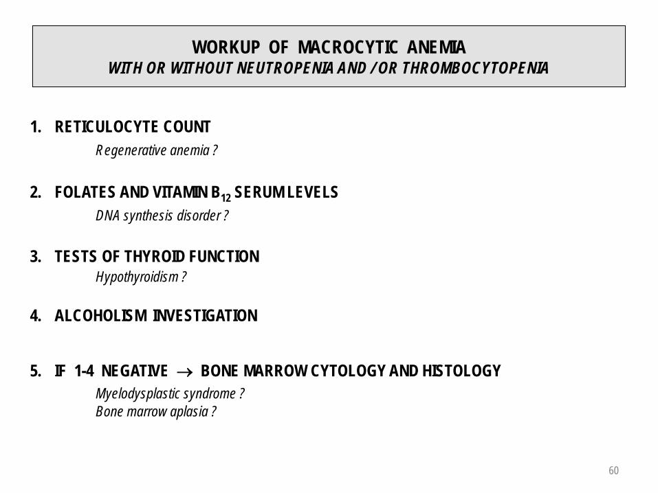
WORKUP OF MACROCYTIC ANEMIAWITH OR WITHOUT NEUTROPENIA AND / OR THROMBOCYTOPENIA
1. RETICULOCYTE COUNTRegenerative anemia ?
2. FOLATES AND VITAMIN B12 SERUM LEVELS DNA synthesis disorder ?
3. TESTS OF THYROID FUNCTIONHypothyroidism ?
4. ALCOHOLISM INVESTIGATION
5. IF 1-4 NEGATIVE → BONE MARROW CYTOLOGY AND HISTOLOGYMyelodysplastic syndrome ?Bone marrow aplasia ?
60

NORMOCYTIC NORMOCHROMIC REGENERATIVE ANEMIA
ACUTE BLOOD LOSS (1)
BLOOD LOSS % BLOOD VOLUME SYMPTOMS
0.5 – 1.0 L 10 – 20 Possible vaso-vagal signs
1.0 – 1.5 L 20 – 30 Tachycardia / hypotension
1.5 – 2.0 L 30 – 40 Reversible hypovolemic shock
> 2.0 L > 40 Irreversible hypovolemic shock
MCV : normal 81 – 99 fL
MCH : normal 27 – 34 pg
MCHC : normal 310 – 360 g / L
Reticulocyte count : > 120 G / L
61

ACUTE BLOOD LOSS (2)
Evolution in 2 phases :
1. Hypovolemia (1-3 days)2. Volemia normalization
Anemia is only found during phase of volemia correction
Anemia normocytic normochromic as far as iron stores not exhausted
To be remembered : 1 L of blood = 500 mg of iron
Increase of the reticulocyte count from the 4th day, possibly neutrophilic leukocytosis with left shift,myelocytosis (presence of some peripheral blood myelocytes and metamyelocytes), thrombocytosis
Treatment :
Phase 1 : packed red cells and plasmaPhase 2 : packed red cells
62

HEMOLYTIC ANEMIABASIC DATA (1)
HISTORYEthnic origin, family historyStay in a foreign countryDrug treatmentPrior transfusion(s), pregnancy(-ies)
CLINICAL FEATURESJaundiceSplenomegaly
HEMOGRAMNormocytic normochromic anemia
Particular situations : Absence of anemia in case of compensated hemolysisMicrocytic anemia : thalassemia, hemoglobinopathies E, C, PNH1
Macrocytic anemia : high reticulocyte count, associated folate deficiencyRegeneration signs
PolychromasiaIncreased reticulocyte countPresence of peripheral blood erythroblasts
Red blood cell morphologySpherocytes, schistocytes, sickle cells, target cells
1 PNH : Paroxysmal Nocturnal Hemoglobinuria (iron deficiency due to chronic hemoglobinuria)
63

HEMOLYTIC ANEMIABASIC DATA (2)
BLOOD CHEMISTRY unconjugated bilirubin L D H haptoglobin fecal stercobilinogenUrobilinuria
ISOTOPIC TESTS ( 51Cr ) : cf. following page
EXTRAVASCULAR HEMOLYSIS"Sensitization" of circulating RBC and destruction by the monocyte / macrophage system(spleen, lymph nodes, bone marrow)
INTRAVASCULAR HEMOLYSIS plasmatic Hb (> 50 mg / L)HemoglobinuriaHemosiderinuria
HEMOLYSIS DUE TO CORPUSCULAR ANOMALYHereditary (except PNH1)Homozygous or heterozygous
HEMOLYSIS DUE TO EXTRACORPUSCULAR ANOMALYAcquired 1 PNH : Paroxysmal Nocturnal Hemoglobinuria
64

MEASURE OF RED BLOOD CELLS HALF LIFE 51 Cr LABELLING
51Cr % activity
Hemolytic
anemia
Measure of RBC half life with 51Cr labeling (51CrT50)o- -o- -o : Theoretical curve•—•—• : Normal curve with half life of 30 ± 2 days
Pathological curve with half life < 10 days
External counts during 51Cr test :a) Predominant splenic sequestration
(hereditary spherocytosis)b) Predominant hepatic sequestration
(sickle cell disease)c) Mixed sequestration (splenic and hepatic)
(some forms of immune hemolytic anemia)
Spleen
Liver Spleen
Liver
Liver
Spleen
Modified from Hoffbrand A.V., Pettit J.E. : Essential Haematology, 3th edition 1993; Blackwell Science : p. 76.
Radioactivity
65

HEMOLYTIC ANEMIA DUE TO CORPUSCULAR DEFECT
ENZYMOPATHY
RBC MEMBRANE ANOMALY
HEMOGLOBINOPATHY
Diminution (or absence) of globin chains synthesis
THALASSEMIAS (cf. pages 44-46)
Substitution (or deletion) of a residue on a globin chain
SICKLE CELL DISEASE
HEMOGLOBINS E, C
UNSTABLE HEMOGLOBINS
HEMOGLOBINS M1
HEMOGLOBINS WITH INCREASED OR REDUCED OXYGEN AFFINITY
1 M : Methemoglobin
66

ERYTHROCYTIC GLYCOLYSIS (1)
Glucose
Glucose 6-P
Fructose 6-P
Fructose 1-6-DP
Glyceraldehyde-3P
1,3-DP-Glycerate
3-P-Glycerate
2-P-Glycerate
P-Enolpyruvate
Pyruvate
LACTATE
MAIN GLYCOLYTICPATHWAY
(Embden-Meyerhof)
HbMethemoglobinreductase
1. Methemoglobinreduction
2. Synthesis of2 ATP
3. NADP reductionPentose shunt
Luebering-Rapoportshunt 4. 2,3-DPG synthesis
FUNCTIONS OF ERYTHROCYTICGLYCOLYSIS
Dihydroxyaceton-P
MetHb
ATPADP
ATP
ATP
ATPADP
ADP
ADP
NADNADH
ERYTHROCYTE
ENERGETIC METABOLISM (2)PROTECTION AGAINST OXYDATIVE STRESS (1, 3)
STRUCTURE AND FUNCTIONS OF THE RBCMEMBRANE (2, 3)HEMOGLOBIN (4)
67

GLYCOLYSIS (2) / STRUCTURE OF THE RBC MEMBRANE
Main glycolyticpathway
Pentose shunt (protection against oxydation of hemoglobin
and RBC membrane)
Luebering-Rapoport shunt(synthesis of 2,3-DPG)
STRUCTURE OF RBCMEMBRANE
Iolascon, A. et al. Haematologica 2008;93:1283-1288
Composite structure with double layer lipidic membrane anchored to a two-dimensional elastic network (cytoskeleton) with tethering sites (transmembrane proteins) Vertical fixation involves cytoplasmic part of Band 3 protein, Ankyrin, Protein 4,2 and Spectrin. The horizontal interaction involves Spectrin (α- and β-chains), Protein 4.1.R, Actin, Tropomodulin, tropomyosin and Adducin. Protein 4.1R interacts with Glycophorin C (GPC) and P55
RhAG : Rhesus Antigens GPA : Glyocophorin A
68

RED BLOOD CELL ENZYMOPATHY
FREQUENT
PENTOSE SHUNT
Glucose-6-phosphate dehydrogenase (G-6-PD) deficiency(> 400 .106 cases, > 300 variants)
EMBDEN-MEYERHOF PATHWAY
Pyruvate kinase deficiency (< 1'000 cases)Glucose phosphate isomerase deficiency (< 200 cases)
UNCOMMON
EMBDEN-MEYERHOF PATHWAY
Deficiency in : Hexokinase, phosphofructokinase, aldolase, triose phosphateisomerase, diphosphoglycerate mutase, phosphoglycerate kinase (< 20 cases)
69

GLUCOSE-6-PHOSPHATE DEHYDROGENASE DEFICIENCY (G-6-PD) (1)
Amino acid substitution in some variants of G-6-PD
Variants Position of residue
68 126 188 227 323
B (+) Valine Asparagine Serine Arginine Leucine
A (+) Aspartic acid
A (-) Methionine
A (-) Leucine
A (-) Proline
Mediterranean Phenylalanine
B (+) : Usual form : predominantA (+) : 30% African colored : normal activityA (-) : 11% African American : activity 5-15% of normalMediterranean [formerly B (-)] : Activity < 1%X-linked recessive deficiencyHemolysis : Chronic (uncommon)
Usually induced by : drugs, fever, fava beans (Favism)
70

GLUCOSE-6-PHOSPHATE DEHYDROGENASE DEFICIENCY (G-6-PD) (2)PATHOPHYSIOLOGY
G-6-PDCatalase NADPH
Reduced glutathione (GSH) protects the -SH groups of the RBC membrane and hemoglobin
During hemolytic crisis, presence of Heinz bodies in the RBC after staining with brilliant cresyl blue : denatured hemoglobin (oxidized)
Decrease in hemolysis during reticulocyte response (young RBC are relatively enzyme rich)
71

GLUCOSE-6-PHOSPHATE DEHYDROGENASE DEFICIENCY (G-6-PD) (3)
Main substances able to induce hemolytic crisis in G-6-PD deficiency1
ANTIMALARIAL DRUGSPrimaquine, pamaquine, pentaquine, quinine
SULFONAMIDESSulfacetamide, sulfamethoxazole, sulfanilamide, sulfapyrine, sulfoxone, thiazosulfone
ANTIBIOTICS AND BACTERIOSTATIC AGENTSPara-aminosalicylic acid, nalidixic acid, nitrofurantoin, chloramphenicol, methylene blue,niridazole
ANALGESICSAcetanilide, amidopyrine, paracetamol
OTHERSToluidin blue, naphtalene, phenylhydrazine, probenecid, trinitrotoluen
FOODBeans (fava beans…)
Modified from Wajcman H., Lantz B., Girot R. : Les maladies du globule rouge 1992; Médecine-Sciences Flammarion : p. 262.
1 Because of disease polymorphism, these substances are not necessarily dangerous for all G-6-PD deficient subjects. Nevertheless they should be avoided because of the unpredictable tolerance of each subject
72

ANOMALY OF RED BLOOD CELL MEMBRANE
HEREDITARY SPHEROCYTOSIS AUTOSOMAL DOMINANT (cf. following pages)
AUTOSOMAL RECESSIVE (frequent in Japan; protein 4.2 mutations)
AUTOSOMAL DOMINANT WITH ACANTHOCYTOSIS
HEREDITARY ELLIPTOCYTOSISAnomaly of spectrin, protein 4.1
HEREDITARY STOMATOCYTOSIS
ABETALIPOPROTEINEMIA WITH ACANTHOCYTOSIS1
1 Not to be mistaken for acanthocytosis secondary to severe liver disorder
73

HEREDITARY SPHEROCYTOSISAUTOSOMAL DOMINANT (1)
PATHOPHYSIOLOGYAnomalies of spectrin, ankyrin, band 3, which may be combinedSpherocytes with loss of plasticity and splenic trapping (sequestration)
Volume generally normalDiameter Surface
Increase of membrane permeability for Na+ (glycolytic activity )
CLINICAL FEATURESChronic hemolytic anemia
if : pregnancyexerciseintercurrent viral infection (EBV, etc)
SplenomegalyNegative Coombs test osmotic resistance autohemolysis, corrected by glucosePure splenic RBC destructionAplastic crises (Parvovirus B19)Frequent cholelithiasis
TREATMENTSplenectomy (severe forms only)
74

AUTOSOMAL DOMINANT HEREDITARY SPHEROCYTOSIS (2)
Clinical classification of hereditary spherocytosis (HS)
Trait Light HS Moderate HS Moderate tosevere HS1 Severe HS1
Hb (g / L) Normal 110 – 150 80 – 120 60 – 80 < 60
Reticulocyte count (‰) 1 – 30 30 – 80 ≥ 80 ≥ 100 ≥ 100
Spectrin content2
(% of normal) 100 80 – 100 50 – 80 40 – 80 20 – 50
Spherocytes - + + + + with poikilocytosis
Osmotic resistance normal normal /
Autohemolysis slightly
1 Values in absence of transfusion. Patients with severe HS are transfusion dependent
2 Reference values (± SD) : 245 ± 27 x 105 spectrin dimers / RBCIn most patients ankyrin content is reduced in parallel. A low number of patients present with absence of band 3 or protein 4.2;in this case HS is light to moderate with normal amounts of spectrin and ankyrin
Modified from Eber S.W., Armbrust R., Schroter W., J Pediatr 1990; 117 : 409-416, & Pekrun A., Eber S.W.,Kuhlmey A., Schroter W., Ann Hematol 1993; 67 : 89-93.
75

PAROXYSMAL NOCTURNAL HEMOGLOBINURIA (PNH) (1)PATHOPHYSIOLOGY
Mutation of a gene on chromosome X coding for the glycosyl phosphatidyl inositols (membrane anchoring proteins) named PIGA (= Phosphatidyl Inositol Glycan complementation class A )with deficiency of membrane anchor proteins
3 types of RBC : PNH I : normalPNH II : intermediatePNH III : abnormal
RBC lysis by complement due to membrane protein anomalies like :CD55 : Decay Accelerating Factor (DAF)CD59 : Membrane Inhibitor of Reactive Lysis (MIRL) or Homologous Restriction Factor (HRF)
Clonal anomaly of hematopoietic stem cell
Lysis affects also neutrophils and platelets which also present functional anomalies
Relation with aplastic anemia
76

PAROXYSMAL NOCTURNAL HEMOGLOBINURIA (PNH) (2)
C3
C3b
C1 C2
C4
C3 CONVERTASE CD55
CD59C5
CONVERTASE
C5*
OPSONIZATIONPHAGOCYTOSIS
C3b / C4b
INFLAMMATIONANAPHYLAXIS
C3a / C5a
MEMBRANE ATTACK COMPLEX
CELL LYSIS
C5b - C9
CLASSICAL PATHWAY
ANTIGEN-ANTIBODYCOMPLEXES
LECTIN PATHWAY ALTERNATIVE PATHWAY
POLYSACCHARIDES BACTERIASALTERATED CELL MEMBRANES
Outline of the complement activation pathways (classical and alternative)
The 2 membrane regulatory proteins CD55 (DAF) and CD59 (NIRL/HRF) play an inhibitory role of the complement activation by the alternative pathway. They are missing on RBC in PNH
* Target for monoclonal antibody Eculizumab for treatment of PNH. Cf. page 78
77

PAROXYSMAL NOCTURNAL HEMOGLOBINURIA (PNH) (3)
CLINICAL FEATURESHemolytic anemia with hemoglobinuria (nocturnal)
of pH during sleep ? (controversial)Depending on the size of the PNH III clone. Promoted by infections, surgery, violent exercise, alcohol,transfusions
SplenomegalyThromboembolic manifestations (Budd-Chiari syndrome : thrombosis of hepatic veins)Median survival : 14.6 years (Socié G. et al., Lancet 1996; 348 : 573-577.)Causes of death : Thromboses
HemorrhagePossible evolution : Aplastic anemia
Acute leukemia DIAGNOSIS
Immunophenotyping : Deficiency(-ies) of CD55 (DAF), CD59 (MIRL / HRF), CD58 (LFA-3) on RBC; CD55, CD59, CD58, CD16, CD24 and CD66b on neutrophils : markers anchored on the cellular membrane by the way of Glycosyl Phosphatidylinositols (GPI-linked)FLAER test (Sutherland D.R. et al., Cytometry Part B (Clinical Cytometry) 2007; 72B : 167-77 and
Am J Clin Pathol 2009; 132 : 564-72)Ham-Dacie test (acid test1)Sucrose test1
TREATMENTTransfusionEculizumab (monoclonal antibody anti-C5) Iron substitution if deficiency (may increase hemolysis by stimulation of PNH III clone)Allogeneic stem cell transplantation (ev. bone marrow) in severe cases
1 These tests are obsolete and should be replaced by immunophenotyping 78

ANOMALY OF HEMOGLOBINHEMOGLOBINOPATHY
Approximately 1'000 mutants (2008)Frequent mutants : S, E, C
SICKLE CELL DISEASE (Hb S) : cf. following pages
HEMOGLOBIN Eβ26 Glu → LysSouth-East AsiaMicrocytic anemia with target cells
HEMOGLOBIN Cβ6 Glu → LysAfricaMicrocytic anemia with target cells
UNSTABLE HEMOGLOBINSHb Zurich (β63 His → Arg)Hemolysis with Heinz bodies after intake of oxidant drugs (sulfonamides)
HEMOGLOBINS MCyanosis due to methemoglobinemia
HEMOGLOBINS WITH INCREASED OR REDUCED OXYGEN AFFINITY
79

SICKLE CELL DISEASE (1)PATHOPHYSIOLOGY
Autosomal recessive transmissionHemoglobin S : β6 Glu → Val Polymerization in deoxygenated form : shape alteration of RBC to drepanocytes ("sickling") with loss of plasticity
Polymerization of Deoxy-Hb S
RBC sickling
Vascular stasis
Acidosis Hypoxia
Conditions triggeringvaso-occlusive
accident :
HypoxiaFeverAcidosisDehydration
"Vicious circle" of sickle cell disease
Modified from Wajcman H., Lantz B., Girot R. : Les maladies du globule rouge 1992; Médecine-Sciences Flammarion : p. 184.
Vaso-occlusive accidents
80

SICKLE CELL DISEASE (2)Africa, Arabia, India, Mediterranean region, African Americans
CLINICAL FEATURES
HETEROZYGOUS VARIETY (A - S)Approximately 30% of Hemoglobin SAsymptomatic, occasionally kidneys may be affected with hyposthenuria, hematuria (microinfarctions of medullary zone)Avoid severe hypoxemia (apnea diving, general anesthesia)Protection against malaria
HOMOZYGOUS VARIETY (S - S)Symptomatic since the age of 6 months : Hb F → Hb S5 typical clinical manifestations :
1. Vaso-occlusive crises2. Splenic sequestration crises (children < 4 years)3. Aplastic crises4. Hemolytic crises5. Infectious complications
DIAGNOSISHemoglobin electrophoresisScreening by Emmel test or in vitro RBC sickling test (Sodium metabisulfite as reducing agent)
TREATMENTRest / hydration / analgesia / exchange transfusion(s)Hydroxyurea (increased synthesis of Hb F)
81

HEMOLYTIC ANEMIA DUE TO EXTRACORPUSCULAR DEFECT
IMMUNOLOGICALAUTOIMMUNE (AIHA)
Warm autoantibodies : IgG, IgA ± C3, C3 aloneIdiopathic AIHA (20%)Secondary AIHA (80%)
Lymphoid neoplasm (50%)Infectious disease (30%)Lupus erythematosus, other systemic autoimmune disease (15%)Cancer (ovary, stomach), drugs, others (5%)
Cold autoantibodies (cold agglutinins) : IgM + C3Polyclonal (idiopathic, EBV, CMV, Mycoplasma pneumoniae)Monoclonal (lymphoid neoplasm, cold agglutinins disease)
ALLOIMMUNETransfusion accident (ABO or Rhesus incompatibility)Neonatal hemolytic anemiaOrgan or bone marrow graft with ABO incompatibility
IMMUNOALLERGICDrugs (penicillin and derivatives)
TOXICINFECTIOUSMECHANICALHYPERSPLENISM
All causes of splenomegaly, e.g. hepatic cirrhosis with portal hypertension. Presence of associated other cytopeniasHEMOPHAGOCYTOSIS
Viral, bacterial, mycotic and parasitic infections in immunodeficient patients
82

TOXIC HEMOLYTIC ANEMIA (1)OXIDATIVE ORIGIN
PATHOPHYSIOLOGYHemoglobin oxidation to methemoglobin, then transformation to hemichromes which precipitate to formHeinz bodies. Oxidation of RBC membrane components
RESPONSIBLE SUBSTANCESIndustrial chemicals (nitrites, chlorates, naphtalene, aniline derivatives)Drugs
MAIN DRUGS SUSCEPTIBLE TO INDUCE OXYDATIVE HEMOLYTIC CRISIS
ANTIMALARIALS :Pamaquine, pentaquine, primaquine, quinine
SULFONAMIDES :Sulfacetamide, sulfamethoxazole, sulfanilamide, sulfapyridine, sulfoxone, thiazosulfone, etc.
ANTIBIOTICS AND BACTERIOSTATIC AGENTS :Para-aminosalicylic acid, nalidixic acid, nitrofurantoin, chloramphenicol, etc.
ANTIPARASITIC DRUGS :Niridazole
ANALGESICS :Acetanilide, amidopyrine, paracetamol, phenacetin, etc.
OTHERS :Chloramine, formaldehyde, chlorates, nitrites, methylene blue, toluidine blue, naphtalene, phenylhydrazine, probenecid, trinitrotoluene
83

TOXIC HEMOLYTIC ANEMIA (2)MULTIFACTORIAL ORIGIN
LEAD POISONINGPathophysiology
Heme synthesis defect (inhibition of porphyrin metabolism enzymes)Inhibition of pyrimidine-5-nucleotidaseInhibition of membrane pumps activity
Clinical featuresAcute abdominal painNeurological signs (central and peripheral)Articular, renal, hepatic manifestations, arterial hypertension
RBC morphologyCoarse basophilic stippling
COPPER POISONINGPathophysiology
Enzymatic inhibition (G-6-PD in particular)Clinical features
Vomiting, abdominal painHepatic cytolysis, renal failure
EtiologyVine treatmentWilson diseaseContamination of dialysis fluids
VENOMS (spiders, snakes, scorpions)
84

HEMOLYTIC ANEMIA OF INFECTIOUS ORIGIN
DIRECT ACTION ON RED BLOOD CELL
PARASITES
MALARIAPlasmodium falciparum, vivax, malariae, ovaleProtection by : Enzymopathy
HemoglobinopathyMembrane anomalyBlood group Duffy (-) : Pl. vivax
BABESIOSIS
BACTERIASCLOSTRIDIUM PERFRINGENS (septic abortion)
BARTONELLOSIS (Oroya fever)
OTHER PATHOPHYSIOLOGICAL MECHANISMImmunological (cold agglutinins due to Mycoplasma pneumoniae, EBV infection)
Microangiopathic hemolysis (HIV)
85

HEMOLYTIC ANEMIA DUE TO MECHANIC RBC FRAGMENTATION (1)SCHISTOCYTES
CARDIOVASCULAR DISORDERSValvular heart disease, operated or notAnomalies of great blood vessels (aortic coarctation)Extracorporeal circulation
MICROANGIOPATHYTHROMBOTIC THROMBOCYTOPENIC PURPURA (TTP1) (Moschcowitz syndrome)
ADAMTS 13 deficiency (metalloproteinase cleaving high molecular weight von Willebrand factor multimers)Clinical features : Fever
Hemolytic anemiaThrombocytopeniaNeurological symptomsRenal failure
Treatment : Plasma exchanges (3 – 4 L / 24 h)
HEMOLYTIC UREMIC SYNDROME (HUS2) Sporadic form (D* –HUS) : ± 10% pediatric casesEpidemic form (D* + HUS) : Verotoxin associated (Escherichia coli O157 : H7) : children ± 85%, adults ± 15%
Clinical features : Predominant renal failureGastroenteritis with bloody diarrheas (D+ HUS)
Treatment : Dialysis
DISSEMINATED INTRAVASCULAR COAGULATIONTRAUMATIC ORIGIN (march hemoglobinuria)
* Diarrheas
1TTP : Thrombotic Thrombocytopenic Purpura2HUS : Hemolytic Uremic Syndrome
86

HEMOLYTIC ANEMIA DUE TO MECHANIC RBC FRAGMENTATION (2)SCHISTOCYTES
THROMBOTIC MICROANGIOPATHY
ADAMTS 13 normal ADAMTS 13 < 6% or absent
ADAMTS 13 dubiousHUS TTP
TTP-HUSSPORADIC
( Autoantibodies + )
FAMILIAL( Autoantibodies – )
Pregnancy
Mitomycine C
DrugsCyclosporine
Quinine
HIV infection
Cancer
VTEC D (+)
Atypical D (-)
Idiopathic
Familial Factor H or MCP
Bone marrow graft
TTP : Thrombotic Thrombocytopenic PurpuraHUS : Hemolytic Uremic SyndromeADAMTS 13 : MetalloproteinaseVTEC : Verotoxin-E. Coli (0157 : H7)D : DiarrheasH : Complement factorMCP : Membrane Cofactor Protein
Modified from Liu J., J Thromb Thrombolysis 2001; 11 : 261-272, quoted inHoffman et al. : Hematology, Basic Principles and Practice 4th edition 2005; Elsevier : p. 2288. 87

Part 2
WHITE BLOOD CELL PATHOLOGY
88
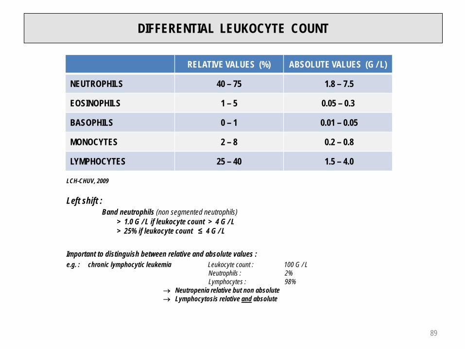
DIFFERENTIAL LEUKOCYTE COUNT
RELATIVE VALUES (%) ABSOLUTE VALUES (G / L)
NEUTROPHILS 40 – 75 1.8 – 7.5
EOSINOPHILS 1 – 5 0.05 – 0.3
BASOPHILS 0 – 1 0.01 – 0.05
MONOCYTES 2 – 8 0.2 – 0.8
LYMPHOCYTES 25 – 40 1.5 – 4.0
LCH-CHUV, 2009
Left shift :Band neutrophils (non segmented neutrophils)
> 1.0 G / L if leukocyte count > 4 G / L> 25% if leukocyte count ≤ 4 G / L
Important to distinguish between relative and absolute values :e.g. : chronic lymphocytic leukemia Leukocyte count : 100 G / L
Neutrophils : 2%Lymphocytes : 98%
→ Neutropenia relative but non absolute→ Lymphocytosis relative and absolute
89

MITOTIC POOL
STROMALCELLS STEM CELLS PROGENITOR
CELLSMYELOBLASTS
PROMYELOCYTESMYELOCYTES
POSTMITOTICPOOL
CIRCULATINGNEUTROPHILS
50%
MARGINATINGNEUTROPHILS
50%
TISSUES
BONE MARROW
6 -10 DAYS
BLOOD
6 -10 HOURS
SCF
IL-3
GM-CSF
IL-5G-CSF
METAMYELOCYTESBAND / SEGMENTED
NEUTROPHILS
IL-8
TISSUE MIGRATION
NEUTROPHIL GRANULOCYTESKINETICS
SCF : Stem Cell FactorIL : InterleukinCSF : Colony-Stimulating FactorG : GranulocyteM : Monocyte 90

ETIOLOGY OF NEUTROPHILIC LEUKOCYTOSIS (NEUTROPHILIA)(NEUTROPHIL COUNT > 7.5 G / L)
PHYSIOLOGICAL, USUALLY MODERATE
NeonateViolent exerciseMenstruationPregnancy
PATHOLOGICAL
Inflammatory process
Bacterial infection localized (abscess) or generalized (septicemia)CancerInflammatory arthritis
Tissue necrosis (myocardial infarction, pancreatitis, etc.)
Regenerative phase of acute blood loss or hemolytic anemia
Tobacco smoking, stress
Drugs (Steroids, G-CSF, GM-CSF, lithium)
Myeloproliferative neoplasms
91

TOXIC CHANGES OF NEUTROPHILS
Leukocytosis (leukocyte count > 10 G / L)
Neutrophilia (neutrophil count > 7.5 G / L)
Neutrophil left shift : band neutrophil count > 1.0 G / L (or > 25% if leukocyte count ≤ 4.0 G / L)
Coarse granules or neutrophils, toxic granules
Doehle bodies (basophilic cytoplasmic inclusions)
Cytoplasmic vacuoles
Myelocytosis (usually moderate)
Toxic changes are seen in inflammatory process (acute or chronic bacterial infection, cancer, inflammatory
arthritis and tissue necrosis)
Possible exceptions : neutropenia of salmonellosis, lymphocytosis of brucellosis and pertussis
92

MYELOCYTOSIS AND ERYTHROBLASTOSIS
DEFINITION
Presence in the peripheral blood of immature cells of neutrophilic lineage (metamyelocytes, myelocytes, promyelocytes) with or without erythroblasts
Erythroblasts Myelocytosis
Inflammatory process (bacterial infection, cancer, etc.1) – +
Rupture of bone marrow-blood barrier (skeletal cancer metastasis with bone marrow infiltration) + +
Chronic myelogenous leukemia – / + +++
Primary myelofibrosis + (+) + (+)
Regeneration phase after acute blood loss or hemolysis + to +++ +
Recovery from agranulocytosis, G-CSF, GM-CSF – + (+)
1 An important leukocytosis associated with toxic changes of neutrophils and myelocytosis is called leukemoid reaction
93

NEUTROPENIA
DEFINITIONSRELATIVE NEUTROPENIA : < 40%
ABSOLUTE NEUTROPENIA : < 1.8 G / L
AGRANULOCYTOSIS : < 0.5 G / L(major risk of infection)
CLASSIFICATION OF ABSOLUTE NEUTROPENIAS
PSEUDONEUTROPENIAExcess neutrophil margination (fasting patient, correction after meal)Splenic sequestration ("pooling") - Hypersplenism
TRUE NEUTROPENIA
Reduced production and / or excessive destruction / demand
94

TRUE NEUTROPENIA (1)REDUCED PRODUCTION
QUANTITATIVEBone marrow aplasia
Bone marrow infiltration
Bone marrow fibrosis
T-cell large granular lymphocytic leukemia
Cyclic neutropenia
Chronic ethnic or idiopathic neutropenia
QUALITITIVEVitamin B12 and / or folate deficiency
Myelodysplastic syndrome
95

TRUE NEUTROPENIA (2)REDUCED PRODUCTION AND / OR EXCESSIVE DESTRUCTION
INFECTIOUS NEUTROPENIA1
Viral (influenza, hepatitis, varicella, measles, rubeola, EBV, HIV)
Bacterial (salmonellosis, brucellosis, sepsis with Gram negative germs)
Parasitic (malaria)
IMMUNE NEUTROPENIAAlloimmune (neonatal neutropenia)
Autoimmune (disseminated lupus erythematosus, rheumatoid arthritis, drugs)
Immunoallergic
Drugs Mianserin (antidepressant), sulfasalazine, phenylbutazone (antiinflammatory agents), cotrimoxazole (antiinfective), metamizole (analgesic), carbamazepine (anticonvulsant), carbimazole (antithyroid drug)
1 Immune pathogenic mechanism possible
96

HEREDITARY MORPHOLOGICAL NEUTROPHIL ANOMALIES
PELGER-HUET ANOMALYNeutrophils with bilobate nucleus (not to be mistaken for neutrophil left shift !)Autosomal dominant anomaly1
MAY-HEGGLIN ANOMALYBasophilic cytoplasmic inclusions (RNA)2
Moderate thrombocytopenia with giant plateletsAutosomal dominant anomaly
ALDER-REILLY ANOMALYCoarse purple granules in neutrophils, monocytes and lymphocytesAutosomal recessive anomaly
CHEDIAK-HIGASHI SYNDROMEGiant granules in neutrophils, eosinophils, monocytes and lymphocytesNeutropenia (infection)Thrombocytopenia (hemorrhage)HepatosplenomegalyAutosomal recessive anomaly
1 Acquired variety in myelodysplastic syndrome : "pelgeroid" nuclei = pseudo-Pelger2 Döhle bodies
97
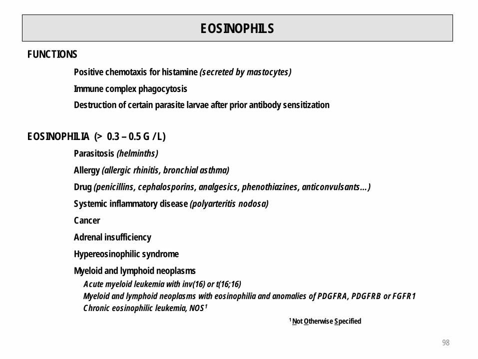
EOSINOPHILS
FUNCTIONSPositive chemotaxis for histamine (secreted by mastocytes)
Immune complex phagocytosisDestruction of certain parasite larvae after prior antibody sensitization
EOSINOPHILIA (> 0.3 – 0.5 G / L)Parasitosis (helminths)
Allergy (allergic rhinitis, bronchial asthma)
Drug (penicillins, cephalosporins, analgesics, phenothiazines, anticonvulsants…)
Systemic inflammatory disease (polyarteritis nodosa)
Cancer
Adrenal insufficiency
Hypereosinophilic syndrome
Myeloid and lymphoid neoplasmsAcute myeloid leukemia with inv(16) or t(16;16)Myeloid and lymphoid neoplasms with eosinophilia and anomalies of PDGFRA, PDGFRB or FGFR1Chronic eosinophilic leukemia, NOS1
1 Not Otherwise Specified
98

BASOPHILS / MASTOCYTES
DEFINITIONBlood : basophilic granulocytes
Tissues : tissue basophils or mastocytes
FUNCTIONS
Surface receptors for IgE Fc fragment"Bridging" effect of several IgE molecules by the specific allergen with degranulationand release of histamine (bronchospasm in asthma bronchiale), heparin and a chemotactic factor for eosinophils
BASOPHILIA (> 0.05 – 0.1 G / L)
Myeloproliferative neoplasm AllergyHypothyroidism
MASTOCYTOSIS (cf. Myeloproliferative neoplasms / WHO classification 2008 on page 118)
99

MONOCYTES / MACROPHAGES (1)FUNCTIONS
Chemotaxis, phagocytosis, killing
Antigen presentation to lymphocytes with help of HLA class I (T CD8 +) or class II(T CD4 +, B) molecules
Secretion Hydrolases (acid phosphatase)LysozymeComplement fractionsTumor Necrosis Factor (TNF)Interleukin-1 (IL-1)
Brain : FeverLiver : CRPNeutrophils : ActivationT lymphocytes : GM-CSF, G-CSF, M-CSF, IL-2-7NK lymphocytes : ActivationEndothelial cells : Proliferation, GM-CSF, M-CSF, IL-1, IL-5-7
Activation by γ-Interferon, TNF and GM-CSFCRP : C-Reactive ProteinIL : InterleukinCSF : Colony-Stimulating FactorG : GranulocyteM : Monocyte
100

MONOCYTES / MACROPHAGES (2)
ABSOLUTE MONOCYTOSIS (> 0.8 – 1.0 G / L)
REACTIVE
Infectious disease (tuberculosis, bacterial endocarditis, salmonellosis, brucellosis, malaria)
Recovery phase of bacterial infection
Recovery from agranulocytosis
Alcoholic hepatic disease
G-CSF or GM-CSF treatment
MALIGNANT
Chronic myelomonocytic leukemiaAcute myeloid leukemia with t(9;11), acute myelomonocytic leukemia, acute monocytic leukemia
MONOCYTOPENIAHairy cell leukemia
101

LYMPHOCYTES / LYMPHOID ORGANS
LYMPHOID ORGANS
Primary : Bone marrow (lymphoid stem cells : CFU-L, B-cell differentiation and maturation)
Thymus (T-cell differentiation and maturation, thymic selection)
Secondary : Lymph node
(B and T) Spleen
Digestive tract mucosa
Respiratory tract mucosa
PROPORTION OF B- AND T-LYMPHOCYTES IN BONE MARROW AND PERIPHERAL BLOOD
BONE MARROW PERIPHERAL BLOODB ≥ T T > B
CD8 > CD4 CD4 > CD8
102

B-LYMPHOCYTES
BONE MARROW
PRECURSORS : CFU-L CD34 +
PRO-B : CD34 +, TdT +, HLA-DR +EARLY PRE-B : Rearrangement of immunoglobulins genes (heavy chains
then light chains) CD19 and CD20 expression
PRE-B : Intracytoplasmic µ chains expression
IMMATURE B : Surface IgM expression
MIGRATION TO BLOOD AND SECONDARY LYMPHOID ORGANS → MATURE B CELLS (surface IgM and IgD expression)
103

STEPS OF B-LYMPHOCYTE MATURATION IN SECONDARY LYMPHOID ORGANS
PRE-B (intracytoplasmic µ chains)
B mature (surface Ig)
IMMUNOBLAST CENTROBLAST
LYMPHOPLASMACYTIC CELL*
PLASMA CELL *
CENTROCYTE
B MEMORY LYMPHOCYTE
* Plasmatic immunoglobulin (Ig) secretion
IgG IgA IgM IgD IgE
Molecular weight (x 1'000) 140 1601 (4002) 900 170 190
Sedimentation constant 7 S 7 S1 (11 S2) 19 S 6.5 S 8 S
Placental transfer Yes No No No No
Serum level (g / L) 8 – 12 1.4 – 4.0 0.5 – 1.9 0.03 – 0.4 0.0001
Half life (d) 21 7 5 2.8 2.3
Heavy chain γ (1-4) α (1-2) µ δ ε
Light chain κ or λ
Examples :IgG γ2κ2 or γ2λ 2IgM (µ2κ2)5 or (µ2λ2)5
(pentamers)
1 Serum IgA2 Secretory IgA
104

T-LYMPHOCYTES / THYMIC SELECTION
MEDULLARY PRECURSORS (CFU-L) CD34 +
MIGRATION TO THYMUSCORTICAL ZONE :
TCR expression (T-Cell Receptor), CD2, CD3
TCR gene rearrangement (γδ then αβ)Positive selection1 : amplification of CD4 + CD8 + thymocytes with affinity for " self " class I and II molecules of the HLA system
MEDULLARY ZONE :Negative selection1 : elimination of thymocytes with affinity for class I and II HLA molecules incontact with " self " antigens (clonal deletion)Expression of CD2, CD3, CD4 + CD8 - or CD4 - CD8 +
MIGRATION TO PERIPHERAL BLOOD AND SECONDARY LYMPHOID ORGANS
1 During positive and negative selections approximately 90% of T-lymphocytes (thymocytes) are eliminated through apoptosis (cell death)
105

B- AND T-LYMPHOCYTE DIFFERENTIATION MARKERS
B-LYMPHOCYTE DIFFERENTIATION T-LYMPHOCYTE DIFFERENTIATION
PRO- B EARLY PRE-B PRE-B B MATURE
Ig genes rearrangement (heavy chain κ, λ)
HLA-DR, TdT
CD34
CD19
CD20
CD10
cCD221
CD22
cIgM3
sIgM4
PRE-T EARLY T T CORTICAL T MATURE
TCR genes rearrangement (γδ, β, α)
TdT
CD7
CD2
CD5
CD1a
cCD32
CD3
CD4 + CD8
CD4 or CD8
1 cCD22 : intracytoplasmic CD222 cCD3 : intracytoplasmic CD33 cIgM : intracytoplasmic IgM4 sIgM : surface IgM 106

NK-LYMPHOCYTES (NATURAL KILLER LYMPHOCYTES)
Large granular lymphocytes (LGL variety)
CD3 - , CD8 + / - , CD16 +, CD56 + , CD57 + / - , absence of TCR
Cytotoxicity 1. Inhibited by the presence of surface receptors for HLA class I molecules expressed by
"self " cellsStimulated by reduced synthesis (or transport) of HLA class I molecules (virus infected cells, tumor cells)
2. CD16 + (Fc receptor) : binding of antibody to surface antigen → binding of a NK lymphocyte by the Fc, leading to activation
107

LYMPHOCYTES / IMMUNE RESPONSE (1)
IMMUNE RESPONSE (2)
Functionally, the adaptive immune system can be divided into two arms : cell-mediated and humoral immunity. B cells areresponsible for the humoral response. B cells interact directly with antigen (Ag) and then differentiate into antibody-secreting cells.T cells are responsible for the cell-mediated immunity. They recognize antigens as short antigen fragments presented on the surface ofantigen-presenting cells (APC)T cells exist as two main functional groups : the Helper T cells (Th), which respond to antigen by producing cytokines and thecytotoxic T cells (CTL) which respond to antigen by releasing cytotoxins. Depending on signals they receive from APC, the helper Tcells can differentiate into four main subsets, with distinct profile of cytokines (Th1, Th2, Th17 and iTreg)
IMMUNE RESPONSE (3)Figure reproduced with authorization of HSeT
108

LYMPHOCYTES / IMMUNE RESPONSE (2)
Th1 cells are required for defense against intracellular pathogens. They arecharacterized by the production of IFN-γ and IL-2. IFN-γ activates themicrobicidal activity of macrophages, stimulates B cells to produce antibodiesthat are involved in the opsonization and phagocytosis of particulate microbes,and enhances the development of long-term memory CD8 T cells.IL-2 increases the cytolytic activity of natural killer cells (CTL NK)
Th2 cells are required for defense against extracellular pathogens.They are characterized by the production of IL-4, IL-5 and IL-13. IL-4stimulates B cell proliferation and induces isotype class switch toIgG1 and IgE and so plays a role in IgE-dependent mast cell-mediated reactions. IL-5 acts largely on eosinophils. IL-13 ishomologous to IL-4 and induces many of the same functions,including inducing IgE isotype switching
Figures reproduced with authorization of HSeT109
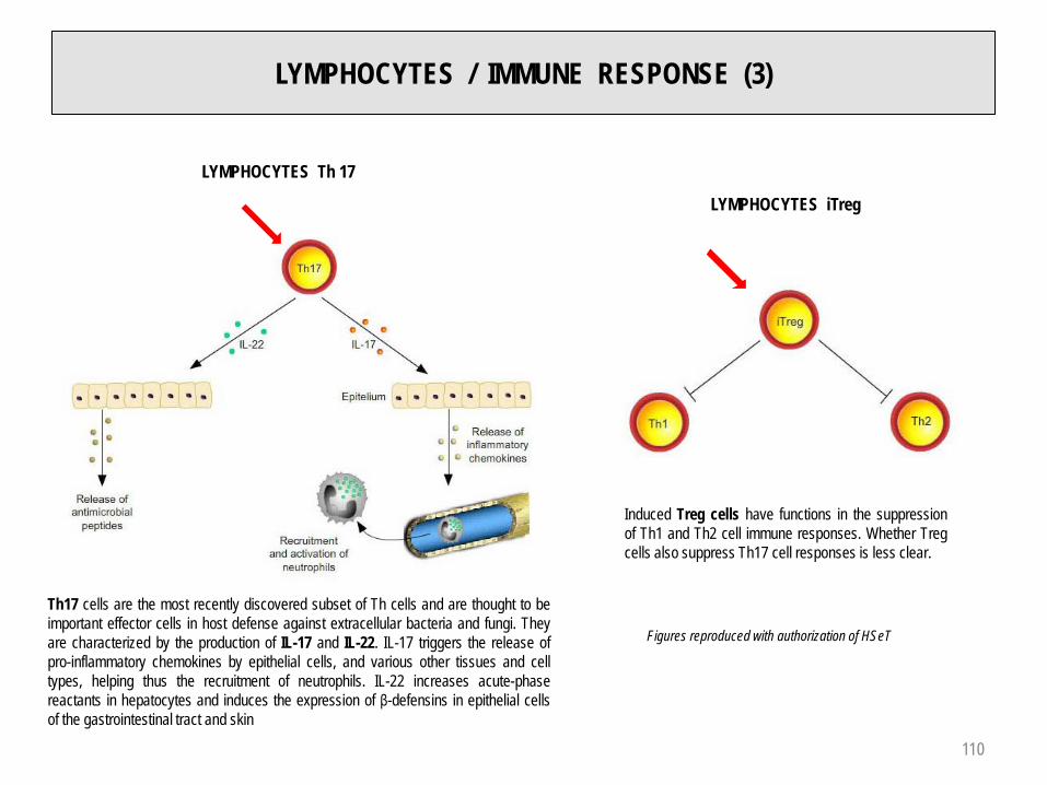
LYMPHOCYTES / IMMUNE RESPONSE (3)
LYMPHOCYTES Th 17
Th17 cells are the most recently discovered subset of Th cells and are thought to beimportant effector cells in host defense against extracellular bacteria and fungi. Theyare characterized by the production of IL-17 and IL-22. IL-17 triggers the release ofpro-inflammatory chemokines by epithelial cells, and various other tissues and celltypes, helping thus the recruitment of neutrophils. IL-22 increases acute-phasereactants in hepatocytes and induces the expression of β-defensins in epithelial cellsof the gastrointestinal tract and skin
LYMPHOCYTES iTreg
Induced Treg cells have functions in the suppressionof Th1 and Th2 cell immune responses. Whether Tregcells also suppress Th17 cell responses is less clear.
Figures reproduced with authorization of HSeT
110

LYMPHOCYTES / IMMUNE RESPONSE (4)CD 4 ET CD 8 CO-RECEPTORS OF T-LYMPHOCYTES
CD4 is a monomer that interacts via its two distal Ig domains (D1 and D2) with the b2 domain of MHC class II
CD8 is a dimer (either homodimer αα or hetedimer αβ) that interacts via its α chain with the α3 domain of MHC class I
Figures reproduced with authorization of HSeTAPC : Antigen Presenting Cell111

LYMPHOCYTOSIS / LYMPHOPENIA
LYMPHOCYTOSISRELATIVE : > 40%ABSOLUTE : > 4.0 G / L
REACTIVEInfection : viral
bacterial (pertussis, tuberculosis, brucellosis, syphilis)ThyrotoxicosisHyposplenism
MALIGNANTLymphoid leukemia
ABSOLUTE LYMPHOPENIA < 1.5 G / L
ACQUIREDHIV, Hodgkin lymphoma, chemotherapy, radiotherapy, steroids, ATG (Antithymocyte Globulin), autoimmune disorder
CONGENITALSCID (Severe Combined Immune Deficiency)
IDIOPATHIC
112

PLASMACYTOSIS / MONONUCLEOSIS SYNDROME
PLASMACYTOSISREACTIVE : Rubella (German measles)
Other viral infectionMALIGNANT : Plasma cell leukemia
Plasma cell myeloma
MONONUCLEOSIS SYNDROMEAbsolute lymphocytosis with polymorphic lymphocytes (T-lymphocytes reactive to theinfected B-lymphocytes)
Etiology : EBV1 (infectious mononucleosis)Lymphadenopathy 100%Fatigue 90%Pharyngitis syndrome 80%Splenomegaly > 50%Possibly hemolytic anemia and / or autoimmune thrombocytopenia, agranulocytosis,cardiac / neurological / respiratory complications, splenic rupture
CMV (cytomegalovirus infection, frequently promoted by immunosuppression) HIV (primary infection)Other virus (e.g. hepatitis)Toxoplasmosis
1 Also involved in the pathogenesis of certain lymphoid neoplasms (African Burkitt, Hodgkin lymphoma, lymphoid neoplasms + HIV)113
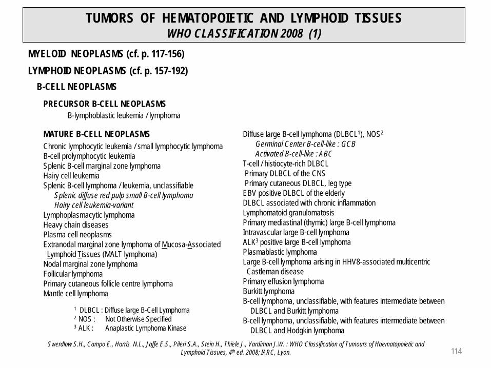
TUMORS OF HEMATOPOIETIC AND LYMPHOID TISSUESWHO CLASSIFICATION 2008 (1)
MYELOID NEOPLASMS (cf. p. 117-156)LYMPHOID NEOPLASMS (cf. p. 157-192)
B-CELL NEOPLASMS
MATURE B-CELL NEOPLASMSChronic lymphocytic leukemia / small lymphocytic lymphomaB-cell prolymphocytic leukemiaSplenic B-cell marginal zone lymphomaHairy cell leukemiaSplenic B-cell lymphoma / leukemia, unclassifiable
Splenic diffuse red pulp small B-cell lymphomaHairy cell leukemia-variant
Lymphoplasmacytic lymphomaHeavy chain diseasesPlasma cell neoplasmsExtranodal marginal zone lymphoma of Mucosa-AssociatedLymphoid Tissues (MALT lymphoma)
Nodal marginal zone lymphomaFollicular lymphomaPrimary cutaneous follicle centre lymphomaMantle cell lymphoma
Diffuse large B-cell lymphoma (DLBCL1), NOS2
Germinal Center B-cell-like : GCBActivated B-cell-like : ABC
T-cell / histiocyte-rich DLBCLPrimary DLBCL of the CNSPrimary cutaneous DLBCL, leg typeEBV positive DLBCL of the elderlyDLBCL associated with chronic inflammationLymphomatoid granulomatosis Primary mediastinal (thymic) large B-cell lymphomaIntravascular large B-cell lymphomaALK3 positive large B-cell lymphomaPlasmablastic lymphomaLarge B-cell lymphoma arising in HHV8-associated multicentricCastleman disease
Primary effusion lymphomaBurkitt lymphomaB-cell lymphoma, unclassifiable, with features intermediate between
DLBCL and Burkitt lymphomaB-cell lymphoma, unclassifiable, with features intermediate between
DLBCL and Hodgkin lymphomaSwerdlow S.H., Campo E., Harris N.L., Jaffe E.S., Pileri S.A., Stein H., Thiele J., Vardiman J.W. : WHO Classification of Tumours of Haematopoietic and
Lymphoid Tissues, 4th ed. 2008; IARC, Lyon.
1 DLBCL : Diffuse large B-Cell Lymphoma2 NOS : Not Otherwise Specified 3 ALK : Anaplastic Lymphoma Kinase
PRECURSOR B-CELL NEOPLASMSB-lymphoblastic leukemia / lymphoma
114

TUMORS OF HEMATOPOIETIC AND LYMPHOID TISSUES WHO CLASSIFICATION 2008 (2)
T-CELL AND NK-CELL NEOPLASMSPRECURSORS T-CELL NEOPLASMS
T-cell lymphoblastic lymphoma / leukemia
MATURE T-CELL AND NK-CELL NEOPLASMST-cell prolymphocytic leukemiaT-cell large granular lymphocytic leukemiaChronic lymphoproliferative disorders of NK-cellsAggressive NK-cell leukemiaSystemic EBV-positive T-cell lymphoproliferative disorders of childhoodHydroa vacciniforme-like lymphomaAdult T-cell lymphoma / leukemia Extranodal NK / T-cell lymphoma, nasal typeEnteropathy-associated T-cell lymphomaHepatosplenic T-cell lymphomaSubcutaneous panniculitis-like T-cell lymphomaMycosis fungoidesSézary syndromePrimary cutaneous CD30 positive T-cell lymphoproliferative disordersPrimary cutaneous gamma-delta T-cell lymphomaPeripheral T-cell lymphoma not otherwise specifiedAngioimmunoblastic T-cell lymphomaAnaplastic large cell lymphoma (ALCL), ALK1 positiveAnaplastic large cell lymphoma (ALCL), ALK1 negative
HODGKIN LYMPHOMA (HODGKIN DISEASE) (cf. p. 189-192)
Swerdlow S.H., Campo E., Harris N.L., Jaffe E.S., Pileri S.A., Stein H., Thiele J., Vardiman J.W. : WHO Classification of Tumours of Haematopoietic and Lymphoid Tissues, 4th ed. 2008; IARC, Lyon.
1ALK : Anaplastic Lymphoma Kinase
115

TUMORS OF HEMATOPOIETIC AND LYMPHOID TISSUES WHO CLASSIFICATION 2008 (3)
IMMUNODEFICIENCY-ASSOCIATED LYMPHOPROLIFERATIVE DISORDERS
Lymphoproliferative diseases associated with primary immune disordersLymphomas associated with HIV infectionPost-Transplant Lymphoproliferative Disorders (PTLD)
Early lesionsPlasmacytic hyperplasia Infectious mononucleosis-like PTLD
Polymorphic PTLDMonomorphic PTLD (criteria for one of the B-cell or T / NK-cell neoplasms of immunocompetent host)Classical Hodgkin lymphoma-type PTLD
Other iatrogenic immunodeficiency-associated lymphoproliferative disorders
HISTIOCYTIC AND DENDRITIC CELL NEOPLASMS
Histiocytic sarcomaLangerhans cell histiocytosisLangerhans cell sarcomaInterdigitating dendritic cell sarcomaFollicular dendritic cell sarcomaFibroblastic reticular cell tumorIndeterminate dendritic cell tumorDisseminated juvenile xanthogranuloma
Swerdlow S.H., Campo E., Harris N.L., Jaffe E.S., Pileri S.A., Stein H., Thiele J., Vardiman J.W. : WHO Classification of Tumours of Haematopoietic and Lymphoid Tissues, 4th ed. 2008; IARC, Lyon. 116

MYELOID NEOPLASMS
MYELOPROLIFERATIVE NEOPLASMS (MPN)
MYELOID AND LYMPHOID NEOPLASMS WITH EOSINOPHILIA AND ANOMALIES OF PDGFRA, PDGFRB OR FGFR1
MYELODYSPLASTIC SYNDROMES (MDS)
MYELODYSPLASTIC / MYELOPROLIFERATIVE NEOPLASMS (MDS / MPN)
ACUTE MYELOID LEUKEMIAS (AML) AND RELATED PRECURSOR NEOPLASMS
ACUTE LEUKEMIAS OF AMBIGUOUS LINEAGE
STEM CELL PROLIFERATION AND DIFFERENTIATIONIN MYELOID NEOPLASMS
STEM CELLGenetic mutationHumoral factors
Cellular interactions
Proliferation Differentiation
Myeloproliferative neoplasms + +Myelodysplastic syndromesMyelodysplastic / myeloproliferative neoplasms ± ±Acute myeloid leukemias (AML) and related precursor neoplasmsAcute leukemias of ambiguous lineage
+ –117

MYELOPROLIFERATIVE NEOPLASMS
GENERAL CHARACTERISTICSStem cell somatic mutation upstream from the myeloid precursor cellProliferation and maturationIncrease in peripheral blood of cells arising from one or more lineagesMyeloid metaplasia (extramedullary hematopoiesis)Frequent bone marrow fibrosisPlatelet function disordersHyperuricemiaPossible transformation in acute leukemia
WHO CLASSIFICATION 2008Polycythemia VeraChronic myelogenous leukemia (CML) BCR-ABL 1 +Essential thrombocythemiaPrimary myelofibrosisChronic neutrophilic leukemiaChronic eosinophilic leukemia, NOS1
Mastocytosis Cutaneous mastocytosisSystemic mastocytosisMast cell leukemiaMast cell sarcomaExtracutaneous mastocytoma
Myeloproliferative neoplasm, unclassifiable1 NOS : Not Otherwise Specified
118

POLYCYTHEMIA VERA (1)
SYMPTOMS AND CLINICAL SIGNSFacial erythrocyanosis Water pruritusEpigastralgiaHyperviscosity (thromboembolic manifestations, headache, dizziness paresthesias)Splenomegaly
MAJORA1 Hb > 185 g / L (men), > 165 g / L (women)1 or
increased isotopic RBC mass > 25% of predicted value
A2 Presence of JAK2V617F or other functionally similar mutation such as JAK2 exon 12 mutation
MINOR
B1Bone marrow biopsy showing hypercellularity for age with trilineage growth (panmyelosis) with prominent erythroid, granulocytic and megakaryocytic hyperplasia
B2 Serum erythropoietin level below the reference range for normal
B3 Endogenous erythroid colony formation in vitro (without EPO)
PV established if :
A1 + A2 and one minor criterionor :A1 and 2 minor criteria
Swerdlow S.H., Campo E., Harris N.L., Jaffe E.S., Pileri S.A., Stein H., Thiele J., Vardiman J.W. : WHO Classification of Tumours of Haematopoietic and Lymphoid Tissues, 4th ed. 2008; IARC, Lyon.
1 Hemoglobin or hematocrit > 99th percentile of method-specific reference range for age, sex, altitude of residence or hemoglobin > 170 g / L in men, > 150 g / L in women if associated with a documented and sustained increase of at least 20 g / L from an individual's baseline value that cannot be attributed to correction of iron deficiency
DIAGNOSTIC CRITERIA
119

POLYCYTHEMIA VERA (2)
COMPLICATIONSThromboembolicHemorrhagicEvolution to myelofibrosis, ∼10% (post-polycythemic phase), cf. page 128Transformation in myelodysplastic syndrome or acute leukemia (> 10% after treatment withcytotoxic drugs)
PROGNOSISMedian survival : > 10 years
TREATMENT :Phlebotomies
Hydroxyurea, Pipobroman32P : age > 70 years (increased risk of leukemic transformation !)
120

DIFFERENTIAL DIAGNOSIS OF ERYTHROCYTOSISRBC VOLUME AND PLASMA VOLUME
PV45 mL / kg
RCV30 mL / kg
RCV
PV
RCV
PV
RCV
PV
NORMAL TRUEERYTHROCYTOSIS
RELATIVEERYTHROCYTOSIS
MICRO-ERYTHROCYTOSIS
Thalassemia minorPV : PLASMA VOLUMERCV : RED CELL VOLUME
DehydrationContraction of plasma volume
Gaisböck syndrome (stresspseudoerythrocytosis)
Male prevalenceSedentary way of lifeLight obesityTobacco smokingHyperlipidemiaHyperproteinemiaThromboembolic risk
121

DIFFERENTIAL DIAGNOSIS OF TRUE ERYTHROCYTOSIS (1)
PRIMARYERYTHROCYTOSIS
Congenital EPO receptor mutationEPO ↓
Acquired Anomaly of erythroid precursors(Polycythemia Vera)
SECONDARYERYTHROCYTOSIS
CongenitalAbsence of erythroid precursors anomalyMutations impairing the system of tissue oxygenation sensingHigh O2-affinity hemoglobins
EPO ↑or normal
Acquired Appropriate or abnormal EPO secretion
HIF : Hypoxia Inducible Factor pVHL : von Hippel-Lindau protein ProH : Prolin-HydroxylaseU : Ubiquitin VEGF : Vascular Endothelial Growth Factor PDGF : Platelet-Derived Growth FactorTGF : Tissue Growth Factor
SENSING PROCESS OF TISSULAR OXYGENATION
In state of normal oxygenation HIF-α protein is rapidely degraded by the action of prolin-hydroxylase and von Hippel-Lindau protein, followed by ubiquitination et destruction in the proteasome
In hypoxic state HIF-α degradation is blocked. The protein is activated by dimerization with HIF-β. The complex acts as a promoter of various genes involved in synthesis of growth factors like EPO
Modified after McMullin M.F. : EHA Hematology Education, 2009; 3 : 238-241. 122

DIFFERENTIAL DIAGNOSIS OF TRUE ERYTHROCYTOSIS (2)
PRIMARY ERYTHROCYTOSIS
CONGENITALMutation of EPO1 receptor
ACQUIREDPolycythemia Vera
SECONDARY ERYTHROCYTOSISCONGENITAL
Mutation of VHL2 gene (Chuvash erythrocytosis)Mutation of PHD23
Mutation of HIF-2-α4
O2 high-affinity hemoglobins2,3-diphosphoglyceromutase deficiency
ACQUIREDAppropriate EPO1 production
Central hypoxiaChronic pulmonary disorder, cardio-pulmonary right-left shunt, CO intoxication, chronicsmoking, hypoventilation syndromes incl.sleep apnea, prolonged stay at high altitude
Local renal hypoxiaRenal artery stenosis, terminal renal failure,hydronephrosis, polycystic kidneys, post renal transplantation erythrocytosis
Abnormal EPO1 productionTumors : cerebellar hemangioblastoma, meningioma, parathyoid carcinoma / adenoma, hepatocellular carcinoma, renal cell carcinoma, pheochromocytoma, uterine leiomyoma
Drugs : androgens
Exogenous EPO1 applicationTherapeutical indicationIllicit application (doping !)
IDIOPATHIC ERYTHROCYTOSIS
1 EPO : Erythropoietin2 VHL : Von Hippel-Lindau (recessive mutations)3 PHD2 : Prolyl-Hydroxylase Domain (dominant mutations)4 HIF : Hypoxia Inducible Factor (dominant mutations)
Modified after McMullin M.F. : EHA Hematology Education, 2009; 3 : 238-241. 123

CHRONIC MYELOGENOUS LEUKEMIA (CML) (1)
SYMPTOMS AND CLINICAL FEATURESFortuitous diagnosis - asymptomatic patientDigestive symptoms (abdominal heaviness, bloating)SplenomegalyThrombosisHemorrhageLeucostasis (CML with very high leukocyte count)
BLOOD PICTURE Leukocytosis with neutrophiliaNeutrophil left shiftMyelocytosis (20-50%)BasophiliaFrequent thrombocytosisLow leukocyte alkaline phosphatase score (obsolete test)
CYTOGENETIC Philadelphia chromosome (Ph) = t(9;22)(q34;q11.2) : 90-95% of casesBCR-ABL 1 fusion gene : 100% of cases
124

CHRONIC MYELOGENOUS LEUKEMIA (CML) (2)
COURSE IN 3 PHASES
CHRONIC : 4-5 years
ACCELERATION : < 6-8 monthsPersistent increase of WBC counts (> 10 G / L) and / or of spleen size unresponsive to therapyPersistent thrombocytosis (> 1'000 G / L) uncontrolled by therapyPersistent thrombocytopenia (< 100 G / L) unrelated to therapyClonal cytogenetic evolutionBasophils ≥ 20% in peripheral bloodBlast cells : 10-19% in peripheral blood and / or of the nucleated cells of bone marrowOften hypercellular bone marrow, morphological signs of myelodysplasia Large clusters or sheets of small, abnormal megakaryocytes + reticulin or collagen fibrosis
TRANSFORMATIONBlast cells : ≥ 20% of peripheral blood cells and / or of the nucleated bone marrow cellsExtramedullary blast proliferation
125

CHRONIC MYELOGENOUS LEUKEMIA (CML) (3)
TREATMENTImatinib mesylate (Glivec) : Tyrosine Kinase inhibitor (TK)
proliferation and apoptosis induction of the BCR-ABL 1 + cell lineagesIn case of primary or secondary drug resistance : Dasatinib (Sprycel), Nilotinib (Tasigna) (other TK inhibitors)
Hydroxyurea (HU)
α-Interferon (α-IFN)
Allogeneic hemopoietic stem cell / bone marrow transplantation
AGE BASED THERAPEUTIC SELECTION
< 60 years : in case of insufficient response toTK inhibitor allogeneic hemopoietic stem cell / bone marrow transplantation (only curative treatment). Probability of HLA compatible sibling donor 20-30%.Possible graft from unrelated donor. 5 year survival rate : 50-70%. Relapse after transplantationtreated by infusion of donor lymphocytes (GVL effect1)
> 60 years : and for patient not suitable for transplantation : Imatinib mesylate, α-Interferon (+ ARA-C2),Hydroxyurea
1 GVL : Graft-Versus-Leukemia 2 ARA-C : Cytosine Arabinoside
126

ESSENTIAL THROMBOCYTHEMIA (1)
SYMPTOMS AND CLINICAL FEATURESArterial or venous thrombosisHemorrhage through thrombopathyErythromelalgiaSplenomegaly (< 50%)
DIAGNOSTIC CRITERIA
1 Sustained platelet count ≥ 450 G / L1
2Bone marrow biopsy : proliferation mainly of megakaryocytic lineagewith increased numbers of enlarged mature megakaryocytes No significant increase or left-shift of neutrophil granulopoiesis or erythropoiesis
3Exclusion of :Polycythemia Vera2, primary myelofibrosis3, BCR-ABL1 positive CML4,myelodysplastic syndrome5 or other myeloid neoplasm
4JAK2V617F mutation present or other clonal markerIn absence of JAK2V617F mutation, exclusion of reactive thrombocytosis6
1 Sustained during the work-up process2 Requires failure of iron replacement therapy to increase Hb level to
PV range if decreased serum ferritinExclusion of PV based on Hb and Hct levels. Measure of RBCmass not required
3 Absence of relevant reticulin fibrosis, collagen fibrosis, peripheralblood leukoerythroblastosis or hypercellular marrow with megakaryocyte morphology typical for primary myelofibrosis(small to large megakaryocytes in dense clusters with aberrant nuclear / cytoplasmic ratio and hyperchromatic, bulbous or irregularly folded nuclei)
4 Absence of BCR-ABL 15 Absence of dyserythropoiesis and dysgranulopoiesis6 Exclusion of secondary thrombocytosis (cf. page 129)
(The presence of a condition associated with secondary thrombocytosis may not exclude the diagnosis of ET if the first 3 criteria are met)
DIAGNOSIS REQUIRES MEETING ALL 4 CRITERIA
Swerdlow S.H., Campo E., Harris N.L., Jaffe E.S., Pileri S.A., Stein H., Thiele J., Vardiman J.W. : WHO Classification of Tumours of Haematopoietic and Lymphoid Tissues, 4th ed. 2008; IARC, Lyon. 127

ESSENTIAL THROMBOCYTHEMIA (2)POSSIBLE COURSE
Polycythemia VeraPrimary myelofibrosisAcute leukemia (3-10%)
TREATMENTHydroxyureaPipobromanAnagrelide (could potentially favor evolution to myelofibrosis)Aspirin (platelet antiaggregant)
Diagnostic criteria for post-PV and post-ET myelofibrosis (MF)
REQUIRED CRITERIA
1 Documentation of a previous diagnosis of WHO-defined (2008) PV or ET
2 Bone marrow fibrosis grade 2-3 (on 0-3 scale). See page 131
ADDITIONALCRITERIA(2 required)
1Post-PV MF : Anemia1 or sustained loss of either phlebotomy alone or cytoreductive treatment requirement for erythrocytosisPost-ET MF : Anemia1 or ≥ 20 g / L decrease from baseline hemoglobin level
2 Leukoerythroblastic peripheral blood picture
3 Increasing palpable splenomegaly of > 5cm from baseline (distance from the left costal margin) or newly palpable splenomegaly
4 Post-ET MF : Increased LDH
5 Development of > 1 of 3 constitutional symptoms : > 10% weight loss in 6 months, night sweats, unexplained fever (> 37.5°C)
1 Below the reference range for appropriate age, gender and altitude
Swerdlow S.H., Campo E., Harris N.L., Jaffe E.S., Pileri S.A., Stein H., Thiele J., Vardiman J.W. : WHO Classification of Tumours of Haematopoietic and Lymphoid Tissues, 4th ed. 2008; IARC, Lyon. 128

DIFFERENTIAL DIAGNOSIS OF THROMBOCYTOSIS
DEFINITIONPlatelet count > 350 - 400 G / L
CAUSE OF ERRORImportant RBC microcytosis, presence of numerous schistocytes
CLASSIFICATIONPRIMARY THROMBOCYTOSIS
Myeloproliferative neoplasm (cf. pages 118-132)Essential thrombocytosis, Polycythemia Vera, chronic myelogenous leukemia, primary myelofibrosis
Myelodysplastic syndrome (cf. pages 134-141)5q- syndrome
SECONDARY THROMBOCYTOSISIron deficiency Metastatic cancerSplenectomy, asplenia1 Lymphoid neoplasmSurgery Acute phase / regeneration of acute hemorrhageInfection, inflammation or hemolysisAutoimmune disorder
1 Presence of Howell-Jolly bodies in RBC129

PRIMARY MYELOFIBROSIS (1)DIAGNOSIS
MAJORCRITERIA
1
Proliferation of atypical megakaryocytes1 with either reticulinand / or collagen fibrosis or :In absence of significant reticulin fibrosis, megakaryocytechanges + increased marrow cellularity with granulocytic proliferation and often decreased erythropoiesis (i.e. prefibrotic cellular-phase disease)
2 Exclusion of : PV2, BCR-ABL1 positive CML3, MDS4 or othermyeloid neoplasms
3
Presence of JAK2V617F mutation or other clonal marker (e.g. MPL W515K/L) orIn absence of clonal marker, exclusion of bone marrow fibrosis or changes secondary to infection, autoimmune disorder orother chronic inflammatory condition, hairy cell leukemia or other lymphoid neoplasm, metastatic malignancy or toxic (chronic) myelopathy5
MINORCRITERIA
1 Leukoerythroblastosis
2 Increased serum lactate dehydrogenase (LDH) level
3 Anemia6
4 Splenomegaly6
1 Small to large megakaryocytes in dense clusters with aberrant nuclear / cytoplasmic ratio and hyperchromatic, bulbous, or irregularly folded nuclei
2 Requires failure of iron replacement therapy toincrease Hb level to the PV range if ferritin level isdecreased. Exclusion of PV is based on Hb and Hctlevels. RBC mass measure not required
3 Absence of BCR-ABL1
4 Absence of dyserythropoiesis and dysgranulopoiesis
5 Conditions associated with reactive myelofibrosis donot exclude PMF. Diagnosis to be considered if othercriteria are met
6 Degree of anomaly borderline or marked
DIAGNOSIS : ALL 3 MAJOR + 2 MINOR CRITERIA
Swerdlow S.H., Campo E., Harris N.L., Jaffe E.S., Pileri S.A., Stein H., Thiele J., Vardiman J.W. : WHO Classification of Tumours of Haematopoietic and Lymphoid Tissues, 4th ed. 2008; IARC, Lyon.
130

PRIMARY MYELOFIBROSIS (2)
MF - 0 Scattered linear reticulin with no intersections (cross-overs), corresponding to normal bone marrow
MF - 1 Loose network of reticulin with many intersections, especially in perivascular areas
MF - 2 Diffuse and dense increase in reticulin with extensive intersections, occasionally with focal bundles of collagen and / or focal osteosclerosis
MF - 3 Diffuse and dense increase in reticulin with extensive intersections and coarse bundles of collagen, often associated with osteosclerosis
BLOOD COUNT : RBC, WBC and platelet counts in relation with disease stageTear drop RBC (dacryocytes)Erythroblastosis and myelocytosisPlatelet anisocytosis
COMPLICATIONS : Splenic infarctionInfection (neutropenia)Hemorrhage (thrombocytopenia and platelet function disorder)Acute leukemia (5-30%)
PROGNOSIS : Mean survival : 2-15 years (depends of the stage in which PMF is first diagnosed)
TREATMENT : No treatment ("wait and watch")HydroxyureaTransfusion supportSectorial splenic radiotherapy Splenectomy
SEMIQUANTITATIVE GRADING OF BONE MARROW FIBROSIS (MF)
131

CHRONIC NEUTROPHILIC LEUKEMIA
1 Peripheral blood : WBC ≥ 25 G / L, neutrophils > 80% WBC, immature granulocytes < 10% WBC, myeloblasts < 1% WBC
2 Bone marrow : percentage and number of neutrophilic granulocytes increased, normal maturation, myeloblasts < 5% of nucleated marrow cells, megakaryocytes normal or left shifted
3 Hepatosplenomegaly
4 No cause of physiological neutrophilia. If present, demonstration of clonality of myeloid cells
5 No BCR-ABL1 fusion gene, no rearrangement of PDGFRA, PDGFRB, FGFR1
6 No evidence of other myeloproliferative neoplasm, or myelodysplastic syndrome or myelodysplastic / myeloproliferative neoplasm. Monocytes < 1 G / L
CHRONIC EOSINOPHILIC LEUKEMIA, NOS1
1 Eosinophilia ≥ 1.5 G / L
2 No BCR-ABL1 fusion gene or other myeloproliferative neoplasm or myelodysplastic / myeloproliferative neoplasm
3 No FIP1L1-PDGFRA fusion gene (or other rearrangement of PDGFRA), no rearrangement of PDGFRB or FGFR1
4 Blast cell count in peripheral blood and bone marrow < 20%, no inv(16)(p13.1q22), t(16;16)(p13.1;q22), no other feature diagnostic of acute myeloid leukemia (AML)
5 Presence of a clonal or molecular genetic abnormality or blasts > 2% in PB or > 5% in bone marrow
1 If these criteria are not met, the diagnosis may be reactive eosinophilia, idiopathic hypereosinophilia or idiopathic hypereosinophilicsyndrome (HES). (See page 98 ) 1NOS : Not Otherwise Specified 132

MYELOID AND LYMPHOID NEOPLASMS WITH EOSINOPHILIA AND ANOMALIES OF PDGFRA, PDGFRB OR FGFR1
MYELOID AND LYMPHOID NEOPLASMS WITH PDGFRA REARRANGEMENT1 Myeloproliferative neoplasm with prominent eosinophilia
2 Presence of FIP1L1-PDGFRA fusion gene
Acute myeloid leukemia and lymphoblastic leukemia / lymphoma with eosinophilia and FIP1L1-PDGFRA are also assigned to this category. If molecular analysis is not available, diagnosis is suspected if : 1) Ph-negative myeloproliferative neoplasm with features of chronic eosinophilic leukemia; 2) splenomegaly; 3) high level of vitamin B12; 4) increase of serum tryptase; 5) increase of BM mast cells
Tyrosine Kinase activity : disease is responsive to TK- inhibitors (Imatinib mesylate)
MYELOID NEOPLASMS WITH PDGFRB REARRANGEMENT1 Myeloproliferative neoplasm often with prominent eosinophilia, sometimes neutrophilia or monocytosis
2 Presence of t(5;12)(q31~q33;p12) or variant translocation. Demonstration of ETV6-PDGFRB fusion gene or of rearragement of PDGFRB
Hematological features : chronic eosinophilic leukemia, chronic basophilic leukemia, chronic myelomonocytic leukemia with / without eosinophilia, Ph-negative chronic myeloid leukemia with eosinophilia, myelodysplastic / myeloproliferative neoplasm with eosinophilia
MYELOID AND LYMPHOID NEOPLASMS WITH FGFR1 ANOMALIES
1Myeloproliferative neoplasm with prominent eosinophilia and sometimes neutrophilia or monocytosis or acute myeloid leukemia or precursor T- or B-cell lymphoblastic leukemia / lymphoma (often associated with peripheral blood or bone marrow eosinophilia)
2 Presence of t(8;13)(p11;q12) or variant translocation with FGFR1 rearrangement in myeloid cells, lymphoblasts or both
133

MYELODYSPLASTIC SYNDROMES (MDS)GENERAL FEATURES
Somatic mutation of a hemopoietic stem cell upstream of myeloid precursor cells
Myelodyplasia (dysmyelopoiesis) : Proliferation + / -
Maturation + / -
Apoptosis +
Peripheral blood with 1-3 cytopenia(s)
WHO classification considering :Presence of signs of dysplasia affecting only one ("unilineage") or morecell lineages ("multilineage")Blast cells in peripheral blood or bone marrow : < 20%
Presence or absence of Auer rods
Presence or absence of ring sideroblasts : < 15% or ≥ 15% (bone marrow)
Peripheral blood monocytosis < 1 G / L
Possible transformation in acute leukemia
134

MYELODYSPLASIA
NON CLONAL CLONAL
Vitamin B12 deficiency
Folate deficiency
Chemotherapy, G-CSF
Arsenic
Parvovirus B19
HIV
Myelodysplastic syndrome
Paroxysmal NocturnalHemoglobinuria
(PNH)
Primary or"de novo"
Therapy-related MDS (t-MDS1)Radiotherapy
ChemotherapySolvents, agricultural and industrial
chemicalsSome inherited hematological
disorders
MYELODYSPLASIA
1 In WHO classification 2008 included in separate category under :Therapy-related myeloid neoplasms
135

MORPHOLOGICAL SIGNS OF MYELODYSPLASIADYSMYELOPOIESIS
PERIPHERAL BLOOD BONE MARROW
Dyserythropoiesis
Macrocytosis (frequent)AnisocytosisPoikilocytosis
AnisochromasiaCoarse basophilic granules
NuclearMegaloblastic changesNuclear budding, internuclear bridgingKaryorrhexis, hyperlobation
CytoplasmicVacuolizationRing Sideroblasts (RS)Periodic acid-Schiff (PAS) staining +
Dysgranulopoiesis
Small or unusually large sizePseudo-Pelger
Irregular hypersegmentationDecreased granules or agranularityPseudo Chediak-Higashi granules
Auer rods
Dysmegakaryopoiesis(platelets)
Giant plateletsLack of granules
MicromegakaryocytesHypolobated nuclei
Multinucleated megakaryocytes
136

CLASSIFICATION OF MDS PERIPHERAL BLOOD AND BONE MARROW FEATURES
DISEASE BLOOD FEATURES BONE MARROW FEATURES
Refractory Cytopenias with Unilineage Dysplasia (RCUD) : RA, RN, RT1
Unicytopenia (rarely bicytopenia)no or rare blasts (< 1%)2
Unilineage dysplasia : ≥ 10% of cells in one myeloid lineage; blasts < 5%Ring Sideroblasts (RS) : < 15%
Refractory Anemia with Ring Sideroblasts (RARS)
Anemiano blasts
Erythroid dysplasia onlyRing Sideroblasts ≥ 15%, blasts < 5%
Refractory Cytopenia with MultilineageDysplasia (RCMD)
Cytopenia(s), no or rare blasts (< 1%)2
no Auer rodsmonocytes < 1 G / L
Dysplasia in ≥ 10% of cells in ≥ 2 myeloid lineages, blasts < 5%, no Auer rodsRing Sideroblasts ± 15%
Refractory Anemia with Excess Blasts-1(RAEB-1)
Cytopenia(s), blasts : < 5%, no Auer rods monocytes < 1 G / L
Uni- or multilineage dysplasia, blasts : 5-9% no Auer rods
Refractory Anemia with Excess Blasts-2(RAEB-2)
Cytopenia(s), blasts : 5-19%, Auer rods ±3
monocytes < 1 G / LUni- or multilineage dysplasia blasts : 10-19%, Auer rods ±3
Myelodysplastic Syndrome - Unclassified (MDS-U)
Cytopeniasblasts : ≤ 1%
Evident dysplasia in less than 10% of cells in one or more myeloid cell lines with MDS cytogenetic anomaly, blasts < 5%
Myelodysplastic Syndrome associated with isolated del(5q)
Anemia,normal or increased platelet count no or rare blasts (< 1%)
Normal or increased megakaryocytes with hypolobulated nuclei, blasts : < 5%, no Auer rods, isolated del(5q)
1 RA : Refractory Anemia; RN : Refractory Neutropenia; RT : Refractory Thrombocytopenia2 If bone marrow blast percentage < 5%, but 2-4% blasts are present in the blood, the diagnostic is RAEB-1. RCUD and RCMD with 1% blasts in blood are classified as MDS-U3 Cases with Auer rods and < 5% blasts in blood and < 10% in bone marrow are classified as RAEB-2
137

DIFFERENTIAL DIAGNOSIS OF MYELODYSPLASTIC SYNDROME AND ACUTE MYELOID LEUKEMIA
IMPORTANCE OF BONE MARROW ERYTHROBLASTS PERCENTAGE
ERYTHROBLASTS (in % of total nucleated bone marrow cells)
< 50% ≥ 50%Blasts in % of total nucleated bone
marrow cellsBlasts in % of non erythroid nucleated bone marrow cells
≥ 20% < 20% < 20% ≥ 20%
AML MDS MDS AML
AML : Acute Myeloid Leukemia MDS : Myelodysplastic Syndrome
ANOMALIES RELATED TO MYELODYSPLASTIC SYNDROME
FUNCTIONAL ALTERATIONS Neutrophils : Motility, adhesion, phagocytosis, bactericidal abilityPlatelets : Aggregation
IMMUNOLOGICAL DISORDERS Polyclonal gammopathyHypogammaglobulinemiaParaproteinAutoantibodiesDecreased counts of CD4 + and NK lymphocytes
Modified from Bennett J.M. & al. : Proposed revised criteria for the classification of acute myeloid leukemia. Ann Intern Med 1985; 103 : 620-625. Modifications according to WHO
classification 2008.
138

MYELODYSPLASTIC SYNDROMEINTERNATIONAL PROGNOSTIC SCORE
Score 0 0.5 1.0 1.5 2.0
Cytopenia(s) 0 – 1 2 – 3
Blasts1 (%) < 5 5 – 10 – 11 – 19 20 – 302
Karyotype Favorable Intermediate Unfavorable
1 Blasts in bone marrow2 This group is classified as AML according to WHO 2008
Cytopenia(s) : Hemoglobin < 100 g / LNeutrophils < 1.8 G / LPlatelets < 100 G / L
Karyotype : Favorable : Normal karyotype, -Y, del(5q), del(20q)Unfavorable : Chromosome 7 anomalies, complex anomalies ( ≥ 3)Intermediate : Other anomalies
Risk groups ScoreLow 0
Intermediate-1 0.5 – 1.0
Intermediate-2 1.5 – 2.0
High ≥ 2.5
Greenberg P., Cox C., LeBeau M.M., Fenaux P., Morel P., Sanz G., Sanz M., Vallespi T., Hamblin T., Oscier D., Oyashiki K., Toyama K., Aul C., Mufti G., Bennett J. : Blood 1997; 89 : 2079-2088 & Erratum, Blood 1998; 91 : 1100. 139

OTHER ADVERSE PROGNOSTIC FACTORS IN MDS1
Age > 60 yearsPerformance statusWhite blood cells > 20 G / LTransfusion dependenceHigh percentage of CD34 positivebone marrow precursor cellsMCV > 100 fL expression of WT1 (Wilms' tumor gene)
Serum β2-microglobulin concentrationMutations of FLT3 gene levels of TNF- α platelet mass (mean platelet volume x platelet count)Presence of bone marrow fibrosisLower levels of circulating endothelial cells Abnormal Localization of Immature Precursors (ALIP) onbone marrow histology
COMPLICATIONS / EVOLUTION / SURVIVALComplications : Recurrent infection
Bleeding manifestationImmunological disorder
Evolution to acute leukemia : RA : 2% (at 5 years), RAS : 1-2%, RCMD : ~10% (at 2 y) , RAEB-1 : 25%, RAEB-2 : 33%
Survival related to prognostic scores1 : score 0 : 5.7 years, score 0.5-1 : 3.5 y, score 1.5-2.0 : 1.2 y,score ≥ 2.5 : 0.4 y
RA : Refractory Anemia; RAS : Refractory Anemia with Ringed Sideroblasts; RCMD : Refractory Cytopenia with Multilineage Dysplasia;RAEB : Refractory Anemia with Excess Blasts
1Estey E.H., Schrier S.L. : Treatment and Prognosis of the Myelodysplastic Syndromes; October 2008, UpToDate.140

TREATMENT OF MYELODYSPLASTIC SYNDROME
SYMPTOMATIC TREATMENTTransfusional supportive care (RBC, platelets)Iron chelatorsAntibioticsErythropoietin + G-CSF, IL-11 ( platelets)
CHEMOTHERAPYAntimetabolites : Cytarabine, Azacitidine, DecitabineAntiangiogenic, anticytokine drugs : Thalidomide, Lenalidomide (5q- syndrome)
IMMUNOSUPPRESSIVE THERAPY (Hypocellular MDS) : ATG (Anti-Thymocyte-Globulin) ± cyclosporin
ALLOGENEIC STEM CELL / BONE MARROW TRANSPLANTATION (< 60 years, HLA identical donor)
Investigational1 :Histone deacetylase inhibitors (valproic acid)Farnesyltransferase inhibitorsTyrosine kinase receptor inhibitors
1 Myelodysplastic Syndrome : Etiology, Natural History, Current and Future Therapies, Rowe J.M. ed., Clinical Haematology 2004; 17 : 535-661.
141

MYELODYSPLASTIC / MYELOPROLIFERATIVE NEOPLASMS
CLASSIFICATIONCHRONIC MYELOMONOCYTIC LEUKEMIAATYPICAL CHRONIC MYELOID LEUKEMIA, BCR-ABL1 NEGATIVEJUVENILE MYELOMONOCYTIC LEUKEMIAMYELODYSPLASTIC / MYELOPROLIFERATIVE NEOPLASM, UNCLASSIFIABLE
Refractory anemia with ring sideroblasts (RARS) associated with marked thrombocytosis
CHRONIC MYELOMONOCYTIC LEUKEMIADIAGNOSTIC CRITERIA
1. Persistent peripheral blood monocytosis > 1.0 G / L2. Absence of Philadelphia chromosome or BCR-ABL1 fusion gene3. No rearrangement of PDGFRA, PDGFRB (should be specifically excluded in cases with eosinophilia)4. < 20% blasts (myeloblasts, monoblasts and promonocytes) in peripheral blood and in the bone marrow5. Signs of dysplasia in one or more myeloid lineage(s)
If dysplasia minimal or absent : 1 + 2 + 3 + 4 with :Presence of acquired cytogenetic or molecular anomaly or :persisting monocytosis (> 3 months) and exclusion of any other cause of monocytosis (see p. 101)
VARIANTS : CMML-1 : blasts (and promonocytes) < 5% (peripheral blood), < 10% (bone marrow)CMML-2 : blasts (and promonocytes) 5-19% (peripheral blood), 10-19% (bone marrow) or presence of Auer rods
UNFAVORABLE PROGNOSTIC CRITERIA : Severe anemia + high leukocytosis (leukostasis !) + splenomegaly
EVOLUTION : Progression to acute myeloid leukemia : 15-30%Median survival : 20-40 months
142

ACUTE MYELOID LEUKEMIA (AML)EPIDEMIOLOGY
IONIZING RADIATION
ALKYLATING AGENTS
BENZENE AND DERIVATIVES
MYELOPROLIFERATIVE NEOPLASMS (MPN)
MYELODYSPLASTIC SYNDROMES (MDS)
MYELODYSPLASTIC / MYELOPROLIFERATIVE NEOPLASMS (MDS / MPN)
TRISOMY 21
PRIMITIVE IMMUNODEFICIENCY
FANCONI ANEMIA (bone marrow aplasia of genetic origin)
PAROXYSMAL NOCTURNAL HEMOGLOBINURIA
143

CLINICAL FEATURES OF ACUTE MYELOID LEUKEMIA (1)
SIGNS OF BONE MARROW FAILURE
Anemia → fatigue, dyspneaNeutropenia → infectionThrombocytopenia → hemorrhage
TUMORAL SIGNS DUE TO BLASTIC INFILTRATION
Frequently absentGingival involvement1
Cutaneous involvement1
Neuromeningeal involvement1
Lymphadenopathy, splenomegaly
OTHER DISORDERS
Lysozyme tubulopathy1
Uric nephropathyElectrolytic disorder ( K+, Ca++)
1 Acute myelomonocytic leukemia, acute monoblastic and monocytic leukemia
144

CLINICAL FEATURES OF ACUTE MYELOID LEUKEMIA (2)
DISSEMINATED INTRAVASCULAR COAGULATION : DIC
Mainly acute promyelocytic leukemia with t(15;17)(q22;q12); PML-RARA
LEUKOSTASISMainly acute myelomonocytic, acute monoblastic and monocytic leukemia
Modified from Lichtman M.A., Baillière's Clinical Haematology 1987; 1 : 725-746. 145
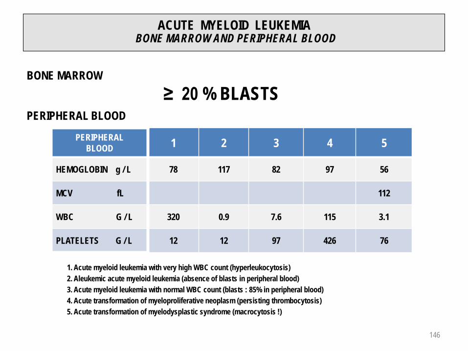
ACUTE MYELOID LEUKEMIABONE MARROW AND PERIPHERAL BLOOD
PERIPHERALBLOOD 1 2 3 4 5
HEMOGLOBIN g / L 78 117 82 97 56
MCV fL 112
WBC G / L 320 0.9 7.6 115 3.1
PLATELETS G / L 12 12 97 426 76
BONE MARROW≥ 20 % BLASTS
PERIPHERAL BLOOD
1. Acute myeloid leukemia with very high WBC count (hyperleukocytosis)2. Aleukemic acute myeloid leukemia (absence of blasts in peripheral blood)3. Acute myeloid leukemia with normal WBC count (blasts : 85% in peripheral blood)4. Acute transformation of myeloproliferative neoplasm (persisting thrombocytosis)5. Acute transformation of myelodysplastic syndrome (macrocytosis !)
146
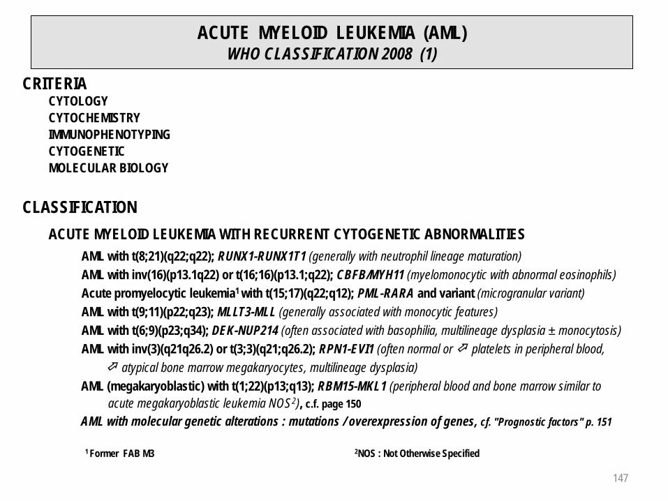
ACUTE MYELOID LEUKEMIA (AML)WHO CLASSIFICATION 2008 (1)
CRITERIA CYTOLOGYCYTOCHEMISTRYIMMUNOPHENOTYPINGCYTOGENETICMOLECULAR BIOLOGY
CLASSIFICATIONACUTE MYELOID LEUKEMIA WITH RECURRENT CYTOGENETIC ABNORMALITIES
AML with t(8;21)(q22;q22); RUNX1-RUNX1T1 (generally with neutrophil lineage maturation)AML with inv(16)(p13.1q22) or t(16;16)(p13.1;q22); CBFB/MYH11 (myelomonocytic with abnormal eosinophils)Acute promyelocytic leukemia1 with t(15;17)(q22;q12); PML-RARA and variant (microgranular variant)AML with t(9;11)(p22;q23); MLLT3-MLL (generally associated with monocytic features)AML with t(6;9)(p23;q34); DEK-NUP214 (often associated with basophilia, multilineage dysplasia ± monocytosis)AML with inv(3)(q21q26.2) or t(3;3)(q21;q26.2); RPN1-EVI1 (often normal or platelets in peripheral blood, atypical bone marrow megakaryocytes, multilineage dysplasia)
AML (megakaryoblastic) with t(1;22)(p13;q13); RBM15-MKL1 (peripheral blood and bone marrow similar toacute megakaryoblastic leukemia NOS2), c.f. page 150
AML with molecular genetic alterations : mutations / overexpression of genes, cf. "Prognostic factors" p. 151
1 Former FAB M3 2NOS : Not Otherwise Specified
147

ACUTE MYELOID LEUKEMIA (AML)WHO CLASSIFICATION 2008 (2)
ACUTE MYELOID LEUKEMIA WITH MYELODYSPLASIA RELATED CHANGES AML from previous MDS or MDS / MPNAML with MDS-related cytogenetic anomalyAML with multilineage dysplasia
THERAPY-RELATED MYELOID NEOPLASMS (t-AML, t-MDS, t-MDS / MPN)Alkylating agents, ionizing radiation therapy, topoisomerase II inhibitors, antimetabolites, antitubulin agents
ACUTE MYELOID LEUKEMIA, NOS1
cf. pages 149-150Acute basophilic leukemiaAcute panmyelosis with myelofibrosis
MYELOID SARCOMAMYELOID PROLIFERATIONS RELATED TO DOWN SYNDROMEBLASTIC PLASMACYTOID DENDRITIC CELL NEOPLASM
ACUTE LEUKEMIAS OF AMIBIGUOUS LINEAGEAcute undifferentiated leukemiaMixed phenotype acute leukemia with t(9;22)(q34;q11.2); BCR-ABL1 : B (or T) and myeloid lineagesMixed phenotype acute leukemia with t(v;11q23); MLL rearrangedMixed phenotype acute leukemia B / myeloid, NOS1
Mixed phenotype acute leukemia T / myeloid, NOS1 1 NOS : Not Otherwise Specified
148

ACUTE MYELOID LEUKEMIA (AML)WHO CLASSIFICATION 2008 (3)
ACUTE MYELOID LEUKEMIA , NOS* (1)
With minimal differentiation : Blasts ≥ 20% of NMC1, P2 + and SB3 + < 3%, presence of myeloid markers : CD13 and / or CD117, CD33 (60%); T-marker : CD7 (40%)
Without maturation : Blasts ≥ 90% of NENC4, P + and SB + ≥ 3%, promyelocytes →neutrophils ≤ 10% of NENC, CD13 +, CD33 +, CD117 +, generally CD15 -, CD65 -
With maturation : Blasts 20-89% of NENC, P +, SB +, promyelocytes → neutrophils ≥ 10% ofNENC, CD13 +, CD33 +, CD65 +, CD11b +, CD15 +
Acute myelomonocytic Blasts 20-79% of NENC. Monoblasts → monocytes ≥ 20% of NENC and / leukemia : or monocytosis in peripheral blood ≥ 5 G / L, P +, ANBE5 +, DE6 +, CD13 +,
CD33 +, CD65 +, CD15 + (monocytic differentiation : CD14 +, CD4 +, CD11b +, CD11c +, CD64 +, CD36 +, CD68 + (PGM17), CD163 +, lysozyme +)
1 NMC : Nucleated Marrow Cells; 2 P : Peroxydase; 3 SB : Sudan Black; 4 NENC : Non Erythroid Nucleated Cells5 ANBE : α-naphtyl-butyrate esterase; 6 DE : double esterase ANBE + CAE (chloroacetate esterase); 7 PGM1 : phosphoglucomutase 1
* Former FAB M0-M2, M4149

ACUTE MYELOID LEUKEMIA (AML)WHO CLASSIFICATION 2008 (4)
ACUTE MYELOID LEUKEMIA, NOS* (2)
Acute monoblastic and Monoblastic : Monoblasts ≥ 80% of NENC1
monocytic leukemia : Monocytic : Monoblasts < 80% of NENC, presence of promonocytes andmonocytes, P2 ± , ANBE3 +, CD13 +, CD33 +, CD15 +, CD65 +, CD14 +, CD4 +, CD11b +, CD11c +, CD64 +, CD68 +, CD36 +, lysozyme +
Acute erythroid Erythroleukemia (Erythroid / myeloid) : ≥ 50% erythroid precursors (with signs of leukemia : dysplasia, PAS4 ±, glycophorin +) of NMC5 , ≥ 20% myeloblasts of NENC (myeloid
markers of AML minimal / without differentiation)Pure erythroid leukemia : ≥ 80% of dysplastic erythroid precursors (basophilia,vacuoles, PAS +, glycophorin +), without myeloblastic component
Acute megakaryoblastic Blasts ≥ 20% of NMC; ≥ 50% of blasts must express markers of megakaryocyticleukemia : lineage : CD41 + (glycoprotein IIb/IIIa) and / or CD61 + (glycoprotein IIIa),
CD42 ± (glycoprotein Ib), vW6 +. Other markers : CD13 ±, CD33 ±, CD36 +
1 NENC : Non Erythroid Nucleated Cells; 2 P : Peroxydase; 3 ANBE : α-naphtyl-butyrate esterase; 4 PAS : Periodic acid-Schiff)5 NMC : Nucleated Marrow Cells; 6 vW : von Willebrand
* Former FAB M5-M7
150

PROGNOSTIC FACTORS IN ACUTE MYELOID LEUKEMIA (AML)
FAVORABLE UNFAVORABLEAge < 50 y > 60 y
Karnofsky1 Index > 60% < 60%
Phenotype CD34 -MDR12 neg
CD34 +MDR1 pos
Leukocytes (WBC) < 30 G / L > 30 G / L
Post chemo- and / orradiotherapy
Prior hematological disorder (MPN, MDS, other)
No Yes
Genetic t(8;21), inv(16) / t(16;16), t(15;17)
Complex karyotypic anomalies, -5, -7, 3q26 aberrations, t(6;9), 11q23 aberrations except t(9;11)
Molecular genetic
alterations
Mutations NPM13 ,CEBPA4
NPM1 & FLT3-ITD
KIT : t(8;21), exon 17; inv16, t(16;16), exon 8, FLT3-ITD5 , FLT3-TKD6, WT17 & FLT3-ITD
MLL-PTD8
Overexpression BAALC9, ERG10, MN111
Schiffer C.A. : Prognosis of acute myeloid leukemia; August 2008, UpToDate Modified from Mrozek & al. : Blood 2007; 109 : 431-448.
1 Karnofsky Index : patient performance index cf. next page; 2 MDR : Multidrug Resistance; 3 NPM1: Nucleophosmin, Member 1; 4 CEBPA : CCAAT / Enhancer Binding Protein α ; 5 FLT3-ITD : Fms-Like Tyrosine Kinase 3-Internal Tandem Duplication (Tyrosine Kinase Receptor); 6 FLT3-TKD : FLT3-2nd TK Domain; 7 WT1 : Wilms' Tumor; 8 MLL-PTD : Myeloid / Lymphoid or Mixed Lineage Leukemia-Partial Tandem Duplication; 9 BAALC : Brain and Acute Leukemia, Cytoplasmic; 10 ERG : ETS (Erythroblast Transformation Specific)-Related Gene; 11MN1 : Meningioma 1
151

KARNOFSKY PERFORMANCE STATUS
% CRITERIA
Normal activityNo assistance needed
100 Normal, no complaints; no evidence of disease
90 Able to carry on normal activity; minor signs or symptoms of disease
80 Normal activity with effort; some signs or symptoms of disease
Impaired activityAmbulatoryAssistance needed
70 Cares for self; unable to carry on normal activity or to do active work
60 Requires occasional assistance but is able to care for most of his / her needs
50 Requires considerable assistance and frequent medical care
Assistance dependantHospital care desirable
40 Disabled; requires special care and assistance
30 Severely disabled; hospitalization is indicated although death not imminent
20 Very sick; hospitalization necessary; active supportive treatment necessary
Terminal care10 Moribund; fatal processes progressing rapidly
0 Dead
152152
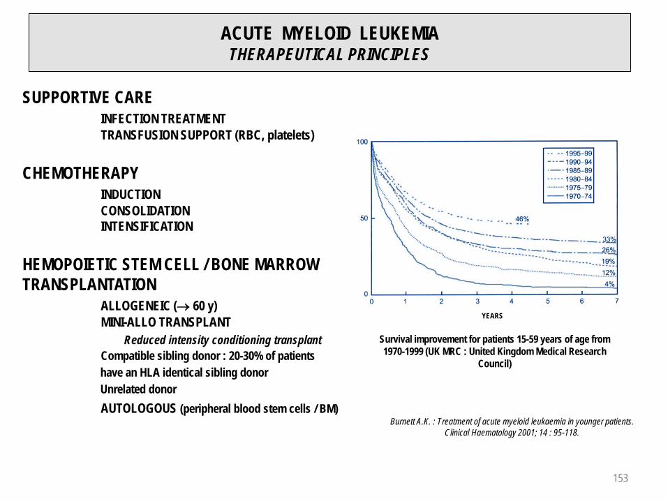
ACUTE MYELOID LEUKEMIA THERAPEUTICAL PRINCIPLES
YEARS
Survival improvement for patients 15-59 years of age from 1970-1999 (UK MRC : United Kingdom Medical Research
Council)
Burnett A.K. : Treatment of acute myeloid leukaemia in younger patients. Clinical Haematology 2001; 14 : 95-118.
SUPPORTIVE CAREINFECTION TREATMENTTRANSFUSION SUPPORT (RBC, platelets)
CHEMOTHERAPYINDUCTIONCONSOLIDATIONINTENSIFICATION
HEMOPOIETIC STEM CELL / BONE MARROW TRANSPLANTATION
ALLOGENEIC (→ 60 y)MINI-ALLO TRANSPLANT
Reduced intensity conditioning transplantCompatible sibling donor : 20-30% of patientshave an HLA identical sibling donorUnrelated donorAUTOLOGOUS (peripheral blood stem cells / BM)
153

TREATMENT OF ACUTE MYELOID LEUKEMIACHEMOTHERAPY
ARA-C (Cytosine arabinoside)ANTHRACYCLINES (Daunorubicin, Idarubicin, Mitoxantrone, Amsacrine)
6-THIOGUANINE
ETOPOSIDE60-70% Complete Remissions (CR)(30-40% with 2nd induction cycle)Then, 2-3 consolidation cycles5 years relapse free survival rate : 20-25%Improvement of survival rate with intensification, autologous or allogeneic transplantation(5 years relapse free survival rate : 40-50%)
ATRA (all-trans retinoic acid) + ARA-C and Anthracycline : Acute promyelocytic leukemia t(15;17)(q22;q12); PML-RARA
Investigational :Stratification by risk factorsTreatment of relapse : Arsenic trioxyde; Farnesyltransferase inhibitors, MDR11, BCL22 , FLT33 andTyrosine Kinases inhibitors, antiangiogenic drugs, anti-CD33 (Gemtuzumab)
1 MDR : Multidrug Resistance2 BCL2 : B-Cell Leukemia / Lymphoma 2 (protooncogene, inhibitor of apoptosis)3 FLT3 : Fms-Like Tyrosine Kinase 3 (Tyrosine Kinase receptor)
154

KINETICS OF LEUKEMIC CELLS RELATED TO TREATMENT
Number of malignant cells
1012
1011
1010
109
108
107
106
105
104
103
102
10
0
DETECTION THRESHOLD
DIAGNOSIS EARLY RELAPSE RELAPSE
Morphological, cytogenetical
Immunophenotypical (cytometry)
Molecular (PCR)
MOLECULARREMISSION
CURE ?
Time
HEMATOLOGICALREMISSION
1
2
3
1
2
3
CHEMOTHERAPY COURSES
??
PROLONGED REMISSION
1 kg
1 g
1 mg
1 µg
155

ACUTE MYELOID LEUKEMIA : ALLOGENEIC TRANSPLANTATION
A : CR-1 (n = 5'192)B : CR-2 (n = 1'122)C : Advanced phase
(n = 1'787)
A : CR-1 (n = 831)B : CR-2 (n = 880)
C : Advanced phase(n = 854)
Modified from Labopin M. : Acute Leukemia Working Party of the European Group for Blood and Marrow Transplantation (EBMT) Chaidos A. et al. : Clinical Haematology 2007; 20 : p. 133. 156

LYMPHOID NEOPLASMS1 (1)(WHO CLASSIFICATION 2008)
SIMPLIFIED CLASSIFICATION2
B-CELL NEOPLASMSPRECURSOR B-CELL NEOPLASMS
B-lymphoblastic leukemia / lymphoma (former FAB L1-L2)2
MATURE B-CELL NEOPLASMSB-cell lymphoid leukemiasBurkitt leukemia variant (former FAB L3)2
T-CELL AND NK-CELL NEOPLASMSPRECURSOR T-CELL
T-lymphoblastic leukemia / lymphoma (former FAB L1-L2)2
MATURE T-CELL OR NK-CELL NEOPLASMST-cell and NK-cell lymphoid leukemias
HODGKIN LYMPHOMA
IMMUNODEFICIENCY-ASSOCIATED LYMPHOPROLIFERATIVE DISORDERS
1 Former lymphoproliferative syndromes, malignant lymphomas2 Lymphoblastic leukemias / lymphomas cf. pages 178-183
157

LYMPHOID NEOPLASMS (2)
PROOF OF MONOCLONALITYExpression of one type only of light chain (κ or λ) on the lymphocyte surface (B)Rearrangement of Ig genes (B)Presence of paraprotein (B)Rearrangement of TCR1 genes (T)Cytogenetics (B,T, NK)
CLINICAL CONDITION PERFORMANCE STATUS OF THE EASTERNCOOPERATIVE ONCOLOGY GROUP (ECOG)GRADE ECOG
0 Fully active, able to carry on all pre-disease performance without restriction
1Restricted in physically strenuous activity butambulatory and able to carry out work of a light or sedentary nature
2Ambulatory and capable of all selfcare but unable to carry out any work activities. Up and about < 50% of waking hours
3 Only capable of limited selfcare, confined to bed or chair > 50% of waking hours
4 Completely disabled. Cannot carry on any selfcare.Totally confined to bed or chair
PROGNOSTIC FACTORSHistology (low grade → high grade)StagingTumor volume ("bulky")Performance status (ECOG score)LDH serum levelPresence or not of inflammatory syndrome
CLINICAL BEHAVIOUR (survival without treatment)Indolent yearsAggressive monthsHighly aggressive weeks
1 TCR : T-Cell Receptor
158

LYMPHOID NEOPLASMS (3)STAGING (ANN ARBOR CLASSIFICATION)
STAGESI Involvement of single lymph node region
IE Limited involvement of single extralymphatic organ or site
II Involvement of two or more lymph node regions on the same side of the diaphragm alone
IIE With involvement of limited contiguous extralymphatic organ or tissue
III Involvement of lymph node regions on both sides of the diaphragm
IIIS With spleen involvement
IIIE With limited, contiguous extralymphatic organ or site
IIIES With limited involvement of contiguous extralymphatic organ or site and spleen
IVDiffuse or disseminated foci of involvement of one or more extralymphatic organ(s) or tissue(s) (digestive tract, liver, lung, bone marrow, bone…) with or without associated lymphatic involvement
159
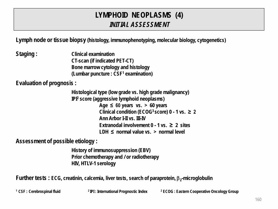
LYMPHOID NEOPLASMS (4)INITIAL ASSESSMENT
Lymph node or tissue biopsy (histology, immunophenotyping, molecular biology, cytogenetics)
Staging : Clinical examinationCT-scan (if indicated PET-CT)Bone marrow cytology and histology(Lumbar puncture : CSF1 examination)
Evaluation of prognosis :Histological type (low grade vs. high grade malignancy)IPI2 score (aggressive lymphoid neoplasms)
Age ≤ 60 years vs. > 60 yearsClinical condition (ECOG3 score) 0 - 1 vs. ≥ 2Ann Arbor I-II vs. III-IVExtranodal involvement 0 - 1 vs. ≥ 2 sitesLDH ≤ normal value vs. > normal level
Assessment of possible etiology :History of immunosuppression (EBV)Prior chemotherapy and / or radiotherapyHIV, HTLV-1 serology
Further tests : ECG, creatinin, calcemia, liver tests, search of paraprotein, β2-microglobulin
1 CSF : Cerebrospinal fluid 2 IPI : International Prognostic Index 2 ECOG : Eastern Cooperative Oncology Group
160

LYMPHOID NEOPLASMS (5)TREATMENT
HIGHLY AGGRESSIVE LYMPHOID NEOPLASM (e.g. Precursor B- or T-cell lymphoblastic leukemia / lymphoma)
CHOP1, DHAP2…Intensification with autologous transplantation or stem cell reinfusion
Overall 5 years survival about 25%
AGGRESSIVE LYMPHOID NEOPLASM (e.g. diffuse large B-cell lymphoma)
CHOP, MACOP-B3, BACOP4, CHOP + Rituximab (anti-CD20)Intensification + autologous transplant
Overall 5 years survival about 30-40%
INDOLENT LYMPHOID NEOPLASM (e.g. follicular lymphoma grade 1-2)
Radiation therapy, α-Interferon, purine analogues (Fludarabine, Cladribine), monoclonal antibodies : Rituximab (Mabthera) alone or in combination, radioimmunoconjugates : Ibritumomab (Zevalin), CVP5, CHOP
Overall 5 years survival about 50-70%1 CHOP : Cyclophosphamide + Doxorubicin + Vincristine + Prednisone2 DHAP : Dexamethasone + Cisplatin + Cytarabine3 MACOP-B : Methotrexate + Doxorubicin + Cyclophosphamide + Vincristine + Bleomycin + Prednisone4 BACOP : Cyclophosphamide + Doxorubicin + Vincristine + Bleomycin + Prednisone5 CVP : Cyclophosphamide + Vincristine + Prednisone
161

B-CELL DIFFERENTIATIONRELATIONSHIP TO MAJOR B-CELL NEOPLASMS
MARGINAL ZONE
MANTLEZONE
GERMINAL CENTER (GC)
NAIVEB-CELL
PROGENITOR B-CELL
PRE-B-CELL
IMMATUREB-CELL
MEMORY B-CELL(Mutated IgV gene)
BONE MARROW
LYMPHO-PLASMACYTIC
CELL
PLASMA CELL
Chronic lymphocytic leukemia(Unmutated IgV gene)
Follicular lymphomaBurkitt lymphoma
Hodgkin lymphomaPost-transplant
lymphomas
Mantle cell lymphomaChronic lymphocytic leukemia
(Unmutated IgV gene)
Lymphoplasmacyticlymphoma
Primary effusionlymphoma
Plasma cell myeloma
Chronic lymphocytic leukemia
(Mutated IgV gene)
Diffuse large B-cell lymphoma (DLBCL)
Primary mediastinal largeB-cell lymphoma
Splenic B-cell marginal zone lymphomaMALT lymphoma
B lymphoblastic leukemia / lymphoma
Hairy cell leukemiaB-cell prolymphocytic
leukemia
Classical Hodgkin lymphoma
LYMPH FOLLICLE
GC B-CELL
162

LYMPHOID LEUKEMIAS
B-CELL PROLIFERATIONChronic lymphocytic leukemia (CLL )B-cell prolymphocytic leukemia (B-PLL)Hairy cell leukemia and variant (HCL, HCL-v)Splenic B-cell marginal zone lymphoma (SMZL)Splenic B-cell marginal zone lymphoma / leukemia, unclassifiableLymphoplasmacytic lymphoma (LPL) - Waldenström macroglobulinemia (WM)Leukemic form of follicular lymphoma (FL)Leukemic form of mantle cell lymphoma (MCL)Plasma cell leukemia (PCL)
T- AND NK-CELL PROLIFERATIONT-cell prolymphocytic leukemia (T-PLL)T-cell large granular lymphocytic leukemia (T-LGL)Chronic lymphoproliferative disorders of NK-cells (CLPD-NK)Aggressive NK-cell leukemiaAdult T-cell leukemia / lymphoma (ATLL)Sézary syndrome (SS)
163

B-CELL LYMPHOID LEUKEMIAS
CHRONIC LYMPHOCYTIC LEUKEMIA (CLL) (1)
DEFINITIONMonoclonal B-cell lymphoid proliferation
SYMPTOMS AND CLINICAL FEATURESFortuitous diagnosisLymph node enlargementSplenomegalyRelapsing infectionsSevere anemic syndromeHemorrhagic manifestations
BLOOD PICTURERelative and absolute lymphocytosisMonoclonality shown by cell surface markers :
Coexpression of CD5 / CD19κ or λ expression
CLASSIFICATION (cf. next page)RaiBinet
164

CHRONIC LYMPHOCYTIC LEUKEMIA (2)
RAI CLASSIFICATION (1975) STAGE CRITERIA MEDIAN SURVIVAL (MONTHS)
0 Isolated monoclonal lymphocytosis (peripheral blood and bone marrow) 150
I 0 + lymphadenopathies1 101
II 0 and I + splenomegaly2 and / or hepatomegaly2 71
III 0 and Hb < 100 g / L ± tumoral syndrome 19
IV 0 and platelets < 100 G / L ± tumoral syndrome 19
BINET CLASSIFICATION (1981)
STAGE LYMPHOID SITES3 Hb AND PLATELETS MEDIAN SURVIVAL (MONTHS)
A < 3 Hb ≥ 100 g / LPlatelets ≥ 100 G / L
Comparable to age-matched control
B ≥ 3 84
C Irrelevant Hb < 100 g / L orPlatelets < 100 G / L 24
1 Cervical, axillary, inguinal lymph nodes on clinical examination2 On abdominal palpation3 Cervical, axillary, inguinal lymph nodes, splenomegaly and hepatomegaly on clinical examination
Rai K.R., Keating M.J. : Staging and prognosis of chronic lymphocytic leukemia; october 2008, UpToDate. 165

CHRONIC LYMPHOCYTIC LEUKEMIA (3)
COURSE AND COMPLICATIONSInfection secondary to :
B-cell immunological defectPotential neutropenia (mainly secondary to chemotherapy)
Autoimmune manifestationHemolytic anemia with positive direct Coombs test (10-15%)
Immune thrombocytopenia (5%)
Pure red cell aplasia (Erythroblastopenia)
Transformation to diffuse large B-cell lymphoma (Richter syndrome)
DIFFERENTIAL DIAGNOSIS
Viral or bacterial lymphocytosis (cf. page 112)
Other lymphoid leukemia
166
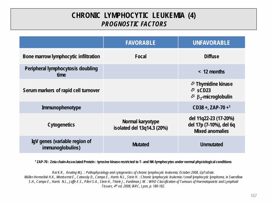
CHRONIC LYMPHOCYTIC LEUKEMIA (4)PROGNOSTIC FACTORS
FAVORABLE UNFAVORABLE
Bone marrow lymphocytic infiltration Focal Diffuse
Peripheral lymphocytosis doubling time < 12 months
Serum markers of rapid cell turnover Thymidine kinase sCD23 β2-microglobulin
Immunophenotype CD38 +, ZAP-70 +1
Cytogenetics Normal karyotypeisolated del 13q14.3 (20%)
del 11q22-23 (17-20%) del 17p (7-10%), del 6q
Mixed anomalies
IgV genes (variable region ofimmunoglobulins) Mutated Unmutated
1 ZAP-70 : Zeta chain-Associated Protein : tyrosine kinase restricted to T- and NK-lymphocytes under normal physiological conditions
Rai K.R., Keating M.J. : Pathophysiology and cytogenetics of chronic lymphocytic leukemia; October 2008, UpToDate.Müller-Hermelink H.K., Montserrat E., Catovsky D., Campo E., Harris N.L., Stein H. : Chronic lymphocytic leukemia / small lymphocytic lymphoma, in Swerdlow
S.H., Campo E., Harris N.L., Jaffe E.S., Pileri S.A., Stein H., Thiele J., Vardiman J.W. : WHO Classification of Tumours of Haematopoietic and Lymphoid Tissues, 4th ed. 2008; IARC, Lyon, p. 180-182.
167

CHRONIC LYMPHOCYTIC LEUKEMIA (5)TREATMENT
"Wait and watch" as long as possible
Alkylating agents (Chlorambucil)
Purine analogs (Fludarabine, Cladribine)
Polychemotherapy (CVP1, CHOP1)
Proapoptotic drugs (monoclonal antibodies) : Rituximab : anti-CD20, Alemtuzumab (MabCampath) : humanized anti-CD52, Ofatumumab : humanized anti-CD20 (↑ affinity for CD20)
Lenalidomide (relapsing or refractory CLL)
Polyvalent immunoglobulin concentrates (in case of relapsing infections related to B immunological defect)
Allogeneic transplantation (< 50 years, HLA identical donor, disease with rapid evolution. 5 years relapse free survival : 40%)
1 See p. 161
168

OTHER B-CELL LYMPHOID LEUKEMIAS (1)
B-CELL PROLYMPHOCYTIC LEUKEMIALarge splenomegaly, few or absent lymphadenopathiesLymphocytosis > 100 G / L, anemia and thrombocytopenia (50% of cases)Large cells with prominent nucleolus : CD19 +, CD20 +, CD22 +, CD79a +, CD79b +, CD5 + (20-30%),
CD23 + (10-20%)Treatment : CHOP (see p. 161), purine analogs (fludarabine, cladribine), chemotherapy + Rituximab, splenectomyMedian survival : 30-50 months
HAIRY CELL LEUKEMIASplenomegaly without lymphadenopathyPancytopeniaWBC usually < 4 G / L, > 10 G / L (10-20%), rarely > 200 G / L, monocytopeniaPresence of hairy cells (TRAP +), CD19 +, CD11c +, CD25 +, CD103 +, CD123 +Bone marrow fibrosisComplications : Recurrent opportunistic Infections
Vasculitis or other immune dysfunctionNeurologic disordersBleeding disordersBone lesions
Treatment : Purine analogs (+ Rituximab), α-Interferon, splenectomy, anti-CD22, anti-CD25immunotoxins
Overall 10-year survival rate : > 90%
169

OTHER B-CELL LYMPHOID LEUKEMIAS (2)
SPLENIC B-CELL MARGINAL ZONE LYMPHOMA (SMZL)SplenomegalyVariable presence in peripheral blood of villous lymphocytes CD20 +, CD79a +, CD5 -, CD25 + / -, CD11c + / -, CD103 usually -, CD123 rarely +Occasionally autoimmune thrombocytopenia or anemiaSmall monoclonal serum paraprotein (1/3 of cases)Clinical course indolentTreatment : splenectomy
SPLENIC B-CELL MARGINAL ZONE LYMPHOMA / LEUKEMIA, UNCLASSIFIABLESplenic diffuse red pulp small B-cell lymphoma (SMZL-diffuse variant)
Frequently massive splenomegalyUsually low lymphocytosis, presence of villous lymphocytesSometimes cutaneous infiltration (pruritic papules)
Hairy cell leukemia-variant (HCL-v) - "Prolymphocytic variant of HCL"Average WBC count ∼ 35 G / L, ↓ platelets (~ 50%), ↓ RBC (~ 25%)Lymphocytes : hybrid features of prolymphocytic leukemia and classical hairy cell leukemiaAbsence of monocytopeniaTreatment : Rituximab, anti-CD22 immunotoxin
Usually no response to purine analogues and to α-Interferon
170

OTHER B-CELL LYMPHOID LEUKEMIAS (3)
LYMPHOPLASMACYTIC LYMPHOMA - WALDENSTRÖM MACROGLOBULINEMIA (WM)
Lymphoplasmacytic bone marrow infiltrationLymphocytosis : generally < 10 G / L (mixture of small and large lymphocytes, sometimes with eccentric nucleus and pronounced cytoplasmic basophilia)Lymphadenopathies : 40%, splenomegaly or hepatomegaly : 30%, hepatosplenomegaly : 25%Mainly IgM paraproteinemia (WM) : hyperviscosity syndrome (IgM > 30 g / L)Possible cryoglobulinemia (Raynaud phenomenon, vasculitis)Anemia of variable severity
Hemodilution Bone marrow failure Autoimmune hemolytic anemia (cold agglutinins)
Polyneuropathy with sensory and motor defect (anti-MAG1 antibodies)Bleeding tendency (thrombocytopenia + thrombopathy)Indolent lymphoid neoplasm
Treatment : Plasmapheresis if hyperviscosity syndromeAlkylating agents, Rituximab, purine analogs, CHOP2 + Rituximab,corticosteroids, splenectomy
Median survival : 5-10 years
1 Myelin Associated Glycoprotein2 See p. 161
171

B-CELL LYMPHOID LEUKEMIAContribution of immunological markers, cytogenetics and molecular biology
sIg CD19 CD5 CD23 CYTOGENETICS OTHERS
CLL + / - + + +B-PLL + + - / + - / +
HCL + + - -TRAP +CD11c +CD25 +
CD103 +
SMZL + + - / + -MCL + + + - t(11;14) Cyclin D1
FL + + - - t(14;18) CD10 +BCL2
CD1231 CD25 CD11c CD103
HCL 22 / 2395%
24 / 2596%
25 / 25100%
25 / 25100%
HCL VARIANT
1 / 119%
0 / 110%
11 / 11100%
4 / 1136%
SMZL 1 / 293%
18 / 2864%
10 / 2638%
0 / 250%
The contribution of morphology
remains paramount for the
differential diagnosis of B-cell
prolymphocytic leukemia, hairy cell
leukemia and its variant form as for
splenic marginal zone lymphoma
CLL : Chronic lymphocytic leukemiaB-PLL : B-cell prolymphocytic leukemia
HCL : Hairy cell leukemia
SMZL : Splenic marginal zonelymphoma
MCL : Mantle cell lymphoma
FL : Follicular lymphoma
BCL2 : B-cell Leukemia / Lymphoma 2Protooncogene, inhibitor ofapoptosis or cell death
1 Del Giudice I. et coll. : The diagnostic value of CD123 in B-cell disorders with hairy or villous lymphocytes. Haematologica 2004; 89 : 303-308.
172

T-CELL AND NK-CELL LYMPHOID LEUKEMIAS (1)
T-CELL PROLYMPHOCYTIC LEUKEMIA (T-PLL)Hepatosplenomegaly, generalized lymphadenopathy High WBC count > 100 G / L (> 200 G / L in 50% of patients)Skin involvement (20% of cases)CD2 +, CD3 + / -, CD7 +, usual expression of CD4 (60%), CD8 + / -, CD52 +Cytogenetic anomalies : inv(14), t(14;14), idic(8p11), t(8;8), trisomy 8q, del 12p13, del 11q23Aggressive disease, median survival < 1 yearTreatment : anti-CD52 (alemtuzumab)
T-CELL LARGE GRANULAR LYMPHOCYTE LEUKEMIA (T-LGL)Serious neutropenia, variable anemia (sometimes severe due to red cell aplasia)Moderate splenomegalyFrequent autoantibodies, immune complexes and hypergammaglobulinemiaAssociation with rheumatoid arthritisCD3 +, TCRαβ +, CD4 - / +, CD8 +; CD57 +, CD 16 + : > 80% of casesIndolent clinical course, median survival ~ 13 years
173

T-CELL AND NK-CELL LYMPHOID LEUKEMIAS (2)
CHRONIC LYMPHOPROLIFERATIVE DISORDERS OF NK-CELLS (CLPD-NK)Usually asymptomatic, some cases with systemic symptoms, cytopenia(s)Sometimes in association with solid tumors, vasculitis, neuropathy, autoimmune disordersCD3 -, CD4 -, CD8 -, TCRαβ -, CD16 +, CD56 + (usually weak), CD57 -
AGGRESSIVE NK-CELL LEUKEMIARare, prevalent Asians, median age : 42 yearsPrincipal involved sites : peripheral blood, bone marrow, spleen, liverCD2 +, CD3 -, CD56 +Fulminant clinical course (coagulopathy, hemophagocytic syndrome) Median survival : < 2 months
174

T-CELL AND NK-CELL LYMPHOID LEUKEMIAS (3)
ADULT T-CELL LEUKEMIA / LYMPHOMA (ATLL)
Japan (1977), Caribbean region, Central Africa
Clinical variants : 1) acute (most common); 2) lymphomatous; 3) chronic; 4) smoldering
Lymphadenopathy, hepatosplenomegaly
Skin involvement (erythematous rushes, papules, nodules)
Leukocytes : 5 – 100 G / L
Lymphocytes with lobated nucleus, CD2 +, CD3 +, CD5 +, usually CD4 +, CD 7 -, CD8 -
Association with HTLV-1 virus
Hypercalcemia
Survival for acute and lymphomatous variants : 2 weeks to > 1 year
175

T AND NK LYMPHOID LEUKEMIAS (4)
SEZARY SYNDROME (SS)Skin involvement (Mycosis fungoides)
Erythema, pruritus, generalized erythrodermaPautrier's microabscesses (epidermotropism)
Presence of Sézary cells in peripheral blood (> 5%)Lymphocytes with convoluted, cerebriform nucleus (cleft) Variable phenotype : CD2 +, CD3 +, TCRβ +, CD5 +, usually CD4 +, CD8 -
Secondary infiltration of tissues and organsLymph nodes, bone marrow, lungs, heart, kidneys, bone
Aggressive diseaseOverall survival rate : 10-20% at 5 years
Stages of mycosis fungoides and Sézary syndrome
Stages Extension
I A / BExclusive skin involvment (patch / plaque)A : skin < 10% of cutaneous surfaceB : skin > 10% of cutaneous surface
II A / BStage I with :A : clinical lymph node involvment or :B : cutaneous tumors
III Erythrodermia : > 80% of cutaneous surface
IV A / B A : histological lymph node involvment or Sézary cells in peripheral bloodB : secondary infiltration of tissues and organs
Modifié d'après Baumann Conzett K. et coll. : Lymphomes cutanés. Classification et recommandations thérapeutiques. Forum Med Suisse 2009, 42 : 744-749.176

T-CELL AND NK-CELL LYMPHOID LEUKEMIAS (5)Contribution of immunological markers, cytogenetics and molecular biology
CD4 CD8 CD56 RTCR OTHERS
T-PLL + + / - - + inv(14)
T-LGL - / + + - + CD3 +
CLPD-NK - - + - CD3 -
ATLL + - - + -
SS + - - + -
RTCR : Rearrangement of genes coding for variable part of TCR (T-Cell Receptor)
T-PLL : T-cell prolymphocytic leukemia
T-LGL : T-cell large granular lymphocytic leukemia
CLPD-NK : Chronic lymphoproliferative disorders of NK-cells
ATLL : Adult T-cell leukemia / lymphoma
SS : Sézary syndrome
177

LYMPHOBLASTIC LEUKEMIA / LYMPHOMA (1) WHO CLASSIFICATION 2008
PRECURSOR B and T LYMPHOID NEOPLASMS
B lymphoblastic leukemia / lymphoma, not otherwise specified (B-ALL / B-LBL)
B lymphoblastic leukemia / lymphoma with recurrent genetic anomalies
T lymphoblastic leukemia / lymphoma
MATURE B CELL NEOPLASMS
Acute lymphoblastic leukemia Burkitt type
178

LYMPHOBLASTIC LEUKEMIA / LYMPHOMA (2) CLINICAL FEATURES (1)
B ACUTE LYMPHOBLASTIC LEUKEMIA (B-ALL)
Bone marrow usually involved, peripheral
blood frequently
Extramedullary involvement
Central nervous system
Lymph nodes, spleen, liver
Testes
Pancytopenia
Leukocyte count decreased, normal or highly
elevated
B LYMPHOBLASTIC LYMPHOMA (B-LBL)
Most frequent sites of involvement
Skin
Soft tissues
Bone marrow
Lymph nodes
179

LYMPHOBLASTIC LEUKEMIA / LYMPHOMA (3)RELATION BETWEEN GENETIC ANOMALY AND PROGNOSIS
CYTOGENETICS FUSION TRANSCRIPT PROGNOSIS
t(9;22)(q34;q11.2) BCR-ABL 1 worst of ALL
t(v;11q23) MLL rearranged poor
t(12;21)(p13;q22) TEL-AML1 (ETV6-RUNX1) good1
Hyperdiploidy (50-66 chromosomes) good1
Hypodiploidy (< 45 chromosomes) poor
t(5;14)(q31;q32) IL3-IGH intermediate
t(1;19)(q23;p13.3) E2A-PBX1 (TCF3-PBX1) poor
1 In absence of adverse prognostic factors : age > 10 years, higher initial WBC count, slow response to initial therapy, minimal residual disease after therapy,CNS involvment at diagnosis
LYMPHOBLASTIC LEUKEMIA / LYMPHOMA WITH RECURRENT GENETIC ANOMALIES
180

LYMPHOBLASTIC LEUKEMIA / LYMPHOMA (4) CLINICAL FEATURES (2)
T LYMPHOBLASTIC LEUKEMIA / LYMPHOMA
Frequent mediastinal (thymic) involvement
Lymphadenopathies
Extranodal sites : skin, tonsils, liver, spleen,
central nervous system, testes
High leukocyte count
High risk disease in childhood (induction failure,
early relapse, isolated CNS relapse)
In adults, better prognosis than for B-ALL with
prognostic adverse cytogenetic anomalies
MATURE B-CELL BURKITT LEUKEMIA VARIANT (former FAB L3)
Frequent involvement of CNS at diagnosis
Blasts with deeply basophilic cytoplasm with
prominent vacuoles
Extreme chemosensitivity (risk of acute tumor lysis syndrome)
181

LYMPHOBLASTIC LEUKEMIA / LYMPHOMA (5)IMMUNOLOGICAL MARKERS
MARKERS PRO-B EARLY PRE-B PRE-B B MATURE
CD19 + + + +
CD10 - + + -
CD20 - + / - + +
CD22 + cyto + + +
CD34 + + + - -
HLA-DR + + + +
TdT + + + + + + + / -
cIgM1 - - +
sIgM2 - - - +
MARKERS PRE-T EARLY-T T CORTICAL T MATURE
CD7 + + + +
CD2 - + + +
CD5 - + + +
CD1a - - + -
cCD31 + + - -
CD3 - - + / - +
CD4 & CD8 - - + -
CD4 or CD8 - - - +
TdT + + + +
B-ALL :
PRO-B or EARLY PRE-B CD10 -
EARLY PRE-B or EARLY PRE-B CD10 +or COMMON PRE-B ALL
PRE-B
B MATURE ALL (type Burkitt leukemia variant, former FAB L3)
T-ALL :
PRE-T
EARLY-T
T CORTICAL
T MATURE OR MARROW T
1 cIgM, cCD3 : Intracytoplasmic IgM, CD32 sIgM : IgM expressed on cell surface
182

TREATMENT OF LYMPHOBLASTIC LEUKEMIA / LYMPHOMA
PREDNISONE - VINCRISTINE - ANTHRACYCLINES - ASPARAGINASE
PRINCIPLES : Induction - Consolidation - MaintenanceRESULTS : Adults1 (1991-2000) : CR* : 69-91%
DFS** : 17- 42%Children2 : CR* : 88-96% (2 children / 3 cured at 5 years)
1 Hoelzer D., Gökbuget N. : Acute lymphocytic leukemia in adults, in Hoffman R. et al., Hematology : Basic Principles and Practice 2005; Elsevier : p. 1181.2 Rivera G.K., Crist W.M. : Acute Lymphoblastic Leukemia, in Handin R.I. et al., Blood : Principles & Practice of Hematology 1995; J.P. Lippincott : p. 758.3 Larson R.A. : Induction therapy for acute lymphoblastic leukemia. UpToDate, mai 2009.4 Labarthe A. et al. : Imatinib combined with induction or consolidation chemotherapy in patients with de novo Philadelphia chromosome-positive acute lymphoblasticleukemia : results of the GRAAPH-2003 study. Blood 2007; 109 : 1408-1413
5 Thomas D.A. et al. : New agents in the treatment of acute lymphocytic leukaemia. Clinical Haematology 2002; 15 : 771-790.
Developments of therapeutical possibilities :Stratification for risk factorsAllograft in patient with unfavorable risk factors, early autologous transplantation with peripheral blood progenitor cellsClofarabine, Nelarabine, FMdC, Trimetrexate, liposomal Vincristine, Flavopiridol, Bryostatin, monoclonal antibodies (anti-CD20, anti-CD52)Arsenic trioxide, proteasome or tyrosine kinase inhibitors5
ALL BCR-ABL 1 + Chemottherapy alone(historical controls)3
Chemotherapy + Imatinib (%)(n = 45)4
Hematological CR 71 96
Molecular CR 29
Overall survival (at 18 months) 39 65
DFS * (at 18 months) 31 51 *CR : Complete Remission**DFS : Disease Free Survival
Followed, if possible, (age ≤ 55 years, related or
unrelated donor) by bone marrow / stem cell transplantation in CR
183
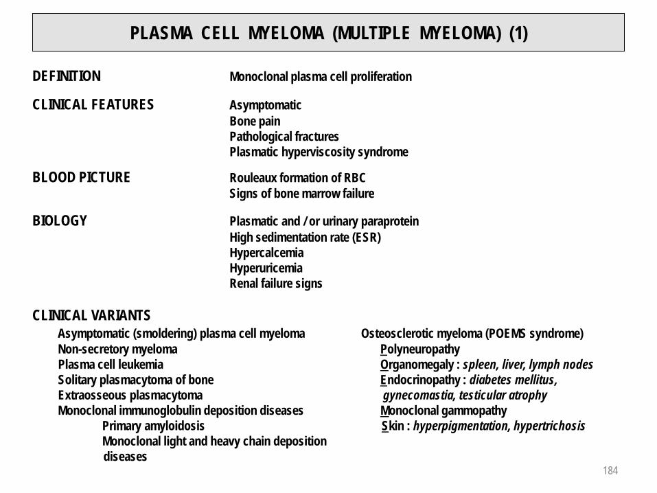
PLASMA CELL MYELOMA (MULTIPLE MYELOMA) (1)
DEFINITION Monoclonal plasma cell proliferation
CLINICAL FEATURES AsymptomaticBone painPathological fracturesPlasmatic hyperviscosity syndrome
BLOOD PICTURE Rouleaux formation of RBCSigns of bone marrow failure
BIOLOGY Plasmatic and / or urinary paraproteinHigh sedimentation rate (ESR)HypercalcemiaHyperuricemiaRenal failure signs
CLINICAL VARIANTSAsymptomatic (smoldering) plasma cell myeloma Osteosclerotic myeloma (POEMS syndrome) Non-secretory myeloma PolyneuropathyPlasma cell leukemia Organomegaly : spleen, liver, lymph nodesSolitary plasmacytoma of bone Endocrinopathy : diabetes mellitus, Extraosseous plasmacytoma gynecomastia, testicular atrophyMonoclonal immunoglobulin deposition diseases Monoclonal gammopathy
Primary amyloidosis Skin : hyperpigmentation, hypertrichosisMonoclonal light and heavy chain deposition diseases
184

PLASMA CELL MYELOMA (MULTIPLE MYELOMA) (2)
DIAGNOSTIC CRITERIA OF SYMPTOMATIC PLASMA CELL MYELOMA
1) Paraprotein in serum or urine. No level included. In most cases, IgG > 30 g / L, IgA > 25 g / L,urine light chain > 1 g / 24 hr. Some patients have lower levels
2) Clonal plasma cells in bone marrow or plasmacytoma. No minimal level. Usually > 10% of nucleated BM cells. 5% patients with < 10% BM plasma cells
3) Related organ or tissue impairment CRAB : Hypercalcemia, renal insufficiency, anemia, bone lesionsHyperviscosity, amyloidosis, recurrent infections
STAGE I(< 0.6 x 10¹² cells/m2)
STAGE II(0.6 – 1.2 x 10¹² cells/m2
STAGE III(> 1.2 x 10¹² cells/m2)
IgG < 50 g / L, IgA < 30 g / L, urinary light chains < 4.0 g / 24 hrAbsent or solitary bone lesionNormal hemoglobin, serum calcium, Ig levels (non-M protein)
Intermediate
IgG > 70 g / L, IgA > 50 g / L, urinary light chains > 12 g / 24 hrMultiple lytic bone lesionsHemoglobin < 85 g / LSerum calcium > 3 mmol / L
A : Creatinin < 177 µmol / L B : Creatinin ≥ 177 µmol / L
Modified from : Criteria for the classification of monoclonal gammopathies, multiple myeloma and related disorders : a report of the InternationalMyeloma Working Group. Br J Haematol 2003; 121 : 749-757.
INTERNATIONAL STAGING SYSTEM FOR PLASMA CELL MYELOMA
Modified from : Durie B.G., Salmon S.E. : A clinical staging system for multiple myeloma. Correlation of measured myeloma cell masswith presenting clinical features, response to treatment and survival. Cancer 1975; 36 : 842-854.
185

PLASMA CELL MYELOMA (MULTIPLE MYELOMA) (3)
PARAPROTEINS = MONOCLONAL IMMUNOGLOBULINS
TYPE % TYPE %
IgG 50 IgD, IgMBiclonal < 10
IgA 20 No monoclonal Ig ~ 3
Light chain 20 IgE < 1
STAGE PARAMETERS MEDIAN SURVIVAL(MONTHS)
1 β2-m < 3.5 mg / LAlbumin ≥ 35 g / L 62
2β2-m < 3.5 mg / L
Albumin < 35 g / Lor β2-m ≥ 3.5 - < 5.5 mg / L
44
3 β2-m ≥ 5.5 mg / L 29
SURVIVAL : ISS (International Staging System) 8'449 patients2
FACTORS OF POOR PROGNOSIS Creatinin, β2-m1, Albumin, CRP, LDH, Hb, Ca++
Important plasmacytic infiltration (bone marrow)Poorly differentiated plasmacytesCytogenetic anomalies :
del 13, hypodiploidyFISH : t(4;14)(p16.3;q32), t(14;16)(q32;q23), t(14;20)(q32;q12)
del 17p13 (p53)Deletions of Rb (retinoblastoma) geneKi-67 expressionPresence of MDR (Multidrug Resistance)
COMPLICATIONSHyperviscosity syndrome (mostly IgA, IgG3)Neurological : nerve compression (radicular or spinal)Renal : light chain, calcium or uric nephropathy,
amyloidosis, plasmacytic infiltrationInfectiousHematological : bone marrow failure, thrombopathy
1 β2-m : β2-microglobulin
2 Modified from : Greipp P.R. et al. : International staging system for multiple myeloma. J Clin Oncol 2005; 23 : 3412-3420.
186

PLASMA CELL MYELOMA (MULTIPLE MYELOMA) (4)DIFFERENTIAL DIAGNOSIS
1. MGUS : Monoclonal Gammopathy of Undetermined Significance (evolution to plasma cell myeloma,lymphoplasmacytic lymphoma - Waldenström macroglobulinemia or amyloidosis : 22% after 19 years)
Differential diagnosis between MGUS and smoldering myeloma
MGUS SMOLDERING MYELOMA
Plasma cells (bone marrow) < 10% ≥ 10%
Serum monoclonal Ig < 30 g / L of uninvolved Ig : 30-40% of cases
> 30 g / L of uninvolved Ig : > 90% of cases
Lytic bone lesions 0 0
Symptoms / Infections 0 0
For both entities, levels of hemoglobin, creatinin and calcium within normal range
2. Primary amyloidosis (amyloidosis AL)3. Lymphoplasmacytic lymphoma - Waldenström macroglobulinemia, cf. page 1714. Heavy chain diseases
HISTOLOGY CLINICAL LOCALISATION
γ heavy chain disease Lymphoplasmacytic lymphoma Lymph nodes, Waldeyer's ring, BM, spleen, liver, blood
µ heavy chain disease Chronic lymphocytic leukemia Spleen, liver, BM, blood
α heavy chain disease (IPSID)1 Extranodal marginal zone lymphoma (MALT)2 Small bowel, mesenteric lymph nodes
5. Reactional plasmacytosis : German measles, HIV, tuberculosis, immunological disorders, alcoholism, see page 1131 IPSID : Immunoproliferative small intestinal disease 2 MALT : Mucosa-Associated Lymphoid Tissue
187

PLASMA CELL MYELOMA (MULTIPLE MYELOMA) (5)TREATMENT
Plasmapheresis (in case of hyperviscosity syndrome)Melphalan + Prednisone, VBAP1, VMCP2 (60% of responses, no complete remission)VAD (Vincristine + Doxorubicin + Dexamethasone high dose)Radiotherapy (solitary plasmacytoma)Supportive care (RBC, platelet transfusions, antibiotics, analgesics, bisphosphonates)Intensification with autologous transplant (PB stem cells or BM) Allogeneic transplantation : PB stem cells or BM (< 50 years, possible cure, important transplant related mortality, GvH +++)Thalidomide, Lenalidomide, Bortezomib (proteasome inhibitor), reduced intensity conditioning transplant (mini-allotransplant with non myeloablative conditioning)
1 VBAP : Vincristine + BCNU + Doxorubicin + Prednisone2 VMCP : Vincristine + Melphalan + Cyclophosphamide + Prednisone
188

HODGKIN LYMPHOMA (1)
SYMPTOMS AND CLINICAL FEATURESB symptoms:
Unexplained persistent and recurrent fever > 38°C during the previous monthRecurrent drenching nights sweats during the previous monthUnexplained loss of > 10% of body weight during the 6 months before initial staging
Other symptoms : pruritus alcohol-induced pain (usually abdominal)
Lymphadenopathy(-ies)Mediastinal involvement mainly in nodular sclerosis subtypeAbdominal (and splenic) involvement mainly in mixed cellularity subtype
HISTOLOGYReed-Sternberg cells (most often of B-cell origin)5 histological types : Nodular lymphocyte predominant Hodgkin lymphoma
Classical Hodgkin lymphomaNodular sclerosis classical Hodgkin lymphomaLymphocyte-rich classical Hodgkin lymphoma Mixed cellularity classical Hodgkin lymphomaLymphocyte-depleted classical Hodgkin lymphoma
189
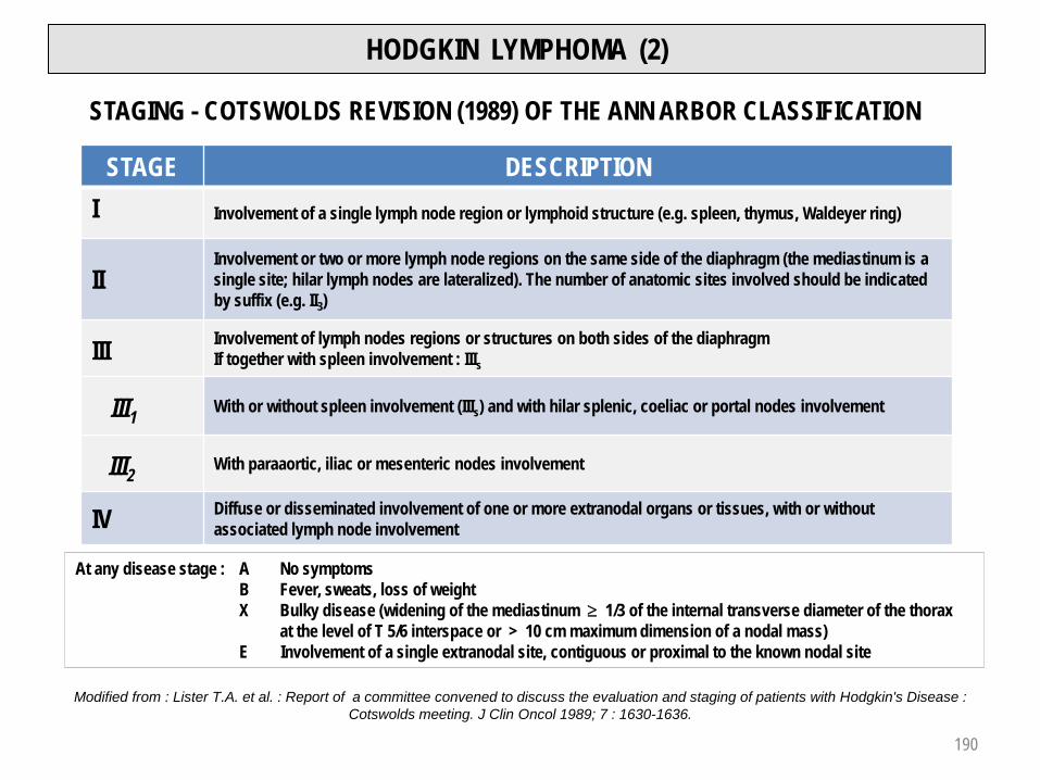
HODGKIN LYMPHOMA (2)
STAGING - COTSWOLDS REVISION (1989) OF THE ANN ARBOR CLASSIFICATION
STAGE DESCRIPTIONI Involvement of a single lymph node region or lymphoid structure (e.g. spleen, thymus, Waldeyer ring)
IIInvolvement or two or more lymph node regions on the same side of the diaphragm (the mediastinum is a single site; hilar lymph nodes are lateralized). The number of anatomic sites involved should be indicated by suffix (e.g. II3)
III Involvement of lymph nodes regions or structures on both sides of the diaphragmIf together with spleen involvement : IIIs
III1 With or without spleen involvement (IIIs) and with hilar splenic, coeliac or portal nodes involvement
III2 With paraaortic, iliac or mesenteric nodes involvement
IV Diffuse or disseminated involvement of one or more extranodal organs or tissues, with or without associated lymph node involvement
At any disease stage : A No symptomsB Fever, sweats, loss of weightX Bulky disease (widening of the mediastinum ≥ 1/3 of the internal transverse diameter of the thorax
at the level of T 5/6 interspace or > 10 cm maximum dimension of a nodal mass)E Involvement of a single extranodal site, contiguous or proximal to the known nodal site
Modified from : Lister T.A. et al. : Report of a committee convened to discuss the evaluation and staging of patients with Hodgkin's Disease : Cotswolds meeting. J Clin Oncol 1989; 7 : 1630-1636.
190

HODGKIN LYMPHOMA (3)
DIFFERENTIAL DIAGNOSISAnaplastic large T cell lymphoma : t(2;5)
UNFAVORABLE PROGNOSTIC FACTORSLarge tumor mass (e.g. : bulky mediastinal)Presence of B symptomsPrimary refractory formIPS = International Prognostic score (advanced stages of disease)
Serum albumin < 40 g / LHemoglobin < 105 g / LMale genderStage IV diseaseAge ≥ 45 yearsWBC count > 15 G / LLymphocyte count < 0.6 G / L (or < 8% of leukocyte differential count)
COMPLICATIONSImmediate, treatment relatedInfection(s)Azoospermia, early menopauseSecondary leukemia / cancer
191

HODGKIN LYMPHOMA (4)
TREATMENTRadiotherapyChemotherapy
M(C)OPP, ABVD, M(C)OPP + ABVDMIME, CEP, DHAP, BEACOPPAutologous / allogeneic transplant
PROGNOSIS AND PREDICTIVE FACTORSCurable disease in more than 85% of cases by modern radiation and chemotherapyPrognosis is function of staging, clinical and laboratory parametersResponse after 2 courses of ABVD by FDG-PET imaging is a relevant prognosticindicator in advanced stage disease1
M(C)OPP : Mustard gas analog (Cyclophosphamide) + Vincristine + Procarbazine + PrednisoneABVD : Adriamycin + Bleomycin + Vinblastine + Dacarbazine (DTIC)MIME : Mitoguazone + Ifosfamid + Methotrexate + EtoposideCEP : Lomustine + Etoposide + PrednimustineDHAP : Dexamethasone + Cisplatin + CytarabineBEACOPP : Bleomycin + Etoposide + Doxorubicin + Cyclophosphamide + Vincristine +
Procarbazine + Prednisone1 Gallamani A. et al. : Early interim 2-(18F)fluoro-2-deoxy-D-glucose positron emission tomography is prognostically superior to international prognostic score
in advanced-stage Hodgkin's lymphoma : a report from a joint Italian-Danish study. J clin Oncol 2007; 25 :3746-3752. 192

Part 3
HEMOSTASIS
193

HEMOSTASISEXPLORATION METHODS
PRIMARY HEMOSTASIS Capillary resistancePlatelet count (RI : 150 – 350 G / L)PFA-100TM 1
Platelet functions (ADP, arachidonic acid, adrenalin-heparin, collagen, ristocetin)
SECONDARY HEMOSTASIS Prothrombin time (PT, Quick) (Exploration of extrinsic pathway)
(Coagulation) Activated partial thromboplastin time (aPTT) (Exploration of intrinsic pathway)
Thrombin time (TT) (Exploration of fibrin formation)
Fibrinogen dosageInvestigation of factor XIII deficiency (fibrin stabilizing factor)Investigation of activation (Fibrin monomers and D-dimers)
TERTIARY HEMOSTASIS Euglobulins lysis time(Fibrinolysis)1 PFA-100TM (Platelet Function Analyzer) : in vitro measure of the time to occlusion of a membrane (measure of platelet adhesion and aggregation process). Replaces, if device available, the classical bleeding time
194

THROMBUS AND EMBOLUS
Thrombus : inappropriate clot formation in a blood vessel (artery or vein)Embolus : migrating thrombus
195

MAIN ACTORS OF HEMOSTASIS
Blood vesselsPlateletsCoagulation proteins
196

STEPS OF HEMOSTASIS
PRIMARY HEMOSTASISVascular time
Vasoconstriction (vascular spasm)
Platelet timePlatelet adhesion to the vessel lesionPlatelet plug formation
SECONDARY HEMOSTASIS (coagulation)Coagulation cascadeClot formation
TERTIARY HEMOSTASIS (fibrinolysis)Clot lysis
197

STEPS OF PRIMARY HEMOSTASIS
Platelet adhesion
Platelet activation
Platelet aggregation
Formation of platelet plug
198

VON WILLEBRAND FACTOR
Synthetized by endothelial cells and megakaryocytesComposed of a series of multimers : the very high molecular weight multimers are physiologicallydegraded by a specific protease (ADAMTS13), leading to prevention of spontaneous platelet aggregates formation (cf. TTP, p. 86-87)Involved, in vitro, in the process of platelet adhesion to subendothelial fibersMandatory for in vitro ristocetin induced platelet aggregationTransport of factor VIII to vascular lesionBound to factor VIII, it prolongs its life expectancy
TxA2 : Thromboxane A2FVW : von Willebrand factorADP : Adenosin DiphosphateFVIII : Factor VIII
199

PLATELET PRODUCTION FROM THE MEGAKARYOCYTE
1 mature megakaryocyte produces 2'000 - 3'000 platelets200

SECONDARY HEMOSTASISCOAGULATION
Coagulation (blood clotting) needs interaction of :
Plasmatic proteins (coagulation factors and inhibitors)A tissular protein (tissue factor)
Platelets
Calcium
201

COAGULATION FACTORS
FACTOR NAME HALF-LIFE(hours) PRODUCTION VITAMINE K
DEPENDENCE
High molecular weightkininogen Fitzgerald factor 150 Liver –
Prekallikrein Fletcher factor 35 Liver –Factor I Fibrinogen 90 Liver –Factor II Prothrombin 65 Liver +Factor V Proaccelerin 15 Liver –Factor VII Proconvertin 5 Liver +
Factor VIII Antihemophilic factor A 12 Liver (sinusoidal cells) –
Factor IX Christmas factor or antihemophilic factor B 24 Liver +
Factor X Stuart-Prower factor 40 Liver +Factor XI Antihemophilic factor C 45 Liver –Factor XII Hageman factor 50 Liver –Factor XIII Fibrin stabilizing factor 200 Liver –
Factor vW von Willebrand factor 15 EndotheliumMegakaryocytes –
202

VITAMIN K DEPENDENT FACTORS
These coagulation factors are synthetized by hepatocytes
Vitamin K is necessary for complete functional synthesis
Vitamin K (liposoluble), in reduced state, works as a cofactor to a carboxylase which transforms 10-12 glutamic acid (Glu) residues in γ-carboxyglutamic acid (Gla)
Vitamin K dependent factors bind to the cell membranes through this Gla domain, in presence of Ca++
203

COAGULATION CASCADE (1)CLASSICAL SCHEME
FibrinogenFunctional or quantitative dosage 204
aPTTActivated partial
thromboplastin time
PTProthrombin time
(or Quick time)
TTThrombin time

COAGULATION CASCADE (2)CONCEPTUAL CHANGES
Factor XI may be activated by thrombin as well as by factor XIIaFactor XI deficiency is responsible for bleeding whereas deficiencies in factor XII, prekallikrein or high molecular weight kininogen do not cause bleedingIn experimental models factor XI and factor XII deficiencies have antithrombotic effectFactor XII is activated by negatively charged surfaces, activated platelets and clot surface
205

COAGULATION CASCADE (3)CONCEPTUAL CHANGES (2)
206

FACTOR XIII AND FIBRIN STABILIZATION
207

NATURAL ANTICOAGULANTS
TFPI (Tissue Factor Pathway Inhibitor) is an effective inhibitor of factor VII - Tissue factor complexAntithrombin neutralizes all procoagulant serine proteases (thrombin, factors IXa, Xa and XIa)The protein C - protein S system inhibits factors Va and VIIIa
208

TERTIARY HEMOSTASISFIBRINOLYSIS
tPA : Tissular Plasminogen ActivatorPAI : Plasminogen Activators Inhibitors 1 and 2FDP : Fibrin Degradation ProductsTAFI : Thrombin Activatable Fibrinolysis InhibitorAP : α2-antiplasmin
Profibrinolytic proteins
Antifibrinolytic proteins
FDP
FDP
209
Intravascular fibrinolysis

HEMORRHAGIC SYNDROMEPRIMARY HEMOSTASIS (1)
Reduced capillary resistance with platelet count1, PFA-100™2, tests of platelet function, coagulation, and fibrinolysis in normal range
VASCULAR PURPURANON INFLAMMATORY
Senile purpuraEhlers-Danlos syndrome (collagen abnormality)Vitamin A deficiencyTreatment with steroids, Cushing diseaseChronic and pigmented dermatitisOsler disease (hereditary hemorrhagic telangiectasia)
INFLAMMATORY (VASCULITIS)Drug induced (Penicillin, non steroidal antiinflammatory drugs)Autoimmune disease SLE, RA, PAN, Crohn's disease)Bacterial infectionViral infection (hepatitis B, CMV, EBV, parvovirus)Lymphoid neoplasmCancerRheumatoid purpura (Henoch-Schoenlein)CryoglobulinemiaHypergammaglobulinemia Idiopathic
1 In case of vasculitis, immune thrombocytopenia may be found 2 Replaces bleeding time
SLE : Systemic Lupus Erythematosus
RA : Rheumatoid arthritis
PAN : Panarteritis nodosa
EBV : Epstein-Barr Virus
CMV : Cytomegalovirus
210

HEMORRHAGIC SYNDROMEPRIMARY HEMOSTASIS (2)
Prolonged occlusion time (PFA-100TM) 1
With normal platelet function testsThrombocytopeniaSecondary thrombocytosis
With platelet function anomaly and aPTT within normal rangeThrombopathy : acquired
hereditaryThrombocytosis of myeloproliferative neoplasm
With platelet function anomaly and prolonged aPTTvon Willebrand disease
Normal(seconds)1 Aspirin von Willebrand Glanzmann2 Bernard-Soulier2
Col / EPI3 84 – 160
Col / ADP4 68 – 121 normal
1Occlusion time (PFA-100TM)
2 cf. following page3 Col / EPI : Collagen / Epinephrin4 Col / ADP : Collagen / Adenosin-5'-diphosphate
LCH-CHUV, 2009
211
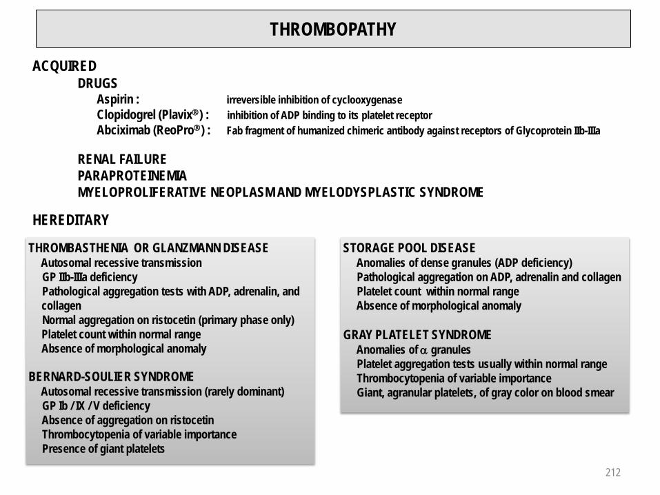
THROMBOPATHY
ACQUIREDDRUGS
Aspirin : irreversible inhibition of cyclooxygenaseClopidogrel (Plavix) : inhibition of ADP binding to its platelet receptorAbciximab (ReoPro) : Fab fragment of humanized chimeric antibody against receptors of Glycoprotein IIb-IIIa
RENAL FAILUREPARAPROTEINEMIAMYELOPROLIFERATIVE NEOPLASM AND MYELODYSPLASTIC SYNDROME
HEREDITARY
THROMBASTHENIA OR GLANZMANN DISEASEAutosomal recessive transmissionGP IIb-IIIa deficiencyPathological aggregation tests with ADP, adrenalin, and collagenNormal aggregation on ristocetin (primary phase only) Platelet count within normal rangeAbsence of morphological anomaly
BERNARD-SOULIER SYNDROMEAutosomal recessive transmission (rarely dominant)GP Ib / IX / V deficiencyAbsence of aggregation on ristocetinThrombocytopenia of variable importance Presence of giant platelets
STORAGE POOL DISEASEAnomalies of dense granules (ADP deficiency)Pathological aggregation on ADP, adrenalin and collagenPlatelet count within normal rangeAbsence of morphological anomaly
GRAY PLATELET SYNDROMEAnomalies of α granulesPlatelet aggregation tests usually within normal rangeThrombocytopenia of variable importanceGiant, agranular platelets, of gray color on blood smear
212

THROMBOCYTOPENIA (1)
DEFINITIONPlatelet count < 150 G / L
HEMORRHAGIC RISK(In case of normal platelet function)
Low if platelet count in range of 50 to 150 G / L
High by platelet count < 20 G / L
SOME RULES OR RECOMMENDATIONS
Every thrombocytopenia has to be controlled on a blood smear (eliminate pseudothrombocytopenia due to EDTA anticoagulation of the probe)
By platelet count < 50 G / L, measure of occlusion time (PFA-100TM) is useless
If platelet functions are correct, the occlusion time on PFA-100TM becomes prolonged from platelet counts < 100 G / L. Platelet count at 70 G / L with normal occlusion time does not allow exclusion of hemorrhagicrisk in case of surgical intervention
At similar platelet levels the hemorrhagic risk is higher in case of "central" thrombocytopenia than inthrombocytopenia of "peripheral" origin
213

THROMBOCYTOPENIA (2)IN THE SETTING OF BICYTOPENIA OR PANCYTOPENIA
Hypersplenism (e.g. severe hepatic failure)
Bone marrow dysfunctionAplasiaInfiltration : Myeloid or lymphoid neoplasm, osteomedullary cancer metastasisDysplasia : Reversible (Vitamin B12 or folate deficiency)
Refractory (myelodysplastic syndrome)Fibrosis
Reduction of thrombopoietin synthesis (e.g. severe hepatic failure)
SOLITARY THROMBOCYTOPENIA
CENTRAL PERIPHERALMegakaryocytes Usually
Mean platelet volume (MPV) 1
Etiology Thiazide Alcohol cf. pages 215-216
1 Frequently increased in myeloproliferative neoplasm and myelodysplastic syndrome214

SOLITARY PERIPHERAL THROMBOCYTOPENIA (1)NON IMMUNOLOGICAL
BY ANOMALY OF PLATELET DISTRIBUTIONHypersplenism
BY PLATELET DESTRUCTIONAlcoholDisseminated Intravascular Coagulation (DIC)Extracorporeal circulationThrombotic Thrombocytopenic Purpura (TTP)Hemolytic Uremic Syndrome (HUS)HELLP1 syndrome (10% of preeclampsias)Renal transplant rejectionAllogeneic stem cell or bone marrow transplantation
1HELLP : Hemolysis, Elevated Liver function tests, Low Platelets (in pregnancy)
215

SOLITARY PERIPHERAL THROMBOCYTOPENIA (2)IMMUNE
PRIMARYPrimary immune thrombocytopenia (PIT)
SECONDARY
Due to autoantibody or immune complexesDrugs (Heparin, Quinine)Infection (bacteria, virus, parasite)Autoimmune disease (SLE1, Evans syndrome2)Lymphoid neoplasmCancer
Due to alloantibodyNeonatal thrombocytopeniaPosttransfusion purpura
1 Systemic lupus erythematosus2 Autoimmune hemolytic anemia and thrombocytopenia
216

INVESTIGATION OF THROMBOCYTOPENIA
Full blood countBlood smear examination
PseudothrombocytopeniaRBC fragmentation (schistocytes)Toxic changes of neutrophilsLymphocyte stimulationAbsolute lymphocytosisErythroblastosis and / or myelocytosisParasites
Complete coagulation tests with search for coagulation activation (DIC)Bone marrow examination (cytology and histology)Direct Coombs test (antiglobulin test)Viral serology (HIV, EBV, CMV)SLE1 serologyThyroid function testsAnti-HLA antibodiesAntiplatelet antibodies
217
1 Systemic lupus erythematosus

HEMORRHAGIC SYNDROMESECONDARY HEMOSTASIS (COAGULATION)
CONSTITUTIONAL ANOMALIESHemophilias (factors VIII, IX), cf. pages 219-221Fibrinogen, factors II, V, VII, X, XI, XIII deficiencies
ACQUIRED ANOMALIESHepatocellular failure (deficiencies of fibrinogen, factors II, V, VII, X)Vitamin K deficiency (deficiencies of factors II, VII, IX, X)Disseminated intravascular coagulation (DIC)
Bacterial or parasitic infectionsCancer (lung, pancreas, prostate)Acute leukemia, particularly Acute Promyelocytic Leukemia, t(15;17)(q22;q12)Obstetrical complications
Amniotic liquid embolismPlacental retentionEclampsiaSeptic abortion
Invasive surgeryExtended burnsTransfusion complicationsVascular malformations (Kasabach-Merritt syndrom)
Coagulation inhibitors (circulating anticoagulants)Alloaantibodies against factor VIII (5-10% of hemophilia patients)Autoantibodies against factor VIII (acquired hemophilia A) : pregnancy, postpartumRhumatoïd arthritis, lupus erythematosus, cancer, drugs
218

HEMOPHILIA (1)
Recessive X-linked transmissionAbsence of familial context in 30% of
hemophilia patients : de novo mutation
Risk for offsprings of a couple of a carrier woman and a normal man:
50% of the sons with hemophilia50% of daughters are carriers
219

HEMOPHILIA (2)
INCIDENCE Hemophilia A : 1 / 10'000, 5 x more frequent than hemophilia B
HEMOPHILIA FACTOR LEVEL (%) HEMORRHAGIC SYNDROME
Light1 5 – 40Surgery
Dental extractionImportant trauma / injury
Moderate 1 – 5 Light trauma (e.g. sport)
Severe2 < 1%Several bleeding episodes / month
Frequent spontaneous hemorrhagesFrequent hemarthrosis episodes
1 Carrier female may have occasionally light symptoms2 Females may only have severe symptoms if the father is hemophiliac and the mother carrier3 NSAID : Non Steroidal Antiinflammatory Drugs
TREATMENTAnalgesia (paracetamol, tramadol, codein, opiates; aspirine and NSAID3 absolutely contra-indicatedFactors concentrates or recombinant factors. Desmopressin (DDAVP) : light forms.
Factor VIII : distribution ½ -life 4 hours, plasmatic ½-life 12 hoursFactor IX : distribution ½-life 2 hours, plasmatic ½-life 24 hours
Orthopedic surgery : hemarthrosisIn case of inhibitors : recombinant factor VIIa, FEIBA ("Factor Eight Inhibitor By-passing Activity")
220

VON WILLEBRAND DISEASE
Quantitative or qualitative anomaly of von Willebrand factor
Transmission autosomal, dominant or recessive
The most common constitutional hemorrhagic disorder (incidence ∼ 1% of whole population)
Mucosal and cutaneous bleeding (epistaxis, menorrhagia)
Biological signs : PFA-100TM prolonged, PT (Prothrombin time) normal, aPTTprolonged, ↓ Factor VIII
221

THROMBOEMBOLIC DISEASE
VIRCHOW'S TRIAD Stasis + vascular lesion + blood hypercoagulability
MAIN RISK FACTORSArterial thrombosis : Arterial hypertension
Hyperlipidemia, diabetes mellitusTobacco smoking
Venous thrombosis : Stasis (bed rest, dehydration, plasma viscosity, varicose veinsSurgery (in particular hip and abdomen)Pregnancy and post-partumEstrogens, contraceptive pillsCancerBehçet diseaseConstitutional coagulations anomalies (cf. table)
Deficiency / anomaly Prevalence (healthy european individuals (%)
Prevalence (patients with deep vein thrombosis ) (%)
Estimated relative risk
Antithrombin III, protein C, protein S 1 – 2 1 – 3 8 –10
Factor V Leiden heterogygoushomozygoous
3 – 100,06 – 0,25
151.5
3 – 750 – 80
Mutation G20210A F. IIheterozygous 1 – 3 5 – 6 2 – 4
222
Venous or arterial thrombosis :
Myeloproliferative neoplasmLupus anticoagulant, antiphospholipid syndromeHyperhomocysteinemiaHeparin induced thrombocytopenia (HIT)

THROMBOEMBOLIC DISEASETREATMENT AND PREVENTION (1)
ANTIPLATELET DRUGSAspirin blocks synthesis of Thromboxane A2 by irreversible acetylation of cyclooxygenases (COX)Clopidogrel (Plavix) causes irreversible inhibition of P2Y12 receptor of ADPDipyridamole increases platelet cyclic AMP through inhibition of phosphodiesterases(Asasantine : dipyridamole + aspirin)Abciximab (ReoPro ) is an antagonist of GP IIb/IIIa receptor
HEPARINS, THROMBIN AND FACTOR Xa INHIBITORSHeparins
Unfractioned : Liquemin , CalciparinFixation and activation of AT III1, inhibition of factors Xa and Iia,inhibition of platelets, interaction with endothelium
HeparinsLow molecular weight : Nadroparin (Fraxiparin or Fraxiforte), Dalteparin (Fragmin ), Enoxaparin (Clexane ), Certoparin (Sandoparin )
Fixation and activation of AT III, inhibition of factor Xa, very lowinhibition of factor IIa, absence of platelet inhibition, few interactions with endothelium
Danaparoid : Orgaran High affinity for AT III, anti-Xa activity, no effect on platelets
Hirudin analogs : Lepirudin (Refludan ), Bivalirudin (Angiox)Direct inhibition of thrombin
Argatroban : Argatra
Pentasaccharide : Fondaparinux (Arixtra)Rivaroxaban (Xarelto) Pure anti-Xa activity
1AT III : Antithrombin III223

THROMBOEMBOLIC DISEASETREATMENT AND PREVENTION (2)
VITAMIN K ANTAGONISTSTherapeutic agents
Acenocoumarol (Sintrom)(½ life : 8-11 hours)Phenprocoumon (Marcoumar)(½ life : 32-46 hours)
Inhibition of γ-carboxylation of vitamin Kdependant factors (FII, FVII, FIX, FX)
Biological monitoring of treatment with vitamin K antagonists (INR : International Normalized Ratio)
INR = (PT patient [seconds] / PT control [seconds])ISI
ISI = International Sensitivity Index : sensitivity index of employed reagent compared to international reference reagent
Low limit Target High limit
Primary and secondary prevention of venousthromboembolic disease 2.0 2.5 3.0
Mechanical prosthetic cardiac valves1 2.5 3.0 3.5
FIBRINOLYTIC AGENTS Tissular plasminogen activator, t-PA (Actilyse), Streptokinase (Streptase), Urokinase (Urokinase HS medac)
Therapeutical ranges
1 For more information : Salem D.N. et al. : Valvular and Structural Heart Disease : American College of Chest Physicians Evidence-Based Clinical Practice Guidelines (8th Edition). Chest 2008; 133 : 593-629.
224

VENOUS TRHOMBOEMBOLIC DISEASEANTICOAGULATION GUIDELINES
1 Activated partial thrombopoplastin time (aPTT) controls must be 1.5 - 2.5 time over basic value. Daily heparin dosis is consequently adapted2 Heparin administration has to be kept as short as possible (↑ risk of heparin induced thrombocytopenia / HIT with prolonged heparin treatment)
INITIAL (Options , depending on situation)
FONDAPARINUX (Arixtra®) : 7,5 mg SC / d 5 mg by body weight (BW) < 50 kg, 10 mg if BW > 100 kg.Contraindication:creatinin clearance < 30mL / min. No laboratory control test needed.
LOW MOLECULAR WEIGHT HEPARIN e.g. : Enoxaparine = Clexane® : 2 mg / kg / 24 h in2 SC inj. In elderly patients, by BW < 50 kg or > 100 kg : dosage of plasmatic anti-Xa activity after 2nd or 3d dose, 3-5 h after SC injection. No laboratory control tests neededCaution by creatinin clearance < 30 mL / min.
UNFRACTIONATED HEPARIN1,2 :Bolus IV 80 UI / kg (2'500-5'000 UI), then 400-600 UI / kg / 24 h (usually : 25'000-40'000 UI / 24 h) as continuous IV infusion. As priority in case of severe renal failure
EARLY SWITCH TO ANTIVITAMIN K DRUGS (Acenocoumarol : Sintrom®)3 mg / d orally from the first or second treatment day (2 mg / d by age > 70 ans, BW < 50 kg or initial PT < 85%). INR control after the first 2 dosesBy INR > 1.8 : ↓ dosis of 3d dayBy INR between 1.2 et 1.8 : same dosis on 3d dayBy INR < 1.2 : light dosis ↑ on 3d dayTarget : allow stopping of the in initial anticoagulation (SC ou IV) < 5 days and / or after 2 consecutive INR at 24 h interval > 2.0
DURATION OF ANTICOAGULATIONPostoperative limited deep vein thrombosis of the leg, increased bleeding riskProximal deep vein thrombosis / secondary pulmonary embolismDeep vein thromobosis / Idiopathic pulmonary embolism
Relapsing deep vein thrombosis and / or pulmonary embolism
6 weeks3 months6-12 months (or more if persisting risk factor without increased bleeding risk)
Long term
225

Part 4
ALGORITHMS
226

ANEMIAHb < 117 g / L : woman, child
Hb < 133 g / L : man
RETICULOCYTESHYPOREGENERATIVE REGENERATIVE< 120 G / L > 120 G / L
SETTING OFPANCYTOPENIA SOLITARY
NORMOCYTICNORMOCHROMIC
MICROCYTICHYPOCHROMIC
MACROCYTICNORMOCHROMIC
MCVMCHMCHC
ACUTE BLOOD LOSS HEMOLYTIC ANEMIA
Bone marrow aplasiaBone marrow infiltrationMyelodysplastic syndromeBone marrow fibrosis
ANEMIA
227
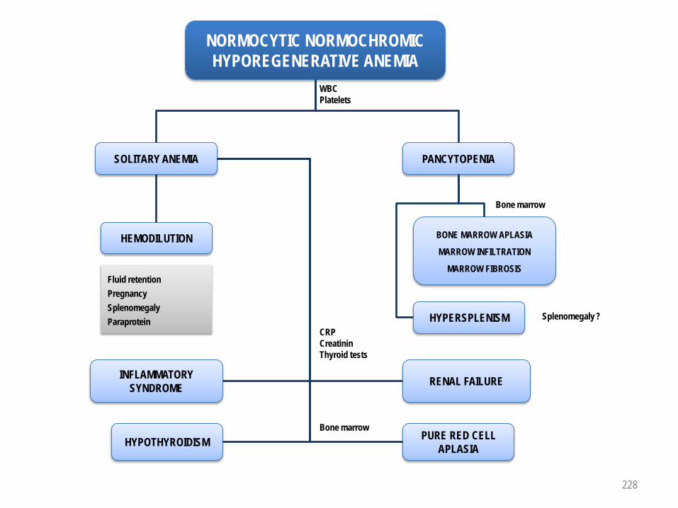
NORMOCYTIC NORMOCHROMIC HYPOREGENERATIVE ANEMIA
WBCPlatelets
SOLITARY ANEMIA PANCYTOPENIA
Bone marrow
BONE MARROW APLASIA
MARROW INFILTRATION
MARROW FIBROSIS
HYPERSPLENISM Splenomegaly ?
HEMODILUTION
INFLAMMATORY SYNDROME RENAL FAILURE
HYPOTHYROIDISM PURE RED CELL APLASIA
CRPCreatininThyroid tests
Bone marrow
Fluid retentionPregnancySplenomegalyParaprotein
NORMOCYTIC NORMOCHROMIC HYPOREGENERATIVE ANEMIA
228

MICROCYTIC HYPOCHROMIC ANEMIA
Serum iron Transferrin Ferritin
Serum iron Transferrin Ferritin
Serum iron Transferrin no / Ferritin
IRON DEFICIENCY
IRON UTILIZATIONDISORDER
INFLAMMATORYANEMIA
SIDEROBLASTIC ANEMIA HEMOGLOBINOPATHY
CRP ESR Fibrinogen α2-globulins Hepcidin
Lead intoxicationDrugs
ThalassemiaHb E, C
Chronic bleedingIncreased demandMalabsorptionPoor diet
Acute and chronic infectionCancerInflammatory arthritis
MICROCYTIC HYPOCHROMIC ANEMIA
Bone marrow (ring sideroblasts)
Hemoglobin electrophoresisMolecular biology
229

MACROCYTIC ANEMIA
Reticulocyte response(after 4 days) No reticulocyte response
B12 DEFICIENCY FOLATE DEFICIENCY
Malabsorption of gastric origin :AchlorhydriaPernicious anemia
Malabsorption of intestinal origin :Gluten enteropathyCrohn's diseaseFish tapeworm1
Poor dietIncreased demand (pregnancy)DrugsAlcoholism
FOLATE AND / ORVITAMIN B12 DEFICIENCY
FOLATE AND VITAMIN B12IN NORMAL RANGE
Association with :Bone marrow infiltration2
Inflammatory syndrome
AlcoholismHypothyroidismMyelodysplastic syndrome2
Vitamin B12 and folate levels1 mg B12 q.d. IM3 mg folate q.d. orally
1 Diphyllobothrium latum2 Indication to bone marrow examination :
CytologyHistologyImmunological markersCytogeneticsMolecular biology
MACROCYTIC ANEMIA
230

REGENERATIVE ANEMIA
ACUTE BLEEDINGHEMOLYTIC
ANEMIA
CORPUSCULAREXTRACORPUSCULAR
MEMBRANE ANOMALYHereditary spherocytosis
ENZYMOPATHYGlucose-6-PD deficiency
HEMOGLOBINOPATHYSickle cell anemia
IMMUNE HEMOLYTIC ANEMIA
TOXIC HEMOLYSISLead intoxication
INFECTIOUS HEMOLYSISMalaria
MECHANICAL HEMOLYSISMicroangiopathy
History : Ethnic originFamily historyStay in foreign countryTransfusionsPregnancies
RBC morphology :SpherocytesSchistocytesSickle cells
Coagulation tests (thrombocytopenia ?)Search for parasitesAntiglobulin test, autohemolysisHemoglobin electrophoresisTest for enzymopathy
Bilirubin LDH Haptoglobin
REGENERATIVE ANEMIA
231

ERYTHROCYTOSISTRUE "FALSE"
JAK2V617F mutation EPO serum level
V617F +EPO
V617F +EPO no /
PV VERY LIKELY
Adequate EPO secretion Inadequate EPO secretion
Stay at high altitudeRespiratory failureCardiopathy with cyanosisSmoking (carboxyhemoglobin)CO intoxicationSleep apneaHemoglobinopathy
Benign tumors (renal cyst, uterine myoma, pheochromocytoma, meningioma, cerebellar hemangioblastoma)Malignant tumors (kidney, liver, parathyroid carcinoma)
1 or : Hb or Hct > 99th percentile of reference range for age, gender oraltitude of residence
or : Hb > 170 g / L (men) or > 150 g / L (women) in case of sustained increase of ≥ 20 g / L from baseline that is not caused bycorrection of iron deficiency
or : RCV : Red Cell Volume > 25% over normal predicted value
2 Hypercellularity, increased number of megakaryocytes with morphological anomalies, reticulin fibrosis
Hb > 185 g / L (man)1
Hb > 165 g / L (woman)1
RCV1 increased
DehydrationGaisböck syndromeMicropolycythemia of heterozygous thalassemia
POLYCYTHEMIA
V617F –EPO
V617F –EPO no /
BM BIOPSY2
(NOT ESSENTIAL)
PV LIKELY
BM BIOPSY2
(CONFIRMATION)
PV POSSIBLE
BM BIOPSY2
JAK2 EXON 12
PV UNLIKELY
+ -
232

ABSOLUTE NEUTROPENIAAgranulocytosis : neutrophils < 0.5 G / L
CONFIRMED NORMALIZED
Pseudoneutropenia due tomargination excess (fasting patient)
SOLITARY NEUTROPENIA PANCYTOPENIA
BONE MARROW APLASIABONE MARROW INFILTRATION
MYELODYSPLASIABONE MARROW FIBROSIS
HYPERSPLENISM
B12 and / orFOLATE DEFICIENCY
VIRAL, BACTERIAL INFECTION(salmonellosis, brucellosis,
tuberculosis)
AUTOIMMUNE DISEASE(SLE)
DRUGS(Phenylbutazone, antithyroid agents,
Chlorpromazine)
CYCLICAL
ETHNICAL
Postprandial control
Bone marrow
Splenomegaly ?
B12 / Folate level ?
NEUTROPENIA
233
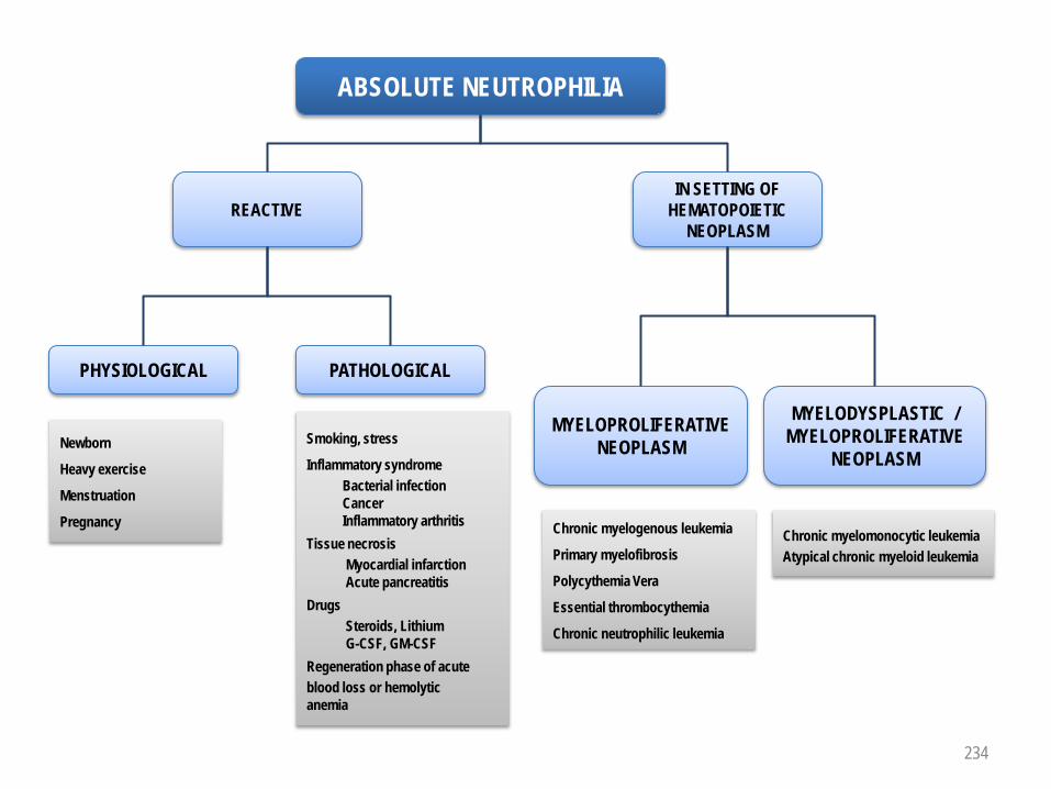
ABSOLUTE NEUTROPHILIA
REACTIVEIN SETTING OF
HEMATOPOIETIC NEOPLASM
MYELOPROLIFERATIVENEOPLASM
MYELODYSPLASTIC /MYELOPROLIFERATIVE
NEOPLASM
PHYSIOLOGICAL PATHOLOGICAL
Chronic myelogenous leukemia
Primary myelofibrosis
Polycythemia Vera
Essential thrombocythemia
Chronic neutrophilic leukemia
Chronic myelomonocytic leukemiaAtypical chronic myeloid leukemia
Newborn
Heavy exercise
Menstruation
Pregnancy
Smoking, stress
Inflammatory syndromeBacterial infectionCancerInflammatory arthritis
Tissue necrosisMyocardial infarctionAcute pancreatitis
DrugsSteroids, LithiumG-CSF, GM-CSF
Regeneration phase of acuteblood loss or hemolyticanemia
NEUTROPHILIA
234
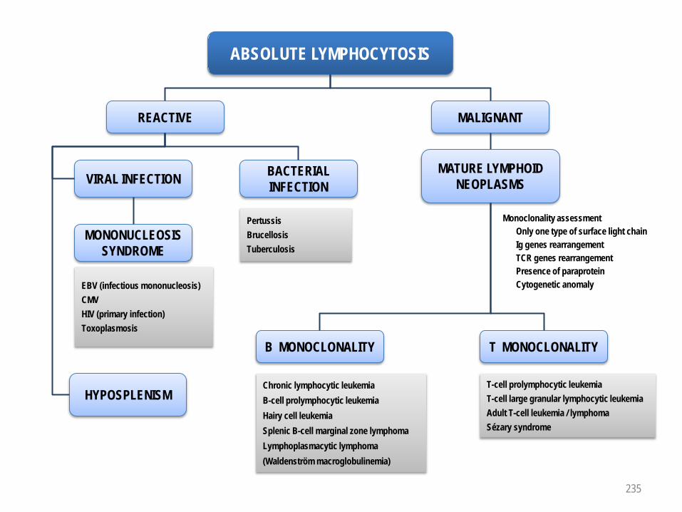
ABSOLUTE LYMPHOCYTOSIS
REACTIVE
EBV (infectious mononucleosis)CMVHIV (primary infection)Toxoplasmosis
HYPOSPLENISM
MALIGNANT
VIRAL INFECTION BACTERIAL INFECTION
MONONUCLEOSIS SYNDROME
MATURE LYMPHOIDNEOPLASMS
PertussisBrucellosisTuberculosis
B MONOCLONALITY T MONOCLONALITY
Monoclonality assessmentOnly one type of surface light chainIg genes rearrangementTCR genes rearrangementPresence of paraproteinCytogenetic anomaly
Chronic lymphocytic leukemiaB-cell prolymphocytic leukemiaHairy cell leukemiaSplenic B-cell marginal zone lymphomaLymphoplasmacytic lymphoma (Waldenström macroglobulinemia)
T-cell prolymphocytic leukemiaT-cell large granular lymphocytic leukemia Adult T-cell leukemia / lymphomaSézary syndrome
LYMPHOCYTOSIS
235

ABSOLUTE EOSINOPHILIA
REACTIVE MALIGNANT
PARASITES ALLERGIES
SYSTEMIC DISEASES MISCELLANEOUS
MYELOPROLIFERATIVENEOPLASM
ACUTE LEUKEMIA
Nematodes (oxyuriasis, ascariasis, trichinosis, filariasis, ancilostomiasis)Trematodes (schizostomiasis, fascioliasis)Cestodes (teniasis, echinococcosis)
Allergic rhinitisAsthma bronchialeUrticaria, atopic dermatitisDrugs (penicillin, carbamazepine,gold salts)
Panarteritis nodosaAllergic granulomatosis angiitis (Churg-Strausssyndrome)Eosinophilic fasciitis (Shulman syndrome)Vasculitis
Recovery phase after acute infectionAdrenal failureChronic enteropathyGM-CSF treatmentHodgkin lymphomaHypereosinophilic syndrome1
Chronic eosinophilic leukemiaChronic myelogenous leukemia
Acute myeloid leukemia with inv(16)
EOSINOPHILIA
MYELOID AND LYMPHOIDNEOPLASMS WITH EOSINOPHILIA
With PDGFRA gene rearrangementWith PDGFRB gene rearrangementWith FGFR1 anomalies
1 Eosinophilia ≥ 1.5 G / L without any evidence for myeloproliferative neoplasm, myeloid and lymphoid neoplasm with eosinophilia and PDGFRA, PDGFRB or FGFR1 anomaly, or AML 236

ABSOLUTE MONOCYTOSIS
REACTIVE MALIGNANT
BACTERIAL INFECTIONTuberculosisSalmonellosis
BrucellosisBacterial endocarditis
PARASITIC INFECTIONMalaria
RECOVERY PHASE AFTER INFECTION
RECOVERY PHASE AFTER AGRANULOCYTOSIS
ALCOHOLIC HEPATOPATHY
HODGKIN LYMPHOMA
G-CSF or GM-CSF TREATMENT
MYELODYSPLASTIC / MYELOPROLIFERATIVE
NEOPLASMChronic myelomonocytic leukemia
ACUTE LEUKEMIA
MONOCYTOSIS
Acute myeloid leukemia with t(9;11) Acute myelomonocytic leukemiaAcute monocytic leukemia
237

THROMBOCYTOPENIA
PSEUDO THROMBOCYTOPENIA TRUE THROMBOCYTOPENIA
SOLITARYTHROMBOCYTOPENIA
PANCYTOPENIA
CENTRALTHROMBOCYTOPENIA
INFECTION AUTOIMMUNEDISEASE
D I C
PRIMARY IMMUNETHROMBOCYTOPENIA
DRUG
PERIPHERALTHROMBOCYTOPENIA
EBVHIVMalaria
Heparin
SLELymphoid neoplasm
Due to EDTA (anticoagulant)
Blood smear examinationPlatelet aggregates
Thiazide, alcohol
Megakaryocytes
Bone marrowSplenomegaly ?B12, folates ?
BONE MARROW APLASIA
BONE MARROW INFILTRATION
MYELODYSPLASIA
BONE MARROW FIBROSIS
B12 OR FOLATE DEFICIENCY
HYPERSPLENISM
THROMBOCYTOPENIA
238

THROMBOCYTOSIS
SOLITARY WITH POLYCYTHEMIA AND / ORNEUTROPHILIA
INFLAMMATORYSYNDROME
MYELOPROLIFERATIVENEOPLASM
HEMORRHAGE ORACUTE HEMOLYSIS
IRON DEFICIENCY AFTER SPLENECTOMY
Essential thrombocythemia
Chronic myelogenous leukemia
Polycythemia Vera
Primary myelofibrosis
C R PSerum ironTransferrinFerritin
Reticulocyte countUnconjugated bilirubinLDHHaptoglobin
Spleen ?
JAK2
Karyotype
Red cell volume
Erythroid cell cultures
THROMBOCYTOSIS
239

PROLONGEDPROTHROMBIN TIME
(PT OR QUICK)
REPEATED BLEEDINGEPISODES
(POSITIVE FAMILY HISTORY)
ISOLATED HEREDITARY
FACTOR DEFICIENCY
II, V, VII, X
TP allongé
1 Mixing test : PT / Quick on a 1:1 mixture of patient plasma with normal plasma
Exploration of extrinsic pathwayFactors II, V, VII, X
DRUGS
Oral anticoagulants (↓ II, VII, IX, X)Acenocoumarol (Sintrom®)Phenprocoumon (Marcoumar®)
LIVERFAILURE
DISSEMINATEDINTRAVASCULAR
COAGULATION (DIC)
↓ II, V, VII, X
↓ II, V, VII, X, fibrinogenD-dimers +, thrombocytopenia
VITAMIN K DEFICIENCY
No PT correctionin mixing test1
INHIBITOR OF EXTRINSIC PATHWAY FACTOR
YES NO
240

PROLONGATION OF ACTIVATED PARTIAL THROMBOPLASTIN TIME (aPTT)
REPEATED BLEEDINGEPISODES
(POSITIVE FAMILY HISTORY)
VON WILLEBRANDDISEASE
Hemophilia A
aPTTCORRECTED
aPTT PROLONGATION
aPTTNOT CORRECTED
1 Mixing test : aPTT on a 1:1 mixture of patient plasma with normal plasma2 PFA-100TM (Platelet Function Analyzer) : in vitro measure of the time to occlusion
of a membrane (measure of platelet adhesion and aggregation process). Replaces, if device available, the classical bleeding time
Exploration of intrinsic pathway
FXI
DRUGS
Heparins
Factor XII deficiencyPrekallikrein deficiencyHigh molecular weight kininogen deficiency
Mixing test1
Lupus anticoagulant
Prolongationof PFA-100TM 2
↓ Factor VIII
YES NO
INHIBITOR OF INTRINSIC PATHWAY FACTOR
F VIII
aPTTNOT CORRECTED
Mixing test1
F VIII / IX / XI NORMAL
FactorXI deficiency
Dosage F VIII
F IX
Hemophilia B
Dosage F IX
Dosage F XI
241

BY WAY OF CONCLUSION
242
Authors and Collaborators :
Pierre-Michel Schmidt, MD, Hematology Service, Centre Hospitalier Universitaire Vaudois (CHUV), Lausanne (Switzerland)
Pierre Cornu, MD, Chairman, Board for Postgraduate and Continuous Medical Education, Swiss Society of Hematology
Anne Angelillo-Scherrer, MD and PhD, Assistant Professor, Hematology Service, CHUV
Stéphane Quarroz, Technician in Biomedical Analyses, Head of Unit, Central Hematology Laboratory (LCH), CHUV
Pieter Canham van Dijken, MD
Transfusion Medicine is presently not covered in this synopsis
Related morphological inconography may be found on :http://ashimagebank.hematologylibrary.org
Remarks or suggestions for improvement of this document are welcome and may be addressed to the authors : Pierre-Michel Schmidt : [email protected]
Pierre Cornu : [email protected]
Anne Angelillo-Scherrer : [email protected]
January 2010