Embed Size (px)
Citation preview

Published: July 06, 2011
r 2011 American Chemical Society 5786 dx.doi.org/10.1021/jm200450h | J. Med. Chem. 2011, 54, 5786–5795
ARTICLE
pubs.acs.org/jmc
A Synthetic Antimicrobial Peptidomimetic (LTX 109): StereochemicalImpact on Membrane DisruptionJohan Isaksson,†,^ Bjørn O. Brandsdal,‡,^ Magnus Engqvist,|| Gøril Eide Flaten,§ John S. Mjøen Svendsen,†,||
and Wenche Stensen*,||
†Department of Chemistry, University of Tromsø, N-9037 Tromsø, Norway‡The Norwegian Structural Biology Centre and the Centre for Theoretical and Computational Chemistry, Department of Chemistry,University of Tromsø, N-9037 Tromsø, Norway§Drug Transport and Delivery Research Group, Department of Pharmacy, University of Tromsø, N-9037 Tromsø, Norway
)Lytix Biopharma AS, Tromsø Research Park, N-9294 Tromsø, Norway
bS Supporting Information
’ INTRODUCTION
Ever since the first discovery of cationic antimicrobial peptides(CAPs) in the early 1980s through the identification of cecropinfrom silk moths (Hyalophora cecropia) by Boman’s group1 andmagainin from the claw footed frog (Xenopus laevis) by Zasloff,2
workers in the field have been aware of their unique properties aspotential antimicrobial agents. The CAPs proved to be activeagainst an unusually broad spectrum of microbes, including bothGram positive and Gram negative bacteria as well as severalfungi.2 Even viruses and cancer cells could be preferentially killedby some CAPs. The conventionally accepted Shai�Matsuzaki�Huang model3�5 explains that the CAPs exert their action bybinding to the cell membrane, causing cell death by direct mem-brane disruption or by translocation into the cytosol wherepotential internal targets are affected. The current opinions aboutthemore detailed interactions betweenCAPs and lipidmembraneshave recently been comprehensively reviewed.6,7 Interestingly,many of the established observations in terms of hydrophobicity,amphipathicity, and charge for the more frequently studiedlonger CAPs (12�50 residues) are also valid for the shortSAMPs8 of interest to this work (three to four residues).
CAPs are an important component of the innate immunesystem in most species where they act as a first line of defenseto delay any infection until a specific immune response towardthe infection has had time to take effect.9 The apparent unspecific
mode of action of CAPs is thus different from most antibio-tics in clinical use today, which are specifically targeting bacte-rial components, often making CAPs equally active againstmultiresistant bacteria as they are against antibiotic-susceptiblestrains.
The proposed general mechanism of action is further con-sidered to make the development of bacterial resistance againstantimicrobial peptides unlikely.10 Even though this view has latelybeen somewhat revised,11,12 the current consensus is still thatresistance development is significantly more difficult against thisclass of compounds compared to antibiotics used in the clinictoday. It is of special interest to this work that the short SAMPsthat 1 (LTX 109) (Figure 1) belongs to are poor substrates forproteolytic cleavage because of its small size and syntheticmodifications.13,14 This leaves bacteria with the demandingtask of modifying its surface by changing lipid composition orneutralizing the surface molecules chemically15,16 as the onlyremaining recognized general defense mechanism. The morespecific mechanisms of trapping molecule secretion17,18 orATP-driven efflux pumps19�21 are also possible but come ata great efficiency cost for bacterial proliferation, requiring greatnumbers of generations of steady selection pressure in order to
Received: April 14, 2011
ABSTRACT: LTX 109 is a synthetic antimicrobial peptidomi-metic (SAMP) currently in clinical phase II trials for topicaltreatment of infections of multiresistant bacterial strains. Allpossible eight stereoisomers of the peptidomimetic have beensynthesized and tested for antimicrobial effect, hemolysis, andhydrophobicity, revealing a strong and unusual dependenceon the stereochemistry for a molecule proposed to act on ageneral membrane mechanism. The three-dimensional structureswere assessed using nuclear magnetic resonance spectroscopy(NMR) and molecular dynamics (MD) simulations in aqueous solution and in phospholipid bilayers. The solution structures ofthe most active stereoisomers are perfectly preorganized for insertion into the membrane, whereas the less active isomers need topay an energy penalty in order to enter the lipid bilayer. This effect is also found to be reinforced by a significantly improved watersolubility of the less active isomers due to a guanidyl-π stacking that helps to solvate the hydrophobic surfaces.

5787 dx.doi.org/10.1021/jm200450h |J. Med. Chem. 2011, 54, 5786–5795
Journal of Medicinal Chemistry ARTICLE
develop at all. For these reasons, SAMPs in general and 1 inparticular are expected to belong to the top tier of antibioticsbeing the most resilient to microbial resistance development.
The first attempts at developing CAPs into clinically usefuldrugs started 20 years ago. Many antimicrobial peptides arecurrently in drug development; a review from 2006 lists 14pharmaceutical companies as active in this field.9 Despiteserious efforts, to date, no CAP has yet reached the clinic. Thisreflects the difficulty of developing peptide drugs, mainly due tounfavorable pharmacokinetics ascribed to the inherent liabilityof peptides toward proteases. This is particularly the casefor CAPs, as their obligate cationic residues make them goodsubstrates for the chymotrypsin family of endoproteases.22 Inaddition, the production costs quickly become commerciallyunacceptable for the synthesis of larger peptides. For thisreason we have for a number of years investigated a minima-listic approach to the CAP concept. This endeavor has involv-ed the identification of essential residues in lactoferricin B(Lfcin B) through a full alanine scan23 and substitution of singleresidues,24 leading to the discovery of a pharmacophore modelfor very short CAPs.25 The resulting pharmacophore model iscomposed of units of cationic charge and lipophilic bulk. Wehave also established that lipophilic bulk can be boosted byincorporating nongenetically coded synthetic amino acids intoCAPs,26�28 which can be introduced into the pharmacophoremodel to construct highly active CAP mimetics using as few astwo or three amino acids.29 This novel class of very shortantimicrobial peptides has been described as synthetic anti-microbial peptidomimetics, SAMPs,8 and one SAMPmolecule,1, has entered phase II clinical studies. Peptide 1 is a tripeptidecontaining a modified tryptophan residue and is capped at theC-terminal by an ethylphenyl group. The small size and thepresence of just three chiral carbon atoms of 1makes it feasibleto prepare all eight possible stereoisomers, allowing for detailedprobing of the effect of stereochemistry on the antimicrobialefficacy and the toxicity on mammalian cells. The present studyexamines all the eight possible stereoisomers, denoted com-pounds 1�8 (Figure 1), and how the stereochemistry affectstheir structural properties in solution and when inserted intophospholipid bilayers using NMR spectroscopy and theoreticalmethodologies.
’EXPERIMENTAL SECTION
Peptide Synthesis. L- and D-2,5,7-tri(tert-butyl)tryptophan (Tbt)was prepared from L- and D-tryptophan, respectively, as described in theliterature.28 The L- and D-tryptophan and the L- and D-Boc-arginine werepurchased from Bachem or Sigma Aldrich.
TheC-terminal peptide part with a free amino group (1 equiv) and Bocprotected amino acid (1.05 equiv) were dissolved in DMF (2�4 mL/mmol amino component) before addition of DIPEA (4.8 equiv). Themixture was cooled on ice before HBTU (1.2 equiv) and HOBt (1.8equiv) were added, and the reaction mixture was agitated at ambienttemperature for 4 h. The reaction mixture was diluted with ethyl acetate,washed with citric acid solution (5% m/m), NaHCO3 (5% m/m)solution, and predried with saturated brine. The organic phase was driedover Na2SO4, filtered, and the solvent was removed under vacuum. Theresulting peptide was kept in a freezer at�18 �C (30min) before the Bocprotecting group was removed by using 8 �C 4 M HCl in dioxane.
The peptides were purified using reversed phase HPLC on a Delta-Pak (Waters) C18 column (100 Å, 15 μm, 25 mm � 100 mm) with amixture of water and acetonitrile (both containing 0.1% TFA) as eluent.The purity of the peptides was further detemined to be 95�100% by RP-HPLC using an analytical Delta-Pak (Waters) C18 column (100 Å,5 μm, 3.9 mm � 150 mm) and positive ion electrospray mass spectro-metry on a Waters Quattro micro quadrupole mass spectrometer andsubsequently confirmed qualitatively by NMR to be >95%.
The peptides (1�8) were assembled in solution by stepwise aminoacid coupling using standard Boc protecting strategy and subsequentlypurified using reverse phase HPLC. The procedure is described infurther detail in the Supporting Information.Antibacterial and Hemolytic Assays. MIC determinations on
Staphylococcus aureus, strain ATCC 25923, Escherichia coli, strain ATCC25922, and Pseudomonas aeruginosa, strain ATCC 27853, were performedby using standard methods.30 The activity of the peptides against humanerythrocytes was measured as reported previously.31
Liposome Preparation. 50 mg/mL 1-palmitoyl-2-oleoylglycero-3-phosphoethanolamine (POPE)/1,2-dimyristoylglycero-3-phosphatidyl-choline (DMPC) (8:2), 1,2-dioleoylglycero-3-phosphocholine (DOPC),and 1,2-dimyristoylglycero-3-phosphatidylcholine (DMPC) liposomedispersions in 10 mM phosphate buffer in D2O, pH 7.6, were made bythe film hydration method.32 Probe sonication (Vibracell high intensityultrasonic processor from Sonics and Materials, Newtown, CT, U.S.)was used to prepare small unilamellar vesicles (SUVs) with a meandiameter of about 35 nm (number weighted distribution measured onPCS, submicrometer particle sizer, model 370, Nicomp, U.S.).NMR Spectroscopy. All NMR experiments were acquired on a
Varian Inova spectrometer operating at 600MHz 1H frequency, equippedwith cryogenically cooled inverse triple resonance probe (second gen-eration). All experiments were acquired using standard pulse sequencesincorporated in the VnmrJ 2.2D package.
Fully deuterated solvents were chosen in order to improve thespectral quality near the water line and avoid the need for water signalsuppression schemes while still allowing the observation of the slowlyexchanging amide resonances. Approximately 5 mg of each peptide(1�8) was dissolved in 0.5 mL of D2O, pH adjusted to 6.0 using NaOD.The peptide solutions were assigned and subsequently titrated into theliposome dispersions. All experiments on the liposome dispersions wereacquired at 310 K. Typically, data matrices of 1400� 512 complex datapoints were collected using up to 32 transients and presaturation of thewater resonance during the relaxation delay. NOESY spectra wereacquired with mixing times of 100 and 300 ms.Computational Details. Molecular models of all peptides
were built with Maestro, version 9.0.33 The OPLS2005 force field34,35
was used for all calculations. All molecular dynamics simulations ofpeptides in solvent were carried out using theMD program package Q.36
Figure 1. Chemical structure of Arg-Tbt-Arg-NH-EtPh (1, LTX 109)with atom numbering for NMR assignment. The peptides (1�8) arelabeled with respect to their stereochemistry according to the right panel.
5788 dx.doi.org/10.1021/jm200450h |J. Med. Chem. 2011, 54, 5786–5795
Journal of Medicinal Chemistry ARTICLE
MacroModel37 was used to assign missing parameters and charges to allpeptides according to the OPLS2005 all-atoms force field.34,35 Thesimulation center was defined as the CR atom of the center amino acidresidue. Each peptide was immersed into a spherical droplet of watermolecules with a 25 Å radius centered at the simulation center. Watermolecules were described using the TIP3P potential.38 Atoms in theoutermost 4.2 Å were weakly restrained to their initial positions with aharmonic potential of 5.0 kcal 3mol�1
3�2. The nonbonded potential
was truncated at 10 Å for solvent�solvent interactions. Long-rangeelectrostatics was treated using a multipole expansion method.39 Inter-actions between peptides and solvent were not truncated, and they werethus allowed to interact with the entire system. All systems were heatedfrom 1 to 300 K during 100 ps, using a stepwise scheme, followed byan equilibration period of 500 ps. SHAKE40 was used to constrain bondsand angles on solvent molecules. A time step of 2 fs was used for theproduction phase, and the temperature was maintained at 300 K using aweak coupling to an external bath. The production phase consisted of 50ns and conformations were sampled every ps. A total of three suchsimulations were run for each peptide, differing in the distribution ofinitial velocities, giving a final simulation time of 150 ns and a total of150 000 structures. The resulting trajectories were visually examinedusing the Visual Molecular Dynamics program.41
Peptide�membrane interactions were studied using MD simulationsand the Desmond program package.42�44 The systemwas prepared witha POPE membrane and explicit SPC water molecules. The simulationwas performed in an orthorhombic box (30 Å� 36 Å� 100 Å). In orderto study the mechanism of how the peptides approach and enter themembrane, the peptides were manually placed outside the membrane,leaving a 3 Å gap between the peptide and the nearest lipid molecules.Simulations of 50 ns were carried out.
’RESULTS
1 is a SAMP8 tripeptide composed around a central 2,5,7-tri(tert-butyl)tryptophan residue (Tbt) flanked by two argininesand a C-terminal phenethyl modification. The peptide containsthree chiral centers; thus, eight different stereoisomers arepossible, where 1 itself is the all-L-enantiomer. All eight stereo-isomers (peptides 1�8, Figure 1) were prepared and analyzed.
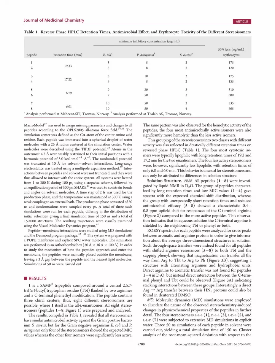
The results, compiled in Table 1, revealed that all stereoisomershave similar antimicrobial activity against the Gram positive bacter-ium S. aureus, but for the Gram negative organisms E. coli and P.aeruginosa only four of the stereoisomers showed the expectedMICvalues whereas the other four isomers were significantly less active.
The same pattern was also observed for the hemolytic activity of thepeptides; the four most antimicrobially active isomers were alsosignificantly more hemolytic than the less active isomers.
This grouping of the stereoisomers into two classeswith differentactivity was also reflected in drastically different retention times onreversed phase HPLC (Table 1). The four most cytotoxic iso-mers were typically lipophilic with long retention times of 19.3 and17.2min for the two enantiomers. The four less active stereoisomerswere, however, significantly less lipophilic with retention times ofonly 6.8 and 6.0min. This behavior is unusual for stereoisomers andcan only be attributed to differences in solution structure.Solution Structure. NMR. All peptides (1�8) were investi-
gated by liquid NMR in D2O. The group of peptides character-ized by long retention times and low MIC values (1�4) gavespectra with the expected chemical shift distribution, whereasthe group with unexpectedly short retention times and reducedantimicrobial efficacy (5�8) showed a characteristic 0.4�0.8 ppm upfield shift for resonances of the C-terminal arginine(Figure 2) compared to the more active peptides. This observa-tion indicates that in aqueous solution the C-terminal arginine isshielded by the neighboring Tbt or phenyl or both.ROESY spectra for each peptide were analyzed for cross-peaks
between aromatic and arginine protons in order to give informa-tion about the average three-dimensional structures in solution.Such through-space transfers were indeed found for all peptideswith shifted arginine resonances (5�8) to both Tbt and thecapping phenyl, showing that magnetization can transfer all theway from Arg to Tbt to Arg to Ph (Figure 3B), suggesting astructure with alternating arginines and hydrophobic units.Direct arginine to aromatic transfer was not found for peptides1�4 in D2O, but instead direct interaction between the C-term-inal phenyl and Tbt could be observed (Figure 3A) indicatingstacking interactions between these groups. Interestingly, a directArg f Arg transfer between their HNε protons could also befound in deuterated DMSO.MD. Molecular dynamics (MD) simulations were employed
to elucidate the nature of the observed stereochemistry-inducedchanges in physicochemical properties of the peptides in furtherdetail. The four stereoisomers L-L-L (1), D-L-L (3), L-D-L (5), andL-L-D (7) were subjected to extensive MD simulations in explicitwater. Three 50 ns simulations of each peptide in solvent werecarried out, yielding a total simulation time of 150 ns. Clusteranalysis of the root-mean-squared deviation with respect to the
Table 1. Reverse Phase HPLC Retention Times, Antimicrobial Effect, and Erythrocyte Toxicity of the Different Stereoisomers
minimum inhibitory concentration (μg/mL)
peptide retention time (min) E. colia P. aeruginosab S. aureusa50% lysis (μg/mL)
erythrocytes
119.33
3 8 2 175
2 3 8 3 120
317.22
3 10 2 175
4 4 8 2 135
56.79
9 30 3 510
6 7 50 3 600
76.01
10 50 3 535
8 10 50 4 605aAnalysis performed at Mabcent-SFI, Tromsø, Norway. bAnalysis performed at Toslab AS, Tromsø, Norway.

5789 dx.doi.org/10.1021/jm200450h |J. Med. Chem. 2011, 54, 5786–5795
Journal of Medicinal Chemistry ARTICLE
starting structure was used to form groups of conformations withsimilar three-dimensional structures for each simulation. Analysisof the most populated groups revealed that the selected stereo-isomers indeed have different conformations in solution that canbe characterized as either amphipathic or nonamphipathic. The
two groups are illustrated in Figure 4 by representative snapshotsfrom the MD trajectories of the L-L-L (1) and L-L-D (7) stereo-isomers. These two structures represent the group that has byfar the highest population in their respective simulations. Inorder to compare the stacking between Tbt and the C-terminal
Figure 3. 300 ms mixing time ROESY spectra of the L-L-L peptide (1) (A) and the L-L-D peptide (7) (B) in D2O at 25 �C. The L-L-L peptide (1) showsphenyl-Tbt contacts (H38TH30 using Figure 1 notation), while the L-L-D peptide (7) shows unexpected C-terminal arginine-Tbt contacts (H13b0/00 TH27, using Figure 1 notation).
Figure 2. Proton assignment of the four different isomers in D2O. The peptides change stereochemical sense at the last peptide step, and peptides 5�8display a distinct upfield shift of its second arginine. Note that peptides 1 and 2, peptides 3 and 4, peptides 5 and 6, peptides and 7 and 8 are mirror imagepairs that give identical NMR spectra.

5790 dx.doi.org/10.1021/jm200450h |J. Med. Chem. 2011, 54, 5786–5795
Journal of Medicinal Chemistry ARTICLE
capping phenyl group with the stacking between Tbt and theC-terminal arginine, representative atomic distances were cal-culated as a function of simulation time. The plots in Figure 5illustrate the time dependence of the interactions during the tra-jectories of both stereoisomers. There is clearly a preference forTbt-phenyl stacking in the L-L-L (1) peptide while the L-L-D (7)peptide shows a strong preference for Tbt-arginine stacking, thusrevealing the source of the magnetic shielding experienced by theC-terminal arginine and confirming the stacking pathways forpeptides 5�8 suggested by the results from the NMR studies.Peptide�Membrane Interactions. NMR.NMRstudies of the
L-L-L (1) and the L-L-D (7) stereoisomers in small unilamellar vesicle(SUV) dispersions composed of POPE/DMPC (8:2), DMPC, andDOPC have been carried out to examine the conformationalbehavior of the peptides upon insertion into a membrane.Since the theoretical simulations were all performed in POPE,
attempts were first made to study the peptides in liposomes
consisting of as high a POPE content as our liposome methodswould allow. Reasonably sized liposomes could be made using an8:2 POPE/DMPC ratio, but these SUVs turned out to be unstableupon peptide titration and provided poor spectra (Figure S6 in theSupporting Information). Titration and NMR experiments weretherefore repeated in pure DMPC and DOPC that, similar toPOPE, also carry no net charge but provide stable liposomes thatcan host sufficiently high concentrations of titrated peptide.Qualitatively, the peptide peaks show the same pattern in POPE/DMPC, DMPC, and DOPC bilayers. Both DMPC and DOPCdata were used in the assignment process, and since DOPC bi-layers provide the best resolved spectra, all further NMR datadiscussed will refer to spectra acquired in DOPC liposomes.Assignment of the peptide resonances inside the phospholipid
bilayers is not straightforward because of spectral overlap and veryfast relaxation (rendering heteronuclear experiments useless).Using deuterated phospholipids helped but was not enough to
Figure 4. Representative snapshots from the MD simulations of L-L-L (1) (top) and L-L-D (7) (lower) illustrating the amphipathic and nonamphipathicconformations, respectively. The intercarbon distances of interest that are plotted in Figure 5, whose attached protons give rise to the key correlationsshown in Figure 3, are represented with dashed lines.

5791 dx.doi.org/10.1021/jm200450h |J. Med. Chem. 2011, 54, 5786–5795
Journal of Medicinal Chemistry ARTICLE
unambiguously assign all resonances. However, for the scope ofthis work it was only necessary to focus on the resonances in thearomatic region of the spectra where there is no spectral overlap.This region holds the resonances of the two Tbt protons 25 and27, the three symmetric phenyl resonances 38�40 originatingfrom five protons, and any nonsubstituted HN protons on thepeptide backbone. The Tbt resonances were separated from thephenyl resonances by subtracting the spectra of 1 with a penta-fluorinated phenyl (1-F5) from the original 1, and theHN10 signalwas identified by subtracting the spectra of the uncapped 1 from1-F5 (Figure S1 in the Supporting Information). By use of theseNOESY spectra of these “edited” peptides, it was possible tounambiguously assign the Tbt methyl resonances and separatethe phenyl resonances from the Tbt resonances, which is the keyto be able to confirm phenyl-Tbt contacts inside the membranes(Figures S2�S5 in the Supporting Information). For practicalreasons, the phenyl protons are individually denoted in Figure 6according to the most likely assignment based on NOESY buildupfrom the ethyl protons to the phenyl protons, even though theassignment is not completely unambiguous. All conclusions in thiswork were, however, based on treating all resonances originating
from the phenyl as one set of resonances, and as such, there areno ambiguities.Inspection of the interactions between the aromatic signals
of the C-terminal phenethyl group and the methyl signals of thetert-butyl groups of Tbt (Figure 6) reveals that the amphipathiccharacter of the molecule, here probed by the phenyl/Tbt NOESYcross-peaks, is reinforced by the insertion into the SUVs (com-pare to Figure 3). This is observed for both the more active 1�4and for the less active stereoisomers 5�8. Interestingly there isno trace of any shielded arginine resonances for peptides 5�8after insertion into the membranes, and there are no detectableinteractions between arginine and the aromatic units, furthersupporting the view that the less active isomers 5�8 are forcedinto a more amphipathic structure when entering the membrane.MD. MD simulations of the L-L-L (1) and L-L-D (7) stereo-
isomers in the presence of a phospholipid bilayer were carriedout in order to examine the structures of these peptides whenincorporated into a membrane. A representative snapshot of eachpeptide was selected from the simulation of the peptide in solventas starting structure and manually placed at a distance of approxi-mately 3 Å from the nearest atom of the lipid bilayer. Fourindependent simulations were conducted for each peptide usingdifferent random starting velocities.For the L-L-L isomer (1), the peptide rapidly forms electrostatic
interactions with the negatively charged phosphate headgroupsat the membrane surface in two out of four simulations. Oncethese have been formed, the peptide does not diffuse into solventbut starts to bury its hydrophobic elements (Tbt and C-terminalcapping) in the membrane, and after approximately 15�20 nsthe peptide is effectively inserted into the lipid bilayer (Figure 7Aand Figure 7B).In contrast, the L-L-D isomer (7) did not manage to penetrate
into the membrane in any of the trajectories simulated, eventhough the arginines started out at approximately the samedistance from the lipid molecules as in the simulations of peptide1 and managed to make occasional contact with the chargedphosphate groups on the membrane surface.In order to speed the process, the starting structure of peptide
7 was also manually placed inside a membrane model, replacingsome the phospholipid molecules. The initial structure was asnapshot from the MD simulation of peptide 7 in solvent, repre-senting the nonamphipathic conformation as shown in Figure 4.The MD simulations revealed an immediate structural reorgani-zation leading to an amphipathic conformation (Figure 7C andFigure 7D), where the peptide remains inside the membraneduring the remainder of the simulation.It is noteworthy that regardless of the starting structure and the
stereochemistry of the peptide, no nonamphipathic conformationsare ever observed for any length of time inside the membranemodel, highlighting that a peptide needs to be able to take up anamphipathic conformation in order to readily reside inside a lipidbilayer.
’DISCUSSION
The apparent interpretation is that the separation of the eightisomers into two classes of compounds differing in antimicro-bial efficacy, cytotoxicity, and lipophilicity (Table 1) is due to theunderlying structural features of the different peptides. Thecommon factor is that the isomers characterized by high activityall have the same chiral sense in the Tbt residue and theC-terminal arginine, whereas the isomers with low activity and
Figure 5. Carbon�carbon distances (Å) calculated between the TbtandC-terminal capping (EtPh) and theC-terminal arginine as a functionof time (ns) from the MD simulation of L-L-L (1) (top) and L-L-D (7)(bottom). By use of atom numbering from Figure 1, the former distanceis calculated between 13b and 27 and the latter is calculated between 29and 38.

5792 dx.doi.org/10.1021/jm200450h |J. Med. Chem. 2011, 54, 5786–5795
Journal of Medicinal Chemistry ARTICLE
short retention times have opposite stereochemical sense in thesetwo residues; i.e., a shift of sense between these two residuesappears to have a negative effect on activity.
The effect of diastereomeric CAPs has been extensively studied,in particular by Shai,45�47 and the general finding is that only smalldifferences in antibacterial efficacy are found between the differentdiastereoisomers tested whereas the unwanted activity againstmammalian cells may be more affected.46 This trend is, however,not readily seen for 1, as lower cytotoxicity against mammaliancells also results in a proportional loss of antimicrobial activity.One possible reason why this observation is not reproduced bythe peptides in the current study may be that Shai’s studies arebased on a series of much longer peptides (15 amino acids)composed of genetically encoded amino acids, whereas the presentstudy concerns synthetically modified peptides only three residueslong. For this reason, it was somewhat unexpected to seemoleculesthis small be so noticeably affected by the stereochemistry. Thedifference in reverse phase HPLC retention times of 18 vs 6 min istruly remarkable, and the different patterns seen in the NMR studyconfirm that the two structural groups have completely different“folds” in aqueous solution. The molecular dynamics simulationsof 1 in explicit water provide an explanation of why this small andnormally unconstrained molecule so poorly compensates for itsstereochemical sense inmolecular detail. The presence of the Tbtmoiety significantly reduces the conformational freedom in thepeptide, in particular for the amide bond linking the Tbt residue
and the C-terminal arginine residue. The resulting steric rigidityof this part of the 1 sequence gives the stereoisomeric peptideswith the same stereochemical sense of the Tbt and the C-term-inal arginine (1�4) completely different properties compared tothose with the opposite sense (5�8). The preorganization of thebackbone determines which interactions are energetically avail-able between the side chains and the C-terminal modification ofthe peptide. In the L-L-L isomer (and its D-D-D enantiomer) therigidified backbone is set up to make a perfect amphipathicmolecule where the bulky and lipophilic parts of the molecule areoriented on one side of the backbone, whereas the hydrophilicand positively charged arginine residues point in the oppositedirection (see Figure 4). The existence of arginine�arginineinteraction is confirmed by ROESY peaks between the methylresonances of Tbt and the aromatic phenyl resonances in D2O, aswell as weak peaks between the NH of the C-terminal arginineand the δ- and ε-protons of the N-terminal arginine in DMSO.Unfortunately these protons are not directly observable in D2Obecause of chemical exchange. The importance and nature of thearginine�arginine interaction is under more thorough investiga-tion and is not pursued further in this work.
NMR patterns indicate that a similar strongly amphipathicconformation is also the dominant form for the D-L-L (3) and L-D-D (4) isomers. While this conformation is unsuitable with respectto water solubility, it is expected to interact very efficiently withthe column support in reverse phase HPLC, leading to longer
Figure 6. NOESY spectra of the L-L-L peptide (1) (A, 100 ms; C, 300 ms mixing time) and the L-L-D peptide (7) (B, 100 ms; D, 300 ms mixing time) inDOPC liposomes. Both peptides show contacts between the phenylmoiety (denoted 38, 39, and 40) and the Tbtmethyls (denoted 30, 32, and 34) wheninserted into the membrane, despite that peptide 4 lacks this contact in aqueous solution.

5793 dx.doi.org/10.1021/jm200450h |J. Med. Chem. 2011, 54, 5786–5795
Journal of Medicinal Chemistry ARTICLE
retention times. On the other hand, the L-L-D and D-D-L isomerscannot easily adopt the same perfect amphipathic conformation.Rather, because of cation�π interactions between the modifiedtryptophan and the C-terminal arginine and a possible similarinteraction between the N-terminal arginine and the C-terminalphenethyl modification, the L-L-D isomer can adopt a conforma-tion where the lipophilicity of the Tbt and the phenethyl groupsare “hidden” from the bulkwater by the guanidiniummoieties of thearginines (see Figure 4), effectively increasing the water solubility ofthemolecule. Other studies have shown that a parallel (i.e., stacked)arrangement between arginine and Trp residues is energeticallyfavorable in aqueous solution,48 and a similar cation�π interactionbetween theN-terminal arginine and the phenethyl groupsmimick-ing the side chain of a phenylalanine is also viable.49 ExperimentalNMR support is found for similar conformations for the L-D-L(5) and D-L-D (6) isomers as well as the L-L-D (7) and D-D-L (8)isomers. Examination of the modeled conformations shows thatthe guanidinium side chains of the arginines are able to formalmost as many hydrogen bonds with the surrounding watermolecules as when they are not involved in any cation�πinteractions,50 thereby retaining their overall hydrophilic nature.The lipophilic residues on the other hand are partly covered bythe hydrophilic guanidiniun side chains, thus effectively reducingthe overall lipophilicity of the peptides. These observationsprovide a rational explanation for the remarkably long retention
times for peptides 5�8 on reverse phase HPLC compared topeptides 1�4.
The NMR and MD results in liposome membranes shedfurther light on the double nature of the reduced antimicrobialactivity of peptides 5�8. First, the importance of an amphipathicstructure as a prerequisite for effective insertion into membranes ishighlighted. The MD simulations indicate that nonamphipathicconformations are highly unfavored inside the membrane, aseither a hydrophilic or lipophilic side chain inevitably ends up inits unpreferred compartment of the membrane. From the NMRspectra in liposomes we do, however, clearly see that all eightpeptides are inserted into the membranes on the macroscopicexperimental time scale (milliseconds to days), and when doingso, all peptides show amphipathic character, even peptides 5�8that are preorganized differently in solution. In addition to theamphipathic prerequisite that favors peptides 1�4, the overallwater solubility of these peptides further works in favor of theirantimicrobial activity. If we consider the lipid bilayer as a solventand the liposome system as a two-phase solvent system, the amp-hipathic structure further pushes the equilibrium toward mem-brane insertion of peptides 1�4 because of lower solubility inwater than peptides 5�8. This makes 1 an interesting case to studybecause it emphasizes the importance of the solution structure forits potency as an antimicrobic agent even though it exerts its effecton the bacterial cell membrane.
Figure 7. Instantaneous structures as observed in the simulations of a lipid bilayer with the L-L-L stereoisomer (1) seen from the side (A) and from thetop (B) and with the L-L-D stereoisomer (7) seen from the side (C) and from the top (D). The lipid bilayer is shown as transparent spheres.

5794 dx.doi.org/10.1021/jm200450h |J. Med. Chem. 2011, 54, 5786–5795
Journal of Medicinal Chemistry ARTICLE
’ASSOCIATED CONTENT
bS Supporting Information. Spectra used in the peptideassignment inside lipid bilayers (Figures S1�S5) and spectra of1 in POPE/DMPC, 8:2 (Figure S6). This material is available freeof charge via the Internet at http://pubs.acs.org.
’AUTHOR INFORMATION
Corresponding Author*Phone: +47 776 44112. E-mail: [email protected].
Author Contributions^These authors contributed equally.
’ACKNOWLEDGMENT
Financial support from the Research Council of Norway isgratefully acknowledged. The Norwegian Structural Biology Cen-tre is supported by the Functional Genomics Program (FUGE) ofthe Research Council of Norway.
’ABBREVIATIONS USED
NMR, nuclear magnetic resonance;MD, molecular dynamics;CAP, cationic antimicrobial peptide; SAMP, synthetic antimi-crobial peptidomimetic; NOESY, nuclear Overhauser effect spe-ctroscopy; ROESY, rotating frame Overhauser spectroscopy;MS, mass spectrometry; HPLC, high performance liquid chro-matography; Tbt, 2,5,7-tri(tert-butyl)tryptophan; EtPh, ethyl-phenyl/phenethyl; POPE, 1-palmitoyl-2-oleoylglycero-3-phosp-hoethanolamine; DMPC, 1,2-dimyristoylglycero-3-phosphatidy-lcholine; DOPC, 1,2-dioleoylglycero-3-phosphocholine
’REFERENCES
(1) Steiner, H.; Hultmark, D.; Engstrom, A.; Bennich, H.; Boman,H. G. Sequence and specificity of two antibacterial proteins involved ininsect immunity. Nature 1981, 292, 246–248.(2) Zasloff, M. Magainins, a class of antimicrobial peptides from
xenopus skin—isolation, characterization of 2 active forms, and partialcDNA sequence of a precursor. Proc. Natl. Acad. Sci. U.S.A. 1987,84, 5449–5453.(3) Yang, L.; Weiss, T.; Lehrer, R.; Huang, H. Crystallization of
antimicrobial pores in membranes: magainin and protegrin. Biophys.J. 2000, 79, 2002–2009.(4) Shai, Y. Mechanism of the binding, insertion and destabilization
of phospholipid bilayer membranes by alpha-helical antimicrobial andcell non-selective membrane-lytic peptides. Biochim. Biophys. Acta,Biomembr. 1999, 1462, 55–70.(5) Matsuzaki, K. Why and how are peptide�lipid interactions
utilized for self-defense? Magainins and tachyplesins as archetypes.Biochim. Biophys. Acta, Biomembr. 1999, 1462, 1–10.(6) Str€omstedt, A. A.; Ringstad, L.; Schmidtchen, A.; Malmsten, M.
Interaction between amphiphilic peptides and phospholipid mem-branes. Curr. Opin. Colloid Interface Sci. 2010, 15, 467–478.(7) Wimley, W.; Hristova, K. Antimicrobial peptides: successes,
challenges and unanswered questions. J. Membr. Biol. 2011, 239, 27–34.(8) Haug, B. E.; Stensen, W.; Kalaaji, M.; Rekdal, O.; Svendsen, J. S.
Synthetic antimicrobial peptidomimetics with therapeutic potential.J. Med. Chem. 2008, 51, 4306–4314.(9) Hancock, R. E. W.; Sahl, H.-G. Antimicrobial and host-defense
peptides as new anti-infective therapeutic strategies. Nat. Biotechnol.2006, 24, 1551–1557.
(10) Perron, G. G.; Zasloff, M.; Bell, G. Experimental evolution ofresistance to an antimicrobial peptide. Proc. R. Soc. B 2006, 273,251–256.
(11) Shafer, W. M.; Kraus, D.; Peschel, A. Molecular Mechanisms ofBacterial Resistance to Antimicrobial Peptides. In Antimicrobial Peptidesand Human Disease; Springer: Berlin, Germany: 2006; Vol. 306,pp 231�250.
(12) Ginsburg, I.; Koren, E. Are cationic antimicrobial peptides also“double-edged swords”?. Expert Rev. Anti-Infect. Ther. 2008, 6, 453–462.
(13) Svenson, J.; Stensen, W.; Brandsdal, B. r.-O.; Haug, B. E.;Monrad, J.; Svendsen, J. S. Antimicrobial peptides with stability towardtryptic degradation. Biochemistry 2008, 47, 3777–3788.
(14) Karstad, R.; Isaksen, G.; Brandsdal, B. r.-O.; Svendsen, J. S.;Svenson, J. Unnatural amino acid side chains as S1, S10, and S20 probesyield cationic antimicrobial peptides with stability toward chymotrypticdegradation. J. Med. Chem. 2010, 53, 5558–5566.
(15) Peschel, A.; Otto,M.; Jack, R.W.; Kalbacher, H.; Jung, G.; G€otz,F. Inactivation of the dlt operon in Staphylococcus aureus conferssensitivity to defensins, protegrins, and other antimicrobial peptides.J. Biol. Chem. 1999, 274, 8405–8410.
(16) Weidenmaier, C.; Kristian, S. A.; Peschel, A. Bacterial resistanceto antimicrobial host defenses—an emerging target for novel antiinfec-tive strategies?. Curr. Drug Targets 2003, 4, 643–649.
(17) Frick, I.-M.; Åkesson, P.; Rasmussen, M.; Schmidtchen, A.;Bj€orck, L. SIC, a Secreted protein of Streptococcus pyogenes thatinactivates antibacterial peptides. J. Biol. Chem. 2003, 278, 16561–16566.
(18) Jin, T.; Bokarewa, M.; Foster, T.; Mitchell, J.; Higgins, J.;Tarkowski, A. Staphylococcus aureus resists human defensins by produc-tion of staphylokinase, a novel bacterial evasion mechanism. J. Immunol.2004, 172, 1169–1176.
(19) Borges-Walmsley, M. I.; McKeegan, K. S.; Walmsley, A. R.Structure and function of efflux pumps that confer resistance to drugs.Biochem. J. 2003, 376, 313–338.
(20) van Veen, H. W. Towards the molecular mechanism ofprokaryotic and eukaryotic multidrug transporters. Semin. Cell Dev. Biol.2001, 12, 239–245.
(21) Shafer,W.M.; Qu, X.-D.;Waring, A. J.; Lehrer, R. I.ModulationofNeisseria gonorrhoeae susceptibility to vertebrate antibacterial peptidesdue to a member of the resistance/nodulation/division efflux pumpfamily. Proc. Natl. Acad. Sci. U.S.A. 1998, 95, 1829–1833.
(22) Perona, J. J.; Craik, C. S. Evolutionary divergence of substratespecificity within the chymotrypsin-like serine protease fold. J. Biol.Chem. 1997, 272, 29987–29990.
(23) Strom, M.; Rekdal, O.; Svendsen, J. Antibacterial activity of 15-residue lactoferricin derivatives. J. Pept. Res. 2000, 56, 265–274.
(24) Strom, M.; Rekdal, O.; Stensen, W.; Svendsen, J. Increasedantibacterial activity of 15-residue murine lactoferricin derivatives.J. Pept. Res. 2001, 57, 127–139.
(25) Strom, M.; Haug, B.; Skar, M.; Stensen, W.; Stiberg, T.;Svendsen, J. The pharmacophore of short cationic antibacterial peptides.J. Med. Chem. 2003, 46, 1567–1570.
(26) Haug, B. E.; Stensen, W.; Svendsen, J. S. Application of theSuzuki�Miyaura cross-coupling to increase antimicrobial potency gen-erates promising novel antibacterials. Bioorg. Med. Chem. Lett. 2007,17, 2361–2364.
(27) Haug, B.; Andersen, J.; Rekdal, O.; Svendsen, J. Synthesis of a2-aryisulphonylated tryptophan: the antibacterial activity of bovinelactoferricin peptides containing Trp(2-Pmc). J. Pept. Sci. 2002, 8,307–313.
(28) Haug, B.; Skar, M.; Svendsen, J. Bulky aromatic amino acidsincrease the antibacterial activity of 15-residue bovine lactoferricinderivatives. J. Pept. Sci. 2001, 7, 425–432.
(29) Haug, B.; Stensen, W.; Stiberg, T.; Svendsen, J. Bulky non-proteinogenic amino acids permit the design of very small and effectivecationic antibacterial peptides. J. Med. Chem. 2004, 47, 4159–4162.
(30) Amsterdam, D. Susceptibility Testing of Antimicrobials inLiquid Media. In Antibiotics in Laboratory Medicine, 4th ed.; Lorian, V.,Ed.; Williams and Wilkins Co: Baltimore, MD, 1996; pp 75�78.
5795 dx.doi.org/10.1021/jm200450h |J. Med. Chem. 2011, 54, 5786–5795
Journal of Medicinal Chemistry ARTICLE
(31) Strøm, M. B.; Haug, B. E.; Skar, M. L.; Stensen, W.; Stiberg, T.;Svendsen, J. S. The pharmacophore of short cationic antibacterialpeptides. J. Med. Chem. 2003, 46, 1567–1570.(32) Brandl, M. Liposomes as drug carriers: a technological ap-
proach. Biotechnol. Annu. Rev. 2001, 7, 59–85.(33) Maestro, version 9.0; Schr€odinger, LCC: New York, NY, 2009.(34) Kaminski, G. A.; Friesner, R. A.; Tirado-Rives, J.; Jorgensen,
W. L. Evaluation and reparametrization of the OPLS-AA force field forproteins via comparison with accurate quantum chemical calculations onpeptides. J. Phys. Chem. B 2001, 105, 6474–6487.(35) Jorgensen, W. L.; Maxwell, D. S.; TiradoRives, J. Development
and testing of the OPLS all-atom force field on conformationalenergetics and properties of organic liquids. J. Am. Chem. Soc. 1996,118, 11225–11236.(36) Marelius, J.; Kolmodin, K.; Feierberg, I.; Aqvist, J. Q: a molecular
dynamics program for free energy calculations and empirical valence bondsimulations in biomolecular systems. J. Mol. Graphics Modell. 1998, 16(213�225), 261.(37) MacroModel, version 9.6; Schr€odinger: New York, NY, 2008.(38) Jorgensen, W. L.; Chandrasekhar, J.; Madura, J. D.; Impey,
R. W.; Klein, M. L. Comparison of simple potential functions forsimulating liquid water. J. Chem. Phys. 1983, 79, 926–935.(39) Lee, F. S.; Warshel, A. A local reaction field method for fast
evaluation of long-range electrostatic interactions in molecular simula-tions. J. Chem. Phys. 1992, 97, 3100–3107.(40) Ryckaert, J. P.; Ciccotti, G.; Berendsen, H. J. C. Numerical-
integration of Cartesian equations ofmotion of a systemwith constraints—molecular-dynamics of N-alkanes. J. Comput. Phys. 1977, 23, 327–341.(41) Humphrey, W.; Dalke, A.; Schulten, K. VMD: Visual molecular
dynamics. J. Mol. Graphics 1996, 14, 33–38.(42) Desmond Molecular Dynamics System, version 2.2; D.E. Shaw
Research: New York, NY, 2009.(43) Maestro-Desmond Interoperability Tools, version 2.2; Schr€odin-
ger: New York, NY, 2009.(44) Bowers, K. J.; Chow, E.; Xu, H.; Dror, R. O.; Eastwood, M. P.;
Gregersen, B. A.; Klepeis, J. L.; Kolossvary, I.; Moraes, M. A.; Sacerdoti,F. D.; Salmon, J. K.; Shan, Y.; Shaw, D. E. Scalable Algrotihms forMolecular Dynamics Simulations on Commodity Clusters. Proceedingsof the ACM/IEEE Conference on Supercomputing (SC06), Tampa, FL,2006; ACM: New York, NY, 2006.(45) Pag, U.; Oedenkoven, M.; Papo, N.; Oren, Z.; Shai, Y.; Sahl, H.
In vitro activity and mode of action of diastereomeric antimicrobialpeptides against bacterial clinical isolates. J. Antimicrob. Chemother.2004, 53, 230–239.(46) Shai, Y.; Oren, Z. From “carpet” mechanism to de-novo
designed diastereomeric cell-selective antimicrobial peptides. Peptides2001, 22, 1629–1641.(47) Oren, Z.; Hong, J.; Shai, Y. A repertoire of novel antibacterial
diastereomeric peptides with selective cytolytic activity. J. Biol. Chem.1997, 272, 14643–14649.(48) Minoux, H.; Chipot, C. Cation�pi interactions in proteins:
Can simple models provide an accurate description?. J. Am. Chem. Soc.1999, 121, 10366–10372.(49) Gallivan, J. P.; Dougherty, D. A. Cation�pi interactions in
structural biology. Proc. Natl. Acad. Sci. U.S.A. 1999, 96, 9459–9464.(50) Aliste, M. P.; MacCallum, J. L.; Tieleman, D. P. Molecular
dynamics simulations of pentapeptides at interfaces: salt bridge andcation�pi interactions. Biochemistry 2003, 42, 8976–8987.


