Embed Size (px)
Citation preview
a
PATTERNS & PHENOTYPES
A Systematic Survey of Carbonic AnhydrasemRNA Expression During Mammalian InnerEar DevelopmentLing Wu,1,2,3 Borum Sagong,4 Jae Young Choi,1,3 Un-Kyung Kim,4 and Jinwoong Bok1,2,3*
Background: Carbonic anhydrases (CAs), which catalyze CO2 hydration to bicarbonate and protons, have beensuggested to regulate potassium homeostasis and endocochlear potential in the mammalian cochlea. Sixteenmammalian CA isozymes are currently known. To understand the specific roles of CA isozymes in the innerear, a systematic survey was conducted to reveal temporal and spatial expression patterns of all 16 CA iso-zymes during inner ear development. Results: Our quantitative reverse transcriptase-polymerase chain reac-tion results showed that different tissues express unique combinations of CA isozymes. During inner eardevelopment, transcripts of four cytosolic isozymes (Car1, Car2, Car3, and Car13), two membrane-bound iso-zymes (Car12 and Car14), and two CA-related proteins (Car8 and Car11) were expressed at higher levels thanother isozymes. Spatial expression patterns of these isozymes within developing inner ears were determinedby in situ hybridization. Each isozyme showed a unique expression pattern during development. For example,Car12 and Car13 expression closely overlapped with Pendrin, an anion exchanger, while Car2 overlapped withNa-K-ATPase in type II and IV otic fibrocytes, suggesting functional relationships in the inner ear. Conclusions:The temporal and spatial expression patterns of each CA isozyme suggest unique and differential roles ininner ear development and function. Developmental Dynamics 242:269–280, 2013. VC 2012 Wiley Periodicals, Inc.
Key words: carbonic anhydrase; inner ear; cochlea; mouse; development
Key findings:� A systematic survey for transcripts of all 16 mammalian carbonic anhydrase isozymes in various organs
showed that each organ expresses unique combinations of carbonic anhydrase isozymes.� During inner ear development, transcripts of four cytosolic isozymes (Car1, Car2, Car3, and Car13), two mem-
brane-bound isozymes (Car12 and Car14), and two CA-related proteins (Car8 and Car11) were expressed athigher levels than other isozymes.
� Each isozyme showed a unique temporal and spatial expression pattern during inner ear development.� Expression patterns for some isozymes closely overlapped with genes previously associated with inner ear de-
velopment or function, such as Pendrin, Pou3f4, or Bmp4.
Accepted 3 December 2012
INTRODUCTION
Carbonic anhydrases (CAs) are zinc-containing metalloenzymes that
catalyze the reversible conversion ofcarbon dioxide to bicarbonate ion anda proton. These enzymes, found in all
types of organisms, are encoded byfive independent gene families: the a-,b-, g-, d-, and e-CAs (Supuran, 2008b;
1Department of Anatomy, Yonsei University College of Medicine, Seoul, South Korea2Department of Otorhinolaryngology, Yonsei University College of Medicine, Seoul, South Korea3BK21 Project for Medical Science, Yonsei University College of Medicine, Seoul, South Korea4Department of Biology, Kyungpook National University, Daegu, South KoreaGrant sponsor: Korea Healthcare Technology R&D Project, Ministry for Health, Welfare & Family Affairs; Grand number: A100493.Grant sponsor: Basic Science Research Program through the National Research Foundation of Korea (NRF) funded by the Ministry ofEducation, Science and Technology; Grand number: 2011-0028066.*Correspondence to: Jinwoong Bok, Department of Anatomy, Yonsei University College of Medicine, Seoul, South Korea.E-mail: [email protected]
DOI: 10.1002/dvdy.23917Published online 10 December 2012 in Wiley Online Library (wileyonlinelibrary.com).
DEVELOPMENTAL DYNAMICS 242:269–280, 2013
VC 2012 Wiley Periodicals, Inc.
Xu et al., 2008). Only a-CA familymembers have been identified inmammals (Henry and Swenson, 2000;Esbaugh and Tufts, 2006). Thus far,16 a-CA isozymes have beendescribed, with different catalyticactivities and subcellular localiza-tions: CAI, II, III, VII, and XIII arecytoplasmic; CAIV, IX, XII, XIV, andXV are membrane-bound; CAVa andVb are mitochondrial; and CAVI issecreted (Supuran, 2008a,b). CAVIII,X, and XI are called CA-related pro-teins (CA-RPs) because they lack clas-sical CA enzymatic activity and theirbiological functions remain unclear.
The importance of carbonic anhy-drases has been shown in many phys-iological and pathological processes,including pH and CO2 homeostasis,electrolyte secretion, respiration,CO2/HCO3
� transport, and boneresorption and calcification (Supuran,2008a). In the respiratory system,CAs are involved in most CO2 trans-port and excretion from metabolicallyactive tissues to red blood cells, andfinally to gas exchanging organs, suchas the lungs (Henry and Swenson,2000). Cytoplasmic CAI and II are themost abundant CA isozymes in thered blood cells, while both cytoplasmic(CAII) and membrane-bound CAs(CAIV) are found in the lung (Henryand Swenson, 2000; Esbaugh andTufts, 2006). The role of CAs has alsobeen studied in detail in regulatingrenal physiology, such as acid/basehomeostasis and bicarbonate reab-sorption (Purkerson and Schwartz,2007). CAs are expressed in most kid-ney segments. Cytoplasmic CAIIaccounts for most CA activity in thekidney, and different combinations ofmembrane-bound CAs, includingCAIV, CAXII, CAXIV, and CAXV, arealso expressed in different species(Purkerson and Schwartz, 2007).CAII is involved in bone physiology,such as osteoclast differentiation andbone resorption, and CAII-deficiencyleads to osteopetrosis in humans andmice (Sly and Hu, 1995; Lehenkariet al., 1998; Margolis et al., 2008). Inaddition, various CAs expressed inthe gastrointestinal canal and diges-tive glands are involved in ammoniadetoxification, saliva production, gas-tric acid production, bile production,pancreatic juice production, and in-testinal ion transport (Pan et al.,
2007; Supuran, 2008a). MitochondrialCAV has been associated with molecu-lar signaling, such as insulin secre-tion in pancreatic b cells (Parkkilaet al., 1998). Some CA isozymes aredown- or up-regulated in varioustumors and have been associated withoncogenesis and tumor progression(Supuran, 2008a,b). Consistently, twohypoxia-inducible CA isozymes, CAIXand XII, were shown to promote tu-mor growth by regulating pH in anacidic and hypoxic microenvironment(Jarvela et al., 2008; Chiche et al.,2009). In the brain, CAIV and XIVwere shown to regulate pH in extrac-ellular fluid (Shah et al., 2005).Lastly, CAIII, which is highlyexpressed in many tissues, includingskeletal muscle, has relatively low CAactivity. Its physiological role isunclear because mice lacking CAIIIdo not have noticeable anatomical orphysiological abnormalities (Kimet al., 2004). CAIII can, however, pro-tect cells from oxidative damage by S-glutathiolation on two cysteine resi-dues in response to oxidative stress,thereby functioning as an oxyradicalscavenger (Raisanen et al., 1999; Mal-lis et al., 2002, 2000; Gailly et al.,2008).
CA activity in the inner ears hasbeen demonstrated biochemically byvisualizing CO2 hydration in guineapigs, cats, and chinchillas (Erulkarand Maren, 1961; Lim et al., 1983;Hsu and Nomura, 1985; Okamuraet al., 1996). Although the specificlocation of CA activity in the innerear differs slightly depending on thespecies and detection method, CA ac-tivity is present in the organ of Corti,outer sulcus cells and their associatedroot processes, otic fibrocytes in thespiral ligament, as well as spiral gan-glion neurons. Consistently, immuno-histochemical studies have detectedCAII and CAIII immunoreactivity inthe otic fibrocytes, spiral limbus, andReissner’s membrane in gerbil,guinea pig, and human cochlea, withslight differences among species anddetection methods.
The role of CA activity in the innerear was first suggested by Erulkarand Maren (Erulkar and Maren,1961). In cat inner ears, CA activity isdistributed along the cochlear duct,with the highest concentrations in theapical turn, progressively lower con-
centrations toward the basal turns,and lowest concentrations in the ves-tibule (Erulkar and Maren, 1961).Administering intravenous acetazol-amide, a specific CA inhibitor, consid-erably reduced the perilymph andendolymph volumes and pressures,and eliminated the high potassiumconcentration in the endolymph(Erulkar and Maren, 1961). Consis-tently, inhibiting CA activity affectsthe generation of endocochlear poten-tial in guinea pigs and rats, suggest-ing a critical role of CA activity in thesound transduction (Prazma, 1978;Sterkers et al., 1984; Ikeda et al.,1987). Furthermore, CA activity hasalso been associated with normal oto-lith (otoconia) development in mice,chicken, and fish (Purichia andErway, 1972; Kido et al., 1991; Tohseet al., 2004).
Despite the critical roles in the au-ditory and vestibular functions, nosystematic study has been conductedto determine the expression patternsof all known CA isozymes in the mam-malian inner ear. Although CAII andIII localization have been described insome cochlear regions (Spicer andSchulte, 1991; Ichimiya et al., 1994;Weber et al., 2001), they do notaccount for all CA activities in theinner ear (Erulkar and Maren, 1961;Lim et al., 1983; Hsu and Nomura,1985; Okamura et al., 1996). In thisstudy, we examined the temporal andspatial expression of all known CAisozymes by quantitative real-timepolymerase chain reaction (qRT-PCR)and in situ hybridization in mouseembryonic and postnatal inner ears.Our study provides comprehensiveexpression profiles for the CA iso-zymes and their potential role ininner ear development and function.
RESULTS AND DISCUSSION
Expression Profiles of
Carbonic Anhydrases in
Various Tissues in Mice
The expression levels of all known CAisozymes were analyzed by semi-quantitative reverse transcriptasePCR (RT-PCR) of RNAs isolated fromvarious tissues, including inner ear,brain, kidney, and liver of 3-week-oldmice (Fig. 1A). In the inner ear, tran-scripts of four cytosolic isozymes
270 WU ET AL.
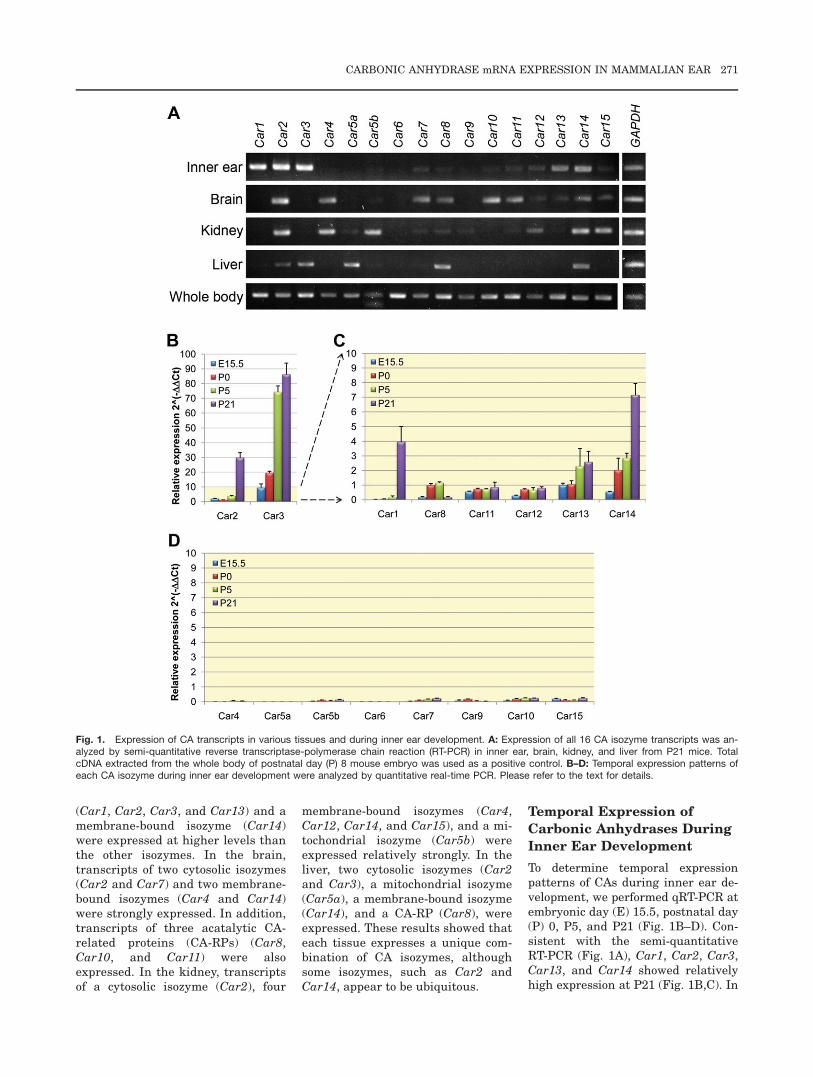
(Car1, Car2, Car3, and Car13) and amembrane-bound isozyme (Car14)were expressed at higher levels thanthe other isozymes. In the brain,transcripts of two cytosolic isozymes(Car2 and Car7) and two membrane-bound isozymes (Car4 and Car14)were strongly expressed. In addition,transcripts of three acatalytic CA-related proteins (CA-RPs) (Car8,Car10, and Car11) were alsoexpressed. In the kidney, transcriptsof a cytosolic isozyme (Car2), four
membrane-bound isozymes (Car4,Car12, Car14, and Car15), and a mi-tochondrial isozyme (Car5b) wereexpressed relatively strongly. In theliver, two cytosolic isozymes (Car2and Car3), a mitochondrial isozyme(Car5a), a membrane-bound isozyme(Car14), and a CA-RP (Car8), wereexpressed. These results showed thateach tissue expresses a unique com-bination of CA isozymes, althoughsome isozymes, such as Car2 andCar14, appear to be ubiquitous.
Temporal Expression of
Carbonic Anhydrases During
Inner Ear Development
To determine temporal expressionpatterns of CAs during inner ear de-velopment, we performed qRT-PCR atembryonic day (E) 15.5, postnatal day(P) 0, P5, and P21 (Fig. 1B–D). Con-sistent with the semi-quantitativeRT-PCR (Fig. 1A), Car1, Car2, Car3,Car13, and Car14 showed relativelyhigh expression at P21 (Fig. 1B,C). In
Fig. 1. Expression of CA transcripts in various tissues and during inner ear development. A: Expression of all 16 CA isozyme transcripts was an-alyzed by semi-quantitative reverse transcriptase-polymerase chain reaction (RT-PCR) in inner ear, brain, kidney, and liver from P21 mice. TotalcDNA extracted from the whole body of postnatal day (P) 8 mouse embryo was used as a positive control. B–D: Temporal expression patterns ofeach CA isozyme during inner ear development were analyzed by quantitative real-time PCR. Please refer to the text for details.
CARBONIC ANHYDRASE mRNA EXPRESSION IN MAMMALIAN EAR 271
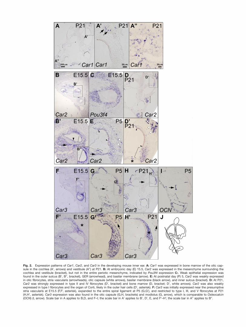
Fig. 2. Expression patterns of Car1, Car2, and Car3 in the developing mouse inner ear. A: Car1 was expressed in bone marrow of the otic cap-sule in the cochlea (A0, arrows) and vestibule (A00) at P21. B: At embryonic day (E) 15.5, Car2 was expressed in the mesenchyme surrounding thecochlea and vestibule (bracket), but not in the entire periotic mesenchyme, indicated by Pou3f4 expression C:. Weak epithelial expression wasfound in the outer sulcus (B0, B00, bracket), GER (arrowhead), and basilar membrane (arrow). E: At postnatal day (P) 5, Car2 was weakly expressedin otic fibrocytes, stria vascularis (arrowheads), otic capsule (white arrows), basilar membrane (black arrow), and inner sulcus (bracket). D: At P21,Car2 was strongly expressed in type II and IV fibrocytes (D0, bracket) and bone marrow (D, bracket; D0, white arrows). Car2 was also weaklyexpressed in type I fibrocytes and the organ of Corti, likely in the outer hair cells (D0, asterisk). F: Car3 was initially expressed near the presumptivestria vascularis at E15.5 (F,F0, asterisk), expanded to the entire spiral ligament at P5 (G,G0), and restricted to type I, III, and V fibrocytes at P21(H,H0, asterisk). Car3 expression was also found in the otic capsule (G,H, brackets) and modiolus (G, arrow), which is comparable to Osteocalcin(OCN) (I, arrow). Scale bar in A applies to B,D, and F–I; the scale bar in A0 applies to B0, D0, E, and F0–H0; the scale bar in A00 applies to B00.
addition, our qRT-PCR data showedthat Car8, Car11, and Car12 werealso expressed at moderate levels dur-ing inner ear development (Fig. 1C),while the remaining isozymes (Car4,Car5a, Car5b, Car6, Car7, Car9,Car10, and Car15) were expressed atundetectable or extremely low levels(Fig. 1D).
Car3 was strongly expressed in theinner ears throughout developmentalstages, suggesting an important roleof Car3 in inner ear development andfunction. Interestingly, acatalytic CA-RPs, such as Car8 and Car11, werealso expressed at moderate levels indeveloping inner ears, although Car8became undetectable in the matureinner ear at P21 (Fig. 1C). Theseresults suggest that CA isozymes mayplay specific roles in different stagesof inner ear development.
Spatial Expression Patterns
of Carbonic Anhydrases
During Inner Ear
Development
The localization of expression for eachCA transcript was determined by insitu hybridization in developing(E15.5, P0, P5) and mature (P21)inner ears.
Abundant cytosolic isozymes,
Car1 and Car2
Consistent with qRT-PCR results(Fig. 1), Car1 transcripts were barelydetectable in the inner ear during em-bryonic and postnatal development(E15.5, P0, and P5; data not shown)and were present in the bone marrowof the otic capsule at P21 (Fig. 2A–A00,arrows). Recently, it has been shownthat CAI is associated with ankylos-ing spondylitis, characterized byabnormal bone formation, in humansand CAI function is involved in boneformation in vitro (Chang et al.,2012).
Car2 transcripts were expressed inmesenchymal cells adjacent to thestria vascularis, spiral limbus, andbasilar membrane (Fig. 2B). WeakCar2 expression was also present inepithelial cells in the outer sulcus andgreater epithelial ridge (GER) (Fig.2B0,B00, bracket and arrowhead). Thisbroad Car2 expression continued dur-
ing neonatal development (Fig. 2E),but by P21, Car2 expression wasmainly present in type II and IV oticfibrocytes (Fig. 2D0, bracket) and bonemarrow of the otic capsule (Fig. 2D0,white arrows). Weaker Car2 expres-sion was present in type I fibrocytes,the apical and basal margins of striavascularis (arrowheads), and outerhair cells in the organ of Corti (aster-isk) at P21. In the vestibules, weakand broad Car2 expression was pres-ent in mesenchymal tissues surround-ing the sensory organs at E15.5 (Fig.2B, bracket), and stronger expressionin the otic capsule at P5, and bonemarrows at P21 (Fig. 2D, brackets).
Previously, CAII immunoreactivityhas been detected in type I, III, andIV otic fibrocytes in the spiral liga-ment of the gerbil cochlea (Spicer andSchulte, 1991) and in type I, II, andIV fibrocytes in guinea pigs (Ichimiyaet al., 1994). These expression pat-terns differ slightly from our results,which show strong Car2 expression intype II and IV fibrocytes (Fig. 2). Thisdifference could be due to differentspecies or detection methods.
Potential role of Car3 as a free
radical scavenger in otic
fibrocytes
Initially, Car3 expression was closelyassociated with prospective type I oticfibrocytes at E15.5 (Fig. 2F,F0, aster-isk), but gradually expanded to theentire spiral ligament, encompassingall types of otic fibrocytes by P5 (Fig.2G,G0), and later restricted to type I,III, and V otic fibrocytes at P21 (Fig.2H,H0, asterisk). Interestingly, Car3expression in the cochlear lateral wallat P21 was complementary to Car2,that is Car3 was more abundant intype I and III fibrocytes while Car2was stronger in type II and IV fibro-cytes (Fig. 2D0,H0, asterisk and brack-ets). Weak Car3 expression was alsodetected in the otic capsule (Fig.2G0,H0, arrows) and bony modiolusarea at P5 (Fig. 2G,I, arrows), whichdisappeared by P21 (Fig. 2H, arrow).Car3 expression was also present inthe vestibular mesenchymal regionsthroughout development (Fig. 2F–H,brackets). Consistent with our data,CAIII immunoreactivity was alsopresent in type I and III fibrocytes
from human and gerbil cochlea(Spicer and Schulte, 1991; Weberet al., 2001).
Unlike the other isozymes, CAIIIhas relatively little CA activity(Sanyal et al., 1982; Engberg et al.,1985), but mainly functions as an oxy-radical scavenger and protects cellsfrom oxidative damage by S-gluta-thiolation on two cysteine residues inresponse to oxidative stress (Raisanenet al., 1999; Mallis et al., 2002, 2000;Gailly et al., 2008). Nevertheless,introducing CAIII into hepatocellularcarcinoma cells accelerated the speedat which culture medium was acidi-fied, suggesting that CAIII can stillcatalyze carbon dioxide hydration,although at a reduced rate (Dai et al.,2008). The in vivo function of CAIII isunclear because Car3 null mutants donot display obvious anatomical orphysiological abnormalities (Kimet al., 2004). A recent study, however,showed that a lack of CAIII functionattenuates ATP synthesis by mito-chondria in skeletal muscle, althoughits underlying mechanism is unclear(Liu et al., 2007). Therefore, CAIIImay play dual roles in otic fibrocytes:facilitating mitochondrial ATP syn-thesis as well as detoxifying free radi-cals resulting from active ATPsynthesis.
Possible Functional Association
of Car12 and Car13 With
Pendrin
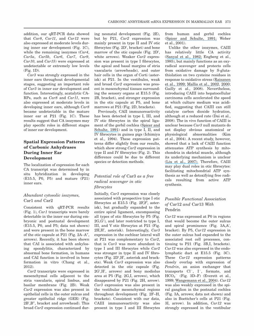
Car12 was expressed at P0 in regionsthat would become the outer sulcusand spiral prominence (Fig. 3A,A0,bracket). By P5, Car12 expression inthe outer sulcus had expanded to theassociated root cell processes, con-tinuing to P21 (Fig. 3B,I, brackets).Car12 was also expressed in the endo-lymphatic duct at E15.5 (Fig. 3C).These Car12 expression patternsclosely overlap with expression ofPendrin, an anion exchanger thattransports Cl�, I�, formate, andHCO3
� (Fig. 3D–F) (Everett et al.,1999; Wangemann et al., 2004). Car12was also weakly expressed in the spi-ral ganglion in the postnatal cochlea(Fig. 3A, arrows; data not shown) andalso in Boettcher’s cells at P21 (Fig.3I, arrow). In addition, Car12 wasstrongly expressed in the vestibular
CARBONIC ANHYDRASE mRNA EXPRESSION IN MAMMALIAN EAR 273
hair cells of cristae and maculae (Fig.3G,H), but not in the auditory haircells of the organ of Corti in the coch-lea (Fig. 3I, asterisk).
Car13 transcripts were observed inthe lesser epithelial ridge (LER) areaof E15.5 cochlea, which closely over-laps with Bmp4 expression (Fig.3J,M, brackets) (Hwang et al., 2010).At P0, Car13 expression was
restricted to the epithelial cells of thespiral prominence and outer sulcus,with faint expression in prospectivetype III fibrocytes (Fig. 3K, bracket,arrowheads), which was maintaineduntil P21 (Fig. 3L, bracket and arrow-heads; data not shown). In the vesti-bule, weak Car13 expression wasobserved in the mesenchymal tissuesalong the otic capsule at E15.5 and
P5, which is similar with the Car13expression pattern in the type IIIfibrocytes of the cochlea (Fig.3K,L,N,O, arrows).
Previously, cytosolic and mem-brane-bound CAs have been shown toassociate directly with ion transport-ers, forming a membrane protein com-plex called a biocarbonate transportmetabolon. This complex can
Fig. 3. Expression of Car12 and Car13 in the developing mouse inner ear. A,B: Car12 was expressed in the spiral prominence and associatedroot cell processes (brackets) and also in the spiral ganglion (arrows). C: Car12 was expressed in the endolymphatic duct at embryonic day (E)15.5. D–F: Car12 expression closely overlapped with Pendrin. G–I: At P21, Car12 was expressed in the spiral prominence and associated root cellprocesses (I, bracket). In addition, Car12 was also expressed in hair cells of cristae (G) and maculae (H) and Boettcher’s cells (I, arrow), but not inhair cells of organ of Corti in the P21 cochlea (I, asterisk). J–M: Car13 was weakly expressed on the lateral side of the lesser epithelial ridge (LER)(brackets), similar to Bmp4 (M) at E15.5. Car13 was also expressed in the spiral prominence and presumptive outer sulcus during neonatal devel-opment (brackets). N,O: Car13 was weakly expressed in prospective type III fibrocytes during neonatal stages (K,L, arrowheads) and in the mes-enchymal tissues along the otic capsule at E15.5 and P5 (N,O, arrowheads). Scale bar in A applies to D; the scale bar in A0 applies to B–C, D0, E–H, and J–O.
274 WU ET AL.
significantly enhance Hþ/HCO3�
transport by mass CA action supply-ing and dissipating the reaction prod-ucts (Alvarez et al., 2003; Purkersonand Schwartz, 2007). For example, inthe renal tube, a cytosolic CAII and amembrane-bound CAIV form trans-port metabolons with bicarbonatetransporters such as AE1, NBC1,NBC3, and SCL26A6 and a protonantiporter such as NHE1 (Purkersonand Schwartz, 2007).
Based on expression of the tran-scripts of membrane-bound Car12, cy-tosolic Car13, and the anionexchanger Pendrin in the outer sulcusand root cell processes, it is possiblethat CAXII and CAXIII may physi-cally associate with Pendrin or othertransporters to form metabolons inthe outer sulcus and root cell proc-esses and regulate ion and pH homeo-stasis of endolymphatic fluid. Consis-tently, the unique biochemical proper-ties of CAXIII, which catalyzes CO2
hydration even in the presence ofhigh HCO3
� concentrations, suggeststhat CAXIII may form metabolons intissues that require tight bicarbonateregulation. For example, the femalereproductive tract requires an alka-line environment to maintain spermmotility (Innocenti et al., 2004).
Further experiments should examinewhether CAXII and CAXIII indeedphysically or functionally associatewith Pendrin to regulate endolymphhomeostasis.
A membrane-bound isozyme
Car14
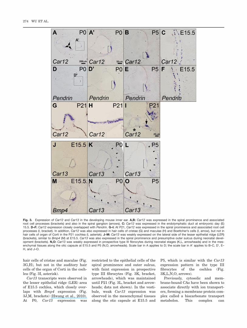
Car14 was broadly expressed in bothepithelial and mesenchymal tissuesin the cochlea at P0 (Fig. 4A,A0). AtP5, Car14 expression was stronger inthe otic fibrocytes, stria vascularis,and supporting cells in the organ ofCorti (Fig. 4B,B0). Car14 expressionwas maintained in the otic fibrocytesand greatly reduced in the stria vas-cularis at P21 (Fig. 4C). Car14expression in Reissner’s membrane atP0 decreased by P5 (Fig. 4A0,B, whitearrow). Car14 expression in the organof Corti at P5 appeared to be in pillarcells and Deiters’ cells when com-pared with Smpx, a hair cell-specificgene (Fig. 4B0) (Yoon et al., 2011). AtP21, Car14 expression was observedin the inner phalangeal cells andinner pillar cells located at the medialside of the tunnel of Corti, and threeDeiters’ cells at the lateral side butnot in the outer pillar cells (Fig. 4D).Car14 was also weakly expressed in
the spiral ganglion region (Fig. 4C,arrows). In the vestibule, weak Car14expression was broadly observed inthe mesenchymal tissues in the vesti-bule, but not in the otic capsule (Fig.4A, bracket).
It was shown in the kidney that themembrane-bound CAXIV (in rodents;CAIV in humans) is expressed on theluminal surface of the renal tube andfacilitates bicarbonate and fluidtransport by ion transporters such asNBCI (Alvarez et al., 2003; Purkersonand Schwartz, 2007). Previously, ithas been suggested that CO2 in theouter hair cells is hydroxylated bymeans of intracellular CA in the outerhair cells, and exchanged to the endo-lymph or diffused to the regionbetween the outer hair cells and Dei-ters’ cells (Thalmann et al., 1970;Kimura, 1975; Ikeda et al., 1992; Oka-mura et al., 1996). Thus, it is also pos-sible that CAII expressed in the outerhair cells may cooperate with CAXIVexpressed on the extracellular surfaceof Deiters’ cells to facilitate thehydroxylation and transport of CO2,which is the major product of high cel-lular metabolism in the hair cells, andplay roles in optimal pH regulationand fluid homeostasis in the organ ofCorti.
Fig. 4. Expression of Car14 in the developing mouse cochlea. A–C: Car14 was broadly expressed in both epithelial and mesenchymal tissues inthe cochlea and vestibule during embryonic and neonatal development. Car14 expression in the otic fibrocytes, Reissner’s membrane (A0, whitearrow), and stria vascularis (A0–C, arrowheads) at postnatal day (P) 0 and P5 became weaker at P21. Car14 was also expressed in the organ ofCorti in pillar cells and Deiters’ cells at P5 (B,B0). The inset in B0 showed Smpx expression in the hair cells. At P21, Car14 was expressed in theinner phalangeal cells, inner pillar cells, and Deiters’ cells (C,D). The inset in D illustrates various cell types in the organ of Corti for comparisonwith the Car14 expression domains. Car14 was also weakly expressed in the spiral ganglion (C, arrows). The scale bar in A applies to B and C;the scale bar in D applies to B0.
CARBONIC ANHYDRASE mRNA EXPRESSION IN MAMMALIAN EAR 275
Acatalytic CA-related proteins
Car8 and Car11
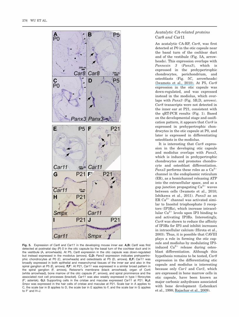
An acatalytic CA-RP, Car8, was firstdetected at P0 in the otic capsule nearthe basal turn of the cochlear ductand of the vestibule (Fig. 5A, arrow-heads). This expression overlaps withPannexin 3 (Panx3), which isexpressed in the prehypertrophicchondrocytes, perichondrium, andosteoblasts (Fig. 5C, arrowheads)(Iwamoto et al., 2010). At P5, Car8expression in the otic capsule wasdown-regulated, and was expressedinstead in the modiolus, which over-laps with Panx3 (Fig. 5B,D, arrows).Car8 transcripts were not detected inthe inner ear at P21, consistent withthe qRT-PCR results (Fig. 1). Basedon the developmental stage and ossifi-cation pattern, it appears that Car8 isexpressed in prehypertrophic chon-drocytes in the otic capsule at P0, andlater is expressed in differentiatingosteoblasts in the modiolus.
It is interesting that Car8 expres-sion in the developing otic capsuleand modiolus overlaps with Panx3,which is induced in prehypertrophicchondrocytes and promotes chondro-cyte and osteoblast differentiation.Panx3 performs these roles as a Ca2þ
channel in the endoplasmic reticulum(ER), as a hemichannel releasing ATPinto the extracellular space, and as agap junction propagating Ca2þ wavesbetween cells (Iwamoto et al., 2010;Ishikawa et al., 2011). Panx3 as anER Ca2þ channel was activated simi-lar to Inositol trisphosphate 3 recep-tors (IP3Rs), which increase intracel-lular Ca2þ levels upon IP3 binding toand activating IP3Rs. Interestingly,Car8 was shown to reduce the affinityof IP3Rs for IP3 and inhibit increasesin intracellular calcium (Hirota et al.,2003). Thus, it is possible that CAVIIIplays a role in forming the otic cap-sule and modiolus by modulating IP3-induced Ca2þ release during osteo-blast differentiation. Although thishypothesis remains to be tested, Car8expression in the differentiating oticcapsule and modiolus is interestingbecause only Car1 and Car2, whichare expressed in bone marrow cells inotic capsule, have been known asmajor carbonic anhydrases associatedwith bone development (Lehenkariet al., 1998; Rajachar et al., 2009).
Fig. 5. Expression of Car8 and Car11 in the developing mouse inner ear. A,B: Car8 was firstdetected at postnatal day (P) 0 in the otic capsule by the basal turn of the cochlear duct and inthe vestibule (A, arrowheads). At P5, Car8 expression in the otic capsule was down-regulatedbut instead expressed in the modiolus (arrows). C,D: Panx3 expression indicates prehypertro-phic chondrocytes at P0 (C, arrowheads) and osteoblasts at P5 (D, arrows). E,F: Car11 wasbroadly expressed in both epithelial and mesenchymal tissues of the inner ear and also in thespiral ganglion at P5 (E, arrows). F,F0: At P21, Car11 was expressed in a similar broad pattern inthe spiral ganglion (F, arrows), Reissner’s membrane (black arrowhead), organ of Corti(white arrowhead), bone marrow of the otic capsule (F0, arrows), and spiral prominence and theassociated root cell processes (bracket). Car11 was also weakly expressed in type I fibrocytes(F0, asterisk). G,I: Supporting cells in the cristae and maculae expressed Car11 at P21. H,J:Smpx was expressed in the hair cells of cristae and maculae at P21. Scale bar in A applies toC, the scale bar in B applies to D, the scale bar in E applies to F, and the scale bar in G appliesto F0 and H–J.
276 WU ET AL.
Another CA-RP, Car11, was broadlyexpressed in the inner ear at P5, withslightly stronger expression in the spi-ral ganglion (Fig. 5E, arrows). At P21,Car11 was expressed in the spiral gan-glion (Fig. 5F, arrows), Reissner’smembrane (Fig. 5F0, black arrowhead),organ of Corti (Fig. 5F0, white arrow-head), bone marrow of the otic capsule(Fig. 5F0, arrows), spiral prominence,and root cell processes (Fig. 5F0,bracket). Car11 was also weaklyexpressed in type I otic fibrocytes (Fig.5F0, asterisk). In the vestibule, Car11expression was observed in the apicalportion of cristae and maculae, mostlikely in the supporting cells, based onthe comparison with the hair cell spe-cific expression of Smpx (Fig. 5G–J).Although CAXI immunoreactivity hasbeen shown in the developing andmature human brain (Taniuchi et al.,2002) and in gastrointestinal stromaltumors (Morimoto et al., 2005), thephysiological roles of Car11 in themouse inner ear remains to beelucidated.
Conclusions
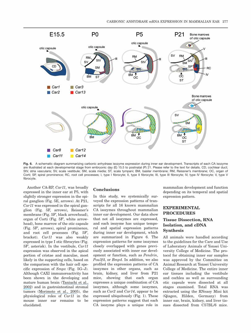
In this study, we systemically sur-veyed the expression patterns of tran-scripts for all 16 known mammalianCA isozymes throughout mammalianinner ear development. Our data showthat not all isozymes are expressed,and each isozyme has unique tempo-ral and spatial expression patternsduring inner ear development, whichare summarized in Figure 6. Theexpression patterns for some isozymesclosely overlapped with genes previ-ously associated with inner ear devel-opment or function, such as Pendrin,Pou3f4, or Bmp4. In addition, we alsoprofiled the expression patterns of CAisozymes in other organs, such asbrain, kidney, and liver from P21mice, showing that each organexpresses a unique combination of CAisozymes, although some isozymes,such as Car2 and Car14, appear to beexpressed ubiquitously (Fig. 1). Theseexpression patterns suggest that eachCA isozyme plays a unique role in
mammalian development and functiondepending on its temporal and spatialexpression pattern.
EXPERIMENTAL
PROCEDURES
Tissue Dissection, RNA
Isolation, and cDNA
Synthesis
All animals were handled accordingto the guidelines for the Care and Useof Laboratory Animals of Yonsei Uni-versity College of Medicine. The pro-tocol for obtaining inner ear sampleswas approved by the Committee onAnimal Research at Yonsei UniversityCollege of Medicine. The entire innerear tissues including the vestibuleand cochlea as well as surroundingotic capsule were dissected at allstages examined. Total RNA wasextracted using the RNeasy Mini kit(Qiagen, Hilden, Germany) frominner ear, brain, kidney, and liver tis-sues dissected from C57BL/6 mice.
Fig. 6. A schematic diagram summarizing carbonic anhydrase isozyme expression during inner ear development. Transcripts of each CA isozymeare illustrated at each developmental stage from embryonic day (E) 15.5 to postnatal (P) 21. Please refer to the text for details. CD, cochlear duct;StV, stria vascularis; SV, scala vestibule; SM, scala media; ST, scala tympani; BM, basilar membrane; RM, Reissner’s membrane; OC, organ ofCorti; SP, spiral prominence; RC, root cell processes; I, type I fibrocyte; II, type II fibrocyte; III, type III fibrocyte; IV, type IV fibrocyte; V, type Vfibrocyte.
CARBONIC ANHYDRASE mRNA EXPRESSION IN MAMMALIAN EAR 277
RNase-free DNase was used to digestof genomic DNA during RNA purifica-tion (Qiagen, Hilden, Germany). Theconcentration and purity of theextracted RNAs were determinedusing both the spectrophotometricmethod at 260 and 280 nm and RNAelectrophoresis. One microgram oftotal RNA was subjected to reversetranscription with oligod(T)19 primerusing the High Capacity cDNAReverse Transcription Kit (AppliedBiosystems, Foster City, CA).
Semi-quantitative Reverse
Transcription PCR Analysis
Synthesized cDNAs were subjected tosemi-quantitative RT-PCR to analyzerelative expression levels of CA iso-zymes among tissues. CA isozyme spe-cific PCR primers previously reportedwere used for RT-PCR (Lacruz et al.,2010). PCR conditions were as follows:28 cycles of denaturation at 94�C for 20sec, annealing at 55�C for 40 sec, andextension at 72�C for 40 sec. The firstdenaturation step and the last exten-sion step were performed at 95�C for15 min and 72�C for 5 min, respec-tively. PCR products were separatedand visualized on a 2% agarose gel.
Quantitative Real-Time PCR
qRT-PCR was performed to analyzethe expression of each CA isozyme
during inner ear development atE15.5, P0, P5, and P21. RNA purifiedfrom three replicates of each innerear sample was used for qRT-PCR.PCR primers were designed based onmRNA sequences in the GenBankdatabase (Table 1). Forward andreverse primers from each primer setwere designed from different exons todistinguish transcripts from genomicDNA. GAPDH (glyceraldehyde-3-phosphate dehydrogenase) was usedas an internal control for normaliza-tion. qRT-PCR for each CA isozymewas performed in triplicate usingSYBR Green PCR Master Mix andthe Applied Biosystems StepOnePlusReal-Time PCR Systems (Applied Bio-systems). A melting curve analysiswas always performed after theamplification to check PCR specificity.The results were analyzed by Ste-pOne Software v2.1 (Applied Biosys-tems). The normalization and relativequantification were calculated using2�DDCt method (Livak and Schmitt-gen, 2001).
In Situ Hybridization
For E15.5 embryos and neonatal (P0and P5) pups, animals were killed bydecapitation and then hemi-sectioned.After removing the brain, the speci-mens were fixed in 4% paraformalde-hyde in phosphate buffered saline(PBS) overnight at 4�C, dehydrated in
30% sucrose in PBS overnight at 4�C,embedded in OCT compound (Sakura,Tokyo, Japan), and stored at �80�Cuntil use. For P21 mice, the entireinner ear tissues including the oticcapsule were dissected. For betterpenetration of fixatives, parts of thesemicircular canals were cut off, theoval and round windows were openedup, and the apex of the cochlea waspunctured. After overnight fixation,the specimens were decalcified in 0.2M EDTA in DEPC-PBS at 4�C for 2days, followed by dehydration andmounting. Tissues were sectioned at12 mm thickness for in situ hybridiza-tion, which was performed as previ-ously described (Morsli et al., 1998).At least three animals were tested foreach CA isozyme at each developmen-tal stage. Sense RNA probes were alsoincluded as controls, which showed nosignal anywhere in the inner ear.
Probes for Bmp4 (Morsli et al.,1998), Pou3f4 (Phippard et al., 1998),Smpx (Yoon et al., 2011), and Pendrin(Everett et al., 1999) were preparedas previously described. RNA probesfor Car1 were generated from a 381base pair (bp) mouse Car1 cDNA con-taining the þ594 to þ786 codingregion and the 188 bp 30 untraslatedregion (NM_009799.4); for Car2, froma 410 bp mouse Car2 cDNA contain-ing the þ654 to þ783 coding regionand the 280 bp 30untranslated region(NM_009801.4); for Car3, from a 404
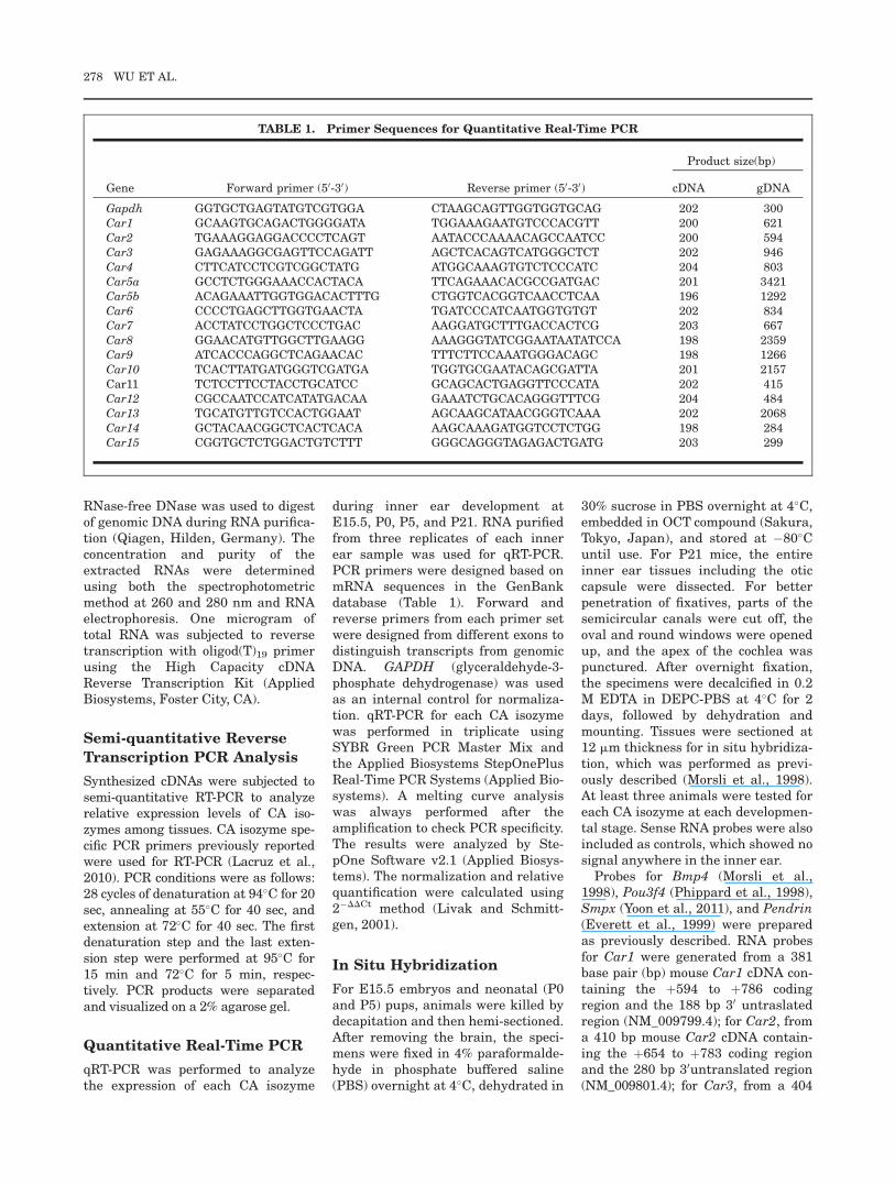
TABLE 1. Primer Sequences for Quantitative Real-Time PCR
Gene Forward primer (50-30) Reverse primer (50-30)
Product size(bp)
cDNA gDNA
Gapdh GGTGCTGAGTATGTCGTGGA CTAAGCAGTTGGTGGTGCAG 202 300Car1 GCAAGTGCAGACTGGGGATA TGGAAAGAATGTCCCACGTT 200 621Car2 TGAAAGGAGGACCCCTCAGT AATACCCAAAACAGCCAATCC 200 594Car3 GAGAAAGGCGAGTTCCAGATT AGCTCACAGTCATGGGCTCT 202 946Car4 CTTCATCCTCGTCGGCTATG ATGGCAAAGTGTCTCCCATC 204 803Car5a GCCTCTGGGAAACCACTACA TTCAGAAACACGCCGATGAC 201 3421Car5b ACAGAAATTGGTGGACACTTTG CTGGTCACGGTCAACCTCAA 196 1292Car6 CCCCTGAGCTTGGTGAACTA TGATCCCATCAATGGTGTGT 202 834Car7 ACCTATCCTGGCTCCCTGAC AAGGATGCTTTGACCACTCG 203 667Car8 GGAACATGTTGGCTTGAAGG AAAGGGTATCGGAATAATATCCA 198 2359Car9 ATCACCCAGGCTCAGAACAC TTTCTTCCAAATGGGACAGC 198 1266Car10 TCACTTATGATGGGTCGATGA TGGTGCGAATACAGCGATTA 201 2157Car11 TCTCCTTCCTACCTGCATCC GCAGCACTGAGGTTCCCATA 202 415Car12 CGCCAATCCATCATATGACAA GAAATCTGCACAGGGTTTCG 204 484Car13 TGCATGTTGTCCACTGGAAT AGCAAGCATAACGGGTCAAA 202 2068Car14 GCTACAACGGCTCACTCACA AAGCAAAGATGGTCCTCTGG 198 284Car15 CGGTGCTCTGGACTGTCTTT GGGCAGGGTAGAGACTGATG 203 299
278 WU ET AL.
bp mouse Car3 cDNA containing theþ687 to þ783 coding region and the334 bp 30 untranslated region(NM_007606.3); for Car8, from a621 bp mouse Car8 cDNA containingthe þ212 to þ832 coding region andthe 12 bp 50 untranslated region(NM_007592.3); for Car12, from a 471bp mouse Car12 cDNA containing theþ947 to þ1,065 coding region and the352 bp 50 untranslated region(NM_178396.4); for Car13, from a 404bp mouse Car13 cDNA containing theþ664 to þ789 coding region and the278 bp 50 untranslated region(NM_024495.5); for Car14, from a 856bp mouse Car14 cDNA containing theþ1 to þ821 coding region and the 35 bp50 untranslated region (NM_011797.2);for Car11, from a 916 bp mouse Car11cDNA containing 634 bp coding regionand the 282 bp 50 untranslated region(NM_009800.4); for Osteocalcin, from a33 bp mouse Osteocalcin cDNA con-taining the 47 bp 50 untranslatedregion and the entire open readingframe (NM_007541.2); for Pannexin3,from a 559 bp mouse Pannexin3 cDNAcontaining the þ565 to þ1,123 codingregion (NM_172454.2).
REFERENCES
Alvarez BV, Loiselle FB, Supuran CT,Schwartz GJ, Casey JR. 2003. Directextracellular interaction between car-bonic anhydrase IV and the humanNBC1 sodium/bicarbonate co-trans-porter. Biochemistry 42:12321–12329.
Chang X, Zheng Y, Yang Q, Wang L, PanJ, Xia Y, Yan X, Han J. 2012. Carbonicanhydrase I is involved in the processof bone formation and is susceptible toankylosing spondylitis. Arthritis ResTher 14:R176.
Chiche J, Ilc K, Laferriere J, Trottier E,Dayan F, Mazure NM, Brahimi-HornMC, Pouyssegur J. 2009. Hypoxia-induci-ble carbonic anhydrase IX and XII pro-mote tumor cell growth by counteractingacidosis through the regulation of the in-tracellular pH. Cancer Res 69:358–368.
Dai HY, Hong CC, Liang SC, Yan MD,Lai GM, Cheng AL, Chuang SE. 2008.Carbonic anhydrase III promotes trans-formation and invasion capability inhepatoma cells through FAK signalingpathway. Mol Carcinog 47:956–963.
Engberg P, Millqvist E, Pohl G, LindskogS. 1985. Purification and some proper-ties of carbonic anhydrase from bovineskeletal muscle. Arch Biochem Biophys241:628–638.
Erulkar SD, Maren TH. 1961. Carbonicanhydrase and the inner ear. Nature189:459–460.
Esbaugh AJ, Tufts BL. 2006. The struc-ture and function of carbonic anhydraseisozymes in the respiratory system ofvertebrates. Respir Physiol Neurobiol154:185–198.
Everett LA, Morsli H, Wu DK, Green ED.1999. Expression pattern of the mouseortholog of the Pendred’s syndromegene (Pds) suggests a key role for pen-drin in the inner ear. Proc Natl AcadSci U S A 96:9727–9732.
Gailly P, Jouret F, Martin D, Debaix H,Parreira KS, Nishita T, Blanchard A,Antignac C, Willnow TE, Courtoy PJ,Scheinman SJ, Christensen EI, DevuystO. 2008. A novel renal carbonic anhy-drase type III plays a role in proximaltubule dysfunction. Kidney Int 74:52–61.
Henry RP, Swenson ER. 2000. The distri-bution and physiological significance ofcarbonic anhydrase in vertebrate gasexchange organs. Respir Physiol 121:1–12.
Hirota J, Ando H, Hamada K, MikoshibaK. 2003. Carbonic anhydrase-relatedprotein is a novel binding protein forinositol 1,4,5-trisphosphate receptortype 1. Biochem J 372:435–441.
Hsu CJ, Nomura Y. 1985. Carbonic anhy-drase activity in the inner ear. ActaOtolaryngol Suppl 418:1–42.
Hwang CH, Guo D, Harris MA, HowardO, Mishina Y, Gan L, Harris SE, WuDK. 2010. Role of bone morphogeneticproteins on cochlear hair cell formation:analyses of Noggin and Bmp2 mutantmice. Dev Dyn 239:505–513.
Ichimiya I, Adams JC, Kimura RS. 1994.Immunolocalization of Naþ, K(þ)-ATPase, Ca(þþ)-ATPase, calcium-bind-ing proteins, and carbonic anhydrase inthe guinea pig inner ear. Acta Otolaryn-gol 114:167–176.
Ikeda K, Kusakari J, Takasaka T, Saito Y.1987. Early effects of acetazolamide onanionic activities of the guinea pigendolymph: evidence for active functionof carbonic anhydrase in the cochlea.Hear Res 31:211–216.
Ikeda K, Saito Y, Nishiyama A, TakasakaT. 1992. Intracellular pH regulation inisolated cochlear outer hair cells of theguinea-pig. J Physiol 447:627–648.
Innocenti A, Lehtonen JM, Parkkila S,Scozzafava A, Supuran CT. 2004. Car-bonic anhydrase inhibitors. Inhibitionof the newly isolated murine isozymeXIII with anions. Bioorg Med ChemLett 14:5435–5439.
Ishikawa M, Iwamoto T, Nakamura T,Doyle A, Fukumoto S, Yamada Y. 2011.Pannexin 3 functions as an ER Ca(2þ)channel, hemichannel, and gap junctionto promote osteoblast differentiation. JCell Biol 193:1257–1274.
Iwamoto T, Nakamura T, Doyle A, Ishi-kawa M, de Vega S, Fukumoto S,Yamada Y. 2010. Pannexin 3 regulatesintracellular ATP/cAMP levels and pro-motes chondrocyte differentiation. JBiol Chem 285:18948–19958.
Jarvela S, Parkkila S, Bragge H, Kahko-nen M, Parkkila AK, Soini Y, Pastore-
kova S, Pastorek J, Haapasalo H. 2008.Carbonic anhydrase IX in oligodendro-glial brain tumors. BMC Cancer 8:1.
Kido T, Sekitani T, Yamashita H, Endo S,Masumitsu Y, Shimogori H. 1991.Effects of carbonic anhydrase inhibitoron the otolithic organs of developingchick embryos. Am J Otolaryngol12:191–195.
Kim G, Lee TH, Wetzel P, Geers C, Robin-son MA, Myers TG, Owens JW, Wehr NB,Eckhaus MW, Gros G, Wynshaw-Boris A,Levine RL. 2004. Carbonic anhydrase IIIis not required in the mouse for normalgrowth, development, and life span. MolCell Biol 24:9942–9947.
Kimura RS. 1975. The ultrastructure ofthe organ of Corti. Int Rev Cytol42:173–222.
Lacruz RS, Hilvo M, Kurtz I, Paine ML.2010. A survey of carbonic anhydrasemRNA expression in enamel cells. Bio-chem Biophys Res Commun 393:883–887.
Lehenkari P, Hentunen TA, Laitala-Leino-nen T, Tuukkanen J, Vaananen HK.1998. Carbonic anhydrase II plays amajor role in osteoclast differentiationand bone resorption by effecting thesteady state intracellular pH and Ca2þ.Exp Cell Res 242:128–137.
Lim DJ, Karabinas C, Trune DR. 1983.Histochemical localization of carbonicanhydrase in the inner ear. Am J Oto-laryngol 4:33–42.
Liu M, Walter GA, Pathare NC, ForsterRE, Vandenborne K. 2007. A quantita-tive study of bioenergetics in skeletalmuscle lacking carbonic anhydrase IIIusing 31P magnetic resonance spectros-copy. Proc Natl Acad Sci U S A104:371–376.
Livak KJ, Schmittgen TD. 2001. Analysisof relative gene expression data usingreal-time quantitative PCR and the 2(-Delta Delta C(T)) Method. Methods25:402–408.
Mallis RJ, Hamann MJ, Zhao W, ZhangT, Hendrich S, Thomas JA. 2002. Irre-versible thiol oxidation in carbonicanhydrase III: protection by S-gluta-thiolation and detection in aging rats.Biol Chem 383:649–662.
Mallis RJ, Poland BW, Chatterjee TK,Fisher RA, Darmawan S, Honzatko RB,Thomas JA. 2000. Crystal structure ofS-glutathiolated carbonic anhydrase III.FEBS Lett 482:237–241.
Margolis DS, Szivek JA, Lai LW, LienYH. 2008. Phenotypic characteristics ofbone in carbonic anhydrase II-deficientmice. Calcif Tissue Int 82:66–76.
Morimoto K, Nishimori I, Takeuchi T,Kohsaki T, Okamoto N, Taguchi T,Yunoki S, Watanabe R, Ohtsuki Y,Onishi S. 2005. Overexpression of car-bonic anhydrase-related protein XI pro-motes proliferation and invasion ofgastrointestinal stromal tumors. Virch-ows Arch 447:66–73.
Morsli H, Choo D, Ryan A, Johnson R,Wu DK. 1998. Development of themouse inner ear and origin of its sen-sory organs. J Neurosci 18:3327–3335.
CARBONIC ANHYDRASE mRNA EXPRESSION IN MAMMALIAN EAR 279
Okamura HO, Sugai N, Suzuki K, OhtaniI. 1996. Enzyme-histochemical localiza-tion of carbonic anhydrase in the innerear of the guinea pig and severalimprovements of the technique. Histo-chem Cell Biol 106:425–430.
Pan PW, Rodriguez A, Parkkila S. 2007.A systematic quantification of carbonicanhydrase transcripts in the mouse di-gestive system. BMC Mol Biol 8:22.
Parkkila AK, Scarim AL, Parkkila S,Waheed A, Corbett JA, Sly WS. 1998.Expression of carbonic anhydrase V inpancreatic beta cells suggests role for mi-tochondrial carbonic anhydrase in insulinsecretion. J Biol Chem 273:24620–24623.
Phippard D, Heydemann A, Lechner M,Lu L, Lee D, Kyin T, Crenshaw EB,3rd. 1998. Changes in the subcellularlocalization of the Brn4 gene productprecede mesenchymal remodeling of theotic capsule. Hear Res 120:77–85.
Prazma J. 1978. Carbonic anhydrase inthe generation of cochlear potentials.Am J Physiol 235:F317–F320.
Purichia N, Erway LC. 1972. Effects ofdichlorophenamide, zinc, and manga-nese on otolith development in mice.Dev Biol 27:395–405.
Purkerson JM, Schwartz GJ. 2007. Therole of carbonic anhydrases in renalphysiology. Kidney Int 71:103–115.
Raisanen SR, Lehenkari P, Tasanen M,Rahkila P, Harkonen PL, VaananenHK. 1999. Carbonic anhydrase III pro-tects cells from hydrogen peroxide-induced apoptosis. FASEB J 13:513–522.
Rajachar RM, Tung E, Truong AQ, LookA, Giachelli CM. 2009. Role of carbonicanhydrase II in ectopic calcification.Cardiovasc Pathol 18:77–82.
Sanyal G, Swenson ER, Pessah NI,Maren TH. 1982. The carbon dioxidehydration activity of skeletal musclecarbonic anhydrase. Inhibition by sulfo-namides and anions. Mol Pharmacol22:211–220.
Shah GN, Ulmasov B, Waheed A, BeckerT, Makani S, Svichar N, Chesler M, SlyWS. 2005. Carbonic anhydrase IV andXIV knockout mice: roles of the respec-tive carbonic anhydrases in bufferingthe extracellular space in brain. ProcNatl Acad Sci U S A 102:16771–16776.
Sly WS, Hu PY. 1995. Human carbonicanhydrases and carbonic anhydrase defi-ciencies. Annu Rev Biochem 64:375–401.
Spicer SS, Schulte BA. 1991. Differentia-tion of inner ear fibrocytes according totheir ion transport related activity.Hear Res 56:53–64.
Sterkers O, Saumon G, Tran Ba Huy P,Ferrary E, Amiel C. 1984. Electrochem-ical heterogeneity of the cochlear endo-lymph: effect of acetazolamide. Am JPhysiol 246:F47–F53.
Supuran CT. 2008a. Carbonic anhy-drases–an overview. Curr Pharm Des14:603–614.
Supuran CT. 2008b. Carbonic anhydrases:novel therapeutic applications for inhib-itors and activators. Nat Rev Drug Dis-cov 7:168–181.
Taniuchi K, Nishimori I, Takeuchi T, Fuji-kawa-Adachi K, Ohtsuki Y, Onishi S.
2002. Developmental expression of car-bonic anhydrase-related proteins VIII,X, and XI in the human brain. Neuro-science 112:93–99.
Thalmann I, Matschinsky FM, ThalmannR. 1970. Quantitative study of selectedenzymes involved in energy metabolismof the cochlear duct. Ann Otol RhinolLaryngol 79:12–29.
Tohse H, Ando H, Mugiya Y. 2004. Bio-chemical properties and immunohisto-chemical localization of carbonicanhydrase in the sacculus of the innerear in the salmon Oncorhynchusmasou. Comp Biochem Physiol A MolIntegr Physiol 137:87–94.
Wangemann P, Itza EM, Albrecht B, WuT, Jabba SV, Maganti RJ, Lee JH, Ever-ett LA, Wall SM, Royaux IE, Green ED,Marcus DC. 2004. Loss of KCNJ10 pro-tein expression abolishes endocochlearpotential and causes deafness in Pen-dred syndrome mouse model. BMC Med2:30.
Weber PC, Cunningham CD III, SchulteBA. 2001. Potassium recycling path-ways in the human cochlea. Laryngo-scope 111:1156–1165.
Xu Y, Feng L, Jeffrey PD, Shi Y, MorelFM. 2008. Structure and metalexchange in the cadmium carbonicanhydrase of marine diatoms. Nature452:56–61.
Yoon H, Lee DJ, Kim MH, Bok J.2011. Identification of genes concord-antly expressed with Atoh1 duringinner ear development. Anat Cell Biol44:69–78.
280 WU ET AL.