Embed Size (px)
Citation preview
A transgenic Xenopus laevis reporter model to studylymphangiogenesis
Annelii Ny1,2,*, Wouter Vandevelde1,2,*, Philipp Hohensinner1,2, Manu Beerens3, Ilse Geudens1,2,Antonio Diez-Juan1,2,`, Katleen Brepoels1,2, Stephane Plaisance1,2, Paul A. Krieg4, Tobias Langenberg1,2,Stefan Vinckier1,2, Aernout Luttun3, Peter Carmeliet1,2 and Mieke Dewerchin1,2,§
1Laboratory of Angiogenesis and Neurovascular link, Vesalius Research Center, VIB, 3000 Leuven, Belgium2Laboratory of Angiogenesis and Neurovascular link, Vesalius Research Center, KU Leuven, 3000 Leuven, Belgium3Center for Molecular and Vascular Biology, KU Leuven, 3000 Leuven, Belgium4Department of Cell Biology and Anatomy, University of Arizona College of Medicine, Tucson, AZ 85724-5044, USA
*These authors contributed equally to this work`Present address: Instituto de Investigacion Sanitaria INCLIVA, 46010 Valencia, Spain§Author for correspondence ([email protected])
Biology Open 2, 882–890doi: 10.1242/bio.20134739Received 11th March 2013Accepted 10th June 2013
SummaryThe importance of the blood- and lymph vessels in the
transport of essential fluids, gases, macromolecules and cells
in vertebrates warrants optimal insight into the regulatory
mechanisms underlying their development. Mouse and
zebrafish models of lymphatic development are
instrumental for gene discovery and gene characterization
but are challenging for certain aspects, e.g. no direct
accessibility of embryonic stages, or non-straightforward
visualization of early lymphatic sprouting, respectively. We
previously demonstrated that the Xenopus tadpole is a
valuable model to study the processes of lymphatic
development. However, a fluorescent Xenopus reporter
directly visualizing the lymph vessels was lacking. Here, we
created transgenic Tg(Flk1:eGFP) Xenopus laevis reporter
lines expressing green fluorescent protein (GFP) in blood-
and lymph vessels driven by the Flk1 (VEGFR-2) promoter.
We also established a high-resolution fluorescent dye labeling
technique selectively and persistently visualizing lymphatic
endothelial cells, even in conditions of impaired lymph vessel
formation or drainage function upon silencing of
lymphangiogenic factors. Next, we applied the model to
dynamically document blood and lymphatic sprouting and
patterning of the initially avascular tadpole fin. Furthermore,
quantifiable models of spontaneous or induced lymphatic
sprouting into the tadpole fin were developed for dynamic
analysis of loss-of-function and gain-of-function phenotypes
using pharmacologic or genetic manipulation. Together with
angiography and lymphangiography to assess functionality,
Tg(Flk1:eGFP) reporter tadpoles readily allowed detailed
lymphatic phenotyping of live tadpoles by fluorescence
microscopy. The Tg(Flk1:eGFP) tadpoles represent a
versatile model for functional lymph/angiogenomics and
drug screening.
� 2013. Published by The Company of Biologists Ltd. This is an
Open Access article distributed under the terms of the Creative
Commons Attribution License (http://creativecommons.org/
licenses/by/3.0), which permits unrestricted use, distribution
and reproduction in any medium provided that the original
work is properly attributed.
Key words: Xenopus, Lymphangiogenesis, Imaging
IntroductionBlood- and lymph vessels are essential for the transport of fluids,
gases, macromolecules and cells within vertebrates (Adams and
Alitalo, 2007; Alitalo, 2011). Numerous serious disorders, such as
cancer, lymphedema, inflammation and diabetic complications are
exacerbated or caused by impaired formation or dysfunction of
these vasculatures (Alitalo, 2011; Carmeliet and Jain, 2011; Jurisic
and Detmar, 2009; Tammela and Alitalo, 2010; Wang and Oliver,
2010). While several anti-angiogenic strategies have been
approved or are under clinical trial for the treatment of human
malignancies (Carmeliet and Jain, 2011), strategies for the specific
modulation of lymphatic growth remain scarce (Alitalo, 2011;
Jurisic and Detmar, 2009; Tammela and Alitalo, 2010; Wang and
Oliver, 2010). This is in part due to the still limited knowledge of
the molecular regulation of lymphatic development, a prerequisite
to identify pro- or anti-lymphangiogenic candidates.
Small vertebrate models such as zebrafish and frog (Xenopus)
embryos have greatly contributed to the molecular deciphering of
biological processes, including vascular development (De Smet
et al., 2006; Ny et al., 2006; Robert and Cohen, 2011; Wheeler
and Brandli, 2009). We previously established the Xenopus laevis
tadpole as a genetic model for lymphangiogenesis research,
phenocopying deficiencies of known mammalian lymphatic
genes (Ny et al., 2005). We and others further applied the
tadpole model to investigate molecular regulation of lymphatic
vascular development, including its use in chemical library
screens to identify anti-lymph/angiogenesis compounds (Kalin et
al., 2009; Marino et al., 2011; Ny et al., 2008; Leslie Pedrioli et
al., 2010). In these studies, visualization of the blood- and
lymphatic vasculature depended on staining by in situ
hybridization (ISH). Although an excellent tool as such,
drawbacks of ISH include its lengthy protocol (days), poor
882 Research Article
Bio
logy
Open
cellular resolution, inappropriateness for dynamic live imaging,and technical difficulties for whole mount ISH staining beyond a
certain developmental stage (stage 42, i.e. 4 days postfertilization(dpf)). Fluorescent reporters could circumvent these problems.Indeed, in zebrafish for instance, transgenic lines with fluorescent
reporter expression in blood and/or lymphatic endothelial cellshave facilitated the identification or characterization of(lymph)angiogenic genes (Bussmann et al., 2010; Cha et al.,2012; Hogan et al., 2009; Kuchler et al., 2006; Lawson and
Weinstein, 2002; Tao et al., 2011; Yaniv et al., 2006). TheXenopus tadpole is a powerful complementary model as itpossesses specific advantages over zebrafish embryos (among
others the development of a complex and functional lymphaticnetwork within 4 to 5 days of embryonic development andallowing lymphatic commitment, sprouting and migration
studies; larger size allowing easier functional lymph/angiography; evolutionary closer to humans). Here, we reportthe first transgenic Xenopus laevis reporter line expressing GFPin both the blood- and lymphatic vasculature under the xFlk1
promoter (Tg(Flk1:eGFP)). We validated the model usinggenetic or pharmacological inhibition, further applied it tophenotype lymphangiogenic processes and established novel in
vivo models of spontaneous and induced lymphatic/vascularsprouting, in combination with a labeling method selectivelyvisualizing lymphatics with cellular resolution up to the sprouting
lymphatic tip cell.
ResultsGeneration of Tg(Flk1:eGFP) Xenopus laevis
We used restriction enzyme mediated integration (REMI) (Kroll
and Amaya, 1996) to generate transgenic Xenopus laevis reportertadpoles with vascular endothelial GFP expression. The Xenopus
laevis Flk1 (VEGFR-2) promoter and first intron were used to
drive reporter expression (supplementary material Fig. S1). Atstage 45, tadpoles were screened for vascular GFP signal andpositive tadpoles were raised, yielding 14 GFP+ F0 animals thatsurvived to adulthood. Nine of these (all males) were crossed
with wild type females to determine germline transmission of thetransgene and to profile GFP expression in the F1 offspring(supplementary material Table S1). Five of the founders sired
offspring with strong and exclusive expression in the vasculature.All further experiments were performed using offspring fromthese founders, collectively referred to as Tg(Flk1:eGFP) lines.
Tg(Flk1:eGFP) tadpoles express GFP in blood and lymphvessels
To characterize transgenic GFP expression in the vasculature, F1tadpoles were monitored by fluorescent microscopy. Upongradual disappearance of autofluorescent signal in the embryo
and yolk, the first identifiable GFP+ vascular structures were theposterial cardinal vein (PCV) and intersomitic vessels (ISVs) atstage 35–36 (2.5 dpf) (not shown). The spatio-temporal onset of
the fluorescent signal corresponded to the VEGFR-2 expressionpattern as demonstrated by in situ hybridization (Cleaver et al.,1997). At stage 40 and beyond, when the autofluorescent yolk
was further retracted and the tadpoles became more transparent,the GFP signal was detectable in the vascular network in the tailand the head, and was present in the entire blood vasculature by
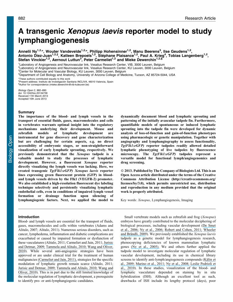
stage 45 (Fig. 1A). In all Tg(Flk1:eGFP) tadpoles, GFP was alsoexpressed in the lymphatic vasculature. Close examination atstage 40–45 (3–5 dpf) revealed that the developing lymph heart
was GFP+ (Fig. 1A,B). In addition, the connecting lymph vessels,such as the cephalic lymph duct (CLD) and the lateral lymph duct
(LLD) (Ny et al., 2005) were GFP+ (Fig. 1B), as well as the large
axial lymph vessels, the ventral caudal lymph vessel (VCLV)
ventrally of the PCV, and the dorsal caudal lymph vessel (DCLV)dorsally of the dorsal longitudinal anastomosing vessel (DLAV)
(Fig. 1C).
The identity of the GFP+ vessels was further confirmed using
functional assays that specifically double label blood- versus
lymph vessels. Upon angiography by intracardial micro-injectionof a red fluorescent dye (tetramethylrhodamine-dextran, TRITC-
dextran, Mr 26106 Da), the otherwise green blood vessels in
Tg(Flk1:eGFP) tadpoles became orange, while the VCLV and
DCLV remained green (Fig. 1D). Conversely, followinglymphangiography by subcutaneous injection of TRITC-dextran
in developing tadpoles, lymph vessels in Tg(Flk1:eGFP) tadpoles
became orange, while blood vessels remained green (Fig. 1E).Thus, both blood- and lymph vessels can be readily visualized in
the Tg(Flk1:eGFP) tadpoles by their fluorescence.
Fig. 1. GFP expression in blood and lymphatic vessels of Tg(Flk1:eGFP)
tadpoles. All panels depict lateral views of the tadpoles, head facing left.(A) Stage 40–45 Tg(Flk1:eGFP) tadpole showing GFP signal in the entireblood and lymphatic vasculature. (B) Higher magnification of fluorescent LH
(encircled) and connecting lymphatic vessels in a stage 45 tadpole. The regiondemarcated by the left square in panel A is shown. (C) Higher magnification ofGFP+ blood and lymphatic vessels in the trunk of a stage 45 tadpole. The regiondemarcated by the right square in panel A is shown. (D) Angiography byintracardial injection of high molecular TRITC-dextran exclusively labeled theblood vasculature (shown in orange). (E) Reversely, lymphangiography byinjecting the TRITC-dextran dye in the fin adjacent to the DCLV (black
asterisk denotes site of injection), showing specific uptake of the dye by thelymphatics (shown in orange) and draining towards the lymph heart. Insetshows higher magnification of the zone proximal to the injection site. CLD,cephalic lymph duct; DA, dorsal aorta; DCLV, dorsal caudal lymph vessel;DLAV, dorsal longitudinal anastomosing vessel; ISV, intersomitic vessel; LH,lymph heart; LLD, lateral lymph duct; PCV, posterior cardinal vein; VCLV,
ventral caudal lymphatic vessel. Scale bars: 500 mm (A), 200 mm (B–E).
Lymphatic reporter frogs 883
Bio
logy
Open
A novel method for selective and prolonged labeling of lymphvessels
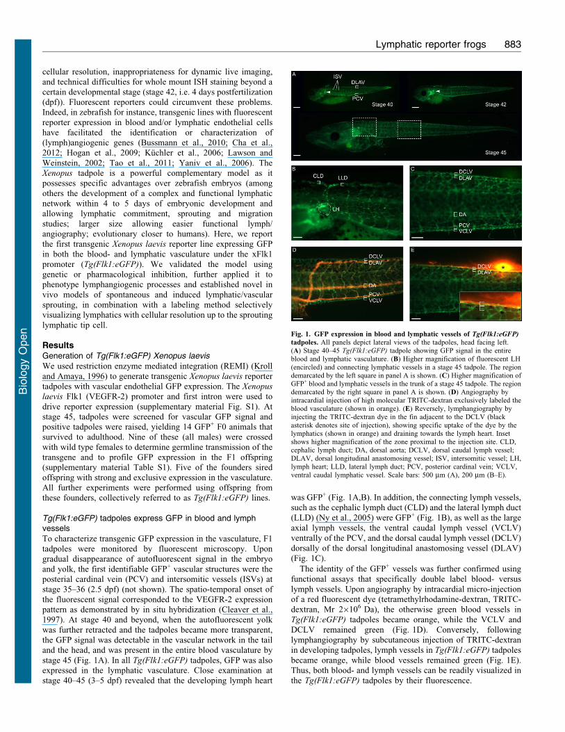
We also established an additional method to selectively labellymphatic endothelial cells (LECs) for protracted periods. Thiswas achieved by intracardial injection of TRITC-dextran, initially
labeling only the blood vessels, but allowing the dye toextravasate into the interstitial space. After 24 hours, the dye isthen taken up by the LECs, likely by a process of pinocytosis, and
was stably retained inside the LECs as well as their daughter cellsfor prolonged times ($2 weeks). This LEC labeling methodselectively labeled lymphatic structures such as the lymph hearts,
head lymphatics, axial lymphatics of the tail (Fig. 2A,B), asfurther confirmed by confocal imaging of cross sections(Fig. 2C–C0). qRT-PCR analysis of FACS sorted GFP+ bloodvascular ECs (BECs) and GFP+TRITC-dextran+ LECs revealed
higher levels of the LEC marker genes Prox1, VEGFR-3, LYVE-1
and reelin in the LECs, confirming their lymphatic lineage(supplementary material Fig. S2). Thus, the LEC labeling method
allowed color distinction between blood and lymph vessels in afast and easy manner and for prolonged times, enabling dynamicmonitoring of vascular development in a single tadpole. Of note,
LEC labeling was also possible in conditions where lymphaticvessels were malformed and dysfunctional (see below).
Documentation of lymphangiogenic sprouting and patterning inthe fin
Follow-up of vascular development in older stage embryosrevealed that lymph vessels branched off into the fin by
sprouting, just like axial blood vessels did (Rhodin andLametschwandtner, 1993). As the fin is originally devoid oflymphatics, and lymphangiogenesis into the fin was not describedto date, we documented day-by-day for 14 days the sequence and
pattern of fin vascularization; to distinguish lymphatic from bloodvessel sprouts by color, LECs were labeled by TRITC-dextran(supplementary material Fig. S3). Blood vessel sprouting preceded
lymphatic sprouting and first occurred in the ventral fin startingfrom stage 46 onwards (6 dpf; supplementary material Fig. S3A9).Blood vessel sprouting was not stereotyped but occurred in a
random pattern, however the final sprout number was comparablebetween tadpoles (not shown). Blood vessel sprouts then turnedback towards the axial vessels forming closed loops(supplementary material Fig. S3B,B9). Later on, existing loops
developed secondary angiogenic branches, forming a networkcovering the entire fin. Lymph vessel sprouting started only fromstage 48 (8 dpf) onwards. Lymphatics sprouted from the VCLV
and DCLV without evidence of a stereotyped spatio-temporalpattern of branch formation. Compared to blood vessels, fewerlymphatic sprouts developed, in turn forming only a few secondary
branches that at a later stage interconnected to form the mature finlymphatic network (supplementary material Fig. S3B–D9).
Confocal microscopy showed that blood vessels in the fin hada smooth contour and shape (Fig. 2D), while lymph vessels
appeared more spiky (Fig. 2D,H,J). Furthermore, the axiallymphatics in the tadpole body displayed LECs with manysmall protrusions, a feature that was not observed in axial blood
vessels (Fig. 2E,F). High-magnification confocal imaging andtime-lapse video-recording showed that the leading cells of bothblood and lymphatic sprouts displayed a typical ‘‘tip cell’’
phenotype, with several filopodia-like structures to sense theenvironment (Fig. 2E,G–I9; supplementary material Movie 1).Occasionally, lymph vessels sprouted close to blood vessel
sprouts, possibly using the latter as a scaffold for further
elongation into the fin (Fig. 2J).
Tg(Flk1:eGFP) tadpoles as a tool for functionallymphangiogenomics
To validate the Tg(Flk1:eGFP) line for lymphangiogenesisresearch, we first performed morpholino oligomer-mediated
Fig. 2. In vivo labeling of lymphendothelial cells allows parallel blood- and
lymph vessel analysis. (A,B) Specific ‘‘LEC labeling’’ of lymph vessels(orange) by TRITC-dextran 24 hours after angiography of stage 46 tadpoles,allowing prolonged visualization of the vessels in the lymph heart (asterisk)area (A) and tail (B). (C–C0) Confocal imaging of cross sections of a LEClabeled tadpole confirmed exclusive labeling of lymphatics (DCLV, VCLV),
while blood vessels (PCV, DA) remain green. C9–C0 are higher magnificationsof the dorsal (C9) and ventral (C0) frames in panel C. (D) High magnification ofvascular sprouting into the fin showed distinct morphology between smoothblood vessels (green) and the spiky lymphatics (red/orange).(E,F) Blood vessel (PCV, green) and lymph vessel (VCLV, yellow/organge) inclose proximity. LECs of the VCLV exhibit small protrusions and filopodia(arrowheads). (G–G0) Newly forming blood vessel sprouts display a typical tip
cell phenotype (arrowheads) with several filopodia. G9–G0 are highermagnifications of angiogenic tip cells. (H) Lymphatic tip cell with protrudingfilopodia (arrowhead). (I,I9) High magnification of lymphatic tip cells withfilopodia (arrowheads) at the forefront of a lymph vessel in the fin (I) or of anew sprout forming from the DCLV (I9). (J) Lymphatics (red) occasionallysprouted at the same site as blood vessels (green), seemingly using them as a
scaffold for further elongation into the fin. CLD, cephalic lymph duct; DA,dorsal aorta; DCLV, dorsal caudal lymph vessel; LLD, lateral lymph duct; NC,notochord; PCV, posterior cardinal vein; VCLV, ventral caudal lymph vessel.Scale bars: 200 mm (A,B), 100 mm (C), 50 mm (C9,C0,D,G), 20 mm(E,F,H,I9,J), 10 mm (G9,G0,I).
Lymphatic reporter frogs 884
Bio
logy
Open
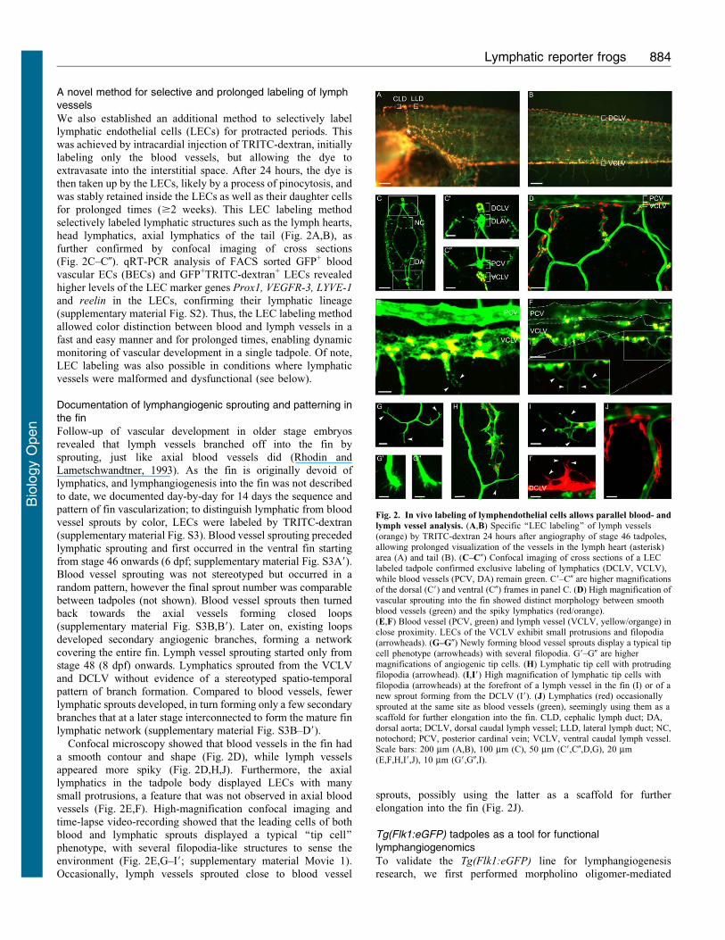
silencing of two known lymphangiogenic genes, Prox1 and
VEGF-C, previously shown to cause severe lymphatic defects in
non-reporter tadpoles (Ny et al., 2005; Ny et al., 2008). All
tadpoles injected with control morpholino developed normally
with well-structured and functional blood vessels and lymphatics
as readily visualized by fluorescent stereomicroscopy in
combination with LEC labeling (Fig. 3A–C). In contrast,
knockdown of xProx1 in Tg(Flk1:eGFP) tadpoles resulted in
edema formation at stage 45/46 (5–6 dpf) (Fig. 3D) and
dysfunctional lymphatics at an incidence similar to what we
previously observed in non-reporter tadpoles (Ny et al., 2005)
(not shown). Fluorescent microscopy for GFP showed that both
the VCLV and the DCLV were severely compromised and hardly
detectable (Fig. 3E). LEC labeling further revealed a highly
disorganized and dilated appearance with few and loosely
attached LECs (Fig. 3F). As observed in non-reporter tadpoles
(Ny et al., 2005), lymphatic defects were more severe for the
DCLV (often completely absent) than for the VCLV (Fig. 3E,F).
Likewise, silencing of xVEGF-C in Tg(Flk1:eGFP) tadpoles
caused edema (Fig. 3G) and severe lymph vessel anomalies
(fewer and more dispersed and scattered LECs) which were
readily visualized by fluorescence microscopy (Fig. 3G–I). These
results are in accordance with previous observations in VEGF-C
deficient mice and in VEGF-C silenced non-reporter tadpoles
(Karkkainen et al., 2004; Ny et al., 2005; Ny et al., 2008). Thus,
morphant phenotypes in Tg(Flk1:eGFP) and non-reporter
tadpoles were comparable (Ny et al., 2005). However, the
Tg(Flk1:eGFP) model enabled superior imaging of vascular
morphology and function for both blood and lymph vessels, and
in a dynamic manner in live tadpoles.
Tg(Flk1:eGFP) tadpoles as a tool for chemicogenetics
We also evaluated the Tg(Flk1:eGFP) line to facilitate screening
of anti-lymphangiogenic chemical compounds. As proof-of-
principle, we used MAZ51, a chemical inhibitor with a .10-
fold higher selectivity for VEGFR-3, the receptor for the
lymphangiogenic factors VEGF-C and VEGF-D (Tammela and
Alitalo, 2010) than for the other (angiogenic) VEGF receptors
(Kirkin et al., 2001). We previously documented the lymphatic
phenotype of MAZ51 in non-reporter tadpoles (Ny et al., 2008).
To minimize any adverse effect of MAZ51 on general
development, we exposed tadpoles to this compound only
beyond stage 26/28, i.e. just prior to the formation of the major
blood and lymph vessels (Levine et al., 2003; Ny et al., 2005),
and at a concentration (10 mM) that affected the blood
vasculature only minimally (Ny et al., 2008). Tg(Flk1:eGFP)
tadpoles treated with MAZ51 displayed edema around the heart
and gut (Fig. 3J). Microscopic inspection of LEC labeled
tadpoles revealed a fragmented DCLV with dispersed and
scattered LECs (Fig. 3K,L). Lymphangiography confirmed that
the DCLV was dysfunctional (not shown), in accordance with
previous observations (Ny et al., 2008). Thus, Tg(Flk1:eGFP)
tadpoles are useful models for screening compounds targeting the
lymphatic (or blood) vasculature.
Fig. 3. Tg(Flk1:eGFP) transgenic tadpoles as a tool to study developmental lymphangiogenesis. All panels depict lateral views of stage 45 tadpoles, headfacing left. All insets show higher magnification of the dorsal side, with top and lower bracket denoting DCLV and DLAV, respectively. (A–C) Control embryoshowing normal morphology (A) and correctly formed GFP+ (B) and LEC labeled lymphatics (C). (D–F) Morpholino knockdown (KD) of xProx1 resulted in edemaaround heart, gut and cloaca (arrowheads in panel D). GFP fluorescent microscopy showing that Prox1KD tadpoles possess few and disorganized LECs failing toassemble into the dorsal and ventral lymphatics (E). LEC labeling showing fewer LECs in xProx1KD tadpoles. Arrowheads denote sites where longitudinal lymphvessels are missing or malformed (green: DLAV, orange: DCLV) (F). (G–I) Morpholino knockdown (KD) of xVEGF-C resulted in edema in the heart and gut
(arrowheads in panel G). Reporter VEGF-CKD tadpoles revealing a fragmented DCLV consisting of dispersed and scattered LECs on the dorsal side while the VCLVappears grossly normal as shown by fluorescent microscopy (H) and LEC labeling (I). Arrowheads denote sites where longitudinal lymph vessel is missing.(J–L) Chemical inhibition of VEGFR-3 by MAZ51 treatment (10 mM) resulted in edema in the heart and gut (arrowheads in panel J). Fluorescent microscopy (K) andLEC labeling (L) revealing a disorganized and fragmented DCLV consisting of few LECs, while the VCLV appeared normal. Arrowheads denote where longitudinallymph vessels are missing. DA, dorsal aorta; DCLV, dorsal caudal lymph vessel; DLAV, dorsal longitudinal anastomosing vessel; PCV, posterior cardinal vein;VCLV, ventral caudal lymph vessel. Scale bars: 1 mm (A,D,G,J), 250 mm (B,E,H,K).
Lymphatic reporter frogs 885
Bio
logy
Open
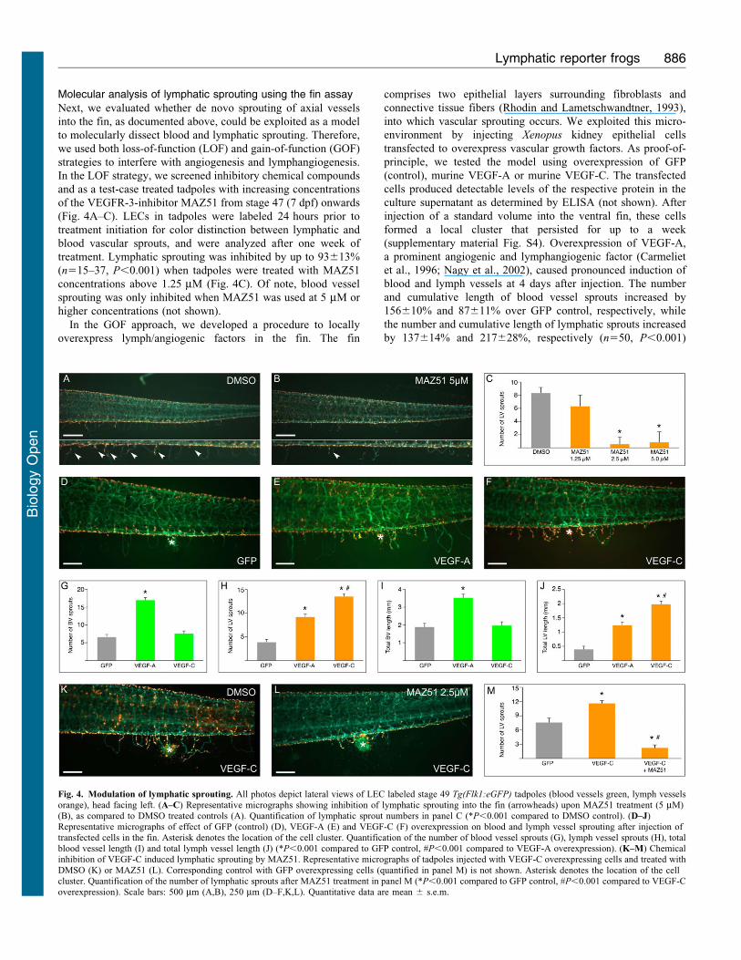
Molecular analysis of lymphatic sprouting using the fin assay
Next, we evaluated whether de novo sprouting of axial vessels
into the fin, as documented above, could be exploited as a model
to molecularly dissect blood and lymphatic sprouting. Therefore,
we used both loss-of-function (LOF) and gain-of-function (GOF)
strategies to interfere with angiogenesis and lymphangiogenesis.
In the LOF strategy, we screened inhibitory chemical compounds
and as a test-case treated tadpoles with increasing concentrations
of the VEGFR-3-inhibitor MAZ51 from stage 47 (7 dpf) onwards
(Fig. 4A–C). LECs in tadpoles were labeled 24 hours prior to
treatment initiation for color distinction between lymphatic and
blood vascular sprouts, and were analyzed after one week of
treatment. Lymphatic sprouting was inhibited by up to 93613%
(n515–37, P,0.001) when tadpoles were treated with MAZ51
concentrations above 1.25 mM (Fig. 4C). Of note, blood vessel
sprouting was only inhibited when MAZ51 was used at 5 mM or
higher concentrations (not shown).
In the GOF approach, we developed a procedure to locally
overexpress lymph/angiogenic factors in the fin. The fin
comprises two epithelial layers surrounding fibroblasts and
connective tissue fibers (Rhodin and Lametschwandtner, 1993),
into which vascular sprouting occurs. We exploited this micro-
environment by injecting Xenopus kidney epithelial cells
transfected to overexpress vascular growth factors. As proof-of-
principle, we tested the model using overexpression of GFP
(control), murine VEGF-A or murine VEGF-C. The transfected
cells produced detectable levels of the respective protein in the
culture supernatant as determined by ELISA (not shown). After
injection of a standard volume into the ventral fin, these cells
formed a local cluster that persisted for up to a week
(supplementary material Fig. S4). Overexpression of VEGF-A,
a prominent angiogenic and lymphangiogenic factor (Carmeliet
et al., 1996; Nagy et al., 2002), caused pronounced induction of
blood and lymph vessels at 4 days after injection. The number
and cumulative length of blood vessel sprouts increased by
156610% and 87611% over GFP control, respectively, while
the number and cumulative length of lymphatic sprouts increased
by 137614% and 217628%, respectively (n550, P,0.001)
Fig. 4. Modulation of lymphatic sprouting. All photos depict lateral views of LEC labeled stage 49 Tg(Flk1:eGFP) tadpoles (blood vessels green, lymph vesselsorange), head facing left. (A–C) Representative micrographs showing inhibition of lymphatic sprouting into the fin (arrowheads) upon MAZ51 treatment (5 mM)(B), as compared to DMSO treated controls (A). Quantification of lymphatic sprout numbers in panel C (*P,0.001 compared to DMSO control). (D–J)Representative micrographs of effect of GFP (control) (D), VEGF-A (E) and VEGF-C (F) overexpression on blood and lymph vessel sprouting after injection oftransfected cells in the fin. Asterisk denotes the location of the cell cluster. Quantification of the number of blood vessel sprouts (G), lymph vessel sprouts (H), totalblood vessel length (I) and total lymph vessel length (J) (*P,0.001 compared to GFP control, #P,0.001 compared to VEGF-A overexpression). (K–M) Chemicalinhibition of VEGF-C induced lymphatic sprouting by MAZ51. Representative micrographs of tadpoles injected with VEGF-C overexpressing cells and treated with
DMSO (K) or MAZ51 (L). Corresponding control with GFP overexpressing cells (quantified in panel M) is not shown. Asterisk denotes the location of the cellcluster. Quantification of the number of lymphatic sprouts after MAZ51 treatment in panel M (*P,0.001 compared to GFP control, #P,0.001 compared to VEGF-Coverexpression). Scale bars: 500 mm (A,B), 250 mm (D–F,K,L). Quantitative data are mean 6 s.e.m.
Lymphatic reporter frogs 886
Bio
logy
Open
(Fig. 4D,E,G–J). Overexpression of the lymphangiogenic growth
factor VEGF-C increased lymphatic sprouting, as the number andcumulative length of lymphatic sprouts increased by 247615%and 406629%, respectively (n550, P,0.001) (Fig. 4F,H,J). Of
note, this increase was significantly higher than that induced byVEGF-A overexpression (P,0.001, Fig. 4H,J). VEGF-C over-expression did, however, not affect blood vessel sprouting(P5NS versus GFP control; Fig. 4G,I). Next, we evaluated if
the effects of VEGF-C on lymphatic sprouting could be reversedby compound inhibitor treatment. Indeed, treatment with 2.5 mMMAZ51 inhibited the VEGF-C-induced increase in lymphatic
sprouts by 8165.4% (n530–37, P,0.001; Fig. 4M) andcumulative length by 8765.7% (1,713687 mm for VEGF-Cversus 219697 mm for VEGF-C/MAZ51; n530–37, P,0.001).
Thus, two new methods based on vascular sprouting into the finwere established, suitable to genetically dissect lymph/angiogenicsprouting or to screen for modulating chemical compounds.
DiscussionXenopus and zebrafish embryos have emerged as powerful smallanimal models for vascular research (Karpanen and Schulte-Merker, 2011; Kuchler et al., 2006; Ny et al., 2006; Tammela and
Alitalo, 2010; Wheeler and Brandli, 2009). While sharing severalfeatures, Xenopus possesses specific advantages, including highergenomic and developmental similarity to mammals, and might
therefore constitute a particularly useful model for the study ofcertain vascular aspects (Isogai et al., 2001; Levine et al., 2003;Ny et al., 2005). We previously demonstrated that the Xenopus
tadpole is a valuable model to study lymphatic development (Nyet al., 2005). While lymph/angiogenesis research benefitted fromthe use of transgenic vascular reporter zebrafish (Bussmann et al.,
2010; Cha et al., 2012; Hogan et al., 2009; Kuchler et al., 2006;Lawson and Weinstein, 2002; Yaniv et al., 2006), no suchtransgenics were available in Xenopus. We therefore generated aXenopus laevis reporter line with GFP expression in the blood-
and lymphatic vasculature. This was achieved by REMItransgenesis (Kroll and Amaya, 1996) using a Xenopus laevis
Flk1 promoter/enhancer construct (Meadows et al., 2009),
yielding Tg(Flk1:eGFP) frogs and tadpoles.
As confirmed by fluorescent microscopy and functional dyeinjection assays (angiography and lymphangiography),Tg(Flk1:eGFP) tadpoles express GFP in both blood and lymph
vessels. At the early stages when LEC commitment andmigration is initiated (around stage 30–35), the tadpoles arestill only semi-transparant and more difficult to image. Shortly
after that however, when the tadpoles have become more fullytransparent, reporter signal is easily monitored. Previously, atransgenic Xenopus laevis reporter line was generated using a
similar xflk-1:GFP construct, but no reporter expression wasdescribed in the lymphatics (Doherty et al., 2007). Possibly, theuse of slightly different promoter or intron fragments yielded
BEC-restricted versus our BEC/LEC GFP expression.
We also established a method to fluorescently label thedeveloping lymphatic network in vivo, which enabled easy andpersistent color distinction of the two vasculatures in the
Tg(Flk1:eGFP) lines. Twenty-four hours following a singleintra-cardiac injection of fluorescent TRITC-dextran, the LECsof developing lymph vessels have taken up the dye, often retained
in vesicle-like structures and appearing as red dots within thecell. Likely, micropinocytotic vesicles, abundant in LECcytoplasm and participating in normal lymph formation, have
taken up the dye via pinocytosis (Ji, 2005). The dye is stablyretained for at least 2 weeks, and new lymphatics sprouting from
existing labeled ones are automatically also labeled. We observeda variable degree of labeling of LECs between tadpoles, likelydue to slight differences in amount of injected dye and explainingwhy in some images the double-labeled lymphatics appear
yellow–orange while red in others (e.g. Fig. 2E,H,I, versusFig. 2D,I9,J). The expression of LEC specific marker genes byFACS sorted double-labeled GFP+TRITC+ endothelial cells
confirmed their lymphatic identity. Sorted GFP+TRITC2 BECand GFP+TRITC+ LEC populations from the Tg(Flk1:eGFP)tadpoles combined with expression profiling may provide a
useful tool to screen for novel lymph/angiogenic genes and studydifferential gene expression in vivo.
We combined reporter expression and LEC labeling tovisualize and document in detail the sprouting and patterning
of lymphatics from the axial lymph vessels into the fin. Highmagnification imaging and time-lapse video-recording revealedlymphatic tips cells with several filopodia sensing the
environment. Occasionally, lymphatic sprouting occurred inclose proximity of the blood vessel sprouts, suggesting thatcommon initiating or guiding cues may be involved.
We validated the Tg(Flk1:eGFP) tadpoles as a model formolecular lymphangiogenesis studies, using gene knockdown orcompound inhibition of factors critically involved in lymphaticdevelopment (Prox1, VEGF-C, VEGFR-3). The phenotypes in
the Tg(Flk1:eGFP) line and in non-reporter tadpoles (Ny et al.,2005; Ny et al., 2008) were comparable, demonstrating theaccuracy and integrity of the model. The added value of the
Tg(Flk1:eGFP) line for lymph/angiogenesis phenotyping wasexemplified by i) the significantly faster procedures as it onlyrequires fluorescent microscopy as compared to lengthy whole
mount ISH staining protocols; ii) the unique possibility toanalyze defects at developmental stages (.stage 42) non-permissive to whole mount ISH; iii) the significantly higher
imaging resolution, enabling analysis of more subtle defects atthe cellular level. For instance, the abnormalities of the DCLV/VCLV upon Prox1, VEGF-C or VEGFR-3 blockage could nowbe seen at a cellular level in the Tg(Flk1:eGFP) line, revealing
dilated, interrupted vessel-like trunks consisting of sparse andloosely attached LECs. These details were unnoticeable in thecounterpart non-reporter morphant tadpoles (Ny et al., 2005; Ny
et al., 2008). Recently, the Tg(Flk1:eGFP) tadpoles have helpedin elucidating the role of novel candidate genes in lymphaticdevelopment (Aranguren et al., 2011; Fu et al., 2008; Hermans et
al., 2010; Norrmen et al., 2010; Saharinen et al., 2010). Theseadvantages of the model, together with the possibility of dynamiclive imaging, and of FACS-sorting of BEC and LEC populations
for gene profiling and expression modulation studies, representsignificant assets.
We further explored the potential of the de novo vascularsprouting into the fin as an in vivo physiological sprouting model.
To that end, we established loss-of-function (LOF) and gain-of-function (GOF) strategies to interfere with sprout formation. LOFexperiments demonstrated blockage of de novo sprouting of
lymph vessels in the fin by the VEGFR-3 inhibitor MAZ51,exposed to 7-day-old tadpoles. Higher inhibitor concentrationsalso inhibited blood vessel sprouting. In a second strategy, we
explored GOF approaches to modulate fin sprouting. In vivoelectroporation with expression plasmids or adenoviraloverexpression of known lymphangiogenic factors, while
Lymphatic reporter frogs 887
Bio
logy
Open
possible in the tail, were not successful in the fin, probably due to
its scarce cell content. Implantation of beads coated with
recombinant lymphangiogenic proteins was technically
challenging, as the fin is very thin and delicate. In contrast,
implantation of a dense suspension of Xenopus laevis renal cells
overexpressing murine lymph/angiogenic factors (VEGF-A or
VEGF-C), worked successfully. The transfected cells persisted as
a local cluster and induced sprouting of blood and/or lymph
vessels. These data also demonstrate that murine VEGF-A and
VEGF-C are active in frog with the same specificity toward
blood and/or lymphatic endothelial cells as in mouse and humans.
Quantification of sprouting (inhibition) in the LEC labeled
Tg(Flk1:eGFP) tadpoles in both the GOF and LOF model
required no more than mere fluorescent microscopy. Sprouting
lymphangiogenesis models during embryogenesis in vivo in other
species demand either tissue dissection/fixation and staining like
in the mouse, or high-resolution confocal/multiphoton imaging
such as in the zebrafish embryo (Cha et al., 2012; Geudens et al.,
2010; Hogan et al., 2009; Okuda et al., 2012; Tao et al., 2011),
and are thus more challenging for screening purposes. The in
vivo sprouting models in the tadpole fin are suited to evaluate
candidate chemical inhibitors or overexpressed candidate
modulating factors in an easy and quantifiable manner, and
enable dynamic monitoring of sprouting over several days. In
addition, the cell cluster-based fin sprouting assay allows
screening for chemical inhibitors for a specific target protein,
extending the versatility of the model. Furthermore, whereas the
fin sprouting model spans later embryonic stages (up to 14 dpf)
and therefore is less compatible with morpholino knockdown
approaches, it will also be applicable in future knockout
experiments, given the promising recent successes of zinc
finger nuclease or TALEn knockout in aquatic species
including Xenopus (Cade et al., 2012; Lei et al., 2012; Moore
et al., 2012; Young et al., 2011).
We conclude that the Tg(Flk1:eGFP) reporter line and the
assays and models established and validated in this study, provide
powerful tools for functional lymphangiogenomics as well as for
drug screening, with easy detection, morphological and
quantitative characterization of lymphatic phenotypes with
resolution up to tip cell and filopodia level.
Materials and MethodsGeneration of transgenic Tg(Flk1:eGFP) reporter tadpolesThe transgenic construct used to generate the Tg(Flk1:eGFP) reporter tadpoleswas engineered from the Xenopus laevis (xl) VEGFR-2/Flk1 gene. Therefore, agenomic fragment containing ,2.5 kb of promoter sequence up to exon 2,including the complete intron 1 containing enhancer elements essential for thecorrect expression pattern, was isolated and cloned into pBSK(+) (Meadows et al.,2009). Next, the eGFP cDNA fused to the SV40 polyA was inserted into exon 1 todrive reporter expression under the control of the xlFlk1 promoter. The constructwas linearized using KpnI and used in restriction enzyme mediated integration(REMI) transgenesis (Kroll and Amaya, 1996) using purified sperm nuclei andunfertilized eggs from wildtype frogs purchased from Nasco Biology (FortAtkinson, WI). Transgenic tadpoles were screened for eGFP signal in thevasculature at stage 45 by fluorescence microscopy and positive tadpoles wereraised to adulthood. F1 tadpoles were generated by natural breeding ofTg(Flk1:eGFP) F0 males with wild type females; F2 tadpoles by interbreedingmale and female Tg(Flk1:eGFP) F1 frogs. All breeding was done using hormonalinduction. All animal procedures were approved by the ethical committee foranimal experimentation of the KU Leuven, Leuven, Belgium.
In vivo imaging of blood- and lymphatic vasculature inTg(Flk1:eGFP) tadpolesStage 45–46 tadpoles were anesthetized in 0.02% 3-aminobenzoic acid ethyl ester,and placed on agarose gel. Tetramethylrhodamine-dextran (TRITC-dextran, Mr
26106, Invitrogen, Merelbeke, Belgium) dye was injected close to the dorsal orventral caudal lymph vessel (DCLV or VCLV, respectively) or in the heart withglass capillaries using a micromanipulator and a Zeiss SV11 stereomicroscope tomonitor lymphatic function (lymphangiography) and to label functional bloodvasculature (angiography), respectively. Lymphendothelial cell (LEC) labelingwas performed by injecting 10–20 nl of TRITC-dextran in or around the heart ofanesthetized tadpoles. After 24 hours, the injected dye is taken up inside the LECsof the lymphatic vasculature. Fluorescent images (of anesthetized tadpoles) wereacquired with the Zeiss AxioVision 4.6 software on a Zeiss Lumar V.12fluorescence stereomicroscope equipped with a Zeiss AxioCam MrC5 digitalcamera (Zeiss, Zaventem, Belgium). Confocal images of blood- and lymph vesselsprouts and tip cells were obtained from anesthetized and agarose embedded (1%low gelling agarose) tadpoles with a Zeiss CLSM510 NLO META mounted on anAxioVert200M (Zeiss, Zaventem, Belgium) inverted microscope. Confocalimaging of GFP was performed using 920 nm pulsed mode-locked laseremission from a tunable Ti:Sapphire Chameleon laser (Coherent, Utrecht, TheNetherlands); TRITC-dextran imaging was performed using a DPSS 561-10 laser.Time-lapse imaging (2 hours) was performed with minimal necessary laser power.Stacks of frame-averaged (four frames) confocal optical slices were collecteddigitally, at 5 minute intervals for time-lapse sequences. Three-dimensional aswell as four-dimensional reconstructions of image data were prepared using LSMsoftware package.
FACS sorting of BECs and LECsFive-day-old tadpoles were injected with 10 nl TRITC dextran into the heart sacand further processed 24 hours later to obtain single cell suspensions. Briefly,tadpoles were bleached for 5 minutes, rinsed with distilled water, macerated, andincubated in 0.25% trypsin at 28 C until almost fully digested. A couple of dropsof fetal bovine serum (FBS) were added to inhibit the trypsin activity and thedigest was diluted with PBS to approximately 25 ml before filtering over a 70 mmnylon mesh (BD Biosciences, Erembodegem-Aalst, Belgium). Cells were pelletedby centrifugation at 600 g for 7 minutes and the cell pellet was resuspended in4 ml PBS containing 1% bovine serum albumin (BSA). Cells were sorted on aFACSAria (BD Biosciences, Erembodegem-Aalst, Belgium), taking care toexclude possible doublets or cell clusters. Non-injected GFP+ and TRITC-injected GFP2 tadpoles were used as controls for proper compensation and gatesetting. On average 25,000 GFP+TRITC+ LECs and 50,000 GFP+TRITC2 bloodendothelial cells (BECs) were sorted directly in lysis buffer (RLT containing 1%b-mercaptoethanol; Qiagen, Venlo, The Netherlands) from 60–100 pooledtadpoles. RNA was prepared from these samples using the QIAGEN mini kitand cDNA was prepared using the SuperscriptIII kit (Ambion, Lennik, Belgium).Gene expression was analyzed by qRT-PCR using SYBR Green (AppliedBiosystems, Lennik, Belgium) and home-designed primer sets (prox1-fwd:GTCGGAGTGCGGAGACATG, prox1-rev: 59-GGC CTT TTT CAA GTG ATTTGG A-39, VEGFR-3-fwd: 59-CCC CAG CCC TCA TTC CA-39, VEGFR-3-rev:59-GCT GGG ACT GAC GA TAT TTG C-39, lyve-1-fwd: 59-CAT TCT GTGGCT CAA GGT GTC ATT AC-39, lyve-1-rev: 59-GCA TTT CTC ATT AGGCTG GAT ACG AG-39, reelin-fwd: 59-TAC AGT GGG TGG AAC CGA AT-39,reelin-rev: 59-GCT GGG CCA GAA AAT CCA GG-39, ef1a-fwd: 59-GAA CCATCG AAA AGT TCG AGA AG-39, ef1a-rev: 59-TCC AAG ACC CAG GCA TACTTG-39.
Morpholino injectionsFertilized Xenopus eggs from natural matings between hormone inducedTg(Flk1:eGFP) males and wild type females were injected at two-cell stagewith xPROX1 (25 ng), xVEGF-C (35 ng) or standard control (35 ng) morpholino(Gene Tools, LLC, Philomath, OR). The ATG-targeted antisense morpholinoswere designed based on published GenBank Xenopus laevis sequences of xProx1
(#AB008773) and xVEGF-C (#CA973641) (Ny et al., 2005) and were as follows,xProx1: 59-CAG GCA TCA CTG GAC TGT TAT TGT G-39; xVEGF-C: 59-GCTCCC TCC AGC AAG TAC ATT TTC C-39; standard control morpholino: 59-CCTCTT ACC TCA GTT ACA ATT TAT A-39. Injected embryos were cultured in0.16 MMR (Ny et al., 2005; Ny et al., 2008) at 18 C until gastrulation andthereafter at 22 C. Developmental stages of tadpoles were determined according toNieuwkoop and Faber (Nieuwkoop and Faber, 1994). At stage 45 (5 days postfertilization (dpf)) tadpoles were subjected to lymphangiographies, angiographiesand LEC labeling.
Chemical compound treatment during developmentAt stage 26/28, Tg(Flk1:eGFP) tadpoles were placed in 6-well dishes (15–20tadpoles/well) and up to 10 mM of MAZ51 (3-(4-dimethylamino-naphthelen-1-ylmethylene)-1,3-dihydroindol-2-one, Calbiochem–Merck Biosciences, Overijse,Belgium) was added to the tadpole growth media, as described previously (Ny etal., 2008). Control tadpoles were treated with the corresponding amount of DMSO.Compound/DMSO and growth medium were refreshed every day. At stage 45 (5dpf), lymphangiographies and LEC labeling were performed to visualize theblood- and lymphatic vasculature.
Lymphatic reporter frogs 888
Bio
logy
Open
Pharmacological modulation of lymphatic sprouting in the finTo visualize blood- and lymphatic sprouts from stage 47 onwards, Tg(Flk1:eGFP)
tadpoles were LEC labeled as described above, 24 hours before the start of theexperiment. MAZ51 (concentrations ranging from 1.25–5 mM) was added to LEClabeled tadpoles of stage 47 (placed in 10 cm petri dishes, with approximately 20tadpoles per condition) in the tadpole growth media. Chemical compounds orbuffer were refreshed daily. As a negative control tadpoles were treated withcorresponding amounts of DMSO. After 7 days the number of lymphatic sproutswas counted over the entire length of the ventral axial vessels, and the cumulativelength of the lymphatic sprouts was measured per tadpole with computer-assistedmorphometry using the Zeiss KS300 software (Zeiss, Zaventem, Belgium) or theImageJ software.
Cell cluster-mediated modulation of fin sproutingThe Xenopus laevis kidney cell line A6 (CCL-102) was purchased from ATCC(LGC Standards, Teddington, UK). Cells were maintained in 75% NCTC 109medium (Sigma–Aldrich, Bornem, Belgium), 15% distilled water (Invitrogen,Merelbeke, Belgium) and 10% fetal calf serum, supplemented with 2 mMglutamine, 100 U/ml Penicillin/Streptomycin, 0.01 M HEPES and 1 mM sodiumpyruvate (all Invitrogen) at room temperature. Cells were passaged 1:3 twice aweek. For transfection, cells were seeded at 70% confluency into 6-well plates andtransfected using lipofectamine (Invitrogen, Merelbeke, Belgium) according to themanufacturer’s instruction with plasmids coding for GFP, murine vascularendothelial growth factor (VEGF)-A or VEGF-C. Expression was verified byELISA using Quantikine ELISA kits (R&D Systems, Abingdon, UK). Eighteen to24 hours after transfection, cells were trypsinized, centrifuged and resuspended ina minimal amount of medium. Approximately 1,500 cells were injected as a densecell suspension using a glass capillary, generating a local cell cluster within thetwo epithelial leaflets of the tadpole fin. Cells were never injected in one singleinjection, but were rather introduced using multiple low-pressure injections(typically 6–10) at the same site, thereby generating progressively larger cellclusters. The number of cells injected was controlled by verifying the diameter ofthe cell cluster. Only tadpoles with comparable cell cluster size were used in theexperimental analysis. Sustained expression of the transgene by the injected cellswas demonstrated by the green fluorescent signal of cells transfected with the GFPexpression plasmid, persisting for a least 1 week. Injected tadpoles were allowed torecover from anesthesia and treatment with 2.5 mM MAZ51 was initiated bysupplementation of the chemicals to the tadpole growth media. Buffers andchemicals were refreshed daily. After 4 days, the local effect on vessel sproutingwas documented by pictures of the fin surrounding the central cell cluster. Thesepictures were taken at 506magnification, representing approximately 1.4 mm onboth sides of the cell cluster. The numbers of blood- and lymphatic sprouts werecounted and the cumulative length of the sprouts was measured by computer-assisted morphometry using the Zeiss Axiovision KS300 software (Zeiss,Zaventem, Belgium) or the ImageJ software.
Statistical analysisAll fin sprouting and cell injection experiments were analyzed using IBM SPSSStatistics 19. Data shown are estimated means and s.e.m. calculated from thecombined results of at least 3 different experiments (except for the experimentusing MAZ51 inhibition of VEGF-C overexpression effects; 2 experiments used).Estimated means, s.e.m. and significance levels were calculated with the generallinear model multivariate statistical model, considering the number of bloodvessels, number of lymph vessels, total blood vessel length and total lymph vessellength as dependent variables, compound dose and/or growth factor overexpressedas fixed factors, and experiment as co-variate. Pairwise comparisons wereperformed between the different doses, after Bonferroni correction for multipletesting. P,0.05 was considered statistically significant.
AcknowledgementsWe thank A. Cobut, H. Laisnez, S. Louwette, W.Y. Man, and A. VanNuffelen for their technical assistance. A.N. is sponsored by theEuropean Union Framework 6 Program – Marie Curie IntraEuropean Fellowship; W.V., I.G. and M.B. by the FlemishInstitute for the promotion of scientific research (IWT), Belgium;P.H. by a Federation of European Biochemical Societies (FEBS)fellowship; A.D.-J. by the Centro Nacional de InvestigacionesCardiovasculares (CNIC), Spain. This work is supported in part bythe Flemish Foundation for Scientific Research [FWO grant numberG.0567.05], the European Union Framework 6 Program [grantnumber LSHG-CT-2004-503573], an unrestricted Bristol-Myers-Squibb grant, and by long-term structural funding: Methusalemfunding by the Flemish Government to P.C., by the Belgian SciencePolicy [grant number IAP P6-20] to M.D., and by funding from the
Concerted Research Activities, Belgium [grant numbers GOA2001/09, GOA2006/11] to M.D. and P.C.
Author ContributionsA.N., W.V., P.C. and M.D. designed experiments and wrote themanuscript; A.N. and W.V. performed experiments and allquantification; P.H., I.G., A.D.-J., K.B., S.P. and T.L. assisted withexperiments; M.B. and A.L. performed the FACS sorting; S.V.helped with confocal microscopy; P.A.K. provided essentialreagents.
Competing InterestsThe authors have no competing interests to declare.
ReferencesAdams, R. H. and Alitalo, K. (2007). Molecular regulation of angiogenesis and
lymphangiogenesis. Nat. Rev. Mol. Cell Biol. 8, 464-478.
Alitalo, K. (2011). The lymphatic vasculature in disease. Nat. Med. 17, 1371-1380.
Aranguren, X. L., Beerens, M., Vandevelde, W., Dewerchin, M., Carmeliet, P. and
Luttun, A. (2011). Transcription factor COUP-TFII is indispensable for venous andlymphatic development in zebrafish and Xenopus laevis. Biochem. Biophys. Res.
Commun. 410, 121-126.
Bussmann, J., Bos, F. L., Urasaki, A., Kawakami, K., Duckers, H. J. and Schulte-
Merker, S. (2010). Arteries provide essential guidance cues for lymphatic endothelialcells in the zebrafish trunk. Development 137, 2653-2657.
Cade, L., Reyon, D., Hwang, W. Y., Tsai, S. Q., Patel, S., Khayter, C., Joung, J. K.,
Sander, J. D., Peterson, R. T. and Yeh, J. R. (2012). Highly efficient generation ofheritable zebrafish gene mutations using homo- and heterodimeric TALENs. Nucleic
Acids Res. 40, 8001-8010.
Carmeliet, P. and Jain, R. K. (2011). Molecular mechanisms and clinical applicationsof angiogenesis. Nature 473, 298-307.
Carmeliet, P., Ferreira, V., Breier, G., Pollefeyt, S., Kieckens, L., Gertsenstein, M.,
Fahrig, M., Vandenhoeck, A., Harpal, K., Eberhardt, C. et al. (1996). Abnormalblood vessel development and lethality in embryos lacking a single VEGF allele.Nature 380, 435-439.
Cha, Y. R., Fujita, M., Butler, M., Isogai, S., Kochhan, E., Siekmann, A. F. and
Weinstein, B. M. (2012). Chemokine signaling directs trunk lymphatic networkformation along the preexisting blood vasculature. Dev. Cell 22, 824-836.
Cleaver, O., Tonissen, K. F., Saha, M. S. and Krieg, P. A. (1997). Neovascularizationof the Xenopus embryo. Dev. Dyn. 210, 66-77.
De Smet, F., Carmeliet, P. and Autiero, M. (2006). Fishing and frogging for anti-angiogenic drugs. Nat. Chem. Biol. 2, 228-229.
Doherty, J. R., Johnson Hamlet, M. R., Kuliyev, E. and Mead, P. E. (2007). A flk-1promoter/enhancer reporter transgenic Xenopus laevis generated using the SleepingBeauty transposon system: an in vivo model for vascular studies. Dev. Dyn. 236,2808-2817.
Fu, J., Gerhardt, H., McDaniel, J. M., Xia, B., Liu, X., Ivanciu, L., Ny, A., Hermans,
K., Silasi-Mansat, R., McGee, S. et al. (2008). Endothelial cell O-glycan deficiencycauses blood/lymphatic misconnections and consequent fatty liver disease in mice.J. Clin. Invest. 118, 3725-3737.
Geudens, I., Herpers, R., Hermans, K., Segura, I., Ruiz de Almodovar, C.,
Bussmann, J., De Smet, F., Vandevelde, W., Hogan, B. M., Siekmann, A. et al.
(2010). Role of delta-like-4/Notch in the formation and wiring of the lymphaticnetwork in zebrafish. Arterioscler. Thromb. Vasc. Biol. 30, 1695-1702.
Hermans, K., Claes, F., Vandevelde, W., Zheng, W., Geudens, I., Orsenigo, F., De
Smet, F., Gjini, E., Anthonis, K., Ren, B. et al. (2010). Role of synectin inlymphatic development in zebrafish and frogs. Blood 116, 3356-3366.
Hogan, B. M., Bos, F. L., Bussmann, J., Witte, M., Chi, N. C., Duckers, H. J. and
Schulte-Merker, S. (2009). Ccbe1 is required for embryonic lymphangiogenesis andvenous sprouting. Nat. Genet. 41, 396-398.
Isogai, S., Horiguchi, M. and Weinstein, B. M. (2001). The vascular anatomy of thedeveloping zebrafish: an atlas of embryonic and early larval development. Dev. Biol.
230, 278-301.
Ji, R. C. (2005). Characteristics of lymphatic endothelial cells in physiological andpathological conditions. Histol. Histopathol. 20, 155-175.
Jurisic, G. and Detmar, M. (2009). Lymphatic endothelium in health and disease. Cell
Tissue Res. 335, 97-108.
Kalin, R. E., Banziger-Tobler, N. E., Detmar, M. and Brandli, A. W. (2009). An invivo chemical library screen in Xenopus tadpoles reveals novel pathways involved inangiogenesis and lymphangiogenesis. Blood 114, 1110-1122.
Karkkainen, M. J., Haiko, P., Sainio, K., Partanen, J., Taipale, J., Petrova, T. V.,
Jeltsch, M., Jackson, D. G., Talikka, M., Rauvala, H. et al. (2004). Vascularendothelial growth factor C is required for sprouting of the first lymphatic vesselsfrom embryonic veins. Nat. Immunol. 5, 74-80.
Karpanen, T. and Schulte-Merker, S. (2011). Zebrafish provides a novel model forlymphatic vascular research. Methods Cell Biol. 105, 223-238.
Kirkin, V., Mazitschek, R., Krishnan, J., Steffen, A., Waltenberger, J., Pepper,
M. S., Giannis, A. and Sleeman, J. P. (2001). Characterization of indolinones which
Lymphatic reporter frogs 889
Bio
logy
Open
preferentially inhibit VEGF-C- and VEGF-D-induced activation of VEGFR-3 ratherthan VEGFR-2. Eur. J. Biochem. 268, 5530-5540.
Kroll, K. L. and Amaya, E. (1996). Transgenic Xenopus embryos from sperm nucleartransplantations reveal FGF signaling requirements during gastrulation. Development
122, 3173-3183.Kuchler, A. M., Gjini, E., Peterson-Maduro, J., Cancilla, B., Wolburg, H. and
Schulte-Merker, S. (2006). Development of the zebrafish lymphatic system requiresVEGFC signaling. Curr. Biol. 16, 1244-1248.
Lawson, N. D. and Weinstein, B. M. (2002). In vivo imaging of embryonic vasculardevelopment using transgenic zebrafish. Dev. Biol. 248, 307-318.
Lei, Y., Guo, X., Liu, Y., Cao, Y., Deng, Y., Chen, X., Cheng, C. H., Dawid, I. B.,Chen, Y. and Zhao, H. (2012). Efficient targeted gene disruption in Xenopusembryos using engineered transcription activator-like effector nucleases (TALENs).Proc. Natl. Acad. Sci. USA 109, 17484-17489.
Leslie Pedrioli, D. M., Karpanen, T., Dabouras, V., Jurisic, G., van de Hoek, G.,Shin, J. W., Marino, D., Kalin, R. E., Leidel, S., Cinelli, P. et al. (2010). miR-31functions as a negative regulator of lymphatic vascular lineage-specific differentiationin vitro and vascular development in vivo. Mol. Cell. Biol. 30, 3620-3634.
Levine, A. J., Munoz-Sanjuan, I., Bell, E., North, A. J. and Brivanlou, A. H. (2003).Fluorescent labeling of endothelial cells allows in vivo, continuous characterization ofthe vascular development of Xenopus laevis. Dev. Biol. 254, 50-67.
Marino, D., Dabouras, V., Brandli, A. W. and Detmar, M. (2011). A role for all-trans-retinoic acid in the early steps of lymphatic vasculature development. J. Vasc. Res. 48,236-251.
Meadows, S. M., Salanga, M. C. and Krieg, P. A. (2009). Kruppel-like factor 2cooperates with the ETS family protein ERG to activate Flk1 expression duringvascular development. Development 136, 1115-1125.
Moore, F. E., Reyon, D., Sander, J. D., Martinez, S. A., Blackburn, J. S., Khayter,
C., Ramirez, C. L., Joung, J. K. and Langenau, D. M. (2012). Improved somaticmutagenesis in zebrafish using transcription activator-like effector nucleases(TALENs). PLoS ONE 7, e37877.
Nagy, J. A., Vasile, E., Feng, D., Sundberg, C., Brown, L. F., Detmar, M. J., Lawitts,J. A., Benjamin, L., Tan, X., Manseau, E. J. et al. (2002). Vascular permeabilityfactor/vascular endothelial growth factor induces lymphangiogenesis as well asangiogenesis. J. Exp. Med. 196, 1497-1506.
Nieuwkoop, P. D. and Faber, J. (1994). Normal Table Of Xenopus Laevis
(Daudin): A Systematical And Chronological Survey Of The Development From
The Fertilized Egg Till The End Of Metamorphosis. New York, NY: GarlandPublishing Inc.
Norrmen, C., Vandevelde, W., Ny, A., Saharinen, P., Gentile, M., Haraldsen, G.,Puolakkainen, P., Lukanidin, E., Dewerchin, M., Alitalo, K. et al. (2010). Liprin(beta)1 is highly expressed in lymphatic vasculature and is important for lymphaticvessel integrity. Blood 115, 906-909.
Ny, A., Koch, M., Schneider, M., Neven, E., Tong, R. T., Maity, S., Fischer, C.,
Plaisance, S., Lambrechts, D., Heligon, C. et al. (2005). A genetic Xenopus laevis
tadpole model to study lymphangiogenesis. Nat. Med. 11, 998-1004.
Ny, A., Autiero, M. and Carmeliet, P. (2006). Zebrafish and Xenopus tadpoles: small
animal models to study angiogenesis and lymphangiogenesis. Exp. Cell Res. 312,
684-693.
Ny, A., Koch, M., Vandevelde, W., Schneider, M., Fischer, C., Diez-Juan, A., Neven,
E., Geudens, I., Maity, S., Moons, L. et al. (2008). Role of VEGF-D and VEGFR-3
in developmental lymphangiogenesis, a chemicogenetic study in Xenopus tadpoles.
Blood 112, 1740-1749.
Okuda, K. S., Astin, J. W., Misa, J. P., Flores, M. V., Crosier, K. E. and Crosier,
P. S. (2012). lyve1 expression reveals novel lymphatic vessels and new mechanisms
for lymphatic vessel development in zebrafish. Development 139, 2381-2391.
Rhodin, J. A. and Lametschwandtner, A. (1993). Circulatory pattern and structure in
the tail and tail fins of Xenopus laevis tadpoles. J. Submicrosc. Cytol. Pathol. 25, 297-
318.
Robert, J. and Cohen, N. (2011). The genus Xenopus as a multispecies model for
evolutionary and comparative immunobiology of the 21st century. Dev. Comp.
Immunol. 35, 916-923.
Saharinen, P., Helotera, H., Miettinen, J., Norrmen, C., D’Amico, G., Jeltsch, M.,
Langenberg, T., Vandevelde, W., Ny, A., Dewerchin, M. et al. (2010). Claudin-like
protein 24 interacts with the VEGFR-2 and VEGFR-3 pathways and regulates
lymphatic vessel development. Genes Dev. 24, 875-880.
Tammela, T. and Alitalo, K. (2010). Lymphangiogenesis: Molecular mechanisms and
future promise. Cell 140, 460-476.
Tao, S., Witte, M., Bryson-Richardson, R. J., Currie, P. D., Hogan, B. M. and
Schulte-Merker, S. (2011). Zebrafish prox1b mutants develop a lymphatic
vasculature, and prox1b does not specifically mark lymphatic endothelial cells.
PLoS ONE 6, e28934.
Wang, Y. and Oliver, G. (2010). Current views on the function of the lymphatic
vasculature in health and disease. Genes Dev. 24, 2115-2126.
Wheeler, G. N. and Brandli, A. W. (2009). Simple vertebrate models for chemical
genetics and drug discovery screens: lessons from zebrafish and Xenopus. Dev. Dyn.
238, 1287-1308.
Yaniv, K., Isogai, S., Castranova, D., Dye, L., Hitomi, J. and Weinstein, B. M.
(2006). Live imaging of lymphatic development in the zebrafish. Nat. Med. 12, 711-
716.
Young, J. J., Cherone, J. M., Doyon, Y., Ankoudinova, I., Faraji, F. M., Lee, A. H.,
Ngo, C., Guschin, D. Y., Paschon, D. E., Miller, J. C. et al. (2011). Efficient
targeted gene disruption in the soma and germ line of the frog Xenopus tropicalis
using engineered zinc-finger nucleases. Proc. Natl. Acad. Sci. USA 108, 7052-7057.
Lymphatic reporter frogs 890
Bio
logy
Open