Embed Size (px)
Citation preview

1
A venom-derived neurotoxin, CsTx-1, from the spider Cupiennius salei exhibits cytolytic
activities
Lucia Kuhn-Nentwig1, Irina M. Fedorova
2, Benjamin P. Lüscher
3, Lukas S. Kopp
4, Christian
Trachsel4, Johann Schaller
4, Xuan Lan Vu
5, Thomas Seebeck
5, Kathrin Streitberger
1, Wolfgang
Nentwig1, Erwin Sigel
3, and Lev G. Magazanik
2,6
From the 1Institute of Ecology and Evolution, University of Bern, Baltzerstrasse 6, CH-3012 Bern,
Switzerland 2I.M. Sechenov Institute of Evolutionary Physiology and Biochemistry, Saint-Petersburg; Russian
Academy of Sciences, Thorez pr. 44, 194223 Saint-Petersburg, Russia 3Institute of Biochemistry and Molecular Medicine, University of Bern, Bühlstrasse 28, CH-3012 Bern,
Switzerland 4Department of Chemistry and Biochemistry, University of Bern, Freiestrasse 3, CH-3012 Bern,
Switzerland 5Institute of Cell Biology, University of Bern, Baltzerstrasse 4, 3012 Bern, Switzerland
6Medical Faculty, 199034 Saint-Petersburg University, 7-9 University embank., Russia
*Running title: CsTx-1, a pore-former
To whom the correspondence should be addressed: Lucia Kuhn-Nentwig, Institute of Ecology and
Evolution, University of Bern, Baltzerstrasse 6, CH-3012 Bern, Switzerland, Tel: +41 31 631 4532 Fax:
+41 31 631 4888, Email: [email protected]
Keywords: CsTx-1, Cupiennius salei, cytolytic activity, Calliphora larvae muscle preparation, Rana
neuromusclular preparations, Xenopus oocytes, L-type Ca2+
channel inhibitor
Background: CsTx-1, an ICK motif containing
neurotoxin, acts as L-type Ca2+
-channel inhibitor.
Results: The partial α-helical C-terminus of CsTx-
1 exhibits cytolytic activity towards prokaryotic
and eukaryotic cell membranes.
Conclusion: one peptide with different domains
for ion channel inhibition and cytolytic activity.
Significance: An important new mechanism for
the evolution of spider venomous peptides.
CsTx-1, the main neurotoxic acting peptide
in the venom of the spider Cupiennius salei is
composed of 74 amino acid residues, exhibits an
ICK motif and is further characterised by its
highly cationic charged C-terminus. Venom
gland cDNA library analysis predicted for
CsTx-1 precursor a prepropeptide structure. In
the presence of trifluoroethanol, CsTx-1 and
the long C-terminal part alone (CT1-long;
Gly45-Lys74) exhibit an α-helical structure, as
determined by CD-measurements. CsTx-1 and
CT1-long are insecticidal towards Drosophila
flies and destroys Escherichia coli SBS 363 cells.
CsTx-1 causes a stable and irreversible
depolarisation of insect larvae muscle cells and
frog neuromuscular preparations which seem
to be receptor independent. Furthermore, this
membranolytic activity could be measured for
Xenopus oocytes, in which CsTx-1 and CT1-
long increase ion permeability non-specifically.
These results support our assumption that the
membranolytic activities of CsTx-1 are caused
by its C-terminal tail, CT1-long. Together,
CsTx-1 exhibits two different functions: as a
neurotoxin it inhibits L-type Ca2+
-channels and
as a membranolytic peptide it destroys a
variety of prokaryotic and eukaryotic cell
membranes. Such a dualism is discussed as an
important new mechanism for the evolution of
spider venomous peptides.
Spiders evolved some 300 million years ago
(1). With currently 42,055 species, spiders
represent the second most abundant group of
terrestrial organisms, after the insects (2). The
majority of spiders rely on the potency of their
venom for immediate prey immobilisation or to
repel aggressors. For fast paralysing or killing a
prey item, spiders very successfully developed a
variety of multicomponent venoms in which
components usually act synergistically. It seems

2
that araneomorph spiders have evolved a much
greater variety of different substance
combinations, which provide likewise immediate
paralysis of prey than the ancient mygalomorph
spiders. Additive interactions between different
venom compounds of the same group, or
synergistic interactions between different venom
compound groups, such as ions, low molecular
mass compounds, enzymes, neurotoxins, small
cationic peptides (SCPs) and α-helical small
cationic peptides (α-SCPs), have been identified
recently (for review see (3)).
The venom strategy of species in the wolf
spider superfamily such as Lycosa singoriensis (4-
6), Oxyopes takobius (7-9), and Cupiennius salei
(3,10-13) is based on synergistic interactions
between low molecular mass compounds,
neurotoxins, and α-SCPs with cytolytic activities
(3). Moreover, first results indicate that two
different venomous functions can even be
combined within one peptide. The spider
Cheiracanthium punctorium, also from this
superfamily, contains a large two-domain modular
protein (CpTx 1a; 15.1 kDa) forming a putative
amphipathic structure, that exhibits a pronounced
insecticidal and cytolytic effect. This protein is
composed of two similar domains, both exhibiting
the putative inhibitory cysteine knot (ICK) motif
and additional C-terminal putative α-helical parts
(14).
CsTx-1 (ω-ctenitoxin-Cs1a, [UniprotKB
P81694]) represents the prevalent and most active
neurotoxic peptide in the C. salei venom (10,15).
The peptide is composed of 74 amino acid
residues with an amidated C-terminus and four
disulfide bridges adopting the ICK motif. CsTx-1
blocks L-type Ca2+
channels in mammalian
neurons at nanomolar concentrations.
Furthermore, CsTx-1 produces a slow voltage-
independent block of mid/low (M-LVA) and high-
voltage-activated (HVA) Ca2+
channels in
cockroach neurons (16). Previous investigations
showed that the loss of the highly positively
charged C-terminal 13 amino acid residues,
resulting in CsTx-2a (ctenitoxin-Cs2a; Ser1-
Arg61) or of the last 14 amino acid residues
(CsTx-2b; ctenitoxin-Cs2b; Ser1-Phe60)
dramatically reduces its insecticidal activity (17).
Nevertheless, the synthetic C-terminal cationic
peptide (CT1-short, Gly62-Lys74) exhibits neither
insecticidal nor bactericidal activity at up to
millimolar concentrations (17). Obviously, 13 to
14 amino acid residues fragments are too short to
expect membranolytic activities. However, the
secondary structure prediction of the C-terminal
last 30 amino acid residues of CT1-long (Gly45-
Lys74) reveals a possible α-helical structure. This
could indeed indicate that CsTx-1 is a peptide with
two structurally different domains exerting two
different biological functions. To explore the
relationship of structure and function of CsTx-1
and its shorter variant CsTx-2a, transcriptomic
investigations into possible polymorphisms,
especially in its C-terminal part, are essential.
Here, we report on the mRNA structure of
CsTx-1, which underlines the importance of the
correct transcription and translation responsible
for the high bioactivity of the peptide. Also the
effects of CsTx-1, CT1-long, CT1-short, and
CsTx-2a have been investigated on different
membrane systems and bioassays. Our results
show that in addition to its published L-type Ca2+
channel blocking activity (16), CsTx-1
additionally exhibits cytolytic activity.
EXPERIMENTAL PROCEDURES
Spider Maintenance, Venom Collection, and
Peptide Purification — Spider breeding, venom
collection, and purification of CsTx-1 by RP-
HPLC in a four step protocol were done as
previously described (15). CsTx-2a was obtained
by digesting CsTx-1 with coagulation factor Xa as
reported (17). CT1-short and CT1-long were
synthesised using Fmoc solid phase chemistry and
were purified by GeneCust (Laboratoire de
Biotechnologie du Luxembourg S.A.). The
concentrations of CsTx-1, CsTx-2a, CT1-long and
CT1-short were determined in duplicate by amino
acid analysis.
cDNA Library of Venom Glands of C. salei —
From 20 adult female spiders venom glands were
prepared after milking at different time intervals
(24 h, 48 h, 62 h, 8 d and 14 d), stored in
RNAlater (Qiagen) and sent on dry ice to
SKULDTECH (Montpellier, France) to generate
the cDNA library by 454 sequencing. CsTx-1 was
identified in the venom gland cDNA library
(202,877 ESTs; 34,107 consensus sequences; 98%
assembly) using the SKULDTECH generated
database screening with BLASTp and analysis of
the cDNA sequences.
Circular Dichroism (CD) Measurements — For
CD measurements, samples (40 µM) were

3
dissolved in a 5 mM Na2HPO4/NaH2PO4 pH 7.2,
and 150 mM NaF or in same buffer containing
50% (v/v) 2,2,2,-trifluoroethanol (TFE).
Measurements were performed with a Jasco J-715
spectropolarimeter (Jasco, Japan) in a Suprasil R
110-QS 0.1 cm quartz cell (Hellma Analytics,
Germany) in the range of 178 - 260 nm at 20°C.
Three independent measurements were recorded
per sample and each spectrum was the average of
three scans to improve the signal-to-noise ratio.
All spectra were corrected for buffer or buffer/TFE
blank measurements. Secondary structure content
was deconvoluted using Dichroweb server,
applying the analysis program CDSSTR and
reference set 1 (18-20).
Insecticidal Activity — Drosophila
melanogaster were used to determine the
insecticidal activity of CT1-short and CT1-long.
Four different peptide concentrations of CT1-short
between 200 and 500 pmol / mg fly (injected in a
total volume of 0.05 l of insect ringer) and four
different peptide concentrations of CT1-long
between 36 and 130 pmol / mg fly were tested on
each of 20 flies and 20 flies were used as controls
(0.05 l of insect ringer only). Calculations of the
lethal doses LD50 (50% of the test flies died of
intoxication 24 h post injection) were performed
as described elsewhere (17).
Antimicrobial Activity — Antimicrobial activity
of CsTx-1, CsTx-2a, CT1-long, and CT1-short
against Escherichia coli ATCC 25922,
Escherichia coli souchier bactériologique de
Saclay (SBS) 363, Staphylococcus aureus ATCC
29213, and Trypanosoma brucei brucei
MiTat1.2(221) were determined as described in
(21).
Insect (Calliphora vicina) and Frog (Rana
temporaria) Neuromuscular Preparations and
Electrophysiological Experiments — Late third
stage larvae of Calliphora vicina (22,23) were
used in all experiments. After dissection, the
internal organs and the ventral ganglion were
removed so that the preparation consisted only of
muscles attached to the cuticle. The segmental
nerves were stimulated through the suction
electrode. Recordings of the resting membrane
potential were made by glass intracellular
microelectrodes from ventral longitudinal fibres.
The resting membrane potential of muscle fibres
was measured in several cells in control and after
30 and 60 min of continuous perfusion with saline
at room temperature (22°C). Saline was composed
of 172 mM NaCl, 2.5 mM KCl, 0.6 mM CaCl2, 4
mM MgCl2, 5 mM HEPES (pH 7.2). Different
concentrations of CsTx-1 and albumin (0.01%,
Sigma) were added to the bath. By nerve
stimulation excitatory postsynaptic currents were
evoked and recorded by a conventional two-
electrode voltage clamp (Axoclamp-2B amplifier,
Axon Instruments) and the data were filtered at 2
kHz.
To investigate the ionic nature of the current
induced by CsTx-1 on C. vicina muscle fibres, the
cells were clamped by conventional two electrode
method at -70 mV. Three series of experiments
were performed: (I) in saline (172 mM NaCl); (II)
95% of Na+ substituted by sucrose; (3) 95% of Na
+
substituted by N-methyl-D-glucamine (NMDG)
chloride. Changes in holding current and input
resistance were simultaneously recorded before
and up to 30 min after application of 100 nM
CsTx-1. Periodically (approximately, each 5 min)
a value of membrane potential by temporal
reduction of current to zero level was estimated.
The glass microelectrodes were filled with KCl
and had a resistance of 10-15 MΩ.
Frog muscle (musculus sartorius) preparations
of Rana temporaria were placed into a 1.5 ml
plastic chamber and super fused with saline at
22°C. Saline was composed of 117 mM NaCl, 2.5
mM KCl, 0.6 mM CaCl2, 4 mM MgCl2, 5 mM
HEPES (pH 7.2). Different concentrations of
CsTx-1 and albumin (0.01%, Sigma) were added
to the bath.
Frog (Xenopus laevis) Oocyte Preparations
and Electrophysiological Experiments — Female
Xenopus laevis were kept under a 12 h day/night
cycle. Other conditions were as described in
several links of www.xenopus.com/links. The
animals were anesthetized by immersion until loss
of all reflexes (~10–15 min) in prechilled water
containing 0.2% ethyl 3-aminobenzoate methane
sulphate (A5040; Sigma, St. Louis, MO, USA).
The female frogs were then laid on wet tissues
placed on an ice bed (ventral face up) and kept wet
by covering the animal with soaked tissue. The
nose of the animal was exposed to air to enable
breathing. Through a small abdominal incision
(0.5–0.8 cm) lobes of the ovary were pulled out
carefully. At least two, but not all lobes of the
ovary were removed to ensure oocyte
regeneration. Follicles were singled out from an
ovary lobe using a platinum loop. Follicles were

4
then stored at 18°C in sterile filtered Barth’s
medium containing NaCl (88 mM), KCl (1 mM),
NaHCO3 (2.4 mM), HEPES (10 mM, pH 7.5),
MgSO4 x 7H2O (0.82 mM), Ca(NO3)2 x 4H2O
(0.34 mM), CaCl2 x 2H2O (0.41 mM), and
penicillin/streptomycin (100 U/ml).
Peeling of the oocytes were carried out as
previously described (24). Briefly, follicles were
exposed for 20 min at 36°C to ~1 mg/ml
collagenase (Type IA, C-9891, 800 U/mL; Sigma),
0.1 mg/mL trypsin inhibitor (Type I-S, Sigma T-
9003) in Barth’s solution in borosilicate glass
tubes. Subsequently, follicles were exposed for 4
min at room temperature to a doubly concentrated
Barth’s solution containing 4 mM Na-EGTA.
Oocytes were then conveniently freed from the
surrounding layers by rolling them in a plastic
culture dish.
Currents were measured using a modified two-
electrode voltage clamp amplifier oocyte clamp
OC-725 (Warner Instruments Corp.) in
combination with a XY-recorder (90% response
time 0.1s) or digitized at 100 Hz using a PowerLab
2/20 (AD Instruments). Voltage protocols to elicit
reversal potential, and data recordings were
performed using the computer programs Chart and
Scope (ADInstruments GmbH, Spechbach,
Germany). Tests with a model oocyte were
performed to ensure linearity in the larger current
range. The response was linear up to 15 µA.
Electrophysiological experiments were carried out
in the media specified in (Online resource 1) at a
holding potential of -80 mV. The perfusion
solution (6 ml/min) was applied through a glass
capillary with an inner diameter of 1.35 mm, the
mouth of which was placed about 0.4 mm from the
surface of the oocyte (25). Perfusion was stopped
for 5 min to perform electrophysiological
experiments on oocytes exposed to the toxin. 100
µl of a toxin were applied directly to the bath
(volume 200 µl).
RESULTS
cDNA Structure of CsTx-1 — Scanning our
venom gland cDNA library, we analysed several
contigs to elucidate the complete cDNA sequence
encoding CsTx-1. The cDNA sequence starts with
a 5’-UTR of 71 bps, followed by an ORF of 369
bps and a 3’-UTR of 102 bps. The predicted
polypeptide consists of the signal peptide
comprising 20 amino acid residues, followed by an
acidic prosequence of 27 amino acid residues, the
premature peptide of 75 amino acid residues and
the stop codon. Three different posttranslational
processing steps are involved in the maturation
process of CsTx-1: (1) cleavage of the signal
peptide, (2) limited proteolysis of the acidic
propeptide at the processing quadruplet motif
(PQM: 44EQAR47) according to the EtoR rule
(26) and (3) additionally, a C-terminal amidation
taking place in which Gly75 is removed and Lys74
simultaneously amidated (27) (Fig. 1).
Remarkably, the codons encoding the different
amino acid residues of the mature peptide CsTx-1
are highly conserved. Screening 782 EST
sequences encoding mature CsTx-1, and focusing
on the C-terminal part, two silent mutations by
substitution in the third codon position for Asp33
ga(c/t) and Lys67 aa(g/a) have been detected. For
Asp33 the point mutation ‘GAT’ behave to 36.2%
and the point mutation of Lys67 ‘AAA’ amounts
to 8.4% (Fig. 1).
Interestingly, CsTx-2a as well as CsTx-2b
seem to be posttranslational modification products
of CsTx-1 because no cDNA sequence could be
identified with clear stop codons behind Phe-60
(CsTx-2b) or Arg-61 (CsTx-2a). In spite of an
amidation of CsTx-2a isolated from the venom
(28) no stop codon could be identified behind Gly-
62. The amidation could be a posttranslational
modification product in which Gly 62 erroneously
could serve as NH2 donator.
Circular Dichroism Spectroscopy of CsTx-1,
CsTx-2a, CT1-short and CT1-long — In order to
assess the secondary structure of the different
peptides, the CD-spectra of CsTx-1 were recorded
in sodium phosphate buffer adopting mainly a β-
sheet, β-turn and unordered conformation (Fig. 2,
Table 1). These findings are consistent with the
secondary structure of ICK motif containing
peptides (14). The addition of TFE induces
pronounced spectral changes of CsTx-1. In TFE
solution the peptides are considered to adopt α-
helical structures and the TFE-induced helicity of
the peptides is a measure of their helix propensity
(29). The α-helical structure content of the peptide
increases from 2 to 42% with a simultaneous
decrease of the β-sheet from 38 to 19% and
unordered structure content from 40 to 18%. Only
a minor increase of the α-helical structure with
simultaneously minor transformations of the β-
sheet, β-turn and unordered structure content is

5
visible in CsTx-2a after TFE addition (Fig. 2,
Table 1).
The prediction of α-helical structures
(http://www.expasy.ch, (30)) for CsTx-1 resulted
in the identification of a putative α-helical segment
(Ala-52 to Lys-65) in the C-terminal cysteine-free
part of CsTx-1 (Fig. 3a). As expected, CT1-short
exhibits a non-α-helical conformation even in the
presence of TFE (Fig. 2, Table 1). CD-
measurements of CT1-long in PBS buffer suggest
non-α-helical structure (Fig. 2, Table 1). However,
addition of TFE resulted in a high α-helical
conformation of CT1-long (66%) and
simultaneously decreases of the β-sheet from 28 to
16%, and unordered structure content from 48 to
11% (Fig. 2, Table 1).
Insecticidal Activity of CsTx-1, CsTx-2a, CsTx-
2b, CT1-short and CT1-long — Truncation of the
last 13 C-terminal amino acids of CsTx-1 (CsTx-
2a) decreases its insecticidal activity about seven-
fold, and a further truncation of Arg 61 (CsTx-2b)
provokes an activity loss of about 190-fold (17).
CT1-short is not insecticidal up to a concentration
of 500 pmol / mg fly. Remarkable, CT1-long
exhibits an insecticidal activity with an LD50 of
82.64 pmol / mg fly (Table 2).
Antimicrobial Activity of CsTx-1, CT1-short
and CT1-long — No bactericidal activity of CsTx-
1 (250 µM), CT1-long (149 µM) and CT1-short
(250 µM) against E. coli (ATCC 25922) and S.
aureus (ATCC 29213) is observable.
Nevertheless, CT1-long (149 µM) reduced the
growth of S. aureus four-fold when compared with
the bacterial control group without peptide.
Surprisingly, CsTx-1 destroys the E. coli mutant
SBS 363 in a concentration of 31.25 µM and CT1-
long in one third of this concentration.
Furthermore, CT1-long exhibits a trypanocidal
activity in a concentration of 5 µM. CT1-short is
up to a concentration of 250 µM neither
bactericidal nor trypanocidal.
Effects of CsTx-1 on Calliphora and Frog
Neuromuscular Preparations — Spontaneous and
nerve evoked postsynaptic currents of C. vicina
late third stage larvae were unaffected by CsTx-1
at concentrations between 50 and 200 nM.
Depolarising effects of CsTx-1 on C. vicina larvae
and frog neuromuscular preparations were
investigated at 50-900 nM. Fly muscle fibres were
depolarised at 100 nM, whereas frog muscle fibres
exhibit this effect only in a three-fold higher
concentration (300 nM) of the peptide. The drop
of the resting membrane potential for both types of
muscle fibres was irreversible and could not be
removed by long-lasting washing (30-60 min)
(Table 3). In the presence of 300 nM CsTx-1, the
depolarisation of fly muscle is about 33% and was
accompanied with muscle contractions which
ceased at a very low (~ 30 mV) membrane
potential.
Furthermore, three different series of
experiments under voltage clamp conditions were
performed to elucidate the effects of CsTx-1 (100
nM) on fly muscle cells: (I) in saline (172 mM
NaCl), (II) 95% of Na+ has been substituted by
sucrose, and (III) 95% of Na+ has been substituted
by NMDG, which is known to block a high
diversity of Na+, K
+, Ca
2+ and other ion channels
(31). In the presence of 172 mM NaCl an
increasing inward current, a decreasing cell input
resistance (Fig. 4a) and a strong depolarisation
were observed after application of CsTx-1 (Fig.
4b). Increasing the Na+ concentration to 277 mM
did not intensify the depolarising effect of CsTx-1.
However, a ten-fold elevation of Ca2+
from 0.6 to
6 mM in the bathing solution substantially damped
the depolarising effect of CsTx-1. Interestingly, an
unspecific blockade of Ca2+
channels by 5 mM
Co2+
diminished the depolarizing effect of CsTx-1
(Fig. 4d). After replacement of Na+ (172 mM)
with sucrose the depolarising effect was very
small. In contrast, CsTx-1 induced a strong
depolarisation in the presence of NMDG alone
(Fig. 4B). A clear drop of the cell input resistance
was observed in the presence of Na+ or NMDG
alone, when compared with the input resistance in
the presence of sucrose (Fig. 4c).
Effects of CsTx-1 on Xenopus Oocyte Plasma
Membranes — We investigated possible effects of
these peptides on the permeability of Xenopus
oocytes. The membrane potential was maintained
at -80 mV and the oocytes exposed to different
concentrations of CsTx-1. Submicromolar
concentrations (0.05-0.5 µM) induce ion currents
amounting to 8-32 µA (Fig. 5a). The current
showed a variability of up to 10-fold in amplitude
and often a lag phase of 10-60 sec upon exposure
to CsTx-1. Furthermore, we analysed the effect of
pH and divalent cations on the membrane
permeability induced by CsTx-1 (0.5 µM).
Decreasing the pH of pH 7.4 to pH 6.4 was
without significant effect. In contrast, at pH 8.4
the conductance induced by CsTx-1 amounted to
only about 30% of that at pH 7.4.
6
To exclude a contribution of the endogenous
Ca2+
activated Cl-channel to the conductance
increase, experiments in Ca2+
free medium
(medium 6, online resource 1) were performed. It
should be noted that the concentration of Ca2+
in
the medium is crucial for the size of the induced
permeability increase (Table 4). Decreasing the
Ca2+
concentration from 1 mM (medium 1, online
resource 1) to below 10-9
M (medium 6, online
resource 1) resulted in an about 5 to 10-fold
enhancement of the permeability increase induced
by CsTx-1 in spite of the presence of 5 mM of the
divalent cation Mg2+
. In a medium containing 40
mM of the divalent cation Ba2+
(medium 5, online
resource 1), 0.5 µM CsTx-1 failed to increase the
membrane permeability.
To determine the relative permeability of
different ions, current induced by continuous
voltage ramps from -80 to +80 mV were
monitored in the absence and presence of 0.5 µM
CsTx-1 (Fig. 6). Such experiments were repeated
in media of different ion compositions (not shown)
and reversal potentials (Er) were determined
(Table 4). From these values, relative ion
permeabilities were determined using the
Goldman-Hodking-Katz (GHK) voltage equation
(32). The following relative permeabilities were
found: Cl- (1) >K
+(0.8) >Na
+(0.7) >Choline
+ (0.6)
>methansulfonate- (0.2) and small anions are
preferred to cations.
Identification of the Domain of CsTx-1
Responsible for the Permeability Increase —
Several fragments of CsTx-1 were used for this
purpose. Applying CsTx-2a and CT1-short alone
at a concentration of 0.5 µM or 5 µM to oocytes,
did not induce a permeability increase.
Additionally, a combination of CsTx-2a and CT1-
short at a concentration of 1 µM or 5 µM did not
increase the oocyte membrane permeability.
Remarkably, 5 µM CT1-long induced an increase
in membrane permeability (Fig. 5b).
DISCUSSION
Insecticidal and Antimicrobial Activity of
CsTx-1 and CT1-long — The inhibitory activity of
CsTx-1 toward L-type Ca2+
channels in
mammalian neurons, as well as on mid/low and
high-voltage-activated Ca2+
channels in cockroach
neurons clearly define the neurotoxic activity of
CsTx-1 (16). This insecticidal activity is strongly
dependent on the intact structure of CsTx-1 (Table
2) and the last 14 or 13 C-terminal amino acids
(CT1-short) have been postulated to be important
for the toxicity (10,17). The cationic C-terminal
part of CsTx-1 could act as an anchor and the
inhibition of ion channels could be the result of a
direct contact of the ICK containing structure of
CsTx-1 with the target ion channel. In the same
way, an interaction of CsTx-1 with the ion channel
surrounding lipid layer is also thinkable. Such a
case could be shown for GsMTx4, a specific
inhibitor for pro- and eukaryotic stretch-activated
mechanosensitive channels acting via bilayer
tension (33,34). The neurotoxic activity of the ICK
structure of CsTx-1 is than further synergistically
assisted by the pore-forming activity of the
peptides’ C-terminal α-helical part.
Several biological activities of CT1-long
support the proposed combined acting mechanism.
The insecticidal activity of CT1-long and CsTx-2a
are comparable, whereas CT1-long is only about
threefold less active when compared with the
cytolytic peptide cupiennin 1a (Table 2).
Especially for CsTx-1 and CT1-long, the
bactericidal activity depends strongly on the
lipopolysaccharide (LPS) chain length which is
connected to the outer membrane of Gram
negative bacteria. In contrast to E. coli ATCC
22592 which was not susceptible below 250 µM
towards CsTx-1 and CT1-long, the E. coli mutant
SBS 363 exhibits a high susceptibility. CsTx-1
was only three-fold less bactericidal than CT1-
long. Access to the negatively charged
phospholipids of the outer membrane is more
pronounced towards shorter LPS chains in the case
of E. coli SBS 363 (35).When compared with the
bactericidal activity of cupiennin 1a, CT1-long is
15-fold- and CsTx-1 50-fold less active.
Differences in the activity towards Gram negative
and Gram positive bacteria may reflect different
access to negatively charged membrane structures
due to peptide size and its amphipathic domain.
Target Specific or Broad Cytolytic Effects on
Excitable Membrane Systems? — CsTx-1 causes
irreversible and concentration dependent
depolarisation of fly larvae or frog muscle fibres
resulting in fly larvae muscle contractions and
subsequent damage of the fibres. However,
spontaneous and nerve evoked postsynaptic
currents of fly larvae muscle fibres were
unaffected. To elucidate more in detail a proposed
membranolytic effect of CsTx-1, voltage clamp
experiments revealed that after CsTx-1 application

7
the transmembrane current increased with a
simultaneous drop of the cell input resistance
which was also measured, when Na+ was
substituted by NMDG. In contrast, when Na+ was
substituted by sucrose, no depolarisation was
measured. Thus, we have reliable evidence that
CsTx-1 increases unspecifically the permeability
of a membrane for ions because the rather large
organic cation NMDG becomes able to enter a
cell. These findings are similar to the results of
Vassilevski and coworkers concerning CpTx-1
which also increased the membrane permeability
of frog muscle fibres in a comparable manner (14).
A reduced depolarisation effect caused by
increasing Ca2+
or Co2+
ion concentrations maybe
explained by occupying negatively charged
membrane structures which prevent an attraction
of the cationic C-terminus of CsTx-1 and possibly
the induction of the α–helix. Thus, positively
charged divalent cations seem to protect the
membrane from the toxin.
Function of the C-Terminal α–Helical Part of
CsTx-1 — Similar as shown above for excitable
membranes, CsTx-1 also increases the
permeability of Xenopus oocyte plasma
membranes. No permeability increase was
detected when administering CsTx-2a, CT1-short
or the combination of CsTx-2a and CT1-short.
This confirmed previously performed insect
bioassays which clearly demonstrated that CT1-
short has to be covalently linked to CsTx-2a to
cause toxicity (17). Remarkably, CT1-long alone
induces membrane permeability even though
about a ten-fold higher concentration than CsTx-1
is needed. These results and the above mentioned
CD-measurements of CsTx-1, CsTx-2a, CT1-long
and CT1-short confirm our hypothesis that without
the last 13 C-terminal cationic amino acids no
helix formation is possible. Depending on
membrane access and structure, CsTx-1 seems to
be more successful in increasing the membrane
permeability of oocyte membranes whereas CT1-
long is more successful in E. coli SBS 363.
To exclude a contribution of the endogenous
Ca2+
activated Cl- channel to conductance increase,
experiments in Ca2+
free medium (medium 6,
online resource 1) were performed. Under Ca2+
free conditions, this channel is not activated.
Interestingly, the permeability increase was even
larger in this medium as compared to the medium
containing 1mM Ca2+
. In medium containing large
concentration of the divalent cation Ba2+
(medium
5, online resource 1), the effect of CsTx-1 was
completely blocked which is similar to the
findings described for fly larvae muscle fibres. An
exception is Mg2+
that was present at 5 mM in the
Ca2+
free medium. The permeability increase for
monovalent ions induced by CsTx-1 has relatively
low ion selectivity, but small anions are preferred
over cations.
Secondary Structure of the C-Terminal α-
Helical Part of CsTx-1 — Secondary structure
predictions (http://www.expasy.ch/tools/, (30))
reveal an α-helical structure for the C-terminal part
of CsTx-1 from Ala 52 to Lys 65 (Fig. 3a,c). The
adjoining highly cationic section seems to be a
more random coiled structure. Likewise, we could
show by CD-measurements, that α-helical
structures are formed in CsTx-1 and CT1-long
after addition of 50% TFE. In contrast, no α-
helical structures were detectable in CsTx-2a and
CT1-short after administration of 50% TFE (Fig.
2) which shows the important role of the Gly 62 to
Lys 69 segment in helix formation induction of
CsTx-1 (Fig. 3b,c). These results point to a dual
role for the cationic C-terminus of CsTx-1: first,
the attraction of CsTx-1 at negatively charged
membranes by the cluster of Lys 67, 68, 69, 71,
72, 74 and second, simultaneously inducing the
formation of an α-helical structure. The
hydrophobic face which builds an amphipathic
structure is defined mainly by the α -helical
structure derived from Met 48, Gly 49, Ala 52, Ile
53, Gly 56, Leu 57, Ile 59, Phe 60, Leu 63, and
Phe 64 (Fig. 3b,c), as predicted by HELIQUEST
(36).
Importance of a Correct Transcription and
Translation of CsTx-1 — About 1,000
neurotoxically or cytolytically acting peptides
have been described from different spider venoms,
but up to now CsTx-1 seems to be unique
concerning its highly charged C-terminal part (3).
Surprisingly, neither in the venom nor in the
cDNA library of C. salei further related peptides
with such a highly cationic C-terminal part have
been identified. CsTx-1 is the prominent and most
insecticidal acting neurotoxin in this venom,
responsible for the main part of venom toxicity
(10,13).
It seems that the C-terminal α-helical part of
CsTx-1 is evolutionary optimised: we did not
detect mutations in its C-terminal part, probably
because already small changes within the amino
acid sequence result in a dramatical loss of the

8
cytolytic and neurotoxic activities, as
demonstrated by the cases of CsTx-2a and CsTx-
2b (17).
Structurally Similar Venomous Peptides —
BLASTn and BLASTp results as well as ClustalW
2.1 sequence alignments of CsTx-1 exhibit only
for CsTx-9, a further neurotoxically acting peptide
from C. salei, 52 % sequence similarity (10).
Remarkably, the toxin-like structure LsTx-A53
[UniprotKB B6DCP2], identified in a cDNA
library of Lycosa singoriensis (6), exhibits also 53
% sequence similarity. However, both peptides do
not possess such a highly cationic C-terminal part
as CsTx-1.
CpTx 1a, a large two-domain modular protein
(15.1 kDa, [UniprotKB D5GSJ8]) is composed of
two similar modules, both exhibiting the putative
inhibitory cysteine knot (ICK) motif and an
additional C-terminal putative α-helical part (14).
The second module of this peptide (amino acid
residues 65-134) exhibits similarity of only 37 %
with CsTx-1 (Fig. 3a). Nevertheless, the protein
exhibits a secondary structure, insecticidal and
cytolytic properties comparable to CsTx-1.
Though we know only few examples of
modular or two-domain containing neurotoxic
acting peptides from spider venoms (14,37,38),
they were also found in some scorpion venoms
(39,40). Scorpine, isolated from the venom of
Pandinus imperator, exhibits an α-helical N-
terminal domain and a cysteine-stabilized α/β (CS-
αβ) motif located in the C-terminal part. The N-
terminal part itself exhibits antimicrobial activity
as verified for a synthetic peptide based on this
sequence (40). The multifunctional family of the
β-KTx polypeptides identified in venoms from
different scorpions are further such two-domain
peptides. They consist of 45-68 amino acids and
contain three disulfide bridges. The putative α-
helical N-terminal part is followed by the C-
terminal region, which is structured according to
the CS-αβ motif (41). Different members of this
family exhibit both activities: cytolytic in the N-
terminal part and Kv-channel blocking in the C-
terminal part (41,42).
Conclusions — The discovery of cytolytic
activity and its localisation in the C-terminal part
of CsTx-1, in addition to its L-type Ca2+
channel
inhibitory effect, highlights the evolutionary trend
to combine two venomous functions in one
compound: ion channel inhibitor and
membranolytic activity. This trend is not new or
restricted to spiders, since the older arachnid group
of scorpions also give some examples, as
previously assumed (41-43). The strategy of
spiders, to combine different venom compounds to
enhance synergistically the toxicity of single
compounds is evolutionarily optimized in the case
of CsTx-1 and CpTx 1a (3,14) with a proposed
synergistic interaction even within one peptide.
Such mechanisms probably enable spiders to
subdue a broader range of prey even if some of
them do not express specific ion channels which
are targeted by these spider neurotoxins. At the
same time this mechanism will impede the
development of resistance to a single venom
compound. If the combination of two venomous
functions in one compound is an evolutionary
fascinating strategy, one may ask why no more
examples are known. This may be due to the still
limited knowledge of spider toxins and their
functions, so, we encourage focusing specifically
on such dual function peptides in the future
research.
Acknowledgements — We thank the Swiss National Science Foundation (grants 310030_127500 and
31003A-113681) for funding. The work was further supported by a grant from the Russian Academy of
Science "Molecular and cell biology" and a grant by the Ministry of Education and Science of the
Russian Federation (State contract n.16.512.11.2197). We are grateful to Dr. D. Destoumieux-Garzón-for
the provided E. coli SBS 363 strain. Special thanks to Prof. E. Grishin and Dr. A. Vassilevski (Shemyakin
& Ovchinnikov Institute, Moscow) for helpful exchange of views and to Prof. J. Tytgat (University of
Leuven) who hypothesized ten years ago a possible cytolytic activity of CsTx-1.
REFERENCES
1. Selden, P. A., Shear, W. A., and Sutton, M. D. (2008) Fossil evidence for the origin of spider
spinnerets, and a proposed arachnid order. Proc. Natl. Acad. Sci. U.S.A. 105, 20781-20785

9
2. Platnick, N. I. (2011) The world spider catalog, version 11.5. online at
http://research.amnh.org/entomology/spiders/catalog/index.html.
3. Kuhn-Nentwig, L., Stöcklin, R., and Nentwig, W. (2011) Venom composition and strategies in
spiders: is everything possible? Adv. Insect Physiol. 40, 1-86
4. Xu, K., Ji, Y., and Qu, X. (1989) Purification and characterization of an antibacterial peptide
from venom of Lycosa singoriensis. Acta Zool. Sin. 35, 300-305
5. Budnik, B. A., Olsen, J. V., Egorov, T. A., Anisimova, V. E., Galkina, T. G., Musolyamov, A. K.,
Grishin, E. V., and Zubarev, R. A. (2004) De novo sequencing of antimicrobial peptides isolated
from the venom glands of the wolf spider Lycosa singoriensis. J. Mass Spectrom. 39, 193-201
6. Zhang, Y., Chen, J., Tang, X., Wang, F., Jiang, L., Xiong, X., Wang, M., Rong, M., Liu, Z., and
Liang, S. (2010) Transcriptome analysis of the venom glands of the Chinese wolf spider Lycosa
singoriensis. Zoology (Jena) 113, 10-18
7. Villegas, E., Adachi-Akahane, S., Bosmans, F., Tytgat, J., Nakajima, T., and Corzo, G. (2008)
Biochemical characterization of cysteine-rich peptides from Oxyopes sp. venom that block
calcium ion channels. Toxicon 52, 228-236
8. Corzo, G., Villegas, E., Gomez-Lagunas, F., Possani, L. D., Belokoneva, O. S., and Nakajima, T.
(2002) Oxyopinins, large amphipathic peptides isolated from the venom of the wolf spider
Oxyopes kitabensis with cytolytic properties and positive insecticidal cooperativity with spider
neurotoxins. J. Biol. Chem. 277, 23627-23637
9. Dubovskii, P. V., Vassilevski, A. A., Samsonova, O. V., Egorova, N. S., Kozlov, S. A., Feofanov,
A. V., Arseniev, A. S., and Grishin, E. V. (2011) Novel lynx spider toxin shares common
molecular architecture with defense peptides from frog skin. FEBS J. 278, 4382-4393
10. Kuhn-Nentwig, L., Schaller, J., and Nentwig, W. (2004) Biochemistry, toxicology and ecology of
the venom of the spider Cupiennius salei (Ctenidae). Toxicon 43, 543-553
11. Kuhn-Nentwig, L., Trachsel, C., Nentwig, W. (2009) Spider venom and hemolymph-derived
cytolytic and antimicrobial peptides. in Bioactive Peptides (Howl, J., Jones, S. ed.), CRC Press,
Taylor & Francis Group, Boca Raton. pp 447-464
12. Wullschleger, B., Kuhn-Nentwig, L., Tromp, J., Kämpfer, U., Schaller, J., Schürch, S., and
Nentwig, W. (2004) CSTX-13, a highly synergistically acting two-chain neurotoxic enhancer in
the venom of the spider Cupiennius salei (Ctenidae). Proc. Natl. Acad. Sci. U.S.A. 101, 11251-
11256
13. Wullschleger, B., Nentwig, W., and Kuhn-Nentwig, L. (2005) Spider venom: enhancement of
venom efficacy mediated by different synergistic strategies in Cupiennius salei. J. Exp. Biol. 208,
2115-2121
14. Vassilevski, A. A., Fedorova, I. M., Maleeva, E. E., Korolkova, Y. V., Efimova, S. S.,
Samsonova, O. V., Schagina, L. V., Feofanov, A. V., Magazanik, L. G., and Grishin, E. V. (2010)
Novel class of spider toxin: active principle from the yellow sac spider Cheiracanthium
punctorium venom is a unique two-domain polypeptide. J. Biol. Chem. 285, 32293-32302
15. Kuhn-Nentwig, L., Schaller, J., and Nentwig, W. (1994) Purification of toxic peptides and the
amino acid sequence of CSTX-1 from the multicomponent venom of Cupiennius salei
(Araneae:Ctenidae). Toxicon 32, 287-302
16. Kubista, H., Mafra, R. A., Chong, Y., Nicholson, G. M., Beirao, P. S., Cruz, J. S., Boehm, S.,
Nentwig, W., and Kuhn-Nentwig, L. (2007) CSTX-1, a toxin from the venom of the hunting
spider Cupiennius salei, is a selective blocker of L-type calcium channels in mammalian neurons.
Neuropharmacology 52, 1650-1662
17. Kuhn-Nentwig, L., Schaller, J., Kämpfer, U., Imboden, H., Malli, H., and Nentwig, W. (2000) A
lysine rich C-terminal tail is directly involved in the toxicity of CSTX-1, a neurotoxic peptide
from the venom of the spider Cupiennius salei. Arch. Insect Biochem. Physiol. 44, 101-111
10
18. Sreerama, N., and Woody, R. W. (2000) Estimation of protein secondary structure from circular
dichroism spectra: comparison of CONTIN, SELCON, and CDSSTR methods with an expanded
reference set. Anal. Biochem. 287, 252-260
19. Whitmore, L., and Wallace, B. A. (2004) DICHROWEB, an online server for protein secondary
structure analyses from circular dichroism spectroscopic data. Nucleic Acids Res 32, W668-673
20. Whitmore, L., and Wallace, B. A. (2008) Protein secondary structure analyses from circular
dichroism spectroscopy: methods and reference databases. Biopolymers 89, 392-400
21. Kuhn-Nentwig, L., Willems, J., Seebeck, T., Shalaby, T., Kaiser, M., and Nentwig, W. (2011)
Cupiennin 1a exhibits a remarkably broad, non-stereospecific cytolytic activity on bacteria,
protozoan parasites, insects, and human cancer cells. Amino Acids 40, 69-76
22. Fedorova, I. M., Magazanik, L. G., and Tikhonov, D. B. (2009) Characterization of ionotropic
glutamate receptors in insect neuro-muscular junction. Comp. Biochem. Physiol. C Toxicol.
Pharmacol. 149, 275-280
23. Magazanik, L. G., Fedorova, I. M., Kovalevskaya, G. I., Pashkov, V. N., Bulgakov, O. V., and
Grishin, E. V. (1992) Selective presynaptic insectotoxin (alpha-latroinsectotoxin) isolated from
black widow spider venom. Neuroscience 46, 181-188
24. Sigel, E. (1987) Properties of single sodium channels translated by Xenopus oocytes after
injection with messenger ribonucleic acid. J. Physiol. (Lond.) 386, 73 -90
25. Baur, R., and Sigel, E. (2007) Replacement of histidine in position 105 in the α5 subunit by
cysteine stimulates zolpidem sensitivity of α5β2γ2 GABAA receptors. J. Neurochem. 103, 2556-
2564
26. Kozlov, S. A., and Grishin, E. V. (2007) The universal algorithm of maturation for secretory and
excretory protein precursors. Toxicon 49, 721-726
27. Eipper, B. A., Milgram, S. L., Husten, E. J., Yun, H. Y., and Mains, R. E. (1993) Peptidylglycine
alpha-amidating monooxygenase: a multifunctional protein with catalytic, processing, and routing
domains. Protein Sci. 2, 489-497
28. Trachsel, C. (2010) Biochemical analysis and recombinant expression of venom components of
the Central American hunting spider Cupiennius salei. PhD thesis, University of Bern,
Switzerland
29. Lehrman, S. R., Tuls, J. L., and Lund, M. (1990) Peptide alpha-helicity in aqueous
trifluoroethanol: correlations with predicted alpha-helicity and the secondary structure of the
corresponding regions of bovine growth hormone. Biochemistry 29, 5590-5596
30. Garnier, J., Gibrat, J. F., and Robson, B. (1996) GOR method for predicting protein secondary
structure from amino acid sequence. Methods Enzymol. 266, 540-553
31. Wang, Z., Wong, N. C., Cheng, Y., Kehl, S. J., and Fedida, D. (2009) Control of voltage-gated K+
channel permeability to NMDG+ by a residue at the outer pore. J. Gen. Physiol. 133, 361-374
32. Hille, B. (2001 ) Ionic Channels of Excitable Membranes (third edition), Sinauer Associates,
Sunderland, MA
33. Hurst, A. C., Gottlieb, P. A., and Martinac, B. (2009) Concentration dependent effect of GsMTx4
on mechanosensitive channels of small conductance in E. coli spheroplasts. Eur. Biophys. J. 38,
415-425
34. Nishizawa, M., and Nishizawa, K. (2007) Molecular dynamics simulations of a stretch-activated
channel inhibitor GsMTx4 with lipid membranes: two binding modes and effects of lipid
structure. Biophys. J. 92, 4233-4243
35. Gonzalez, M., Gueguen, Y., Destoumieux-Garzon, D., Romestand, B., Fievet, J., Pugniere, M.,
Roquet, F., Escoubas, J. M., Vandenbulcke, F., Levy, O., Saune, L., Bulet, P., and Bachere, E.
(2007) Evidence of a bactericidal permeability increasing protein in an invertebrate, the
Crassostrea gigas Cg-BPI. Proc. Natl. Acad. Sci. U.S.A. 104, 17759-17764
36. Gautier, R., Douguet, D., Antonny, B., and Drin, G. (2008) HELIQUEST: a web server to screen
sequences with specific alpha-helical properties. Bioinformatics 24, 2101-2102

11
37. Bohlen, C. J., Priel, A., Zhou, S., King, D., Siemens, J., and Julius, D. (2010) A bivalent tarantula
toxin activates the capsaicin receptor, TRPV1, by targeting the outer pore domain. Cell 141, 834-
845
38. Vassilevski, A. A., Kozlov, S. A., Samsonova, O. V., Egorova, N. S., Karpunin, D. V.,
Pluzhnikov, K. A., Feofanov, A. V., and Grishin, E. V. (2008) Cyto-insectotoxins, a novel class
of cytolytic and insecticidal peptides from spider venom. Biochem. J. 411, 687-696
39. Zhu, S., and Tytgat, J. (2004) The scorpine family of defensins: gene structure, alternative
polyadenylation and fold recognition. Cell. Mol. Life Sci. 61, 1751-1763
40. Conde, R., Zamudio, F. Z., Rodriguez, M. H., and Possani, L. D. (2000) Scorpine, an anti-malaria
and anti-bacterial agent purified from scorpion venom. FEBS Lett. 471, 165-168
41. Diego-Garcia, E., Abdel-Mottaleb, Y., Schwartz, E. F., de la Vega, R. C., Tytgat, J., and Possani,
L. D. (2008) Cytolytic and K+ channel blocking activities of -KTx and scorpine-like peptides
purified from scorpion venoms. Cell. Mol. Life Sci. 65, 187-200
42. Diego-Garcia, E., Schwartz, E. F., D'Suze, G., Gonzalez, S. A. R., Batista, C. V. F., Garcia, B. I.,
de la Vega, R. C. R., and Possani, L. D. (2007) Wide phylogenetic distribution of scorpine and
long-chain -KTx-like peptides in scorpion venoms: Identification of "orphan" components.
Peptides 28, 31-37
43. Zhu, S., Gao, B., Aumelas, A., del Carmen Rodriguez, M., Lanz-Mendoza, H., Peigneur, S.,
Diego-Garcia, E., Martin-Eauclaire, M. F., Tytgat, J., and Possani, L. D. (2010) MeuTXK1, a
scorpion venom-derived two-domain potassium channel toxin-like peptide with cytolytic activity.
Biochim. Biophys. Acta 1804, 872-883
44. DeLano, W. L. (2002) The PyMOL molecular graphics system, available at http://pymol.org,
2002
Figure Legends
FIGURE 1. cDNA sequence encoding the prepropeptide of CsTx-1. The deduced amino acid
sequence is presented below the nucleotide sequence. The signal peptide is in bold, the prosequence is in
italics and the mature peptide sequence is in bold and underlined. The asterisks mark the stop codon. The
blackish boxed nucleotides indicate silent mutations as described in the text. The dark grey underlined
part of the sequence corresponds to CT1-short. The grey and dark grey underlined part of the sequence
corresponds to CT1-long.
FIGURE 2. CD spectra of CsTx-1, CsTx-2a, CT1-long, and CT1-short. CD characteristics of
CsTx-1, CsTx-2a, CT1-long, and CT1-short (C= 4 x 10⁻⁵ M) in buffer (5 mM sodium phosphate, 150
mM sodium fluoride, pH 7.2) (____) and in 5 mM sodium phosphate, 150 mM sodium fluoride, pH 7.2,
50% TFE (----). Θ is the mean residue ellipticity. Error bars, SD; every 10 nm.
FIGURE 3. Amino acid sequence of CsTx-1, CsTx-2a, CsTx-2b, CT1-short, CT1-long and CpTx
1a. (a) Disulfide bridges forming the ICK motif are represented by lines and the corresponding cysteine
residues are within black boxes. Basic amino acids are within grey boxes and the predicted α-helical parts
(30) of CsTx-1, CT1-long and CpTx 1a (only amino acid residues 65-134) are in italics within boxes.
Asterisks mark an amidated C-terminus. (b) Helical wheel projection of the proposed C-terminal α-helical
part of CsTx-1 (Met48-Lys65). Blue and red circles correspond to charged amino acids; rose and
interrupted circles to polar amino acids. (c) Modelling of the C-terminal part of CsTx-1. The colours
correspond to (b). The figure was produced with PyMOL (44).
12
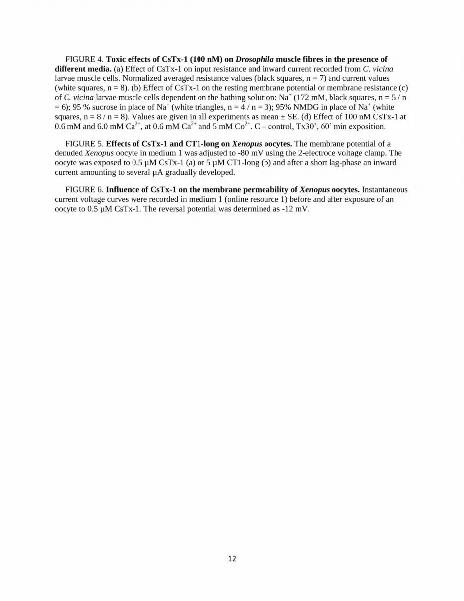
FIGURE 4. Toxic effects of CsTx-1 (100 nM) on Drosophila muscle fibres in the presence of
different media. (a) Effect of CsTx-1 on input resistance and inward current recorded from C. vicina
larvae muscle cells. Normalized averaged resistance values (black squares, n = 7) and current values
(white squares, n = 8). (b) Effect of CsTx-1 on the resting membrane potential or membrane resistance (c)
of C. vicina larvae muscle cells dependent on the bathing solution: Na+ (172 mM, black squares, n = 5 / n
= 6); 95 % sucrose in place of Na+ (white triangles, n = 4 / n = 3); 95% NMDG in place of Na
+ (white
squares, n = 8 / n = 8). Values are given in all experiments as mean ± SE. (d) Effect of 100 nM CsTx-1 at
0.6 mM and 6.0 mM Ca2+
, at 0.6 mM Ca2+
and 5 mM Co2+
. C – control, Tx30’, 60’ min exposition.
FIGURE 5. Effects of CsTx-1 and CT1-long on Xenopus oocytes. The membrane potential of a
denuded Xenopus oocyte in medium 1 was adjusted to -80 mV using the 2-electrode voltage clamp. The
oocyte was exposed to 0.5 µM CsTx-1 (a) or 5 µM CT1-long (b) and after a short lag-phase an inward
current amounting to several µA gradually developed.
FIGURE 6. Influence of CsTx-1 on the membrane permeability of Xenopus oocytes. Instantaneous
current voltage curves were recorded in medium 1 (online resource 1) before and after exposure of an
oocyte to 0.5 µM CsTx-1. The reversal potential was determined as -12 mV.

13
Tables
TABLE 1
Estimation of secondary structure of CsTx-1, CsTx-2a, CT1-long, and CT1-short by circular
dichroism
Secondary structure content (%)
Peptides
in solution α-helix β-sheet turns unordered total NRMSD
§
CsTx-1 PBS* 2 38 18 40 98 0.083
CsTx-1 TFE# 42 19 21 18 99 0.022
CsTx-2a PBS 2 41 19 37 98 0.086
CsTx-2a TFE 8 36 22 35 100 0.074
CT1-long PBS 1 28 23 48 99 0.019
CT1-long TFE 66 16 7 11 100 0.005
CT1-short PBS -2 30 21 49 97 0.010
CT1-short TFE 2 31 22 45 99 0.023
*PBS: 5 mM sodium phosphate, 150 mM sodium fluoride, pH 7.2 #TFE:
5 mM sodium phosphate, 150 mM sodium fluoride, pH 7.2, 50% trifluoroethanol
§NRMSD: normalized root mean square deviation, calculated by DICHROWEB server / CDSSTR,
reference set 1 (19,20)
TABLE 2
Biological activities of CsTx-1, CsTx-2a, CsTx-2b, CT1-long, and CT1-short
Peptide LD50 pmol/mg
Drosophila
EC50 (µM)
Trypanosoma
brucei brucei
MiTat1.2(221)a
MIC (µM)
E. coli S. aureus
SBS 363b ATCC 29213
c
CsTx-1 0.35 n.d. 15.63-31.25 > 250
CsTx-2a 2.58 n.d. n.d. n.d.
CsTx-2b 66.51 n.d. n.d. n.d.
CT1-long 82.64 5.01 4.66-9.32 > 149*
CT1-short > 500 > 40 > 250 > 250
Cu1a 24.4d 0.120
e 0.313-0.625 0.157-0.313
a 1x10
4 cells/ml;
b 6.9x10
2 cfu/ml;
c 2.7x10
3 cfu/ml;
d [41],
e [26]
* the growth of S. aureus is four-fold reduced when compared with the control group without peptide

14
TABLE 3
CsTx-1 causes a concentration dependent irreversible decrease of the resting membrane potential
(MP) of fly and frog muscle fibres
CsTx-1 (nM) MP (mV)
control
MP (mV)
30 min
MP (mV)
60 min
Calliphora vicina
50 -70.0 ± 4.8 (3) -72.0 ± 1.9 (4) -68.4 ± 4.7 (3)
100 -66.3 ± 1.5 (27) -47.0 ±1.5 (28)** -42.0 ± 2.2 (3)**
300 -60.3 ± 1.7 (7) -25.2 ± 4.0 (7)** -20.7 ± 1.7 (4)**
Rana temporaria
100 -80.0 ± 3.2 (3) -80.6 ± 2.0 (3) -77.1 ± 1.5 (3)
300 -84.5 +-2.9 (4) -57.2+-9.2 (4)* -46.8 ± 12 (4)*
900 -84.4 ± 1.6 (3) -64.1 ± 1.6 (3)** -59.0 ± 7.0 (3)*
The numbers of experiments are given in brackets (n)
Significances are given as:*p ≤ 0.05, **p ≤ 0.01
TABLE 4
Reversal potential and conductance of CsTx-1 in different media
Medium CsTx-1
(µM)
Er
(mV)
Conductance
(µS)
M1 0.0 - 0.8-2
M1 0.5 -15 ± 1 10-40
M2 0.5 -11 ± 3 20-40
M3 0.05-0.5 -16 ± 6 40-60
M4 0.5 -2 ± 2 10-20
M5 0.5 n.d. 0.4-1.2
M6 0.05-0.1 -12 ± 2 10-30

15
Figures
FIGURE 1
1-
3-
63-
123-
183-
243-
303-
363-
423-
483-
GA
TTCATACAGAACTTTCTTGAGAAAGTTTAGACTGAGTGAGAGAGAAAGAATTTTCCCTCG
CTAATCATCATGAAAGTTCTCATTATCTCTGCTGTGCTCTTCATAACTATTTTCAGCAAC
M K V L I I S A V L F I T I F S N
ATTTCAGCTGAAATAGAAGATGATTTCTTGGAAGACGAAAGTTTTGAAGCTGAGGACATA
I S A E I E D D F L E D E S F E A E D I
ATACCTTTCTTTGAAAACGAACAAGCCAGAAGCTGCATTCCGAAGCACGAGGAATGTACC
I P F F E N E Q A R S C I P K H E E C T
AACGATAAACACAACTGCTGTAGGAAGGGCCTGTTCAAGTTGAAGTGCCAGTGCTCAACA
N D K H N C C R K G L F K L K C Q C S T
TTTGACGACGAAAGCGGACAGCCAACGGAAAGATGCGCCTGCGGAAGACCGATGGGCCAC
F D D E S G Q P T E R C A C G R P M G H
CAGGCTATTGAAACGGGCCTCAACATCTTCAGGGGTCTTTTTAAAGGGAAGAAGAAGAAT
Q A I E T G L N I F R G L F K G K K K N
AAGAAAACAAAGGGCTAAGAAATTTATTGGAATAGAGTGAATACAAGTCATTGGATCTTA
K K T K G ***
ATTATCTTTTATAATGTTTAATAAATTTTTCAGAAATAGTGAAAACCTTTACTTTGAAAA
- 2
- 62
-122
-182
-242
-302
-362
-422
-482
-542

16
FIGURE 2

17
FIGURE 3

18
FIGURE 4a

19
FIGURE 4b

20
FIGURE 4c

21
FIGURE 4d

22
FIGURE 5

23
FIGURE 6


