Embed Size (px)
Citation preview

Immunity, Vol. 6, 623-632, May, 1997, Copyright ©1997 by Cell Press
Α Väral ER-Resident Glycoprotein Inactivatesthe MHC-Encoded Peptide Transporter
Hartmut Hengel,* Jens-Oliver Koopmann,tThomas Flohr,* Walter Muranyi,* Eis Goulmy,*Günter J. Hämmerling.t Ulrich H. Koszinowski,*and Frank Momburgt*Max von Pettenkofer-InstitutLehrstuhl VirologieGenzentrum der Ludwig-Maximilians-Universität
München81377 MünchenGermanytAbteilung für Molekulare ImmunologieDeutsches Krebsforschungszentrum69120 HeidelbergGermany* Department of Immunohematology and Blood BankUniversity Hospital2300 RC LeidenThe Netherlands
Summary
Human cytomegalovirus inhibits peptide import intothe endoplasmic reticulum (ER) by the MHC-encodedTAP peptide transporter. We identified the open read-ing frame US6 to mediate this effect. Expression ofthe 21 kDa US6 glycoprotein in human cytomegalovi-rus-infected cells correlates with the Inhibition of pep-tide transport during infection. The subcellular local-ization of US6 is ER restricted and is identical withTAP. US6 protein is found in complexes with TAP1/2,MHC class I heavy chain, ß2-microglobulin, calnexin,calreticulin, and tapasin. TAP Inhibition, however, isindependent of the presence of class I heavy chainand tapasin. The results establish a new mechanismfor viral immune escape and a novel role for ER-resi-dent proteins to regulate TAP via its luminal face.
Introduction
Cytomegaloviruses (CMVs) belong to the β subfamily ofherpesviruses, which are large DNA-containing enve-loped viruses. Human CMV (HCMV) is an importantpathogen causing both acute and chronic infections inthe immunologically immature and in the immunocom-promised host (Ho, 1982). CMV genes are expressed ina cascade fashion characteristic of herpesviruses duringthe irnmediate-early (IE), early, and late phases of infec-tion. CMVs have evolved specific functions to escapecellular immune responses (reviewed by York, 1996).Both HCMV and mouse CMV interfere with the surfaceexpression of major histocompatibility (MHC) class Imolecules and antigen presentation to CD8H Τ lympho-cytes at multiple Checkpoints (Barnes and Grundy, 1992;Del Val et al., 1992; Hengel et al., 1995; Jones et al.,1995). In HCMV-infected fibroblasts, the formation ofternary class I heavy chain-ß2-microglobulin (ß2m)-peptide complexes is drastically reduced during theearly and late phase of infection (Beersma et al., 1993;Yamashita et al., 1993; Warren et al., 1994).
In the MHC class I pathway of antigen presentation,antigenic peptides generated by cytosolic proteasesmust be translocated by the ATP-dependent transporterassociated with antigen processing (TAP) across theendoplasmic reticulum (ER) membrane for assemblyinto ternary MHC class I complexes (reviewed by Yew-dell and Bennink, 1992; by Heemels and Ploegh, 1995;and by Koopmann et al., 1997). TAP is a heterodimercomposed of two homologous proteins, TAP1 andTAP2, both encoded in the MHC. Both subunits arepredicted to span the ER membrane 6-10 times withsmall loops penetrating the cytosol and ER lumen andto possess a large cytosolic domain containing an ATP-binding cassette. The transport of peptides by TAP re-quires two coupled but independent events. In the firststep, the peptide is bound to the cytosolic face of TAP,before it is subsequently translocated in an ATP-depen-dent manner and released into the lumen of the ER(Androlewicz et al., 1993; Neefjes et al., 1993; Shepherdet al., 1993; van Endert et al., 1994). Recently, the herpesSimplex virus 1 (HSV-1) ICP47 protein was demonstratedto inhibit the peptide transport by blocking the peptide-binding Site of TAP (Ahn et al., 1996b; Tomazin et al.,1996).
The assembly of MHC class I heavy chain with ß2mand peptide is assisted by transient interactions withmolecular chaperones in the ER. Calnexin has beenshown to interact with free class I heavy chains (Degenand Williams, 1991; Rajagopalan and Brenner, 1994),and calreticulin binds human class l/ß2m dimers (Sadasi-van et al., 1996). MHC class I heterodimers associatewith TAP via the TAP1 subunit (Androlewicz et al., 1994;Ortmann et al., 1994; Suh et al., 1994) mediated by an48 kDa ER glycoprotein, tapasin (Sadasivan et al., 1996).Binding of high-affinity peptides to class I moleculesleads to the dissociation of TAP-class I complexes andthe exit of ternary class I complexes from the ER (Ort-mann et al., 1994; Suh et al., 1994).
The down-regulation of MHC class I expression duringpermissive HCMV infection was attributed to two generegions of the HCMV genome, one of which is the geneUS11 (Jones et al., 1995). We have recently describedthat HCMV infection results in an Inhibition of peptidetranslocation into the ER despite augmented TAP ex-pression in HCMV-infected cells. This effect was notmediated by the gene US11 and was found to be absentfrom cells infected with a HCMV deletion mutant, ts9,lacking the genes US1 through US15 (Hengel et al.,1996). Ploegh and coworkers have elegantly demon-strated that the US11- and L/S2-encoded glycoproteinstarget class I heavy chains from the ER to the cytosolfor rapid proteolytic degradation (Wiertz et al., 1996a,1996b).
Here we describe the Identification of the HCMV geneUS6 encoding a 21 kDa glycoprotein preventing peptidetranslocation by TAP. In US6-expressing HeLa cells,MHC class I molecules do not acquire peptides and lacktransport out of the ER. The subcellular distribution ofgpUS6 shows a pattern identical with TAP1, and gpUS6maintains complete sensitivity to endoglycosidase Η

Immumty624
(endo Η), indicative of ER-resident proteins. gpUS6 isdemonstrated to associate with the TAP-tapasin-MHC-calreticulin complex as well as with calnexin gpUS6prevented the peptide Import into microsomes preparedfrom mutant cell lines deficient for either MHC class Ior for tapasin, indicating that these molecules are notrequired to block TAP. Both the Inhibition of TAP via itsER luminal face and the retamed peptide binding toTAP in the presence of gpUS6 underscore a markedlydifferent behavior from ICP47 of HSV-1 and establish anew rnolecular mechanism to regulate this transporter.
Results
HCMV US6 Affects MHC Class I Surface Expression,Antigen Presentation to CD8+ CytotoxicΤ Lymphocytes, and PeptideTransport into the ERThe absence of peptide transport Inhibition in humanfibroblasts permissively infected with the HCMV AD169-derived deletion mutant ts9 suggested that the putativeInhibitor may reside within the gene region lacking ints9, that is, US1 through US15. To search for the viralgenes that mediate TAP Inhibition, we cloned and stablyexpressed the open readmg frames US1, US2, US3,US4, US5, US6, US7, US8, US9, US10, US12, and US13m HLA-A2+ 293 kidney cells and HeLa cells. Thetransfectants were screened for antigen presentation toHLA-A2 allospecific CD8+ cytotoxic Τ lymphocyte (CTL)clones (Goulmy et al., 1984), class I surface expression,and TAP-mediated peptide transport. The isolatedgenes US2 (data not shown) and US6 proved to reduceboth surface expression of class I molecules and recog-nition by CD8f CTL (Figures 1Α and 1B). In contrast, thesurface expression of CD44 molecules on HeLa cellswas not affected by US6 expression (Figure 1B). In HeLaor 293 cells stably transfected with US6 or infected witha recombinant vaccinia virus expressing US6, a drasticreduction of ATP-dependent peptide transport by TAPwas found (Figure 1C). This Inhibition was similar to theInhibition seen in transfectants stably expressing theTAP Inhibitor ICP47 of HSV-1 (Figure 1C) (Früh et al.,1995; Hill et al., 1995). ükewise the US6 sequencetagged with the hydrophilic FLAG sequence at theC-terminus inhibited peptide translocation by TAP (Fig-ure 1C). We conclude that HCMV US6 is able and suffi-cient to Interrupt the MHC class I pathway of antigenpresentation by reducing the peptide translocation intothe ER.
MHC Class I Molecules in HeLa-US6 TransfectantsDo Not Aquire PeptidesPeptide-filled MHC class I complexes are charactenzedby stability at 37°C in 1 % NP40 lysate and transportto the medial-Golgi where their carbohydrate moietiesacquire resistance to cleavage by endo Η (Townsendet a l , 1990). To determine whether MHC class I mole-cules in HeLa-US6 transfectants are loaded with peptideor not, HeLa control cells and HeLa-US6 cells were met-abohcally labeled with [35S]methionine for 15 min andlysed in 1% NP40 buffer. The lysates were split andaliquots chased for 60 min at 37°C or 4°C, respectively.MHC class I molecules were precipitated with either the
conformation-dependent monoclonal antibody (MAb)W6/32 detecting ß2m-associated class I heavy chains(Parham et al., 1979) or MAb HC10 recognizing nonas-sembled class I molecules (Stam et al., 1986). Half ofeach precipitate was subjected to endo Η digestion andseparated by SDS polyacrylamide gradient gel electro-phoresis (SDS-PAGE). As depicted in Figure 1D, theformation of MHC class I complexes that remained endoΗ sensitive was diminished in HeLa-US6 cells. Moststnkingly, almost all MHC I complexes formed in HeLa-US6 transfectants were unstable at 37°C, while in HeLacontrol cells most ß2m-associated class I heavy chainsremained stable at 37°C and aquired resistance to endoΗ cleavage. Conversely, the level of nonassembled MHCclass I heavy chains recognized by MAb HC10 was m-creased in US6-expressing HeLa cells compared to con-trols (Figure 1D, bottom). Taken together, the resultsconfirm defective peptide loading onto heavy cham/ß2mheterodimers in the presence of the US6 protein re-sulting in a reduced exit of stably formed MHC class Imolecules from the ER.
Synthesis of US6 Protein Correlates with Inhibitionof Peptide Transport dunng PermissiveHCMV InfectionAs in other herpesviruses, CMV replication is tightly reg-ulated in a multiStep process. Dunng productive infec-tion, cellular transcription factors initiate the transcnp-tion of IE genes that induce the expression of severalsets of early genes, most abundantly expressed 6-60hr postinfection. Early proteins are required for viral DNAreplication followed by the synthesis of late proteins(approximately 48-96 hr postinfection), many of whichare incorporated into the vinon or aid the process ofprogeny assembly. The kmetics of US6 protein expres-sion in HCMV wild-type strain AD169-infected fibro-blasts dunng the course of permissive infection wasassessed after metabohc labeling and immunoprecipita-tion with a polyclonal rabbit antiserum raised againstsynthetic peptide corresponding to amino acids 20-29of the US6 sequence. From parallel cultures of the sameexpenment, ATP-dependent peptide translocation byTAP was assessed using the peptide RYWANATRSF.As shown in Figure 1E, the continuous decline in peptidetransport correlated with US6 protein synthesis, whichwas maximal at 72 hr postinfection. Pulse-chase expen-ments indicated that the US6 polypeptide has a halftime of approximately 3 hr (data not shown). We con-clude that US6 protein synthesis Starts dunng the earlyphase and reaches peak levels at 72 hr postinfectionin the late phase of the viral replication cycle, while,inversely, TAP-dependent peptide translocation into theER is progressive^ decreased.
Subcellular Distribution of the US6 ProteinThe putative amino acid sequence of US6 codes for atype la transmembrane protein with a protein core of21 kDa and a Single potential N-hnked glycosylation Site.To study the subcellular distribution of the US6 protein,confocal laser scanning microscopy of L/S6-transfectedHeLa cells was performed using an affinity-punfied rab-bit antiserum recognizing the luminal domain of the pro-tein. In paraformaldehyde-fixed detergent-solubilized

TAP Inhibition by HCMV gpUS6625
Α
Β
293 pcDNAI
60-
| 50-
2 40-
| 30-
£ 20-10-
0 .
$^-~~~~~—"^
/
pcDNAI-US6
0 8 4 20 0 8 4 20 E/T
ilPept de #600 (TNKTRIDGQY)
LLPeplide #802 (BRYQNSTEL)
HeLa ' ' HeLavao HeLa vac'HeLa U S e W a US6unntected contra] 0S6 #12 flag #24
HeLa HeLa-US6
log fluorescence intensity
HeLa HeLa-US6
chase
EndoH
W6/32
12 24 48 72 96 hours ρ ι
time post infection (hours)
293 ' 293 US6 ' 293 US6 ' 293 US6 293 ICP47
untransi #111 1!agff9 iag#10 #S
Figure 1 US6 Expression Prevents CD8+ ΤCell Recognition, IVIHC Class I Surface Ex-pression, and MHC Class I Complex Forma-tion Due to Inhibited Peptide Transport byTAP
(A) 293 cells stably transfected with pcDNAI-US6 plasmid or the vector alone were labeledwith 51Cr and tested in a 4 hr Standard releaseassay with graded number of effector cellsThe effectors were the HLA-A2 allospecificCD8 f CTL clones IE2 (circles) and JS132 (tn-angles)
(B) Cytofluorometnc analysis of MHC class Isurface expression of HeLa cells transfectedwith pcDNAI-US6 and HeLa control cellsCells were stained with MAb W6/32 (boldlines) or anti-CD44 MAb (narrow Imes) fol-lowed by goat-anti mouse IgG-FITC Dottedlines represent control staining with secondantibody only
(C) ATP-dependent peptide translocationwas assessed for permeabilized HeLa cellsand individual US6 transfected clones (topand middle) and 293 cells and 293-US6transfectants, respectively (bottom) HeLacells were infected overnight with US6-recombinant vaccima virus or control vac-cinia virus at a multiplicity of infection (moi)of 3 Filled bars represent transport rates inthe presence of ATP, open bars in the ab-sence of ATP for control The data representmeans of duplicate values
(D) Nontransfected and US6-transfected HeLacells were metabohcally labeled for 15 min
Lysates in 1 % NP40 were either kept at 4°C or incubated at 37°C for 60 min pnorto immunoprecipitation of ahquots with MAb W6/32 (top) and MAb HC-10 (bottom)Half of the precipitated molecules were digested with endo Η or mock treated sindicates MHC class I molecules sensitive and r indicates MHC class I moleculesresistant to endo Η cleavage HC, MHC class I heavy chains(E) Kinetics of peptide translocation by TAP assessed with peptide RYWANATRSF(triangles) dunng permissive infection of MRC-5 fibroblasts with HCMV AD169(moi = 5) In parallel cultures, the level of US6 expression in MRC-5 cells infectedwith HCMV AD169 (moi = 5) was determined by immunoprecipitation with anti-US6 antiserum and analyzed by SDS-PAGE (top) US6 expression (circles) is shownin arbitrary units after phosphoimager quantitation of the US6 bands Peptidetransport is shown as the percentage Inhibition of the transport rate (9 2%) obtainedwith mock-infected cells
HC-10
cells a typical ER-like staining pattern was observed(Figure 2A), while HeLa control cells were negative (datanot shown). The localization of US6 in the ER was con-firmed by a nearly perfect colocalization with the ERmarker protein BiP (Vaux et al., 1990) (data not shown)
and with TAP1, which is pnmarily located in the ER andcan reach cisternae of the cis-Golgi (Kleijmeer et al.,1990; Russ et al., 1995) (Figure 2B). The distnbutionpattem of US6 clearly differed from that of the ER Golgiintermediate compartment (ERGIC) marker ERGIC-p53
Immunity626
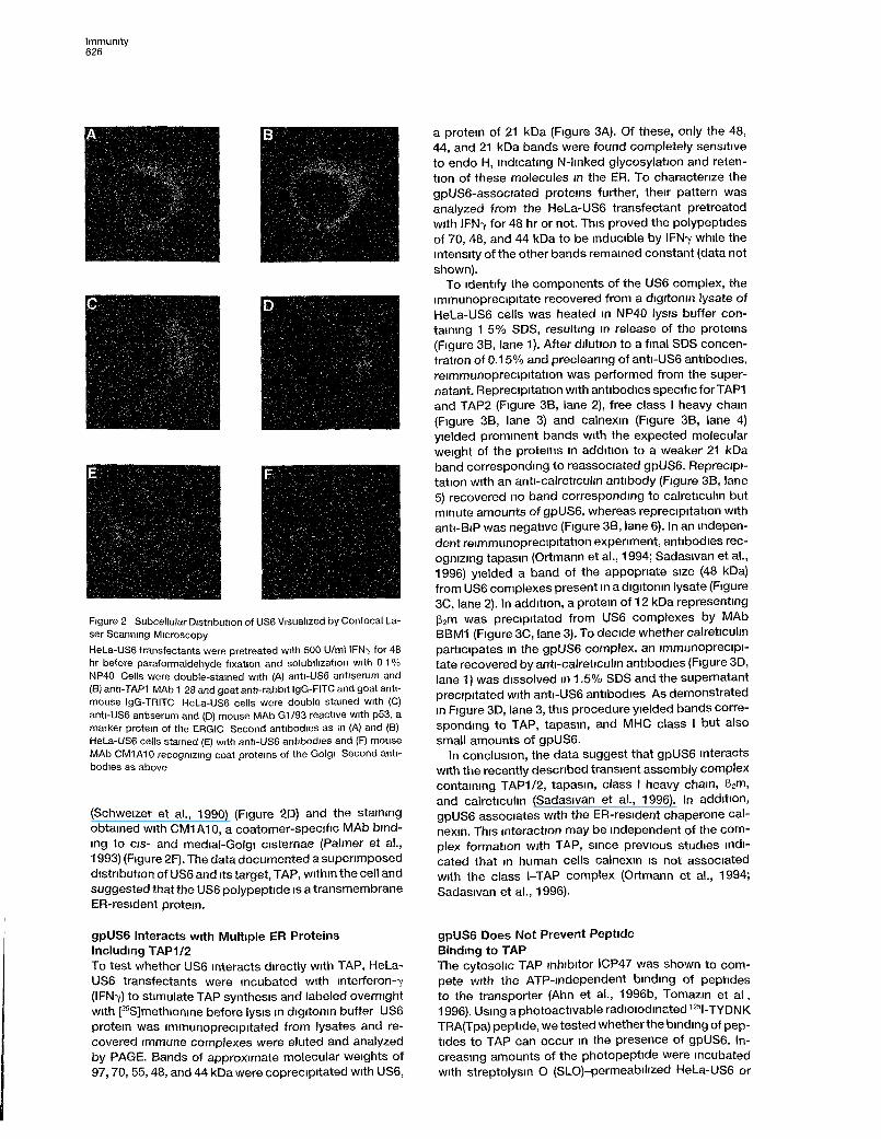
Figure 2 Subcellular Distribution of US6 Visualized by Confocal La-ser Scanning Microscopy
HeLa-US6 transfectants were pretreated with 500 U/ml IFN-y for 48hr before paraformaldehyde fixation and solubilization with 0 1 %NP40 Cells were double-stained with (A) anti-US6 antiserum and(B) anti-TAP1 MAb 1 28 and goat anti-rabbit IgG-FITC and goat anti-mouse IgG-TRITC HeLa-US6 cells were double stained with (C)anti-US6 antiserum and (D) mouse MAb G1/93 reactive with p53, amarker protem of the ERGIC Second antibodies as in (A) and (B)HeLa-US6 cells stained (E) with anti-US6 antibodies and (F) mouseMAb CM1A10 recogmzmg coat proteins of the Golgi Second anti-bodies as above
(Schweizer et al., 1990) (Figure 2D) and the stainingobtained with CM1A10, a coatomer-specific MAb bind-mg to eis- and medial-Golgi cisternae (Palmer et al.,1993) (Figure 2F). The data documented a supenmposeddistnbution of US6 and its target, TAP, within the cell andsuggested that the US6 polypeptide is a transmembraneER-resident protem.
gpUS6 interacts with Multiple ER ProteinsIncluding TAP1/2To test whether US6 interacts directly with TAP, HeLa-US6 transfectants were ineubated with interferon-7(IFN7) to stimulate TAP synthesis and labeled overnightwith [35S]methionine before lysis in digitonin buffer US6protem was immunoprecipitated from lysates and re-covered immune complexes were eluted and analyzedby PAGE. Bands of approximate molecular weights of97,70,55,48, and 44 kDa were coprecipitated with US6,
a protem of 21 kDa (Figure 3A). Of these, only the 48,44, and 21 kDa bands were found completely sensitiveto endo H, indicating N-Iinked glycosylation and reten-tion of these molecules in the ER. To charactenze thegpUS6-associated proteins further, their pattern wasanalyzed from the HeLa-US6 transfeetant pretreatedwith IFN7 for 48 hr or not. This proved the polypeptidesof 70, 48, and 44 kDa to be inducible by IFN7 while theintensity of the other bands remained constant (data notshown).
To identify the components of the US6 complex, theimmunoprecipitate recovered from a digitonin lysate ofHel_a-US6 cells was heated in NP40 lysis buffer con-taining 1 5% SDS, resulting in release of the proteins(Figure 3B, lane 1). After dilution to a final SDS concen-tration of 0.15% and precleanng of anti-US6 antibodies,reimmunoprecipitation was performed from the super-natant. Reprecipitation with antibodies specific forTAPIand TAP2 (Figure 3B, lane 2), free class I heavy chain(Figure 3B, lane 3) and calnexin (Figure 3B, lane 4)yielded prominent bands with the expected molecularweight of the proteins in addition to a weaker 21 kDaband correspondmg to reassociateel gpUS6. Reprecipi-tation with an anti-calreticulm antibody (Figure 3B, lane5) recovered no band correspondmg to calreticulm butminute amounts of gpUS6, whereas reprecipitation withanti-ΒιΡ was negative (Figure 3B, lane 6). In an indepen-dent reimmunoprecipitation expenment, antibodies rec-ognizing tapasin (Ortmann et al., 1994; Sadasivan et al.,1996) yielded a band of the appopriate size (48 kDa)from US6 complexes present in a digitonin lysate (Figure3C, lane 2). In addition, a protem of 12 kDa representingß2m was precipitated from US6 complexes by MAbBBM1 (Figure 3C, lane 3). To decide whether calreticulmparticipates in the gpUS6 complex, an immunoprecipi-tate recovered by anti-calreticulm antibodies (Figure 3D,lane 1) was dissolved in 1.5% SDS and the supernatantprecipitated with anti-US6 antibodies As demonstratedin Figure 3D, lane 3, this procedure yielded bands corre-spondmg to TAP, tapasin, and MHC class I but alsosmall amounts of gpUS6.
In conclusion, the data suggest that gpUS6 interactswith the recently desenbed transient assembly complexcontaming TAP1/2, tapasin, class I heavy chain, ß2m,and calreticulm (Sadasivan et al., 1996). In addition,gpUS6 associates with the ER-resident chaperone cal-nexin. This mteraction may be independent of the com-plex formation with TAP, smee previous studies mdi-cated that in human cells calnexin is not associatedwith the class I-TAP complex (Ortmann et al., 1994;Sadasivan et al., 1996).
gpUS6 Does Not Prevent PeptideBindmg to TAPThe cytosolic TAP mhibitor ICP47 was shown to com-pete with the ATP-mdependent bindmg of peptidesto the transporter (Ahn et al., 1996b, Tomazm et a l ,1996). Usmg aphotoactivable radioiodmated 125I-TYDNKTRA(Tpa) peptide, we tested whether the bindmg of pep-tides to TAP can oeeur in the presence of gpUS6. In-creasing amounts of the photopeptide were ineubatedwith streptolysin Ο (SLO)-permeabilized HeLa-US6 or

TAP Inhibition by HCMV gpUS6627
HeLa HeLa-US6
+ - + Endo Η
B1°:aUS6
pellet supernatant
97 -
69 -
46 •
30 •
14 "
UM
mm •<r US6
<-US6 s
97 -69 .
46 .
3 0 -
14-
S 1Ö Β
λ
8 *3Ö a
mim· *
th
Ö
kD
1°: α US6
pellet supernatantD
1°:aCRN
<r- tapasin
1 4 -
97-
6 9 -
4 6 -
3 0 -
1 4 -
< - gpUS6
Figure 3. Identification and Characterizationof US6-Associated ER Proteins
(A) HeLa and HeLa-US6 transfectants weremetabolically labeled overnight and lysed indigitonine lysis buffer. gpUS6 was immuno-precipitated by rabbit anti-US6 antiserum,and proteins were separated by 10%-15%PAGE. US6-associated proteins are indicatedby arrows. Immune complexes retrieved fromdigitonin lysates were mock-digested or di-gested with endo H. s indicates bands witha mobility shift after endo Η digestion.
(B) HeLa-US6 cells were metabolically la-beled as described in (A) and lysed in digito-nin lysis buffer. Material precipitated withanti-US6 antiserum was heated and dis-solved in 1 % NP40/1.5% SDS buffer. Proteinsnot dissociated from the protein Α Sepharosepellet were analyzed on lane 1. Aliquots ofthe supernatant were reprecipitated with anti-TAP1 MAb TAP1.28 and anti-TAP2 MAbTAP2.70 (lane 2), rabbit anti-heavy chain anti-serum (lane 3), anti-calnexin MAb AF8 (lane4), rabbit anti-calreticulin antiserum (lane 5),and rabbit anti-BiP antiserum (lane 6).
(C and D) Hel_a-US6 cells were pretreatedwith 500 U/ml IFN7 before metabolically la-beled as described in (A) and lysed in digito-nin lysis buffer. (C) One aliquot of the lysatewas precipitated with anti-US6 antiserum.The precipitate was heated and dissolved in1 % SDS. Proteins not dissociated from theprotein Α Sepharose pellet were analyzed inlane 1. Aliquots of the supernatant were re-precipitated with rabbit anti-gp48 (tapasin)(lane 2) antiserum and MAb BBM1 specificfor human ß2m (lane 3). (D) The lysate wasprecipitated with rabbit anti-calreticulin anti-bodies. Aliquots of these immune complexeswere either directly analyzed (lane 1) orheated and dissolved in 1 % SDS. Proteinsnot dissociated from the protein Α Sepharosepellet were analyzed in lane 2. The superna-tant was reprecipitated with anti-US6 antise-rum (lane 3).
HeLa control celte in the absence of ATP at 4°C. Ultravio-let crosslinking and immunoprecipitation with TAP-spe-cific antibodies from HeLa-US6 cells resulted in bandsof about 70 kDa, the intensity of which was not reducedcompared to the HeLa control (Figure 4) but was drasti-cally reduced after addition of recombinant ICP47 pro-tein, which blocks peptide binding to TAP (Figure 4,lanes 4 and 8). This result indicates that gpUS6 does notaffect peptide binding to TAP. Furthermore, the ICP47-mediated competitive Inhibition of peptide binding isindependent of the presence of gpUS6. Thus the mecha-nism employed by US6 for the blockade of peptidetransport is different from ICP47.
gpUSß Does Not Require MHC Class Iand Tapasin to Block TAPTo address the role of class I heavy chains or tapasinfor the inactivation of TAP1/2 by gpUS6, US6 protein wastranslated in vitro in the presence of microsomes preparedfrom HLA-A~, -Fr, -C+ tapasin-negative LCL721.220and HLA-A, -B, -C-negative but tapasin-positive
LCL721.221 mutant cells (DeMars et al., 1985; Green-wood et al., 1994; Grandea et al., 1995; Sadasivan etal., 1996) and the microsomes were assayed for ATP-dependent peptide import (Figure 5). In the presence ofUS6, microsomes of both cell lines completely failed toaccumulate glycosylated peptides, while translation ofSaccharomyces cerevisiae α-factor mRNA as a controlhad no effect. Thus, class I heavy chains and tapasinaredispensableforthefunctional inactivation of TAP1/2.Furthermore, this finding illustrates that in vitro trans-lated US6 protein is able to reach preformed TAP com-plexes to exert its blocking activity.
Discussion
Despite augmented levels of TAP expression, fibro-blasts permissively infected with HCMV exhibit a down-modulation in the capacity to translocate peptidesacross the ER membrane. This effect is detectable notearlier than 12 hr postinfection and progressively in-creases during infection (Hengel et al., 1996). This kinet-ics is consistent with the appearance of a viral inhibitor

Immunity628
HeLa HeU
50 250 500 500 50 250 500 500 Peptide [pmoi]
+ ICP47
66-kD
-TAP2-TAP1
Figure 4. gpUS6 Does Not Prevent Peptide Binding to TAPSLO-permeabilized HeLa or HeLa-US6 cells were incubated withtitrated amounts of the photolabile peptide 125l-TYDNKTRA(Tpa)without (lanes 1 -3 and 5-7) or with recombinant ICP47 protein (lanes4 and 8). After ultraviolet crosslinking, TAP1 and TAP2 moleculeswere immunoprecipitated by MAb TAP1.28 and TAP2.70 and ana-lyzed by SDS-PAGE.
the expression of which is relatively Iow after the onsetof the early phase and peaks in the late phase 72 hrpostinfection. Here we describe the US6 gene productto cause the Inhibition of TAP-mediated peptide trans-port. As a consequence of TAP Inhibition by gpUS6and other independent gene functions expressed earlierduring HCMV infection (vide infra), the formation of MHCclass I complexes, their transport to the cell surface andantigen presentation to CD84 Τ cells is abolished inHCMV-infected cells (Beersma et al., 1993; Yamashitaet al., 1993; Warren et al., 1994; Hengel et al., 1995).Transient expression of the isolated US6 gene by recom-binant vaccinia virus or stable expression of gpUS6 intransfected cells consistently resulted in a diminishedpeptide transport f unction of human cells. The maximumof gpUS6 expression was found to occur at approxi-mately 72 hr postinfection, which perfectly matches theslow kinetics of TAP Inhibition. Α HCMV deletion mutant,ts9, lacking the genomic region encompassing US6, waspreviously shown not to impair peptide transport(Hengel et al., 1996). Finally, in cells expressing theHCMV US11 (Hengel et al., 1996) or US2 genes (datanot shown), which are sufficient to down-regulate MHCclass I expression, the peptide transport function is notaffected. We conclude that in all likelihood, gpUS6 rep-resents the only relevant HCMV gene product mediatingTAP Inhibition in the course of HCMV infection.
The reduced transport capacity of gpUS6-expressingcells is demonstrated in vitro using nonnatural peptidesequences that are retained in the ER through anN-Iinked carbohydrate. Our biochemical analysis ofHeLa-US6 cells provided evidence that the vast majorityof class I molecules expressed in HeLa cells fails to beloaded with peptides as indicated by lacking thermosta-bility and ER retention. Thus, the impairment of peptidetransport is not restricted to selected peptide se-quences but appears to apply to the function of TAP ingeneral. Only a small minority of class I ligands may begenerated in the ER lumen itself or get access to theER independent of TAP (reviewed by Momburg et al.,1994a).
The US6 open reading frame encodes a type I trans-membrane protein of 21 kDa containing a Single N-gly-cosylation site. We found the whole population of gpUS6glycosylated. The carbohydrate moiety may contribute
#55Peptide
#60
Figure 5. Inhibiton of Peptide Transport by US6 Is Independent ofTapasin and MHC Class I Heavy Chain
US6 protein was in vitro translated into microsomes of tapasin-deficient 721.220 cells and HLA-A, -B, -C-deficient 721.221 cells.The transport capacity of microsomes was determined with glycosy-latable peptides 55 (RYWANATRSA) and 60 (RYWANATRSQ). Filledbars represent in vitro-translated control mRNA, transport in thepresence of ATP; open bars represent in vitro translation of US6mRNA, transport in the presence of ATP; hatched bars representin vitro translation of control mRNA, no ATP added.
to the stability of gpUS6 since the half-life of US6 isreduced in tunicamycin-treated cells (Η. Η., unpublisheddata). The complete sensitivity of gpUS6 to endo Ηindicates efficient retention in the ER consistent withthe superimposable immunofluorescence staining forgpUS6 and the ER marker protein BiP. The perfect colo-calization with TAP1 appears to make the latter a dedi-cated target for gpUS6. Since gpUS6 does not containthe C-terminal KKXX consensus motif for ER retentionof transmembrane proteins (Jackson et al., 1990), it isunclear by which mechanism gpUS6 is retained in theER. We have found a prominent association of gpUS6with the ER-resident chaperone calnexin that associateswith incompletely folded glycoproteins through a lectin-like activity (Ou et al., 1993). in contrast to gpUS6 com-plexes with TAP that are severely reduced or undetect-able in NP-40 lysates compared with digitonin lysates,association of gpUS6 and calnexin is less affected bythe stronger detergent NP-40 (data not shown). It isconceivable that gpUS6 molecules containing as manyas 11 cystein residues slowly attain a mature conforma-tion and that calnexin retains immature gpUS6 mole-cules in the ER by high-affinity binding.
Although the functional phenotype of HSV-1 ICP47and HCMV gpUS6 appears similar, these proteins utilizeentirely different mechanism for the Inhibition of TAP.While the soluble 9 kDa protein ICP47 binds to the cyto-solic face of TAP1/2 dimers with high affinity and inhibitspeptide association with the transporter in a competitivefashion (Ahn et al., 1996b; Tomazin et al., 1996), gpUS6does not interfere with peptide binding, which is an ATP-independent and temperature-insensitive step thoughtto precede the energy-consuming translocation ofbound Substrate (van Endert et al., 1994). It is unknownhow TAP facilitates vectorial transport of peptides vary-ing greatly in their amino acid composition and length

TAP Inhibition by HCMV gpUS6629
against a concentration gradient (reviewed by Andro-lewicz and Cresswell, 1996, and by Koopmann et a l ,1997). It can be envisaged that amphipathic membrane-spanning segments form a pore that allows the transitof hydrophilic Substrates through the hpid bilayer. Theenergy dependence of peptide translocation and thepresence of two nucleotide-bmding cassettes within theTAP1/2 dimer suggests that conformational changes ofthe transporter itself are essentially involved. Α stableintercalation of gpUS6 with ER-Iuminal loops or mem-brane-spanning segments of TAP1/2 might disturb con-formational changes leading to abrogation of transport.
In a minimal model, the inactivation of peptide trans-port could be explained by the physical association ofgpUS6 and TAP without further factors being involved.We have shown here that gpUS6 associates with themultimenc TAP-associated complex rather than dis-rupting it. This raises the possibilrty that the interactionwith TAP might not be sufficient to prevent peptidetransport but requires a cellular cofactor to mediate thiseffect. Our results obtained with the tapasm-deficient.220 mutants and with HLA-A, B, C-deficient 221 mutantcell indicate that at least these components are notessential for US6-mediated TAP Inhibition. The role ofcalreticulin and calnexin remains to be addressed. Thedecreased presence of class I heavy chains in the TAP-associated complex at late time pomts dunng HCMVinfection (Hengel et a l , 1996) further strengthens thenotion that class I heavy chains are dispensable forTAP Inhibition by gpUS6. We favor the idea that gpUS6directly contacts the TAP1/2 heterodimer, the latter be-ing simultaneously associated with tapasin, MHC classl/ß2m, and calreticulin (Sadasivan etal., 1996). This phys-ical interaction possibly mvolves a luminal region en-compassing residues 78-96 of gpUS6 because an anti-serum recognizing this region failed to coprecipitateTAP (Η. Η., unpublished data) whereas the N-terminalUS6 epitope 20-29 allows coimmunoprecipitation asshown here.
Available biochemical evidence suggests that cat-nexin dissociates frjm human class l-ß2m heterodimersbefore the latter associate with calreticulin and with ta-pasin-TAP (Ortmann et al., 1994, Sadasivan et al., 1996;Solheim et a l , 1997). Thus, calnexin does not participatein the human TAP-associated complex, which is in clearcontrast to fmdings in munne cells (Suh et al., 1994,1996). Therefore, it seems likely that gpUS6 binds tocalnexin di' ectly and independent of its association withTAP complexes.
Among the herpesviruses, CMVs have evolved themost extensive genetic repertoire to evade the MHCclass l-restncted Τ lymphocyte response of the host.Moüse CMV expresses three early gene functions thatinterfere with the MHC class I pathway of antigen pre-sentation (Thale et a l , 1995; Kleijnen et al., 1997; Ziegleret al., 1997). HCMV is expressing a cascade of fourconsecutive US gene functions interrupting the class Ipathway of antigen presentation in a general manner.The (IE) protein gpUS3, which is expressed in the verybeginning of permissive infection, impairs the transportof MHC class I complexes (Ahn et a l , 1996a; Jones etal., 1996). The US2- and t/Sf 7-encoded glycoproteinsmisdirect nascent class I heavy chains into the cytosol
where they are rapidly degraded by the proteasome(Wiertz et al., 1996a, 1996b). These genes are abundantlyexpressed up to 24 hr postinfection but poorly tran-scnbed at latertimes (Jones and Muzithras, 1991; Ten-ney and Colberg-Poley, 1991). By contrast, the appear-ance of gpUS6 is maximal 48-96 hr postinfection whenother genes interfenng with the MHC class I pathwayof antigen presentation become almost silent. The ac-tion of gpUS6 at late times of infection may limit thepresentation of abundantly expressed structural anti-gens of the vinon like glycoprotein B. Indeed, the CTLresponse against HCMV glycoprotein Β was reportedto be relatively weak and predominantly restricted byMHC class II rather than MHC class I (Borysiewicz etal., 1988; Hopkins et al., 1996).
The multitude of stealth genes in the genomes of CMVmay be required for several reasons: first, to cover theprotracted replication cycle, which takes at least 72 hrin the case of HCMV, and second, to compensate forthe opposite effects on MHC class I by cytokines likeIFN7, type I IFNs, and TNFa (Hengel et al., 1994; Hengelet al., 1996) that are produced in infected tissues. Finally,the great demand to regulate the presentation functionof a high number of MHC class I alleles and their pep-tides in different cell types may have favored the diversi-fication of the HCMV US genes and their functions. Ourscreening procedure failed to identify US3 as an Inhibitorof antigen presentation (Ahn et al., 1996a, Jones et al.,1996). In fact, this could be due to a preference of theUS3 glycoprotein for certain human MHC class I alleles(Joneset al., 1996)
Altogether, it appears a fascinating feature of HCMVto use proteins encoded within a Single cluster of relatedgenes, i.e. gpUS3 (Ahn et al., 1996a; Jones et al., 1996)Controlling ER export of peptide loaded MHC class Imolecules on the one hand and gpUS6 Controlling ERImport of peptides on the other hand, to escape recogni-tion by class l-restncted CTL.
Expenmental Procedures
Cell Lines and AntibodiesHuman fetal lung fibroblasts, MRC-5 (Bio-Whittacker), in passages6-16, 293 human kidney cells (ATCC CRL 1573), and human HeLacells (ATCC CCL-2) were grown in Dulbecco's modified Eagle'smedium supplemented with 10% fetal calf serum, penicilhn, strepto-mycin, and 2 mW! glutamine The human lymphoblastoid cell ImesLCL721 220 and LCL 721 221 (DeMars et a l , 1985, Greenwood eta l , 1994) were grown in RPMI 1640
Antibodies used were the following MAb W6/32 recognizing HLAclass I heavy chain/ß2m dimers (Parham et a l , 1979) was obtainedfrom the ATCC (HB 95), MAb HC-10 recognizing unassembled classI heavy chains with a preference for HLA-B and -C alleles but alsoHLA-A locus products (Stam et a l , 1986), MAb BBM1 detectinghuman ß2m (ATCC HB-28), anti-CD44 MAb BA06 was from Onco-gene Science (Umondale, NY), polyclonal rabbit antibodies to calre-ticulin and BiP were from StressGen (Victoria, British Columbia,Canada), MHC I heavy chain rabbit antiserum was a kind gift fromDr Η L Ploegh (Cambridge, MA), MAb AF8 to calnexin was a kindgift from Dr Μ Brenner (Boston, MA), anti-TAP1 MAb TAP1 28 andanti-TAP2 MAb TAP2 70 have been described (Nijenhuis et a l , 1996),polyclonal rabbit antibodies to tapasin (Rgp48N) (Sadasivan et a l ,1996) were kindly donated by Dr Ρ Cresswell (New Haven, CT)Polyclonal rabbit antiserum was raised by immunization of rabbitswith KLH-coupIed synthetic peptides of amino acids 20-29 of theUS6 sequence

Immunity630
Viruses and Infection ConditionVirus Stocks of HCMV strain AD169 were prepared as described(Hengel et al., 1995) For mfections, subconfiuent monolayers offibroblasts were incubated with HCMV at an multiplicity of infectionof 5 and centnfuged at 800 χ g for 30 min to increase the efficiencyof infection. Infections of suboonfluent HeLa cells with vaccima viruswas performed at an multiplicity of infection of 3 ovemight.
Cytolytic AssayThe generation and maintenance of the CD8h CTL clone IE2 wasdescribed in detail earlier (Goulmy et al., 1984). Clone JS132 waskindly donated by Dr. Jannie Borst, Amsterdam. Target cells werelabeled with 51Cr and tested in a 4 hr Standard release assay withgraded numbers of effector cells. In all expenments, effector-to-target ratios ranged from 20 1 to 0.8:1 Spontaneous 51Cr release inthe expenments given did not exceed 30% of the maximal releasevalues measured in the presence of 1 % Triton X-100
Cloning and Expression of the HCMV US6 Gene andConstruction of a US6 Vaccima Virus RecombinantThe open reading frame of the US6 gene was cloned afterPCR amphfication from HCMV AD169 DNA (forward primer 5'-CGCGGGGGATCCGCCGCCATGGATCTCTTGATTCGTCTC-3', backwardprimer 5 -CGCGGGTCTAGAGAATTCGCATCAGGAGCCACAACGTCG-3 , resulting in an amphfication product of 591 bp) into thepcDNAIneo expression vector (Invitrogen, San Diego, CA). The US6construct containing the 3 24 bp FLAG sequence (Eastman Kodak,New Haven, CT) was obtained using the backward primer 5'-CGCCCCTCTAGATTACTACTTGTCATCGTCGTCCTTGTAGTCCT CGAGGATATCGGAGCCACAACGTCG AATGGGACG-3' The PCR productwas cloned into the 5' BamHI and 3' Xbal restnction sites ofpcDNAIneo Intervened by a Short spacer (DILE), the hydrophilicFLAG sequence (DYKDDDDK) was fused to the US6 coding se-quence Human 293 kidney cells and HeLa cells were transfectedwith plasmid DNA by calcium phosphate precipitation. Cell cloneswere selected in the presence of 0.5 mg/ml G418 and tested forgpUS6 protein expression by immunoprecipitation with US6-spe-cific antibodies.
The open reading frame of the US6 gene sequence was clonedafter PCR amplification from HCMV AD169 DNA (forward pnmer5 -CGCGGGGGATCCGCCGCCATGGATCTCTTGATT CGTCTC-3',backward pnmer 5 -CGCGGGTCTAGAGAATTCGCATCAGGAGCCACAACGTCG-3' into the 5' BamHI and 3 EcoRI Sites of plasmidp7 5K131 (Schlicht and Schaller, 1989) This plasmid was used forthe construction of the vaccima recombinant virus vacUS6 by ho-mologous recombination with the vaccima strain Copenhagen. Therecombinant vaccima virus vacUS6 expressing US6 were selectedby infecting tk-143 cells as described (Volkmer et a l , 1987)
Peptide Translocation AssayThe transport assays were performed essentially as described(Neefjes et a l , 1993; Momburg et a l , 1994b) Peptides 67 (RYWANATRSF), 600 (TNKTRIDGQY), and 802 (RRYQNSTEL) were radio-labeled with 125I by chloramine-T-catalyzed lodination After trypsin-ization, HCMV-infected fibroblasts, vaccima virus-infected HeLacells, ortransfectants were permeabilized with SLO (2.5 U/ml) Next,1 25 X 106 cells per sample were incubated with peptide (1 μΜ) and10 mM ATP in 0 1 ml mcubation buffer (130 mM KCI, 5 mM HEPES[pH 7 3], 10 mM NaCI, 1 mM CaCI2, 2 mM EGTA, 2 mM MgCI?) for20 min at 37°C Following lysis in 1 % NP40, the glycosylated peptidefraction was isolated with 30 μΙ concanavalin A-Sepharose slurryand quantified by 7-counting Concanavalin Α recovered counts perminute were expressed as percentage of input counts per minute.
Preparation of Microsomes, In Vitro Translations,and Peptide Transport AssayMicrosomes were prepared according to Scheele (1983). In vitrotranslations were performed using microsomes from the humanlymphoblastoid cell lines LCL721 220 and LCL 721.221 US6 mRNAwas transcnbed from pcDNAIneo-US6 using T7 RNA polymerase(Promega, Heidelberg, Germany) according to the instructions ofthe supplier. US6 mRNA or, for control, S cerevisiae α-factor mRNA
was translated in the presence of microsomes. 50 pmol radioiodin-ated peptide and 10 mM ATP were added in a final volume of 50 μΙmcubation buffer containing 0.1 % BSA and incubated for 20 min at37°C. Then microsomes were lysed in 1 % NP40 and glycosylatedpeptides recovered with concanavalin A-Sepharose and quanti-tated by 7-counting
Metabolie Labeling and ImmunoprecipitationImmunoprecipitation was performed as described previously(Hengel et al., 1995, 1996). In bnef, semiconfluent layers of HeLacells were incubated with IFN7 (500 U/ml) and labeled ovemightwith [35S]methionine and [35S]cysteme (1200 Ci/mmol; Amersham,Braunschweig, Germany) at a concentration of 350 μΟ/ηηΙ and lysedin lysis buffer (140 mM NaCI, 20 mM Tns [pH 7.6], 5 mM MgCI2, 0.2mM phenylmethylsulfonyl fluonde, leupeptin and leustatin) with 1 %digitonin 35S incorporation into proteins was quantitated in all exper-iments by liquid scintillation counting of a TCA precipitate or analiquot of the lysate All lysates used for immunoprecipitation wereadjusted to ensure comparabihty in quantitative terms. After removalof nuclei by centrifugation, lysates were precleared with preimmunerabbit serum and protein Α Sepharose. Immune complexes wereeluted with sample buffer and analyzed by 10%-15% PAGE. Gelswere treated with Amplify (Amersham), dned, and exposed to Bio-MaxMR films (Kodak) at -70°C for 1-7 days In some expenments,bands were quantitated using a Storm 860 Molecular Imager (Molec-ular Dynamics, Sunnyville, CA). Digestion of immune complexeswith 2 mU per sample endo Η (Boehnnger Mannheim, Germany)was performed at 37°C ovemight
In reimmunoprecipitation expenments, HeLa-US6 and controlcells were metabohcally labeled ovemight and lysed in 1 % digitoninlysis buffer followed by immunopreciptitation with anti-US6 antibod-ies. After washing precipitated proteins were dissolved in 1 % NP40lysis buffer containing 1.5% SDS and heated to 65°C for 35 min.After dilution to a final SDS concentration of 0 15% anti-US6 anti-bodies were removed by two rounds of mcubation with protein ΑSepharose before reimmunoprecipitation with the appropriate anti-bodies and protein Α Sepharose.
Photocrosslinking of PeptideWe used 107 SLO-permeabilized HeLa or HeLa-US6 cells for photo-crosslinking with the radioiodinated peptide 125l-TYDNKTRA(Tpa)(4-[trifuoromethyl-diazinnyl]phenylalamn) 4°C by Irradiation of theSuspension at 254 nm with an ultraviolet lamp as described (Nijen-huis et al., 1996). After 5 min exposure, cells were lysed with buffercontaining 1 % NP40 for 30 min at 4°C Nuclei were pelleted for 5min at 2000 x g TAP was immunoprecipitated from the supernatantand immunoprecipitated using MAbs TAP1.28 and TAP2.70 andanalyzed by 10% SDS-PAGE. As an Inhibitor of peptide binding toTAP, recombinant ICP47 protein (Früh et al., 1995) was used at aconcentration of 30 \ug/m\.
Flow CytometryHeLa cells were premeubated in 5% goat serum and then stainedwith MAbs Bound antibodies were visuahzed by addition of fluores-cein-conjugated goat anti-mouse antibodies (Dianova, Hamburg,Germany). As a negative control cells were incubated with the sec-ond antibody alone Α total of 10" cells was analyzed for each histo-gram on a FACScan IV (Becton Dickinson, San Jose, CA)
Confocal Laser Scannmg Microscopy
Subconfiuent layers of HeLa-US6 cells were grown on glass cov-erslips. Cells were nnsed with phosphate-buffered sahne (PBS) andfixed with 3% (wt/vol) paraformaldehyde in PBS for 20 min. Afterblocking unreactable aldehyde groups with 50 mM NH„CI and 20mM glycine in PBS, cells were permeabilized with 0 2% Tricon X-100in PBS. To block nonspecific binding of antibodies, covershps wereincubated in 0.2% (wt/vol) fish skin gelatm in PBS (Sigma, St. Louis,MO) Double immunofluorescence was performed by ineubating pn-mary antibodies together with 0.2% gelatm in PBS for 45 min. Afterextensive washing with PBS, the cells were incubated again with0 2% gelatme and stained with second antibodies, fluorescein iso-thiocyanate (FlTC)-conjugated goat anti-rabbit immunoglobulin G

TAP Inhibition by HCMV gpUS6631
(IgG) (Dianova) and rhodamine-conjugated goat anti-mouse IgG (Di-anova), in 0.2% gelatinefor45 min. After washing with PBS, the glasscoverslips were mounted on glass slides with Histosafe (Camon,Wiesbaden, Germany). The mounted cells were analyzed with alaser scanning confocal microscope (Leitz DM IRB, Leica ScannerTCS 4D).
Acknowledgments
Correspondence should be addressed to Η. Η. We are grateful to DrΜ. Β Brenner, Dr. P. Cresswell, Dr. R. DeMars, Dr. H.-P. Haun, Dr.H. L. Ploegh, and Dr. J. E. Rothman for their generous gifts ofantibodies or cell Imes; Dr. Τ Ruppert and Dr. P. Lucin for producingUS6 antisera; Dr. S Kohlstadt for the HSV-1 ICP47 expressingplasmid; Dr. J Brunner for providing us with the photolabile aminoacid tpa; and Dr K. Früh (The R W Johnson Pharmaeutical Re-search Institute, La Jolla, CA) for providing recombinant ICP47 pro-tein The skillful technical assistance of M. Post and N. Bulbuc isgratefully acknowledged. The data were presented in pari at theTwenty-first International Herpesvirus Workshop, poster 380, De-Kalb/USA, July 26 to August 2, 1996 This work was supported bythe Forschungsschwerpunkt Transplantation Heidelberg, Sonder-forschungsbereich 352 of the Deutsche Forschungsgemeinschaftand the J A. Cohen Institute for Radiopathology and RadiationProtection (IRS).
Received January 21, 1997; revised April 4, 1997.
References
Ahn, K., Angulo, Α., Ghazal, P., Peterson, P.A, Yang, Y., and Früh,Κ (1996). Human cytomegalovirus inhibits antigen presentation bya sequential multiStep process. Proc. Natl. Acad. Sei USA 93,10990-10995
Ahn, K., Meyer, Τ Η., Uebel, S., Sempe, P., Djaballah, H., Yang, Υ ,Peterson, P.A., Früh, Κ , and Tampe, R. (1996) Molecular mecha-nism and species specificity of TAP Inhibition by herpes SimplexVirus protein ICP47 EMBO J. 15, 3247-3255.
Androlewicz, M.J , Anderson, K.S., and Cresswell, Ρ (1993). Ενι-dence that transporters associated with antigen processing translo-cate a major histocompatibility complex class l-binding peptide intothe endoplasmic reticulum in an ATP-dependent manner. Proc Natl.Acad. Sei USA 90, 9130-9134.
Androlewicz, Μ J., Ortmann, B., van Endert, P.M., Spies, T., andCresswell, P. (1994) Charactenstics of peptide and major histocom-patibility complex olass l/ß2-microglobuhn binding to the transport-ers associated with antigen processing (TAP1 and TAP2). Proc. Natl.Acad. Sei 91, 12176-12720.
Androlewicz, M.J , and Cresswell, P. (1996). How selective is thetransporter associated with antigen processing? Immunity 5, 1-5.Barnes, P.D., and Grundy, J Ε (1992) Down-regulation of the class
HLA heterodimer and ß2-microglobulin on the surface of cells in-fected with cytomegalovirus. J Gen Virol. 73, 2395-2403.Beersna, Μ F.C , Bijlmakers, M.J E., and Ploegh, Η L (1993). Human
cytomegalovirus down-regulates HLA class I expression by reduc-
ing the stability of class Ι Η chains. J Immunol. 151, 4455-4464.
Borysiewicz, L K., Hickling, J.K., Graham, S., Sinclair, J., Cranage,
M.P., Smith, G.L., and Sissons, J G.P. (1988). Human cytomegalovi-
rus-specrfic cytotoxic Τ cells. Relative frequency of stage-specific
CTL recognising the 72-kD immediate early protein and glycoprotein
Β expressed by recombinant vaccinia viruses. J Exp. Med. 168,
919-931.
Degen, E., and Williams, D.B. (1991). Participation of a novel 88-kD protein in the biogenesis of murine class I histocompatibilitymolecules. J. Cell. Biol 112, 1099-1115.
Del Val, M., Hengel, H., Hacker, H., Hartlaub, U., Ruppert, T., Lucin,
P., and Koszinowski, U H. (1992) Cytomegalovirus prevents antigen
presentation by blocking the transport of peptide-loaded major his-
tocompatibility complex class I molecules into the medial-Golgi
compartment. J. Exp Med. 772, 729-738
DeMars, R., Rudersdorf, R., Chang, C, Petersen, J., Strandtmann,
J., Korn, N., Sidwell, B., and Orr, H.T. (1985). Mutations that impaira post-transcnptional Step in expression of HLA-A and -B antigens.Proc. Natl. Acad. Sei. USA 82, 8183-8187.
Früh, K., Ahn, K., Djaballah, H., Sampe, P., van Endert, P.M., Tampe,R., Peterson, P.A., and Yang, Y. (1995). Α viral Inhibitor of peptidetransporters for antigen presentation. Nature 375, 415-418Goulmy, E., van der Poel, J , Giphart, M., and van Rood, J.J. (1984).Analysis of functional epitopes on different HLA-A2 molecules Im-munogenetics 20,13-21.
Grandea, A.G. III, Androlewicz, M.J., Athwal, R.S., Geraghty, D.E ,
and Spies, Τ (1995). Dependence of peptide bindmg by MHC class
I molecules on their interaction with TAP. Science 270, 105-108.
Greenwood, R., Shimizu, Y., Sedhon, G.S., and DeMars, R. (1994).
Novel allele-specific, post-translational reduction in HLA class I sur-
face expression in a mutant Β cell line. J. Immunol. 153, 5525-5536.
Heemels, M.T., and Ploegh, Η L. (1995) Generation, translocation,
and presentation of MHC class l-restncted peptides. Annu. Rev.
Biochem. 64, 463-491.
Hengel, H., Lucin, P., Jonjic, S., Ruppert, T., and Koszinowski, U.H(1994). Restoration of cytomegalovirus antigen presentation bygamma Interferon combats viral escape. J Virol. 68, 289-297.Hengel, Η , Eßlinger, C, Pool, J., Goulmy, E., and Koszinowski, U.H(1995). Cytokines restore MHC class I complex formation and controlantigen presentation in human cytomegalovirus-infected cells. JGen. Virol. 76, 2987-2997.
Hengel, H., Flohr, T , Hammerling, G J., Koszinowski, U.H., andMomburg, F. (1996). Human cytomegalovirus inhibits peptide trans-location into the endoplasmic reticulum for MHC class I assembly.J. Gen. Virol. 77, 2287-2296
Hill, Α , Jugovic, Ρ , York, I., Russ, G., Bennink, J , Yewdell, J.,
Ploegh, Η , and Johnson, D (1995). Herpes Simplex virus turns off
the TAP to evade host immunity Nature 375, 411-415.
Ho, M. (1982). Human cytomegalovirus infections in immunosup-
pressed patients In Cytomegalovirus Biology and Infection: Current
Topics in Infectious Disease, W.B. Greenough and Τ C. Merngan,
eds. (New York: Plenum Press), pp 171-204.
Hopkins, J.I., Fiander, A.N., Evans, A.S , Delchambre, M., Gheysen,
D., and Borysiewicz, L.K. (1996). Cytotoxic Τ cell immunity to human
cytomegalovirus glycoprotein B. J Med Virol. 49, 124-131
Jackson, M.R., Nilsson, T., and Peterson, P.A. (1990). Identification
of a consensus motif for retention of transmembrane proteins in the
endoplasmic reticulum. EMBO J. 9, 3153-3162.
Jones, T.R , and Muzithras, V.P (1991). Fine mapping of transenpts
expressed from the US6 gene family of human cytomegaolvirus
strain AD169 J. Virol. 65, 2024-2036.
Jones, Τ R., Hanson, L K., Sun, L., Slater, J.S., Stenberg, R.S., andCampbell, A.E. (1995). Multiple independent loci within the humancytomegalovirus unique short region down-regulate expression ofmajor histocompatibility complex class I heavy chains J. Virol. 69,4830-4841.
Jones, T.R., Wiertz, E.J.H.J., Sun, L, Fish, K.N., Nelson, J.A, andPloegh, H.L. (1996). Human cytomegalovirus US3 impairs transportand maturation of major histocompatibility complex class I heavychains Proc Natl. Acad. Sei. USA 93, 11327-11333Kleijmeer, M., Kelly, Α , Geuze, H.J , Slot, J W , Townsend, Α., andTrowsdale, J (1992) Location of MHC-encoded transporters in theendoplasmic reticulum and cis-Golgi. Nature 357, 342-344.Kleijnen, M., Huppa, J.B., Lucin, P., Mukherjee, S , Farrell, H., Camp-bell, Α., Koszinowski, U.H., Hill, A.B , and Ploegh, H.L (1997). Αmouse cytomegalovirus glycoprotein, gp34, forms a complex withfolded class I MHC molecules in the ER which is not retained buttransported to the cell surface. EMBO J. 16, 685-694.Koopmann, J.-O , Hammerling, G.J., and Momburg, F. (1997). Gener-ation, intracellular transport and loading of peptides associated withMHC class I molecules. Curr. Opin. Immunol 9, 80-88Momburg, F., Neefjes, J.J., and Hammerling, G.J. (1994a). Peptideselection by MHC-encoded TAP transporters. Curr. Opin. Immunol6, 32-37
Momburg, F., Roelse, J., Hammerling, G.J., and Neefjes, J.J. (1994b).

Immunity632
Peptide size selection by the major histocompatibihty complex-encoded peptide transporter. J. Exp. Med. 179, 1613-1623.
Neefjes, J J., Momburg, F., and Hammerling, G.H. (1993). Selectiveand ATP-dependent translocation of peptides by the MHC-encodedtransporter. Science 267, 769-771.
Nljenhuis, Μ , Schmitt, S., Armandola, E.A., Obst, R., Brunner, J.,and Hammerling, G.J. (1996) Identification of a contact region forpeptide on the TAP1 chain of transporter associated with antigenpresentation. J. Immunol. 756, 2186-2195.
Ortmann, B., Androlewicz, M., and Cresswell, Ρ (1994) MHC classl/ß2-rmcroglobulin complexes associate with TAP transporters be-fore peptide binding. Nature 368, 864-867.
Ou, W -J., Cameron, P.H., Thomas, D.Y., and Bergeron, J.J.M.(1993). Association of folding intermediates of glycoproteins withcalnexin during protein maturation. Nature 364, 771-776
Palmer, DJ , Helms, J.B , Beckers, C.J., Orci, L., and Rothman, J.E.(1993). Binding of coatomer to Golgi membranes requires ADP-nbosylation factor. J. Biol. Chem. 268, 12083-12089
Parham, Ρ , Barnstable, C J., and Bodmer, W.F. (1979) Use of mono-clonal antibody (w6/32) in structural studies of HLA-A, B, C antigens.J Immunol. 123, 342-349
Rajagopalan, S , and Brenner, M.B (1994) Calnexin retains unas-sembled major histocompatibihty complex class I free heavy chainsin the endoplasmic reticulum J. Exp. Med. 780, 407-412.
Russ, G., Esquivel, F , Yewdell, J.W., Cresswell, P., Spies, Τ , andBennink, J.R. (1995). Assembly, intracellular localization, and nucle-otide binding properties of the human peptide transporters TAP1and TAP2 expressed by recombinant vaccinia virus. J. Biol Chem.270, 21312-21318.
Sadasivan, B., Lehner, P.J., Ortmann, B., Spies, T., and Cresswell,P. (1996) Roles for calreticuhn and a novel glycoprotein, tapasin,in the interaction of MHC class I molecules with TAP. Immunity 5,103-114.
Scheele, G (1983). Methods for the study of protein translocationacross the RER membrane using the reticulocyte lysate Systemand canine pancreatic microsomal membranes. Meth. Enzymol. 96,94-111.
Schlicht, H.-J., and Schaller, H. (1989) The secretory core proteinof human hepatitis Β virus is expressed on the cell surface. J. Virol.63, 5399-5404
Schweizer, Α , Fransen, J.A.M., Matter, Κ , Kreis, T.E., Gmsel, L, andHauri, H.-P. (1990). Identification of an interrnediate compartmentinvolved in protein transport from endoplasmic reticulum to Golgiapparatus Eur J. Cell Biol 53,185-196.
Shepherd, J C, Schumacher, T.N.M., Ashton-Rickardt, P.G., Im-aeda, S., Ploegh, H.L, Janeway, CA., and Tonegawa, S (1993).TAP1 -dependent peptide translocation in vitro is ATP dependentand peptide selectice Cell 74, 577-584.
Solheim, J.C., Harris, M.R., Kmdle, C.S , and Hansen, T.H. (1997)Prominence of ß2-microglobuhn, class I heavy chain conformation,and tapasin in the interactions of class I heavy chain with calreticuhnand the transporter associated with antigen processing. J. Immunol.758, 2236-2241.Stam, N.J , Spits, H., and Ploegh, H.L. (1986). Monoclonal antibodiesraised against denatured HLA-B locus H-chains permit biochemicalcharactenzation of certain HLA-C locus products. J. Immunol. 737,2299-2306Suh, W -K., Cohen-Doyle, M.F , Früh, K., Wang, K., Peterson, Ρ Α ,and Williams, D B. (1994). Interaction of MHC class I molecules withthe transporter associated with antigen processing. Science 264,1322-1326.
Suh, W.-K , Mitchell, E.K., Young, Y., Peterson, P.A , Wanek, G., andWihams, D.B (1996). MHC class I molecules form ternary complexeswith calnexin and TAP and undergo peptide-regulated interactionwith TAP via their extracellular domains J Exp. Med 784,337-348.
Tenney, DJ., and Colberg-Poley, Α Μ (1991). Human cytomegalovi-rus UL36-38 and US3 immediate-early genes: temorally regulatedexpression of nuclear, cytoplasmic, and polysome-associated tran-scripts during mfection. J Virol. 65, 6724-6734.
Thale, R., Szepan, U., Hengel, H., Geginat, G., Lucin, P., and Koszi-nowski, U.H. (1995). Identification of the mouse cytomegalovirus
genomic region affecting major histocompatibihty complex class Imolecule transport. J. Virol. 69, 6098-6105.
Tomazin, R., Hill, A.B., Jugovic, P., York, I., van Endert, P., Ploegh,H.L., Andrews, D.W., and Johnson, D.C. (1996) Stable binding ofthe herpes Simplex virus ICP47 protein to the peptide binding Siteof TAP. EMBO J. 75, 3256-3266
Townsend, Α., Elhot, T., Cerundolo, V , Foster, L., Barber, Β , andTse, A. (1990). Assembly of MHC class I molecules analyzed in vitro.Cell 62, 285-295.
van Endert, R., Tampe, R., Meyer, T.H , Tisch, R., Bach, J.-F., andMcDevitt, H.O (1994) Α sequential model for peptide binding andtransport by the transporters associated with antigen processing.Immunity 7, 491-500.
Vaux, D., Tooze, J., and Füller, S. (1990). Identification by anti-idio-type antibodies of an intracellular membrane protein that recogmzesa mammahan endoplasmic reticulum retention Signal. Nature 345,495-502.
Volkmer, H., Bertholet, C , Jonjic, S., Wittek, R., and Koszinowski,U.H. (1987). Cytolytic Τ lymphocyte recognition of the mouse cyto-megalovirus nonstructural immediate-early protein pp89 expressedby recombinant vaccinia virus J Exp. Med. 766, 668-677Warren, A.P., Ducroq, D.H , Lehner, P.J., and Borysiewicz, L.K.(1994). Human-cytomegalovims-infected cells have unstable as-sembly of major histocompatibihty complex class I complexes andare resistant to lysis by cytotoxic Τ lymphocytes. J. Virol. 68, 2822-2829.
Wiertz, E.J.H.J., Jones, T.R., Sun, L., Bogyo, M., Geuze, HJ., andPloegh, H.L. (1996a). The human cytomegalovirus US11 gene prod-uct dislocates MHC class I heavy chains from the endoplasmicreticulum to the cytosol. Cell 84, 769-779.
Wiertz, E.J.H.J., Tortorella, D., Bogyo, Μ , Yu, J., Mothes, W., Jones,T.R , Rapoport, T.A., and Ploegh, H.L (1996b). Sec61-mediatedtransfer of a membrane protein from the endoplasmic reticulum tothe proteasome for destruction. Nature 384, 432-438.Yamashita, Υ , Shimokata, K., Mizuno, S., Yamaguchi, H., and Nishi-yama, Y. (1993) Down-regulation of the surface expression of classI IvIHC antigens by human cytomegalovirus. Virology 793, 727-736.Yewdell, J.W., and Bennink, J.R. (1992). Cell biology of antigen pro-cessing and presentation to MHC class I molecule-restricted Τ lym-phocytes. Adv. Immunol. 52,1-123
York, I. (1996) Immune evasion strategies of the herpesviruses.
Chem. Biol 3, 331-335.
Ziegler, H., Thale, R., Lucin, P., Muranyi, W., Flohr, T., Hengel, H.,Farell, H., Rawhnson, W., and Koszinowski, U.H (1997). Α mousecytomegalovirus glycoprotein retains MHC class I complexes in theERGIC/cis-Golgi. Immunity 6, 57-66.