Embed Size (px)
Citation preview

A
fgIme©
K
1
4eodonns
dttme
1d
Spectrochimica Acta Part A 66 (2007) 405–412
Ab initio and density functional computations of the vibrationalspectrum, molecular geometry and some molecular properties
of the antidepressant drug sertraline (Zoloft) hydrochloride
Seda Sagdinc a, Fatma Kandemirli b, Sevgi Haman Bayari c,∗a Department of Physics, Faculty of Science, Kocaeli University, Kocaeli, Turkey
b Department of Chemistry, Faculty of Science, Kocaeli University, Kocaeli, Turkeyc Department of Physics, Faculty of Education, Hacettepe University, Ankara, Turkey
Received 25 August 2005; received in revised form 21 December 2005; accepted 6 March 2006
bstract
Sertraline hydrochloride is a highly potent and selective inhibitor of serotonin (5HT). It is a basic compound of pharmaceutical applicationor antidepressant treatment (brand name: Zoloft). Ab initio and density functional computations of the vibrational (IR) spectrum, the moleculareometry, the atomic charges and polarizabilities were carried out. The infrared spectrum of sertraline is recorded in the solid state. The observed
R wave numbers were analysed in light of the computed vibrational spectrum. On the basis of the comparison between calculated and experi-ental results and the comparison with related molecules, assignments of fundamental vibrational modes are examined. The X-ray geometry andxperimental frequencies are compared with the results of our theoretical calculations.2006 Elsevier B.V. All rights reserved.
actod
eywords: DFT and HF calculations; Sertraline hydrochloride; IR spectra
. Introduction
Sertraline hydrochloride (1) (4-(3,4-dichlorophenyl)-1,2,3,,-tetrahydro-N-methyl-1-naphthalenamine hydrochloride) sel-ctively blocks serotonin reuptake and is used for the treatmentf depression, as well as dependency- and other anxiety-relatedisorders. Sertraline belongs to those medicinal agents havingne or more asymmetric centers in which the isomers show sig-ificant differences in their biological activity, and therefore, it isecessary to produce the biologically active 1S,4S-enantiomer,ertraline, with high optical purity [1].
Sertraline.HCl is the active ingredient in the antidepressantrug Zoloft. Due to the enormous commercial value of the drug,he patent literature on crystal forms of sertraline is substan-ial. Almarsson et al. reported 28 phases for sertraline (poly-orphs, solvates, hydrates and an amorphous phase) which have
xtracted from five patents from 1992 to 2001 [2].
∗ Corresponding author. Tel.: +90 312 297 86 06; fax: +90 312 297 86 00.E-mail address: [email protected] (S.H. Bayari).
oc
m
386-1425/$ – see front matter © 2006 Elsevier B.V. All rights reserved.oi:10.1016/j.saa.2006.03.013
In this study, density functional theory (DFT) by using BLYPnd B3LYP hybrid functionals and ab initio Hartree–Fock (HF)omputations of the vibrational spectrum, the molecular geome-ry, the atomic charges and molecular polarizability were carriedut for sertraline·HCl molecule. The experimental geometricata of the molecule were taken from the crystallographic results
f sertraline·HBr [Cambridge Crystallographic Database: CSDode: CAVVUQ [3]].The main objective of this paper is to find effective theoreticalethods that would offer a higher certainty of finding molecular

4 ica Acta Part A 66 (2007) 405–412
pmelmsitte(f
2
aR
ar
3
gBtts
(hbBwta
4
4
edsswIaro
imo
F(
tcia
tme(Bgwim
tcf(oer
06 S. Sagdinc et al. / Spectrochim
arameters and vibrational wavenumbers. The calculated har-onic frequencies are usually higher than the corresponding
xperimental quantities, due to a combination of electron corre-ation effects and basis set deficiencies. It is well known that HF
ethod tends to overestimate vibrational frequencies. In den-ity functional theory (DFT) some correlation effects are takennto account through the effective exchange-correlation poten-ial. Becke’s three parameter exchange functional in combina-ion with the LYP correlation functional (B3-LYP) and Becke’sxchange functional in combination with the Lee, Yang and ParLYP) correlation functional (BLYP) were the most widely usedor molecular calculations by a fairly large margin [4,5].
. Experimental
Sertraline·HCl, pharmaceutical grade, was kindly provideds a gift by Pfizer Pharmaceuticals Production Corporationingaskiddy, Co. Cork, Ireland.
The FTIR spectra were recorded using Shimadzu 8201PCnd Mattson 1000 spectrometer with the KBr technique, in theegion 4000–400 cm−1 that was calibrated by polystyrene.
. Computational methods
Experimental molecular geometry [3] was used as the initialuess for optimization. In this structure, we have changed to ther atom with the Cl atom. All the theoretical calculations (vibra-
ional wavenumbers, geometric parameters, atomic charges andhe other molecular properties) were performed using the Gaus-ian 03 program package [6].
Ab initio calculations were carried out using small basis setHF/3-21G) and larger basis set with polarization functions oneavy atoms (HF/6-31G(d)). DFT calculations were carried outy using B3LYP/STO-3G, B3LYP/3-21G, B3LYP/6-31 G(d),LYP/STO-3G, BLYP/3-21G. Semi-empirical AM1 methodas also used for comparison. The harmonic unscaled vibra-
ional frequencies were calculated by analytical differentiationlgorithms, for every each completely optimized geometry.
. Results and discussion
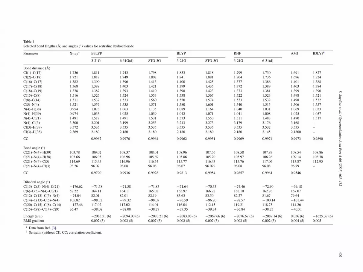
.1. Geometric parameters
Crystallographic studies on sertraline·HCl have shown thexistence of several forms depending on the crystal. No obviousifferences were observed between the sertraline HCl and HBralts that could explain the increased polymorphism of the HClalt [7]. Therefore, the experimental X-ray data of sertraline·HBrere used to calculate geometrical parameters of sertraline·HCl.
n structure, sertraline has two planar phenyl rings that arepproximately perpendicular to each other and an unsaturateding in a half-chair conformation. The distance from the ‘center-f-mass’ to an extreme halogen is approximately 6 A [8].
The labelling of the atoms in the X-ray structure [3] is shownn Fig. 1(a). Fig. 1(b) presents the geometry of molecule opti-ized using DFT method at the B3LYP/6-31G(d) level. The
ptimized geometrical parameters determined using the ab ini-
bp
i
ig. 1. (a) X-ray structure of sertraline hydrochloride (CSD code: CAVVUQ).b) Geometry of the sertraline optimized at the B3LYP/6-31G(d) level.
io, density functional and AM1 methods for molecule areollected in Table 1. In the second column of Table 1, the exper-mental data obtained by the X-ray study [3] on molecule arelso included.
As discussed by Jonson et al. [9] the BLYP functional leadso bond lengths which are systematically too long. The B3LYP
ethod leads to geometry parameters which are much closer toxperimental data [9–11]. A statistical treatment of these datasee at the bottom of Table 1) shows that for the bond lengths3LYP/6-31G(d) (Fig. 2) is slightly better than the HF/6-31G(d)eometry. The lowest correlation coefficient for bond lengthsas 0.9951 for BLYP/STO-3G method Electron correlation is
mportant for on the overall geometry of the complete molecularodel geometrical parameters.The agreement between the calculated and the experimen-
al bond angles is worse than for the bond lengths. The largesthange between calculated and experimental bond angles areound in the C(21)–N(4)–H(39) angle which varying from 109◦optimized) to 103◦ (in the crystal). The reason for this is that theptimization is performed in an isolated condition, whereas thexperimental X-ray structure was affected by the crystal envi-onment. Torsion angles, as expected, are somewhat different
ecause of the numerous degrees of conformational freedomresent in the molecule.The largest discrepancies are for the dihedral angles whichnvolve amino nitrogen atom.
S.Sagdincetal./Spectrochim
icaA
ctaPartA
66(2007)
405–412407
Table 1Selected bond lengths (A) and angles (◦) values for sertraline hydrochloride
Parameter X-raya B3LYP BLYP RHF AM1 B3LYPb
3-21G 6-31G(d) STO-3G 3-21G STO-3G 3-21G 6-31(d)
Bond distance (A)Cl(1)–C(17) 1.736 1.811 1.743 1.798 1.833 1.818 1.799 1.730 1.691 1.827Cl(2)–C(18) 1.721 1.818 1.749 1.802 1.841 1.881 1.804 1.736 1.696 1.824C(16)–C(17) 1.382 1.390 1.396 1.413 1.400 1.425 1.377 1.386 1.401 1.388C(17)–C(18) 1.368 1.388 1.403 1.421 1.399 1.435 1.372 1.389 1.403 1.384C(18)–C(19) 1.378 1.387 1.393 1.410 1.398 1.423 1.373 1.381 1.399 1.390C(15)–C(8) 1.516 1.526 1.524 1.553 1.538 1.567 1.522 1.523 1.498 1.521C(8)–C(14) 1.511 1.537 1.533 1.560 1.550 1.574 1.533 1.532 1.498 1.532C(5)–N(4) 1.521 1.557 1.535 1.571 1.580 1.601 1.540 1.515 1.506 1.557N(4)–H(38) 0.954 1.073 1.063 1.135 1.089 1.164 1.040 1.031 1.069 1.033N(4)–H(39) 0.974 1.033 1.025 1.059 1.042 1.071 1.041 1.008 1.025 1.057N(4)–C(21) 1.491 1.517 1.491 1.531 1.533 1.550 1.511 1.483 1.470 1.517N(4)–Cl(3) 3.300 3.201 3.199 3.253 3.213 3.273 3.179 3.176 3.168 –Cl(3)–H(39) 3.572 3.535 3.535 3.535 3.535 3.535 3.535 3.532 3.535 –Cl(3)–H(38) 2.369 2.180 2.180 2.180 2.180 2.180 2.180 2.145 2.1800 –
CC 0.9967 0.9976 0.9964 0.9962 0.9951 0.9969 0.9974 0.9973 0.9890
Bond angle (◦)C(21)–N(4)–H(39) 103.78 109.02 108.37 108.01 108.96 107.56 108.58 107.89 108.54 108.86C(21)–N(4)–H(38) 103.66 106.05 106.96 105.69 105.86 105.70 105.97 106.26 109.14 108.38C(21)–N(4)–C(5) 114.69 115.45 116.96 116.54 115.77 116.43 115.76 117.06 113.87 112.95C(21)–N(4)–Cl(3) 93.26 96.07 96.08 96.08 96.07 96.079 96.08 96.08 96.79 –
CC 0.9790 0.9936 0.9928 0.9813 0.9954 0.9857 0.9961 0.9546
Dihedral angle (◦)C(13)–C(5)–N(4)–C(21) −176.62 −71.58 −71.58 −71.83 −71.64 −70.33 −74.46 −72.90 −69.18C(6)–C(5)–N(4)–C(21) 52.22 164.11 164.11 165.02 165.97 166.72 162.10 162.76 167.07C(12)–C(13)–C(5)–N(4) −74.04 82.01 82.01 82.19 83.63 83.50 82.27 81.67 79.64C(14)–C(13)–C(5)–N(4) 105.82 −98.32 −99.32 −98.07 −96.59 −96.70 −98.57 −100.14 −101.44C(20)–C(15)–C(8)–C(14) −127.46 117.02 117.02 114.01 116.04 112.15 119.21 118.73 114.26C(15)–C(8)–C(14)–C(9) 36.47 −38.08 −38.08 −38.27 −37.35 −39.24 −36.84 −38.25 −40.51
Energy (a.u.) −2083.51 (6) −2094.00 (6) −2070.21 (6) −2083.08 (6) −2069.66 (6) −2076.67 (6) −2087.14 (6) 0.056 (6) −1625.37 (6)RMS gradient 0.002 (5) 0.002 (5) 0.007 (5) 0.002 (5) 0.007 (5) 0.002 (5) 0.002 (5) 0.004 (5) 0.005
a Data from Ref. [3].b Sertralin (without Cl), CC: correlation coefficient.

408 S. Sagdinc et al. / Spectrochimica A
Fm
iCC[cma
Ftt
or(Xlt
mfiTiamsan
(tc
4
ttdtc(
pcbrFu
TS
A
CCNHHCCCCCCC
R
ig. 2. Calculated (B3LYP/6-31G(d)) bond lengths in comparison with experi-ental data.
Various polymorphic forms of 1 have been describedn the literature and found as 64◦ for dihedral angle(5)–C(6)–C(7)–C(8) of Forms I (2) and III (3) using solid-stateP/MAS 13C NMR spectra [12–14]. According to X-ray study
8], sertraline hydrochloride has an unsaturated ring in a half-hair conformation. The C(5)–C(6)–C(7)–C(8) dihedral valueeasured as 62.3◦ by X-ray crystallography. We calculated this
ngle as 63.88◦.◦
The patent reports that as 68.8 for C(6)–C(5)–N–CH3 fororm III (3) whereas only Form I (2) has an antiperplanarype value (162.6◦). In solid-state CP/MAS 13C NMR spec-ra [14] of conformational polymorphs Forms I (2) and III
ta
c
able 2elected atomic charges of sertraline hydrochloride for different levels and basis sets
tom B3LYP/3-21G B3LYP/6-31G(d) RB3LYP/STO-3G RBLYP/3-21G R
(19) −0.142754 −0.127742 −0.072780 −0.127539 −(16) −0.163004 −0.179073 −0.073652 −0.158881 −(4) −0.623164 −0.600919 −0.246940 −0.559245 −(39) 0.359991 0.376477 0.255503 0.344116(38) 0.389893 0.380334 0.247767 0.358549l(3) −0.767375 −0.746970 −0.751342 −0.732422 −(21) −0.443595 −0.337946 −0.134093 −0.426096 −(5) −0.150620 −0.086012 0.029077 −0.149034(17) −0.277355 0.095855 0.030222 −0.247565l(2) 0.135265 0.004868 −0.100369 0.113507 −l(1) 0.175380 0.039314 −0.078772 0.152550 −(18) −0.257603 −0.075415 0.026514 −0.227916
: restricted; U: unrestricted.
cta Part A 66 (2007) 405–412
f sertraline·HCl had also correlated with a �-gauche effectesulting from the respective 162.6◦ antiperiplanar and 68.8◦+)-synclinal C(6)–C(5)–N–CH3 torsion angles as measured by-ray crystallography. As can be seen from Table 1, we calcu-
ated ca 164◦ for this angle. According to calculation results,wo phenyl groups are nearly perpendicular to each other.
In order to find molecular geometry of Form III (3), theolecule was constructed by keeping C(6)–C(5)–N–CH3 anglexed at 70◦. We have not found a minimum for this structure.he results obtained were unrealistic, with the IR spectra hav-
ng negative frequencies. If there are negative frequencies inn IR spectrum, it is a sign that you have not obtained the mini-um energy for the analysed structure. A valid minimum energy
tructure has only positive frequencies. Moreover, the measurednd calculated bond lengths and bond angles for two models areearly the same as those appearing in X-ray study [3].
Sertraline is in the form of a pharmaceutically acceptable salte.g., chloride, lactate, acetate, aspartate). Comparison purpose,he molecular structure of sertraline without Cl− ion was alsoalculated by using B3LYP/6-31G(d) basis set (Table 1).
.2. Other molecular properties
The calculation of effective atomic charges plays an impor-ant role in the application of quantum mechanical calculationso molecular systems. Our interest here is in the comparison ofifferent methods to describe the electron distribution in ser-raline as broadly as possible, and assess the sensitivity of thealculated charges to changes in (1) the choice of the basis set;2) the choice of the quantum mechanical method.
Mulliken charges are calculated by determining the electronopulation of each atom as defined by the basis functions. Thealculated Mulliken charge values using different levels andasis sets are listed in Table 2. The results can, however, better beepresented in graphical form as has been done in Figs. 3 and 4.ig. 3 shows that the Mulliken charge of atoms for B3LYP levelssing STO-3G, 3-21G and 6-31G(d) basic sets and Fig. 4 shows
hat the Mulliken charge of the same atoms for B3LYP, BLYPnd HF levels.From those results, it will be possible to say to the change ofharge distribution by a change basic set. The charges depending
BLYP/STO-3G RHF/3-21G RHF/6-31G(d) AM1 UBLYP/6-31G
0.072318 −0.177364 −0.175062 −0.126944 −0.1491340.072585 −0.179957 −0.191607 −0.097985 −0.1448570.224727 −0.805426 −0.765850 −0.058491 −0.5856220.242913 0.399963 0.423217 0.217213 0.2877780.213338 0.479992 0.473132 0.310760 0.3639140.682117 −0.857591 −0.837619 −0.8799830.133449 0.421207 −0.338245 −0.185599 −0.4484240.023222 −0.095634 −0.054169 −0.016399 −0.2093140.026637 −0.316995 −0.145137 −0.055550 −0.2527890.094861 0.151629 0.029560 0.004683 0.1182920.074451 0.193671 0.063434 0.040442 0.1105810.023138 −0.299433 −0.129651 −0.062863
S. Sagdinc et al. / Spectrochimica Acta Part A 66 (2007) 405–412 409
Fig. 3. Variance of the atomic charges for different basis sets of B3LYP level.
ot3Bf−
eHmsvs
m
〈
st3
4
rbrttosTocw
nrtv
The fundamental vibrational modes were calculated on the
TC
B
BBBBBHH
Fig. 4. Comparison of different methods for calculated atomic charges.
n basis set are changed due to polarization. For example,he charge of C19 atom is −0.072780e− for B3LYP/STO-G, −0.127742e− for B3LYP/6-31G(d), −0.142754e− for3LYP/3-21G and the charge of C16 atom is −0.073652e−
or B3LYP/STO-3G, −0.163004e− for B3LYP/6-31G(d),0.179073e− for B3LYP/3-21G.One of the objectives of this investigation is to study the
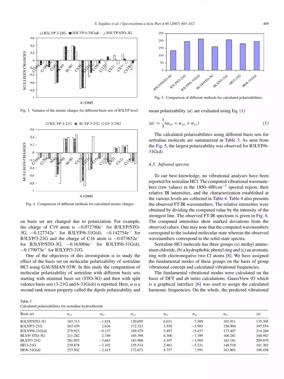
ffect of the basis set on molecular polarizability of sertralineCl using GAUSSIAN 03W. In this study the computation ofolecular polarizability of sertraline with different basis sets
tarting with minimal basis set (STO-3G) and then with splitalence basis sets (3-21G and 6-31G(d)) is reported. Here, α is aecond rank tensor property called the dipole polarizability and
bih
able 3alculated polarizabilities for sertraline hydrochloride
asis set αxx αxy αyy
3LYP/STO-3G 183.313 −1.818 120.6953LYP/3-21G 263.439 2.636 172.3213LYP/6-31G(d) 279.923 −0.137 189.479LYP/ STO-3G 211.282 2.749 165.398LYP/3-21G 281.092 −3.683 183.966F/3-21G 239.878 −3.102 155.514F/6-31G(d) 253.502 −2.415 172.071
Fig. 5. Comparison of different methods for calculated polarizabilities.
ean polarizability 〈α〉 are evaluated using Eq. (1)
α〉 = 1
3(αxx + αyy + αzz) (1)
The calculated polarizabilities using different basis sets forertraline molecule are summarized in Table 3. As seen fromhe Fig. 5, the largest polarizability was observed for B3LYP/6-1G(d).
.3. Infrared spectra
To our best knowledge, no vibrational analyses have beeneported for sertraline HCl. The computed vibrational wavenum-ers (raw values) in the 1850–400 cm−1 spectral region, theirelative IR intensities, and the characterization established athe various levels are collected in Table 4. Table 4 also presentshe observed FT-IR wavenumbers. The relative intensities werebtained by dividing the computed value by the intensity of thetrongest line. The observed FT-IR spectrum is given in Fig. 6.he computed intensities show marked deviations from thebserved values. One may note that the computed wavenumbersorrespond to the isolated molecular state whereas the observedavenumbers correspond to the solid-state spectra.Sertraline·HCl molecule has three groups (a) methyl ammo-
ium chloride, (b) a hydrophobic phenyl ring and (c) an aromaticing with electronegative two Cl atoms [8]. We have assignedhe fundamental modes of these groups on the basis of groupibrational concept and calculated vibrational frequencies.
asis of DFT and ab initio calculations. GaussView 03 whichs a graphical interface [6] was used to assign the calculatedarmonic frequencies. On the whole, the predicted vibrational
αxz αyz αzz 〈α〉6.031 7.589 101.911 135.3053.558 −5.993 156.904 197.5545.497 −5.437 173.407 214.2696.306 −7.399 106.281 160.9874.107 −1.505 162.181 209.0792.461 −5.221 148.518 181.3034.757 7.591 163.801 196.458

410 S. Sagdinc et al. / Spectrochimica Acta Part A 66 (2007) 405–412
Table 4Observed and calculated IR wavenumbers (relative intensities) for sertraline hydrochloride using B3LYP/3-21G, B3LYP/ 6-31G(d), HF/3-21G(d), HF/6-31G(d),BLYP/3-21G methods
Tentative assignment Observed B3LYP HF BLYP B3LYP
3-21G 6-31G(d) 3-21G 6-31G(d) 3-21G 6-31G(d)a
δs(N+H2) 1582 (mw) 1680 (68) 1658 (53) 1831 (100) 1820 (66) 1623 (74) 1623 (12)νring 1564 (vw) 1630 (6) 1641 (4) 1773 (7) 1810 (17) 1565 (8) 1573 (100)νring 1485 (w) 1594 (11) 1610 (11) 1736 (10) 1666 (39) 1542 (3) 1553 (28)δa(CH3) + δs(CH2) 1468 (vs) 1573 (17) 1539 (41) 1682 (82) 1663 (100) 1535 (33) 1549 (5)δs(CH2) + δs(NH2) 1428 (mw) 1563 (20) 1537 (26) 1669 (37) 1649 (41) 1528 (20) 1540 (3)δνring + δ(CH) 1401 (s) 1524 (65) 1517 (82) 1658 (68) 1652 (74) 1469 (79) 1506 (74)δ(CH2) + δ(CH3) + δ(NH2) 1505 (11) 1510 (10) 1652 (11) 1634 (22) 1431 (24) 1415 (22)�(CH3)+ ν(N-C) + δ(CH3) 1365 vw 1478 (17) 1469 (28) 1624 (34) 1624 (24) 1394 (4)C–NH2 + δ(CH3) + δ(CH) 1338 (mw) 1472 (100) 1460 (69) 1611 (92) 1612 (62) 1418 (100) 1357 (11)ω(CH2) 1454 (21) 1446 (26) 1577 (25) 1572 (30) 1408 (24)ω(NH2) + �(CH) 1310 (vw) 1430 (32) 1432 (46) 1551 (20) 1503 (16) 1384 (37) 1344 (20)δ(CH3) + δ(CH) + νring + ν(C–N) 1270 (w) 1373 (18) 1374 (26) 1493 (11) 1494 (22) 1363 (18) 1325 (33)ω(CH2) + νring + r(NH2) 1250 w 1259 (8) 1307 (4) 1358 (7) 1352 (16) 1288 (7) 1228 (2)r(CH3) + δ(CH) + ν(CNC) 1212 (mw) 1245 (35) 1264 (8) 1259 (33) 1268 (9) 1230 (14) 1214 (25)δ(CH) + t(CH2) + ω(N–H) 1171 vw 1173 (27)δring + ν(Ph–Cl) 1138 (s) 1162 (38) 1160 (100) 1160 (13) 1204 (12) 1152 (86) 1134 (20)δ(CH) 1076 vwδ(CH3) + δring + ν(C–N) 1058 vw 1140 (8) 1132 (6) 1150 (39) 1132 (55) 1100 (12)δ(CH) + ν(Ph–Cl) 1025 (m) 1097 (6) 1096 (5) 1020 (43)CN + ω(CH2) 1082 (13) 1108 (13) 1114 (41)γ(CH) + r(CH2) + δCCC 1005 (86) sh 1064 (10) 1080 (9) 1061 (59) 1105 (12) 1049 (8)γ(CH) + δring 975 vw 993 (10)δCCC + γ(CH) 955 m 1061 (25) 1041 (42) 1006 (17) 1047 (23) 981 (8) 1047 (7)ω(NH2) + r(CH2) + γ(CH) 922 (mw) 1029 (8) 1033 (26) 990 (46) 998 (10) 943 (21) 964 (3)γ(CH) 901 vw 996 (12) 1020 (6) 957 (17) 972 (3) 930 (12) 986 (12)r(CH2) + γ(CH) 891 (w) 969 (13) 970 (18) 944 (18) 932 (46) 941 (7)r(CH2) + γ(CH) 824 (m) 928 (21) 925 (13) 894 (13) 914 (7) 893 (21) 928 (19)γ(NH2) + γ(CH) 803 (w) 852 (37) 838 (14) 885 (39) 873 (35) 779 (30) 852 (34)r(NH2) + r(CH3) + r(CH2) 787 (s) 814 (27) 808 (24) 860 (37) 844 (25) 814 (25)γ(CH) + γ(NH2)γ ring 762 (mw) 795 (9) 786 (10) 812 812 (3) 809 (39) 795 (8)γ(CH) + γ(NH2) 743 (vw)γPh 713 shγ(CH) + γPh 704 (mw) 782 (55) 772 (38) 775 (19) 800 (3) 748 (70) 782 (51)γ ring + γ(NH) 672 (mw) 772 (3) 750 (1) 751 (10) 764 (17) 689 (18) 772 (3)γ ring + γ(CH) 621 (w) 716 (16) 725 (11) 739 (19) 670 (13) 716 (15)γ ring 590 (mw) 694 (11) 685 (18) 691 (9) 633 (10) 694 (10)γ ring 565 (w) 613 (6) 600 (5) 537 (13) 540 (26) 585 (8) 613 (6)γ ring 512 (vw) 513 (6) 511 (9) 534 (13) 494 (7) 513 (6)γ ring 495 (mw) 484 (14) 499 (24) 516 (18) 462 (16) 484 (13)τ 466 (γ 428 (
fTobe
or
da
om
hmtld
46fl
(ring) 448 (w) 483 (12)(CH) + γ(C–Cl) 424 (w) 477 (10)
a For sertraline without chloride.
requencies are in agreement with the experimental results.he calculated vibrational wavenumbers using different meth-ds were compared with experimentally observed values. Someands found in the predicted IR spectra were not observed in thexperimental spectrum of sertraline·HCl.
The correlation graphs between the unscaled calculated andbserved results for the assigned fundamentals in the fingerprintegion (400–1600 cm−l) are shown in Fig. 7.
NH and C–H stretching frequencies were not used as inputata to the corelasyon procedure. These neglected fundamentals
re which often show significant uncertainties.Theoretical harmonic frequencies typically overestimatebserved fundamentals due to the neglect of mechanical anhar-onicity, electron correlation and basis set effects Theoretical
[r
s
3) 442 (4) 462 (4) 456 (21) 483 (11)5) 414 (6) 404 (5) 477 (9)
armonic frequencies are often scaled to compare with experi-ental wavenumbers. Unscaled harmonic frequencies show a
endency to overestimate experimental fundamentals, with aarge number of frequencies overestimating the experimentalata by more than 70 cm−1.
The root mean square deviation of vibrations in the00–1600 cm−l range is 75.17 cm−l for the BLYP/321-G and9.22 for B3LYP/6-31G(d). In a second step an overall scalingactor has been applied to the calculated frequencies for theseevels. The scaling factor is 1 for the BLYP method and 0.962
15] for the B3LYP functional. With use of the scaling factor, theoot mean square deviation decreased to 26.22 cm−l for B3LYP.For CH3NH2 group [16] the vibrational modes are the C–Htretching modes, the NH2 scissors, N–H stretching modes, NH2

S. Sagdinc et al. / Spectrochimica Ac
wbTa
sgshvib
g1abmia
Fig. 6. Infrared spectrum of sertralin hydrochloride (KBr disc).
ag CH3 rock C–N stretching and the torsion about the C–Nond. NH2 symmetric stretching vibrations occur at 3361 cm−1.he band at 780 cm−1 corresponds to the NH2 wag and the bandt 1616 cm−1 corresponds to the NH bending motions. The C–N
aabr
Fig. 7. Unscaled calculated vibrational frequencies in compariso
ta Part A 66 (2007) 405–412 411
tretching is located at 1044 cm−1. The absorption of the N+H2roup are lower by about 200 cm−1. In this region also occurstretching modes of CH3. The asymmetric stretching of N+H2as been assigned to the ranges 2920–2915 cm−1. We observedery weak band at 2918 cm−1. The band observed at 2751 cm−1
s assigned to the symmetric stretching of N+H2. The calculatedand at ca. 2620 cm−1 is assigned to the ν(NH· · ·Cl).
The deformation vibration (scissors) of methyl bonded N+H2roup is found in the region 1620–1560 cm−1 [17]. The bands at582, 1428 and 803 cm−1 are assigned to the δS(N+H2), waggingnd rocking modes of sertraline hydrochloride, respectively. Theand at 891 cm−1 is assigned to ν(CN) stretching mode. Thisode showed also coupling between the other modes. Table 4
ndicates that most of the vibrational wave numbers arise onccount of mixing of different normal modes.
The weak bands observed at 3076, 3037 and 3008 cm−1 are
ssigned to C–H stretching frequencies. The bands observedt 2817 and 2940 cm−1 are assigned to C–H stretching of Nonded CH3 group and CH2, respectively. The CH2 group (foring) gives rice to a band near 1465 cm−1 due to the scissoringn to the experimentally obtained data. All units in cm−1.

4 ica A
vagoCc
1pNictT
Cshs
5
fDmsdda
tRBTtT
A
PC
R
[[[
[
[[
12 S. Sagdinc et al. / Spectrochim
ibration. The asymmetrical CH3 deformation is also foundround 1460 cm−1. A methyl group attached to a nitrogen atomives rice to a band at 1408 cm−1 as a symmetric scissors. Thebserved IR band at 1468 cm−1 is assigned to the asymmetricalH3 and CH2 scissoring vibration modes, corresponding to theomputed spectra [18].
Vucis et al. [1] reported that the frequencies at 1130 and077 cm−1 were observed Ph–Cl bands in N-[4-(3,4-dichloro-henyl)-3,4-dihydro-1(2H)-naphthalenylidene]-methanamine-oxide. In the IR spectrum of sertraline·HCl, the correspond-
ng bands are observed at 1138 and 1025 cm−1. The theoreticalomputations at the ab initio and density functional levels ofhe other vibrational modes of molecule studied are given inable 4.
The calculated vibrational spectrum of sertraline withoutl− atom using B3LYP/6-31G(d) is also evaluated. As can be
een from Table 4, computed wavenumbers of this moleculeave lower wavenumbers and intensities than that of theertraline·HCl.
. Conclusions
The frequency assignments were performed for the first timerom FT-IR spectrum recorded for sertraline·HCl. TheoreticalFT and ab initio calculations of the vibrational spectra of theolecule presented in this paper are compared with the infrared
pectrum of the solid sertraline·HCl. Geometries were repro-uced within the limits of accuracy of available experimentalata The molecular geometry of sertralin.HCl is best reproducedt the B3LYP levels of DFT theory.
All frequencies of the bands can be approached practically byheoretical calculation performed for a single isolated molecule.egarding the harmonic vibrational frequencies, we found that3LYP is better for the frequencies compared to BLYP and HF.he Mulliken charges and molecular polarizability values of ser-
raline using different levels and basis sets were also calculated.he largest polarizability was observed for B3LYP/6-31G(d).
cknowledgements
The authors are grateful to the referee for helpful comments.fizer Pharmaceuticals Production Corporation Ringaskiddy,o. is thanked for their gift of sertraline·HCl molecules.
[[
[
cta Part A 66 (2007) 405–412
eferences
[1] K. Vukics, T. Fodor, J. Fischer, I. Fellegvari, S. Levai, Organ. Process Res.Dev. 6 (2002) 82.
[2] O. Almarsson, M.B. Hickey, M.L. Peterson, S.L. Morissette, S. Soukassene,C. McNulty, M. Tawa, J.M. MacPhee, J.F. Remenar, Cryst. Growth Des. 3(6) (2003) 927 (patent information documents: (a) US 5,248,699 (Pfizer);(b) EP 0 928 784 A1 (Torcan); (c) WO 00/32551 A1 (Teva); (d) WO01/32601 A1 (CIBA Specialty Chemicals Holding Inc.); (e) WO01/45692A1 (Teva)).
[3] Cambridge Crystallographic DataBase, Cambridge Crystallographic DataCenter, Cambridge UK, Coden, CAVVUQ.
[4] D.C. Young, Computational Chemistry: A Practical Guide for ApplyingTechniques to Real World Problems., John Wiley & Sons Inc., 2001, p. 42.
[5] V. Chis, S. Filip, V. Miclaus, A. Pirnau, C. Tanaselia, V. Almasan, M.Vasilescu, J. Mol. Struct. 744–747 (2005) 363.
[6] M.J. Frisch, G.W. Trucks, H.B. Schlegel, G.E. Scuseria, M.A. Robb, J.R.Cheeseman, J.A. Montgomery Jr., T. Vreven, K.N. Kudin, J.C. Burant, J.M.Millam, S.S. Iyengar, J. Tomasi, V. Barone, B. Mennucci, M. Cossi, G. Scal-mani, N. Rega, G.A. Petersson, H. Nakatsuji, M. Hada, M. Ehara, K. Toyota,R. Fukuda, J. Hasegawa, M. Ishida, T. Nakajima, Y. Honda, O. Kitao, H.Nakai, M. Klene, X. Li, J.E. Knox, H.P. Hratchian, J.B. Cross, C. Adamo,J. Jaramillo, R. Gomperts, R.E. Stratmann, O. Yazyev, A.J. Austin, R.Cammi, C. Pomelli, J.W. Ochterski, P.Y. Ayala, K. Morokuma, G.A. Voth,P. Salvador, J.J. Dannenberg, V.G. Zakrzewski, S. Dapprich, A.D. Daniels,M.C. Strain, O. Farkas, D.K. Malick, A.D. Rabuck, K. Raghavachari, J.B.Foresman, J.V. Ortiz, Q. Cui, A.G. Baboul, S. Clifford, J. Cioslowski, B.B.Stefanov, G. Liu, A. Liashenko, P. Piskorz, I. Komaromi, R.L. Martin,D.J. Fox, T. Keith, M.A. Al-Laham, C.Y. Peng, A. Nanayakkara, M. Chal-lacombe, P.M.W. Gill, B. Johnson, W. Chen, M.W. Wong, C. Gonzalez,J.A. Pople, GAUSSIAN 03, Revision B. 04, Gaussian Inc., Pittsburgh PA,2003.
[7] J.F. Remenar, J.M. MacPhee, B.K. Larson, V.A. Tyagi, J.H. Ho, D.A. McIl-roy, M.B. Hickey, P.B. Shaw, O. Almarsson, Organ. Process Res. Dev. 7(2003) 990.
[8] F. Caruso, A. Besmer, M. Rossi, Acta Cryst. C 55 (1999) 1712.[9] B.G. Johnson, P.M.W. Gill, J.A. Pople, J. Chem. Phys. 98 (1993)
5612.10] A.P. Scott, L. Radom, J. Phys. Chem. 100 (41) (1996) 16502.11] G. Rauhut, P. Pulay, J. Phys. Chem. 99 (10) (1995) 3093.12] R.J. Sysko, J.M. Allen, US Patent 5,248,699, 1993 (28 September);
R.J. Sysko, J.M. Allen, Chem. Abstr. 120 (1994) 38134f.13] B.M. Johnson, P.-T.L. Chang, Anal. Profiles Drug Subs. 24 (1996)
443.14] A. Novoselsky, R. Glaser, Magn. Reson. Chem. 40 (2002) 723.15] A.P. Scott, L. Radom, J. Phys. Chem. 100 (1996) 16502.
16] D. Zerokaa, J.O. Jensen, J. Mol. Struct. (Theochem.) 425 (1998) 181.17] N.B. Colthup, L.H. Daly, S.H. Wiberley, Introduction to Infrared andRaman Spectroscopy, Academic Press, 1990.18] C.N.R. Rao, Chemical Applications of Infrared Spectroscopy, Academic
Press, 1963, p. 152.


