Embed Size (px)
Citation preview

Eur. J. Immunol. 2013. 43: 371–381 Cellular immune responseDOI: 10.1002/eji.201242809 371
Aberrant antibody affinity selection in SHIP-deficientB cells
Wai-Hang Leung, Tatiana Tarasenko, Zuzana Biesova, Hemanta Kole,Elizabeth R. Walsh and Silvia Bolland
Laboratory of Immunogenetics, National Institute of Allergy and Infectious Diseases, NationalInstitutes of Health, Rockville, MD, USA
The strength of the Ag receptor signal influences development and negative selection ofB cells, and it might also affect B-cell survival and selection in the GC. Here, we have usedmice with B-cell-specific deletion of the 5′-inositol phosphatase SHIP as a model to studyaffinity selection in cells that are hyperresponsive to Ag and cytokine receptor stimula-tion. In the absence of SHIP, B cells have lower thresholds for Ag- and interferon (IFN)-induced activation, resulting in augmented negative selection in the BM and enhancedB-cell maturation in the periphery. Despite a tendency to spontaneously downregulatesurface IgM expression, SHIP deficiency does not alter anergy induction in responseto soluble hen-egg lysozyme Ag in the MDA4 transgenic model. SHIP-deficient B cellsspontaneously produce isotype-switched antibodies; however, they are poor respondersin immunization and infection models. While SHIP-deficient B cells form GCs and undergomutation, they are not properly selected for high-affinity antibodies. These resultsillustrate the importance of negative regulation of B-cell responses, as lower thresh-olds for B-cell activation promote survival of low affinity and deleterious receptors to thedetriment of optimal Ab affinity maturation.
Keywords: Affinity maturation � B cell � Negative selection � SHIP
� Additional supporting information may be found in the online version of this article at thepublisher’s web-site
Introduction
BCR signaling is essential for the initiation of humoral responsesand regulation of B-cell development and maturation [1–3]. It isgenerally believed that in the absence of foreign Ags, BCR con-tinuously receives activation signals [4], presumably through thebinding of self-ligands. The strength of BCR signaling is deter-mined both by the extent of Ag engagement and by the involve-ment of coreceptors and intracellular effector molecules that mod-ulate signaling events. Modifications that alter the strength of
Correspondence: Dr. Silvia Bollande-mail: [email protected]
BCR signaling are expected to have an effect on final B-cell acti-vation outcomes; however, these alterations also affect devel-opment, activation, and interactions with other cell types, mak-ing it difficult to ascertain whether each particular stage in theB-cell activation process requires modulation of BCR signal [5–7].Enhancement of BCR signaling intensity in several KO mice leadsto alterations in humoral responses that have been interpreted tobe a consequence of developmental skewing or homing defects.For example, deficiency in the tyrosine phosphatase SHP-1 resultsin reduced isotype switched B-cell responses, which has beenattributed to severe skewing of the B-cell repertoire toward aB1 cell differentiation path and reduced development of moreeffective responders, B2 cells [8]. CD22 deficiency also detrimen-tally affects humoral responses, though in this case alterations areattributed to the lower production of marginal zone (MZ) B cells
C© 2012 WILEY-VCH Verlag GmbH & Co. KGaA, Weinheim www.eji-journal.eu

372 Wai-Hang Leung et al. Eur. J. Immunol. 2013. 43: 371–381
and lack of recirculating B-cell populations [9]. Targeted deletionof Grb2, an adaptor protein that plays a significant role in nega-tive regulatory processes, to the B-cell lineage results in reducedAb responses that has been ascribed to the lack of lymphoid orga-nization and GC defects [10, 11]. These examples illustrate therange of functional dysregulation that accompanies removal ofvarious negative regulators of B-cell activation, making it difficultto discern which of these effects is responsible for the poor overallresponse.
The 5′-inositol phosphatase SHIP is another well-characterizedmodulatory factor in B cells that can associate with Grb2 and CD22[12,13]. SHIP regulates cell responses in lymphocytes and myeloidcells by its ability to hydrolyze the second messenger PI(3,4,5)trisphosphate and prevent downstream signaling pathways thatlead to activation [12, 14–16]. In B cells, SHIP is recruited to thephosphorylated immunoreceptor tyrosine-based inhibitory motifof FcγRIIB upon coaggregation with the BCR [17, 18] where itsenzymatic activity depletes PI(3,4,5) trisphosphate and preventsmembrane localization of PH-domain-containing factors such asTec kinases, Akt, and PLCγ [19–22]. SHIP can also regulate BCRsignaling in the absence of FcγRIIB engagement [23–26]. Overall,SHIP’s inhibitory activity leads to the modulation of BCR inducedcalcium influx and prevention of cellular activation [23]. B cellspurified from SHIP-deficient mice are hyperresponsive to acti-vating signals and more resistant to apoptosis in vitro [26–30].SHIP-null mice develop alterations in B-cell development, such asreduced immature B-cell numbers in BM and increased numbersof mature B cells in the spleen [26]. However, there are caveatsto studies performed on B cells purified from mice with germlinedeletion of SHIP (SHIPnull/null). Those mice exhibit early mortal-ity from a myeloproliferative-like syndrome characterized by pro-found splenomegaly and massive myeloid infiltration of the lung[31], making it difficult to study the role of SHIP in lymphocyteswithout the influence of the inflammatory environment. For ourcurrent studies, we have analyzed mice with B-cell-specific dele-tion of SHIP as a model system to assess the effect of enhancedBCR signaling strength and enhanced responses to cytokines, inthe Ab selection process. Our analysis confirms a role for SHIPas a negative regulator of B-cell selection and activation, whileit also uncovers a requirement for SHIP in ensuring efficient GCresponses and appropriate Ab affinity maturation.
Results
Increased negative selection and B-cell maturation inmice with B-cell-specific deletion of SHIP
Mice with B-cell-specific deletion of SHIP were generated by cross-ing floxed-ship mice [32] with mice in which the Cre recombi-nase is driven by the CD19 promoter [33]. The resulting micewere designated shipfl/flcd19cre/+, while shipfl/fl littermates wereused as controls. We observed that SHIP protein expression wasgreatly reduced at the pro-B-cell stage, although a few cells main-tained SHIP expression (Fig. 1A). This may be a result of a
small contaminating population of CD19− pre-pro B cells in ourCD43+B220+HSA+ gate or it may indicate less than 100% effi-ciency of ship gene deletion at this stage. However, SHIP expres-sion in mature CD19+ splenic B cells is completely ablated inshipfl/flcd19cre/+ mice (Fig. 1B). We tested whether B-cell-specificdeletion of SHIP would alter B-cell development. Immature B-cellpopulations were found at the same frequency, while the numberof recirculating B cells was reduced in BM of shipfl/flcd19cre/+ micecompared with control shipfl/fl (Fig. 1C and Supporting Informa-tion Fig. 1). B-cell-specific deletion of SHIP did not alter the num-ber of transitional or follicular B cells in the spleen, but it reducedthe number of MZ B cells and increased the number of B1, GCB cells, and isotype switched plasmablasts (CD138+IgM−) (Fig. 1Dand Supporting Information Fig. 2).
SHIP-deficient B cells could have repopulated back to normallevels even if their development was somewhat impaired. To testthis possibility, we performed a BM reconstitution experimentin which shipfl/flcd19cre/+cells compete with WT cells throughoutdevelopment. An equal mix of shipfl/flcd19cre/+ and WT BM cellswas injected into lethally irradiated WT recipients. Donor WT cellsharbored the CD45.1 allotype to differentiate them from SHIP-deficient cells. Two months after the transfer, mice contained anequal ratio of WT:SHIP-deficient cells at the pre-pro-B stage, buta 2:1 ratio in all the subsequent populations (Fig. 1E). This skew-ing in favor of WT cells was even more pronounced (16:1) in therecirculating population in the BM. In contrast, the majority of theB cells present in spontaneous GCs (∼80%) were SHIP-deficient.Thus, we determined that SHIP-deficient B cells are more likely tobe negatively selected at the immature stage and in the MZ, butthey are also more likely to spontaneously mature into GC B cells,possibly precluding their homing back to the BM upon activation.
Spontaneous IgG2a/b switch and enhanced interferonsensibility in SHIP-deficient B cells
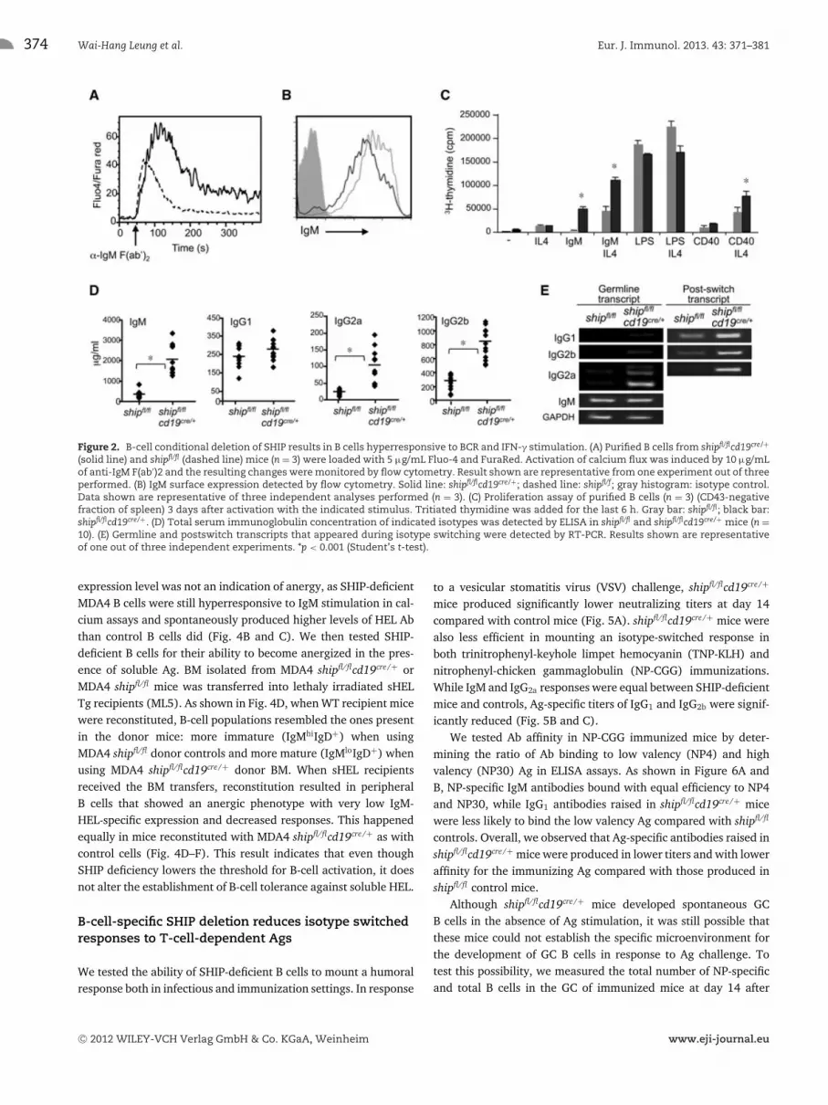
As previously reported for Ship−/− DT40 B cells [23], we observedenhanced Ca2+ flux in SHIP-deficient B cells upon stimulationwith anti-IgM F(ab’)2 (Fig. 2A) even though they expressedlower surface levels of IgM (Fig. 2B). SHIP-deficient B cells fromshipfl/flcd19cre/+ were hyperproliferative upon IgM and CD40 stim-ulation (Fig. 2C). Consistent with their lower threshold for acti-vation, SHIP-deficient B cells spontaneously produced antibodiesin vivo: steady state levels of serum IgM, IgG2a, and IgG2b werethree- to fivefold higher in shipfl/flcd19cre/+ than in shipfl/fl con-trol mice. In contrast, IgG1 levels in serum were not significantlydifferent (Fig. 2D). Serum Ab levels correlated with transcriptexpression of the various isotypes in these mice, both for germlineand postswitch transcripts (Fig. 2E).
The observed IgG isotype preference in shipfl/flcd19cre/+ mice,mainly of IgG2a/b, indicated a Th1 skewing in the humoralresponse. Given that SHIP deletion is B-cell specific in these mice,we sought to find a reason for Th1 skewing that was B-cell intrin-sic. We tested expression of T-bet and STAT1, both of which aretranscription factors linked to Th1 bias. We observed that B cells,
C© 2012 WILEY-VCH Verlag GmbH & Co. KGaA, Weinheim www.eji-journal.eu

Eur. J. Immunol. 2013. 43: 371–381 Cellular immune response 373
Figure 1. B-cell-specific deletion of SHIP uncovers its role in negative selection. (A) Intracellular flow cytometric analysis of the expression ofSHIP in BM cells (HSC and Pro-B cells) isolated from shipfl/flcd19cre/+ (solid line) or shipfl/f (dashed line). (B) Western blot analysis of SHIP expressionin B cells (CD43 negative selection) and T cells (pan T cells isolation) isolated from spleen of mice of the indicated genotype. (C and D) B-celldevelopment in (C) BM and (D) spleen was compared between shipfl/flcd19cre/+ and shipfl/f mice. Data are shown as mean + SD of n = 4. *p < 0.05(Student’s t-test). (E) BM cells isolated from shipfl/flcd19cre/+ (expressing CD45.2) and B6.Ptprca/b mice (expressing CD45.1 and CD45.2) were mixed ina ratio of 1:1 and transferred to lethally irradiated (960 rads) C57BL/6 mice. Reconstituted BM cells (top histograms and bar graph) and total spleencells (bottom histograms and bar graph) were collected after 2 months and analyzed by flow cytometry. Data are shown as mean + SD of n = 4and are representative of four independent BM transfers.*p < 0.05, **p < 0.0005 (Student’s t-test). See Supporting Information Fig. 1 and 2 for theflow cytometric gating of different developmental stages and types of B cells from BM and spleen.
but not T cells, of shipfl/flcd19cre/+ had increased basal level expres-sion of T-bet and STAT1 compared with shipfl/fl controls (Fig. 3Aand B). These expression differences could be general to all B cellsor due to changes in the proportion of certain B-cell populationsthat are more likely to express these transcription factors. We thentested the LPS/IFN-γ sensitivity of purified B cells for switchingto Th1 IgG isotypes by incubating them for 24 h with LPS andIFN-γ. Indeed, SHIP-deficient cells were much more likely toinduce IgG2a production (Fig. 3C and D), and to upregulate AIDand T-bet expression upon LPS + IFN-γ activation compared withcontrol B cells (Fig. 3E and F).
SHIP deficiency does not alter B-cell tolerance tosoluble Ag
Mutations that alter the threshold for B-cell activation as SHIPdeficiency does might override an anergic state driven by chronicstimulation with soluble Ag. To test the role of SHIP in B-celltolerance to soluble Ag, we bred hen-egg lysozyme (HEL)-specificIgM/D transgenic mice (MDA4) to shipfl/flcd19cre/+ mice. As shownin Fig. 4A, when mice lacked soluble HEL Ag (sHEL), MDA4shipfl/flcd19cre/+ were more likely to fully mature to IgMlo IgD+
B cells than control MDA4 shipfl/fl cells. Their lower surface IgM
C© 2012 WILEY-VCH Verlag GmbH & Co. KGaA, Weinheim www.eji-journal.eu
374 Wai-Hang Leung et al. Eur. J. Immunol. 2013. 43: 371–381
Figure 2. B-cell conditional deletion of SHIP results in B cells hyperresponsive to BCR and IFN-γ stimulation. (A) Purified B cells from shipfl/flcd19cre/+
(solid line) and shipfl/fl (dashed line) mice (n = 3) were loaded with 5 μg/mL Fluo-4 and FuraRed. Activation of calcium flux was induced by 10 μg/mLof anti-IgM F(ab′)2 and the resulting changes were monitored by flow cytometry. Result shown are representative from one experiment out of threeperformed. (B) IgM surface expression detected by flow cytometry. Solid line: shipfl/flcd19cre/+; dashed line: shipfl/f; gray histogram: isotype control.Data shown are representative of three independent analyses performed (n = 3). (C) Proliferation assay of purified B cells (n = 3) (CD43-negativefraction of spleen) 3 days after activation with the indicated stimulus. Tritiated thymidine was added for the last 6 h. Gray bar: shipfl/fl; black bar:shipfl/flcd19cre/+. (D) Total serum immunoglobulin concentration of indicated isotypes was detected by ELISA in shipfl/fl and shipfl/flcd19cre/+ mice (n =10). (E) Germline and postswitch transcripts that appeared during isotype switching were detected by RT-PCR. Results shown are representativeof one out of three independent experiments. *p < 0.001 (Student’s t-test).
expression level was not an indication of anergy, as SHIP-deficientMDA4 B cells were still hyperresponsive to IgM stimulation in cal-cium assays and spontaneously produced higher levels of HEL Abthan control B cells did (Fig. 4B and C). We then tested SHIP-deficient B cells for their ability to become anergized in the pres-ence of soluble Ag. BM isolated from MDA4 shipfl/flcd19cre/+ orMDA4 shipfl/fl mice was transferred into lethaly irradiated sHELTg recipients (ML5). As shown in Fig. 4D, when WT recipient micewere reconstituted, B-cell populations resembled the ones presentin the donor mice: more immature (IgMhiIgD+) when usingMDA4 shipfl/fl donor controls and more mature (IgMloIgD+) whenusing MDA4 shipfl/flcd19cre/+ donor BM. When sHEL recipientsreceived the BM transfers, reconstitution resulted in peripheralB cells that showed an anergic phenotype with very low IgM-HEL-specific expression and decreased responses. This happenedequally in mice reconstituted with MDA4 shipfl/flcd19cre/+ as withcontrol cells (Fig. 4D–F). This result indicates that even thoughSHIP deficiency lowers the threshold for B-cell activation, it doesnot alter the establishment of B-cell tolerance against soluble HEL.
B-cell-specific SHIP deletion reduces isotype switchedresponses to T-cell-dependent Ags
We tested the ability of SHIP-deficient B cells to mount a humoralresponse both in infectious and immunization settings. In response
to a vesicular stomatitis virus (VSV) challenge, shipfl/flcd19cre/+
mice produced significantly lower neutralizing titers at day 14compared with control mice (Fig. 5A). shipfl/flcd19cre/+ mice werealso less efficient in mounting an isotype-switched response inboth trinitrophenyl-keyhole limpet hemocyanin (TNP-KLH) andnitrophenyl-chicken gammaglobulin (NP-CGG) immunizations.While IgM and IgG2a responses were equal between SHIP-deficientmice and controls, Ag-specific titers of IgG1 and IgG2b were signif-icantly reduced (Fig. 5B and C).
We tested Ab affinity in NP-CGG immunized mice by deter-mining the ratio of Ab binding to low valency (NP4) and highvalency (NP30) Ag in ELISA assays. As shown in Figure 6A andB, NP-specific IgM antibodies bound with equal efficiency to NP4and NP30, while IgG1 antibodies raised in shipfl/flcd19cre/+ micewere less likely to bind the low valency Ag compared with shipfl/fl
controls. Overall, we observed that Ag-specific antibodies raised inshipfl/flcd19cre/+ mice were produced in lower titers and with loweraffinity for the immunizing Ag compared with those produced inshipfl/fl control mice.
Although shipfl/flcd19cre/+ mice developed spontaneous GCB cells in the absence of Ag stimulation, it was still possible thatthese mice could not establish the specific microenvironment forthe development of GC B cells in response to Ag challenge. Totest this possibility, we measured the total number of NP-specificand total B cells in the GC of immunized mice at day 14 after
C© 2012 WILEY-VCH Verlag GmbH & Co. KGaA, Weinheim www.eji-journal.eu

Eur. J. Immunol. 2013. 43: 371–381 Cellular immune response 375
Figure 3. SHIP negatively regulates IFN-γ-induced class switching. (A and B) Western blot analysis of T-bet and STAT-1 expression levels inlymphocytes from mice of the indicated genotype. Results shown are from one experiment representative of two independent experimentsperformed. (C) Germline and postswitch transcripts generated in B cells stimulated by IFN-γ and/or LPS during IgG2a class switching were detectedby RT-PCR. (D) Surface IgG2a expression on B cells stimulated with IFN-γ and LPS. Solid line: shipfl/flcd19cre/+; dashed line: shipfl/fl, gray histogram:isotype control. (E and F) The expression of AID and T-bet in B cells upon LPS ± IFN-γ stimulation was analyzed by qRT-PCR. Data were normalizedto GAPDH mRNA levels. (E and F) Data shown as mean + SD and (C–F) are representative of two independent experiments, n = 4 mice/group. *p <
0.001 (Student’s t-test).
injection. GC B cells were identified by flow cytometry asGL7+FAShi and B220+ cells. As shown in Fig. 6C–E, 60% of theB cells in shipfl/fl GC controls were NP+, but only 25% of theGC B cells in shipfl/flcd19cre/+ mice were NP-specific, even thoughthe total number of GC B cells was comparable in both cases.Long-term responses were also impaired in shipfl/flcd19cre/+ mice,as measured by the number of NP-specific IgG1 memory B cellsdetected 55 days after the second challenge (Fig. 6F and G).
SHIP-deficient B cells have aberrant Ab affinityselection
To test if the lower affinity of antibodies generated inshipfl/flcd19cre/+ mice was due to an abnormal rate of somatichypermutation, we sequenced antibodies generated in the courseof an NP-CGG immunization. We purified GC B cells fromshipfl/flcd19cre/+ and shipfl/fl controls 14 days after NP-CGG immu-nization and the V186.2 heavy chain was amplified by PCR andsequenced. V186.2 has been previously shown to dominate in
response to T-dependent NP immunization; frequently joining theD segment DFL16.1 and the JH segment JH2 [34–37]. The extentof somatic hypermutation of individual clones was determined bycomparing its sequence with the original nonmutated germlineV186.2 gene sequence (Supporting Information Fig. 3) and theresult was summarized in Table 1. Shipfl/flcd19cre/+ B cells hadsimilar percentages of clones containing at least one mutation ingermline V186.2 sequences compared with controls (78% versus90%) and somewhat lower average number of mutations (2.0 ±1.6 versus 5.3 ± 4.05). Remarkably, we observed that none of theshipfl/flcd19cre/+ GC B cells had a mutation at CDR 1 compared with60% of GC B cells in control mice. In particular, the tryptophanto leucine substitution at position 33, which has been shown toincrease the affinity of anti-NP antibodies by tenfold [38, 39],was present in five out of ten sequences in control mice but wasdetected in none of the shipfl/flcd19cre/+ sequences. We then com-pared CDR 3 sequences, which contain the rearranged D and JH
segments (Supporting Information Fig. 4). Consistent with previ-ous studies that showed DFL16.1 as the predominant D segmentin combination with V186.2 in the anti-NP response [35,40], 60%
C© 2012 WILEY-VCH Verlag GmbH & Co. KGaA, Weinheim www.eji-journal.eu

376 Wai-Hang Leung et al. Eur. J. Immunol. 2013. 43: 371–381
Figure 4. Deletion of SHIP does not influence the establishment of B-cell tolerance to soluble HEL Ag. (A) FACS histogram and bar graph analysisof splenocyte populations: T1 (B220+, IgDlow, IgMahigh), T2 (B220+, IgDhigh, IgMahigh), and mature (B220+, IgDhigh, IgMalow) B cells.*p < 0.01, **p <
0.0005, ***p < 0.00001 (Student’s t-test). (B) Calcium release assay of MDA4 shipfl/flcd19cre/+(solid line) and MDA4 shipfl/fl (dashed line), performed asin Figure 2A. (C) Anti-HEL Ab levels in serum from MDA4 shipfl/flcd19cre/+ (solid line) and MDA4 shipfl/fl (dashed line) mice were analyzed by ELISA.(D and E) BM cells were isolated from MDA4 shipfl/flcd19cre/+ or MDA4 shipfl/fl mice. A total of 1 × 10⁷ cells were then transferred to lethally irradiated(960 rads) C57BL/6 (top) or ML5 (bottom) mice. Total spleen cells were collected after 2 months and the development of anergic B cells was analyzedby flow cytometry. (F) Surface anti-HEL expression on B cells isolated from the mice in (D) was analyzed by flow cytometry. (A and E) Data shownas mean + SD and (A–F) are representative of four independent experiments, n = 3 mice/group.
of the GC B cells from our control mice contained this segment. Incontrast, only 22% of the shipfl/flcd19cre/+ GC B cells included theDFL16.1 region. Uncharacteristically, 33% of the antibodies raisedin shipfl/flcd19cre/+ mice contained an early stop codon mutation(Supporting Information Fig. 3), while no such mutation occurredin controls. Thus, SHIP-deficient B cells are more likely to encodenonfunctional antibodies, a result that points toward a defect inGC selection and affinity maturation mechanisms.
Discussion
Modulation of B-cell activation by inhibitory pathways allows forfine-tuning and flexibility of responses to adapt them to specificneeds. Mutational studies in inhibitory molecules of the BCR path-way such as SHIP, SHP-1, or Grb2 have established the impor-tance of this regulation in B-cell development and activation[8, 10, 11, 26]. Most experiments aimed at elucidating the roleof SHIP in B cells were performed in mice with complete dele-tion of the gene, usually giving rise to a number of pleiotropicphenotypes and a prominent inflammatory condition that com-plicates interpretation of B-cell-specific effects. [31]. In ship-nullmice, lymphoid cell development is inhibited by increased pro-duction of IL-6 [41], thus preventing an accurate assessment ofthe role of SHIP in B cells. By analyzing shipfl/flcd19cre/+ mice, we
have confirmed the B-cell intrinsic role for SHIP, both in regulatingnegative selection in the BM and activation in the periphery.
B-cell-specific SHIP deletion allowed us to study the conse-quences of hyperactive Ag- and cytokine-receptor pathways in theAb selection process. We found that B-cell SHIP deficiency resultsin a lower BCR stimulation threshold and enhanced proliferativeresponses, consistent with the phenotype already observed in cellsobtained from SHIP-null mice and another model of B-cell-specificSHIP deficiency [28, 29]. However, our data differ in some wayswhen compared with the recently described Cd79a-cre SHIP abla-tion model [42] as we did not find that anergy is SHIP depen-dent in B cells. This discrepancy may be explained by differingmechanisms of anergy induction in the mouse models that wereused, as anergy in MD4 B cells has been suggested to be depen-dent on PTEN expression, while Ars/A1 transgenic B cells used inO’Neill et al. appear to be more SHIP dependent [42]. Our miceexpressing SHIP-deficient B cells also developed autoantibodies ata much later timepoint than those in the Cd79a-cre system (datanot shown). Overall, our data reinforce the view that the strengthof BCR signal correlates with the rate of B-cell development. Wealso confirm the B-cell-intrinsic role of SHIP in balancing the devel-opment between Fo and Mz B cells in positive selection of the pri-mary immune repertoire and in setting a threshold to preventB cells from spontaneous activation. Additionally, we haveextended the role of SHIP in B cells beyond the regulation of
C© 2012 WILEY-VCH Verlag GmbH & Co. KGaA, Weinheim www.eji-journal.eu

Eur. J. Immunol. 2013. 43: 371–381 Cellular immune response 377
Figure 5. B-cell conditional deletion of SHIP reduces humoralB-cell responses. (A) Serum analysis of VSV-specific Ab production inshipfl/flcd19cre/+or shipfl/f mice (n = 5 per group) 14 days after VSV injec-tion was performed by neutralization assay. (B–C) Six- to eight-week-old shipfl/flcd19cre/+or shipfl/f mice (four mice per group) were immunizedwith 50 μg of TNP-KLH in RIBI (B) or NP-CGG in Alum (C). Ag-specificserum IgM was determined 2 weeks after the primary immunizationand specific serum IgG was determined 1 week after a booster given onday 28. (A–C) Results shown are representative of three experiments.*p < 0.05 (Student’s t-test).
BCR signaling, as IFN-γ signaling was similarly affected by dele-tion of SHIP. This cytokine regulation might occur through SHIPbinding to Shc by a mechanism similar to what has been reportedfor GM-CSF or IL-3 regulation by SHIP [43].
We detected reduced humoral responses in mice with B-cell-specific deletion of SHIP, a phenotype reminiscent of the onesobserved in mice with B-cell-specific deletion of SHP-1 or Grb2[8, 10, 11]. One main difference is that in the case of SHIPdeficiency, the formation of GCs is unaltered or even enhancedin spontaneous conditions and thus this phenotype cannot beexplained by a disruption of GC structure as suggested for Grb2 Ab
responses. Also, SHIP deficiency in B cells does not lead to B1-typeskewing to the extreme degree that B-cell-specific PTEN or SHP-1deletion does and does not expand MZ B-cell populations as occursin PTEN-deficient B cells [8,44]. Thus, reduced numbers of follic-ular B cells cannot explain defective humoral responses. Althoughboth SHIP-deficient B cells and PTEN-deficient B cells have defectsin CSR and IgG production, they differ in that SHIP-deficientB cells have increased expression of AID upon stimulation, whereasPTEN-deficient B cells do not properly induce AID [44,45]. Thus,our preferred hypothesis is that impaired affinity maturation leadsto less effective humoral responses in SHIP-deficient B cells.
SHIP deficiency results in enhanced negative selection of B cellsat immature stages, likely causing a shift in clonality of the B-cellrepertoire toward cells bearing low-affinity BCRs. Enhancement ofBCR signaling strength likely upregulates B-cell apoptosis in nega-tive selection of auto-reactive B cells supporting the possible occur-rence and rapid disappearance of high-affinity B cells in responseto T-dependent immunization. We also cannot exclude the possi-bility that high-affinity B cells are able to escape to the periphery,but upon Ag encounter, they are subjected to apoptosis as a resultof hyperactive BCR and/or cytokine signaling augmented by theabsence of SHIP. However, absence of B cells with high-affinityBCRs from the starting pool of B-cell clones should not preventthe production of high-affinity antibodies in the GC. Accordingto a study by Shih et al., B cells in GCs actually undergo a fixedmutation program which is independent of the initial affinity ofthe BCR [46], suggesting that the tendency for a B-cell to developa high-affinity BCR is the same as that to acquire a low-affinityone in the GC. As our data show that SHIP-deficient B cells haveincreased AID expression upon IFN-γ + LPS stimulation but havea reduced propensity to acquire high-affinity BCRs upon Ag chal-lenge when compared with control B cells, it is more likely thatSHIP-deficient B cells are exempt from processes that normallydictate B-cell survival and selection of high-affinity clones.
Overall, it seems counterintuitive that SHIP deficiencyenhances spontaneous switched Ab generation but reduces high-affinity GC responses. These results suggest that very low affinityresponses drive spontaneous Ab production and would be favoredby lack of negative regulation, while hyperactive low-affinity SHIP-deficient B cells may be deleted in the GC because they do not meetthe minimum Ag affinity requirement for selection.
Materials and methods
Mice
The generation of shipfl/fl mice has been described previously [32].C57BL/6 mice, cd19cre/+ mice, ML5 mice (mice expressing solubleHEL), MD4 mice (mice containing the anti-HEL IgMa transgene)and B6. Ptprca mice were obtained from The Jackson laboratory(Bar Harbor, ME, USA). All experiments involving mice were per-formed in accordance with National Institutes of Health (NIH)guidelines.
C© 2012 WILEY-VCH Verlag GmbH & Co. KGaA, Weinheim www.eji-journal.eu

378 Wai-Hang Leung et al. Eur. J. Immunol. 2013. 43: 371–381
Figure 6. Reduced production of NP-specific GC B cells and memory B cells in SHIP-deficient mice. (A, B) ELISA assay to detect NP-specific antibodiesusing either NP30-BSA or NP4-BSA as the coating Ags was performed 5 days (for IgM) and 14 days (for IgG1) after NP-CGG (Alum) immunization(n = 5 samples per group). The affinities of the NP-specific IgM or IgG1 were determined by the ratio NP4/NP30. (C–E) Total and NP-specific GCB cells were identified by gating GC B cells B220+GL7+ splenocytes 14 days after NP-CGG (Alum) immunization and staining with NP-PE (n = 4 miceper group). (F–G) NP-specific memory B cells were determined on day 55 after NP-CGG (Alum) immunization, defined as CD90mix−, NP+, IgG1+,and CD38+ cells. (A–G) Data are shown as mean ± SD and are representative of two independent experiments, (n = 4–5 per group). *p < 0.05, **p <
0.001 (Student’s t-test).
Cells and antibodies
Purified B cells were obtained by negative selection of splenocytesisolated from 6- to 8-week-old mice with magnetic beads spe-cific for CD43 (Miltenyi Biotec, Auburn, CA, USA). For all assays,cells were maintained and stimulated in DMEM (Invitrogen, Carls-bad, CA, USA) with 10% heat-inactivated fetal bovine serum,100 U/mL penicillin, and 100 μg/mL streptomycin. All of theantibodies for FACS analysis were from BD Biosciences (San Jose,CA, USA).
Intracellular calcium measurements
Purified B cells were loaded with 5 μg/mL Fluo-4 and 5 μg/mLFuraRed (Molecular Probes, Eugene, OR, USA). Ca2+ mobilizationwas triggered by 10 μg/mL F(ab′)2 fragment of anti-IgM andthe resulting changes were monitored by FACS. The data wereexpressed as the ratio of Fluo-4/FuraRed.
IFN-γ and LPS B-cell stimulations
Purified B cells from shipfl/flcd19cre/+ or shipfl/fl mice were incu-bated with LPS (20 μg/mL) with or without IFN-γ (100 ng/mL)for 24 h. Stimulated B cells were analyzed for Th1 isotype switch-ing by flow cytometry and RT-PCR. B-cell total RNA was extractedand cDNA was prepared for real-time PCR analysis of the expres-sion of AID and T-bet.
Immunization and serum analysis
Mice were injected with 50 μg of TNP-KLH with RIBI (Sigma,St. Louis, MO, USA) or NP-CGG with Alum (Pierce, Rockford, IL,USA). Sera were collected 2 weeks after the immunization. Themice were given a second booster on day 28 and sera were col-lected 1 week after the booster. The levels of Ag-specific antibodiesof different isotypes were determined by ELISA using TNP-BSA orNP-BSA (NP4-BSA or NP30-BSA) (5 μg/mL; Biosearch Technolo-gies, Novato, CA, USA) coated 96-well plates and the ClonotypingSystem-HRP (SouthernBiotech, Birmingham, AL, USA).
VSV immunization and neutralization assay
VSV serotype Indiana (VSV-IND) was obtained from the labora-tory of Drs. Jack R. Bennink and Jonathan W. Yewdell. For immu-nization, 6- to 8-week-old mice were i.p. injected with 2 × 10⁶
PFU of VSV in 200 μL of PBS. Sera were collected on day 14after immunization. Neutralizing titers of sera were determinedas described [47, 48]. Briefly, the sera were heat inactivated for30 min at 56◦C. Serial diluted sera were mixed with 500 PFU/mLof VSV and incubated for 90 min at 37◦C. The serum–virus mixturewas subsequently transferred onto Vero cell monolayers in 96-wellplates and incubated for 1 h at 37◦C. An overlay of 100 μL MEMcontaining 1% methylcellulose (Sigma) was added. After incuba-tion for 24 h at 37◦C, the cells were fixed and stained with 0.5%crystal violet (Sigma) and the number of plaques was counted.Neutralization titer was determined as the dilution of serum thatreduces the number of plaques by 50%.
C© 2012 WILEY-VCH Verlag GmbH & Co. KGaA, Weinheim www.eji-journal.eu

Eur. J. Immunol. 2013. 43: 371–381 Cellular immune response 379
Table 1. VH 186.2-D—JH2 gene sequence summary from GC B cells of NP immunized micea)
Cell type No. of VH Sequencesb) Mutationsc) Sequences Sequencesgenes mutated per VH gene mutated at mutated atsequenced Ratio (%) Average CDR1 CDR2
± SD Ratio (%) Ratio (%)
shipfl/fl 10 9/10 (90) 5.3 ± 4.05 6/10 (60) 5/10 (50)
shipfl/flcd19cre/+ 9 7/9 (78) 2.0 ± 1.6 0/9 (0) 1/9 (11)
Cell type Position 33 Stop codond) R/S ratioe) R/S ratiof) DFL16.1 geneTrp → Leu mutation CDR1 and 2 FWR usage (%)Ratio (%) Ratio (%)
shipfl/fl 5/10 (50) 0/10 (0) 16/1 1.1/1 60
shipfl/flcd19cre/+ 0/9 (0) 3/9 (33) 2/0 6.5/1 22
a) Data presented in Supporting Information Fig. 3 and 4.b) The percentage of VH186.2 genes which had more than one mutation.c) SD = standard deviation.d) The percentage of genes containing mutations which resulted in the coding of early stop codon (TGA).e) The ratio of replacement (R) to silent (S) mutations.f) FWR: framework region.
Somatic hypermutation analysis
GC B cells from NP-immunized mice were sorted based onthe expression of GL7, Fas, and B220 using FACS Aria (BDBiosciences). Genomic DNA was isolated with the DNeasy 96kit from Qiagen. V186.2 genes were amplified by using iProofHigh-Fidelity DNA polymerase (Biorad) with primers V186.2outer 5′-TCTTTACAGTTACTGAGCACACAGGAC-3′ and JH2 5′-GGGTCTAGAGG TGTCCCTAGTCCTTCATGACC-3′ for 20 cycles.Three microliters of product were used as template for a sec-ond round (nested PCR) of PCR with primers V186.2inner 5′-CAGTAGCAGGCTTGAGGTCTGGAC-3′ and JH2 in 30 cycles. PCRproducts were cloned using TOPO TA cloning kit (Invitrogen) andsequenced using the T3 universal primer.
BM reconstitution
Eight- to ten-week-old recipient mice (C57BL/6 or ML5 mice) werelethally irradiated (960 rads) and reconstituted 16–20 h later byintravenous injection with 1 × 10⁷ BM cells isolated from shipfl/fl
or shipfl/flcd19cre/+ mice. All mice were analyzed 2 months afterreconstitution.
Acknowledgments: We thank Drs. Jack R. Bennink and JonathanW. Yewdell (NIAID) for providing the VSV-IND, Bethany Scottfor managing the mouse work. This work was supported by theIntramural Research Program of the National Institute of Allergyand Infectious Diseases, National Institutes of Health.
Conflict of interest: The authors declare no financial or commer-cial conflict of interest.
References
1 Benschop, R. J. and Cambier, J. C., B cell development: signal transduction
by antigen receptors and their surrogates. Curr. Opin. Immunol. 1999. 11:
143–151.
2 Rajewsky, K., Clonal selection and learning in the antibody system.
Nature 1996. 381: 751–758.
3 Casola, S., Otipoby, K. L., Alimzhanov, M., Humme, S., Uyttersprot, N.,
Kutok, J. L., Carroll, M. C. et al., B cell receptor signal strength determines
B cell fate. Nat. Immunol. 2004. 5: 317–327.
4 Lam, K. P., Kuhn, R. and Rajewsky, K., In vivo ablation of surface
immunoglobulin on mature B cells by inducible gene targeting results
in rapid cell death. Cell 1997. 90: 1073–1083.
5 King, L. B. and Monroe, J. G., Immunobiology of the immature B cell: plas-
ticity in the B-cell antigen receptor-induced response fine tunes negative
selection. Immunol. Rev. 2000. 176: 86–104.
6 Kurosaki, T., Functional dissection of BCR signaling pathways. Curr. Opin.
Immunol. 2000. 12: 276–281.
7 Niiro, H. and Clark, E. A., Regulation of B-cell fate by antigen-receptor
signals. Nat. Rev. Immunol. 2002. 2: 945–956.
8 Pao, L. I., Lam, K. P., Henderson, J. M., Kutok, J. L., Alimzhanov, M.,
Nitschke, L., Thomas, M. L. et al., B cell-specific deletion of protein-
tyrosine phosphatase Shp1 promotes B-1a cell development and causes
systemic autoimmunity. Immunity 2007. 27: 35–48.
9 Samardzic, T., Marinkovic, D., Danzer, C. P., Gerlach, J., Nitschke, L. and
Wirth, T., Reduction of marginal zone B cells in CD22-deficient mice. Eur.
J. Immunol. 2002. 32: 561–567.
10 Jang, I. K., Cronshaw, D. G., Xie, L. K., Fang, G., Zhang, J., Oh, H., Fu,
Y. X. et al., Growth-factor receptor-bound protein-2 (Grb2) signaling in
B cells controls lymphoid follicle organization and germinal center reac-
tion. Proc. Natl. Acad. Sci. USA 2011. 108: 7926–7931.
C© 2012 WILEY-VCH Verlag GmbH & Co. KGaA, Weinheim www.eji-journal.eu

380 Wai-Hang Leung et al. Eur. J. Immunol. 2013. 43: 371–381
11 Ackermann, J. A., Radtke, D., Maurberger, A., Winkler, T. H. and
Nitschke, L., Grb2 regulates B-cell maturation, B-cell memory responses
and inhibits B-cell Ca2+ signalling. EMBO J. 2011. 30: 1621–1633.
12 Rohrschneider, L. R., Fuller, J. F., Wolf, I., Liu, Y. and Lucas, D. M., Struc-
ture, function, and biology of SHIP proteins. Genes Dev. 2000. 14: 505–520.
13 Poe, J. C., Fujimoto, M., Jansen, P. J., Miller, A. S. and Tedder, T. F., CD22
forms a quaternary complex with SHIP, Grb2, and Shc. A pathway for
regulation of B lymphocyte antigen receptor-induced calcium flux. J. Biol.
Chem. 2000. 275: 17420–17427.
14 Ono, M., Okada, H., Bolland, S., Yanagi, S., Kurosaki, T. and Ravetch,
J. V., Deletion of SHIP or SHP-1 reveals two distinct pathways for
inhibitory signaling. Cell 1997. 90: 293–301.
15 Kalesnikoff, J., Sly, L. M., Hughes, M. R., Buchse, T., Rauh, M. J., Cao, L. P.,
Lam, V. et al., The role of SHIP in cytokine-induced signaling. Rev. Phys.
Biochem. Pharm. 2003. 149: 87–103.
16 Rauh, M. J., Kalesnikoff, J., Hughes, M., Sly, L., Lam, V. and Krystal, G.,
Role of Src homology 2-containing-inositol 5′-phosphatase (SHIP) in mast
cells and macrophages. Biochem. Soc. Trans. 2003. 31: 286–291.
17 Ono, M., Bolland, S., Tempst, P. and Ravetch, J. V., Role of the inositol
phosphatase SHIP in negative regulation of the immune system by the
receptor Fc(gamma)RIIB. Nature 1996. 383: 263–266.
18 Nakamura, K., Brauweiler, A. and Cambier, J. C., Effects of Src homology
domain 2 (SH2)-containing inositol phosphatase (SHIP), SH2-containing
phosphotyrosine phosphatase (SHP)-1, and SHP-2 SH2 decoy proteins
on Fc gamma RIIB1-effector interactions and inhibitory functions. J.
Immunol. 2000. 164: 631–638.
19 Bolland, S., Pearse, R. N., Kurosaki, T. and Ravetch, J. V., SHIP modulates
immune receptor responses by regulating membrane association of Btk.
Immunity 1998. 8: 509–516.
20 Scharenberg, A. M., El-Hillal, O., Fruman, D. A., Beitz, L. O., Li, Z., Lin,
S., Gout, I. et al., Phosphatidylinositol-3,4,5-trisphosphate (PtdIns-3,4,5-
P3)/Tec kinase-dependent calcium signaling pathway: a target for SHIP-
mediated inhibitory signals. EMBO J. 1998. 17: 1961–1972.
21 Carver, D. J., Aman, M. J. and Ravichandran, K. S., SHIP inhibits Akt
activation in B cells through regulation of Akt membrane localization.
Blood 2000. 96: 1449–1456.
22 Galandrini, R., Tassi, I., Mattia, G., Lenti, L., Piccoli, M., Frati, L. and
Santoni, A., SH2-containing inositol phosphatase (SHIP-1) transiently
translocates to raft domains and modulates CD16-mediated cytotoxic-
ity in human NK cells. Blood 2002. 100: 4581–4589.
23 Okada, H., Bolland, S., Hashimoto, A., Kurosaki, M., Kabuyama, Y., Iino,
M., Ravetch, J. V. et al., Role of the inositol phosphatase SHIP in B
cell receptor-induced Ca2+ oscillatory response. J. Immunol. 1998. 161:
5129–5132.
24 Petrie, R. J., Schnetkamp, P. P., Patel, K. D., Awasthi-Kalia, M. and Deans,
J. P., Transient translocation of the B cell receptor and Src homology 2
domain-containing inositol phosphatase to lipid rafts: evidence toward
a role in calcium regulation. J. Immunol. 2000. 165: 1220–1227.
25 Hashimoto, A., Hirose, K., Okada, H., Kurosaki, T. and Iino, M., Inhibitory
modulation of B cell receptor-mediated Ca2+ mobilization by Src homol-
ogy 2 domain-containing inositol 5′-phosphatase (SHIP). J. Biol. Chem.
1999. 274: 11203–11208.
26 Brauweiler, A., Tamir, I., Dal Porto, J., Benschop, R. J., Helgason, C. D.,
Humphries, R. K., Freed, J. H. et al., Differential regulation of B cell devel-
opment, activation, and death by the src homology 2 domain-containing
5′ inositol phosphatase (SHIP). J. Exp. Med. 2000. 191: 1545–1554.
27 Brauweiler, A. M., Tamir, I. and Cambier, J. C., Bilevel control of B-cell
activation by the inositol 5-phosphatase SHIP. Immunol. Rev. 2000. 176:
69–74.
28 Helgason, C. D., Kalberer, C. P., Damen, J. E., Chappel, S. M., Pineault,
N., Krystal, G. and Humphries, R. K., A dual role for Src homology 2
domain-containing inositol-5-phosphatase (SHIP) in immunity: aberrant
development and enhanced function of b lymphocytes in ship -/- mice.
J. Exp. Med. 2000. 191: 781–794.
29 Liu, Q., Oliveira-Dos-Santos, A. J., Mariathasan, S., Bouchard, D., Jones, J.,
Sarao, R., Kozieradzki, I. et al., The inositol polyphosphate 5-phosphatase
ship is a crucial negative regulator of B cell antigen receptor signaling. J.
Exp. Med. 1998. 188: 1333–1342.
30 Liu, L., Damen, J. E., Hughes, M. R., Babic, I., Jirik, F. R. and Krystal,
G., The Src homology 2 (SH2) domain of SH2-containing inositol phos-
phatase (SHIP) is essential for tyrosine phosphorylation of SHIP, its asso-
ciation with Shc, and its induction of apoptosis. J. Biol. Chem. 1997. 272:
8983–8988.
31 Helgason, C. D., Damen, J. E., Rosten, P., Grewal, R., Sorensen, P.,
Chappel, S. M., Borowski, A. et al., Targeted disruption of SHIP leads
to hemopoietic perturbations, lung pathology, and a shortened life span.
Genes Dev. 1998. 12: 1610–1620.
32 Karlsson, M. C., Guinamard, R., Bolland, S., Sankala, M., Steinman, R. M.
and Ravetch, J. V., Macrophages control the retention and trafficking of B
lymphocytes in the splenic marginal zone. J. Exp. Med. 2003. 198: 333–340.
33 Rickert, R. C., Roes, J. and Rajewsky, K., B lymphocyte-specific,
Cre-mediated mutagenesis in mice. Nucleic Acids Res. 1997. 25:
1317–1318.
34 Brady, J., Radonovich, M., Vodkin, M., Natarajan, V., Thoren, M., Das, G.,
Janik, J. et al., Site-specific base substitution and deletion mutations that
enhance or suppress transcription of the SV40 major late RNA. Cell 1982.
31: 625–633.
35 Cumano, A. and Rajewsky, K., Structure of primary anti-(4-hydroxy-
3-nitrophenyl)acetyl (NP) antibodies in normal and idiotypically sup-
pressed C57BL/6 mice. Eur. J. Immunol. 1985. 15: 512–520.
36 Cumano, A. and Rajewsky, K., Clonal recruitment and somatic mutation
in the generation of immunological memory to the hapten NP. EMBO J.
1986. 5: 2459–2468.
37 Weiss, U. and Rajewsky, K., The repertoire of somatic antibody mutants
accumulating in the memory compartment after primary immunization
is restricted through affinity maturation and mirrors that expressed in
the secondary response. J. Exp. Med. 1990. 172: 1681–1689.
38 Ohno, S., Mori, N. and Matsunaga, T., Antigen-binding specificities of
antibodies are primarily determined by seven residues of VH. Proc. Natl.
Acad. Sci. USA 1985. 82: 2945–2949.
39 Allen, D., Simon, T., Sablitzky, F., Rajewsky, K. and Cumano, A., Anti-
body engineering for the analysis of affinity maturation of an anti-hapten
response. EMBO J. 1988. 7: 1995–2001.
40 Bothwell, A. L., Paskind, M., Reth, M., Imanishi-Kari, T., Rajewsky, K.
and Baltimore, D., Heavy chain variable region contribution to the NPb
family of antibodies: somatic mutation evident in a gamma 2a variable
region. Cell 1981. 24: 625–637.
41 Nakamura, K., Kouro, T., Kincade, P. W., Malykhin, A., Maeda, K. and
Coggeshall, K. M., Src homology 2-containing 5-inositol phosphatase
(SHIP) suppresses an early stage of lymphoid cell development through
elevated interleukin-6 production by myeloid cells in bone marrow. J.
Exp. Med. 2004. 199: 243–254.
42 O’Neill, S. K., Getahun, A., Gauld, S. B., Merrell, K. T., Tamir, I.,
Smith, M. J., Dal Porto, J. M. et al., Monophosphorylation of CD79a
and CD79b ITAM motifs initiates a SHIP-1 phosphatase-mediated
inhibitory signaling cascade required for B cell anergy. Immunity 2011. 35:
746–756.
43 Ramshaw, H. S., Guthridge, M. A., Stomski, F. C., Barry, E. F., Ooms, L.,
Mitchell, C. A., Begley, C. G. et al., The Shc-binding site of the betac
C© 2012 WILEY-VCH Verlag GmbH & Co. KGaA, Weinheim www.eji-journal.eu

Eur. J. Immunol. 2013. 43: 371–381 Cellular immune response 381
subunit of the GM-CSF/IL-3/IL-5 receptors is a negative regulator of
hematopoiesis. Blood 2007. 110: 3582–3590.
44 Suzuki, A., Kaisho, T., Ohishi, M., Tsukio-Yamaguchi, M., Tsubata, T.,
Koni, P. A., Sasaki, T. et al., Critical roles of Pten in B cell homeostasis
and immunoglobulin class switch recombination. J. Exp. Med. 2003. 197:
657–667.
45 Omori, S. A., Cato, M. H., Anzelon-Mills, A., Puri, K. D., Shapiro-Shelef,
M., Calame, K. and Rickert, R. C., Regulation of class-switch recombi-
nation and plasma cell differentiation by phosphatidylinositol 3-kinase
signaling. Immunity 2006. 25: 545–557.
46 Shih, T. A., Meffre, E., Roederer, M. and Nussenzweig, M. C., Role of BCR
affinity in T cell dependent antibody responses in vivo. Nat. Immunol.
2002. 3: 570–575.
47 Wagner, R. R., Snyder, R. M. and Yamazaki, S., Proteins of vesicular
stomatitis virus: kinetics and cellular sites of synthesis. J. Virol. 1970. 5:
548–558.
48 Fehr, T., Rickert, R. C., Odermatt, B., Roes, J., Rajewsky, K., Hengartner,
H. and Zinkernagel, R. M., Antiviral protection and germinal center for-
mation, but impaired B cell memory in the absence of CD19. J. Exp. Med.
1998. 188: 145–155.
Abbreviations: HEL: hen-egg lysozyme · MZ: marginal zone · NP: nitro-
phenyl · VSV: vesicular stomatitis virus
Full correspondence: Dr. Silvia Bolland, 12441 Parklawn Drive, Rockville,MD 20852, USAFax: +1-301-402-0259e-mail: [email protected]
Received: 9/7/2012Revised: 18/10/2012Accepted: 2/11/2012Accepted article online: 7/11/2012
C© 2012 WILEY-VCH Verlag GmbH & Co. KGaA, Weinheim www.eji-journal.eu


