Embed Size (px)
Citation preview
GENOMICS, TRANSCRIPTOMICS, PROTEOMICS
Acidithiobacillus thiooxidans secretome containing a newlydescribed lipoprotein Licanantase enhances chalcopyritebioleaching rate
Roberto A. Bobadilla Fazzini & Gloria Levican &
Pilar Parada
Received: 16 September 2009 /Revised: 22 November 2010 /Accepted: 5 December 2010 /Published online: 30 December 2010# The Author(s) 2010. This article is published with open access at Springerlink.com
Abstract The nature of the mineral–bacteria interphasewhere electron and mass transfer processes occur is akey element of the bioleaching processes of sulfideminerals. This interphase is composed of proteins,metabolites, and other compounds embedded in extracel-lular polymeric substances mainly consisting of sugarsand lipids (Gehrke et al., Appl Environ Microbiol 64(7):2743–2747, 1998). On this respect, despite Acid-ithiobacilli—a ubiquitous bacterial genera in bioleachingprocesses (Rawlings, Microb Cell Fact 4(1):13, 2005)—has long been recognized as secreting bacteria (Jones andStarkey, J Bacteriol 82:788–789, 1961; Schaeffer andUmbreit, J Bacteriol 85:492–493, 1963), few studies havebeen carried out in order to clarify the nature and the roleof the secreted protein component: the secretome. Thiswork characterizes for the first time the sulfur (meta)secretome of Acidithiobacillus thiooxidans strain DSM17318 in pure and mixed cultures with Acidithiobacillusferrooxidans DSM 16786, identifying the major compo-nent of these secreted fractions as a single lipoproteinnamed here as Licanantase. Bioleaching assays with the
addition of Licanantase-enriched concentrated secretomefractions show that this newly found lipoprotein as anactive protein additive exerts an increasing effect onchalcopyrite bioleaching rate.
Keywords Chalcopyrite . Bioleaching . Acidithiobacillussecretome .Metasecretome . Licanantase
Introduction
The use of microorganisms for metal recovery from oresconstitutes an industrial process of major interest. In thiscontext, bacterial leaching with the corresponding release ofmetal ions from insoluble metal sulfides such as copperbioleaching from the refractory chalcopyrite (CuFeS2)through biooxidative processes represents an area of activeresearch.
Due to the extreme environmental conditions in biol-eaching processes, the predominant bacterial species areextremophiles (particularly acidophilic) able to oxidize ironand sulfur, where proteobacteria belonging to the generaAcidithiobacillus, Acidiphilium, and Leptospirillum arepresent. Particular attention has been given to the speciesAcidithiobacillus ferrooxidans since it is the most studiedbacterial species among the microorganisms found inindustrial bioleaching heaps and dumps together withAcidithiobacillus thiooxidans (Rawlings 2005).
Concerning the role of bacteria in the bioleachingprocess, contact and non-contact mechanisms have beenproposed (Rohwerder et al. 2003). The non-contactmechanism involves planktonic bacteria recycling strongoxidants such as ferric ions. On the other hand, thecontact mechanism considers that most cells attach to thesulfide mineral surface forming specialized biofilms and
Electronic supplementary material The online version of this article(doi:10.1007/s00253-010-3063-8) contains supplementary material,which is available to authorized users.
R. A. Bobadilla Fazzini : P. Parada (*)BioSigma ‘S.A.’,Loteo Los Libertadores, Lote 106,Colina, Chilee-mail: [email protected]
G. LevicanBiology Department, Chemistry and Biology Faculty,University of Santiago of Chile,Avda. Libertador Bernardo O’Higgins 3363, Estación Central,Santiago, Chile
Appl Microbiol Biotechnol (2011) 89:771–780DOI 10.1007/s00253-010-3063-8
that the bioleaching process occurs at the mineral–bacteria interphase. In such case, A. ferrooxidans and A.thiooxidans may act directly catalyzing a straight electrontransfer from the metal sulfide to the formation ofbiomass, or indirectly by means of their ability to oxidize,for example, inorganic sulfur compounds such as ele-mental sulfur using oxygen as electron acceptor, assistingthe solubilization of metal sulfides present in the mineralsurface through the elimination of passivation layers thatact as diffusion barriers for the oxidative process(Rohwerder et al. 2003). The contact mechanism involvesthe formation of biofilms where extracellular polymericsubstances (EPS) play an essential role in bioleachingbacterial attachment to the mineral surface together with aseries of substances that are produced including acidexcretion, slime formation, and surfactant and proteinsecretion among other bacterial products (Sand andGehrke 2006).
An important role of bioleaching bacteria is to generatean appropriate micro-environment at the mineral–bacteriainterphase that allows the occurrence of electron and masstransfer phenomena involved in the bioleaching processes.From this perspective, the production of secreted substan-ces and particularly of EPS has been directly linked to theelectrochemical nature of mineral degradation by surfacepotential measurements, indicating the importance of EPSfor metal sulfide dissolution (Gehrke et al. 1998). More-over, for many years, it has been recognized that Acid-ithiobacilli and particularly A. thiooxidans are able tosecrete organic compounds, some initially identified asamino acids and polypeptides (Jones and Starkey 1961) andothers as metabolites such as phosphotidylinositol whichapparently enhances elemental sulfur oxidation acting as awetting agent (Schaeffer and Umbreit 1963). However,neither the characterization of the secreted protein fractionnor the determination of its potential role as bioleachingenhancers has been reported. Recently, the fraction ofextracellular proteins of A. ferrooxidans grown in sulfurand ferrous sulfate was characterized by standard proteomictechniques (2D SDS-PAGE and MALDI-ToF/ToF), indi-cating the presence of a complex mixture of extracellularproteins in sulfur-grown cultures potentially associated tosulfur solubilization (Zhang et al. 2008) and thereforepossibly related to the bioleaching processes of sulfideminerals.
In this study, based on recent proteomic studiesperformed at BioSigma S.A. of the secreted protein fraction(secretome) of A. thiooxidans DSM 17318 pure culturesand mixtures with A. ferrooxidans DSM 16786 grown inelemental sulfur, we report here the identification of themajor secreted protein component being a lipoproteinnamed here as Licanantase that is able to increase thebioleaching rate of chalcopyrite.
Materials and methods
Strains and culture conditions
A. ferrooxidans DSM 16786 and A. thiooxidans DSM 17318(all strains patented by BioSigma S.A. in their use for bio-leaching purposes, patent registration no. CL 44546 and patentapplication no. CL 2101-2005) were pre-grown at 30°C withshaking (200 rpm) in basal 9Kmedium (990mg/L (NH4)2SO4,145 mg/L NaH2PO4·H2O, 52 mg/L KH2PO4, 100 mg/LMgSO4·7H2O, and 21 mg/L CaCl2), adjusted to pH 1.6 andsupplemented with sulfur 1% w/v. Bacteria were inoculated at1% v/vwith the same conditions as the grown pre-cultures andharvested at the late exponential phase (7–8 days).
Bacterial enumeration
Cell number was determined by chamber counting using amicroscope (Thoma Chamber, depth 0.010 mm). Strainproportion was determined by specific Q-PCR determinationof purified genomic DNA extracted by the phenol/chloro-form/isopropyl alcohol method with primers directed againsta conserved 16S ribosomal DNA (rDNA) gene region fortotal bacteria (forward 5′-GTGCCAGCMGCCGCGGTAA-3′, reverse 5′-CCGTCAATTCCTTTGAGTTT-3′), rusticyaningene rusB for A. ferrooxidans DSM 16786 (forward5 ′ -GGACACCACCTGGAAAAC-3 ′ , reverse 5 ′ -TCCCTTGTTGGTGTTGATG-3′), and 16S rDNA genefor A. thiooxidans DSM 17318 (forward 5′-TAA-TATCGCCTGCTGTTGAC-3′, reverse 5′-TTTCACGACA-GACCTAATG-3′), all patented by Biosigma S.A. for theircommercial use for the identification and quantification ofbiomining microorganisms (patent registration no. CL 46739and patent application no. CL 0660-2007).
Concentration of secreted complex protein mixtures(secretome) and subfraction enriched in Licanantase
One to 5 L of late exponential culture was centrifuged twice at5,000×g for 15 min at 4°C in order to separate solids. Theculture supernatant was filtered with 0.2-μm nitrocellulose-disposable filters to remove any remaining solid and/or celldebris, followed by ultrafiltration in a membrane of 3 kDa(Amicon® Ultra-15, 3 MWCO, Millipore) by centrifugationsteps at 6,000×g for 30 min at 4°C, reducing the volumedown to 0.2–1 L (5× concentration) for bioleaching assays orto 1–5 mL (1,000× concentration) for proteomic analysis. Inorder to obtain different secretome subfractions, the filtratewas passed through an ultrafiltration membrane of 30 kDa(Amicon® Ultra-15, 30 MWCO, Millipore), obtaining aretained subfraction of >30 kDa impoverished on Licanantaseand an ultrafiltrated subfraction of <30 kDa enriched inLicanantase.
772 Appl Microbiol Biotechnol (2011) 89:771–780
2-Keto-3-deoxyoctonate assay
Determination of the integrity of the outer membraneduring the concentration of the secretome was performedby determination of lypopolysaccharide conducted by assayfor 2-keto-3-deoxyoctonate (KDO) as previously described(Lee and Tsai 1999).
First dimension: isoelectrofocusing
Analytical determinations were carried out with 50 μg ofprotein mixture determined by Bradford (Bio-Rad proteinassay, Bio-Rad), diluted up to 125 μL with proteinresuspension buffer (7 M urea, 2 M thiourea, 4% w/vCHAPS, 20 mM Trizma base), in the presence ofampholytes and under reducing conditions on ReadyStripIPG strips, 7 cm, pH 3–10 (Bio-Rad). Passive rehydrationwas carried out for 2 h at 20°C on a focusing tray. Sampleswere covered with silicon oil to avoid dehydration. Activerehydration was performed at 50 V for 12 h. Isoelectricfocusing was done at a final voltage of 10,000 V onProtean® IEF cell (Bio-Rad) until reaching 10 kVh.Focused samples were stored at –80°C until the seconddimension step. Isoelectrofocusing was done at least induplicate for each sample.
Second dimension: SDS-PAGE
Focused ReadyStrip IPG strips were equilibrated first inequilibration buffer containing 6 M urea, 0.375 MTrizma base, pH 8.6, 30% v/v glycerin, 2% w/v SDS,and 2% w/v dithiothreitol (DTT) and later in the samebuffer replacing DTT with 2.5% w/v iodoacetamide. Afterequilibration, second dimension separation was performedon 15% SDS-polyacrylamide 13×9-cm gels with thefocused sample embedded in 0.5% IEF agarose in aCriterionTM Cell (Bio-Rad) at 150 V for 1.5 h. The gelswere directly stained with ProteoBlue Safe (Coomassie™Brilliant Blue G-250, National Diagnostics) overnight andfinally de-stained with distilled water. Images of the 2-DEgels were scanned and processed using Z3 v3.0.7 imageanalysis software (Compugen) for protein differentialexpression analysis. SDS-PAGE was done at least induplicate for each sample.
Protein identification
Protein spots from 2D SDS-PAGE were manually cut andprocessed using In-Gel tryptic digestion kit (Pierce).Tryptic peptides were sent to the Yale Center for FunctionalProteomics and Genomics (University of Yale) for LC/MSand MS/MS analysis with an Orbitrap XL mass spectrom-eter (Thermo Scientific). The generated data were analyzedwith Xcalibur software (Thermo Scientific) producing apeptide mass list and identified with SEQUEST and X!tandem search engines against the NCBInr database.Protein identification reported here comes from at leasttwo independent LC–MS/MS analyses.
Bioleaching assays
Bioleaching assays were done in shake flasks at least induplicate. Each flask contained 100 mL of minimal 9Kmedia (pH 1.6) as described above and supplemented with1.5 g/L of Fe(III) and 2.5 g/L of Fe(II) with 1% w/vchalcopyrite concentrate (85.5% chalcopyrite representingmore than 99% of total Cu in the assay). Culture media wasinoculated with A. thiooxidans and A. ferrooxidans equally(1.00E+007 cell/mL each) and (meta)secretome was addedat a final total protein concentration of 10 mg/L. A non-inoculated control, a control inoculated but without secre-tome addition, and an inoculated heat-inactivated secretomewith the total secreted protein fraction of A. thiooxidansdenatured by boiling it for 5 min at 95°C were included.Shake flasks were incubated for 21 days at 30°C withagitation, and weekly measurements of cell counts (asdescribed above), Fe(II) by the o-phenantroline method(Kolthoff and Sandell 1963), and total iron and Cu(II) byatomic absorption spectrometry (AAnalyst 400, PerkinElmer) were performed.
Results
Culture harvesting at the late exponential phase showedsimilar total number of cells between pure and mixedcultures and a major proportion of A. thiooxidans in themixed culture as shown in Table 1. This result indicates thatA. thiooxidans has better fitness under the culture con-
Culture Q-PCR determinations
Total bacteria/mL A. ferrooxidans/mL A. thiooxidans/mL
A. thiooxidans 2.30×10⁸ n.d. 6.67×10⁸
A. ferrooxidans 2.70×10⁸ 2.56×10⁸ n.d.
A. thiooxidans + A. ferrooxidans 4.31×10⁸ 1.83×10⁵ 5.46×10⁸
Table 1 Species composition atlate exponential phase of pureand mixed cultures used in thisstudy
n.d. not detected
Appl Microbiol Biotechnol (2011) 89:771–780 773
ditions used since it overcomes A. ferrooxidans growth bythree orders of magnitude. Similar results have beenreported previously in natural populations of sulfur-oxidizing acidophilic bacteria (Knickerbocker et al. 2000),indicating that A. thiooxidans has a competitive advantageover A. ferrooxidans under current culture conditions.
The amount of total protein secreted in both cultureswas determined in the range of 2 mg/L for cultures withA. thiooxidans. This can be considered a low amountcompared to previous reports on Bacillus sp. (200 mg/L)(Chu et al. 2000) but closer to the reported proteinsecretion of the biomining microorganism Paenibacilluspolymyxa (20 mg/L) (Patra and Natarajan 2006). Noprotein secretion was detected in the pure culture of A.ferrooxidans using the protocols described here, indicatingthat no cell lysis or protein leakage could be associated tothe observed proteins due to the treatment used. Moreover,KDO assay showed outer membrane integrity duringsecretome concentration (data not shown). This result isin contrast to the one published by Zhang et al. (2008)where a complex mixture is reported. However, it isimportant to highlight that the procedures for extracellularprotein isolation differ significantly since these authorspartially purify the washing solution coming out of an acidincubation at 60°C of the cell pellet obtained aftercentrifugation, while our protocol is restricted only to thesterile-filtered supernatant.
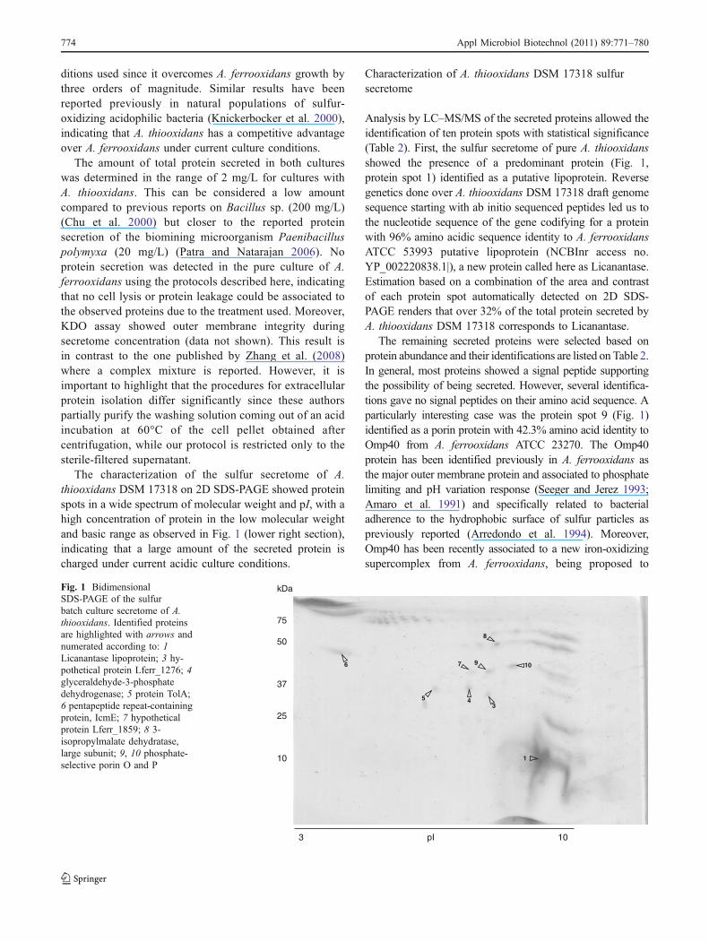
The characterization of the sulfur secretome of A.thiooxidans DSM 17318 on 2D SDS-PAGE showed proteinspots in a wide spectrum of molecular weight and pI, with ahigh concentration of protein in the low molecular weightand basic range as observed in Fig. 1 (lower right section),indicating that a large amount of the secreted protein ischarged under current acidic culture conditions.
Characterization of A. thiooxidans DSM 17318 sulfursecretome
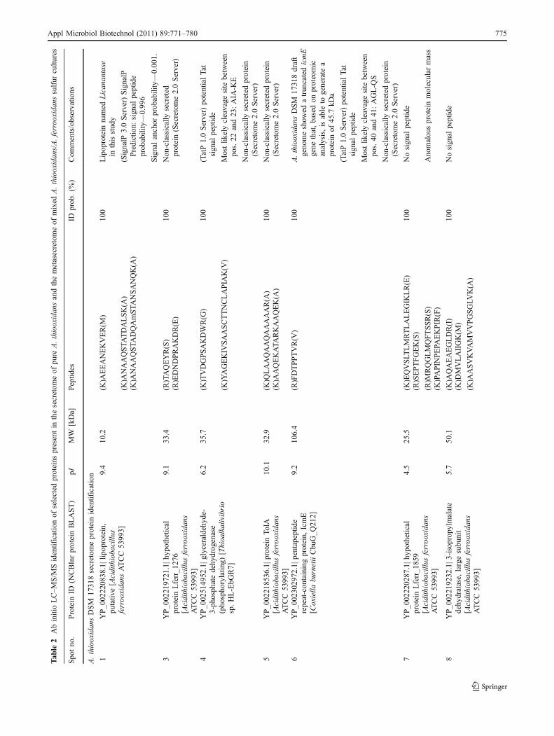
Analysis by LC–MS/MS of the secreted proteins allowed theidentification of ten protein spots with statistical significance(Table 2). First, the sulfur secretome of pure A. thiooxidansshowed the presence of a predominant protein (Fig. 1,protein spot 1) identified as a putative lipoprotein. Reversegenetics done over A. thiooxidans DSM 17318 draft genomesequence starting with ab initio sequenced peptides led us tothe nucleotide sequence of the gene codifying for a proteinwith 96% amino acidic sequence identity to A. ferrooxidansATCC 53993 putative lipoprotein (NCBInr access no.YP_002220838.1|), a new protein called here as Licanantase.Estimation based on a combination of the area and contrastof each protein spot automatically detected on 2D SDS-PAGE renders that over 32% of the total protein secreted byA. thiooxidans DSM 17318 corresponds to Licanantase.
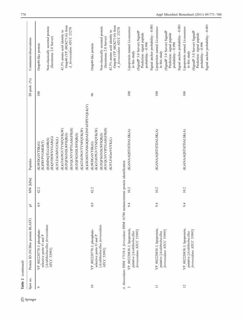
The remaining secreted proteins were selected based onprotein abundance and their identifications are listed on Table 2.In general, most proteins showed a signal peptide supportingthe possibility of being secreted. However, several identifica-tions gave no signal peptides on their amino acid sequence. Aparticularly interesting case was the protein spot 9 (Fig. 1)identified as a porin protein with 42.3% amino acid identity toOmp40 from A. ferrooxidans ATCC 23270. The Omp40protein has been identified previously in A. ferrooxidans asthe major outer membrane protein and associated to phosphatelimiting and pH variation response (Seeger and Jerez 1993;Amaro et al. 1991) and specifically related to bacterialadherence to the hydrophobic surface of sulfur particles aspreviously reported (Arredondo et al. 1994). Moreover,Omp40 has been recently associated to a new iron-oxidizingsupercomplex from A. ferrooxidans, being proposed to
1
3
8
976
5 4
10
3 pI 10
kDa
75
50
37
25
10
Fig. 1 BidimensionalSDS-PAGE of the sulfurbatch culture secretome of A.thiooxidans. Identified proteinsare highlighted with arrows andnumerated according to: 1Licanantase lipoprotein; 3 hy-pothetical protein Lferr_1276; 4glyceraldehyde-3-phosphatedehydrogenase; 5 protein TolA;6 pentapeptide repeat-containingprotein, IcmE; 7 hypotheticalprotein Lferr_1859; 8 3-isopropylmalate dehydratase,large subunit; 9, 10 phosphate-selective porin O and P
774 Appl Microbiol Biotechnol (2011) 89:771–780
Tab
le2
AbinitioLC–M
S/M
Sidentificationof
selected
proteins
presentin
thesecretom
eof
pure
A.thiooxidan
sandthemetasecretomeof
mixed
A.thiooxidan
s/A.ferroo
xida
nssulfur
cultu
res
Spo
tno
.Protein
ID(N
CBInrproteinBLAST)
pIMW
[kDa]
Peptid
esID
prob
.(%
)Com
ments/observatio
ns
A.thiooxidan
sDSM
1731
8secretom
eproteinidentification
1YP_0
0222
0838
.1|lipop
rotein,
putativ
e[Acidithioba
cillu
sferroo
xida
nsATCC53
993]
9.4
10.2
(K)A
EEANEKVER(M
)10
0Lipop
rotein
named
Lican
antase
inthisstud
y
(K)A
NAAQSTA
TDALSK(A
)(SignalP
3.0Server)SignalP
Prediction:
sign
alpeptide
prob
ability—0.99
6(K
)ANAAQSTA
DQAmSTA
NSANQK(A
)
Signalanchor
prob
ability—0.00
1.
3YP_0
0221
9721
.1|h
ypothetical
proteinLferr_1
276
[Acidithioba
cillu
sferroo
xida
nsATCC53
993]
9.1
33.4
(R)TAQEYR(S)
100
Non
-classically
secreted
protein(Secretome2.0Server)
(R)EDNDPRAKDR(E)
4YP_0
0251
4952
.1|g
lyceraldehyd
e-3-ph
osph
atedehy
drog
enase
(pho
spho
rylatin
g)[Thioa
lkalivibrio
sp.HL-EbG
R7]
6.2
35.7
(K)TVDGPSAKDWR(G
)10
0(TatP1.0Server)po
tentialTat
sign
alpeptide
Mostlik
elycleavage
site
between
pos.22
and23
:AIA
-KE
(K)YAGEKIV
SAASCTTNCLAPIA
K(V
)
Non
-classically
secreted
protein
(Secretome2.0Server)
5YP_0
0221
8536
.1|p
rotein
TolA
[Acidithioba
cillu
sferroo
xida
nsATCC53
993]
10.1
32.9
(K)Q
LAAQAAQAAAAAR(A
)10
0Non
-classically
secreted
protein
(Secretome2.0Server)
(K)A
AQEKATA
RKAAQEK(A
)
6YP_0
0230
2972
.1|p
entapeptide
repeat-con
tainingprotein,
IcmE
[Coxiella
burnetiiCbu
G_Q
212]
9.2
106.4
(R)FDTPPTVR(V
)10
0A.thiooxidan
sDSM
1731
8draft
geno
meshow
edatrun
catedicmE
gene
that,basedon
proteomic
analysis,isable
togenerate
aproteinof
45.7
kDa
(TatP1.0Server)po
tentialTat
sign
alpeptide
Mostlik
elycleavage
site
between
pos.40
and41
:AGL-Q
S
Non
-classically
secreted
protein
(Secretome2.0Server)
7YP_0
0222
0287
.1|h
ypothetical
proteinLferr_1
859
[Acidithioba
cillu
sferroo
xida
nsATCC53
993]
4.5
25.5
(K)EQVSLT
LMRTLALEGIK
LR(E)
100
Nosign
alpeptide
(R)SEPTFGEK(S)
(R)M
RQGLMQFTSSR(S)
Ano
malou
sproteinmolecular
mass
(K)PAPIN
PEPA
EKPIR(F)
8YP_0
0221
9232
.1|3
-isoprop
ylmalate
dehydratase,
largesubunit
[Acidithioba
cillu
sferroo
xida
nsATCC53
993]
5.7
50.1
(K)A
QAEAEGLDR(I)
100
Nosign
alpeptide
(K)D
MVLAIIGK(M
)
(K)A
ASVKVAMVVPGSGLV
K(A
)
Appl Microbiol Biotechnol (2011) 89:771–780 775
Tab
le2
(con
tinued)
Spo
tno
.Protein
ID(N
CBInrproteinBLAST)
pIMW
[kDa]
Peptid
esID
prob
.(%
)Com
ments/observatio
ns
9YP_0
0222
0770
.1|p
hosphate-
selectivepo
rinO
andP
[Acidithioba
cillu
sferroo
xida
nsATCC53
993]
4.9
42.2
(K)W
DGAYTSK(G
)10
0Omp4
0-lik
eprotein
(K)FRVPVGMER(T)
(R)SMNQTLGADR(S)
Non
-classically
secreted
protein
(Secretome2.0Server)
(R)Q
VDSWNVGAR(G
)
(K)Y
LFA
GMVGYK(L)
(K)G
TAVPGVTYNFQYK(W
)42
.3%
aminoacid
identityto
Omp4
0(Y
P_0
0242
7116
)from
A.ferroo
xida
nsATCC23
270
(R)TQFSGNDLW
FIQ
R(S)
(R)V
QLNYVIPTGASGFR(H
)
(R)TQFSGNDLW
FIQ
R(S)
(K)G
TAVPGVTYNFQYK(W
)
(K)G
IGSHTGNSGIQ
DAEIN
FAFIPEVQLK(V
)
10YP_0
0222
0770
.1|p
hosphate-
selectivepo
rinO
andP
[Acidithioba
cillu
sferroo
xida
nsATCC53
993]
4.9
42.2
(K)W
DGAYTSK(G
)96
Omp4
0-lik
eprotein
(K)G
TAVPGVTYNFQYK(W
)
(R)TQFSGNDLW
FIQ
R(S)
Non
-classically
secreted
protein
(Secretome2.0Server)
(R)V
QLNYVIPTGASGFR(H
)
42.3%
aminoacid
identityto
Omp4
0(Y
P_0
0242
7116
)from
A.ferroo
xida
nsATCC23
270
(K)Y
LFA
GmVGYK(L)
A.thiooxidan
sDSM
1731
8/A.ferroxidan
sDSM
1678
6metasecretomeproteinidentification
2YP_0
0222
0838
.1|lipop
rotein,
putativ
e[Acidithioba
cillu
sferroo
xida
nsATCC53
993]
9.4
10.2
(K)A
NAAQSTA
TDALSK(A
)10
0Lipop
rotein
named
Lican
antase
inthisstud
y
(SignalP
3.0Server)SignalP
Prediction:
sign
alpeptide
prob
ability—
0.99
6
Signalanchor
prob
ability—0.00
1
11YP_0
0222
0838
.1|lipop
rotein,
putativ
e[Acidithioba
cillu
sferroo
xida
nsATCC53
993]
9.4
10.2
(K)A
NAAQSTA
TDALSK(A
)10
0Lipop
rotein
named
Lican
antase
inthisstud
y
(SignalP
3.0Server)SignalP
Prediction:
sign
alpeptide
prob
ability—
0.99
6
Signalanchor
prob
ability—0.00
1
12YP_0
0222
0838
.1|lipop
rotein,
putativ
e[Acidithioba
cillu
sferroo
xida
nsATCC53
993]
9.4
10.2
(K)A
NAAQSTA
TDALSK(A
)10
0Lipop
rotein
named
Lican
antase
inthisstud
y
(SignalP
3.0Server)SignalP
Prediction:
sign
alpeptide
prob
ability—
0.99
6
Signalanchor
prob
ability—0.00
1
776 Appl Microbiol Biotechnol (2011) 89:771–780
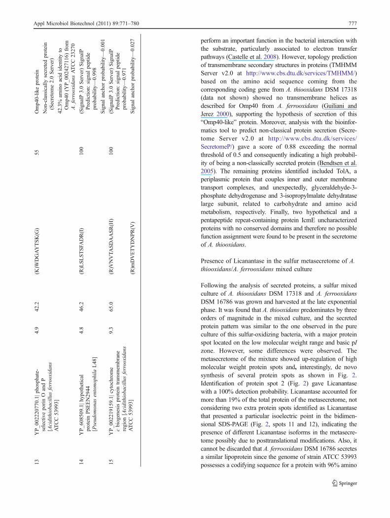
perform an important function in the bacterial interaction withthe substrate, particularly associated to electron transferpathways (Castelle et al. 2008). However, topology predictionof transmembrane secondary structures in proteins (TMHMMServer v2.0 at http://www.cbs.dtu.dk/services/TMHMM/)based on the amino acid sequence coming from thecorresponding coding gene from A. thiooxidans DSM 17318(data not shown) showed no transmembrane helices asdescribed for Omp40 from A. ferrooxidans (Guiliani andJerez 2000), supporting the hypothesis of secretion of this“Omp40-like” protein. Moreover, analysis with the bioinfor-matics tool to predict non-classical protein secretion (Secre-tome Server v2.0 at http://www.cbs.dtu.dk/services/SecretomeP/) gave a score of 0.88 exceeding the normalthreshold of 0.5 and consequently indicating a high probabil-ity of being a non-classically secreted protein (Bendtsen et al.2005). The remaining proteins identified included TolA, aperiplasmic protein that couples inner and outer membranetransport complexes, and unexpectedly, glyceraldehyde-3-phosphate dehydrogenase and 3-isopropylmalate dehydrataselarge subunit, related to carbohydrate and amino acidmetabolism, respectively. Finally, two hypothetical and apentapeptide repeat-containing protein IcmE uncharacterizedproteins with no conserved domains and therefore no possiblefunction assignment were found to be present in the secretomeof A. thiooxidans.
Presence of Licanantase in the sulfur metasecretome of A.thiooxidans/A. ferrooxidans mixed culture
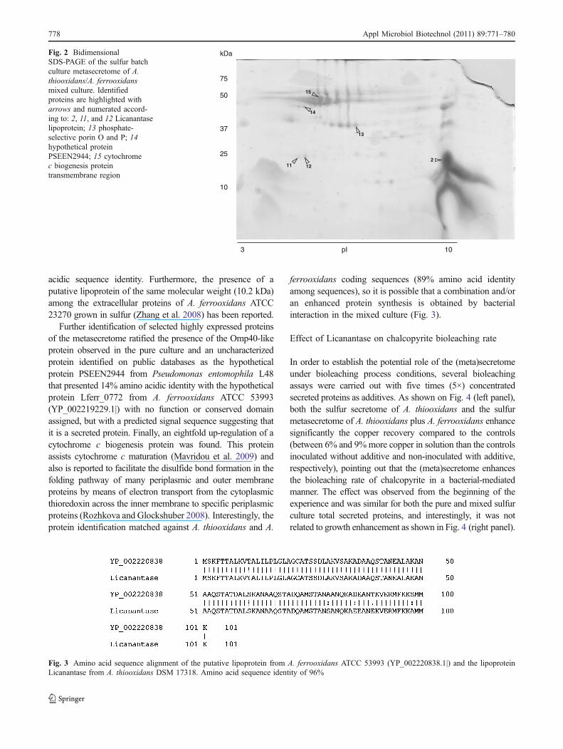
Following the analysis of secreted proteins, a sulfur mixedculture of A. thiooxidans DSM 17318 and A. ferrooxidansDSM 16786 was grown and harvested at the late exponentialphase. It was found that A. thiooxidans predominates by threeorders of magnitude in the mixed culture, and the secretedprotein pattern was similar to the one observed in the pureculture of this sulfur-oxidizing bacteria, with a major proteinspot located on the low molecular weight range and basic pIzone. However, some differences were observed. Themetasecretome of the mixture showed up-regulation of highmolecular weight protein spots and, interestingly, de novosynthesis of several protein spots as shown in Fig. 2.Identification of protein spot 2 (Fig. 2) gave Licanantasewith a 100% detection probability. Licanantase accounted formore than 19% of the total protein of the metasecretome, notconsidering two extra protein spots identified as Licanantasethat presented a particular isoelectric point in the bidimen-sional SDS-PAGE (Fig. 2, spots 11 and 12), indicating thepresence of different Licanantase isoforms in the metasecre-tome possibly due to posttranslational modifications. Also, itcannot be discarded that A. ferrooxidans DSM 16786 secretesa similar lipoprotein since the genome of strain ATCC 53993possesses a codifying sequence for a protein with 96% amino13
YP_0
0222
0770
.1|p
hosphate-
selectivepo
rinO
andP
[Acidithioba
cillu
sferroo
xida
nsATCC53
993]
4.9
42.2
(K)W
DGAYTSK(G
)55
Omp4
0-lik
eprotein
Non
-classically
secreted
protein
(Secretome2.0Server)
42.3%
aminoacid
identityto
Omp4
0(Y
P_0
0242
7116
)from
A.ferroo
xida
nsATCC23
270
14YP_6
0850
9.1|
hypo
thetical
proteinPSEEN29
44[Pseud
omon
asentomop
hila
L48
]
4.8
46.2
(R)LSLSTSFA
DR(I)
100
(SignalP
3.0Server)SignalP
Prediction:
sign
alpeptide
prob
ability—0.99
8
Signalanchor
prob
ability—
0.00
1
15YP_0
0221
9159
.1|cytochrom
ecbiog
enesisproteintransm
embrane
region
[Acidithioba
cillu
sferroo
xida
nsATCC53
993]
9.3
65.0
(R)V
NVTA
SDAASR(H
)10
0(SignalP
3.0Server)SignalP
Prediction:
sign
alpeptide
prob
ability—0.97
1
(R)m
DVETYDNPR(V
)Signalanchor
prob
ability—
0.02
7
Appl Microbiol Biotechnol (2011) 89:771–780 777
acidic sequence identity. Furthermore, the presence of aputative lipoprotein of the same molecular weight (10.2 kDa)among the extracellular proteins of A. ferrooxidans ATCC23270 grown in sulfur (Zhang et al. 2008) has been reported.
Further identification of selected highly expressed proteinsof the metasecretome ratified the presence of the Omp40-likeprotein observed in the pure culture and an uncharacterizedprotein identified on public databases as the hypotheticalprotein PSEEN2944 from Pseudomonas entomophila L48that presented 14% amino acidic identity with the hypotheticalprotein Lferr_0772 from A. ferrooxidans ATCC 53993(YP_002219229.1|) with no function or conserved domainassigned, but with a predicted signal sequence suggesting thatit is a secreted protein. Finally, an eightfold up-regulation of acytochrome c biogenesis protein was found. This proteinassists cytochrome c maturation (Mavridou et al. 2009) andalso is reported to facilitate the disulfide bond formation in thefolding pathway of many periplasmic and outer membraneproteins by means of electron transport from the cytoplasmicthioredoxin across the inner membrane to specific periplasmicproteins (Rozhkova and Glockshuber 2008). Interestingly, theprotein identification matched against A. thiooxidans and A.
ferrooxidans coding sequences (89% amino acid identityamong sequences), so it is possible that a combination and/oran enhanced protein synthesis is obtained by bacterialinteraction in the mixed culture (Fig. 3).
Effect of Licanantase on chalcopyrite bioleaching rate
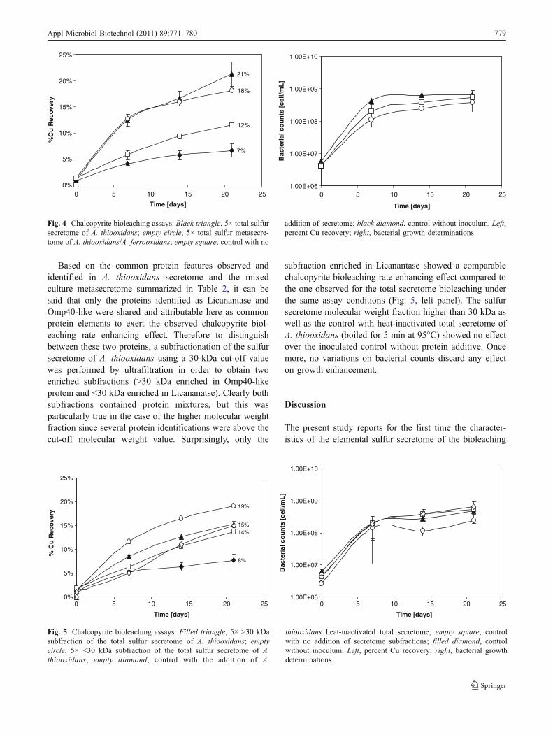
In order to establish the potential role of the (meta)secretomeunder bioleaching process conditions, several bioleachingassays were carried out with five times (5×) concentratedsecreted proteins as additives. As shown on Fig. 4 (left panel),both the sulfur secretome of A. thiooxidans and the sulfurmetasecretome of A. thiooxidans plus A. ferrooxidans enhancesignificantly the copper recovery compared to the controls(between 6% and 9%more copper in solution than the controlsinoculated without additive and non-inoculated with additive,respectively), pointing out that the (meta)secretome enhancesthe bioleaching rate of chalcopyrite in a bacterial-mediatedmanner. The effect was observed from the beginning of theexperience and was similar for both the pure and mixed sulfurculture total secreted proteins, and interestingly, it was notrelated to growth enhancement as shown in Fig. 4 (right panel).
2
13
1211
15
14
3 pI 10
kDa
75
50
37
25
10
Fig. 2 BidimensionalSDS-PAGE of the sulfur batchculture metasecretome of A.thiooxidans/A. ferrooxidansmixed culture. Identifiedproteins are highlighted witharrows and numerated accord-ing to: 2, 11, and 12 Licanantaselipoprotein; 13 phosphate-selective porin O and P; 14hypothetical proteinPSEEN2944; 15 cytochromec biogenesis proteintransmembrane region
Fig. 3 Amino acid sequence alignment of the putative lipoprotein from A. ferrooxidans ATCC 53993 (YP_002220838.1|) and the lipoproteinLicanantase from A. thiooxidans DSM 17318. Amino acid sequence identity of 96%
778 Appl Microbiol Biotechnol (2011) 89:771–780
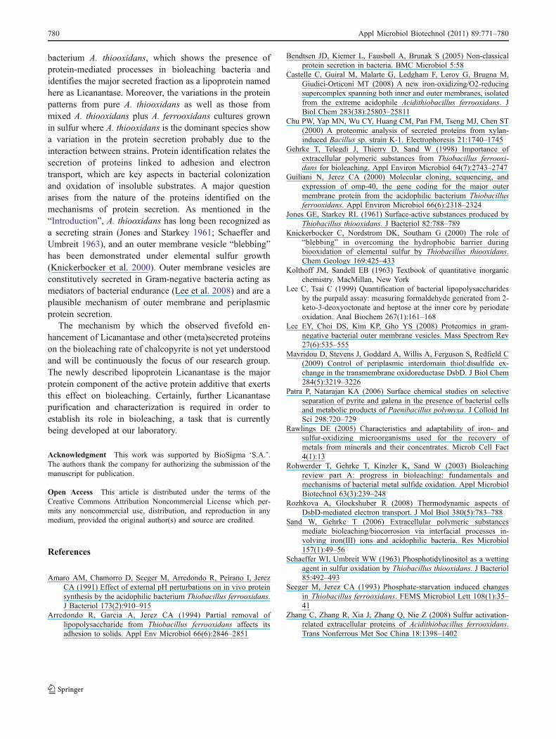
Based on the common protein features observed andidentified in A. thiooxidans secretome and the mixedculture metasecretome summarized in Table 2, it can besaid that only the proteins identified as Licanantase andOmp40-like were shared and attributable here as commonprotein elements to exert the observed chalcopyrite biol-eaching rate enhancing effect. Therefore to distinguishbetween these two proteins, a subfractionation of the sulfursecretome of A. thiooxidans using a 30-kDa cut-off valuewas performed by ultrafiltration in order to obtain twoenriched subfractions (>30 kDa enriched in Omp40-likeprotein and <30 kDa enriched in Licananatse). Clearly bothsubfractions contained protein mixtures, but this wasparticularly true in the case of the higher molecular weightfraction since several protein identifications were above thecut-off molecular weight value. Surprisingly, only the
subfraction enriched in Licanantase showed a comparablechalcopyrite bioleaching rate enhancing effect compared tothe one observed for the total secretome bioleaching underthe same assay conditions (Fig. 5, left panel). The sulfursecretome molecular weight fraction higher than 30 kDa aswell as the control with heat-inactivated total secretome ofA. thiooxidans (boiled for 5 min at 95°C) showed no effectover the inoculated control without protein additive. Oncemore, no variations on bacterial counts discard any effecton growth enhancement.
Discussion
The present study reports for the first time the character-istics of the elemental sulfur secretome of the bioleaching
21%
18%
7%
12%
0%
5%
10%
15%
20%
25%
0 5 10 15 20 25
Time [days]
%C
u R
eco
very
1.00E+06
1.00E+07
1.00E+08
1.00E+09
1.00E+10
0 5 10 15 20 25
Time [days]
Bac
teri
al c
ou
nts
[ce
ll/m
L]
Fig. 4 Chalcopyrite bioleaching assays. Black triangle, 5× total sulfursecretome of A. thiooxidans; empty circle, 5× total sulfur metasecre-tome of A. thiooxidans/A. ferrooxidans; empty square, control with no
addition of secretome; black diamond, control without inoculum. Left,percent Cu recovery; right, bacterial growth determinations
15%
19%
8%
14%
0%
5%
10%
15%
20%
25%
0 5 10 15 20 25
Time [days]
% C
u R
eco
very
1.00E+06
1.00E+07
1.00E+08
1.00E+09
1.00E+10
0 5 10 15 20 25
Time [days]
Bac
teri
al c
ou
nts
[ce
ll/m
L]
Fig. 5 Chalcopyrite bioleaching assays. Filled triangle, 5× >30 kDasubfraction of the total sulfur secretome of A. thiooxidans; emptycircle, 5× <30 kDa subfraction of the total sulfur secretome of A.thiooxidans; empty diamond, control with the addition of A.
thiooxidans heat-inactivated total secretome; empty square, controlwith no addition of secretome subfractions; filled diamond, controlwithout inoculum. Left, percent Cu recovery; right, bacterial growthdeterminations
Appl Microbiol Biotechnol (2011) 89:771–780 779
bacterium A. thiooxidans, which shows the presence ofprotein-mediated processes in bioleaching bacteria andidentifies the major secreted fraction as a lipoprotein namedhere as Licanantase. Moreover, the variations in the proteinpatterns from pure A. thiooxidans as well as those frommixed A. thiooxidans plus A. ferrooxidans cultures grownin sulfur where A. thiooxidans is the dominant species showa variation in the protein secretion probably due to theinteraction between strains. Protein identification relates thesecretion of proteins linked to adhesion and electrontransport, which are key aspects in bacterial colonizationand oxidation of insoluble substrates. A major questionarises from the nature of the proteins identified on themechanisms of protein secretion. As mentioned in the“Introduction”, A. thiooxidans has long been recognized asa secreting strain (Jones and Starkey 1961; Schaeffer andUmbreit 1963), and an outer membrane vesicle “blebbing”has been demonstrated under elemental sulfur growth(Knickerbocker et al. 2000). Outer membrane vesicles areconstitutively secreted in Gram-negative bacteria acting asmediators of bacterial endurance (Lee et al. 2008) and are aplausible mechanism of outer membrane and periplasmicprotein secretion.
The mechanism by which the observed fivefold en-hancement of Licanantase and other (meta)secreted proteinson the bioleaching rate of chalcopyrite is not yet understoodand will be continuously the focus of our research group.The newly described lipoprotein Licanantase is the majorprotein component of the active protein additive that exertsthis effect on bioleaching. Certainly, further Licanantasepurification and characterization is required in order toestablish its role in bioleaching, a task that is currentlybeing developed at our laboratory.
Acknowledgment This work was supported by BioSigma ‘S.A.’.The authors thank the company for authorizing the submission of themanuscript for publication.
Open Access This article is distributed under the terms of theCreative Commons Attribution Noncommercial License which per-mits any noncommercial use, distribution, and reproduction in anymedium, provided the original author(s) and source are credited.
References
Amaro AM, Chamorro D, Seeger M, Arredondo R, Peirano I, JerezCA (1991) Effect of external pH perturbations on in vivo proteinsynthesis by the acidophilic bacterium Thiobacillus ferrooxidans.J Bacteriol 173(2):910–915
Arredondo R, Garcia A, Jerez CA (1994) Partial removal oflipopolysaccharide from Thiobacillus ferrooxidans affects itsadhesion to solids. Appl Env Microbiol 66(6):2846–2851
Bendtsen JD, Kiemer L, Fausbøll A, Brunak S (2005) Non-classicalprotein secretion in bacteria. BMC Microbiol 5:58
Castelle C, Guiral M, Malarte G, Ledgham F, Leroy G, Brugna M,Giudici-Orticoni MT (2008) A new iron-oxidizing/O2-reducingsupercomplex spanning both inner and outer membranes, isolatedfrom the extreme acidophile Acidithiobacillus ferrooxidans. JBiol Chem 283(38):25803–25811
Chu PW, Yap MN, Wu CY, Huang CM, Pan FM, Tseng MJ, Chen ST(2000) A proteomic analysis of secreted proteins from xylan-induced Bacillus sp. strain K-1. Electrophoresis 21:1740–1745
Gehrke T, Telegdi J, Thierry D, Sand W (1998) Importance ofextracellular polymeric substances from Thiobacillus ferrooxi-dans for bioleaching. Appl Environ Microbiol 64(7):2743–2747
Guiliani N, Jerez CA (2000) Molecular cloning, sequencing, andexpression of omp-40, the gene coding for the major outermembrane protein from the acidophilic bacterium Thiobacillusferrooxidans. Appl Environ Microbiol 66(6):2318–2324
Jones GE, Starkey RL (1961) Surface-active substances produced byThiobacillus thiooxidans. J Bacteriol 82:788–789
Knickerbocker C, Nordstrom DK, Southam G (2000) The role of“blebbing” in overcoming the hydrophobic barrier duringbiooxidation of elemental sulfur by Thiobacillus thiooxidans.Chem Geology 169:425–433
Kolthoff JM, Sandell EB (1963) Textbook of quantitative inorganicchemistry. MacMillan, New York
Lee C, Tsai C (1999) Quantification of bacterial lipopolysaccharidesby the purpald assay: measuring formaldehyde generated from 2-keto-3-deoxyoctonate and heptose at the inner core by periodateoxidation. Anal Biochem 267(1):161–168
Lee EY, Choi DS, Kim KP, Gho YS (2008) Proteomics in gram-negative bacterial outer membrane vesicles. Mass Spectrom Rev27(6):535–555
Mavridou D, Stevens J, Goddard A, Willis A, Ferguson S, Redfield C(2009) Control of periplasmic interdomain thiol:disulfide ex-change in the transmembrane oxidoreductase DsbD. J Biol Chem284(5):3219–3226
Patra P, Natarajan KA (2006) Surface chemical studies on selectiveseparation of pyrite and galena in the presence of bacterial cellsand metabolic products of Paenibacillus polymyxa. J Colloid IntSci 298:720–729
Rawlings DE (2005) Characteristics and adaptability of iron- andsulfur-oxidizing microorganisms used for the recovery ofmetals from minerals and their concentrates. Microb Cell Fact4(1):13
Rohwerder T, Gehrke T, Kinzler K, Sand W (2003) Bioleachingreview part A: progress in bioleaching: fundamentals andmechanisms of bacterial metal sulfide oxidation. Appl MicrobiolBiotechnol 63(3):239–248
Rozhkova A, Glockshuber R (2008) Thermodynamic aspects ofDsbD-mediated electron transport. J Mol Biol 380(5):783–788
Sand W, Gehrke T (2006) Extracellular polymeric substancesmediate bioleaching/biocorrosion via interfacial processes in-volving iron(III) ions and acidophilic bacteria. Res Microbiol157(1):49–56
Schaeffer WI, Umbreit WW (1963) Phosphotidylinositol as a wettingagent in sulfur oxidation by Thiobacillus thiooxidans. J Bacteriol85:492–493
Seeger M, Jerez CA (1993) Phosphate-starvation induced changesin Thiobacillus ferrooxidans. FEMS Microbiol Lett 108(1):35–41
Zhang C, Zhang R, Xia J, Zhang Q, Nie Z (2008) Sulfur activation-related extracellular proteins of Acidithiobacillus ferrooxidans.Trans Nonferrous Met Soc China 18:1398–1402
780 Appl Microbiol Biotechnol (2011) 89:771–780


