Embed Size (px)
Citation preview

Cell, Vol. 102, 565–575, September 1, 2000, Copyright 2000 by Cell Press
Activation-Induced Cytidine Deaminase (AID)Deficiency Causes the Autosomal Recessive Formof the Hyper-IgM Syndrome (HIGM2)
HIGM2 patients (and in AID2/2 mice) demonstrates theabsolute requirement for AID in several crucial stepsof B cell terminal differentiation necessary for efficientantibody responses.
Patrick Revy,1 Taro Muto,2 Yves Levy,3
Frederic Geissmann,4 Alessandro Plebani,5
Ozden Sanal,6 Nadia Catalan,1 Monique Forveille,1
Remi Dufourcq-Lagelouse,1 Andrew Gennery,1
Ilhan Tezcan,6 Fugen Ersoy,6 Hulya Kayserili,7
IntroductionAlberto G. Ugazio,5 Nicole Brousse,4
Masamichi Muramatsu,2 Luigi D. Notarangelo,5
Production of highly efficient neutralizing antibodies re-Kazuo Kinoshita,2 Tasuku Honjo,2
quires affinity maturation of antibody responses. ThisAlain Fischer,1,8 and Anne Durandy1
crucial event takes place within lymphoid organs in ger-1 Inserm U429minal centers (GC) and is T cell driven (Liu et al., 1989;Hopital Necker-Enfants MaladesBerek et al., 1991; Kuppers et al., 1993). Four majorParisevents occur, i.e., B cell proliferation, generation of so-Francematic hypermutations in the immunoglobulin (Ig) vari-2 Department of Medical Chemistryable region (V) genes able to increase the affinity ofGraduate School of Medicineantibodies for antigens, positive selection of B cells
Kyoto Universitybearing a B cell receptor (BCR) of high affinity for anti-
Japan gen, and Ig class switch recombination (CSR) toward3 Inserm U474 the production of IgG, IgA, and IgE (Rajewsky, 1996).Hopital Cochin The molecular mechanisms involved in these events areParis not precisely delineated.France A cognate interaction between CD40-ligand (CD40L4 Service d’Anatomo-Pathologie or CD154) expressed on activated helper T cells andHopital Necker-Enfants Malades CD40 constitutively expressed on B cells is requiredParis for B cell terminal maturation. Cross-linking of CD40France molecules leads to B cell proliferation, rescue of B cells
from apoptosis, somatic hypermutations of Ig V genes,5 Clinica Pediatrica and Istituto di Medicinaand CSR (Clark and Ledbetter, 1986; Rousset et al.,Molecolare “Angelo Nocivelli”1991; Lederman et al., 1992; Spriggs et al., 1992; ZanUniversita di Bresciaet al., 1999). This activation pathway requires adaptorItalyproteins, such as the members of the TRAF family (Hu6 Immunology Divisionet al., 1994; Cheng et al., 1995; Sato et al., 1995; IshidaHacettepe Universityet al., 1996a, 1996b) or BLNK (Jumaa et al., 1999; Hay-Ihsan Dogramaci Children’s Hospitalashi et al., 2000) and induces phosphorylation of tyrosineAnkarakinases (Ren et al., 1994) and activation of transcriptionTurkeyfactors, such as NF-kB (Berberich et al., 1994). However,7 Institute of Child Healthnone of these molecules have been shown to be directly
University of Istanbulinvolved in the generation of somatic mutations of Ig V
Turkey genes or CSR. Recently, the Activation Induced cytidine8 Unite d’Immunologie-Hematologie Deaminase (AID) protein has been identified in theHopital Necker-Enfants Malades mouse (Muramatsu et al., 1999). AID expression isParis strictly restricted to germinal center B cells and B cellsFrance induced to switch in vitro in the presence of LPS or
soluble CD40L and IL-4, suggesting a role for AID interminal B cell differentiation. AID is a novel member
Summary of the RNA-editing deaminase family, which containsproteins able to create new functional products frommRNA by base substitution (Scott, 1995; Simpson et al.,The activation-induced cytidine deaminase (AID) gene,1995). An increasing number of human genes are knownspecifically expressed in germinal center B cells into be edited. The most widely studied is the mRNAmice, is a member of the cytidine deaminase family.editing of apolipoprotein B, induced by the cytidine de-We herein report mutations in the human counterpartaminase APOBEC-1 (Navaratnam et al., 1993; Teng etof AID in patients with the autosomal recessive formal., 1993) which is homologous to AID.of hyper-IgM syndrome (HIGM2). Three major abnor-
A rare human immunodeficiency, the Hyper-IgM syn-malities characterize AID deficiency: (1) the absencedrome (HIGM), is characterized by normal or elevatedof immunoglobulin class switch recombination, (2) theserum IgM levels with absence of IgG, IgA, and IgE,
lack of immunoglobulin somatic hypermutations, and resulting in a profound susceptibility to bacterial infec-(3) lymph node hyperplasia caused by the presence of tions (Notarangelo et al., 1992). The molecular basis ofgiant germinal centers. The phenotype observed in the X-linked form (HIGM1) is due to mutations in the
gene coding for CD40L (Allen et al., 1993; Aruffo et al.,1993; Di Santo et al., 1993; Fuleihan et al., 1993; Kor-9 To whom correspondence should be addressed (e-mail: durandy@
necker.fr). thauer et al., 1993). Besides the Ig switch defect, these

Cell566
patients exhibit a remarkable defect in germinal center sequencing was normal and CD40L was normally ex-pressed on activated T cells. CD40 gene sequence and/formation (Facchetti et al., 1995). These observations
emphasize the role of CD40L/CD40 interaction in the or membrane expression revealed no abnormality. Pe-ripheral blood B cell (CD191) counts were normal. All Bterminal differentiation of B cells in secondary lymphoid
organs. In these patients, B cells are intrinsically normal cells expressed sIgM and sIgD while some CD191 Bcells from age-matched controls did not express eithersince they can be normally triggered by CD40 agonists
to CSR in vitro (Allen et al., 1993; Aruffo et al., 1993; sIgM or sIgD (data not shown). CD27, a recently identi-fied marker of memory B cells (Klein et al., 1998) wasDurandy et al., 1993; Korthauer et al., 1993).
Another HIGM syndrome with autosomal recessive expressed on a normal proportion (from 9% to 45%)and with the same intensity on B cells, as compared withinheritance (HIGM2) has been described (Callard et al.,
1994; Conley et al., 1994). In these patients, CD40L gene age-matched controls (Table 2 and data not shown).In vitro activation of peripheral blood mononuclearsequencing and membrane molecule expression are
normal and, in contrast to CD40L-deficient patients, B cells (PBMC) by soluble CD40L (sCD40L) and IL-4, whichregularly induced IgE production by B cells from controlscells do not undergo CSR in vitro in the presence of
CD40-agonists. CD40 gene sequence and CD40 mole- and CD40L-deficient patients (Durandy et al., 1993), wasineffective in the 15 tested HIGM2 patients (Table 2).cule expression are normal in these patients (Durandy
et al., 1997). A defect of other molecules involved in However, in the same culture conditions, Ie-Ce steriletranscripts were inducible (Figure 3C). T cell (CD31) andCD40 signaling such as the TRAF proteins (Hu et al.,
1994; Cheng et al., 1995; Sato et al., 1995; Ishida et al., T cell subset numbers (CD41 and CD81) as well as invitro T cell proliferation to mitogens and antigens was1996a, 1996b) or proteins involved in the activation of
transcription factor NF-kB such as TANK (Cheng and normal (data not shown).Baltimore., 1996; Rothe et al., 1996) or NIK (Malinin etal., 1997; Song et al., 1997) have also been excluded Identification of the HIGM2 Genetic Locus(unpublished data). In addition, activation by CD40 ago- Genetic mapping was investigated by studying the seg-nists of monocytes and dendritic cells from these pa- regation of polymorphic microsatellite markers in 10tients is consistently detectable (Revy et al., 1998), families (8 consanguineous) . The genome was randomlystrongly suggesting that the intrinsic defect in HIGM2 screened by using 280 polymorphic markers. We per-patients originates in B cells, either in a B cell–specific formed homozygosity mapping in the eight consanguin-CD40 triggered event, or in the CSR mechanism itself. eous families and performed a bipoint LOD score analy-
In order to identify the genetic basis of the HIGM2 sis in all affected families. Disease segregation wassyndrome, we performed a genome-wide search for compatible with the D12S372 marker (telomeric regionsusceptibility loci using polymorphic microsatellite of short arm of chromosome 12) in the studied families.markers in consanguineous families. We demonstrated Further analysis with additional markers was performeda strong linkage to chromosome 12p13. As the human and two additional multiplex nonconsanguineous fami-AID (huAID) gene, a possible candidate gene, also maps lies were included. Multipoint analysis with those addi-to 12p13 (Muto et al., 2000), we investigated whether tional markers gave a maximum LOD score of 10.45 forhuman AID gene defects could cause the HIGM2 syn- marker D12S1695 (u 5 0.00). Recombination analysisdrome. We describe 10 independent mutations in the defined the critical genetic interval on 12p13, as no re-huAID gene in 18 patients with HIGM2 from 12 families. combination was found in all tested families betweenWe demonstrate that the HIGM2 syndrome is character- markers D12S397 and D12S1697, encompassing a 4.5ized by defective CSR, defective generation of somatic cM region. Among a YAC contig covering the delineatedmutations of the Ig V genes, and by abnormal germinal region, the 943a9 YAC, which contains both D12S1695centers, showing that AID gene expression plays a key and D12S336 markers, was shown to harbor exon 2 ofrole in terminal B cell differentiation. huAID (Figure 2A).
Results Sequencing of huAID Gene in HIGM2 PatientsBecause the expression of mouse AID is strictly re-stricted to GC B cells and B cells induced to switch inPatients’ Characteristics
Eighteen patients from 12 unrelated families (Figure 1) vitro, and the huAID gene (Muto et al., 2000) and HIGM2locus colocalize on chromosome 12p13, we studiedwere diagnosed with hyper-IgM syndrome (HIGM) de-
fined by markedly diminished serum levels of IgG and huAID as a possible candidate gene. The coding se-quence of huAID gene, including the 5 exons and theIgA with a normal or increased serum level of IgM (Table
1). All patients presented in childhood with recurrent adjacent intronic regions, was sequenced in 18 patients.Ten independent mutations were found (Figures 2B andbacterial sino-respiratory and gastro-intestinal tract in-
fections (Table 1). A characteristic feature observed in 2C). Five homozygous missense mutations, causing dif-ferent amino acid substitutions, were identified in 7 con-most of the patients (13/18) was lymphoid hyperplasia,
leading to tonsillectomy in 5 of them. No opportunistic sanguineous families. The same mutation was observedin exon 2 in patients P7, P11-P12, and P18 from 3 unre-infections occurred, in contrast to those patients with
HIGM1 (Levy et al., 1997). Anti-tetanus IgG antibodies lated families of Turkish origin. Segregation of sur-rounding polymorphic markers clearly indicate that twowere not found despite immunization (data not shown),
while IgM isohemagglutinins, when evaluable, were de- of 3 families (families 4 and 7) have received the samealleles, suggesting a common origin of the mutation. Intected (Table 1). From diagnosis, all patients were
treated with intravenous Ig substitution. As shown in three other Turkish families, three different point muta-tions were found causing amino acid substitutions inFigure 1, both males (n 5 13) and females (n 5 5) were
affected. Family consanguinity was frequent (9 out of exon 3 (patients P9-P10 and P16) or exon 4 (P14-P15).The missense mutation observed in P16 was located12), suggesting an autosomal recessive inheritance pat-
tern. HIGM1 was excluded in all, since CD40L gene within the cytidine deaminase catalytic region in exon

AID Gene Defects Cause HIGM2 Syndrome567
Figure 1. Pedigree of the HIGM2 Families
The figure depicts the pedigrees of the 12 families included in the study (9 consanguineous).
3 (Figures 2B–2D). A homozygous 19 bp deletion was substitution was detected leading to a stop codon inthe cytidine deaminase catalytic region in exon 3. Theobserved in patient P17 at the beginning of exon 2,
causing a frameshift and a premature stop codon. In the mutation was inherited from the father, who was hetero-zygous, but was not detected in the mother. This obser-unrelated patients P6 and P8, an identical homozygous
base change leads to a stop codon in exon 4. In all these vation suggested a genomic deletion spanning exon 3in the mother, confirmed by Southern blot analysis infamilies, parents were heterozygous for the mutation. In
family 1 (P1-P2-P3), a single homozygous nucleotide the mother and affected children (data not shown). The
Table 1. Patients’ Characteristics
Ig levels (g/l) IgM Antibody productionAge atdiagnosis Recurrent Lymphoid Hemaglutinins (titer 3 1021)
Family Origin Patients (year) infections hyperplasia IgM IgG IgA Anti-A Anti-B
1 Morocco P1 6 1 1 4.5 ,0.06 ,0.07 4 4P2 5 1/2 1 1 ,0.06 ,0.07 32 16P3 1 1 1 1 ,0.06 ,0.07 NE 4
2 Morocco P4 5 1 1 2.4 0.4 ,0.07 NE 64P5 1 1/2 2 1.5 ,0.06 ,0.07 NE 64
3 Italy P6 6 1 2 8 ,0.06 ,0.07 16 84 Turkey P7 2 1 2 1.6 0.5 ,0.07 ND ND5 Italy P8 2 1 1 9 0.3 ,0.07 32 46 Turkey P9 12 1 1 10 0.5 ,0.02 16 16
P10 5 1 1 11 0.1 ,0.02 NE 27 Turkey P11 13 1 1 7 ,0.06 ,0.02 512 6
P12 13 1 1 17 ,0.05 ,0.02 NE NE8 Turkey P13 8 1 1 34 0.05 ,0.02 128 5129 Turkey P14 3 1 1 9 0.7 0.1 1,024 512
P15 8 1 1 11 ,0.02 ,0.02 NE 12810 Turkey P16 1 1 2 10 1.3 0.2 NE NE11 Turkey P17 12 1 1 37 ,0.02 ,0.02 32 3212 Turkey P18 2 1 2 5.6 ,0.02 ,0.02 ND ND
Age-matched 1–5 0.5–1.1 5–12 0.3–1.3 4–16 4–16controls 5–13 0.6–1.7 7–15 0.8–2.3 16–128 16–128
ND 5 Not done.NE 5 Not evaluable (because of blood group).
Cell568
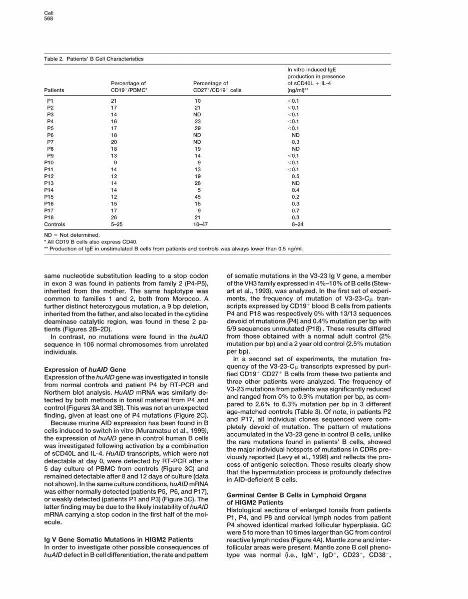
Table 2. Patients’ B Cell Characteristics
In vitro induced IgEproduction in presence
Percentage of Percentage of of sCD40L 1 IL-4Patients CD191/PBMC* CD271/CD191 cells (ng/ml)**
P1 21 10 ,0.1P2 17 21 ,0.1P3 14 ND ,0.1P4 16 23 ,0.1P5 17 29 ,0.1P6 18 ND NDP7 20 ND 0.3P8 18 19 NDP9 13 14 ,0.1
P10 9 9 ,0.1P11 14 13 ,0.1P12 12 19 0.5P13 14 28 NDP14 14 5 0.4P15 12 45 0.2P16 15 15 0.3P17 17 9 0.7P18 26 21 0.3Controls 5–25 10–47 8–24
ND 5 Not determined.* All CD19 B cells also express CD40.** Production of IgE in unstimulated B cells from patients and controls was always lower than 0.5 ng/ml.
same nucleotide substitution leading to a stop codon of somatic mutations in the V3-23 Ig V gene, a memberof the VH3 family expressed in 4%–10% of B cells (Stew-in exon 3 was found in patients from family 2 (P4-P5),art et al., 1993), was analyzed. In the first set of experi-inherited from the mother. The same haplotype wasments, the frequency of mutation of V3-23-Cm tran-common to families 1 and 2, both from Morocco. Ascripts expressed by CD191 blood B cells from patientsfurther distinct heterozygous mutation, a 9 bp deletion,P4 and P18 was respectively 0% with 13/13 sequencesinherited from the father, and also located in the cytidinedevoid of mutations (P4) and 0.4% mutation per bp withdeaminase catalytic region, was found in these 2 pa-5/9 sequences unmutated (P18) . These results differedtients (Figures 2B–2D).from those obtained with a normal adult control (2%In contrast, no mutations were found in the huAIDmutation per bp) and a 2 year old control (2.5% mutationsequence in 106 normal chromosomes from unrelatedper bp).individuals.
In a second set of experiments, the mutation fre-quency of the V3-23-Cm transcripts expressed by puri-Expression of huAID Genefied CD191 CD271 B cells from these two patients andExpression of the huAID gene was investigated in tonsilsthree other patients were analyzed. The frequency offrom normal controls and patient P4 by RT-PCR andV3-23 mutations from patients was significantly reducedNorthern blot analysis. HuAID mRNA was similarly de-and ranged from 0% to 0.9% mutation per bp, as com-tected by both methods in tonsil material from P4 andpared to 2.6% to 6.3% mutation per bp in 3 differentcontrol (Figures 3A and 3B). This was not an unexpectedage-matched controls (Table 3). Of note, in patients P2finding, given at least one of P4 mutations (Figure 2C).and P17, all individual clones sequenced were com-Because murine AID expression has been found in Bpletely devoid of mutation. The pattern of mutations
cells induced to switch in vitro (Muramatsu et al., 1999), accumulated in the V3-23 gene in control B cells, unlikethe expression of huAID gene in control human B cells the rare mutations found in patients’ B cells, showedwas investigated following activation by a combination the major individual hotspots of mutations in CDRs pre-of sCD40L and IL-4. HuAID transcripts, which were not viously reported (Levy et al., 1998) and reflects the pro-detectable at day 0, were detected by RT-PCR after a cess of antigenic selection. These results clearly show5 day culture of PBMC from controls (Figure 3C) and that the hypermutation process is profoundly defectiveremained detectable after 8 and 12 days of culture (data in AID-deficient B cells.not shown). In the same culture conditions, huAID mRNAwas either normally detected (patients P5, P6, and P17), Germinal Center B Cells in Lymphoid Organsor weakly detected (patients P1 and P3) (Figure 3C). The of HIGM2 Patientslatter finding may be due to the likely instability of huAID Histological sections of enlarged tonsils from patientsmRNA carrying a stop codon in the first half of the mol- P1, P4, and P8 and cervical lymph nodes from patientecule. P4 showed identical marked follicular hyperplasia. GC
were 5 to more than 10 times larger than GC from controlIg V Gene Somatic Mutations in HIGM2 Patients reactive lymph nodes (Figure 4A). Mantle zone and inter-In order to investigate other possible consequences of follicular areas were present. Mantle zone B cell pheno-
type was normal (i.e., IgM1, IgD1, CD231, CD382,huAID defect in B cell differentiation, the rate and pattern

AID Gene Defects Cause HIGM2 Syndrome569
Figure 2. Localization and Genetic Analysis of huAID Mutations in HIGM2 Patients
(A) Multipoint LOD score analysis. The telomeric end of chromosome 12 is located on the left. A maximum LOD score of 10.45 was obtainedbetween polymorphic markers D12S397 and D12S89. Among the YAC contig, the 943a9 YAC was shown to harbor markers D12S336 andD12S1695 and huAID exon 2.(B) Localization of mutations in the huAID gene. Nine different mutations were found in the coding sequence (red) in 18 patients tested.Mutations in patients P1-P2-P3, P4-P5, and P16 were localized in the cytidine deaminase catalytic region. In patients P1-P2-P3, a furtherheterozygous deletion was detected by Southern blot (not shown).(C) Mutations in the huAID gene. In 10 out of 12 families, the mutations were homozygous in patients (**), and in two families, heterozygousmutations were found (*). Among these mutations, 7 were nucleotide substitutions causing missense mutations in 5 cases, or a stop codon(X) in 2 others. Two other changes were nucleotide deletions leading to one missense mutation and one stop codon(D) Alignment of the human and mouse amino acid sequences. The conserved cytidine deaminase motif is indicated by an open box. Onlynonidentical residues are indicated for mouse.

Cell570
Table 3. Mutations in V3-23-Cm Transcript Sequences fromCD191CD271 Purified B Cells
Clones Mutations
All Unmutated* Total Per bp, %
2 yr old control 10 0 172 5.93.5 yr old control 7 2 52 2.611 yr old control 10 1 182 6.3
P2 9 9 0 0P4 8 7 3 0.1P8 10 9 22 0.76
P17 12 12 0 0P18 9 3 24 0.9
* Unmutated 5 #1 mutation.
Bcl-21, and Ki672) (Figure 4A, arrows, and data notshown). Giant GC contained a normal follicular dendriticcell network (DRC1, data not shown), and PNA1, CD381,CD231, CD831, CD951, CD401, IgM1, Bcl22, and Ki671
B cells (Figures 4A and 4B and data not shown). Strik-ingly, numerous GC B cells coexpressed sIgD (Figures4A and 4B), contrasting with observations in normalreactive GC in which GC IgD1 cells are scarcely found(Figure 4A). The high proliferation frequency of B cellsin GC was associated with a dense network of macro-phages filled with apoptotic bodies (Figure 4B, openarrows) that gave the GC a starry sky appearance. Adark zone and a lighter zone could be distinguished insome patients’ follicles on Ki67 staining (data notshown). However this “light zone” also contained numer-ous cycling cells and sIgD1 cells. The phenotype andsize of the GC B cells identify them as proliferating(Ki671) germinal founder cells (CD381, sIgM1, andsIgD1) as described by Lebecque et al. (1997). SIgD1
and sIgM1 cells were also numerous in T cell areas(Figure 4A). Occasional CD271 CD32 cells as well asIgM and IgD plasma cells were found in GC and T cellareas. Neither IgG nor IgA plasma cells were observed(data not shown).
Discussion
HuAID Gene Mutations Cause HIGM2 SyndromeIn this study, compelling evidence is provided that muta-tions in the coding sequence of the huAID gene are
Figure 3. Expression of huAID Transcriptsresponsible for the HIGM2 syndrome. We studied 18
(A) HuAID transcripts were analyzed by RT-PCR in tonsils from a HIGM2 patients from 12 families who fulfilled the diag-control (Ctl) and patient P4 as compared with CD19 transcripts. (2),
nostic criteria of HIGM2. Genetic studies clearly demon-no cDNA.strated a strong linkage between the HIGM2 locus and(B) Northern blot analysis: similar amounts of huAID mRNA werea short genetic region on chromosome 12p13, whichfound in patient and control tonsils, although more CD19 mRNAcontains the human counterpart of mouse AID (Muto etwas present in the patient, in accordance with the increased number
of B cells detected by immunohistology. Loading control is shown al., 2000). Sequencing of the coding sequence of huAIDby BET gel staining (lower panel). revealed deleterious mutations in all 12 families, all along(C) hAID, CD19, and Ie-Ce transcripts were detected by RT-PCR on the gene. Widely scattered point mutations in huAID areRNA isolated from PBMC before (d0) in control (Ctl), or after 5 day all defective, indicating the strong structural constraintstimulation (d5) by sCD40L and IL-4 in control (Ctl) and patients. of AID in agreement with its evolutionary conservationThe HuAID RT-PCR product found in patient P6 was smaller due to
between mouse (Muramatsu et al., 1999) and humanan exon “skipping”, leading to a deletion of 35 bp, which includes(Muto et al., 2000). Some of the mutations are predictedthe stop codon detected on the genomic DNA and lets the transcriptto lead to truncated huAID proteins by generating stopout of frame.codons or deletions. Several of the missense mutationsIe-Ce sterile transcripts were normally amplified in patients, although
no IgE production was obtained after 12 days (see also Table 2). were localized within the putative cytidine deaminaseAs a control, CD19 transcripts were similarly amplified. catalytic region of the protein (Muto et al., 2000). Despite
the lack of an accessible experimental model to validatethe effects of the huAID gene mutations, the associationof the HIGM2 phenotype with huAID gene mutationstogether with the similar phenotype observed in AID2/2

AID Gene Defects Cause HIGM2 Syndrome571
Figure 4. Immunohistological Examination of Cervical Lymph Node from Patient P4
(A) Comparison between histology and immunophenotype of P4 cervical lymph node and a control reactive lymph node. On hematoxilin-eosin staining (magnification 325), P4 lymph node shows follicular hyperplasia with giant GC, compared with classical follicular hyperplasia,as control, shown at the same magnification. GC B cells from patient and control express CD38 (blue staining, original magnification 350).Anti-IgD Ab stained numerous cells in interfollicular areas, as well as mantle cells (arrow), and most of the GC B cells from the patient, butnot of control (brown staining, original magnification 3100). Bcl-2 staining indicated that most GC cells did not express Bcl-2, in contrast tothe mantle zone and interfollicular areas, a pattern similar to that observed in control (brown staining, original magnification 3100). Ki67staining reveals that most of the GC B cells from the patient are cycling (brown staining, original magnification 3100). An identical picturewas observed in 2 other patients.(B) Expression of IgM, IgD, Ki67, and Bcl-2 (brown staining) at higher magnification (3400) in a HIGM2 germinal center from P4. On comparablesections, the large majority of GC B cells express IgM and Ki67, numerous GC B cells coexpress IgD, but only occasional small lymphocytes—presumably T cells—express Bcl-2. Note the very numerous macrophages filled with apoptotic bodies (open arrows). An identical picture wasobserved in 2 other patients.
mice (Muramatsu et al., 2000 [this issue of Cell]) demon- machinery (Lorenz et al., 1995; Hein et al., 1998), andthe formation of RNA:DNA complexes with R-loop struc-strates the causative role of AID gene mutations in the
disease. The phenotype observed in this group of pa- ture at the S regions (Daniels and Lieber, 1995; Tracy andLieber, 2000). The role of DNA-PK and the Ku70–Ku80tients was fairly homogeneous and consisted of 3 abnor-
mal features characteristic of the disease, i.e., (1) defec- recombination complex in CSR has also been clearlydemonstrated (Rolink et al., 1996; Casellas et al., 1998;tive CSR, (2) defective Ig variable region gene somatic
mutation generation, and (3) finding of enlarged GC with Manis et al., 1998), whereas RAG molecules are notrequired (Lansford et al., 1998). It is likely that AID playshighly proliferating B cells.a role downstream of the germline transcription sincewe observed normal induction of sterile Ie-Ce transcriptsRole of huAID in CSRby CD40 agonists and IL-4 in HIGM2 patients. In addi-HIGM2 is characterized by absent or very low serumtion, activation-induced de novo protein synthesis isIgG and IgA while B cells are unable to undergo Ig switchrequired to perform CSR (Muramatsu et al., 1999), andin vitro (Callard et al., 1994; Conley et al., 1994; Durandythe existence of a CSR recombinase has been postu-et al., 1997), indicating that AID is involved in CSR. How-lated (Kinoshita and Honjo., 2000). Thus AID could be,ever, the CSR mechanism is complex and not com-if not the CSR recombinase itself, at least an essentialpletely elucidated. CSR requires germline transcriptionpart of the CSR complex machinery (Muramatsu et al.,(Gu et al., 1993; Jung et al., 1993; Xu et al., 1993; Zhang
et al., 1993; Bottaro et al., 1994), an efficient splicing 1999).

Cell572
Role of huAID in Ig Variable Region Gene 1997). One possible explanation for our observation isthat, in absence of functional AID, B cells are continu-Somatic Hypermutations
Peripheral B cells from HIGM2 patients, though they ously triggered to proliferate by antigen (as they nor-mally express the BCR), as long as no successful Ignormally express CD27, carry very rare Ig V gene so-
matic mutations compared with normal controls, based variable region gene somatic mutations have occurred.on the study of V3-23 genes in 5 cases showing that AIDis also involved in the generation of Ig V gene somatic Concluding Remarksmutations. The precise mechanism responsible for so- The observed consequences of AID deficiency in humansmatic mutations is not well understood. The Ig genes together with the phenotype described in the AID2/2
have to be transcriptionally active to undergo somatic mouse (Muramatsu et al., 2000 [this issue of Cell])mutations (Peters and Storb, 1996; Fukita et al., 1998). clearly demonstrate a crucial role of AID in Ig switch, IgIt has been suggested that the DNA mismatch repair variable region gene somatic mutation generation, andsystem, as well as a newly described DNA polymerase normal germinal center formation. HuAID function, al-(Pol m) are involved in somatic mutation generation (Cas- though unidentified, appears to be a key element con-calho et al., 1998; Wiesendanger et al., 1998; Dominguez trolling the steps required for terminal B cell differentia-et al., 2000). tion. These data will enable further molecular delineation
The role of AID in the generation of somatic hypermu- of the mechanisms required for B cell terminal matura-tations is also unknown. Since CSR and somatic muta- tion and efficient antibody responses.tion processes can occur independently (Kaartinen etal., 1983; Jacob and Kelsoe, 1992; Liu et al., 1996), it Experimental Proceduresis unlikely that defective somatic mutation generation
Immunological Study of B Cellsprevents subsequent CSR in HIGM2 patients. HuAIDPeripheral blood mononuclear cells (PBMC) were isolated by Ficoll-could rather act, as an RNA-editing enzyme, on one orHypaque density centrifugation. Immunofluorescent studies wereseveral templates generating yet unidentified factor(s)performed by using specific antibodies: FITC or PE anti-CD19 mono-involved in CSR and/or somatic mutation generationclonal antibodies (mAb), PE-anti-CD27 mAb from Immunotech (Mar-
(Muramatsu et al., 2000). seille, France), FITC-anti-CD40 mAb from Diaclone (Besancon,A residual level of CSR was found in a few patients France), PE-goat anti-hu IgM Ab, and FITC-goat anti-hu IgD Ab from
(for instance, patients P7, P14, and P16). Several hypoth- Caltag, (San Francisco, CA). Samples were analyzed on a FACScaneses can account for this finding, i.e., residual AID activ- (Becton Dickinson, Mountain View, CA) (Durandy et al., 1990). In
vitro IgE switch was assessed in cultures of PBMC stimulated withity associated with missense mutations or a role for AID-soluble CD40L, 1 mg/ml (Morris et al., 1999) (a kind gift from Im-independent factor(s) as suggested in family 9 (patientsmunex, Seattle, WA) and IL-4 (100 U/ml, R&D system, Minneapolis,P14-P15) since no CSR could be detected in P15. Persis-MN). IgE concentrations were evaluated in supernatants by ELISAtence of maternal B cells due to fetal tolerance might beat day 12 (Durandy et al., 1997).one possible factor. Interestingly, detection of residual
CSR did not correlate with occurrence of somatic muta-Linkage Analysis
tions as observed in patient P18. While no serum IgG and Genomic DNA was extracted from peripheral blood leukocytes withIgA were detectable in this case, a low but significant proteinase K, sodium dodecyl sulfate, and a series of phenol-chloro-frequency of somatic mutations was found. Genetic or form extractions. Microsatellite markers were studied using radio-environmental factors may generate some variability as labeled primers from the Genethon collection. The FASTLINK 2.2
package was used for genetic linkage analysis (Cottingham et al.,serum IgG was detectable in patient P7 but not in P18,1993) and the GENEHUNTER 1.1 program was used for multipointwho carries the same AID mutation. However, one canlinkage analysis (Kruglyak et al., 1995). A YAC contig was obtainedsuggest that an AID protein carrying a missense muta-from CEPH, Paris, France (Krauter et al., 1995).tion (R24W) could still be active in one pathway, thus
implying that targets of AID in CSR and somatic hyper-HuAID Gene Sequencingmutation differ.The five exons of huAID were amplified by polymerase chain reaction(PCR) using Thermal Cycler 9700 from Perkin Elmer (Norwalk, CT),specific primers, and Taq High Fidelity (Roche Diagnostic, Mann-Role of huAID in Controlling GC B Cell Proliferationheim, Germany).In sharp contrast to the lack of germinal centers (GC)
Primers for exon 1: 59-CATTAATTGAAGTGAGATTTTTCTGG-39in HIGM1 patients (Facchetti et al., 1995), lymphadenop-(forward) 59-AGC ATTTGTGTGGAAAACTCTGG-39 (reverse) (1 cycleathy and tonsillar hypertrophy were frequently observedat 948C 1 min, then 40 cycles at 948C 30 s, 548C 1 min, 688C 6in HIGM2 patients. Histological examination revealedmin). Primers for exon 2: 59- GAGTTTGAGGTACAAGTTGGACAC-39
giant GC filled with highly proliferating B cells. The pres- (forward) 59-ACAAGC TGATAATATTCTCCCCAT-39 (reverse) (1 cycleence within GC of numerous macrophages filled with at 948C 1 min, then 40 cycles at 948C 30 s, 548C 30 s, 728C 1 min).apoptotic bodies indicate that intense apoptosis also Primers for exons 3, 4, 5: 59-TATCTCCTCTCTCCTAACACGCT-39occurs. Therefore, huAID function is not involved in ei- (forward) 59- GATA CTCTCATTAGGAGGTCC-39 (reverse) (1 cycle at
948C 1 min then 40 cycles at 948C, 30 s, 548C 1 min, 728C 3 min).ther T cell–mediated nor BCR-mediated signaling eventsPCR products were separated by electrophoresis and purifiedrequired for B cell proliferation or apoptosis (Garrone et
(Qiagen GmbH, Hilden, Germany). The 5 exons and adjacent intronical., 1995; Lagresle et al., 1996; Rathmell et al., 1996).regions were sequenced with the dRhodamine dye terminator cycleStrikingly, GC proliferating B cells in HIGM2 patientssequencing kit (ABI prism) and analyzed with the ABI prism 377express IgM, IgD, and CD38. This phenotype has pre-genetic analyzer from Perkin Elmer.
viously been described on a small B cell subset corre-sponding to germinal center founder cells (Lebecque et HuAID Gene Expression Studyal., 1997). Normally, these cells are thought to represent Total RNA from tonsillar tissue, unstimulated PBMC, or PBMC stimu-a transitional stage between follicular mantle and GC B lated for 5–12 days with sCD40L (1 mg/ml) and IL-4 (100 U/ml) wascells, at the onset of Ig variable region gene somatic extracted by disruption in guanidium thiocyanate followed by ultra-
centrifugation through a cesium chloride cushion. Pelleted RNA wasmutation and antigen-driven selection (Lebecque et al.,

AID Gene Defects Cause HIGM2 Syndrome573
extracted with phenol chloroform and precipitated with ethanol. Aruffo, A., Farrington, M., Hollenbaugh, D., Li, X., Milatovich, A.,Nonoyama, S., Bajorath, J., Grosmaire, L.S., Stenkamp, R., Neu-Single-strand cDNA transcription was produced with 5 mg of total
RNA, oligodT and reverse transcriptase (Superscript II, from Gibco bauer, M., et al. (1993). The CD40 ligand, gp39, is defective in acti-vated T cells from patients with X-linked hyper-IgM syndrome. CellLife technologies, Paisley, Scotland)
RT-PCR was performed with: primers for AID: 59- GAGGCAAGAAG 72, 291–300.ACACTCTGG-39 (forward) and 59- GTGACATTCCTGGAAGTTGC-39 Berberich, I., Shu, G., and Clark, E.A. (1994). Cross linking CD40 on(reverse) (1 cycle at 948C 5 min, then 35 cycles at 948C 1 min; 568C B cells rapidly activates nuclear factor-kappa B. J. Immunol. 153,1 min., 728C 2 min). 4357–4367.
Amplification of CD19 and Ie-Ce transcripts was performed (prim-Berek, C., Berger, A., and Appel, M. (1991). Maturation of the immune
ers available if requested).response in germinal centers. Cell 67, 1121–1129.
For Northern blot analysis, 10 mg of total mRNA from tonsils wasBottaro, A., Lansford, R., Xu, L., Zhang, J., Rothman, P., and Alt,electrophoresed on alkaline agarose gels and transferred to chargeF.W. (1994). S region transcription per se promotes basal IgE classnylon membranes overnight. Northern blots were hybridized to theswitch recombination but additional factors regulate the efficiencyradio-labeled huAID probe, then stripped and rehybridized to theof the process. EMBO J. 13, 665–674.radio-labeled CD19 probe as control. Radioactivity was determined
by using a phosphorimager. Callard, R.E., Smith, S.H., Herbert, J., Morgan, G., Padayachee, M.,Lederman, S., Chess, L., Kroczek, R.A., Fanslow, W.C., and Armi-tage, R.J. (1994). CD40 ligand (CD40L) expression and B cell functionCloning and Sequencing of V3-23-Cm Transcriptsin agammaglobulinemia with normal or elevated levels of IgM (HIM).CD191/CD271 and CD191/CD272 B cells were sorted using a FAC-Comparison of X-linked, autosomal recessive, and non-X-linkedSTAR PLUS (Becton-Dickinson). The purity of CD191/CD271 B cellsforms of the disease, and obligate carriers. J. Immunol. 153, 3295–was greater than 99% and contamination by CD271 cells in the3306.CD191/CD272 population was less than 1%. Total RNA extraction
and cDNA synthesis was performed as previously described (Levy Cascalho, M., Wong, J., Steinberg, C., and Wabl, M. (1998). Mis-et al., 1998) with the following modifications: Cm A (59-GAGGCAGCT match repair co-opted by hypermutation. Science 279, 1207–1210.CAGCAATC-39) primer was used for cDNA synthesis. PCR was per- Casellas, R., Nussenzweig, A., Wuerffel, R., Pelanda, R., Reichlin,formed with 0.5 U of Pfu polymerase (Stratagene) and the primers: A., Suh, H., Qin, X.F., Besmer, E., Kenter, A., Rajewsky, K., andV3-23 leader exon (59-GGCTGAGCTGGCTTTTTCTTGTGG-39) and Nussenzweig, M.C. (1998). Ku80 is required for immunoglobulin iso-Cm B (59-TCACAGGAGACGAGGGGGAA-39) (35 cycles at 948C 45 s, type switching. EMBO J. 17, 2404–2411.608C 1.5 min; 728C 2 min). PCR products were cloned using the TA
Cheng, G., Cleary, A.M., Ye, Z.S., Hong, D.I., Lederman, S., andcloning kit (Invitrogen) and V3-23 positive colonies were sequencedBaltimore, D. (1995). Involvement of CRAF1, a relative of TRAF, inwith the dRhodamine dye terminator cycle sequencing kit (ABICD40 signaling. Science 267, 1494–1498.prism) and analyzed with the ABI prism 310 genetic analyzer.Cheng, G., and Baltimore, D. (1996). TANK, a co-inducer with TRAF2of TNF- and CD40L-mediated NF-kappaB activation. Genes Dev.Immunopathology of Lymph Nodes and Tonsils10, 963–973.Immune reactive tonsils from patients P1, P4, and P8 and enlarged
cervical lymph node from P4 were surgically removed for diagnostic Clark, E.A., and Ledbetter, J.A. (1986). Activation of human B cellspurposes. Paraffin embedded sections were stained with hematein- through two distinct cell surface differentiation antigens, Bp35 andeosin and serial cryostat, or, paraffin sections were incubated with Bp50. Proc. Natl. Acad. Sci. USA. 83, 4494–4498.PNA (Vector Laboratories, Burlingame, CA) or mouse IgG1 antibod- Conley, M.E., Larche, M., Bonagura, V.R., Lawton, A.R., 3rd, Buckley,ies to IgM (R1/69), IgD (IgD26), Bcl-2 (124), Ki67 (MiB1 and KI67), R.H., Fu, S.M., Coustan-Smith, E., Herrod, H.G., and Campana, D.CD3 (UCHT1), mouse IgG2a antibodies to CD20 (L26) from DAKO, (1994). Hyper IgM syndrome associated with defective CD40-medi-Glostrup, Denmark; mouse IgM anti follicular dendritic cells (DRC, ated B cell activation. J. Clin. Invest. 94, 1404–1409.R4/23), and mouse IgG1 antibodies to CD38 (T16) from Immunotech
Cordell, J.L., Falini, B., Erber, W.N., Ghosh, A.K., Abdulaziz, Z., Mac-(Marseille, France) followed by a goat anti-mouse alkaline phospha-Donald, S., Pullford, K.A., Stein, H., and Mason, D.Y. (1984). Immu-tase-conjugated or peroxidase-conjugated antibody. Fast bluenoenzymatic labelling of monoclonal antibodies using immune com-(Sigma) and DAB (Sigma) were used as substrates for alkaline phos-plexes of alkaline phosphatase and monoclonal anti-alkalinephatase and peroxidase, respectively (Cordell et al., 1984).phosphatase (APAAP complexes). J. Histochem. Cytochem. 32,219–223.
AcknowledgmentsCottingham, R., Jr., Idury, R., and Schaffer, A. (1993). Faster sequen-tial genetic linkage computations. Am. J. Hum. Genet. 53, 252–263.This work was supported by grants from the Institut National de laDaniels, G.A., and Lieber, M.R. (1995). RNA:DNA complex formationSante et de la Recherche Medicale, Assistance Publique/Hopitauxupon transcription of immunoglobulin switch regions: implicationsde Paris (PHRC: AOM 96104), Association Francaise contre les my-for the mechanism and regulation of class switch recombination.opathies, European Biomed2 Program (PL 963007), Italian TelethonNucleic Acids Res. 25, 5006–5011.(grant E.668 to LDN), and Immune Deficiency Foundation (grant to
L. D. N.). P. R. was supported by a doctoral fellowship from Ministere DiSanto, J.P., Bonnefoy, J.Y., Gauchat, J.F., Fischer, A., and dede l’Education Nationale, de la Recherche et de la Technologie, and Saint Basile, G. (1993). CD40 ligand mutations in x–linked immuno-A.G. was supported by a PPP Healthcare Medical Trust Mid-Career deficiency with hyper-IgM. Nature 361, 541–543.Award. We thank Drs. M. Debre and A. Deville, who take care of
Dominguez, O., Ruiz, J.F., Lain de Lera, T., Garcia-Diaz, M., Gonza-some of the patients. We acknowledge Pr. F. Facchetti, Drs. F. Le
lez, M.A., Kirchhoff, T., Martinez-A, C., Bernad, A., and Bianco, L.Deist, F. Rieux-Laucat, and J. P. de Villartay for helpful discussions,
(2000). DNA polymerase mu (Pol m), homologous to Tdt, could actMrs. F. Seltz for cell sorting experiments and Mrs. Tiouri for excellent
as a DNA mutator in eukaryotic cells. EMBO J. 19, 1731–1742.secretarial assistance. We also thank Dr. Morris from Immunex for
Durandy, A., Thuillier, L., Forveille, M., and Fischer, A. (1990). Pheno-the kind gift of soluble CD40-ligand.typic and functional characteristics of human newborns’ B lympho-cytes. J. Immunol. 144, 60–65.Received May 9, 2000; revised June 15, 2000.Durandy, A., Schiff, C., Bonnefoy, J.Y., Forveille, M., Rousset, F.,Mazzei, G., Milili, M., and Fischer, A. (1993). Induction by anti-CD40Referencesantibody or soluble CD40 ligand and cytokines of IgG, IgA andIgE production by B cells from patients with X-linked hyper IgMAllen, R.C., Armitage, R.J., Conley, M.E., Rosenblatt, H., Jenkins,syndrome. Eur. J. Immunol. 23, 2294–2299.N.A., Copeland, N.G., Bedell, M.A., Edelhoff, S., Disteche, C.M.,
Simoneaux, K., et al. (1993). CD40 ligand gene defects responsible Durandy, A., Hivroz, C., Mazerolles, F., Schiff, C., Bernard, F., Jouan-guy, E., Revy, P., DiSanto, J.P., Gauchat, J.F., Bonnefoy, J.Y., et al.for X-linked hyper-IgM syndrome. Science 259, 990–992.

Cell574
(1997). Abnormal CD40-mediated activation pathway in B lympho- Korthauer, U., Graf, D., Mages, H.W., Briere, F., Padayachee, M.,Malcolm, S., Ugazio, A.G., Notarangelo, L.D., Levinsky, R.J., andcytes from patients with hyper-IgM syndrome and normal CD40
ligand expression. J. Immunol. 158, 2576–2584. Kroczek, R.A. (1993). Defective expression of T-cell CD40 ligandcauses X-linked immunodeficiency with hyper-IgM. Nature 361,Facchetti, F., Apiani, C., Salvi, L., Levy, J., and Notarangelo, L.D.539–541.(1995). Immunohistologic analysis of ineffective CD40–CD40 ligand
interaction in lymphoid tissues from patients with X-linked immuno- Krauter, K., Montgomery, K., Yoon, S.J., LeBlanc-Straceski, J., Re-nault, B., Marondel, I., Herdman, V., Cupelli, L., Banks, A., Lieman,deficiency with hyper-IgM. J. Immunol. 154, 6624–6633.J., et al. (1995). A second-generation YAC contig map of humanFukita, Y., Jacobs, H., and Rajewsky, K. (1998). Somatic hypermuta-chromosome 12. Nature 377, 321–333.tion in the heavy chain locus correlates with transcription. Immunity
9, 105–114. Kruglyak, L., Daly, M., and Lander, E. (1995). Rapid multipoint linkageanalysis of recessive traits in nuclear families including homozygoz-Fuleihan, R., Ramesh, N., Loh, R., Jabara, H., Rosen, R.S., Chatila,ity mapping. Am. J. Hum. Genet. 56, 519–527.T., Fu, S.M., Stamenkovic, I., and Geha, R.S. (1993). Defective ex-
pression of the CD40 ligand in X chromosome-linked immunoglobu- Kuppers, R., Zhao, M., Hansmann, M., and Rajewsky, K. (1993).Tracing B cell development in human germinal centres by molecularlin deficiency with normal or elevated IgM. Proc. Natl. Acad. Sci.
USA 90, 2170–2173. analysis of single cells picked from histological sections. EMBO J.12, 4955–4967.Garrone, P., Neidhardt, E.M., Garcia, E., Galibert, L., van Kooten,
C., and Banchereau, J. (1995). Fas ligation induces apoptosis of Lagresle, C., Mondiere, P., Bella, C., Krammer, P.H., and Defrance,T. (1996). Concurrent engagement of CD40 and the antigen receptorCD40-activated human B lymphocytes. J. Exp. Med. 182, 1265–
1273. protects naive and memory human B cells from APO-1/Fas-medi-ated apoptosis. J. Exp. Med. 183, 1377–1388.Gu, H., Zou, Y.R., and Rajewsky, K. (1993). Independent control of
immunoglobulin switch recombination at individual switch regions Lansford, R., Manis, J.P., Sonoda, E., Rajewsky, K., and Alt, F.W.(1998). Ig heavy chain class switching in Rag-deficient mice. Int.evidenced through Cre-loxP-mediated gene targeting. Cell 73,
1155–1164. Immunol. 10, 325–332.
Lebecque, S., de Bouteiller, O., Arpin, C., Banchereau, J., and Liu,Hayashi, K., Nittono, R., Okamoto, N., Tsuji, S., Hara, Y., Goitsuka,R., and Kitamura, D. (2000). The B cell-restricted adaptor BASH is Y.J. (1997). Germinal center founder cells display propensity for
apoptosis before onset of somatic mutation. J. Exp. Med. 185,required for normal development and antigen receptor-mediatedactivation of B cells. Proc. Natl. Acad. Sci. USA 97, 2755–2760. 563–571.
Lederman, S., Yellin, M.J., Inghirani, G., Lee, J.L., Knowles, D.M., andHein, K., Lorenz, M., Siebenkotten, G., Petry, K., Christine, R., andRadbruch, A. (1998). Processing of switch transcripts is required for Chess, L. (1992). Molecular interactions mediating T-B lymphocyte
collaboration in human lymphoid follicles: role of T cell-B cell activitytargeting of antibody class switch recombination. J. Exp. Med. 188,2369–2374. molecule (5c8 antigen) and CD40 in contact-dependent help. J.
Immunol. 149, 3817–3825.Hu, H.M., O’Rourke, K., Boguski, M.S., and Dixit, V.M. (1994). Anovel RING finger protein interacts with the cytoplasmic domain of Levy, J., Espanol-Boren, T., Thomas, C., Fischer, A., Tovo, P., Bordi-
goni, P., Resnick, I., Fasth, A., Baer, M., Gomez, L., et al. (1997).CD40. J. Biol. Chem. 269, 30069–30072.Clinical spectrum of X-linked hyper-IgM syndrome. J. Pediatr. 131,Ishida, T.K., Tojo, T., Aoki, T., Kobayashi, N., Ohishi, T., Watanabe, T.,47–54.Yamamoto, T., and Inoue, J. (1996a). TRAF5, a novel tumor necrosis
factor receptor-associated factor family protein, mediates CD40 sig- Levy, Y., Gupta, N., Le Deist, F., Garcia, C., Fischer, A., Weill, J.C.,and Reynaud, C.A. (1998). Defect in IgV gene somatic hypermutationnaling. Proc. Natl. Acad. Sci. USA 93, 9437–9442.in common variable immuno-deficiency syndrome. Proc. Natl. Acad.Ishida, T., Mizushima, S., Azuma, S., Kobayashi, N., Tojo, T., Suzuki,Sci. USA 95, 13135–13140.K., Aizawa, S., Watanabe, T., Mosialos, G., Kieff, E., et al. (1996b).
Identification of TRAF6, a novel tumor necrosis factor receptor- Liu, Y.J., Joshua, D.E., Williams, G.T., Smith, C.A., Gordon, J., andMacLennan, I.C.M. (1989). Mechanism of antigen-driven selectionassociated factor protein that mediates signaling from an amino-
terminal domain of the CD40 cytoplasmic region. J. Biol. Chem. 271, in germinal centres. Nature 342, 929–931.28745–28748. Liu, Y.J., Malisan, F., de Bouteillier, O., Guret, C., Lebecque, S.,
Banchereau, J., Mills, F.C., Max, E.E., and Martinez-Valdez, H.Hu, H.M., O’Rourke, K., Boguski, M.S., and Dixit, V.M. (1994). Anovel RING finger protein interacts with the cytoplasmic domain of (1996). Within germinal centers, isotype switching of immunoglobu-
lin genes occurs after the onset of somatic mutation. Immunity 4,CD40. J. Biol. Chem. 269, 30069–30072.241–250.Jacob, J., and Kelsoe, G. (1992). In situ studies of the primary im-
mune response to (4-hydroxy-3-nitrophenyl) acetyl II. A common Lorenz, M., Jung, S., and Radbruch, A. (1995). Switch transcripts inimmunoglobulin class switching. Science 267, 1825–1828.clonal origin for periarteriolar lymphoid sheath-associated foci and
germinal centers. J. Exp. Med. 176, 679–687. Malinin, N.L., Boldin, M.P., Kovalenko, A.V., and Wallach, D. (1997).MAP3K-related kinase involved in NF-kappa B induction by TNF,Jumaa, H., Wollscheid, B., Mitterer, M., Wienands, J., Reth, M.,
and Nielsen, P.J. (1999). Abnormal development and function of B CD95 and IL-1. Nature 385, 540–544.lymphocytes in mice deficient for the signaling adaptor protein SLP- Manis, J.P., van der Stoep, N., Tian, M., Ferrini, R., Davidson, L.,65. Immunity 11, 547–554. Bottaro, A., and Alt, F.W. (1998). Class switching in B cells lacking
39 immunoglobulin heavy chain. J. Exp. Med. 188, 1421–1431.Jung, S., Rajewsky, K., and Radbruch, A. (1993). Shut down of classswitch recombination by deletion of a switch region control element. Morris, A.E., Remmele, R.L., Jr., Klinke, R., Macduff, B.M., Fanslow,Science 259, 984–987. W.C., and Armitage, R.J. (1999). Incorporation of an isoleucine zipper
motif enhances the biological activity of soluble CD40L (CD154). J.Kaartinen, M., Griffiths, G.M., Markham, A.F., and Milstein, C. (1983).mRNA sequences define an unusually restricted IgG response to Biol. Chem. 274, 418–423.2-phenyloxazolone and its early diversification. Nature 304, Muramatsu, M., Sankaranand, V.S., Anant, S., Sugai, M., Kinoshita,320–323. K., Davidson, N.O., and Honjo, T. (1999). Specific expression of
activation-induced cytidine deaminase (AID), a novel member of theKinoshita, K., and Honjo, T. (2000). Unique and unprecedentedmechanisms in class switching. Curr. Opin. Immunol. 12, 195–198. RNA-editing deaminase family in germinal center B cells. J. Biol.
Chem. 274, 18470–18476.Klein, U., Rajewsky, K., and Kuppers, R. (1998). Human immunoglob-ulin (Ig)M1IgD1 peripheral blood B cells expressing the CD27 cell Muramatsu, M., Kinoshita, K., Faragasan, S., Yamada, S., Shinkai, Y.,
and Honjo, T. (2000). Class switch recombination and hypermutationsurface antigen carry somatically mutated variable region genes:CD27 as a general marker for somatically mutated (memory) B cells. require activation-induced cytidine deaminase (AID), a potential
RNA editing enzyme. Cell, 102, this issue, 553–563.J. Exp. Med. 188, 1679–1689.

AID Gene Defects Cause HIGM2 Syndrome575
Muto, T., Muramatsu, M., Taniwaki, M., Kinoshita, K., and Honjo, T. (1993). Replacement of germ-line epsilon promoter by gene tar-geting alters control of immunoglobulin heavy chain class switching.(2000). Isolation, tissue distribution and chromosomal localization
of the human Activation-Induced cytidine Deaminase (hAID) gene. Proc. Natl. Acad. Sci. USA 90, 3705–3709.Genomics, in press. Zan, H., Cerutti, A., Dramitinos, P., Schaffer, A., Li, Z., and Casali,
P. (1999). Induction of Ig somatic hypermutation and class switchingNavaratnam, N., Morrison, J.R., Bhattacharya, S., Patel, D., Funa-in a human monoclonal IgM1 IgD1 B cell line in vitro: definition ofhashi, T., Giannoni, F., Teng, B.B., Davidson, N.O., and Scott, J.the requirements and modalities of hypermutation. J. Immunol. 162,(1993). The p27 catalytic subunit of the apolipoprotein B mRNA3437–3447.editing enzyme is a cytidine deaminase. J. Biol. Chem. 268, 20709–
20712. Zhang, J., Bottaro, A., Li, S., Stewart, V., and Alt, F.W. (1993). Aselective defect in IgG2b switching as a result of targeted mutationNotarangelo, L.D., Duse, M., and Ugazio, A.G. (1992). Immunodefi-of the I gamma 2b promoter and exon. EMBO J. 12, 3529–3537.ciency with Hyper-IgM (HIM). Immunodefic. Rev. 3, 101–122.
Peters, A., and Storb, U. (1996). Somatic hypermutation of immuno-GenBank Accession Numbersglobulin genes is linked to transcription initiation. Immunity 4, 57–65.
Rajewsky, K. (1996). Clonal selection and learning in the antibody The genomic and cDNA sequences of AID reported in this papersystem. Nature 381, 751–758. have been deposited in GenBank with accession numbersRathmell, J.C., Townsend, S.E., Xu, J.C., Flavell, R.A., and Goodnow, AB040430 and AB040431, respectively.C.C. (1996). Expansion or elimination of B cells in vivo: dual rolefor CD40 and Fas (CD95)-ligand modulated by the B cell antigenreceptor. Cell 87, 319–329.
Ren, C.L., Morio, T., Fu, S.M., and Geha, R.S. (1994). Signal transduc-tion via CD40 involved activation of lyn kinase and phosphatidylinos-iyol-3-kinase and phosphorylation of phospholipase-Cg2. J. Exp.Med. 179, 673–680.
Revy, P., Geissmann, F., Debre, M., Fischer, A., and Durandy, A.(1998). Normal CD40-mediated activation of monocytes and den-dritic cells from patients with hyper-IgM syndrome due to a CD40pathway defect in B cells. Eur. J. Immunol. 28, 3648–3654.
Rolink, A., Melchers, F., and Andersson, J. (1996). The SCID but notthe RAG-2 gene product is required for S-mu-S epsilon heavy chainclass switching. Immunity 4, 319–330.
Rothe, M., Xiong, J., Shu, H.B., Williamson, K., Goddard, A., andGoeddel, D.V. (1996). I-TRAF is a novel TRAF-interacting proteinthat regulates TRAF-mediated signal transduction. Proc. Natl. Acad.Sci. USA 93, 8241–8246.
Rousset, F., Garcia, E., and Banchereau, J. (1991). Cytokine inducedproliferation and immunoglobulin production of human B lympho-cytes triggered through their CD40 antigen. J. Exp. Med. 173,705–710.
Sato, T., Irie, S., and Reed, J.C. (1995). A novel member of the TRAFfamily of putative signal transducing proteins binds to the cytosolicdomain of CD40. FEBS Lett. 358, 113–118.
Scott, J. (1995). A place in the world for RNA editing. Cell 81,833–836.
Simpson, L., and Thiemann, O.H. (1995). Sense from nonsense: RNAediting in mitochondria of kinetoplastid protozoa and slime molds.Cell 81, 837–840.
Song, H.Y., Regnier, C.H., Kirschning, C.J., Goeddel, D.V., andRothe, M. (1997). Tumor necrosis factor (TNF)-mediated kinase cas-cades: bifurcation of nuclear factor-kappaB and c-jun N-terminalkinase (JNK/SAPK) pathways at TNF receptor-associated factor 2.Proc. Natl. Acad. Sci. USA 94, 9792–9796.
Spriggs, M.K., Armitage, R.J., Strockbine, L., Clifford, K.N., Macduff,B.M., Sato, T.A., Maliszewski, C.R., and Fanlow, W.C. (1992). Re-combinant human CD40 ligand stimulates B cell proliferation andimmunoglobulin E secretion. J. Exp. Med. 176, 1543–1550.
Stewart, A.K., Huang, C., Stollar, B.D., and Schwartz, R.S. (1993).High frequency representation of a single VH gene in the expressedhuman B cell repertoire. J. Exp. Med. 177, 409–418.
Teng, B., Burant, C.F., and Davidson, N.O. (1993). Molecular cloningof an apolipoprotein B messenger RNA editing protein. Science 260,1816–1819.
Tracy, R.B., Hsieh, C.L., and Lieber, M.R. (2000). Stable RNA/DNAhybrids in the mammalian genome: inducible intermediates in immu-noglobulin class switch recombination. Science 288, 1058–1061.
Wiesendanger, M., Scharff, M.D., and Edelmann, W. (1998). Somatichypermutation, transcription, and DNA mismatch repair. Cell 94,415–418.
Xu, L., Gorham, B., Li, S.C., Bottaro, A., Alt, F.W., and Rothman, P.


