Embed Size (px)
Citation preview
www.neoplasia.com
Volume 16 Number 4 April 2014 pp. 301–318 301
Activation of HER3 Interferes withAntitumor Effects of Axl ReceptorTyrosine Kinase Inhibitors:Suggestion of Combination Therapy1
Robert Torka*,2, Kinga Pénzes*,§,2,Simone Gusenbauer*, Christine Baumann*,István Szabadkai†, LászloŐrfi†,‡,György Kéri†,§ and Axel Ullrich*
*Department of Molecular Biology, Max Planck Institute ofBiochemistry, Martinsried, Germany; †Vichem ChemieResearch Ltd, Budapest, Hungary; ‡Department ofPharmaceutical Chemistry, Semmelweis University,Budapest, Hungary; §MTA-SE (Magyar TudományosAkadémia -Semmelweis Egyetem) PathobiochemistryResearch Group, Department of Medical Chemistry,Semmelweis University, Budapest, Hungary
AbstractThe Axl receptor tyrosine kinase (RTK) has been established as a strong candidate for targeted therapy of cancer.However, the benefits of targeted therapies are limited due to acquired resistance and activation of alternativeRTKs. Therefore, we asked if cancer cells are able to overcome targeted Axl therapies. Here, we demonstrate thatinhibition of Axl by short interfering RNA or the tyrosine kinase inhibitor (TKI) BMS777607 induces the expressionof human epidermal growth factor receptor 3 (HER3) and the neuregulin 1(NRG1)–dependent phosphorylation ofHER3 in MDA-MB231 and Ovcar8 cells. Moreover, analysis of 20 Axl-expressing cancer cell lines of different tissueorigin indicates a low basal phosphorylation of RAC-α serine/threonine-protein kinase (AKT) as a generalrequirement for HER3 activation on Axl inhibition. Consequently, phosphorylation of AKT arises as an independentbiomarker for Axl treatment. Additionally, we introduce phosphorylation of HER3 as an independentpharmacodynamic biomarker for monitoring of anti-Axl therapy response. Inhibition of cell viability byBMS777607 could be rescued by NRG1-dependent activation of HER3, suggesting an escape mechanism bytumor microenvironment. The Axl-TKI MPCD84111 simultaneously blocked Axl and HER2/3 signaling and therebyprohibited HER3 feedback activation. Furthermore, dual inhibition of Axl and HER2/3 using BMS777607 andlapatinib led to a significant inhibition of cell viability in Axl-expressing MDA-MB231 and Ovcar8 cells. Therefore,we conclude that, in patient cohorts with expression of Axl and low basal activity of AKT, a combined inhibition ofAxl and HER2/3 kinase would be beneficial to overcome acquired resistance to Axl-targeted therapies.
Neoplasia (2014) 16, 301–318
Address all correspondence to: Robert Torka, PhD, Department of Molecular Biology,Max Planck Institute of Biochemistry, Am Klopferspitz 18, D-85152, Martinsried,Germany. E-mail: [email protected] work was supported by Max Planck Society. Conflict of interest: The authorsdeclare no conflict of interest.2 These authors contributed equally to this work.Received 14 February 2014; Revised 10 March 2014; Accepted 11 March 2014
Copyright © 2014 Neoplasia Press, Inc. All rights reserved 1476-5586/14http://dx.doi.org/10.1016/j.neo.2014.03.009
IntroductionAxl is a member of the unique Tyro3, Axl, MerTK family of receptortyrosine kinases (RTKs). Axl was first identified as an oncogene inpatients with chronic myelogenous leukemia [1] and was shown tohave transforming activity when transfected into NIH/3T3 cells [2].Axl activation occurs by binding of growth arrest–specific gene 6, itsonly validated ligand [3,4]. Axl signaling stimulates phosphatidyli-nositide 3-kinase/RAC-α serine/threonine-protein kinase (PI3K/AKT), extracellular signal-regulated kinase (ERK) and p38 mito-gen-activated protein kinase cascades, the nuclear factor-kappa B(NF-κB) pathway, and signal transducer and activator of transcriptionsignaling [5]. Consequently, Axl signaling modulates biologic
processes, including invasion, angiogenesis, resistance to chemother-apeutics and targeted drugs, survival, and proliferation, whichrepresent characteristics associated with malignancies [6].

302 Activation of HER3 Interferes with Axl RTK Inhibitors Torka et al. Neoplasia Vol. 16, No. 4, 2014
Subsequent to its original discovery in patients with chronicmyelogenous leukemia, Axl expression was observed in more than 20human tumor entities and is associated in most cases with tumorprogression [7]. High expression of Axl in primary lung andpancreatic adenocarcinomas correlates with lymph node status anddisseminated disease [8–10]. In glioblastoma multiforme, Axlexpression is associated with an actively migrating cell populationthat predicts aggressive behavior [11]. Importantly, primary ovarycarcinoma [12], lung carcinoma [13], breast carcinoma [14], renal cellcarcinoma [15], glioblastoma multiforme [16], or acute myeloidleukemia [17] with high Axl expression levels correlate with shorterprogression-free and overall survival. These studies suggest thatexpression of Axl confers aggressive tumor behavior, leading to tumordissemination and mortality from metastasis.
Axl up-regulation has also been described in cisplatin-resistantovarian cancer [18], doxorubicin-resistant acute myeloid leukemia[19], lapatinib-resistant breast cancer [20], imatinib-resistantgastrointestinal stromal tumors [21], and imatinib-resistant chronicmyeloid leukemia [22]. Lately, Axl activation has been reported as acause of resistance to epidermal growth factor receptor (EGFR)-targeted therapy in non–small cell lung cancers [23,13]. Thesestudies support the well-known fact that the benefits of single-targeted therapies are limited due to resistance formation byactivation of alternative RTKs. Hence, we asked if targeted Axltherapies would result in the activation of other RTKs tocircumvent Axl inhibition. To answer this question, we blockedAxl phosphorylation in MDA-MB231 cells overexpressing Axl byshort interfering RNA-mediated knockdown or by treatment withthree selective Axl tyrosine kinase inhibitors (TKIs), namely,BMS777607, R428, and MPCD84111. BMS777607 was recentlypublished as a Met inhibitor but also targets Axl RTK verypotently at low nanomolar concentrations [24]. R428 is a selectiveTKI published by Holland et al. entering clinical phase I studies inthe near future [25]. MPCD84111 was recently patented as a newAxl TKI by our group.
In contrast to MPCD84111, BMS777607 and R428 induce av-erb-b2 avian erythroblastic leukemia viral oncogene homolog 3/human epidermal growth factor receptor 3 (ERBB3/HER3)-activating feedback loop mechanism depending on inhibition ofPI3K/AKT signaling. Inhibition of cell viability by BMS777607 wasrescued with the HER2/3 ligand neuregulin 1 (NRG1), suggesting apossible mechanism of tumor cell escape. MPCD84111 simulta-neously blocked Axl and HER2/3 signaling, thereby prohibiting thisHER3 feedback loop. Importantly, blocking of Axl by BMS777607and HER2/3 by lapatinib led to synergistic or at least additiveinhibition of cell viability of MDA-MB231 and Ovcar8 cells. Thisprovides a strong rationale for combined inhibition of Axl andHER2/3 kinase signaling in patients with Axl-amplified tumors.
Materials and Methods
Cell Culture TechniquesCell lines were grown in a humidified, 5% CO2 incubator at 37°C.
The human cancer cell lines MDA-MB231, A549, C4-I, A172,U373, Panc-1, SF126, U118, and NIH/3T3-Axl fibroblasts weremaintained in Dulbecco's modified Eagle medium supplementedwith 1% sodium pyruvate and 10% FBS. Hs578T, NCI-H1975,NCI-H1299, NCI-H292, Ovcar8, BxPC3, BT549, MDA-MB436,and MDA-MB468 were routinely passaged in Roswell Park
Memorial Institute 1640 medium supplemented with 1% gluta-mine and 10% FBS. MDA-MB231-D3H2LN and Capan-2 werecultivated in minimum essential media supplemented with 1%sodium pyruvate, 1% glutamine, 1% nonessential amino acids, and10% FBS. McCoy 5A medium (modified) supplemented with 10%FBS was used for SKOV-3 cells. Cell culture media were purchasedfrom Gibco (Invitrogen, Bleiswijk, The Netherlands), andsupplements were ordered from GE Healthcare (Wien, Austria).
Three dimensional (3D) Spheroid CultureBD Matrigel Basement Membrane Matrix (BD Biosciences,
Bedford, MA, No. 354234) was diluted at a concentration of 3mg/ml in cell line–corresponding serum-free medium. Eightymicroliters of precooled Matrigel was pipetted into the wells ofprecooled 96-well plates. The Matrigel polymerized at least 16 hoursat 37°C. Cells (7000 per well) in 120 μl of medium were seeded ontop of the solid Matrigel and formed spheroidlike structures within 72hours. Inhibitor treatments were initiated at the moment of cellseeding into Matrigel.
RNA InterferenceTransfection of 21-nucleotide siRNA duplexes (Axl No. s1845,
Met No. s8701, HER2 No. 540, HER3 No. s4780, and AmbionControl No. 1; Ambion/Invitrogen, Darmstadt, Germany andEGFR, No. J-003114-13; Thermo Scientific Dharmacon, Schwerte,Germany) was carried out using Lipofectamine RNAiMAX (Invitro-gen, Darmstadt, Germany) and OPTI-MEM media (Gibco/Invitro-gen, Bleiswijk, The Netherlands). After 6 hours, medium waschanged to normal medium containing 10% FBS. Cells were used forsubsequent experiments after additional 48 hours of incubation.
Cell ViabilityWe measured cell viability using a luciferase-coupled ATP
quantitation assay (CellTiter-Glo; Promega, Mannheim, Germany).Cells were incubated for 72 hours, and the CellTiter-Glo reagent wasadded according to the manufacturer's instructions. The lumines-cence signal was recorded using a microplate luminometer, LB 96V(Berthold Technologies, Bad Wildbad, Germany). The experimentswere performed at least in triplicate.
Human Phospho-RTK ArrayHuman Phospho-RTK array (R&D Systems GmbH, Wiesbaden-
Nordenstadt, Germany, No. ARY001) was used according to themanufacturer's instructions. Briefly, cells were lysed on ice in lysis buffer[50mMHepes (pH 7.5), 150mMNaCl, 1 mMEGTA, 10% glycerol,1% Triton X-100, 100 mM NaF, 10 mM Na4P2O7
*10 H2O, 1 mMNa3VO4, 1 mM PMSF, and 10 mg/ml aprotinin, all purchased fromSigma-Aldrich, Steinheim, Germany] for 15 minutes. Five hundredmicrograms of lysates was incubated with blocked array membranesovernight. Detection was performed using enhanced chemilumines-cence (Western Lightning; PerkinElmer, Rodgau, Germany).
Western Blot AnalysisCells were lysed in lysis buffer for 15 minutes. Equal amounts of
protein were resolved by sodium dodecyl sulfate–polyacrylamide gelelectrophoresis. Proteins were transferred to nitrocellulose mem-branes (Schleicher and Schuell BioScience GmbH, Dassel, Germany),blocked for 1 hour, and incubated at 4°C overnight with thecorresponding primary antibody. pHER3 Y1289 (No. 4791),pHER2 Y1248 (No. 2247), EGFR (No. 2232), and pAKT S473
Neoplasia Vol. 16, No. 4, 2014 Activation of HER3 Interferes with Axl RTK Inhibitors Torka et al. 303
(No. 9271) antibodies were purchased from Cell SignalingTechnology (Cell Signaling Technology/New England BiolabsGmbH, Frankfurt am Main, Germany). The anti-HER2 (No. 06-562) and anti-HER3 (No. 05-390; Millipore, Schwalbach, Germa-ny), anti-Met (No. sc-161; Santa Cruz Biotechnology, Heidelberg,Germany), and the anti-Tubulin (No. T9026) antibody waspurchased from Sigma-Aldrich. For phospho-tyrosine (p-Tyr)detection, the homemade anti–p-Tyr clone 4G10 was used.Membranes were blocked and incubated with an HRP-conjugatedanti-rabbit or anti-mouse secondary antibody (Jackson ImmunoResearch Europe Ltd., Suffolk, UK) for 1 hour at room temperature.Detection was performed using enhanced chemiluminescence(Western Lightning; PerkinElmer, Rodgau, Germany). Densitomet-ric analysis was conducted using ImageJ software from the NationalInstitutes of Health (Bethesda, MD). The changes of intensityreported in the figures were obtained as a ratio between the DMSOcontrol or siRNA control band intensity and the band intensity ofinterest after indicated treatment. All bands were normalized toTubulin as loading control.
ImmunoprecipitationRespective antibodies were precoupled to 40 μl of A-Sepharose
beads (GE Healthcare, Munich, Germany, No. 17-5280-04) in lysisbuffer for 1 hour and washed three times with lysis buffer. Lysates andprecoupled antibody-beads were incubated at 4°C for 16 hours, andprecipitates were washed three times with 1 ml of lysis buffer,suspended in Laemmli buffer, boiled for 10 minutes, and analyzed byWestern blot. Antibodies used for immunoprecipitation (IP) wereHER3 (No. 05-390) and HER2 (No. 06-562) purchased fromMillipore or HER2 homemade antibody clone 13D1B1.
Enzyme-Linked Immunosorbent Assay (ELISA)Phospho-Axl ELISA. Axl phosphorylation was determined using
phospho-tyrosine Axl ELISA (p-Tyr–Axl ELISA). Onto six-wellplates, 75,000 NIH/3T3-Axl cells were seeded, starved for 24 hours,and then treated with serially diluted inhibitor concentrations for 1hour. Cells were lysed on ice in 400 μl of lysis buffer for 15 minutes.Ninety-six–well Nunc MicroWell plates (Fisher Scientific GmbH,Schwerte, Germany) were coated overnight with 2 μg/ml homemadeanti-Axl capture antibody (homemade clone 259/2, IgG1 isotype) inphosphate-buffered saline (PBS) (100 μl per well). Subsequently, 96-well plates were blocked with PBS/0.05% Tween 20 (Sigma-Aldrich,Steinheim, Germany) + 10% FBS for 4 hours at 37°C. Plates werewashed five times with PBS/0.05% Tween 20, and 95 μl of lysate wastransferred per well for incubation overnight at 4°C. Plates werewashed five times with PBS/0.05% Tween 20. For detection ofphosphorylated tyrosine, we used the biotinylated homemade anti–p-Tyr clone 4G10 antibody (0.5 μg/ml) in PBS/0.05% Tween 20 +10% FBS (100 μl per well) and incubated the 96-well plate for 2hours at room temperature. The 4G10 antibody was biotinylatedwith Sulfo-NHS-Biotin according to the supplier’s protocol (Pierce,Rockford, IL) and purified by Micro Bio-Spin 6 chromatographycolumns (Bio-Rad Laboratories, Inc, Hercules, CA) using PBS asdiluent. Plates were washed five times with PBS/0.05% Tween 20.Alkaline phosphatase–conjugated streptavidin SA110 (Millipore,Schwalbach, Germany) (1:4000) was used in PBS/0.05% Tween20 + 10% FBS (100 μl per well) and incubated for 30 minutes atroom temperature. Plates were washed five times with PBS/0.05%Tween 20. For fluorometric detection of alkaline phosphatase,
AttoPhos substrate set (Roche Diagnostics GmbH, Mannheim,Germany) was used (100 μl per well). The fluorometric signal wasquantified after 90 minutes at a wavelength of 430/560 nm using aTECAN Ultra Evolution plate reader (Tecan Deutschland GmbH,Crailsheim, Germany). The Patent No. WO2011045084A owned bythe Max Planck Society contains a detailed description of the p-Tyr–Axl ELISA and the generation of Axl-expressing NIH/3T3-Axl cellsused for this ELISA.
Neuregulin 1–enzyme-linked immunosorbent assay. We used aspecific human NRG1-ELISA (R&D Systems GmbH, Wiesbaden-Nordenstadt, Germany, No. DY377) to quantify protein amountsaccording to the manufacturer's protocol with the followingmodifications: After the incubation with a biotinylated detectionantibody specific for NRG1 (1:180), we used an alkalinephosphatase–conjugated streptavidin SA110 (Millipore, Schwal-bach, Germany) (1:4000) at room temperature. For fluorometricdetection of alkaline phosphatase, AttoPhos substrate set (RocheDiagnostics GmbH, Mannheim, Germany) was used (100 μl perwell). The fluorometric signal was quantified after 90 minutes at awavelength of 430/560 nm using a TECAN Ultra Evolution platereader (Tecan Deutschland GmbH, Crailsheim, Germany). Allantibodies were diluted in PBS/0.05% Tween 20 + 1% BSA (100 μlper well). The plates were washed five times with PBS/0.05%Tween 20.
Phospho-AKT S473 ELISA. The Pan AKT-specific ELISA kit(R&D Systems GmbH, Wiesbaden-Nordenstadt, Germany, No.DYC887B-5) was used for quantification of phospho-AKT S473according to the manufacturer's protocol with the followingmodifications: After the incubation with a biotinylated phospho-AKT (S473) panspecific detection antibody (1:180), we used analkaline phosphatase–conjugated streptavidin SA110 (Millipore,Schwalbach, Germany) (1:4000) at room temperature. For fluoro-metric detection of alkaline phosphatase, AttoPhos substrate set(Roche Diagnostics GmbH, Mannheim, Germany) was used (100 μlper well). The fluorometric signal was quantified after 90 minutesat a wavelength of 430/560 nm using a TECAN Ultra Evolutionplate reader (Tecan Deutschland GmbH, Crailsheim, Germany).All antibodies were diluted in PBS/0.05% Tween 20 + 1% BSA(100 μl per well). The plates were washed five times with PBS/0.05%Tween 20.
Quantitative Polymerase Chain ReactionMDA-MB231 cells were treated with 10 μM BMS777607 or
DMSO for the indicated periods of time. RNA preparation [RNeasyMini Kit (50); QIAshredder; Qiagen, Hilden, Germany] and first-strand cDNA synthesis (First Strand cDNA Synthesis Kit; Fermentas/Fisher Scientific GmbH) were performed according to the manu-facturer's protocol. For cDNA synthesis, 1 μg of total RNA and200 ng of random hexamer primer were used. The Gene databasesearch on National Center for Biotechnology Information platform(Bethesda, MD) revealed 17 different NRG1 isoforms inHomo sapiens(Gene ID: 3084): No. 1, NM_001159995.1; No. 2,NM_001159996.1; No. 3, NM_001159999.1; No. 4,NM_001160001.1; No. 5, NM_001160002.1; No. 6,NM_001160004.1; No. 7, NM_001160005.1; No. 8,NM_001160007.1; No. 9, NM_001160008.1; No. 10,NM_004495.3; No. 11, NM_013956.3; No. 12, NM_013957.3;No. 13, NM_013958.3; No. 14, NM_013959.3; No. 15,NM_013960.3; No. 16, NM_013962.2; and No. 17,

304 Activation of HER3 Interferes with Axl RTK Inhibitors Torka et al. Neoplasia Vol. 16, No. 4, 2014
NM_013964.3. For HER3 (ERBB3 v-erb-b2 avian erythroblasticleukemia viral oncogene homolog 3; Gene ID: 2065), two differentisoforms are described: No. 1, NM_001982.3 and No. 2,NM_001005915.1. Primers were designed to cover most of the 17isoforms of NRG1. For cDNA quantitation, one sixtieth of the reversetranscriptase reaction was mixed with 0.5 μM gene-specific primers.Primers were given as follows: NRG1 primer set 1 (binding all isoforms
0
10
20
30
40
50
60
70
80
90
100
Axl
PAKIRAK4
PIM1InsR
ZIPK
CHK1
PKCa
PAK1
RockII
ERK1
CDK2
TrkA
IKKbeta
AKT1
JNK1
PLK3mTOR
CDK4
0% F
CS
DM
SO
BM
S
8411
1
BM
S
8411
1
BM
S
0 h 1 h 6 h 1
B
55kD-
250kD-
250kD-
DM
SO
BM
S
8411
1
R42
8
pHER3Y1289
HER3
Tubulin
C
250kD-
250kD-
55kD-
D 10 µM MPCD84111% Inhibition
A NIH/3T3
fold
ch
ang
e
pH
except Isoforms No. 2 and No. 14)—5′- TTCGCATTAACAAAG-CATCACTGG-3′ (forward) and 5′- ATCTCGAGGGGTTTGAAAGGTCTT-3′ (reverse); NRG1 primer set 2 (binding toisoform No. 2, No. 15 and No. 17)—5′- ACCTTTCAAACCCCTC-GAGATAC-3′ (forward) and 5′- TCATGGGCACATTCTCAGTA-CAT-3′ (reverse); and HER3 (binding only isoform No. 1)—5′-TGTGTAGCCAGCTGTCCCCATAAC-3′ (forward) and 5′-
MetSrc
Abl
Kit
Ret
PDGFRbeta
Tie2
AurA
VEGFR-2
BRAF
FGFR3
FLT3
DDR1
CSK
JAK3HER2
Syk4
HER3
Tubulin
8411
1
BM
S
8411
1
BM
S
8411
1
6 h 24 h 48 h
MDA-MB231
-Axl
fold
ch
ang
e
ER3 Y1289 HER3
pHER3Y1289

Neoplasia Vol. 16, No. 4, 2014 Activation of HER3 Interferes with Axl RTK Inhibitors Torka et al. 305
GAGGCCGGTGATCAGAAAGTCC-3′ (reverse); reference geneHPRT1–5′- GCTATAAATTCTTTGCTGACCTGCTG-3′ (for-ward) and 5′ AATTACTTTTATGTCCCCTGTTGACTGG-3′(reverse) and Fast SYBR Green Master Mix (Applied Biosystems) to atotal volume of 12 μl. The polymerase chain reaction (PCR) was carriedout on a StepOnePlus instrument (Applied Biosystems, Darmstadt,Germany), according to the manufacturer's instruction.
Kinase Selectivity ProfilingThe kinase selectivity profiling was performed by Proteros
biostructures GmbH (Martinsried, Germany) according to thecompany's standard operation procedure. The calculations of percent-age of inhibition have been performed using three different types ofassays, namely, IMAP (Molecular Devices, Sunnyvale, CA), binding,and HTRF assays (Molecular Devices, Sunnyvale, CA). The radar blotin Figure 1D contains the data for the selectivity screening of compoundMPCD84111 against 36 protein kinases normalized to a maximalinhibition of 100%.The experiments have been performed in triplicate.
IMAP assay. The IMAP assay (Molecular Devices, Sunnyvale,CA) detects kinase activity in solution. A fluorescently labeled substratepeptide is phosphorylated in the kinase reaction. After the reaction, abinding solution containing large trivalent metal-based nanoparticles isadded, and the phosphorylated substrate binds to these beads. Thisreduces the rotational speed of the substrate, which can be detectedusing fluorescence polarization. The following kinases were used assubstrates: Abl, AKT1, AurA, Axl, Cyclin-dependent kinase 2 (CDK2),CDK4, Serine/threonine-protein kinase Chk1/Checkpoint kinase-1(CHK1), Kit, Met, Tyrosine-protein kinase CSK/C-Src kinase (CSK),Fibroblast growth factor receptor 3 (FGFR3), Receptor-type tyrosine-protein kinase FLT3/Fms-like tyrosine kinase 3 (FLT3), Inhibitor ofnuclear factor kappa-B kinase subunit beta (IKKβ), InsR, Interleukin-1receptor-associated kinase 4 (IRAK4), Tyrosine-protein kinase JAK3/Janus kinase 3 (JAK3), Mitogen-activated protein kinase 8/c-JunN-terminal kinase 1 (JNK1). Mitogen-activated protein kinase 3/Extracellular signal-regulated kinase 1 (ERK1), Serine/threonine-protein kinase PAK 1/p21-activated kinase 1 (PAK1), PAK4,Platelet-derived growth factor receptor beta (PDGFRβ), Serine/threonine-protein kinase pim-1 (PIM1), Protein kinase C alphatype (PKCα), Serine/threonine-protein kinase PLK3/Polo-likekinase 3 (PLK3), Ret, RockII, Src, Syc, Tie2, TrkA, Vascularendothelial growth factor receptor 2 (VEGFR-2), and Death-associatedprotein kinase 3/Zipper-interacting protein kinase (ZIPK).
Binding assay. The binding assay is based on reporter probes thatare designed to bind to the site of interest of the target protein. Thebinding of the reporter probe to the protein results in the emission of an
Figure 1. HER3 activation is a common feedback mechanism of Axcontrast to BMS777607. (A) Inhibition of Axl phosphorylation was detR428 by p-Tyr–Axl ELISA in NIH/3T3-Axl cells. IC50 values were calculain a dose-dependent manner, with an IC50 value of 0.006 μM for BMSInhibitors induce HER3 expression. Western blot analysis of MDA-MBfor 24 hours. BMS777607 and R428 caused an increase in pHER3Treatment with all three Axl inhibitors leads to a six- to 7.5-fold up-reguanalysis of Western blots for pHER3 Y1289 and HER3. Mean values aof HER3 in contrast to BMS777607. Western blot analysis of MDA-Mhours is shown. BMS777607 caused an increase in pHER3 Y1289 aftinduced a significant increase in protein expression of HER3 after 16of 36 human kinases proves HER2 as a direct target of MPCD84111.concentration of 10 μM. The plots indicate the percentages of inhibi
optical signal. Compounds that bind to the same site as the reporterprobe displace the probe, causing signal diminution. Probe displace-ment is calculated in percentage. Signal reflecting 100% probedisplacement is determined in the absence of enzyme, whereas 0%probe displacement is measured in the absence of compound. Thereporter probe is used at a concentration reflecting its own Kd is theequilibrium dissociation constant (probe) value. The following kinaseswere used as substrates: Serine/threonine-protein kinase B-raf/v-Rafmurine sarcoma viral oncogene homolog B1 (BRAF), Epithelialdiscoidin domain-containing receptor 1 (DDR1), and Serine/threo-nine-protein kinase mTOR/Mammalian target of rapamycin (mTOR).
HTRF assay. This assay detects kinase activity with time-resolvedfluorescence transfer (FRET). A biotinylated, kinase-specific peptideis phosphorylated in the kinase reaction. After the reaction, twodetection reagents are added: first, an antibody recognizing thephosphorylated amino acid residue that is labeled with europiumcryptate as a FRET donor and second, streptavidin that binds to thepeptide through its biotin group and carries XL665 as a FRETacceptor. If the substrate becomes phosphorylated, the closeproximity between the FRET donor and acceptor allows for themeasurement of the time-resolved FRET signal. The HER2 kinasewas analyzed by HTRF assay.
TKIs and Therapeutic Monoclonal AntibodiesLapatinib and MPCD84111 were obtained from Vichem Chemie
Research Ltd (Budapest, Hungary). MPCD84111 is patented underapplication example 12 from WO2011045084. BMS777607 andR428 were kind gifts from Lead Discovery Center GmbH (LDC,Dortmund, Germany). All inhibitors were dissolved in DMSO andstored at room temperature in 10 mM stock solution. The therapeuticmonoclonal antibodies Herceptin and Erbitux were purchased fromthe Max Planck Pharmacy (Martinsried, Germany).
Ligands and BatimastatThe recombinant human NRG1 (No. 396-HB-050) and batima-
stat (BB94, No. 2961) were purchased from R&D Systems GmbH.
MicroscopyPhase-contrast images were captured on a Axiovert 300 microscope
(Carl Zeiss, Jena, Germany) using MetaMorph (Molecular Devices,Sunnyvale, CA).
Statistical Data AnalysisAll assays were performed at least in triplicate. Mean values and
SEM are shown. Half maximal inhibitory concentration IC50 values
l inhibitors, and MPCD84111 blocks phosphorylation of HER3 inermined 1 hour posttreatment with BMS777607, MPCD84111, andted by four-parameter log curve fit. Axl kinase activity was inhibited777607, 0.027 μM for MPCD84111, and 0.043 μM for R428. (B) Axl231 cells treated with 10 μM BMS777607, MPCD84111, and R428Y1289 after 24 hours of treatment in contrast to MPCD84111.
lation of HER3 protein levels. The diagrams show the densitometricnd SEM are shown (n= 3). (C) MPCD84111 blocks phosphorylationB231 cells treated with 1 μM BMS777607 or MPCD84111 up to 48er 6 hours of treatment in contrast to MPCD84111. Both inhibitorshours of treatment. (D) The kinase selectivity profile against a panelThe selectivity profiling was performed in triplicate at a compoundtion for each individual kinase.

BM
S 1
µM
BM
S 1
0 µ
M
Axl
siR
NA
ctrl
siR
NA
Axl
siR
NA
ctrl
siR
NA
DM
SO
ctr
l
pHER3Y1289
Axl
250kD-p-Tyr
250kD- pHER3 Y1289
Axl
siR
NA
ctrl
siR
NA
IP: Her3
WB: total lysate
ctrl
Axl siRNA
BMS
MDA-MB231A
DC
B
250kD-
116kD-
HER3 InR Axl
+ - + - - - -10% FBS
Tubulin55kD-
pHER3Y1289
Met
250kD-
116kD-
Tubulin55kD-
ctrl
siR
NA
Met
siR
NA
-BMS
ctrl
siR
NA
Met
siR
NA
+
E
- +
EGFRMet
HER4
NIH/3T3-Axl
HER2 IGF-1R
Tubulin55kD-
Figure 2. Inhibition of the Axl RTK leads to up-regulation of HER3 phosphorylation. (A) Inhibition of Axl phosphorylation was determined 1hour posttreatment with BMS777607 by p-Tyr–Axl ELISA in NIH/3T3-Axl cells. IC50 values were calculated by four-parameter log curve fit.(B) Axl inhibition led to an up-regulation of HER3 phosphorylation as determined by the Human Phospho-RTK Array Kit. MDA-MB231 cellswere incubated with 1 μM BMS777607 for 24 hours or Axl-specific siRNA for 48 hours. The capture antibodies have been spotted induplicates. The coordinates in the membrane for EGFR, HER2, Met, HER3, HER4, InR, IGF1R, and Axl are highlighted by rectangles. Thecorresponding antibody names are labeled at the bottom of the three displayed membranes from left to the right. (C) Validation of HER3phosphorylation by HER3 immunoprecipitation with a HER3-specific antibody (Millipore, No. 05-390) and the Western blot for p-Tyr. Thesite-specific pHER3 Y1289 antibody displayed the same up-regulation of HER3 phosphorylation in Western blot experiments aftertreatment of MDA-MB231 cells with Axl-specific siRNA for 48 hours. Tubulin served as loading control. (D) Validation of HER3phosphorylation by Western blot analysis of MDA-MB231 cells treated with Axl-specific siRNA for 48 hours or with 1 or 10 μMBMS777607 for 24 hours. The depletion of Axl kinase by siRNA was confirmed by anti-Axl Western blot analysis. Independent of theconditions, an increase of pHER3 Y1289 levels was evident in contrast to control treatments. Tubulin served as loading control. (E) Met-specific knockdown displayed no effect on pHER3 Y1289 levels compared to control siRNA treatment. The phosphorylation of HER3Y1289 was induced by 10 μM BMS777607 treatment for 24 hours independent of Met expression. The depletion of Met kinase from thecells was confirmed byWestern blot analysis performed on cell lysates harvested 48 hours after Met-specific siRNA transfection. Tubulinserved as loading control.
306 Activation of HER3 Interferes with Axl RTK Inhibitors Torka et al. Neoplasia Vol. 16, No. 4, 2014
were determined from dose-response curve generated by four-parameter curve fitting. For statistical analysis, we performed aMann-Whitney test or a Kruskal-Wallis test in combination with
Dunn multiple comparison posttest using GraphPad Prism 5(GraphPad Software, Inc, La Jolla, CA). Differences with *P b .05,**P b .01, and ***P b .001 were considered as statistically significant.

Ovcar8
MDA-MB231
Hs578T
SKOV-3
250kD-
250kD-
250kD-
250kD-B
MS
DM
SO
ctr
l
Ovcar8
MDA-MB231
Hs578T
SKOV-3
250kD-
250kD-
250kD-
250kD-
Axl
siR
NA
ctrl
siR
NAA B
C
pAKT S473
pHER3 induction
no pHER3 induction
55kD- Tubulin 55kD- Tubulin
55kD- Tubulin55kD- Tubulin
55kD- Tubulin55kD- Tubulin
55kD- Tubulin55kD- Tubulin
pHER3Y1289
pHER3Y1289
pHER3Y1289
pHER3Y1289
pHER3Y1289
pHER3Y1289
pHER3Y1289
pHER3Y1289
Figure 3. Inhibition of Axl leads to phosphorylation of HER3 in multiple cell lines and correlates with low basal AKT phosphorylation. (A)Western blot analysis of MDA-MB231, Hs578T, Ovcar8, and SKOV-3 cells treated with 10 μM BMS777607 for 24 hours. A significantincrease of pHER3 Y1289 levels was evident in MDA-MB231 and Ovcar8 cells in contrast to Hs578T and SKOV-3 cells. Tubulin served asloading control. (B) Western blot analysis of MDA-MB231, Hs578T, Ovcar8, and SKOV-3 cells treated with Axl-specific siRNA for 48 hours.A significant increase of pHER3 Y1289 levels was evident in MDA-MB231 and Ovcar8 cells in contrast to Hs578T and SKOV-3 cells.Tubulin served as loading control. (C) Low basal levels of AKT S473 phoshorylation correlate with induction of pHER3 Y1289 after Axlinhibition by 10 μM BMS777607 and Axl knockdown with siRNA. The levels of basal AKT S473 phosphorylation for the indicated 20 celllines are shown in ascending order on the abscissas axis. In parallel, we determined the up-regulation of HER3 phosphorylation of those20 cell lines by Western blot analysis for pHER3 Y1289. The cell lines displaying activation of HER3 after Axl depletion were marked withred triangles on the top. The cell lines without HER3 response to Axl inactivation were marked with blue triangles. A positive correlationbetween a low basal phosphorylation of AKT S473 and the induction of HER3 phosphorylation was evident as 7 of 10 cell lines with lowpAKT S473 levels, but only 2 of 10 cell lines with high pAKT S473 levels responded with up-regulation of pHER3 Y1289 to the inhibition ofAxl by BMS777607 or Axl-specific siRNA. AKT phosphorylation was normalized to the highest values represented by SKOV3 cells (100%).Mean values and SEM are shown (n = 3).
Neoplasia Vol. 16, No. 4, 2014 Activation of HER3 Interferes with Axl RTK Inhibitors Torka et al. 307
Results
Inhibition of the Axl RTK Leads to Up-Regulation of HER3PhosphorylationThe Axl RTK activates prosurvival and metastatic pathways, and
Axl overexpression was shown to correlate with aggressive tumorbehavior. Therefore, we evaluated multiple TKI as potential Axlinhibitors and finally selected BMS777607 as the most potent Axlinhibitor published up to now. BMS777607 is currently undergoingphase I clinical trials and was originally described as a Met TKI.However, BMS777607 displays a higher affinity to Axl RTK (IC50 of
1.1 nM) than to Met (IC50 of 3.9 nM) as published by Schreoder etal., 2009 [24]. In the first step, we validated the efficiency ofBMS777607 to inhibit Axl phosphorylation by treating Axl-overexpressing NIH/3T3-Axl cells for 1 hour with serially dilutedcompound concentrations. Subsequently, we performed a phospho-Axl–specific ELISA and calculated the IC50 values by applying afour-parameter logistic curve fit. BMS777607 inhibits Axl autopho-sphorylation at low nanomolar concentrations, displaying an IC50
value of 0.006 μM as shown in Figure 2A. This NIH/3T3-Axl cellline was generated by Axl cDNA transfection because it represents auseful tool to screen the efficacy of Axl inhibitors.

308 Activation of HER3 Interferes with Axl RTK Inhibitors Torka et al. Neoplasia Vol. 16, No. 4, 2014
For the subsequent experiments, we used MDA-MB231 cells as amodel system to study the effect of Axl inhibitors on human cancercells. We selected the triple-negative breast cancer cell line MDA-MB231, first, because it is characterized by overexpression of the AxlRTK, and second, because multiple studies proved in MDA-MB231
A B
1 h 3 h 6 h
BMS - + - + - +
55kD-
250kD-
250kD-
D
E
HER3 N
Sup
70kD-
12 h
NR
G1
[% o
f ct
rl]
0
100
80
60
40
20
0
2
4
6
8
fold
ch
ang
e
1 3 6 16 24 48hours
0
1
2
3
fold
ch
ang
e
1 3 6h
1 3 6 16 24 48hours
15
10
5
0fold
ch
ang
e
fold
ch
ang
e
pHER3 Y1289
Figure 4. Inhibition of the Axl RTK activates HER3 transcription andQuantification of HER3 mRNA induction in MDA-MB231 cells. Cellsthree- to six-fold increase of HER3 mRNA was evident between 16 to(B) Quantification of NRG1mRNA induction with primer set 1 detectintreated with 10 μM BMS777607 for 1 hour up to 48 hours. No significSEM are shown (n = 3). (C) Quantification of NRG1 mRNA inductionNRG1. Cells were treated with 10 μMBMS777607 for 1 hour up to 48Mean values and SEM are shown (n = 3). (D) Quantification of NRGELISA. Cells were treated with 10 μM BMS777607 for 1 to 48 hourvalues and SEM are shown (n= 3). (E) Western blot analysis of MDA-hours. A significant increase of the pHER3 Y1289 levels was evident inprotein expression was evident 16 hours posttreatment. BMS77760S473. Tubulin served as loading control. The diagrams show the denMean values and SEM are shown (n = 3).
cells that aggressive cell behavior depends on the expression of Axl RTK[14,26,27]. To confirm the activity of BMS777607 on MDA-MB231cells and to analyze the activation pattern of other RTKs after Axlinhibition, we performed Human Phospho-RTK arrays. Therefore, wedepleted Axl protein by Axl-specific siRNA knockdown or blocked Axl
C
HER3
Tubulin
16 h 24 h 48 h
- + - + - +
RG1 NRG1
ernatant
pAKT S473
24 h 36 h 48 h
16 24 48ours
0
1
2
3
fold
ch
ang
e
1 3 6 16 24 48hours
1 3 6 16 24 48hours
15
10
5
0
HER3
pHER3Y1289
phosphorylation. HER3 correlates with consumption of NRG1. (A)were treated with 10 μM BMS77607 for 1 hour up to 48 hours. A48 hours posttreatment. Mean values and SEM are shown (n = 3).g all isoforms of NRG1 except isoforms No. 2 and No. 14. Cells wereant increase of NRG1 mRNA levels was detected. Mean values andwith primer set 2 detecting isoforms No. 2, No. 15, and No. 17 ofhours. No significant increase of NRG1 mRNA levels was detected.1 protein levels in conditioned medium was assayed using NRG1-s. A time-dependent consumption of NRG1 was measured. MeanMB231 cells. Cells were treated with 10 μMBMS777607 for 1 to 48MDA-MB231 after 6 hours of treatment. The up-regulation of HER37 treatment continuously suppressed the phosphorylation of AKTsitometric analysis of Western blots for pHER3 Y1289 and HER3.
Neoplasia Vol. 16, No. 4, 2014 Activation of HER3 Interferes with Axl RTK Inhibitors Torka et al. 309
tyrosine kinase activity by BMS777607 treatment. Surprisingly, 24hours after exposure of MDA-MB231 cells to 1 μM BMS777607,phosphorylation of Axl and Met decreased significantly, whereasphosphorylation of HER3 was markedly enhanced. The phosphory-lation of the other three HER family members (EGFR, HER2, andHER4) remained unchanged (Figure 2B). In contrast to the treatmentwith 1 μM BMS777607, the Axl-specific siRNA knockdownadditionally induced the phosphorylation of the insulin receptor(InR) and a slight up-regulation of the insulin-like growth factor 1receptor (IGF1R) and of HER2 levels (Figure 2B). Unfortunately, wewere not able to verify the phosphorylation of InR, IGF1R, and HER2in Western blot experiments (data not shown).To validate this Axl-specific siRNA-induced HER3 phosphoryla-
tion, we performed an immunoprecipitation for HER3 from MDA-MB231 cell lysates under the same conditions as described for theRTK array. Tyrosine phosphorylation of HER3 was quantified byWestern blot analysis using a homemade anti–p-Tyr antibody. Thesame induction of HER3 phosphorylation was evident with the site-specific pHER3 Y1289 antibody (Figure 2C). pHER3 Y1289 is aPI3K-binding site, and this phosphorylation was shown to beessential for PI3K/AKT signaling cascade activation [28]. Therefore,we used this site-specific antibody for all subsequent experiments.To further validate the RTK array results and to elucidate the
impact of serum on HER3 phosphorylation, we treated MDA-MB231 cells with Axl-specific siRNA to induce depletion of Axlprotein either under full-serum conditions (10% FBS) or understarving conditions (0% FBS). The depletion of Axl kinase from thecells was confirmed by Western blot analysis performed on cell lysatesharvested 48 hours after Axl-specific siRNA transfection. Indepen-dent of the serum conditions, a significant increase of pHER3 Y1289levels was evident in contrast to control siRNA treatment(Figure 2D). The treatment with 1 and 10 μM BMS777607 for 24hours led also to an enhancement of HER3 phosphorylation in aconcentration-dependent manner (Figure 2D).As BMS777607 was originally published as Met inhibitor but also
targets Axl RTK very potently at low nanomolar concentrations, itwas necessary to elucidate a possible role of Met in the up-regulationprocess of phospho-HER3 [24]. Therefore, we used Met-specificsiRNA to deplete the protein from MDA-MB231 cells. The Metknockdown was confirmed by Western blot analysis 48 hours afterMet-specific siRNA transfection. Met knockdown displayed no effecton pHER3 Y1289 levels compared to control siRNA treatment(Figure 2E). Additionally, independent from the Met knockdown,the treatment with 10 μM BMS777607 for 24 hours increased thephosphorylation level of HER3. On the basis of this result, weconclude that Met inhibition is not related to the up-regulation ofHER3 phosphorylation.To sum up, HER3 remained the only validated and consistently
up-regulated RTK after treatment with BMS777607 or knockdownwith Axl-specific siRNA in MDA-MB231 cells.
Inhibition of Axl RTK Leads to Phosphorylation of HER3in Multiple Cell Lines and Correlates With Low BasalAKT PhosphorylationBecause we had shown that pHER3 is an activating feedback
mechanism on Axl inhibition inMDA-MB231 cells, we asked if this is ageneral phenomenon in Axl-overexpressing tumor cells. Therefore, weanalyzed the activation of HER3 in different breast and ovary cancer celllines expressing Axl. First, we blocked Axl activity pharmacologically
with 10 μM BMS777607 (Figure 3A), and second, we depleted Axlprotein by Axl-specific siRNA (Figure 3B). Phospho-HER3 wasinducible after Axl protein depletion as well as after inhibition ofphosphorylation by treatment with 10 μM BMS777607 in MDA-MB231 and Ovcar8 cells but not in Hs578T and SKOV-3 cells(Figure 3, A and B). From this finding, we concluded that Axlexpression itself is not a biomarker for the induction of a HER3feedback loop.
To further characterize the HER3 activation loop, we analyzed 20different Axl-expressing cell lines originating from brain, breast,ovary, cervix, lung, and pancreatic tumors. Knowing that the AKTsignaling pathway is influenced by Axl RTK, we performed phospho-AKT S473 ELISAs to determine the basal AKT phosphorylationlevels. The levels of pAKT S473 are shown in ascending order on theaxis of abscissas in Figure 3C. In parallel, we determined theactivation of HER3 in those 20 cell lines after treatment with 10 μMBMS777607 and Axl-specific siRNA by Western blot analysis withthe pHER3 Y1289 antibody. The cell lines displaying activation ofHER3 after Axl depletion were marked with red triangles on the topof Figure 3C. The cell lines without HER3 response to Axlinactivation were marked with blue triangles. Thereby, we discovereda positive correlation between a low basal phosphorylation of AKTS473 and the induction of HER3 activation on Axl inhibition byBMS777607 and Axl-specific siRNA treatment. Seven of 10 cell lineswith low pAKT S473 levels but only 2 of 10 cell lines with highpAKT S473 levels responded with up-regulation of pHER3 Y1289after inhibition of Axl by BMS777607 or Axl-specific siRNAtreatment (Figure 3C).
From these results, we conclude that inhibition of Axl induces anactivation feedback loop of HER3, in various tumor types,characterized by low basal AKT S473 phosphorylation and by ahigh dependency on Axl/PI3K/AKT signaling.
Inhibition of the Axl RTK Activates HER3 Transcription butNot of Its Ligand NRG1
Next, we performed quantitative PCR to determine the levels ofHER3 mRNA. Therefore, we treated MDA-MB231 cells with 10μM BMS777607 and harvested them at different time points. Asignificant induction of HER3 mRNA production became evidentafter 16 hours of treatment reaching a 3.9-fold level, whereas themaximum increase of HER3 mRNA was achieved after 48 hoursreaching 5.9-fold values (Figure 4A). As breast cancer cells oftenexpress high levels of HER RTK activating ligands [29], weperformed quantitative PCR and ELISA analysis of the HER3ligand NRG1 to compare the relative levels after treatment with 10μM BMS777607. The Gene database search on National Center forBiotechnology Information platform revealed 17 different NRG1isoforms in H. sapiens (Gene ID: 3084). Due to the fact that NRG1appears in different isoforms deriving from the NRG1 gene byalternative splicing, we used two different primer sets forquantitative PCR analysis. One primer set binds to the mRNA ofall isoforms except of isoforms No. 2 and No. 14. The secondprimer set binds to the mRNA of isoforms No. 2, No. 15, and No.17. In this way, we were able to cover all isoforms with theexception of isoform No. 14. NRG1 mRNA expression wasdetectable in MDA-MB231 cells, but it was induced only reachingvalues between 1.2- or 1.5-fold within 48 hours (Figure 4, B and C).Accordingly, we excluded NRG1 mRNA up-regulation as beingresponsible for HER3 activation.

310 Activation of HER3 Interferes with Axl RTK Inhibitors Torka et al. Neoplasia Vol. 16, No. 4, 2014
Induction of HER3 Phosphorylation Correlates withConsumption of NRG1
Although we did not expect an up-regulation of NRG1 ligand onthe basis of the quantitative PCR results, we analyzed NRG1 proteinlevels in the supernatant of BMS777607-treated MDA-MB231 cellsby NRG1-ELISA. Strikingly, the levels of NRG1 protein dropped inthe supernatant of 10 μM BMS777607-treated MDA-MB231 cellsin a time-dependent manner compared to DMSO-treated controllevels from 97% after 1 hour to less than 1% after 48 hours(Figure 4D). To determine if there is any connection between thedecrease of NRG1 ligand in the supernatant and HER3 activation, wetreated the MDA-MB231 cells in a time course experiment up to 48hours with 10 μM BMS777607 and analyzed in parallel thesupernatant by Western blot. Consistent with the previous results,
BMS777607 caused a 4.2-fold activation of pHER3 Y1289 after 6hours of treatment (Figure 4E). The activation of pHER3 Y1289correlated with the decrease of NRG1 in the supernatant (Figure 4D).The climax of 11.1-fold HER3 activation was reached after 24 hoursof compound treatment and decreased at the 48-hour time point.Parallel to the pHER3 Y1289 decrease, NRG1 levels of thesupernatant approximated the background level of the NRG1ELISA assay after 48 hours. At the same time, we proved that the7.8- to 8.8-fold increased expression of HER3 protein, after 16 to 48hours of treatment, results from the elevated level of HER3 mRNA asshown in Figure 4A. Simultaneously, BMS777607 was able to inhibitphosphorylation of AKT S473 constantly for up to 48 hours.However, the phosphorylation of HER3 does not result in adownstream activation of pAKT S473. These results reveal that Axl

Neoplasia Vol. 16, No. 4, 2014 Activation of HER3 Interferes with Axl RTK Inhibitors Torka et al. 311
inhibition by BMS777607 leads to transcription of HER3 proteinand to the NRG1 ligand–dependent phosphorylation of HER3.Additionally, these results are in line with the mechanism of AKTinhibition–dependent transcription of HER3 described recently byChandarlapty et al., 2011 and Chakrabarty et al., 2012 [30,31].
HER3 Induction is a Common Feedback Mechanismof Axl InhibitorsBecause we could show that induction of pHER3 is part of an
activation feedback mechanism induced by BMS777607, we askedwhether this is a general phenomenon of Axl inhibitors. Therefore, wecompared BMS777607 to R428, a recently described Axl inhibitor[25], and an Axl inhibitor recently developed by our group, namely,MPCD84111. First, we determined the activity of these Axl TKIs inNIH/3T3 Axl-overexpressing cells. For this reason, we treated thecells for 1 hour with serially diluted compound concentrations ofBMS777607, MPCD84111, and R428 and subsequently performeda phospho-Axl–specific ELISA to calculate the IC50 values byapplying a four-parameter logistic curve fit. As shown in Figure 1A,the compounds inhibited Axl phosphorylation at nanomolarconcentrations, displaying IC50 values for BMS777607 of 0.006μM, MPCD84111 of 0.027 μM, and R428 of 0.043 μM.Next, we analyzed if MPCD84111 and R428 have the same
impact on the HER3 feedback loop as BMS777607 in MDA-MB231cells. Interestingly, treatment with 10 μM BMS777607 and 10 μMR428 induced the phosphorylation of HER3 (6.7- and 2.8-fold),whereas 10 μM MPCD84111 prohibited the phosphorylation ofHER3 by 68% in comparison to DMSO control within 24 hours(Figure 1B). We could also demonstrate that 10 μM BMS777607and 10 μM R428 as well as 10 μM MDCD84111 induce HER3expression 6.1-, 7.5-, and 7.4-fold in comparison to DMSOtreatment. Our data reveal that the transcriptional feedback loop ofHER3 was initiated similarly by BMS777607, R248, andMPCD84111, even though MPCD84111 was able to blockphosphorylation of HER3 Y1289 efficiently.
Figure 5. Inhibition of HER2 blocks HER3 phosphorylation in triple-neMPCD84111. MPCD84111 inhibits phosphorylation of HER2 in concells. Phosphorylation of HER2 Y1248 was analyzed 1 hour after acompounds were used in three-fold serial dilutions starting with 10block HER3 phosphorylation in MDA-MB231 cells. Representative Wfor 24 hours compared to DMSO-treated control cells are shown. CellsErbitux, 10 μg/ml Herceptin, or 5 μM lapatinib for 2 hours. One hundrpolyacrylamide gel electrophoresis, blotted, and probed with correspand Tubulin as loading control. BB94, Herceptin, and lapatinibphosphorylation in contrast to Erbitux. (C) EGFR in combination withY1289 levels compared to control siRNA treatment. MDA-MB231 cellsand HER2-specific siRNA for 48 hours. Subsequently, the phosphorylfor 24 hours. Only HER3 and the combination of EGFR and HER2knockdown leads to a less pronounced reduction of HER3 phosphoractivity by 1.4-fold. The depletion of EGFR, HER2, and HER3 from tas loading control. The diagram shows the densitometric analysis of(n = 3). (D) MDA-MB231 cells were treated with 10 μM BMS77760phosphorylation of HER3. The HER2 RTKwas immunoprecipitated wit2 mg of total lysate. Western blots for p-Tyr and total HER2 are showcompared to DMSO-treated control, and 50 ng/ml NRG1 further enhimmunoprecipitation of HER2. MDA-MB231 cells were treatedimmunoprecipitated with a homemade HER2-specific antibody (cloneaswell as for Tubulin are shown. Twentymicrograms of the total lysatethe immunoprecipitation. BMS777607 treatment increased the phosphNRG1 further enhanced the phosphorylation level. Therefore, HER2 remMDA-MB231 cells.
To elucidate these results in more detail, we treated MDA-MB231cells in a time course experiment for up to 48 hours with 1 μMBMS777607 or MPCD84111 and analyzed the cell lysates byWestern blot. In contrast to BMS777607, MPCD84111 inhibitedthe phosphorylation of HER3 Y1289 during the entire time course(Figure 1C). Interestingly, both inhibitors increased the total proteinlevels of HER3 after 16 hours of treatment. It was published earlier byMyatt and Lam, 2007 that inhibition of AKT has an impact on theexpression and activity of the Forkhead box protein O (FoxO) familymembers [32]. Therefore, the induction of HER3 RTK expressionmight result from FoxO-dependent transcription. Our data point outthat the phosphorylation of HER3 is blocked by MPCD8411independently from transcription cascade of HER3 and might resultfrom a direct inhibition by the compound.
MPCD84111 Inhibits the Dimerization Partnerfor HER3
Subsequently, we addressed the question why MPCD84111, butnot BMS777607, inhibits phosphorylation of HER3. It is widelyaccepted that HER3 has an impaired kinase activity. Because of thisreason, activation of the HER3 RTK occurs only after thedimerization with other RTKs, such as HER2 or EGFR [33]. Weasked if MPCD84111-dependent inhibition of HER3 phosphoryla-tion (Figure 1, B and C) might be a result of blocking HER2 as aheterodimerization partner for HER3. Therefore, we determined thekinase selectivity profile of MPCD84111 using three different typesof assays, namely, IMAP, binding, and HTRF assays. The kinaseprofile of BMS777607 was published earlier by Schroeder et al., 2009[24]. The kinase profile of MPCD84111 displayed an inhibition ofAxl as well as Met, Src, Abl, Kit, Ret, PDGFRβ, Tie2, VEGFR-2, andDDR1 with efficacy of 100% when used at 10 μM concentration(Figure 1D). The comparison of both kinase selectivity profilesidentified common targets of BMS777607 and MDCD84111 suchas Axl, Met, Kit, PDGFRβ, Tie2, AuroraA/B, and VEGFR-2. Incontrast to BMS777607, MPCD84111 uniquely targets HER2, a
gative breast cancer cells. (A) Validation of HER2 being a target oftrast to BMS777607 as demonstrated on HER2-expressing MCF7ddition of BMS777607 and MPCD84111 by Western blot. BothμM. Tubulin served as loading control. (B) Herceptin and lapatinibestern blots of MDA-MB231 cells treated with 10 μM BMS777607were additionally incubated with 5 μMBB94 (batimastat), 10 μg/ml
ed micrograms of lysate was subjected to sodium dodecyl sulfate–onding antibodies for pHER3 Y1289, HER3, pAKT S473, pERK1/2,were able to suppress the BMS777607-induced HER3 Y1289HER2-specific knockdown exhibits a significant effect on pHER3were treated with EGFR, HER2, HER3, and a combination for EGFR
ation of HER3 Y1289 was induced by 10 μMBMS777607 treatmentsiRNA blocked HER3 phosphorylation completely. HER2-specificylation, and EGFR-specific knockdown even induces pHER3 y1289he cells was confirmed by Western blot analysis. Tubulin servedWestern blots for pHER3 Y1289. Mean values and SEM are shown7 for 24 hours to further validate the involvement of HER2 in theh a commercial HER2-specific antibody (Millipore, No. 06-562) fromn. BMS777607 treatment increased the phosphorylation of HER2anced the phosphorylation level. (E) Validation of HER2 activity bywith 10 μM BMS777607 for 24 hours. The HER2 RTK was13D1B1) from 2 mg of total lysate. Western blots for p-Tyr, HER2,was used for the TubulinWestern blot analysis as loading control fororylation of HER2 compared to DMSO-treated control, and 50 ng/mlains a hardly detectable, but active dimerization partner for HER3 in
312 Activation of HER3 Interferes with Axl RTK Inhibitors Torka et al. Neoplasia Vol. 16, No. 4, 2014
dimerization partner of HER3 (Figure 1D). This finding implicatesthat blocking of HER3 phosphorylation by MPCD84111 might beachieved by inhibition of HER2.
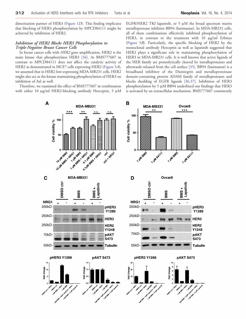
Inhibition of HER2 Blocks HER3 Phosphorylation inTriple-Negative Breast Cancer Cells
In breast cancer cells with HER2 gene amplification, HER2 is themain kinase that phosphorylates HER3 [34]. As BMS777607 incontrast to MPCD84111 does not affect the catalytic activity ofHER2 as demonstrated in MCF7 cells expressing HER2 (Figure 5A),we assumed that in HER2 low-expressing MDA-MB231 cells, HER2might also act as the kinase maintaining phosphorylation of HER3 oninhibition of Axl as well.
Therefore, we examined the effect of BMS777607 in combinationwith either 10 μg/ml HER2-blocking antibody Herceptin, 5 μM
EGFR/HER2 TKI lapatinib, or 5 μM the broad spectrum matrixmetalloprotease inhibitor BB94 (batimastat). In MDA-MB231 cells,all of these combinations effectively inhibited phosphorylation ofHER3, in contrast to the treatment with 10 μg/ml Erbitux(Figure 5B). Particularly, the specific blocking of HER2 by themonoclonal antibody Herceptin as well as lapatinib suggested thatHER2 plays a significant role in maintaining phosphorylation ofHER3 in MDA-MB231 cells. It is well known that active ligands ofthe HER family are proteolytically cleaved by metalloproteases andafterwards released from the cell surface [35]. BB94 (batimastat) is abroadband inhibitor of the Disintegrin and metalloproteinasedomain-containing protein ADAM family of metalloproteases andblocks shedding of EGFR ligands [36,37]. Inhibition of HER3phosphorylation by 5 μM BB94 underlined our findings that HER3is activated by an extracellular mechanism. BMS777607 consistently

Neoplasia Vol. 16, No. 4, 2014 Activation of HER3 Interferes with Axl RTK Inhibitors Torka et al. 313
blocked phosphorylation of AKT S473 but had only a minor effecton the Mitogen-activated protein-kinase pathways as proven bypERK1/2 Western blot analysis (Figure 5B).Knowing that lapatinib was most efficient in blocking HER3
phosphorylation, we evaluated the impact of EGFR and HER2 onHER3 phosphorylation by siRNA experiments. Therefore, wedepleted EGFR, HER2, HER3, and EGFR in combination withHER2 from MDA-MB231 cells by siRNA knockdown andsubsequently induced HER3 phosphorylation by 10 μM BMStreatment for 24 hours (Figure 5C). Only HER3 siRNA and thecombination of EGFR and HER2 siRNA treatment blocked HER3phosphorylation, completely exhibiting less than 5% of phosphor-ylation compared with control siRNA treatment. Comparable toHerceptin, HER2-specific siRNA treatment remained a residualHER3 phosphorylation of 22% (Figure 5C).Although MDA-MB231 cells represent a prototype of triple-
negative breast cancer cell lines characterized by the loss of HER2expression, we investigated HER2 as one of the probable dimerizationpartners of HER3. To elucidate in more detail the phosphorylationstatus of HER2 in triple-negative breast cancer cells, we performedimmunoprecipitation experiments with a vast protein amount of 2mg. We treated MDA-MB231 cells with 10 μM BMS777607 for 24hours, and we further enhanced the phosphorylation of HER2 bystimulation with 50 ng/ml NRG1. The HER2 RTK wasimmunoprecipitated with a homemade anti-HER2–specific antibody(clone 13D1B1) or a commercial anti-HER2 antibody (Millipore,No. 06-562). The subsequent Western blot analysis for p-Tyrresulted in a slight, but reproducible, induction of tyrosinephosphorylation after BMS777607 treatment and NRG1 stimulation(Figure 5, D and E).Hence, we assume that this minimal amount of HER2
phosphorylation might contribute to the pronounced HER3activation and thereby maintains the feedback loop counteractingAxl inhibitor treatment. Additionally, lapatinib emerged as the mostefficient treatment strategy for inhibition of HER3 phosphorylation.
Figure 6. Exogenous application of NRG1 rescues AKT phosphorylatioinhibition of cell viability by BMS777607. Cell viability of MDA-MBtreatment under starving conditions (0% FBS). Cells were incubatedand without 50 ng/ml NRG1. Addition of NRG1 completely compensatThe DMSO control does not react to NRG1 stimulation. Mean values awith *P b .05, **P b .01, and ***P b .001 were considered statisticallyof MDA-MB231 and Ovcar8 cells is inhibited by MPCD84111 in contwas measured by CellTiter-Glo assay after 72 hours of treatment.MPCD84111 using starving conditions (0% FBS). Cell viability was reOvcar8 cells. Mean values and SEM of three independent experimenwere considered statistically significant (Mann-Whitney test). (C) BMSphosphorylation. Western blot analysis of MDA-MB231 cells is showμM BMS777607 and 5 μM lapatinib relative to DMSO control for 24 hsignificant increase of the pHER3 Y1289 levels was evident afterphosphorylation of AKT S473 to 24%. Additional NRG1 stimulaphosphorylation. The combination of BMS777607 and lapatinib blockloading control. The diagrams show the densitometric analysis of Wesare shown (n= 3). (D) BMS777607 and lapatinib exhibit a synergisticOvcar8 cells is shown. Cells were treated with 1 μM BMS777607 orDMSO control for 24 hours with or without 50 ng/ml NRG1 stimulationevident after BMS777607 treatment. BMS777607-induced HER phoAdditional NRG1 stimulation enhances HER3 and pAKTS473 phospcompletely the HER3 and AKT phosphorylation. Tubulin served as lWestern blots for pHER3 Y1289 and pAKT S473. Mean values and S
Exogenous Application of NRG1 Rescues AKTPhosphorylation and Cell Viability
Even though complete recovery of pAKT S473 was not achievedwith a pharmacological dose of 1 μM BMS777607, the feedback up-regulation of HER3 expression and HER3 phosphorylation wasclearly evident, further suggesting that inhibition of the Axl/PI3K/AKT pathway leads to the reactivation of HER3 (Figure 4E). Thesedata imply that HER3 phosphorylation partially restores theprosurvival and proliferation signaling that, in turn, may limit theeffect of Axl inhibitors. To support this hypothesis, we performedrescue experiments in combination with cell viability assays. MDA-MB231 cells were treated with pharmacological concentrations ofBMS777607 under starving conditions for 72 hours. Addition of theHER3 ligand (50 ng/ml NRG1) completely compensated theinhibitory effect of 0.1 and 1 μM BMS777607 on cell viability. Incontrast, the cell viability of the untreated cells was not affected byNRG1 stimulation (Figure 6A). The compensatory up-regulation ofHER3 expression and partial maintenance of HER3 phosphorylationon inhibition of Axl suggested that combined inhibition of Axl andHER2/HER3 could synergistically inhibit tumor cell viability.
Monolayer cell cultures represent oversimplified models for tumorstudies, due to the loss of extracellular matrix rigidity on artificialplastic surfaces and high serum concentrations. These conditionspoorly mimic the tumor cell biology in vivo. A more robust andreproducible test system is the three dimensional (3D) spheroidculture. To test the combined inhibition of Axl and HER2/HER3 ontumor cell viability, we used these 3D spheroid cultures.
We treated two Axl-expressing cell lines, namely MDA-MB231and Ovcar8, with MPCD84111 and with a combination of BMS andlapatinib, to elucidate the hypothesis that an inhibition of Axl andHER2/HER3 might be essential for treatment of Axl overexpressingcells. First, we proved the efficacy of 1 μM MPCD84111 in contrastto 1 μM BMS777607 treatment on MDA-MB231 and Ovcar8 cells.One micromolar MPCD84111 significantly inhibited cell viability to37% in MDA-MB231 spheroids and to 42% in Ovcar8 spheroids in
n and cell viability. (A) Exogenous application of NRG1 rescues the231 cells was measured by CellTiter-Glo assay after 72 hours ofwith increasing concentrations of BMS777607 (0.1, 1, 10 μM) withes the proliferation inhibitory function of 0.1 and 1 μMBMS777607.nd SEM of three independent experiments are shown. Differencessignificant (Mann-Whitney test; ns, nonsignificant). (B) Cell viabilityrast to BMS777607. Cell viability of MDA-MB231 and Ovcar8 cells3D spheroids were incubated with 1 µM BMS777607 or 1 µMduced to 37% by MPCD84111 in MDA-MB231 cells and to 43% ints are shown. Differences with *P b .05, **P b .01, and ***P b .001777607 and lapatinib exhibit a synergistic inhibitory effect on HER3n. Cells were treated with 1 μM BMS777607 or a combination of 1ours with or without 50 ng/ml NRG1 stimulation for 15 minutes. ABMS777607 treatment. BMS777607 treatment suppressed thetion enhances HER3 phosphorylation and restores pAKTS473s completely the HER3 and AKT phosphorylation. Tubulin served astern blots for pHER3 Y1289 and pAKT S473. Mean values and SEMinhibitory effect on HER3 phosphorylation. Western blot analysis ofa combination of 1 μM BMS777607 and 5 μM lapatinib relative tofor 15 minutes. A 2.4-fold increase of the pHER3 Y1289 levels wassphorylation treatment completely rescued AKT phosphorylation.horylation. The combination of BMS777607 and lapatinib blocksoading control. The diagrams show the densitometric analysis ofEM are shown (n = 3).
314 Activation of HER3 Interferes with Axl RTK Inhibitors Torka et al. Neoplasia Vol. 16, No. 4, 2014
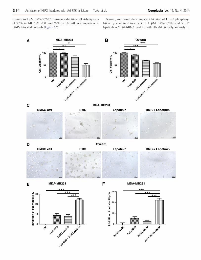
contrast to 1 μMBMS777607 treatment exhibiting cell viability ratesof 97% in MDA-MB231 and 92% in Ovcar8 in comparison toDMSO-treated controls (Figure 6B).
Second, we proved the complete inhibition of HER3 phosphory-lation by combined treatment of 1 μM BMS777607 and 5 μMlapatinib inMDA-MB231 and Ovcar8 cells. Additionally, we analyzed

Neoplasia Vol. 16, No. 4, 2014 Activation of HER3 Interferes with Axl RTK Inhibitors Torka et al. 315
if BMS treatment or the stimulation with HER3 ligand NRG1 mightlead to the reconstitution of the AKT signaling pathway.One micromolar BMS777607 leads to up-regulation of total HER3
protein levels and to a six-fold induction of HER3 phosphorylationafter 24 hours of treatment in MDA-MB231 cells (Figure 6C). Thephospho-AKTS473 levels are reduced to 24% compared with DMSO-treated cells, and additional stimulation with 50 ng/ml NRG1 restorespAKTS473 signal to DMSO control levels. The combination of 1 μMBMS777607 and 5 μM lapatinib inhibited HER3 and AKTphosphorylation to 0.9%, respectively, 6.1% in comparison toBMS777607 single treatment. Especially, NRG1 ligand stimulationwas not able to induce HER3 phosphorylation any more than incontrast to DMSO (two-fold induction) and 1 μM BMS777607treatment (13-fold induction). HER2 phosphorylation was belowdetection threshold in MDA-MB231 cells due to the low expressionlevel of HER2 in this cell line (Figure 6C).TheOvcar8 cells react to 1μMBMS777607 treatment, analogous to
MDA-MB231, by up-regulation of totalHER3protein levels and a 2.4-fold HER3 phosphorylation compared to DMSO-treated cells. ThisHER3 phosphorylation results in a 1.4-fold induction of AKTphosphorylation. Additional 50 ng/ml NRG1 ligand stimulationleads in a 10- or 17.8-fold HER3 phosphorylation in DMSO controland 1 μM BMS777607-treated cells. The combined treatment with 1μM BMS777607 and 5 μM lapatinib completely prevents HER3 andAKT phosphorylation and additionally prohibits the activation byNRG1 ligand. In Ovcar8 cells, HER2 Y1248 exhibits an identicalphosphorylation pattern to pHER3 Y1289, emphasizing the impor-tance of the HER2/HER3 heterodimer in maintaining the Her3phosphorylation in cell lines with significant HER2 levels (Figure 6D).We conclude that BMS777607-induced HER3 phosphorylation is
able to rescue or at least stabilizes theAKT signaling inOvcar8 andMDA-MB231 cells, and vice versa, a combination of 1 μMBMS777607 and 5μM lapatinib completely inhibits HER3 and AKT phosphorylation.
Figure 7. Pharmacological inhibition of Axl sensitizes for lapatinib. (A) Bcell viability. Cell viability of MDA-MB231 cells was measured byspheroids were incubated with 1 μM BMS777607, 5 μM lapatinib, orstarving conditions (0% FBS). Cell viability was reduced to 49.5% by tto either compound administered separately. Mean values and SEM o.05, **P b .01, and ***P b .001 were considered statistically signifilapatinib exhibit an additive cell viability inhibition effect. Cell viability otreatment. Ovcar8 3D spheroids were incubated with 1 μM BMS7776DMSO control under starving conditions (0% FBS). Cell viability wasBMS777607 compared to either compound administered separateshown. Differences with *P b .05, **P b .01, and ***P b .001nonsignificant). (C) Representative phase-contrast images of MDA-MμMBMS777607, 5 μM lapatinib, or a combination of both compoundshours. Scale bars indicate 100 μm. (D) Representative phase-contrawith 1 μM BMS777607, 5 μM lapatinib, or a combination of both coFBS) for 72 hours. Scale bars indicate 100 μm. (E) Validation of the sycell viability. Cell viability of MDA-MB231 cells was measured byspheroids were incubated with 1 μM BMS777607, 5 μM lapatinib, orfull-serum conditions (10% FBS). Inhibition of cell viability was significBMS777607 compared to either compound administered separately.Differences with *P b .05, **P b .01, and ***P b .001 were considereValidation of synergistic inhibitory effect by knockdown of Axl and Hspecific siRNA, HER3-specific siRNA, or a combination of both siRNgrown for 72 hours under full-serum conditions (10% FBS), and cell viawas significantly increased to 22% by a combination of Axl-spectransfected separately. Mean values and SEM of five independent expb .001 were considered statistically significant (Kruskal-Wallis test; n
Pharmacological Inhibition of Axl Sensitizes forLapatinib Treatment
We further tested the combined inhibition of Axl and HER2/HER3 on tumor cell viability in 3D spheroid cultures of MDA-MB231 and Ovcar8 cells. Therefore, we treated MDA-MB231 3Dspheroids with 1 μM BMS777607 and 5 μM lapatinib and assessedtheir viability under starving conditions (0% FBS). Cell viability wassignificantly reduced up to 50% by a combination of 1 μMBMS777607 and 5 μM lapatinib compared to the effect of eitherinhibitor administered alone (Figure 7A). The cell viability of Ovcar8cell spheroids was significantly inhibited by 5 μM labatinib and thecombination of 1 μM BMS777607 and 5 μM lapatibnib. The cellviability was inhibited to 56% by the combination of 1 μMBMS777607 and 5 μM lapatinib (Figure 7B).
The images of Figure 7, C and D, visualize the spheroids formedafter 72 hours in Matrigel. Ovcar8 cell spheroids react more sensitivelyto 5 μM lapatinib treatment alone due to the fact of higher HER2expression levels and resulting dependency on HER2 signaling.
Analogous but less pronounced results were achieved with MDA-MB231 3D spheroids cultured under full-serum conditions (10%FBS). The cell viability of MDA-MB231 spheroids decreasedsignificantly under full-serum conditions by combination treatmentwith 1 μM BMS777607 and 5 μM lapatinib. To better present thedifferences in cell viability, we displayed the inhibition of cell viabilityin Figure 7, E and F. The cell viability of MDA-MB231 3Dspheroids was not significantly affected by 1 μM BMS777607 or 5μM lapatinib alone, whereas MDA-MB231 3D spheroids treatedwith the TKI combination displayed a statistically significantreduction in viability of 24% (Figure 7E). To underline the efficacyof combined targeted therapies toward Axl and HER3, we performedsiRNA experiments to knockdown Axl and HER3 (Figure 7F).Analogous to the pharmacological inhibition, the cell viability ofMDA-MB231 3D spheroids was not significantly affected by Axl
MS777607 and lapatinib exhibit a synergistic effect on inhibition ofCellTiter-Glo assay after 72 hours of treatment. MDA-MB231 3Da combination of both compounds relative to DMSO control underhe combination of 5 μM lapatinib and 1 μM BMS777607 comparedf three independent experiments are shown. Differences with *P bcant (Kruskal-Wallis test; ns, nonsignificant). (B) BMS777607 andf Ovcar8 cells wasmeasured by CellTiter-Glo assay after 72 hours of07, 5 μM lapatinib, or a combination of both compounds relative toreduced to 56% by the combination of 5 μM lapatinib and 1 μM
ly. Mean values and SEM of three independent experiments arewere considered statistically significant (Kruskal-Wallis test; ns,B231 spheroids. MDA-MB231 3D spheroids were incubated with 1relative to DMSO control under starving conditions (0% FBS) for 72st images of Ovcar8 spheroids. Ovcar8 spheroids were incubatedmpounds relative to DMSO control under starving conditions (0%nergistic inhibitory effect of BMS777607 and lapatinib treatment onCellTiter-Glo assay after 72 hours of treatment. MDA-MB231 3Da combination of both compounds relative to DMSO control underantly increased to 24% by a combination of 5 μM lapatinib and 1 μMMean values and SEM of six independent experiments are shown.d statistically significant (Kruskal-Wallis test; ns, nonsignificant). (F)ER3 on cell viability. MDA-MB231 cell were transfected with Axl-As for 48 hours. Subsequently, MDA-MB231 3D spheroids werebility wasmeasured by CellTiter-Glo assay. Inhibition of cell viabilityific siRNA and HER3-specific siRNA compared to either siRNAeriments are shown. Differences with *P b .05, **P b .01, and ***Ps, nonsignificant).

316 Activation of HER3 Interferes with Axl RTK Inhibitors Torka et al. Neoplasia Vol. 16, No. 4, 2014
siRNA or HER3 siRNA, whereas MDA-MB231 3D spheroidstreated with a combination of these two siRNAs displayed astatistically significant reduction in viability of 22% (Figure 7F).
These data suggest that pharmacological inhibition of Axl may notbe an effective single-agent therapy in Axl-expressing tumors, whereasthe simultaneous blockade of Axl and HER2/3 complexes at the cellsurface using lapatinib might be a suitable approach to optimize theantitumor action of inhibitors targeting the Axl RTK.
DiscussionThe benefits of single-target therapies are limited due to resistanceformation by the activation of alternative RTKs [38]. Understandingthe reasons for the acquired resistance would be an important key to amore successful cancer therapy. Hence, we asked whether targeted Axltherapies would also result in the activation of alternative RTKs. In thisstudy, we show for the first time, that on knockdown of Axl expressionby siRNA or pharmacological inhibition with BMS777607 and R428,MDA-MB231 and Ovcar8 cells are able to compensate the loss of Axlactivity by the induction of HER3 phosphorylation.
Furthermore, the analysis of 20 Axl-expressing cell lines from brain,breast, ovary, cervix, lung, and pancreatic cancer provided the firstevidence for a positive correlation between a low basal phosphorylationof AKT S473 and the induction of HER3 activation on Axl inhibitionby BMS777607 or Axl-specific siRNA-mediated knockdown. Accord-ingly, 7 of 10 cell lines with low pAKT S473 basal levels activatedpHER3 Y1289 on treatment with BMS777607 or Axl-specific siRNA-mediated knockdown. In contrast, only 2 of 10 cell lines with highpAKT S473 basal levels showed phosphorylation of HER3 Y1289 onAxl inhibition or knockdown (Figure 3C). The high basal phosphor-ylation of AKT S473 mainly appears in cell lines with loss of PTEN- orPI3K-activating mutations such as the brain tumor cell lines U373,SF126, and U118 [39,40]. These cell lines are less sensitive to the Axl/PI3K/AKT signaling pathway inhibition and do not respond throughHER3 up-regulation to the inhibition of the upstream RTK Axl. Weconclude that inhibition of Axl induces the phosphorylation of HER3,and this induction is not confined to a particular type of tumor but isrestricted to tumors with low basal activation of AKT.We suppose thatAKT phosphorylation is the key sensor for upstream signalingpathways. Thus, we reason that high AKT phosphorylation levels,resulting from PTEN loss, PI3K activating mutations, or manifoldaffecting upstream pathways (RTKs, G-protein coupled receptors(GPCRs), integrins, and cytosolic kinases), reduce the sensitivity of theAKT sensor to upstream RTK inhibition. Vice versa, low basal AKTphosphorylation levels indicate a higher sensitivity of the AKT sensor toupstream RTK inhibition. Axl was shown to influence AKTphosphorylation significantly as reviewed by Verma et al., 2011 andPazzez et al., 2014 [7,41]. In the case of Axl-overexpressing cells, as usedin the present study, this correlates with higher dependency on theupstream Axl-RTK signaling pathway.
Consequently, we assume that the levels of pAKT S473 and pHER3could be suitable biomarkers for Axl therapy response, indicatingpotential clinical use. The induction of HER3 phosphorylation couldbe used to discriminate between responder versus nonresponder patientcohorts as a pharmacodynamic biomarkermonitoring therapy response.Additionally, our results indicate that patient cohorts with expression ofAxl and low basal activity of AKT might benefit from a treatment withAxl inhibitors due to high dependency on the Axl/PI3K/AKT signalingpathway. To our knowledge, this is the first description of anindependent biomarker for Axl treatment regimens.
A comparison of BMS777607 with R428 and with an Axlinhibitor discovered by our group, namely, MPCD84111, indicatesthat only MPCD84111 was able to block the HER3 phosphorylationfeedback loop efficiently (Figure 1B). As a common characteristic, allthree inhibitors efficiently block AKT phosphorylation. AKT hasbeen shown to phosphorylate the FoxO family of transcription factorsand thereby prevents their function [32]. Thus, we assume that AKTregulates the expression of HER3 by inhibiting FoxO-dependenttranscription downstream of the Axl RTK. These results would be inline with observations of Chandarlapaty et al., 2011 and Chakrabartyet al., 2012 using AKT or PI3K inhibitors to induce expression ofHER3 in various tumor cell lines [30,31].
Our experiments with BMS777607 and MPCD84111 revealedthat only MPCD84111 was able to block phosphorylation of HER3completely, although the transcriptional feedback loop was inducedin a similar extent to BMS777607 treatment (Figure 1B). As it iswidely accepted that HER3 has an impaired kinase activity, activationof the HER3 RTK occurs only after its dimerization with other RTKssuch as HER2 or EGFR [33,28]. On the basis of the knowledge thatMPCD84111 targets HER2 (Figure 1D), a potential dimerizationpartner for HER3, we focused primarily on the HER family of RTKs.Although MDA-MB231 cells display a prototype of triple-negativebreast cancer cell lines, characterized by a low HER2 expression, ourstudy provides a line of arguments that HER2 is the essentialdimerization partner for HER3. The inhibition of HER2/3heterocomplexes has been demonstrated in two independent ways.First, in contrast to BMS777607 [24], MPCD84111 potentiallyinhibits HER2, as demonstrated by kinase selectivity profiling(Figure 1D). The HER2 inhibition capability of MPCD84111 wasalso proven in HER2-expressing MCF7 breast cancer cells(Figure 5A). Second, our results clearly demonstrate that Herceptinas well as lapatinib, two U.S. Food and Drug Administration-approved therapeutics for treatment of HER2-amplified breastcancer, are able to inhibit phosphorylation of HER3 (Figure 5B).Particularly, the inhibition of HER3 phosphorylation by Herceptin, ahumanized IgG1 monoclonal antibody raised against HER2, is worthto be emphasized. For the sake of completeness, it is essential tomention that Herceptin- as well as HER2-specific knockdown couldnot prohibit HER3 phosphorylation totally. The complete inhibitionof HER3 phosphorylation, comparable to HER3-specific knock-down, was only achieved by lapatinib, a dual-specific EGFR/HER2TKI [42], or a combination of EGFR and HER2-specific siRNA.Therefore, we cannot exclude a partial impact of EGFR in themaintenance of HER3 phosphorylation in MDA-MB231 cells.However, EGFR-specific knockdown as well as EGFR inhibition byErbitux and erlotinib (data not shown for erlotinib) did not interferewith HER3 phosphorylation in MDA-MB231 cells. Consequently,the dual-specific EGFR/HER2 TKI lapatinib emerged as the mostefficient strategy to prohibit HER3 phosphorylation.
To our knowledge, this is the first study that supports thesignificant role of HER2 in maintaining the phosphorylation ofHER3 in MDA-MB231 cells although its quantities are hardlydetectable with the common immunoprecipitation or Western blotanalysis techniques (Figure 5, C–E).
We could also support our hypothesis of the NRG1/HER2/HER3compensation mechanism by rescue experiments in combinationwith cell viability assays in MDA-MB231 cells. Addition of theHER3 ligand NRG1 completely compensated the inhibitory effect ofBMS777607 on cell viability, whereas proliferation was not

Neoplasia Vol. 16, No. 4, 2014 Activation of HER3 Interferes with Axl RTK Inhibitors Torka et al. 317
stimulated by NRG1 in untreated cells (Figure 6A). Our resultsimplicate that up-regulation of HER3 expression by inhibition of theAxl/PI3K/AKT pathway is a prerequisite for NRG1 ligand–dependent activation of HER3 that might be important in thecontext of the tumor microenvironment and paracrine resistance toTKIs. The paracrine resistance is a common mechanism and has beendescribed recently in patients with chronic myeloid leukemia treatedwith BCR-ABL kinase inhibitors as well as in patients with non–smallcell lung cancer treated with EGFR inhibitors [43]. Wang et al., 2009reported that hepatocyte growth factor (HGF) released by stromalfibroblasts induced resistance to EGFR inhibitors in non–small celllung cancer due to Met activation [44]. A tumor-supportive role formacrophages was described by Vlaicu et al., 2013, where tumor-associated macrophages supply tumor cells with EGFR ligands [45].The expression and secretion of NRG1 has been demonstrated forvarious components of the tumor environment, namely, fibroblasts,endothelial cells, and peripheral monocytes, additionally supportingthe role of NRG1 in a paracrine tumor microenvironment–drivenresistance mechanism in vivo. The expression of NRG1 in fibroblastshas been described by Wen et al., 1992 and Nagata et al., 2006[46,47]. Cote et al., 2005 and Hedhi et al., 2011 nicely described theparacrine function of NRG1 produced by endothelial cells on cardiacmyocytes [48,49]. Another in vivo study focused on mesenchyme-derived NRG1 regulating the epithelial morphogenesis of mouseembryonic submandibular glands [50]. The activation of humanmonocyticlike THP-1 [THP1] (ATCCR TIB-202™) cells andperipheral blood monocytes was companied by up-regulation andsecretion of NRG1, leading to paracrine activation of the erbreceptors and contribution to the epithelial and connective tissueproliferation [51]. The paracrine NRG1/HER3 signal initiated bybone marrow–derived mesenchymal stem cells and tumor-associatedmesenchymal cells was shown to promote colorectal cancer cellprogression and high NRG1 expression and was associated with poorprognosis as shown by De Boeck et al., 2013 [52]. Additionally,human blood serum contains considerable amounts of NRG1 innanogram-per-milliliter concentration range [53]. Lately, NRG1produced by breast tumor–initiating cells was shown to promote theirproliferation and self-renewal in HER2 low-expressing tumors,including triple-negative breast tumors [54]. Our results indicatethat MDA-MB231 cells secret NRG1 in autocrine manner and thatHER3 phosphorylation strongly correlates with the consumption ofNRG1 after Axl inhibitor treatment (Figure 4D). In vivo, anadditionally paracrine mechanism of tumor microenvironment–driven resistance might be considered for the NRG1 and HER3;however, this needs further investigation. The compensatory up-regulation of HER3 expression and the maintenance of HER3phosphorylation on inhibition of Axl suggested that a combinedinhibition of Axl and HER3 would synergistically inhibit tumor cellviability. Cell viability was significantly reduced by a combination of1 μM BMS777607 and 5 μM lapatinib compared to single-inhibitortreatments (Figure 7, A and E). The efficacy of combined therapiestargeting Axl and HER2/3 was underlined by siRNA-mediatedknockdown of Axl and HER3 (Figure 7F). Analogous to thepharmacological intervention, we achieved a significant synergisticinhibition of cell viability by a combined application of both siRNAs.Together, these data suggest that, although pharmacologicalinhibition of Axl may not be an effective single-agent therapy inAxl-expressing cells with low basal AKT levels, blockade of HER2/3complexes using lapatinib might be an effective approach to block the
HER3-activating feedback loop induced by Axl TKI treatment. Weassume that a monotherapy with Axl inhibitors might have limitedclinical activity due to a HER3 bypass mechanism. In cells that exhibitlow expression of HER2/3, up-regulation and phosphorylationof HER3 are relevant mechanisms to counteract inhibition of theAxl/PI3K/AKT pathway. Consequently, Axl inhibitors should be usedin combination with HER2/3 antagonists in Axl-positive tumors withlow basal AKT phosphorylation or with an autocrine NRG1 loop.Therefore, alternatively to BMS777607 or R428, using MPCD84111as an Axl inhibitor, with the capacity to block the HER2/3 bypassmechanism by direct inhibition of HER2, would be beneficial forcancer treatment. The final decision about using an Axl/HER2-selective compound, e.g., MPCD84111 or an alternative strategy ofAxl inhibitors in combination with HER2/3 antagonistic agents, will ofcourse require additional investigation in preclinical and clinical trials.
AcknowledgmentsWe thank Stefano Iacobelli (Department of Oncology and Experi-mental Medicine, University Foundation ‘G D’Annunzio’ Chieti-Pescara) for providing the MDA-MB231 cell line and U3 Pharma forproviding the Hs578T cell line. We also thank Lead Discovery CenterGmbH (LCD, Dortmund, Germany) for providing the BMS777607and R428 Axl TKIs and Gregor Kirfel (Institute for Cell Biology,University of Bonn) for critical reading of the manuscript.
References
[1] O'Bryan JP, Frye RA, Cogswell PC, Neubauer A, Kitch B, Prokop C, EspinosaIII R, Le Beau MM, Earp HS, and Liu ET (1991). axl, a transforming geneisolated from primary human myeloid leukemia cells, encodes a novel receptortyrosine kinase. Mol Cell Biol 11, 5016–5031.
[2] Janssen JW, Schulz AS, Steenvoorden AC, Schmidberger M, Strehl S, AmbrosPF, and Bartram CR (1991). A novel putative tyrosine kinase receptor withoncogenic potential. Oncogene 6, 2113–2120.
[3] Stitt TN, Conn G, Gore M, Lai C, Bruno J, Radziejewski C, Mattsson K, Fisher J,Gies DR, and Jones PF, et al. (1995). The anticoagulation factor protein S and itsrelative, Gas6, are ligands for the Tyro 3/Axl family of receptor tyrosine kinases.Cell 80, 661–670.
[4] Varnum BC, Young C, Elliott G, Garcia A, Bartley TD, Fridell YW, Hunt RW,Trail G, Clogston C, and Toso RJ, et al. (1995). Axl receptor tyrosine kinasestimulated by the vitamin K-dependent protein encoded by growth-arrest-specific gene 6. Nature 373, 623–626.
[5] Hafizi S and Dahlbäck B (2006). Signalling and functional diversity within the Axlsubfamily of receptor tyrosine kinases. Cytokine Growth Factor Rev 17, 295–304.
[6] Linger RM, Keating AK, Earp HS, and Graham DK (2008). TAM receptortyrosine kinases: biologic functions, signaling, and potential therapeutic targetingin human cancer. Adv Cancer Res 100, 35–83.
[7] Verma A, Warner SL, Vankayalapati H, Bearss DJ, and Sharma S (2011).Targeting Axl and Mer kinases in cancer. Mol Cancer Ther 10, 1763–1773.
[8] Shieh YS, Lai CY, Kao YR, Shiah SG, Chu YW, Lee HS, and Wu CW (2005).Expression of axl in lung adenocarcinoma and correlation with tumorprogression. Neoplasia 7, 1058–1064.
[9] Song X, Wang H, Logsdon CD, Rashid A, Fleming JB, Abbruzzese JL,Gomez HF, Evans DB, and Wang H (2011). Overexpression of receptortyrosine kinase Axl promotes tumor cell invasion and survival in pancreaticductal adenocarcinoma. Cancer 117, 734–743.
[10] Koorstra JB, Karikari CA, FeldmannG,Bisht S, Rojas PL,OfferhausGJ, AlvarezH,andMaitra A (2009). The Axl receptor tyrosine kinase confers an adverse prognosticinfluence in pancreatic cancer and represents a new therapeutic target. Cancer BiolTher 8, 618–626.
[11] Vajkoczy P, Knyazev P, Kunkel A, Capelle HH, Behrndt S, von Tengg-Kobligk H,Kiessling F, Eichelsbacher U, Essig M, and Read TA, et al. (2006). Dominant-negative inhibition of the Axl receptor tyrosine kinase suppresses brain tumor cellgrowth and invasion and prolongs survival. Proc Natl Acad Sci U S A 103,5799–5804.

318 Activation of HER3 Interferes with Axl RTK Inhibitors Torka et al. Neoplasia Vol. 16, No. 4, 2014
[12] Chen PX, Li QY, and Yang Z (2013). Axl and prostasin are biomarkers forprognosis of ovarian adenocarcinoma. Ann Diagn Pathol 17, 425–429.
[13] Byers LA, Diao L,Wang J, Saintigny P, Girard L, PeytonM, Shen L, Fan Y,Giri U,and Tumula PK, et al. (2013). An epithelial–mesenchymal transition gene signaturepredicts resistance to EGFR and PI3K inhibitors and identifies Axl as a therapeutictarget for overcoming EGFR inhibitor resistance. Clin Cancer Res 19, 279–290.
[14] Gjerdrum C, Tiron C, Høiby T, Stefansson I, Haugen H, Sandal T, Collett K,Li S, McCormack E, and Gjertsen BT, et al. (2010). Axl is an essentialepithelial-to-mesenchymal transition-induced regulator of breast cancermetastasis and patient survival. Proc Natl Acad Sci U S A 107, 1124–1129.
[15] Gustafsson A,MartuszewskaD, JohanssonM, EkmanC,Hafizi S, Ljungberg B, andDahlbäck B (2009). Differential expression of Axl and Gas6 in renal cell carcinomareflecting tumor advancement and survival. Clin Cancer Res 15, 4742–4749.
[16] Hutterer M, Knyazev P, Abate A, Reschke M, Maier H, Stefanova N, Knyazeva T,Barbieri V, Reindl M, and Muigg A, et al. (2008). Axl and growth arrest–specificgene 6 are frequently overexpressed in human gliomas and predict poor prognosis inpatients with glioblastoma multiforme. Clin Cancer Res 14, 130–138.
[17] Rochlitz C, Lohri A, Bacchi M, Schmidt M, Nagel S, Fopp M, Fey MF,Herrmann R, and Neubauer A (1999). Axl expression is associated with adverseprognosis and with expression of Bcl-2 and CD34 in de novo acute myeloidleukemia (AML): results from a multicenter trial of the Swiss Group for ClinicalCancer Research (SAKK). Leukemia 13, 1352–1358.
[18] Macleod K, Mullen P, Sewell J, Rabiasz G, Lawrie S, Miller E, Smyth JF, andLangdon SP (2005). Altered ErbB receptor signaling and gene expression incisplatin-resistant ovarian cancer. Cancer Res 65, 6789–6800.
[19] Hong CC, Lay JD, Huang JS, Cheng AL, Tang JL, Lin MT, Lai GM, andChuang SE (2008). Receptor tyrosine kinase AXL is induced by chemotherapydrugs and overexpression of AXL confers drug resistance in acute myeloidleukemia. Cancer Lett 268, 314–324.
[20] Liu L, Greger J, Shi H, Liu Y, Greshock J, Annan R, Halsey W, Sathe GM,Martin AM, and Gilmer TM (2009). Novel mechanism of lapatinib resistance inHER2-positive breast tumor cells: activation of AXL. Cancer Res 69, 6871–6878.
[21] Mahadevan D, Cooke L, Riley C, Swart R, Simons B, Della Croce K, Wisner L,Iorio M, Shakalya K, and Garewal H, et al. (2007). A novel tyrosine kinase switchis a mechanism of imatinib resistance in gastrointestinal stromal tumors.Oncogene 26, 3909–3919.
[22] Dufies M, Jacquel A, Belhacene N, Robert G, Cluzeau T, Luciano F, Cassuto JP,Raynaud S, andAuberger P (2011).Mechanisms of AXLoverexpression and functionin Imatinib-resistant chronic myeloid leukemia cells. Oncotarget 2, 874–885.
[23] Zhang Z, Lee JC, Lin L, Olivas V, Au V, Laframboise T, Abdel-RahmanM,WangX, Levine AD, and Rho JK, et al. (2012). Activation of the AXL kinase causesresistance to EGFR-targeted therapy in lung cancer. Nat Genet 44, 852–860.
[24] Schroeder GM, An Y, Cai ZW, Chen XT, Clark C, Cornelius LA, Dai J,Gullo-Brown J, Gupta A, and Henley B, et al. (2009). Discovery of N-(4-(2-amino-3-chloropyridin-4-yloxy)-3-fluorophenyl)-4-ethoxy-1-(4-fluorophenyl)-2-oxo-1,2-dihydropyridine-3-carboxamide (BMS-777607), a selective and orallyefficacious inhibitor of the Met kinase superfamily. J Med Chem 52, 1251–1254.
[25] Holland SJ, Pan A, Franci C, Hu Y, Chang B, Li W, Duan M, Torneros A, Yu J,and Heckrodt TJ, et al. (2010). R428, a selective small molecule inhibitor of Axlkinase, blocks tumor spread and prolongs survival in models of metastatic breastcancer. Cancer Res 70, 1544–1554.
[26] Li Y, Ye X, Tan C, Hongo JA, Zha J, Liu J, Kallop D, Ludlam MJ, and Pei L(2009). Axl as a potential therapeutic target in cancer: role of Axl in tumorgrowth, metastasis and angiogenesis. Oncogene 28, 3442–3455.
[27] Mackiewicz M, Huppi K, Pitt JJ, Dorsey TH, Ambs S, and Caplen NJ (2011).Identification of the receptor tyrosine kinase AXL in breast cancer as a target forthe human miR-34a microRNA. Breast Cancer Res Treat 130, 663–679.
[28] Campbell MR, Amin D, and Moasser MM (2010). HER3 comes of age: newinsights into its functions and role in signaling, tumor biology, and cancertherapy. Clin Cancer Res 16, 1373–1383.
[29] Révillion F, Lhotellier V, Hornez L, Bonneterre J, and Peyrat JP (2008). ErbB/HER ligands in human breast cancer, and relationships with their receptors, thebio-pathological features and prognosis. Ann Oncol 19, 73–80.
[30] Chandarlapaty S, Sawai A, Scaltriti M, Rodrik-Outmezguine V, Grbovic-HuezoO, Serra V, Majumder PK, Baselga J, and Rosen N (2011). AKT inhibitionrelieves feedback suppression of receptor tyrosine kinase expression and activity.Cancer Cell 19, 58–71.
[31] Chakrabarty A, Sánchez V, Kuba MG, Rinehart C, and Arteaga CL (2012).Feedback upregulation of HER3 (ErbB3) expression and activity attenuatesantitumor effect of PI3K inhibitors. Proc Natl Acad Sci U S A 109, 2718–2723.
[32] Myatt SS and Lam EW (2007). The emerging roles of forkhead box (Fox)proteins in cancer. Nat Rev Cancer 7, 847–859.
[33] Hynes NE and Lane HA (2005). ERBB receptors and cancer: the complexity oftargeted inhibitors. Nat Rev Cancer 5, 341–354.
[34] Stern DF (2008). ERBB3/HER3 and ERBB2/HER2 duet in mammarydevelopment and breast cancer. J Mammary Gland Biol Neoplasia 13, 215–223.
[35] Blobel CP (2005). ADAMs: key components in EGFR signalling anddevelopment. Nat Rev Mol Cell Biol 6, 32–43.
[36] Borrell-PagèsM,Rojo F, Albanell J, Baselga J, andArribas J (2003).TACE is requiredfor the activation of the EGFR by TGF-α in tumors. EMBO J 22, 1114–1124.
[37] Dong J, Opresko LK, Dempsey PJ, Lauffenburger DA, Coffey RJ, and Wiley HS(1999). Metalloprotease-mediated ligand release regulates autocrine signaling throughthe epidermal growth factor receptor. Proc Natl Acad Sci U S A 96, 6235–6240.
[38] Engelman JA and Settleman J (2008). Acquired resistance to tyrosine kinaseinhibitors during cancer therapy. Curr Opin Genet Dev 18, 73–79.
[39] Parsa AT,Waldron JS, PannerA,CraneCA, Parney IF, Barry JJ, CacholaKE,MurrayJC, Tihan T, and JensenMC, et al. (2007). Loss of tumor suppressor PTEN functionincreases B7-H1 expression and immunoresistance in glioma. Nat Med 13, 84–88.
[40] Eitel JA, Bijangi-Vishehsaraei K, Saadatzadeh MR, Bhavsar JR, Murphy MP,Pollok KE, and Mayo LD (2009). PTEN and p53 are required for hypoxiainduced expression of maspin in glioblastoma cells. Cell Cycle 8, 896–901.
[41] Paccez JD, Vogelsang M, Parker MI, and Zerbini LF (2014). The receptortyrosine kinase Axl in cancer: biological functions and therapeutic implications.Int J Cancer 134, 1024–1033.
[42] Davis MI, Hunt JP, Herrgard S, Ciceri P, Wodicka LM, Pallares G, Hocker M,Treiber DK, and Zarrinkar PP (2011). Comprehensive analysis of kinaseinhibitor selectivity. Nat Biotechnol 29, 1046–1051.
[43] Hölzel M, Bovier A, and Tüting T (2013). Plasticity of tumour and immunecells: a source of heterogeneity and a cause for therapy resistance? Nat Rev Cancer13, 365–376.
[44] Wang W, Li Q, Yamada T, Matsumoto K, Matsumoto I, Oda M, Watanabe G,Kayano Y, Nishioka Y, and Sone S, et al. (2009). Crosstalk to stromal fibroblastsinduces resistance of lung cancer to epidermal growth factor receptor tyrosinekinase inhibitors. Clin Cancer Res 15, 6630–6638.
[45] Vlaicu P, Mertins P, Mayr T, Widschwendter P, Ataseven B, Högel B, EiermannW, Knyazev P, and Ullrich A (2013). Monocytes/macrophages supportmammary tumor invasivity by co-secreting lineage-specific EGFR ligands anda STAT3 activator. BMC Cancer 13, 197.
[46] Wen D, Peles E, Cupples R, Suggs SV, Bacus SS, Luo Y, Trail G, Hu S, SilbigerSM, and Levy RB, et al. (1992). Neu differentiation factor: a transmembraneglycoprotein containing an EGF domain and an immunoglobulin homologyunit. Cell 69, 559–572.
[47] Nagata K, Wada K, Tatsuguchi A, Futagami S, Gudis K, Miyake K, Tsukui T,and Sakamoto C (2006). Heregulin-α and heregulin-β expression is linked to aCOX-2-PGE2 pathway in human gastric fibroblasts. Am J Physiol GastrointestLiver Physiol 290, G1243–G1251.
[48] Cote GM, Miller TA, Lebrasseur NK, Kuramochi Y, and Sawyer DB (2005).Neuregulin-1α and β isoform expression in cardiac microvascular endothelialcells and function in cardiac myocytes in vitro. Exp Cell Res 311, 135–146.
[49] Hedhli N, Huang Q, Kalinowski A, Palmeri M, Hu X, Russell RR, and RussellKS (2011). Endothelium-derived neuregulin protects the heart against ischemicinjury. Circulation 123, 2254–2262.
[50] Miyazaki Y, Nakanishi Y, and Hieda Y (2004). Tissue interaction mediated byneuregulin-1 and ErbB receptors regulates epithelial morphogenesis of mouseembryonic submandibular gland. Dev Dyn 230, 591–596.
[51] Mograbi B, Rochet N, Imbert V, Bourget I, Bocciardi R, Emiliozzi C, and RossiB (1997). Human monocytes express amphiregulin and heregulin growth factorsupon activation European cytokine network. Eur Cytokine Netw 8, 73–81.
[52] De Boeck A, Pauwels P, Hensen K, Rummens JL, Westbroek W, Hendrix A,Maynard D, Denys H, Lambein K, and Braems G, et al. (2013). Bone marrow-derived mesenchymal stem cells promote colorectal cancer progression throughparacrine neuregulin 1/HER3 signalling. Gut 62, 550–560.
[53] Shibuya M, Komi E, Wang R, Kato T, Watanabe Y, Sakai M, Ozaki M, SomeyaT, and Nawa H (2010). Measurement and comparison of serum neuregulin 1immunoreactivity in control subjects and patients with schizophrenia: aninfluence of its genetic polymorphism. J Neural Transm 117, 887–895.
[54] Lee CY, Lin Y, Bratman S, Feng W, Kuo A, Scheeren F, Engreitz JM, Varma S,West R, and Diehn M (2013). Neuregulin autocrine signaling promotes self-renewal of breast tumor-initiating cells by triggering HER2/HER3 activation.Cancer Res 74, 341–352.


