Embed Size (px)
Citation preview

Behavioral/Systems/Cognitive
Acute and Chronic Ethanol Alter GlutamatergicTransmission in Rat Central Amygdala: an In Vitro and InVivo Analysis
Marisa Roberto, Paul Schweitzer, Samuel G. Madamba, David G. Stouffer, Loren H. Parsons, and George R. SigginsDepartment of Neuropharmacology, The Scripps Research Institute, La Jolla, California 92037
The modulation of glutamatergic transmission by ethanol may contribute to ethanol intoxication, reinforcement, tolerance, and depen-dence. Therefore, we used in vitro electrophysiological and in vivo microdialysis techniques to investigate the effects of acute and chronicethanol on glutamatergic transmission in the central nucleus of amygdala (CeA). Superfusion of 5– 66 mM ethanol decreased compoundglutamatergic EPSPs and EPSCs in CeA neurons, with half-maximal inhibition elicited by 14 mM ethanol. Ethanol (44 mM) decreased bothnon-NMDAR- and NMDAR-mediated EPSPs and EPSCs by 21%. Both the ethanol- and ifenprodil-induced depression of NMDAR-mediated EPSPs and EPSCs was enhanced in rats that received chronic ethanol treatment (CET). Ifenprodil also occluded the ethanoleffect, suggesting that NR2B subunit-containing receptors may be involved. With local applications of NMDA, acute ethanol elicited agreater inhibition of NMDA currents in slices taken from CET (47%) compared with naive (30%) animals, suggesting that CET sensitizesNMDA receptors to ethanol. Acute ethanol also reduced paired pulse facilitation of EPSPs and EPSCs only in CET animals, suggestingacute ethanol-induced increase of glutamate release. This finding was supported by in vivo experiments showing that infusion of ethanol(0.1–1 M) via reverse microdialysis significantly increased glutamate release into the CeA dialysate but only after CET. Moreover, baselineCeA glutamate content was significantly higher in CET compared with naive animals. These combined findings suggest that CET andwithdrawal lead to neuroadaptations of glutamatergic transmission at both presynaptic and postsynaptic sites in CeA, and glutamatergicsynapses in CeA may play an important role in ethanol dependence.
Key words: alcohol; electrophysiology; microdialysis; chronic alcohol treatment; ethanol dependence; paired pulse facilitation; NMDA
IntroductionAlcoholism is a complex behavioral disorder characterized byexcessive consumption of ethanol, the development of toleranceand dependence, and the impairment of social and occupationalfunctioning (Tabakoff and Hoffman, 1996; Koob et al., 1998; Tsaiand Coyle, 1998). Alterations in an individual’s control over al-cohol intake can arise as a consequence of adaptive and compen-satory changes in the brain produced by the chronic presence ofethanol. Many studies using a variety of experimental techniquesindicate that the amygdaloid complex plays a crucial role in drugdependence and ethanol-reinforcing actions (Davis et al., 1994;Koob et al., 1998; Koob and Le Moal, 2001). The central nucleusof amygdala (CeA) is considered particularly important in medi-ating the behavioral effects of ethanol (Rassnick et al., 1993; Hyy-tia and Koob, 1995; Pich et al., 1995; Roberts et al., 1996; Eckardtet al., 1998; Koob et al., 1998).
There is considerable evidence that glutamate-mediated exci-
tatory neurotransmission plays an important role in mediatingthe behavioral actions of acutely administered ethanol and theneuroadaptations associated with prolonged ethanol exposurethat underlie ethanol dependence (Eckardt et al., 1998) (for re-view, see Krystal et al., 2003). The ionotropic glutamate receptorsare classified into three subtypes: NMDA, AMPA, and kainatereceptors. Although the molecular mechanisms remain to be elu-cidated, there is a broad consensus that both NMDA and non-NMDA (AMPA and kainate) glutamate receptors are inhibitedby ethanol in several brain regions (Hoffman et al., 1989; Lov-inger et al., 1989; Nie et al., 1994; Martin et al., 1995; Tabakoff andHoffman, 1996; Calton et al., 1998; Tsai and Coyle, 1998; Wood-ward, 1999; Kumari and Ticku, 2000; Criswell et al., 2003). It hasbeen demonstrated that NR2A and NR2B subunit-containingNMDA receptors (NMDARs) are especially sensitive to ethanolinhibition (Masood et al., 1994). There also is evidence that theactivation of AMPA and kainate and NMDARs within the CeAplays a crucial role in the negative affective component of mor-phine abstinence (Vanderschuren and Kalivas, 2000; Watanabeet al., 2002). In addition, amygdala lesions (Post et al., 1987; Wolfet al., 1995) or injection of an NMDA antagonist into the amyg-dala (Kalivas and Alesdatter, 1993; Robledo et al., 1996) pre-vented behavioral sensitization to cocaine or amphetamine.
Importantly, the effects of ethanol on glutamatergic responsesin CeA neurons, a crucial substrate mediating the action of etha-
Received Sept. 4, 2003; revised Dec. 13, 2003; accepted Dec. 19, 2003.This work was supported by National Institutes of Health Grants AA013517, AA06420, DA03665, DA13658, and
AA12294. We thank Dr. G. F. Koob, Dr. J. R. Henriksen, and Dr. P. J. Kenny for critical comments on this manuscript andDr. W. Frostl and Dr. A. Suter (Novartis Pharma) for CGP-55854A.
Correspondence should be addressed to Dr. George R. Siggins, CVN-12, The Scripps Research Institute, 10550North Torrey Pines Road, La Jolla, CA 92037. E-mail: [email protected].
DOI:10.1523/JNEUROSCI.5077-03.2004Copyright © 2004 Society for Neuroscience 0270-6474/04/241594-10$15.00/0
1594 • The Journal of Neuroscience, February 18, 2004 • 24(7):1594 –1603

nol, to our knowledge have not been investigated. Therefore, wehave now studied the effects of acute and chronic ethanol onglutamatergic transmission within the CeA in a slice preparationand in vivo. In slices taken from rats that received chronic ethanoltreatment (CET), acute ethanol elicited a greater inhibition ofNMDAR-mediated responses than in naive rats and reducedpaired pulse facilitation (PPF), an indicator of increased gluta-mate release. We extended these in vitro findings by demonstrat-ing that in vivo administration of ethanol increases glutamatedialysate content in the CeA only after CET, supporting a presyn-aptic ethanol site of action. Our results indicate that CET inducesneuroadaptative changes in CeA glutamatergic synapses that maybe involved in behavioral signs of ethanol dependence.
Materials and MethodsSlice preparation. We prepared amygdala slices as previously described(Roberto et al., 2003) from male Sprague Dawley rats (120 –300 gm) thatwere anesthetized with halothane (3%) and decapitated. The brains wererapidly removed into ice-cold artificial CSF (ACSF) gassed with 95% O2
and 5% CO2. Transverse slices 400 �m thick were cut on a VibratomeSeries 3000 (Technical Products International, St. Louis, MO), incubatedin an interface configuration for �30 min, and then completely sub-merged and continuously superfused (flow rate of 2– 4 ml/min) withwarm (31°C), gassed ACSF of the following composition (in mM): NaCl,130; KCl, 3.5; NaH2PO4, 1.25; MgSO4�7H2O, 1.5; CaCl2, 2.0; NaHCO3,24; and glucose, 10. In studies of the NMDAR-mediated responses, weused a modified ACSF containing reduced Mg 2� levels (0.75 mM). Theinner chamber had a total volume of 0.8 ml; at the 2– 4 ml/min superfu-sion rates used, 90% replacement of the chamber solution could be ob-tained within 1 min. Drugs were added to the ACSF from stock solutionsto obtain known concentrations.
CET. We used the standard ethanol inhalation method of The ScrippsResearch Institute Alcohol Research Center for developing CET rats(Rogers et al., 1979). This method has been used for �25 years, and allprocedures are conducted in accordance with the National Institutes ofHealth Guide for the Care and Use of Laboratory Animals (Rogers et al.,1979). Briefly, male Sprague Dawley rats were housed two to four percage with a 6 A.M.– 6 P.M. light cycle and with ad libitum access to foodand water. The animals were randomly divided into two groups andplaced into either ethanol vapor chambers or into air-only chambers(sham controls). In the ethanol-treated group, we exposed rats to con-tinuous ethanol vapors for at least 2 weeks. The health of the animals,weight gain, food and water intake, body posture, and locomotor activitywere monitored daily. Sham controls were treated similarly but withoutexposure to ethanol vapor. On experiment days, animals in the CETgroup were maintained in vapor chambers until preparation of theamygdala slices. We made recordings in ethanol-free ACSF from slices ofCET and sham rats 2– 8 hr after cutting the slices, corresponding to theperiod for the hyperexcitable behavioral response to ethanol withdrawalcharacteristic of dependence. Because we found no statistically signifi-cant ( p � 0.1) differences in the basic electrophysiological measures orethanol responses between neurons from naive control and sham controlrats, we therefore pooled measures from these two groups.
Blood alcohol level and body weight. We determined blood alcohollevels (BALs) of the CET animals from tail blood samples taken threetimes/week. Control animals were also routinely bled. When necessary,we adjusted the ethanol vapor concentration after BAL measurement toachieve a constant target BAL of 150 –200 mg/dl. The mean BAL of allCET animals was 185.4 mg/dl (n � 40). The CET animals did not showsigns of deteriorating health, impairment of locomotor activity, or ab-normality in body posture and gait. The mean body weight of CET ani-mals was 250 gm (n � 23), compared with a mean body weight of 270 gm(n � 10) for sham control animals. The mean body weights of 1 and 2week withdrawn animals were 290 (n � 9) and 319 (n � 5) gm,respectively.
Electrophysiology. We recorded from CeA neurons with sharp micropi-pettes (3 M KCl) using the discontinuous voltage- or current-clamp
mode. In the voltage-clamp mode, we used a switching frequency of 3–5kHz and continuously monitored, on a separate oscilloscope, electrodesettling time and capacitance neutralization at the headstage. The datawere acquired with an Axoclamp-2A preamplifier (Axon Instruments,Foster City, CA) and stored for later analysis using pClamp software(Axon Instruments).
We evoked pharmacologically isolated EPSPs and EPSCs by stimulat-ing locally within the CeA through a bipolar stimulating electrode. Wepharmacologically isolated evoked AMPA receptor-mediated EPSPs andEPSCs by adding 30 �M bicuculline (to block GABAA receptors), 1 �M
[1-(S)-3,4-dichlorophenyl)ethyl]amino-2-(S)-hydroxypropyl-p-benzyl-phosphonic acid (CGP 55845A) (to block GABAB receptors), and 30 �M
DL-2-amino-5-phosphonovalerate (D-AP-5; to block NMDARs) to theACSF. We isolated NMDAR-mediated EPSPs and EPSCs in low-Mg 2�
ACSF (0.75 instead of 1.5 mM) using 30 �M bicuculline, 1 �M CGP55845A, and 10 �M 6-cyano-7-nitroquinoxaline-2,3-dione (CNQX; toblock AMPA and kainate receptors).
In most neurons, we held the cells near their resting membrane poten-tial (RMP; �76 mV) and applied hyperpolarizing and depolarizingcurrent steps (200 pA increments, 750 msec duration) to generate V–Icurves. We examined PPF in each neuron using 50, 100, and 180 msecinterpulse intervals. The stimulus strength was adjusted such that theamplitude of the first EPSP or EPSC was 50% of the maximal amplitudedetermined in an input– output (I–O) relationship. We calculated thePPF ratio as the second EPSP or EPSC amplitude over that of the firstEPSP or EPSC. We took all measures before ethanol superfusion (con-trol), during ethanol (5–10 min), and after ethanol washout (20 –30min). We express all values as mean � SEM. We subjected data to abetween-subjects or within-subject ANOVA with repeated measures andto the Newman–Keuls post hoc test, with p � 0.05 considered statisticallysignificant. When appropriate, we used Student’s paired or unpairedt test.
NMDA pressure application. We applied NMDA (10 mM) locally nearthe recorded neuron by pressure from a pipette (tip diameter, 2– 4 �m;pressure, 1–10 psi; duration, 0.5–3 sec). The NMDA currents were re-corded in voltage-clamp mode in low-Mg 2� ACSF and in the presence ofthe GABA receptor blockers CGP 55845A (1 �M) and bicuculline (30�M) together with CNQX (10 �M) and 1 �M TTX (to minimize presyn-aptic effects). The neurons were held near their RMPs (�76 mV). Afterstable NMDA responses were achieved, we took peak current measure-ments at several points before, during, and after ethanol application. Wedefined an ethanol decrease of NMDA responses as a �10% decrease inpeak response.
Drugs. CGP 55845A was a gift from Norvartis Pharma. We purchasedD-AP-5 and CNQX from Tocris Cookson (Holloway Road, MO), bicu-culline and NMDA from Sigma (St. Louis, MO), TTX from Calbiochem(San Diego, CA), ifenprodil tartrate from Research Biochemicals(Natick, MA), and ethanol from Remet (La Mirada, CA). To avoid loss ofethanol by evaporation, the solutions were diluted in gassed ACSF fromsealed stock solutions of reagent grade 95% ethyl alcohol in water imme-diately before administration.
Microdialysis procedures. We conducted all procedures in accordancewith the National Institutes of Health Guide for the Care and Use ofLaboratory Animals. Sprague Dawley rats (250 –350 gm) were anesthe-tized with isofluorane (1–2%) and stereotaxically implanted with a stain-less steel microdialysis guide cannula (21 gauge; Plastics One, Roanoke,VA), which terminated at the dorsal surface of the medial CeA (antero-posterior, �2.3 mm; mediolateral, �4.0 mm; ventral, �6.4 mm, fromdura; Paxinos and Watson, 1986). After a 7 d recovery period, the ani-mals were lightly anesthetized (1–2% isoflourane), and microdialysisprobes (1 mm active length, 0.6 �l/min ACSF perfusate flow rate: fordetails, see Frantz et al., 2002) were inserted and secured to the guidecannulas. Rats regained consciousness within 5 min of probe insertion,and a 3 hr postimplantation period was given before dialysate collection.We collected microdialysate samples at 10 min intervals during a 60 minbaseline period and 30 min periods during which ACSF containing 0.1,0.3, and 1.0 M ethanol was perfused through the probe. A new 40 minbaseline period was obtained between each ethanol pulse, during whichethanol-free ACSF was the perfusate. To avoid technical artifacts arising
Roberto et al. • Ethanol Alters Glutamate in Amygdala J. Neurosci., February 18, 2004 • 24(7):1594 –1603 • 1595
from switching the perfusate solutions, the firstsample collected after each perfusate change wasnot included in the final analyses. On the basis ofprevious work characterizing the delivery of etha-nol by reverse dialysis (Robinson et al., 2000;Gonzales et al., 2002), the efficiency of ethanol de-livery in the present study was estimated to be�10%. Dialysate samples were frozen at –70°Cuntil they were derivatized with a fluorescentprobe (naphthalene-2,3-dicarboxaldehyde) andanalyzed for amino acid content using capillaryelectrophoresis with laser-induced fluorescencedetection (see below).
In the ethanol-treated group, as describedabove for slice experiments, rats were continu-ously exposed to ethanol vapors for at least 2weeks before microdialysis experiments. Themean BAL of these CET animals was 165.8mg/dl (n � 7). Sham controls were treated sim-ilarly but without ethanol vapors. We im-planted the stainless steel microdialysis guidecannula 7 d before the beginning of the CET.On experiment days, animals in the CET groupwere maintained in the ethanol vapor chamberuntil the microdialysis probes were inserted andsecured to the guide cannulas. We collected thedialysate sample 3 hr after probe implantation.
Amino acid analysis. We determined micro-dialysate amino acid content using capillaryelectrophoresis with laser-induced fluorescencedetection. Amino acid derivatization wasachieved by mixing 5 �l of microdialysate with9 �l of 40 mM borate buffer, pH 9.2, containing3.8 mM KCN and 1 �l of 5 mM naphthalene-2,3-dicarboxaldehyde in MeOH. This mixture wasallowed to react at room temperature in thedark for 30 min before placing the samples inthe refrigerated (10°C) sample tray of the capil-lary electrophoresis instrument (Agilent Tech-nologies, Wilmington, DE). Samples were sub-sequently loaded onto a 90 cm fused silicacapillary (30 �m inner diameter; sample load-ing by 50 millibars of pressure for 10 sec), andthe amino acids were separated using �15 kVand a background electrolyte solution consist-ing of 100 mM borate buffer, pH 9.2, containing30 mM SDS and 2 mM hydroxypropyl-�-cyclodextrin. We detected the amino acids us-ing a laser-induced fluorescence detector (Zeta-lif; Picometrics, Ramon Ville, France) with a442 nm HeCd laser (30 mW; Melles Griot,Carlsbad, CA). External calibration standards were run in duplicate andwere interspersed throughout the sample run. The limits of quantizationwere �1 nM for each of the analytes. All reagents and amino acid stan-dards were from Sigma.
ResultsAcute ethanol inhibits glutamatergic EPSPs and EPSCsWe recorded from 141 CeA neurons with a mean RMP of �76 �2 mV and a mean input resistance of 107 � 5 M�. Local electricalstimulation within the CeA evoked multicomponent postsynap-tic responses consisting of glutamatergic EPSPs or EPSCs andGABAergic IPSPs or ISPCs. At RMP, the evoked responses werecomposed mainly of non-NMDA glutamate (AMPA and kai-nate) and GABAAreceptor-mediated synaptic components. Sin-gle stimuli did not evoke additional components such as glycin-ergic, nicotinergic (Nose et al., 1991), or GABAC-like (Delaneyand Sah, 1999) responses.
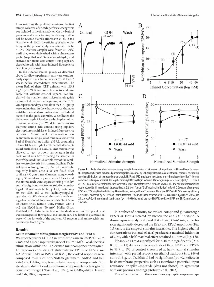
In a subset of neurons, we evoked compound glutamatergicEPSPs or EPSCs isolated by bicuculline and CGP 55845A. Adose–response analysis showed that ethanol (5– 66 mM) superfu-sion significantly decreased the EPSP and EPSC amplitudes (Fig.1A) across the range of stimulus intensities. The highest ethanolconcentrations (44 and 66 mM) produced a maximal inhibitionof 21%, with a half-maximal effect obtained at 14 mM (Fig. 1B).
Ethanol at 44 mM superfused for 7–10 min significantly ( p �0.05; n � 11) decreased the amplitude of these EPSPs and EPSCsto 71.9 � 4% of control (measured at half-maximal stimulusintensity), with partial recovery on ethanol washout (84 � 9% ofcontrol; Fig. 1A,C). Ethanol had no significant ( p � 0.1) effect onbasic membrane properties such as membrane potential, inputresistance, or spike amplitude (data not shown), in agreementwith our previous findings (Roberto et al., 2003).
The ethanol effect on these excitatory synaptic responses ap-
Figure 1. Acute ethanol decreases excitatory synaptic transmission in CeA neurons. A, Superfusion of 44 mM ethanol decreasedthe amplitude of evoked compound glutamatergic EPSCs isolated by GABAergic blockers. B, Concentration–response relationshipfor ethanol inhibition of compound glutamatergic EPSP and EPSC amplitudes in CeA neurons (ethanol superfused for 7–10 min;number of cells in parentheses). The logistic curve is plotted by Origin Software (Microcal) using y � (A1 – A2)/[sqb]1 � (x/xo) �
p � A2]. Parameters of the logistic curve were set at upper asymptote fixed at 21% and lower at 1%. The half-maximal inhibitionwas produced by 14 mM ethanol. Rate was fixed at 2.2, with “center” (half-maximal inhibition) unfixed. C, Decrease of compoundEPSP and EPSC amplitudes elicited by 44 mM ethanol, averaged from 11 neurons. The mean EPSPs and EPSCs were significantly( p � 0.05) decreased by 26 –39%. D, Pooled data from 17 neurons; in the presence of 30 �M bicuculline, 1 �M CGP 55845A, and20 �M D-AP-5, 44 mM ethanol significantly ( p � 0.05) decreased the non-NMDAR-mediated EPSP and EPSC amplitudes by15–26%.
1596 • J. Neurosci., February 18, 2004 • 24(7):1594 –1603 Roberto et al. • Ethanol Alters Glutamate in Amygdala

peared to be mediated primarily by a reduction of the non-NMDAR-mediated component. In fact, D-AP-5 (10 �M) did notsignificantly mitigate the inhibitory effect of ethanol on EPSPsand EPSCs, and in most neurons, the addition of 10 �M CNQXtotally blocked the EPSPs and EPSCs. Thus, in normal ACSF andat RMP, the evoked EPSPs and EPSCs are composed of non-NMDA glutamatergic components. In a subset of experiments(n � 4) without CGP 55845A (but still with bicuculline), therewas no quantitative or qualitative difference in the ethanol-induced inhibition of glutamatergic responses under theses condi-tions (data not shown). Thus, as in our previous report showingethanol augmentation of both GABA IPSPs and GABA currents inCeA in absence of a GABAB receptor antagonist (cf. Wan et al., 1996vs Roberto et al., 2003), GABAB receptors do not appear to play a rolein the ethanol inhibition of EPSP and EPSC amplitudes.
We studied the isolated non-NMDAR-mediated componentin 17 neurons held at RMP (�77 � 1 mV). In the presence ofD-AP-5, CGP 55845A, and bicuculline, ethanol superfusion (44mM, 7–10 min) significantly decreased the non-NMDAR-mediated EPSP and EPSC amplitudes by 15–26% across the stim-ulus strengths used, with recovery on washout (Fig. 1D). The I–Ocurves of the compound glutamatergic EPSPs and EPSCs andnon-NMDAR-mediated EPSPs and EPSCs measured before ap-plication of ethanol were comparable ( p � 0.05; data notshown), again indicating that in this experimental condition, theNMDAR-mediated component at CeA synapses is negligible.However, we could evoke D-AP-5-sensitive, voltage-dependentNMDAR-mediated EPSPs and EPSCs by using a low-Mg 2�
ACSF. Acute ethanol (44 mM) also decreased these pharmacolog-ically isolated NMDAR-mediated EPSPs and EPSCs by 15–20%(n � 17; p � 0.01) over the stimulus strengths used (Fig. 2A,B).
Effects of chronic ethanol on evoked glutamatergic EPSPsand EPSCsBecause glutamate receptors, and especially NMDARs, arethought to be involved in ethanol-related phenomena such astolerance, dependence, and withdrawal, we recorded from slicestaken from rats chronically exposed to ethanol vapors for 2– 4weeks. We prepared the CeA slices from CET rats exactly as forthe acute ethanol experiments and allowed the slices to withdrawfrom ethanol in the recording chamber.
To determine whether the CET animals were ethanol-dependent, at the end of the CET period, we assessed the perfor-mance of a separate sample of CET rats on several behavioral testsfor ethanol withdrawal severity. Withdrawal signs, evaluated by acommon behavioral rating scale for hyperactivity (ventromedialdistal limb flexion response, tail stiffness, and abnormal bodyposture and gait; Macey et al., 1996), were measured at 2, 4, 6, and8 hr after ethanol withdrawal in the CET group and comparedwith similar measures made in the naive group. Behavioral signsof ethanol withdrawal were evident in all six rats tested at 2– 8 hrafter the termination of the CET, a time frame corresponding tothe electrophysiological recordings.
However, we did not observe signs of postsynaptic neuronalhyperexcitability in CeA slices, such as prolonged depolarizationsor spontaneous burst discharges, after withdrawal. In fact, we didnot observe a significant difference between the baseline com-pound EPSP and EPSC I–O curves in slices from naive and CETanimals (data not shown). The baseline I–O curves for NMDAR-mediated EPSPs and EPSCs were also comparable in naive andCET rats (data not shown). However, baseline I–O curves for thenon-NMDAR-mediated EPSPs and EPSCs were significantlylower at the highest stimulus intensities in slices from CET ratscompared with slices from naive and sham rats (Fig. 3A). In slicestaken from CET rats after early withdrawal (2– 8 hr after ethanolexposure), superfusion of 44 mM ethanol significantly ( p � 0.01)decreased the evoked compound and non-NMDAR-mediatedEPSPs and EPSCs (Fig. 3B,C) by 20 � 6 and 19 � 7% of control,respectively, similar to the ethanol effect in slices taken fromnaive rats (compare Fig. 1C,D).
By contrast, the depressant effect of acute 44 mM ethanol onNMDA-EPSP and EPSC amplitudes was significantly ( p � 0.05)stronger in slices from CET rats (EPSPs and EPSCs reduced by25– 40%; Fig. 4A) than in those from naive rats (EPSPs andEPSCs reduced by 15–20%; Fig. 2). Ethanol at 44 mM significantly( p � 0.05) decreased the amplitude of NMDAR-mediated EPSPsin eight neurons from CET rats, with only partial recovery onwashout (Fig. 4B). Although the ethanol inhibition of NMDAR-mediated EPSPs was greater in neurons from CET rats comparedwith naive rats, the washout (recovery) values also were de-pressed (Fig. 4A,B). Therefore, we performed further experi-ments to determine whether the ethanol effect on NMDAR-mediated EPSPs and EPSCs was ethanol-specific and notattributable to NMDAR and channel rundown in CET rats. Firstwe recorded NMDAR-mediated EPSPs or EPSCs in neuronstaken from CET rats and superfused with ACSF without ethanolfor �45 min (Fig. 4C). We observed that the NMDAR-mediatedEPSP and EPSC amplitudes remained unaffected over such re-cording periods in both CET rats (n � 6; Fig. 4C) and naive rats(n � 3; data not shown). We then compared the effect of com-petitive (D-AP-5) and noncompetitive (ifenprodil) NMDAR an-tagonists on the ethanol-induced inhibition of NMDAR-mediated EPSPs and EPSCs. Ifenprodil provides near-maximalblock of NMDARs containing the NR2B subunit (Williams,2001), which is strongly implicated in ethanol effects. In low-
Figure 2. Acute ethanol decreases the amplitude of evoked NMDAR-mediated EPSPs andEPSCs. A, Representative NMDAR-mediated EPSCs evoked in the presence of 30 �M bicuculline,1 �M CGP 55845A, 10 �M CNQX, and low Mg 2� concentrations. Ethanol decreased the NMDAR-mediated EPSC amplitudes with partial recovery on washout. The subsequent addition ofD-AP-5 completely blocked the EPSCs, implicating NMDARs. B, In slices from naive rats, super-fusion of 44 mM ethanol significantly ( p � 0.01; n � 17) reduced evoked NMDAR-mediatedEPSPs and EPSCs by 15–20%.
Roberto et al. • Ethanol Alters Glutamate in Amygdala J. Neurosci., February 18, 2004 • 24(7):1594 –1603 • 1597

Mg 2� ACSF containing GABA receptorblockers and CNQX, D-AP-5 (10 �M)completely blocked NMDAR-mediatedEPSPs and EPSCs (n � 6; data not shown).Ifenprodil (10 �M) had a small effect inneurons of naive rats (5–15%; n � 6), butsignificantly reduced the NMDAR-mediated EPSP and EPSC amplitudes by20 –30% in neurons of CET rats ( p � 0.05compared with baseline or ifenprodil innaive slices; n � 5; Fig. 5). Interestingly,superfusion of 44 mM ethanol in the pres-ence of ifenprodil did not further depressNMDAR-mediated EPSPs and EPSCs.
Effects of acute and chronic ethanolon PPFTo determine whether the inhibitory effectof ethanol on glutamatergic transmissioninvolved presynaptic changes in glutamaterelease, we examined PPF (50, 100, and180 msec interpulse intervals) of com-pound and non-NMDAR- and NMDAR-mediated EPSPs and EPSCs in CeA neu-rons before, during, and after ethanolapplication. Changes in PPF are thoughtto be inversely related to changes in trans-mitter release such that enhanced proba-bility of release is associated with a reduc-tion of PPF (Mennerick and Zorumski,1995; Bonci and Williams, 1997). Wefound that baseline PPF of non-NMDAR-and NMDAR-mediated EPSPs and EPSCsin neurons from naive and CET animalswas similar at all interpulse intervals tested(Fig. 6B). Also, superfusion of 44 mM eth-anol onto control slices had little effect onPPF of NMDAR-mediated EPSPs andEPSCs (Fig. 6C). By contrast, acute 44 mM
ethanol significantly ( p � 0.05) decreasedPPF of NMDAR-mediated EPSPs andEPSCs by 15% in slices from CET rats (n �16; Fig. 6A,C), suggesting an acuteethanol-induced increase in glutamate re-lease and a neuroadaptative change afterCET. Acute ethanol had equivalent effectson PPF of both compound and non-NMDAR-mediated EPSPs and EPSCs;that is, there was no acute ethanol effect onPPF in naive rats, but PPF was decreasedby 15–22% in CET rats (data not shown).This effect returned to control levels after 2weeks of withdrawal (our unpublishedobservations).
Effects of acute and chronic ethanol onpostsynaptic NMDARsBecause acute ethanol reduced NMDAR-mediated EPSP and EPSC amplitudes withlittle effect on PPF in neurons of naive rats,we hypothesized that the ethanol effectmay take place at postsynaptic sites, per-haps on the NMDAR itself. To test this hy-
Figure 3. CET reduces baseline non-NMDA transmission but does not alter acute ethanol effects. A, I–O curves of non-NMDAR-mediated EPSP and EPSC amplitudes evoked by local stimulation. At higher stimulus strengths, the baseline response is signifi-cantly (*p � 0.05) reduced in slices taken from CET rats compared with controls. B, Acute ethanol significantly ( p � 0.05)decreased by 20% compound EPSPs and EPSCs in slices from CET rats, to a similar extent as those from control slices. C, In neuronsof CET rats, acute ethanol significantly ( p �0.05; n �11) reduced non-NMDAR-mediated EPSPs and EPSCs (with partial recoveryon washout) to a similar extent as in control slices.
Figure 4. CET potentiates the inhibitory effect of acute ethanol on NMDAR-mediated EPSPs and EPSCs in CeA. A, Acute ethanolreduced NMDAR-mediated EPSP and EPSC amplitudes by 35– 45% in neurons from CET rats (n � 15) compared with 15–20% inneurons from naive rats (Fig. 2 B), suggesting sensitization to acute ethanol. B, Ethanol superfusion significantly ( p � 0.001; n �8) decreased evoked NMDAR-mediated EPSP amplitudes across the voltage range tested (with only partial recovery on washout),especially at the more depolarized potentials. C, In neurons from CET rats, evoked NMDAR-mediated EPSPs and EPSCs remainedstable for �45 min of superfusion in the absence of ethanol.
Figure 5. The NR2B blocker ifenprodil occludes the depressant effect of acute ethanol. A, Bath application of 10 �M ifenprodil(IFEN) for 10 min decreased the NMDAR-mediated EPSP and EPSC amplitudes (n � 6). Subsequent superfusion of 44 mM ethanol(EtOH) did not further affect the NMDAR-mediated EPSPs and EPSCs in these neurons taken from naive rats. B, Time course recorddepicting the sequential application of 10 �M ifenprodil and 44 mM ethanol on NMDAR-mediated EPSC amplitudes in a neuronfrom a CET rat. C, Summary of the effect of ifenprodil and ethanol on five CeA neurons from CET rats. Ifenprodil significantly ( p �0.05) decreased NMDAR-mediated EPSP and EPSC amplitudes and prevented the ethanol effect, with full recovery on drugwashout.
1598 • J. Neurosci., February 18, 2004 • 24(7):1594 –1603 Roberto et al. • Ethanol Alters Glutamate in Amygdala
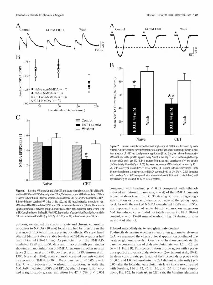
pothesis, we studied the effects of acute and chronic ethanol onresponses to NMDA (10 mM) locally applied by pressure in thepresence of TTX to minimize presynaptic effects. We superfusedethanol (44 mM) after a stable baseline of NMDA responses hadbeen obtained (10 –15 min). As predicted from the NMDAR-mediated EPSP and EPSC data and in accord with past studiesshowing ethanol inhibition of NMDA responses in other neurontypes (Hoffman et al., 1989; Lovinger et al., 1989; Simson et al.,1993; Nie et al., 1994), acute ethanol decreased currents elicitedby exogenous NMDA to 70 � 5% of baseline ( p � 0.05; n � 4;Fig. 7) with recovery on washout. In addition, as with theNMDAR-mediated EPSPs and EPSCs, ethanol superfusion elic-ited a significantly greater inhibition (to 47 � 7%; p � 0.001
compared with baseline; p � 0.05 compared with ethanol-induced inhibition in naive rats; n � 4) of the NMDA currentsevoked in slices taken from CET rats (Fig. 7), again suggesting asensitization or reverse tolerance but now at the postsynapticlevel. As with the evoked NMDAR-mediated EPSPs and EPSCs,the depressant effect of acute 44 mM ethanol on exogenousNMDA-induced currents did not totally recover (to 82 � 10% ofcontrol; n � 3; 15–20 min of washout; Fig. 7) during or afterwashout of ethanol.
Ethanol microdialysis: in vivo glutamate contentTo directly determine whether ethanol alters glutamate release inCeA, we measured the effects of local application of ethanol dia-lysate on glutamate levels in CeA in vivo. In sham control rats, thebaseline concentration of dialysate glutamate was 1.2 � 0.2 �M
(n � 11; Fig. 8B). This concentration profile agrees with a previ-ous report of amygdala dialysate levels (Quertemont et al., 1998).In sham control rats, perfusion of the microdialysis probe with0.1, 0.3, and 1.0 M ethanol into the CeA did not significantly ( p �0.05) alter the local dialysate glutamate levels (increase comparedwith baseline, 114 � 72, 43 � 110, and 153 � 139 nM, respec-tively; Fig. 8C). In contrast, in CET rats, the baseline glutamate
Figure 6. Baseline PPF is unchanged after CET, and acute ethanol decreases PPF of NMDAR-mediated EPSPs and EPSCs but only after CET. A, Voltage records of NMDAR-mediated EPSPs inresponse to two stimuli 100 msec apart in a neuron from a CET rat. Acute ethanol reduced PPF.B, Pooled data of baseline PPF ratios (at 50, 100, and 180 msec interpulse intervals) of non-NMDAR- and NMDAR-mediated EPSPs and EPSCs in neurons of naive and CET rats. There was nosignificant difference between groups. C, Pooled data of PPF ratio expressed as the second EPSPor EPSC amplitude over the first EPSP or EPSC. Superfusion of ethanol significantly decreased thePPF ratio in neurons from CET (by 15%; *p � 0.05; n � 16) but not naive (n � 10) rats.
Figure 7. Inward currents elicited by local application of NMDA are decreased by acuteethanol. A, Representative current records before, during, and after ethanol superfusion (8 min)from a neuron of a CET rat. Local pressure application (2 sec, 6 psi; bars above the records) ofNMDA (10 mM in the pipette, applied every 2 min) in low-Mg 2� ACSF containing GABAergicblockers CNQX and 1 �M TTX. B, In 4 neurons from naive rats, superfusion of 44 mM ethanol(5–10 min) significantly (*p � 0.05) decreased exogenous NMDA-induced currents by 30 �5%, with recovery on washout (95 � 7% of control, 10 –15 min). In four neurons from CET rats,44 mM ethanol more strongly decreased NMDA currents by 53 � 7% (*p � 0.001 comparedwith baseline; #p � 0.05 compared with ethanol-induced inhibition in control slices) withpartial recovery on washout (to 82 � 10% of control).
Roberto et al. • Ethanol Alters Glutamate in Amygdala J. Neurosci., February 18, 2004 • 24(7):1594 –1603 • 1599

microdialysate content was significantly ( p � 0.05; n � 7) in-creased (2.6 � 0.8 �M) compared with that in naive rats (1.2 �0.2 �M; Fig. 8B). Furthermore, local administration of 0.1, 0.3,and 1.0 M ethanol also led to a significant ( p � 0.05) dose-dependent increase in dialysate glutamate levels (increase com-pared with baseline, 413.3 � 107.9, 522 � 125, and 787.4 � 116.0nm, respectively; Fig. 8C). A representative microdialysis timecourse record shows that in each case, dialysate glutamate levelsreturned to preethanol baseline levels when the perfusion me-dium was replaced with ethanol-free ACSF (Fig. 8A).
DiscussionWe have shown that acute ethanol decreases both NMDAR- andnon-NMDAR-mediated responses in CeA slices taken from bothnaive and CET rats. In slices from CET rats, acute ethanol super-fusion selectively inhibited NMDAR-mediated EPSPs and EPSCsto a greater degree than in those from control rats, concomitantwith a decreased PPF suggestive of an increase in glutamate re-lease. Acute ethanol also elicited a more pronounced inhibitionof the NMDA currents evoked by locally applied NMDA (in the
presence of TTX) in slices taken from CET rats compared withthose from naive rats, suggesting a sensitization or reverse toler-ance to ethanol at the postsynaptic level. The inhibition ofNMDAR-mediated EPSPs and EPSCs by the noncompetitiveNR2B-specific antagonist ifenprodil also was increased in neu-rons from CET rats. Ifenprodil also completely occluded the eth-anol inhibition of NMDAR-mediated EPSPs and EPSCs, suggest-ing that the ethanol effect was mediated by NR2B subunits. Usingmicrodialysis, we found that in CET rats the intra-CeA adminis-tration of ethanol (0.1–1.0 M) via reverse microdialysis signifi-cantly increased dialysate glutamate content compared with na-ive rats, supporting the PPF data suggesting a presynaptic action.Overall, our findings support the hypothesis that ethanol intoxi-cation, reinforcement, or both may be associated with depressionof glutamatergic transmission, whereas the dysregulation of glu-tamatergic synapses resulting from the combination of increasedNMDAR sensitivity to ethanol and increased glutamate releasemay contribute to ethanol dependence.
A prominent electrophysiological effect of acute ethanol is adecrease in current generated by NMDAR activation (Kumariand Ticku, 2000; Woodward, 2000; Lovinger, 2002). The abilityof ethanol to inhibit NMDARs may vary among brain regions(Yang et al., 1996; Kumari and Ticku, 2000; Criswell et al., 2003).Although the exact mechanisms underlying such variability re-main to be elucidated at the molecular level, it is likely that theyreflect differences in the various isoforms constituting theNMDA–receptor complex (Koltchine et al., 1993; Mirshahi andWoodward, 1995) as well as other neuron-specific factors (e.g.,subunit phosphorylation) that may modulate the sensitivity ofNMDAR channels to ethanol (Kuner et al., 1993; Masood et al.,1994). For example, the degree of ethanol inhibition may be de-pendent on the NR2 subunit expressed; NMDARs with NR1/2Aand NR1/2B subunits may be the most sensitive to ethanol (Lov-inger, 1995; Mirshahi and Woodward, 1995; Allgaier, 2002). Ad-aptations within the glutamatergic system have been suggested tocontribute to ethanol tolerance and dependence and both acuteand protracted features of ethanol withdrawal (Tabakoff andHoffman, 1996; Tsai and Coyle, 1998; Krystal et al., 2003). CETalters the glutamatergic system in several brain regions, includinghippocampus, cerebral cortex, striatum, and thalamus (Lovinger,1997; Little, 1999; Kumari and Ticku, 2000; Dettmer et al., 2003;Kumari et al., 2003). However, the exact molecular mechanismsresponsible for such effects after CET remain to be determined.CET increases the number of binding sites for several NMDARligands and alters NMDAR subunit expression (Trevisan et al.,1994; Kalluri et al., 1998; Kumari and Ticku, 2000), which couldreflect an adaptive upregulation of NMDAR complexes in re-sponse to long-term ethanol exposure.
We recorded from CeA slices that were allowed to undergowithdrawal 2– 8 hr after removal from CET, and we did not ob-serve postsynaptic hyperexcitability. In these slices, the basalcompound glutamatergic synaptic responses and the baselinePPF were comparable in CeA neurons from naive and CET rats.However, we found a significant difference between CeA neuronsof these two groups in the I–O curves for the non-NMDAR-mediated EPSPs and EPSCs evoked over the highest range ofstimulus strengths. Furthermore, acute superfusion of ethanolalso decreased the amplitudes of both evoked NMDAR-mediatedEPSPs and EPSCs and the responses to locally applied NMDA toa greater extent in CeA slices of CET rats than from naive rats,suggesting sensitization of NMDARs to ethanol after CET. Thisreceptor sensitization could represent the cellular underpinningsfor some aspects of behavioral sensitization seen in dependent
Figure 8. In vivo dialysate levels of glutamate in CeA. A, Representative samples of dialysateglutamate levels from naive (squares) and CET (circles) rats depicting the effect of ethanoladministration (0.1, 0.3, and 1.0 M; bars). B, In CET rats, the baseline dialysate glutamate levelwas increased (2.6 � 0.8 �M; n � 7) compared with that in naive rats (1.2 � 0.2 �M; *p �0.05; n � 11). C, In naive rats, local in vivo ethanol administration (0.1, 0.3, and 1.0 M) into theCeA did not significantly ( p � 0.05) alter local dialysate glutamate (Glu) levels (increase com-pared with baseline, 114 � 72, 43 � 110, and 153 � 139, nM, respectively; n � 11). Inagreement with our in vitro electrophysiological results, only in CET rats did local infusion ofethanol produce a significant dose-dependent increase in dialysate glutamate levels (increasecompared with baseline, 413.3 � 107.9, 522 � 125, and 787.4 � 116.0 nm, respectively; n �7; *p � 0.05). Dialysates were normalized to baseline level.
1600 • J. Neurosci., February 18, 2004 • 24(7):1594 –1603 Roberto et al. • Ethanol Alters Glutamate in Amygdala

animals (Xie et al., 1995; Darbra et al., 2002; Quadros et al., 2002;Broadbent et al., 2003; Meyer and Phillips, 2003). It has beenreported that in the CeA the NMDARs are composed mainly ofNR1 and NR2B subunits, and there is no developmental changein subunit composition (Lopez de Armentia and Sah, 2003; Sahand Lopez De Armentia, 2003). Interestingly, we found that theNR2B subunit-selective antagonist ifenprodil is more effective ininhibiting the NMDAR-mediated EPSPs and EPSCs and in oc-cluding the ethanol effect in CET rats than in naive rats. Thesefindings suggest that an increase in NR2B subunit compositionmay be involved in the acute effect of ethanol after CET.
There may be both direct (on NMDAR subunits) and indirect(on intracellular targets such as kinases and phosphatases) mech-anisms that mediate ethanol effects on the NMDARs. We hy-pothesize that, in CeA, increased ethanol inhibition of NMDARsalso could be attributable to changes in tyrosine phosphorylationof NR2B subunits. Potential mechanisms downstream of changesin phosphorylation include modulation of channel properties,regulation of subunit interactions, and altered receptor localiza-tion (e.g., greater surface expression of the receptors). It is alsopossible that the responsiveness of the trafficking function of thereceptors to acute ethanol may increase during CET and may leadto alterations of internalization or of insertion of the NMDARs.Recent studies of NMDARs have focused on dynamic regulationof surface expression as a mechanism of ethanol action (Maldveet al., 2002; Alvestad et al., 2003; Yaka et al., 2003). These studiesindicate that the inhibitory effect of ethanol on NMDARs in-volves a reduced tyrosine phosphorylation of NR2A and NR2Bsubunits, possibly via activation of a tyrosine phosphatase. Spe-cifically, a Fyn kinase determines NMDAR sensitivity to ethanol(Yaka et al., 2003), and tyrosine phosphorylation of this Fyn ki-nase site is reduced by ethanol (Alvestad et al., 2003). Therefore,ethanol induces a phosphorylation-dependent enhancement ofNMDAR channels. An alternative hypothesis involves the trans-location of NMDARs between synaptic and extrasynaptic sites inresponse to ethanol. Molecular studies are now in progress in ourlaboratory to determine whether this sensitization is associatedwith changes in the phosphorylation status of the NR2B-containing NMDARs (nontranscriptional mechanisms), up-regulation in NR2B mRNA transcription, or both.
A few reports have shown that ethanol decreases glutamaterelease in the hippocampus (Moghaddam and Bolinao, 1994;Reynolds and Brien, 1994; Gonzales and Brown, 1995; Tsai,1998). Here we estimated the interstitial glutamate concentra-tions in the CeA using microdialysis in freely moving rats. Inagreement with our in vitro electrophysiological results, the invivo data showed that acute ethanol had no effect on glutamaterelease in naive rats but increased glutamate release in CET rats.We also found that baseline dialysate levels of glutamate in theCeA were elevated in dependent animals, suggesting that the in-crease in the glutamate content may be related to changes inpresynaptic function after the CET and could represent a com-pensatory response to the continuous inhibitory effect (attribut-able to the reverse tolerance; see above) of ethanol on postsynap-tic glutamate receptors. This enhancement in glutamate levels orrelease could also represent a response by the CeA to oppose theinhibitory effects of acute ethanol caused by the enhancement ofGABAergic neurotransmission previously reported by our labo-ratory (Roberto et al., 2003). The increase in dialysate glutamatecould represent synaptically released glutamate, carrier-mediated release, a change in glial metabolism, or a combinationthereof (Timmerman and Westerink, 1997). Thus, part of theamino acid content of the dialysate may originate from non-
impulse-dependent sources. Further studies (e.g., calcium re-moval or TTX inclusion) will ascertain whether the enhancementof glutamate dialysate is attributable to neuronal release, gluta-mate transporters, or both.
In conclusion, we have shown that acute and chronic ethanolsignificantly alters CeA glutamatergic transmission, involvingboth presynaptic and postsynaptic sites. Whereas acute ethanolappears to act predominantly at postsynaptic sites to decreaseresponses to glutamate, chronic ethanol causes an increased sen-sitivity to acute ethanol of postsynaptic NMDARs and increasedpresynaptic glutamatergic release. These changes with chronicethanol may represent neuroadaptations or allostatic responsesunderlying behavioral signs of ethanol dependence (Koob and LeMoal, 2001). Although these two persistent effects might seem tocounter each other in the presence of ethanol, they could bias thesynapses toward hyperstimulation of non-NMDARs and re-duced NMDAR function, resulting in altered spatial and tempo-ral integrative properties of CeA neurons. The combination ofthese effects with enhanced GABAergic transmission by ethanol(Roberto et al., 2003) may tend to depress neuronal activity ofCeA neurons, especially in the depolarized range. Further studiesof ethanol effects on the integrative network and plasticity char-acteristics of the amygdala may help further elucidate the biolog-ical substrates of the reinforcing effects of ethanol consumptionand how those effects change during the development of depen-dence.
Note added in proof. We overlooked a microdialysis study by Rossettiand Carboni (1995) showing increased glutamate levels in striatum12–24 hr after withdrawal from systemic chronic ethanol treatment.
ReferencesAllgaier C (2002) Ethanol sensitivity of NMDA receptors. Neurochem Int
41:377–382.Alvestad RM, Grosshans DR, Coultrap SJ, Nakazawa T, Yamamoto T, Brown-
ing MD (2003) Tyrosine dephosphorylation and ethanol inhibition ofN-Methyl-D-aspartate receptor function. J Biol Chem 278:11020 –11025.
Bonci A, Williams JT (1997) Increased probability of GABA release duringwithdrawal from morphine. J Neurosci 17:796 – 803.
Broadbent J, Kampmueller KM, Koonse SA (2003) Expression of behavioralsensitization to ethanol by DBA/2J mice: the role of NMDA and non-NMDA glutamate receptors. Psychopharmacology 167:225–234.
Calton JL, Wilson WA, Moore SD (1998) Magnesium-dependent inhibi-tion of N-methyl-D-aspartate receptor-mediated synaptic transmissionby ethanol. J Pharmacol Exp Ther 287:1015–1019.
Criswell HE, Ming Z, Griffith BL, Breese GR (2003) Comparison of effect ofethanol on N-methyl-D-aspartate- and GABA-gated currents fromacutely dissociated neurons: absence of regional differences in sensitivityto ethanol. J Pharmacol Exp Ther 304:192–199.
Darbra S, Prat G, Pallares M, Ferre N (2002) Tolerance and sensitization tothe hypnotic effects of alcohol induced by chronic voluntary alcohol in-take in rats. J Psychopharmacol 16:79 – 83.
Davis M, Rainnie D, Cassell M (1994) Neurotransmission in the rat amyg-dala related to fear and anxiety. Trends Neurosci 17:208 –214.
Delaney AJ, Sah P (1999) GABA receptors inhibited by benzodiazepinesmediate fast inhibitory transmission in the central amygdala. J Neurosci19:9698 –9704.
Dettmer TS, Barnes A, Iqbal U, Bailey CD, Reynolds JN, Brien JF, ValenzuelaCF (2003) Chronic prenatal ethanol exposure alters ionotropic gluta-mate receptor subunit protein levels in the adult guinea pig cerebral cor-tex. Alcohol Clin Exp Res 27:677– 681.
Eckardt MJ, File SE, Gessa GL, Grant KA, Guerri C, Hoffman PL, Kalant H,Koob GF, Li TK, Tabakoff B (1998) Effects of moderate alcohol con-sumption on the central nervous system. Alcohol Clin Exp Res22:998 –1040.
Frantz KJ, Hansson KJ, Stouffer DG, Parsons LH (2002) 5-HT6 receptorantagonism potentiates the behavioral and neurochemical effects of am-phetamine but not cocaine. Neuropharmacology 42:170 –180.
Gonzales RA, Brown LM (1995) Brain regional differences in glycine rever-
Roberto et al. • Ethanol Alters Glutamate in Amygdala J. Neurosci., February 18, 2004 • 24(7):1594 –1603 • 1601

sal of ethanol-induced inhibition of N-methyl-D-aspartate-stimulatedneurotransmitter release. Life Sci 56:571–577.
Gonzales R, Tang A, Robinson D (2002) Quantitative microdialysis for invivo studies of pharmacodynamics. In: Methods in alcohol-related neu-roscience research (Liu Y, Lovinger D, eds), pp 287–317. New York: CRC.
Hoffman PL, Rabe CS, Moses F, Tabakoff B (1989) N-Methyl-D-aspartatereceptors and ethanol: inhibition of calcium flux and cyclic GMP produc-tion. J Neurochem 52:1937–1940.
Hyytia P, Koob GF (1995) GABAA receptor antagonism in the extendedamygdala decreases ethanol self-administration in rats. Eur J Pharmacol283:151–159.
Kalivas PW, Alesdatter JE (1993) Involvement of N-methyl-D-aspartate re-ceptor stimulation in the ventral tegmental area and amygdala in behav-ioral sensitization to cocaine. J Pharmacol Exp Ther 267:486 – 495.
Kalluri HS, Mehta AK, Ticku MK (1998) Up-regulation of NMDA receptorsubunits in rat brain following chronic ethanol treatment. Brain Res MolBrain Res 58:221–224.
Koltchine V, Anantharam V, Wilson A, Bayley H, Treistman SN (1993) Ho-momeric assemblies of NMDAR1 splice variants are sensitive to ethanol.Neurosci Lett 152:13–16.
Koob GF, Le Moal M (2001) Drug addiction, dysregulation of reward, andallostasis. Neuropsychopharmacology 24:97–129.
Koob GF, Roberts AJ, Schulteis G, Parsons LH, Heyser CJ, Hyytia P, Merlo-Pich E, Weiss F (1998) Neurocircuitry targets in ethanol reward anddependence. Alcohol Clin Exp Res 22:3–9.
Krystal JH, Petrakis IL, Mason G, Trevisan L, D’Souza DC (2003)N-Methyl-D-aspartate glutamate receptors and alcoholism: reward, de-pendence, treatment, and vulnerability. Pharmacol Ther 99:79 –94.
Kumari M, Ticku MK (2000) Regulation of NMDA receptors by ethanol.Prog Drug Res 54:152–189.
Kumari M, Anji A, Woods Jr H, Ticku MK (2003) The molecular effects ofalcohol: clues to the enigmatic action of alcohol. Ann NY Acad Sci 993:82–94, 123–124.
Kuner T, Schoepfer R, Korpi ER (1993) Ethanol inhibits glutamate-inducedcurrents in heteromeric NMDA receptor subtypes. NeuroReport5:297–300.
Little HJ (1999) The contribution of electrophysiology to knowledge of theacute and chronic effects of ethanol. Pharmacol Ther 84:333–353.
Lopez de Armentia M, Sah P (2003) Development and subunit compositionof synaptic NMDA receptors in the amygdala: NR2B synapses in the adultcentral amygdala. J Neurosci 23:6876 – 6883.
Lovinger DM (1995) Developmental decrease in ethanol inhibition ofN-methyl-D-aspartate receptors in rat neocortical neurons: relation to theactions of ifenprodil. J Pharmacol Exp Ther 274:164 –172.
Lovinger DM (1997) Alcohols and neurotransmitter gated ion channels:past, present and future. Naunyn Schmiedebergs Arch Pharmacol356:267–282.
Lovinger DM (2002) NMDA receptors lose their inhibitions. Nat Neurosci5:614 – 616.
Lovinger DM, White G, Weight FF (1989) Ethanol inhibits NMDA-activated ion current in hippocampal neurons. Science 243:1721–1724.
Macey DJ, Schulteis G, Heinrichs SC, Koob GF (1996) Time-dependentquantifiable withdrawal from ethanol in the rat: effect of method of de-pendence induction. Alcohol 13:163–170.
Maldve RE, Zhang TA, Ferrani-Kile K, Schreiber SS, Lippmann MJ, SnyderGL, Fienberg AA, Leslie SW, Gonzales RA, Morrisett RA (2002)DARPP-32 and regulation of the ethanol sensitivity of NMDA receptorsin the nucleus accumbens. Nat Neurosci 5:641– 648.
Martin D, Tayyeb MI, Swartzwelder HS (1995) Ethanol inhibition of AMPAand kainate receptor-mediated depolarizations of hippocampal area CA1.Alcohol Clin Exp Res 19:1312–1316.
Masood K, Wu C, Brauneis U, Weight FF (1994) Differential ethanol sensi-tivity of recombinant N-methyl-D-aspartate receptor subunits. Mol Phar-macol 45:324 –329.
Mennerick S, Zorumski CF (1995) Paired-pulse modulation of fast excita-tory synaptic currents in microcultures of rat hippocampal neurons.J Physiol (Lond) 488:85–101.
Meyer PJ, Phillips TJ (2003) Bivalent effects of MK-801 on ethanol-inducedsensitization do not parallel its effects on ethanol-induced tolerance. Be-hav Neurosci 117:641– 649.
Mirshahi T, Woodward JJ (1995) Ethanol sensitivity of heteromeric NMDA
receptors: effects of subunit assembly, glycine and NMDAR1 Mg(2�)-insensitive mutants. Neuropharmacology 34:347–355.
Moghaddam B, Bolinao ML (1994) Biphasic effect of ethanol on extracellu-lar accumulation of glutamate in the hippocampus and the nucleus ac-cumbens. Neurosci Lett 178:99 –102.
Nie Z, Madamba SG, Siggins GR (1994) Ethanol inhibits glutamatergic neu-rotransmission in nucleus accumbens neurons by multiple mechanisms.J Pharmacol Exp Ther 271:1566 –1573.
Nose I, Higashi H, Inokuchi H, Nishi S (1991) Synaptic responses of guineapig and rat central amygdala neurons in vitro. J Neurophysiol65:1227–1241.
Paxinos G, Watson C (1986) The rat brain in sterotaxic coordinates. SanDiego: Academic.
Pich EM, Lorang M, Yeganeh M, Rodriguez de Fonseca F, Raber J, Koob GF,Weiss F (1995) Increase of extracellular corticotropin-releasing factor-like immunoreactivity levels in the amygdala of awake rats during re-straint stress and ethanol withdrawal as measured by microdialysis. J Neu-rosci 15:5439 –5447.
Post RM, Weiss SR, Pert A (1987) The role of context and conditioning inbehavioral sensitization to cocaine. Psychopharmacol Bull 23:425– 429.
Quadros IM, Hipolide DC, Frussa-Filho R, De Lucca EM, Nobrega JN, Souza-Formigoni ML (2002) Resistance to ethanol sensitization is associatedwith increased NMDA receptor binding in specific brain areas. EurJ Pharmacol 442:55– 61.
Quertemont E, de Neuville J, De Witte P (1998) Changes in the amygdalaamino acid microdialysate after conditioning with a cue associated withethanol. Psychopharmacology 139:71–78.
Rassnick S, Heinrichs SC, Britton KT, Koob GF (1993) Microinjection of acorticotropin-releasing factor antagonist into the central nucleus of theamygdala reverses angiogenic-like effects of ethanol withdrawal. BrainRes 605:25–32.
Reynolds JD, Brien JF (1994) Effects of acute ethanol exposure on glutamaterelease in the hippocampus of the fetal and adult guinea pig. Alcohol11:259 –267.
Roberto M, Madamba SG, Moore SD, Tallent MK, Siggins GR (2003) Eth-anol increases GABAergic transmission at both pre- and postsynaptic sitesin rat central amygdala neurons. Proc Natl Acad Sci USA 100:2053–2058.
Roberts AJ, Cole M, Koob GF (1996) Intra-amygdala muscimol decreasesoperant ethanol self-administration in dependent rats. Alcohol Clin ExpRes 20:1289 –1298.
Robinson DL, Lara JA, Brunner LJ, Gonzales RA (2000) Quantification ofethanol concentrations in the extracellular fluid of the rat brain: in vivocalibration of microdialysis probes. J Neurochem 75:1685–1693.
Robledo P, Robbins TW, Everitt BJ (1996) Effects of excitotoxic lesions ofthe central amygdaloid nucleus on the potentiation of reward-relatedstimuli by intra-accumbens amphetamine. Behav Neurosci 110:981–990.
Rogers J, Wiener SG, Bloom FE (1979) Long-term ethanol administrationmethods for rats: advantages of inhalation over intubation or liquid diets.Behav Neural Biol 27:466 – 486.
Rossetti ZL, Carboni S (1995) Ethanol withdrawal is associated with in-creased extracellular glutamate in the rat striatum. Eur J Pharmacol 283:177–183.
Sah P, Lopez De Armentia M (2003) Excitatory synaptic transmission in thelateral and central amygdala. Ann NY Acad Sci 985:67–77.
Simson PE, Criswell HE, Breese GR (1993) Inhibition of NMDA-evokedelectrophysiological activity by ethanol in selected brain regions: evidencefor ethanol-sensitive and ethanol-insensitive NMDA-evoked responses.Brain Res 607:9 –16.
Tabakoff B, Hoffman PL (1996) Alcohol addiction: an enigma among us.Neuron 16:909 –912.
Timmerman W, Westerink BH (1997) Brain microdialysis of GABA andglutamate: what does it signify? Synapse 27:242–261.
Trevisan L, Fitzgerald LW, Brose N, Gasic GP, Heinemann SF, Duman RS,Nestler EJ (1994) Chronic ingestion of ethanol up-regulates NMDAR1receptor subunit immunoreactivity in rat hippocampus. J Neurochem62:1635–1638.
Tsai G (1998) Glutamatergic neurotransmission in alcoholism. J BiomedSci 5:309 –320.
Tsai G, Coyle JT (1998) The role of glutamatergic neurotransmission in thepathophysiology of alcoholism. Annu Rev Med 49:173–184.
1602 • J. Neurosci., February 18, 2004 • 24(7):1594 –1603 Roberto et al. • Ethanol Alters Glutamate in Amygdala

Vanderschuren LJ, Kalivas PW (2000) Alterations in dopaminergic and glu-tamatergic transmission in the induction and expression of behavioralsensitization: a critical review of preclinical studies. Psychopharmacology151:99 –120.
Wan FJ, Berton F, Madamba SG, Francesconi W, Siggins GR (1996) Lowethanol concentrations enhance GABAergic inhibitory postsynaptic po-tentials in hippocampal pyramidal neurons only after block of GABABreceptors. Proc Natl Acad Sci USA 93:5049 –5054.
Watanabe T, Nakagawa T, Yamamoto R, Maeda A, Minami M, Satoh M(2002) Involvement of glutamate receptors within the central nucleus ofthe amygdala in naloxone-precipitated morphine withdrawal-inducedconditioned place aversion in rats. Jpn J Pharmacol 88:399 – 406.
Williams K (2001) Ifenprodil, a novel NMDA receptor antagonist: site andmechanism of action. Curr Drug Targets 2:285–298.
Wolf ME, Dahlin SL, Hu XT, Xue CJ, White K (1995) Effects of lesions ofprefrontal cortex, amygdala, or fornix on behavioral sensitization to
amphetamine: comparison with N-methyl-D-aspartate antagonists. Neu-roscience 69:417– 439.
Woodward JJ (1999) Ionotropic glutamate receptors as sites of action forethanol in the brain. Neurochem Int 35:107–113.
Woodward JJ (2000) Ethanol and NMDA receptor signaling. Crit Rev Neu-robiol 14:69 – 89.
Xie ZC, Buckner E, Commissaris RL (1995) Anticonflict effect of MK-801 inrats: time course and chronic treatment studies. Pharmacol Biochem Be-hav 51:635– 640.
Yaka R, Phamluong K, Ron D (2003) Scaffolding of Fyn kinase to theNMDA receptor determines brain region sensitivity to ethanol. J Neuro-sci 23:3623–3632.
Yang X, Criswell HE, Simson P, Moy S, Breese GR (1996) Evidence for aselective effect of ethanol on N-methyl-D-aspartate responses: ethanolaffects a subtype of the ifenprodil-sensitive N-methyl-D-aspartate recep-tors. J Pharmacol Exp Ther 278:114 –124.
Roberto et al. • Ethanol Alters Glutamate in Amygdala J. Neurosci., February 18, 2004 • 24(7):1594 –1603 • 1603


![Glutamatergic neurotransmission and synaptic plasticity: molecular, clinical, and phylogenetic aspects [Portuguese]](https://img.pdfslide.net/doc/110x75/634c6757738f1906320e170a/glutamatergic-neurotransmission-and-synaptic-plasticity-molecular-clinical-and.jpg)


