Embed Size (px)
Citation preview

lable at ScienceDirect
Fish & Shellfish Immunology 38 (2014) 25e33
Contents lists avai
Fish & Shellfish Immunology
journal homepage: www.elsevier .com/locate / fs i
Full length article
Adaptive and innate immune molecules in developing rainbow trout,Oncorhynchus mykiss eggs and larvae: Expression of genes andoccurrence of effector molecules
Rasmus D. Heinecke*, Jiwan K. Chettri, Kurt BuchmannLaboratory of Aquatic Pathobiology, Department of Veterinary Disease Biology, Faculty of Health and Medical Sciences, University of Copenhagen,Stigbøjlen 7, DK-1870 Frederiksberg C, Denmark
a r t i c l e i n f o
Article history:Received 24 October 2013Received in revised form12 February 2014Accepted 14 February 2014Available online 21 February 2014
Keywords:OntogenyImmune systemRainbow troutEggGene expression
* Corresponding author. Tel.: þ45 35333136; fax: þE-mail addresses: [email protected] (R.D. Heinecke),
[email protected] (K. Buchmann).
http://dx.doi.org/10.1016/j.fsi.2014.02.0101050-4648/� 2014 Elsevier Ltd. All rights reserved.
a b s t r a c t
The ontogenetic development of the immune system was studied during the egg phase and the earlypost-hatch period of rainbow trout. Quantitative real-time PCR (qPCR) was used to assess the timing anddegree of expression of 9 important immune relevant genes and EF1-a. Further, immunohistochemicalstaining using monoclonal antibodies was applied on rainbow trout embryos and larvae in order tolocalize five different protein molecules (MHCII, CD8, IgM, IgT and SAA) in the developing tissue andimmune organs. Maternally transferred transcripts of EF1-a mRNAwere detected in the unfertilized egg.Early onset of expression was seen for all immune genes at very low levels. The amount of mRNA slowlyincreased and peaked around and after hatching. The highest increases were seen for MHCII, C3, C5 andSAA. Immunohistochemistry using five monoclonal antibodies showed positive staining from day 84 postfertilization. Skin, gills, intestine, pseudobranch and thymus showed reactivity for MHCII, thymus forCD8, gill mucus for IgT and pseudobranch and cartilage associated tissue for SAA. The importance ofdetected factors for early protection of eggs and larvae is discussed.
� 2014 Elsevier Ltd. All rights reserved.
1. Introduction
Rainbow trout is an important species in the fish farming in-dustry and disease is the most significant limiting factor in theproduction of this fish species. In order to manage or prevent dis-ease and lower the impact on aquaculture production it is impor-tant to increase our understanding of the ontogenetic developmentof the fish immune system. Rainbow trout is a cold water fishspecies that produces large eggs and has a long developmentalphase from fertilization to hatching.
In salmonids, the development of the thymus and posterior andanterior kidney begins during the early embryonic phase before thelarva hatches. The thymus is the first lymphoid organ to appearduring the ontogenetic development and it appears as an enlarge-ment of the gill chamber epithelium above the first three gill arches[1,2]. In rainbow trout a thymic primordium can be seen before
45 [email protected] (J.K. Chettri),
hatching and at hatching lymphocytes can be observed in thedeveloping thymus [1,3] which is covered by a connective tissuecapsule that is impenetrable to waterborne antigens [4,5]. At anearly stage when only 15-25 pairs of somites have developed theprimitive kidney tubules are formed [6] but large undifferentiatedhematopoietic cells are not seen in the kidney until just prior tohatching andonlyafterhatching small and large lymphocytes canbeseen in the kidney [1,3].
Maternal transfer of innate immune molecules has beendemonstrated to occur in several fish species. In brown trout Salmotrutta, mRNA transcripts of lysozyme was detected in the unfertil-ized egg [7] and in rainbow trout, coho salmonOncorhynchus kisutchand Chinook salmon Oncorhynchus tshawytscha, lysozyme activityhas been demonstrated [8,9] and it was suggested that lysozyme isan early innate defense molecule protecting the oocyte and embryoagainst vertically transmitted bacterial diseases. Maternal transferof IgM and several complement components have been observed inrainbow trout [10] and it was speculated that complement and IgMcould act in concert to protect the developing embryo [11]. Inter-estingly, it was recently shown that phosvitin, a nutritional reserveprotein for the developing embryo and a major constituent of the

R.D. Heinecke et al. / Fish & Shellfish Immunology 38 (2014) 25e3326
maternally transferred yolk substance, acts as an antimicrobialprotein by inhibiting the growth of different species of pathogenicbacteria [12]. In the same study it was also demonstrated thatphosvitin binds directly to bacteria and is a multivalent recognitionmolecule binding to lipopolysaccharide, lipoteichoic acid andpeptidoglycan and thus has a dual function as a nutritional proteinand as a potent innate defense molecule.
Cells positive for membrane IgM (mIgM) appear in the rainbowtrout kidney 4e5 days after hatching [3] but another study onsingle-cell suspensions demonstrated cells positive for surface IgM8 days before hatching [10]. Ontogenetic studies on the expressionof different immune molecules in salmonids have revealed thattranscription is initiated quite early after fertilization. In salmon,the expression of complement factor C3 started at day 14 postfertilization (p.f.) [13] and in rainbow trout several components ofthe complement system were found as early as 7 days post fertil-ization when the early embryo develops inside the egg [11].
We have investigated the ontogenetic development of therainbow trout immune system at the very early embryonic stagesstarting from fertilization to after hatching. This was done bymeasuring the relative expression of a set of important innate andadaptive immune genes and demonstrating immune related pro-teins by immunohistochemistry during the egg phase and the earlypost-hatch period.
2. Materials and methods
2.1. Rearing and sampling of eggs and larvae
Unfertilized and fertilized rainbow trout eggs were collected atFousing fish farm (Jutland) and subsequently transported on ice tothe fish facilities at the Laboratory of Aquatic Pathobiology. Theunfertilized eggs were snap frozen in liquid nitrogen and storedat �80 �C until use. The fertilized eggs were slowly acclimatized to8 �C and gently transferred to the egg incubator. The incubator wastemperature controlled to 8 �C (x ¼ 8.2 � 1.0) and the water wascontinuously aerated. For quantitative real-time PCR (qPCR), wholeeggs and larvae were individually sampled at alternating even daysfrom before fertilization at day 0 to hatching at day 46, corre-sponding to 382� days (d.d.) and after hatching to day 84. Thenumber of time point samples obtained and processed was three(from day 2e38) and five (at day 0 and from day 40e84). Forimmunohistochemical staining, samples of whole eggs and larvaewere taken at four sampling points (at day 26, 38, 40 and 84), cor-responding to 215, 316, 333 and 695 d.d.. One samplewas processedat each sampling point.
2.2. Extraction of total RNA and cDNA production
From each sample consisting of onewhole egg or larva, total RNAwas extracted and purified using the phenol-chloroform method[14]. Tri Reagent (Sigma-Aldrich, Cat. No. T9424, Denmark)was usedaccording to the protocol of the manufacturer. Briefly, each egg orlarva was homogenized in a FastPrep�-24 instrument (MP Bio-medicals) in a 1.5 ml microcentrifuge tube containing 1 ml of TriReagent and approximately 60 mg of 425e600 mm glass beads(Sigma-Aldrich, Cat. No. G8772, Denmark). After homogenization,200 ml of chloroform was added to the homogenate, mixed andincubated for 3min. The sampleswere centrifuged at 4 �C for 15minat 12,000 g. The upper aqueous phase (w600 ml) containing the RNAwas pipetted off and mixed with 500 ml 2-propanol to precipitatethe RNA. To pellet the precipitated RNA the sample was centrifugedat 4 �C for 10 min at 12,000 g. The supernatant was poured off andthe pellet was washed three times in 75% EtOH and re-dissolved in20e50 ml RNase/DNase (Invitrogen Cat. No 10977, Denmark) free
water. To remove any contaminating gDNA, the purified RNA wastreatedwithDNase I (DNase I, Fermentas Cat. NoEN0521, Denmark).Using a NanoDrop spectrophotometer (Saveen and Werner Aps,Denmark) the purity and concentration of the RNA was measured.The integrity of the RNAwas assessed by running a subsample of 2 mlof each sample in a 1.5% agarose gel and the resulting bands wereinspected visually. The samples of purified intact RNA from singlewhole eggs or larvae were kept at �80 �C until use. The TaqMan�
Reverse Transcription kit (Applied Biosystems Cat. No. N8080234,Denmark) with random hexamer primers was used for reversetranscription (RT) and first strand cDNA generation. The cDNA wasgenerated in 20 ml reactions using 800 ng RNA/reaction. Theresulting solution of 20 ml cDNA was diluted 1:10 by addition of180 ml RNase/DNase free water. The RT-PCR reactions were run on aT3 Thermocycler (Biometra, Germany) under the following condi-tions: 25 �C for 10min, 37 �C for 60min and 95 �C for 5min. Controlscontaining no reverse transcriptase enzyme were made for allsamples under the same conditions.
2.3. Real-time quantitative PCR
The qPCR reactions were set up using 6.25 ml Brilliant� II mastermix (Agilent Technologies, cat. No. 600804), 0.5 ml forward primer(10 mM), 0.5 ml reverse primer (10 mM), 0.5 ml fluorescent probe(5 mM), 2.25 ml RNase/DNase free water and 2.5 ml cDNA template.The 12.5 ml qPCR reactionswere run in 96well plates on a StratageneMx3005P real-time thermal cycler (AH Diagnostics, Denmark). Thereactions were initiated by hot start at 95 �C for 15 min followed by45 cycles of 95 �C for 30 s and 60 �C for 30 s. To check for gDNAcontamination all the samples were run as no reverse transcriptase(no RT) controls using IgM primers and probes. Also, 10 samplestaken from each of the early, middle and late developmental phaseswere pooled into three new no RT control samples which were runas no RT controls in each gene assay as well as three no templatecontrols. The fluorescent probes (TAG Copenhagen, Denmark) weredual labeled with BHQ-1 and FAM. The genes studied by qPCR wereIgM, IgT,MHCII, TCR b, CD8, CD4, C3, C5 and SerumAmyloid A (SAA).Also, expression analysis was performed on EF1-a to evaluate EF1-aas an internal reference gene. Details of primers and probes areshown in Table 1.
2.4. Immunohistochemistry
The embryos and larvae sampled for immunohistochemistrywere fixed in 4% formaldehyde in PBS for 24 h at 4 �C and subse-quently transferred to 70% EtOH until embedding in paraffin. Beforeembedding the samples in paraffin they were first dehydrated inincreasing grades of EtOH and cleared in xylene. Tissue sections of4 mmwere cut on a microtome (Leica RM2135, Leica Microsystems,Germany) and placed on adhesive glass slides (SuperFrost� PlusMenzel-Gläser, Germany) and dried for 24 h at 40 �C. Prior tostaining, the slides were deparaffinized in xylene and rehydrated indecreasing grades of EtOH (99.9e70%). The tissue sections wereincubated for 10 min in 1.5% H2O2 in Tris-buffered saline (TBS) toinhibit endogenous peroxidase. Heat induced epitope retrieval wasperformed by boiling slides for 15min in a Tris-EDTA buffer (10mMTris Base, 1 mM EDTA, pH 9.0) in a microwave oven. Slides werecooled for 15 min and transferred to TBS at room temperature (RT).The antibody staining was done using a Shandon rack set up withcoverplates (Axelab, Denmark). The tissue was blocked for unspe-cific binding of antibodies by incubating 10 min in 2% bovine serumalbumin (BSA) in TBS. The tissue was subsequently covered withprimary antibody diluted in 1% BSA in TBS and incubated at 4 �Covernight. Details of the antibodies used can be viewed in Table 2.Next, the slides were incubated 5 min in TBS at RT and HiDef

Table 1Details of forward (FWD) and reverse (REV) primers and probes used to assess mRNA levels of the target genes studied.
Target gene Primers Probes Amplicon Accession no.
EF1-a FWD: ACCCTCCTCTTGGTCGTTTC GCTGTGCGTGACATGAGGCA 63 AF498320REV: TGATGACACCAACAGCAACA
C3 FWD: ATTGGCCTGTCCAAAACACA TGGAATCTGTGTGTCTGAACCCC 85 AF271080REV: AGCTTCAGATCAAGGAAGAAGTTC
C5 FWD: TGGCAAGGACTTTTTCTGCT CTGGCAGGGATTGCATCAAATC 64 AF349001REV: AGCACAGGTATCCAGGGTTG
CD4 FWD: CATTAGCCTGGGTGGTCAAT CAGAAGAGAGAGCTGGATGTCTCCG 89 AY973028REV: CCCTTTCTTTGACAGGGAGA
CD8 FWD: ACACCAATGACCACAACCATAGAG ACCAGCTCTACAACTGCCAAGTCGTGC 74 AF178054REV: GGGTCCACCTTTCCCACTTT
SAA FWD: GGGAGATGATTCAGGGTTCCA TCGAGGACACGAGGACTCAGCA 79 AM422446REV: TTACGTCCCCAGTGGTTAGC
MHCII FWD: TGCCATGCTGATGTGCAG CGCCTATGACTTCTACCCCAAACAAAT 68 AF115533REV: GTCCCTCAGCCAGGTCACT
TCR b FWD: TCACCAGCAGACTGAGAGTCC CCAATGAATGGCACAAACCAGAGAA 73 AF329700REV: AAGCTGACAATGCAGGTGAATC
IgM FWD: CTTGGCTTGTTGACGATGAG TGGAGAGAACGAGCAGTTCAGCA 72 S63348REV: GGCTAGTGGTGTTGAATTGG
IgT FWD: AGCACCAGGGTGAAACCA AGCAAGACGACCTCCAAAACAGAAC 73 AY870265REV: GCGGTGGGTTCAGAGTCA
R.D. Heinecke et al. / Fish & Shellfish Immunology 38 (2014) 25e33 27
Detection� HRP Polymer System (Cell Marque Cat. No. 954D-30)amplifier and polymer were added according to the manufacturer’sprotocol. Bound antibodywas visualized by incubation for 20min ina carbazole solution (50 mM acetic acid buffer, pH 5.0; 0.04% 3-amino-9-ethyl carbazole; 0.015% H2O2) and counterstained withMeyer’s Hematoxylin (Lillie’smodification) for 15e30 s. The stainedslides were mounted in Aquamount (Merck, UK). Negative controlslides were prepared by incubation in 1% BSA with no primaryantibody and for all five mAbs positive controls were prepared onmultiblock slides. The multiblock was made with organs from arainbow trout injected with Freund’s complete adjuvant (0.1 ml)three days prior to sampling of the organs. The stained slides wereexamined in a Leica DMLB microscope (Leica Microsystems, Ger-many) and photos were taken using a Leica DC 300 camera (LeicaMicrosystems, Germany).
2.5. Data analysis
To analyze the Ct values obtained from the real-time qPCR re-actions the 2�Ct method was applied [15]. The mean expression of aspecific gene was calculated as mean 2�Ct at each time point,leaving out individuals showing no expression. To obtain the foldchange in expression, the mean 2�Ct value at each time point wasdivided by the lowest mean 2�Ct value obtained for the same geneduring the experimental period. Since no suitable internal refer-ence gene was found during the developmental stages, normali-zation was performed by accurate measuring of the concentrationof total RNA using a NanoDrop spectrophotometer (Saveen andWerner Aps, Denmark) and all samples were diluted to 400 ng/ml.After diluting the samples, the RNA concentrations of all sampleswere re-measured and during cDNA generation the pipetting wasadjusted for each sample according to the measured RNA concen-tration to ensure the use of 800 ng total RNA for each reaction.
Table 2Details of the applied monoclonal antibodies used for specific staining of protein.
Antigen Antibody clone Antibody dilution Reference
CD8 Sasa F1-29 1:75 [37e40]IgM Onmy F1-18 1:400 [38,40]IgT Onmy F1-8 1:75 [39e41]MHCII Sasa F1-6 1:5000 [38,40,41]SAA Onmy F1-4 1:5 [40,42]
3. Results
3.1. Expression of genes during ontogeny
The expression levels of nine immunologically relevant geneswere analyzed by quantitative real-time PCR from before fertiliza-tion at day 0 and every second day until day 84 post fertilization(p.f.). Also, EF1-a was evaluated as an internal reference gene butdue to high variability EF1-a was not found suitable as a referencegene. Interestingly, the only robust observation of transcripts in theunfertilized egg was for EF1-a where all 5 samples had Ct valuesfrom 30.2 to 32.7. From day 0 to day 4 (29 d.d.) there was a slightdecrease in the amount of transcript in the fertilized eggs buthereafter the amount of transcript increased markedly for the restof the experiment (Fig. 1(A)). Very small amounts of transcript ofCD8, complement factor C3 and C5 (Ct¼ 34.9e38.4) was seen in theunfertilized egg but with only one of five samples being positive.
The levels of expression of IgM started at day 4 (29 d.d.) but at avery low level and many of the individual samples were negative.From day 50 p.f. (415 d.d.) the expression of IgM became morerobust but still very lowuntil the end of the experiment (Fig.1(B)). Asimilar patternwas seen for IgT where the first expressionwas seenat day 26 (215 d.d.) and low levels of transcript were seen until theend of the experiment at day 84 (Fig. 1(C)). The first expression ofMCHIIwas seen at day 10 (80 d.d.) but the amount of transcriptswasquite low and not all individual samples showed MHCII expressionuntil day 48 (399 d.d.) where a large increase was seen (Fig. 1(D)).Hereafter, increasing amounts ofMHCII transcriptswere found untilthe end of the experiment. At day 2, low amounts of C3 transcriptwere observed, disappearing again until day 10 (80 d.d.). Then a lowC3 expression again was seen (Fig. 1(E)). From day 10 p.f. theexpression of C3was steady at a low level till day 28 (231 d.d.)wherea modest increase in expression was seen until around hatching atday 48 (399 d.d.) where a large increase in C3mRNA levelswas seen.From day 50 (415 d.d.) the levels of C3 transcripts slowly increaseduntil day 70 (581 d.d.) from where the C3 mRNA levels slowlydeclined until the end of the study period. Two waves of increasinglevels of C5mRNAwere seen during the study period (Fig.1(F)). Thefirst wave started at day 10 with a peak at day 26e28 (251-231 d.d.)and a decrease until day 38 (316 d.d.). The next wave of increasedmRNA levels began around hatching at day 48 and peaks at day 60(497 d.d.) and hereafter C5 mRNA levels slowly declined until theend of the study period. The first expression of CD4 was seen at day

Fig. 1. Relative amount of mRNA transcripts of EF1-a and the 9 different immune genes analyzed by qPCR. Samples were taken every second day on even days and data on day 0 arefrom unfertilized eggs. Data represents n ¼ 5 samples at day 0 and from day 40e84 p.f. and n ¼ 3 from day 2e38 p.f. Arrow indicate the day of hatching at day 46 (382 d.d.). Theinset graphs are showing the low expression seen early in development. Note: samples on day 16 p.f. were contaminated and no qPCR data are available from that day.
R.D. Heinecke et al. / Fish & Shellfish Immunology 38 (2014) 25e3328

Fig. 1. (continued).
R.D. Heinecke et al. / Fish & Shellfish Immunology 38 (2014) 25e33 29
R.D. Heinecke et al. / Fish & Shellfish Immunology 38 (2014) 25e3330
12 (97 d.d.) and until day 26 the CD4 mRNA levels were very lowwith many individual samples being negative (Fig. 1(G)). From day26 the mRNA levels were still low but all individual samples werepositive for CD4 transcripts. Around hatching at day 48 a significantincrease in CD4 expression was seen and the levels increased upuntil day 60 where expression peaked and after day 60 there was atrend of decreasing mRNA levels until the end of the study period.Transcripts of CD8mRNAweremeasured at day 0 in the unfertilizedeggs and by day 4 until day 8, no CD8mRNAwas detected (Fig.1(H)).From day 10, transcripts of CD8 were seen again at low levels untilday 58 (481 d.d.) except on day 12, 22, 40, 46, and 54 where an in-crease in mRNA levels was seen. In general, low levels of CD8mRNAwere measured during the study and a trend of increasing mRNAlevels were only seen after day 56 (465 d.d.) and lasting till the endof the study. Expression of TCR bwas seen at low levels from day 12to the time of hatching at day 46 and hereafter the expression of TCRb increased until the end of the study (Fig. 1(I)). Expression of SAAstarted at very low levels at day 2 and low levels of mRNAwere seenuntil day 26 (Fig. 1(J)). From day 28 to day 46 a general increase inmRNA levels from around 850 folde1500 fold was seen except forday 40 where expression increased to about 3300 fold. A high in-crease of around 4000 foldwas seen at day 48 and after this a steadyincrease was seen from 1600 fold at day 50e3400 fold at day 66. Atrend of decreasingmRNA levels was seen from day 66 to the end ofthe study at day 84 where a 1246 fold increase was measured.
3.2. Immunohistochemical staining
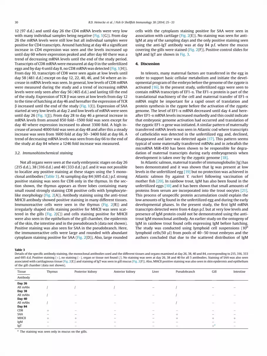
Not all organs were seen at the early embryonic stages on day 26(215 d.d.), 38 (316 d.d.) and 40 (333 d.d.) p.f. and it was not possibleto localize any positive staining at these stages using the 5 mono-clonal antibodies (Table 3). At sampling day 84 (695 d.d.) p.f. strongpositive staining was seen for CD8 only in the thymus. In the sec-tion shown, the thymus appears as three lobes containing manysmall round strongly staining CD8 positive cells with lymphocyte-like morphology (Fig. 2(A)). Antibody staining using the anti-troutMHCII antibody showed positive staining in many different tissues.Immunoreactive cells were seen in the thymus (Fig. 2(B)) andirregularly shaped cells staining positive for MHCII was seen scat-tered in the gills (Fig. 2(C)) and cells staining positive for MHCIIwere also seen in the epithelium of the gill chamber, the epidermisof the skin, the intestine and in the pseudobranch (data not shown).Positive staining was also seen for SAA in the pseudobranch. Here,the immunoreactive cells were large and rounded with abundantcytoplasm staining positive for SAA (Fig. 2(D)). Also, large rounded
Table 3Details of the specific antibody staining, the monoclonal antibodies used and the differentand 695 d.d. Positive staining (þ), no staining (�), organ or tissue not found (/). No stainassociated with cartilaginous tissue (Fig. 2(E)) and staining of IgT was seen in gill mucus (Fof the gill chamber (data not shown).
Tissue Thymus Posterior kidney Anterior kidney
Antibody
Day 26All mAbs / / /Day 38All mAbs / � �Day 40All mAbs / � �Day 84CD8 þ � �SAA � � �MHCII þ þ �IgM � � �IgT � � �a The staining was seen only in mucus on the gills.
cells with the cytoplasm staining positive for SAA were seen inassociation with cartilage (Fig. 2(E)). No staining was seen for anti-IgM at any of the sampling days and the only positive staining seenusing the anti-IgT antibody was at day 84 p.f. where the mucuscovering the gills were stained (Fig. 2(F)). Positive control slides forIgM and IgT are shown in Fig. 3.
4. Discussion
In teleosts, many maternal factors are transferred in the egg inorder to support basic cellular metabolism and initiate the devel-opmental programof the embryo before the genomeof the zygote isactivated [16]. In the present study, unfertilized eggs were seen tocontain mRNA transcripts of EF1-a. The EF1-a protein is part of thetranslational machinery of the cell and maternal transfer of EF1-amRNA might be important for a rapid onset of translation andprotein synthesis in the zygote before the activation of the zygoticgenome. The level of EF1-a mRNA decreased until day 4 and here-after EF1-amRNA levels increasedmarkedly and this could indicatethat embryonic genome activation had occurred and translation ofthe zygotic EF1-a genewas initiated. A similar pattern of maternallytransferred mRNA levels was seen in Atlantic cod where transcriptsof cathelicidin was detected in the unfertilized egg and, declined,disappeared and later was detected again [17]. This pattern seemstypical of some maternally transferred mRNAs and in zebrafish themicroRNA MiR-430 has been shown to be responsible for degra-dation of maternal transcripts during early embryogenesis whendevelopment is taken over by the zygotic genome [18].
In Atlantic salmon,maternal transfer of immunoglobulin (Ig) hasbeen demonstrated and it was shown that Ig was present at lowlevels in the unfertilized egg [19] but no protectionwas achieved inAtlantic salmon fry against Y. ruckeri following vaccination ofmother fish [20]. In rainbow trout, IgM has also been found in theunfertilized eggs [10] and it has been shown that small amounts ofproteins from serum are incorporated into the trout oocytes [21].This process of unspecific protein accumulation could explain thelowamounts of Ig found in the unfertilized egg and during the earlydevelopmental phases. In the present study, the first IgM mRNAtranscripts detected were from 4 days p.f. but at very low levels andpresence of IgM protein could not be demonstrated using the anti-trout IgMmonoclonal antibody. An earlier study on the ontogeny ofIgM in rainbow trout found cells expressing IgM before hatching.The study was conducted using lymphoid cell suspensions (106
lymphoid cells/50 ml) from pools of 40e50 trout embryos and theauthors concluded that due to the scattered distribution of IgM
tissues and organs examined at day 26, 38, 40 and 84, corresponding to 215, 316, 333ing was seen at day 26, 38 and 40 for all 5 antibodies. Staining of SAA was also seenig. 2(F)). Also, MHCII positive stainingwas also seen in skin epidermis and epithelium
Liver Pseudobranch Gill Intestine
� / � �
� / � �
� / � �
� � � �� þ � �� þ þ þ� � � �� � þa �

Fig. 2. Immunohistochemical staining on day 84 (695 d.d.) using monoclonal antibodies raised against CD8, SAA, MHCII, IgM and IgT. Strong positive staining of CD8 was seen in thethymus (A) and staining of MCHII positive cells were seen in multiple tissues occurring in the thymus (B), gill tissue (C) and also the gill cavity epithelium, skin epidermis, intestineand the pseudobranch (data not shown). Strong positive staining of SAA was seen in the pseudobranch (D) and in cells juxtaposed to cartilaginous tissue (E). Staining of IgT showedweak positive reaction in the gill mucus (F). G: gill, GC: gill cavity, PB: Pseudobranch, C: cartilage. Scale bar ¼ 100 mm.
Fig. 3. Positive control slides showing immunoreactive cells in rainbow trout spleen. Cells positive for IgM (A) and IgT (B) are found scattered in the spleen tissue. Scalebar ¼ 100 mm.
R.D. Heinecke et al. / Fish & Shellfish Immunology 38 (2014) 25e33 31

R.D. Heinecke et al. / Fish & Shellfish Immunology 38 (2014) 25e3332
positive cells these cells aremore easily detected in cell suspensions[10]. The presence of mRNA transcripts of IgM or IgT in the unfer-tilized eggs could not be confirmed in this study and accordinglymaternal transfer of mRNA transcripts of Ig frommother to egg wasnot supported.
In general, the 9 immune genes studied in the developing em-bryo exhibited very low levels of expression early in the egg phase.Later in the post-hatch larvae, mRNA transcripts for most geneswere still present at low levels except for MHCII, SAA, C3 and C5.Increases in expression levels were seen around and after the timeof hatching and this could indicate that expression is stimulatedwhen the larvae hatches and are directly exposed to the sur-rounding environment.
It was not possible to locate any positive staining of proteins atthe early time points at day 26, 38 and 40 using the five mono-clonal antibodies and this could be due to the very low level ofexpression at these time points and the challenges of locating thefew, if any, cells expressing the proteins. The expression of mRNAand the translation of the mRNA into protein are highly regulatedprocesses [22] and basic research (using a yeast model organism)has shown that the levels of mRNA and protein are not alwayscorrelated [23]. A lack of correlation between gene expressiontranscripts and levels of protein has been observed previously forthe complement factors C4 and C5 in rainbow trout [11]. Also, inzebrafish a latent period exists between expression of the IgMgene transcripts at day 7e10 post-hatch and until expression ofthe IgM protein at day 28 post-hatch [24]. It is possible that theuncoupling seen between gene expression and protein expressionof the IgM and IgT genes in the present study can be explained bya similar latent period between gene expression and proteinsynthesis. Also, it is becoming increasingly clear that microRNAsand RNA-binding proteins have an important role in regulating theexpression of genes and proteins in the cell [18] and it cannot beexcluded that miRNAs could be involved in rapid degradation ofmRNA or regulating translation by blocking specific mRNA at earlyembryonic stages.
In mammals, CD8 positive T cells develop, mature and undergorestrictive selection in the thymus [25] and the TCR/CD8 complexon the cytotoxic T cells is involved in the recognition of non-selfpeptides of intracellular origin bound to MCH I and the killing ofallogeneic cells [26]. In rainbow trout, CD8 positive cells havelikewise been shown to mediate cytotoxic killing of allogeneic cells[27] and the development, maturation and selection of the CD8positive cells is assumed to occur in the fish thymus. In a previousreport on rainbow trout ontogenesis the expression of CD8 tran-scripts was observed at day 7 post fertilization, which was theearliest time point sampled, and before appearance of the thymus[28] and long before allograft rejection is established [29]. In thepresent study, transcripts of CD8 mRNAwere also detected early atvery low levels and CD8 positive cells were only seen in the thymusat 84 days p.f. In a similar study of CD8a in sea bass Dicentrarchuslabrax mRNA transcripts of CD8a was detected at 51 days post-hatch corresponding to about 742 d.d. post fertilization [30]. Thiswas later than the first CD8 protein detected in the present studyand much later that the early detection of CD8 mRNA. No positivestaining of CD8 was seen in the intestine at any of the time pointssampled. A characteristic pattern of CD8 positive staining of intra-epithelial lymphocytes in the epithelium of the intestine is seenin older stages of rainbow trout (unpublished results) and it is likelythat the colonization of the intestinal epithelium by CD8 positivecells is initiated only after first feeding has commenced when theintestinal epithelium of the larva is exposed to antigens. The initi-ation of influx of CD8 positive cells into the epithelium of the in-testine might be initiated when trabeculae are formed in thethymus and the extensive vascularization of the thymus develops at
14e21 days post-hatch or when the zonation of the thymus isdeveloped at one month [3,4].
The acute phase protein serum amyloid A (SAA) has been shownto possess many functional properties including chemoattraction[31] and opsonization of Gram-negative bacteria [32] which facil-itated phagocytosis by macrophages [33]. High expression of SAAwas seen from day 26 p.f. to the end of the experiment. The highexpression of SAA mRNA correlates with the strong positivestaining of SAA protein seen at day 84 p.f. especially in the pseu-dobranch but also in tissue associated with cartilage. The role of thepseudobranch is still quite an enigma but evidence suggest that it isinvolved in controlling oxygen supply to the retina of the eye [34]but other functional roles of the pseudobranch are possible. Inthe present study, the pseudobranch was found highly positive forSAA staining and it is likely to function as an extrahepatic site ofSAA production. The produced SAA could be specifically supplied tothe developing eye or more likely the pseudobranch is supplyingSAA systemically. In this case SAA could be an important defensemolecule during development due to the opsonizing and chemo-tactic properties of the protein.
We found that staining of MHCII in rainbow trout larvae waswidespread and positive cells occurred in many tissues at day 84p.f. Using rabbit antisera against MHCII, a similar pattern of positiveMHCII staining in gills, intestine, thymus and skin was reported inAtlantic salmon [35]. The two major groups of MHCII expressingcells in mammals are B cells and macrophages [36] and it is likelythat the positive staining cells in the different tissues are tissueresident macrophages expressing MHCII.
In summary, the expression patterns of EF1-a and 9 immunegenes were studied during the ontogenetic development frombefore fertilization to 84 days p.f. In general, the onset of immunegene expression appeared at an early time point and at low levelsand during the ontogenetic development an increase in the tran-scription level was seen around and after the time of hatching withthe highest increases seen for MHCII, C3, C5 and SAA. Maternaltransfer was seen for EF1-awhere abundantmRNA transcriptsweredetected in the unfertilized egg. No positive staining of immuneproteinswas seen at the early sampling time points at 26, 38,40 daysp.f. but positive staining was detected in different tissues for MHCII,CD8 and SAA at 84 days p.f. which was prior to first feed.
Acknowledgments
This study was performed under The Danish Fish ImmunologyResearch Center and Network DAFINET funded by the DanishCouncil for Strategic Research (grant 09-065150). The assistance ofMrs. Moonika Haahr Marana in the immunohistochemical lab isgreatly appreciated and thanks to Dr. Per Walter Kania for technicalassistance and guidance in the qPCR lab.
References
[1] Grace MF, Manning MJ. Histogenesis of the lymphoid organs in rainbow trout,Salmo gairdneri Rich. 1836. Dev Comp Immunol 1980;4:255e64.
[2] Ellis AE. Ontogeny of the immune response in Salmo salar. Histogenesis of thelymphoid organs and appearance of membrane immunoglobulin and mixedleucocyte reactivity. In: Solomon JB, Horton JD, editors. DevelopmentalImmunobiology. Amsterdam: Elsevier/North Holland Biomedical Press; 1977.
[3] Razquin BE, Castillo A, Lopezfierro P, Alvarez F, Zapata A, Villena AJ. Ontogenyof IgM-producing cells in the lymphoid organs of rainbow trout, Salmogairdneri Richardson - an immunohistochemical and enzyme-histochemicalstudy. J Fish Biol 1990;36:159e73.
[4] Castillo A, Lopezfierro P, Zapata A, Villena A, Razquin B. Posthatching devel-opment of the thymic epithelial-cells in the rainbow trout Salmo gairdneri ean ultrastructural study. Am J Anat 1991;190:299e307.
[5] Castillo A, Razquin B, Villena AJ, Zapata AG, Lopez-Fierro P. Thymic barriers toantigen entry during the post-hatching development of the thymus ofrainbow trout, Oncorhynchus mykiss. Fish Shellfish Immunol 1998;8:157e70.

R.D. Heinecke et al. / Fish & Shellfish Immunology 38 (2014) 25e33 33
[6] Ballard WW. Normal embryonic stages for salmonid fishes, based on Salmogairdneri Richardson and Salvelinus fontinalis (Mitchill). J Exp Zool 1973;184:7e25.
[7] Cecchini S, Paciolla M, Biffali E, Borra M, Ursini MV, Lioi MB. Ontogeneticprofile of innate immune related genes and their tissue-specific expression inbrown trout, Salmo trutta (Linnaeus, 1758). Fish Shellfish Immunol 2013;35:988e92.
[8] Yousif AN, Albright LJ, Evelyn TPT. Occurrence of lysozyme in the eggs of cohosalmon Oncorhynchus kisutch. Dis Aquat Org 1991;10:45e9.
[9] Li M, Russell SK, Lumsden JS, Leatherland JF. The influence of oocyte cortisolon the early ontogeny of intelectin and TLR-5, and changes in lysozyme ac-tivity in rainbow trout (Oncorhynchus mykiss) embryos. Comparativebiochemistry and physiology part B. Biochem Mol Biol 2011;160:159e65.
[10] Castillo A, Sanchez C, Dominguez J, Kaattari SL, Villena AJ. Ontogeny of IgMand IgM-bearing cells in rainbow trout. Dev Comp Immunol 1993;17:419e24.
[11] Løvoll M, Kilvik T, Boshra H, Bøgwald J, Sunyer JO, Dalmo RA. Maternaltransfer of complement components C3-1, C3-3, C3-4, C4, C5, C7, Bf, and Df tooffspring in rainbow trout (Oncorhynchus mykiss). Immunogenetics 2006;58:168e79.
[12] Wang SH, Wang Y, Ma J, Ding YC, Zhang SC. Phosvitin plays a critical role inthe immunity of zebrafish embryos via acting as a pattern recognition re-ceptor and an antimicrobial effector. J Biol Chem 2011;286:22653e64.
[13] Løvoll M, Johnsen H, Boshra H, Bøgwald J, Sunyer JO, Dalmo RA. The ontogenyand extrahepatic expression of complement factor C3 in Atlantic salmon(Salmo salar). Fish Shellfish Immunol 2007;23:542e52.
[14] Chomczynski P, Sacchi N. Single-step method of RNA isolation by acid gua-nidinium thiocyanate phenol chloroform extraction. Anal Biochem 1987;162:156e9.
[15] Schmittgen TD, Livak KJ. Analyzing real-time PCR data by the comparative CTmethod. Nat Protoc 2008;3:1101e8.
[16] Pelegri F. Maternal factors in zebrafish development. Dev Dyn e Off Pub AmAssoc Anat 2003;228:535e54.
[17] Seppola M, Johnsen H, Mennen S, Myrnes B, Tveiten H. Maternal transfer andtranscriptional onset of immune genes during ontogenesis in Atlantic cod.Dev Comp Immunol 2009;33:1205e11.
[18] Giraldez AJ, Mishima Y, Rihel J, Grocock RJ, Van Dongen S, Inoue K, et al.Zebrafish MiR-430 promotes deadenylation and clearance of maternalmRNAs. Science 2006;312:75e9.
[19] Olsen YA, Press CM. Degradation kinetics of immunoglobulin in the egg, alevinand fry of Atlantic salmon, Salmo salar L, and the localisation of immuno-globulin in the egg. Fish Shellfish Immunol 1997;7:81e91.
[20] Lillehaug A, Sevatdal S, Endal T. Passive transfer of specific maternal immunitydoes not protect Atlantic salmon (Salmo salar L.) fry against yersiniosis. FishShellfish Immunol 1996;6:521e35.
[21] Campbell CM, Jalabert B. Selective protein incorporation by vitellogenic Salmogairdneri oocytes in vitro. Ann Biol Anim Bioch 1979;19:429e37.
[22] Jackson RJ, Hellen CU, Pestova TV. The mechanism of eukaryotic translationinitiationandprinciples of its regulation.NatRevMolCell Biol 2010;11:113e27.
[23] Gygi SP, Rochon Y, Franza BR, Aebersold R. Correlation between protein andmRNA abundance in yeast. Mol Cell Biol 1999;19:1720e30.
[24] Lam SH, Chua HL, Gong Z, Lam TJ, Sin YM. Development and maturation of theimmune system in zebrafish, Danio rerio: a gene expression profiling, in situhybridization and immunological study. Dev Comp Immunol 2004;28:9e28.
[25] Goldrath AW, Bevan MJ. Selecting and maintaining a diverse T-cell repertoire.Nature 1999;402:255e62.
[26] Harms JS, Splitter GA. CD8þ lymphocytes that kill allogeneic and xenogeneicmajor histocompatibility complex class I targets. Hum Immunol 1995;44:50e7.
[27] Fischer U, Utke K, Ototake M, Dijkstra JM, Koller B. Adaptive cell-mediatedcytotoxicity against allogeneic targets by CD8-positive lymphocytes ofrainbow trout (Oncorhynchus mykiss). Dev Comp Immunol 2003;27:323e37.
[28] Fischer U, Dijkstra JM, Kollner B, Kiryu I, Koppang EO, Hordvik I, et al. Theontogeny of MHC class I expression in rainbow trout (Oncorhynchus mykiss).Fish Shellfish Immunol 2005;18:49e60.
[29] Tatner MF, Manning MJ. The ontogeny of cellular immunity in the rainbowtrout, Salmo gairdneri Richardson, in relation to the stage of development ofthe lymphoid organs. Dev Comp Immunol 1983;7:69e75.
[30] Picchietti S, Guerra L, Selleri L, Buonocore F, Abelli L, Scapigliati G, et al.Compartmentalisation of T cells expressing CD8a and TCRb in developingthymus of sea bass Dicentrarchus labrax (L.). Dev Comp Immunol 2008;32:92e9.
[31] Badolato R, Wang JM, Murphy WJ, Lloyd AR, Michiel DF, Bausserman LL, et al.Serum amyloid A is a chemoattractant: induction of migration, adhesion, andtissue infiltration of monocytes and polymorphonuclear leukocytes. J Exp Med1994;180:203e9.
[32] Hari-Dass R, Shah C, Meyer DJ, Raynes JG. Serum amyloid A protein binds toouter membrane protein A of gram-negative bacteria. J Biol Chem 2005;280:18562e7.
[33] Shah C, Hari-Dass R, Raynes JG. Serum amyloid A is an innate immune opsoninfor Gram-negative bacteria. Blood 2006;108:1751e7.
[34] Bridges CR, Berenbrink M, Muller R, Waser W. Physiology and biochemistry ofthe pseudobranch: an unanswered question? Comparative biochemistry andphysiology Part A. Mol Integr Physiol 1998;119:67e77.
[35] Koppang EO, Hordvik I, Bjerkas I, Torvund J, Aune L, Thevarajan J, et al. Pro-duction of rabbit antisera against recombinant MHC class II beta chain andidentification of immunoreactive cells in Atlantic salmon (Salmo salar). FishShellfish Immunol 2003;14:115e32.
[36] Glimcher LH, Kara CJ. Sequences and factors: a guide to MHC class-II tran-scription. Annu Rev Immunol 1992;10:13e49.
[37] Hetland DL, Jorgensen SM, Skjodt K, Dale OB, Falk K, Xu C, et al. In situlocalisation of major histocompatibility complex class I and class II and CD8positive cells in infectious salmon anaemia virus (ISAV)-infected Atlanticsalmon. Fish Shellfish Immunol 2010;28:30e9.
[38] Olsen MM, Kania PW, Heinecke RD, Skjoedt K, Rasmussen KJ, Buchmann K.Cellular and humoral factors involved in the response of rainbow trout gills toIchthyophthirius multifiliis infections: molecular and immunohistochemicalstudies. Fish Shellfish Immunol 2011;30:859e69.
[39] Chettri JK, Raida MK, Kania PW, Buchmann K. Differential immune response ofrainbow trout (Oncorhynchus mykiss) at early developmental stages (larvaeand fry) against the bacterial pathogen Yersinia ruckerie. Dev Comp Immunol2012;36:463e74.
[40] Heinecke RD, Buchmann K. Inflammatory response of rainbow trout Onco-rhynchus mykiss (Walbaum, 1792) larvae against Ichthyophthirius multifiliis.Fish Shellfish Immunol 2013;34:521e8.
[41] Jørgensen LV, Heinecke RD, Skjødt K, Rasmussen KJ, Buchmann K. Experi-mental evidence for direct in situ binding of IgM and IgT to early trophonts ofIchthyophthirius multifiliis (Fouquet) in the gills of rainbow trout, Onco-rhynchus mykiss (Walbaum). J Fish Dis 2011;34:749e55.
[42] Chettri JK. Innate immune response dynamics in rainbow trout: effects ofpathogen and immunostimulants exposure. University of Copenhagen; 2011.


