Embed Size (px)
Citation preview

Biochimica et Biophysica Acta 1798 (2010) 9–20
Contents lists available at ScienceDirect
Biochimica et Biophysica Acta
j ourna l homepage: www.e lsev ie r.com/ locate /bbamem
Review
Adenosine receptors interacting proteins (ARIPs): Behind the biology ofadenosine signaling
Francisco Ciruela ⁎, Catarina Albergaria, Aroa Soriano, Laura Cuffí, Lourdes Carbonell,Silvia Sánchez, Jorge Gandía, Víctor Fernández-Dueñas ⁎Unitat de Farmacologia, Departament de Patologia i Terapèutica Experimental, Facultat de Medicina-Bellvitge, Pavelló de Govern, Universitat de Barcelona, Av. Feixa Llarga, s/n, 08907L'Hospitalet del Llobregat, Barcelona, Spain
⁎ Corresponding authors. Tel.: +34 934024280/+34E-mail addresses: [email protected] (F. Ciruela), vfern
0005-2736/$ – see front matter © 2009 Elsevier B.V. Adoi:10.1016/j.bbamem.2009.10.016
a b s t r a c t
a r t i c l e i n f oArticle history:Received 6 July 2009Received in revised form 26 September 2009Accepted 27 October 2009Available online 31 October 2009
Keywords:G protein-coupled receptorAdenosine receptorProtein–protein interactionAccessory proteinReceptor anchoringPlasma membrane expression
Adenosine is a well known neuromodulator in the central nervous system. As a consequence, adenosine canbe beneficial in certain disorders and adenosine receptors will be potential targets for therapy in a variety ofdiseases. Adenosine receptors are G protein-coupled receptors, and are also expressed in a large variety ofcells and tissues. Using these receptors as a paradigm of G protein-coupled receptors, the present reviewfocus on how protein–protein interactions might contribute to neurotransmitter/neuromodulator regula-tion, based on the fact that accessory proteins impinge on the receptor/G protein interaction and thereforemodulate receptor functioning. Besides affecting receptor signaling, these accessory components also play akey role in receptor trafficking, internalization and desensitization, as it will be reviewed here. In conclusion,the finding of an increasing number of adenosine receptors interacting proteins, and specially the molecularand functional integration of these accessory proteins into receptorsomes, will open new perspectives in theunderstanding of particular disorders where these receptors have been proved to be involved.
© 2009 Elsevier B.V. All rights reserved.
Contents
1. Introduction . . . . . . . . . . . . . . . . . . . . . . . . . . . . . . . . . . . . . . . . . . . . . . . . . . . . . . . . . . . . . . . 102. Adenosine receptors . . . . . . . . . . . . . . . . . . . . . . . . . . . . . . . . . . . . . . . . . . . . . . . . . . . . . . . . . . . 103. Adenosine receptors interacting proteins (ARIPs) . . . . . . . . . . . . . . . . . . . . . . . . . . . . . . . . . . . . . . . . . . . . . 11
3.1. Adenosine A1 receptor interacting proteins (A1RIPs) . . . . . . . . . . . . . . . . . . . . . . . . . . . . . . . . . . . . . . . . . 113.1.1. Adenosine deaminase (ADA) . . . . . . . . . . . . . . . . . . . . . . . . . . . . . . . . . . . . . . . . . . . . . . . 113.1.2. Heat shock cognate protein 73 (HSC-73) . . . . . . . . . . . . . . . . . . . . . . . . . . . . . . . . . . . . . . . . . . 113.1.3. Caveolin-1 . . . . . . . . . . . . . . . . . . . . . . . . . . . . . . . . . . . . . . . . . . . . . . . . . . . . . . . . 123.1.4. Protein 4.1G . . . . . . . . . . . . . . . . . . . . . . . . . . . . . . . . . . . . . . . . . . . . . . . . . . . . . . . 13
3.2. Adenosine A2A receptor interacting proteins (A2ARIPs) . . . . . . . . . . . . . . . . . . . . . . . . . . . . . . . . . . . . . . . . 133.2.1. α-actinin . . . . . . . . . . . . . . . . . . . . . . . . . . . . . . . . . . . . . . . . . . . . . . . . . . . . . . . . . 133.2.2. Arf nucleotide site opener (ARNO)/cytohesin-2 . . . . . . . . . . . . . . . . . . . . . . . . . . . . . . . . . . . . . . . 143.2.3. Ubiquitin-specific protease 4 (USP4) . . . . . . . . . . . . . . . . . . . . . . . . . . . . . . . . . . . . . . . . . . . . 153.2.4. Translin-associated protein X (TRAX) . . . . . . . . . . . . . . . . . . . . . . . . . . . . . . . . . . . . . . . . . . . 153.2.5. Neuronal Calcium-Binding Protein 2 (NECAB2) . . . . . . . . . . . . . . . . . . . . . . . . . . . . . . . . . . . . . . . 16
3.3. Adenosine A2B receptor interacting proteins (A2BRIPs) . . . . . . . . . . . . . . . . . . . . . . . . . . . . . . . . . . . . . . . . 163.4. Adenosine A3 receptor interacting proteins (A3RIPs) . . . . . . . . . . . . . . . . . . . . . . . . . . . . . . . . . . . . . . . . . 18
4. Concluding remarks . . . . . . . . . . . . . . . . . . . . . . . . . . . . . . . . . . . . . . . . . . . . . . . . . . . . . . . . . . . 18Acknowledgements . . . . . . . . . . . . . . . . . . . . . . . . . . . . . . . . . . . . . . . . . . . . . . . . . . . . . . . . . . . . . . 18References . . . . . . . . . . . . . . . . . . . . . . . . . . . . . . . . . . . . . . . . . . . . . . . . . . . . . . . . . . . . . . . . . . 18
934035820; fax: +34 [email protected] (V. Fernández-Dueñas).
ll rights reserved.

Table 1Adenosine receptors in the brain.
Receptor Adenosineaffinity
Gprotein
Transductionmechanismsa
Physiological actionsin brain
A1R ∼ 70 nM Gi1,2,3 Inhibits AC (↓cAMP)Activates PLC (↑IP3/DAG)
Inhibits synaptictransmission;hyperpolarizesneurons
Activates PLA2 (↑AA)Activates PLD (↑PEtOH)Activates GIRKsInhibits Ca2+ channels
Go
A2AR ∼ 150 nM GSb Activates AC (↑cAMP) Facilitates
transmitter release;regulation ofsensorimotorintegration in basalganglia
Golf Activates AC (↑cAMP)G15,16
c ↑IP3Inhibits Ca2+ channels
A2BR ∼ 5000 nM GSb Activates AC (↑cAMP)
Activates Ca2+ channelsIncreases in cAMPin brain slices
Gq/11c Activates PLC (↑IP3/DAG)
A3R ∼ 6500 nM Gi2,3 Inhibits AC (↓cAMP) Uncouples A1R andmGlu receptorsGq/11 Activates PLC (↑IP3/DAG)
a AC, adenylyl cyclase; PLC, phospholipase C; IP3, inositol triphosphate; DAG,diacylglycerol; PLA2, phospholipase A2; PEtOH, phosphatidylethanol; GIRKs, G protein-dependent inwardly rectifying K+ channels; AA, arachidonic acid.
b Main mechanism of coupling.c Receptor transfected cell system.
10 F. Ciruela et al. / Biochimica et Biophysica Acta 1798 (2010) 9–20
1. Introduction
Since Drury and Szent-Györgyi found 80 years ago that adenosineproduced profound hypotension and bradycardia, as well as it affectedkidney function in mammals [1], the physiological roles and potentialtherapeutic use of adenosine have been largely revised [2,3].Adenosine is mainly produced by degradation of ATP and in lessenextend by S-adenosyl-L-homocysteine (SAH) metabolism. Also,adenosine can be formed extracellularly by the breakdown of ATP.Once it has been generated, it can either be eliminated out of the cellsby means of ubiquitous nitrobenzylthioinosine-sensitive equilibrativenucleoside transporters (ENTs) or intracellularly phosphorylated toform AMP. In addition, adenosine can react with L-homocysteine toform SAH. And finally, both intra- and extracellular adenosine can bedeaminated to form inosine by the action of intra- and ecto-adenosinedeaminase, respectively.
Regarding adenosine functions, it has been largely considered aretaliatory metabolite [4] that increases oxygen supply and decreasesoxygen consumption, thus modulating a large array of physiologicalprocesses including respiratory regulation, hormone action, neuralfunction, platelet aggregation, lymphocyte differentiation and vascu-lar tone. For instance, adenosine is able to induce the dilation ofcoronary arteries, but it can also lead to the contraction of other bloodvessels like the ones in the kidney, resulting in a decreased renalfiltration. Also, adenosine exerts a negative chronotropic and dromo-tropic effect on the heart, as well as it mediates the inhibition ofneurotransmitters release and lipolysis. Overall, it has been assumedthat this purine nucleoside is a mediator of metabolic distress, thushaving considerable impact on homeostatic cellular functioning.
In 1972, it was shown that adenosine was released from brainslices upon electrical stimulation [5]. Interestingly, the releasedadenosine produced an increase in the intracellular levels of cAMP,an effect that was antagonized by methylxanthines such as caffeineand theophylline [6]. This fact was also observed in the heart [7].Collectively, these observations constituted the first evidence sug-gesting that adenosine exerted its effects via specific plasmamembrane receptors. Afterwards, it was demonstrated that the sodescribed antilipolytic effect of adenosine on fat cells occurred with aconcomitant reduction in cAMP [8]. The dual effects of adenosine oncAMP formation were further substantiated when it was demonstrat-ed that adenosine could either inhibit or stimulate adenylyl cyclase.Overall, these observations ended with the first subclassification ofadenosine receptors into Ri and Ra [9], or alternatively, A1 and A2
adenosine receptors [10].In the Central Nervous System (CNS), adenosine has been shown
to play a regulatory role, acting as a presynaptic, postsynaptic and/ornon-synaptic neuromodulator [11]. Extracellular adenosine in thebrain is related to the intracellular concentration of adenosine andnucleotides, such as ATP, AMP and cAMP [12]. In some brain areas, likethe hippocampus, most of the extracellular adenosine seems todepend mostly on intracellular adenosine, the concentration of whichis related to the rate of breakdown and synthesis of ATP [12]. Thus,adenosine is released as a neuromodulator [13] by the effector cells inresponse to an increased metabolic demand [14]. However, in thestriatum, it has been suggested that the main source of extracellularadenosine is intracellular cAMP [15], which is metabolized to AMP bymeans of phosphodiesterases and then to adenosine by the ectoen-zyme 5′nucleotidase. Since cAMP can only be generated by the actionof the enzyme adenylyl cyclase, striatal extracellular adenosine wouldmostly reflect an increased activation of receptors positively linked toadenylyl cyclase.
2. Adenosine receptors
Adenosine mediates its actions by means of activation of specific Gprotein-coupled receptors (GPCRs), for which four subtypes (A1R,
A2AR, A2BR and A3R) have been identified so far. These receptors havea distinctive pharmacological profile, tissue distribution and effectorcoupling [16], and its functioning has been extensively studied in theCNS (Table 1). The GPCRs comprise the largest family of membrane-bound receptors, containing more than 1000 different memberswhich represent over 1% of the genome in vertebrates [17]. From aphylogenetic point of view, these receptors can be classified into fivemain families (GRAFS classification system), namely glutamate,rhodopsin, adhesion, frizzled/taste 2 and secretin [18]. Adenosinereceptors (ARs) belong to the rhodopsin family, also called family I orA [19]. This receptor family has more than 700 different members thatshare some phylogenetic and intrinsic characteristics. For instance,within their sequence, all adenosine receptors contain the widelyconserved NPxxY(x)5,6F and the DRYmotifs [20,21]. Thus, adenosine-mediated conformational change in the ARs core domain affects theconformation of the intracellular loops, which in turn determines thebinding and activation of specific G proteins (Table 1). Stimulation ofG proteins is then responsible for activation of different intracellularsignaling pathways associated to adenosine function (Table 1).
Interestingly, A1Rs and A2ARs are primarily responsible for thecentral effects of adenosine [22]. The most abundant and homoge-neously distributed adenosine receptor in the brain is the A1R, whichis functionally coupled to members of the pertussis toxin-sensitivefamily of G proteins (Gi1, Gi2, Gi3 and Go) and whose activationregulates several membrane and intracellular proteins such asadenylyl cyclase, Ca2+ channels, K+ channels and phospholipase C(Table 1) [23]. On the other hand, A2AR is expressed at high levels inonly a few regions of the brain, namely primarily striatum, olfactorytubercle and nucleus accumbens [15]. A2ARs are mostly coupled toGs/Golf proteins [24], thus activating adenylyl cyclase which in turnconverts ATP into AMPc (Table 1). Depending on the cell type studied,the A2AR may also signal via a pathway that might be independent ofheterotrimeric G proteins; such is the case of the activation of themitogen-activated protein kinase (MAPK) signaling cascade [25].Next, the A2BR is positively coupled to adenylyl cyclase and PLCthrough a Gs and Gq protein, respectively [2] (Table 1). A2BR is thoughtto be fairly ubiquitous in the brain, and the association of this receptorto specific physiological or behavioral responses remains quite

11F. Ciruela et al. / Biochimica et Biophysica Acta 1798 (2010) 9–20
difficult because of the paucity of A2BR agonists or antagonists [26].Lastly, the A3R has also been found to be coupled to classical second-messenger pathways such as inhibition of adenylyl cyclase, stimula-tion of PLC and calcium mobilization (for review see [2]) (Table 1).Finally, it is important to mention at this point that different classes ofproteins, other than G proteins, are being recognized to interact withGPCRs. The binding of GPCRs to some of these proteins will determineinteractions with different intramembrane and intracellular elements,which in turn will result, not only in changes in receptor localizationand function, but also in G protein-independent signaling [27]. Theseproteins include serine–threonine or tyrosine protein kinases, β-arrestins and scaffolding proteins within others.
3. Adenosine receptors interacting proteins (ARIPs)
In the present work, we underscore the ARs ability to interact withproteins different from receptors and how these accessory proteinsaffect receptor function, in the view that these protein–proteininteractions might control receptor trafficking (e.g. cell surfaceexpression, internalization and recycling) and function (e.g. Gprotein-coupling and desensitization) [27]. In addition, the stoichi-ometry, the affinity of these partners and the presence of cellularmodulators (e.g. Ca2+) can determine the dynamics of these protein–protein interactions. Finally, it is proposed that the intracellularportions of adenosine receptors, namely the C-terminal tail andintracellular loops, might become signal integrators where interactingproteins would be associated or dissociated depending on cellularinputs, thus making up the adenosine receptorsome.
3.1. Adenosine A1 receptor interacting proteins (A1RIPs)
Within the numerous neurophysiological actions of adenosine, theinhibition of glutamate neurotransmission has been largely studied inseveral brain regions [22]. This adenosine-mediated glutamate releaseinhibition might be a consequence of the activation of presynapticA1Rs, which on its turn lead to a direct inhibitory effect of G proteinβγ-subunits on voltage-dependent Ca2+ channels [28,29]. Thus,presynaptic A1Rs are the prototype of GPCRs the stimulation ofwhich decreases the probability of neurotransmitter release (Table 1).On the other hand, the A1R turns out to be the predictable example ofGPCR where the identification of interacting proteins other than thetypical GPCR-associated proteins (e.g. G proteins, β-arrestin, GRKs,etc.) became successful. For instance, four A1RIPs have been describedso far, namely adenosine deaminase (ADA), heat shock cognateprotein 73 (HSC-73), Caveolin-1 and Protein 4.1.
3.1.1. Adenosine deaminase (ADA)ADA is the enzyme that metabolizes the physiological agonist of
adenosine receptors, thus reducing the effective concentration ofadenosine in the cellular milieu and precluding adenosine-mediatedreceptor stimulation. In addition, it has been demonstrated that ADAbehaves like an ecto-enzyme that is anchored to the cell surfacethrough different plasmamembrane proteins [30]. Interestingly, apartfrom being the enzyme that can deaminate extracellular adenosine,ADA can also have a non-enzymatically role through direct protein–protein interactions [31]. Firstly, by means of confocal laser micros-copy, co-immunoprecipitation and affinity chromatography experi-ments performed in DDT1MF-2 cells, an ADA/A1R interaction wasdemonstrated [32], and it was shown to occur within the extracellularloops of the receptor (Fig. 1). Subsequently, the existence of thisinteraction in native cell membranes was also confirmed, where itseemed that ADA was necessary for the coupling of A1R toheterotrimeric G proteins [32,33]. Moreover, the ADA/A1R complexwas found in the cell-surface of cortical cultured neurons [34], andafterwards it was suggested that ADAwould be targeted to the plasmamembrane by A1R [35].
The proposed physiological role of such protein–protein interactionis tomake receptorsmore sensitive to adenosine. Thus, this heterotypicinteraction would be necessary for A1Rs to achieve high-affinitybinding of agonists and subsequently for allowing efficient couplingto the signal transduction machinery. In fact, it was recentlydemonstrated that human ADA markedly enhanced the agonist andantagonist affinity and abolished the negative cooperativity on agonistbinding to human striatal A1Rs [36]. In addition, apart from reducingthe adenosine concentration and preventing A1R desensitization, ADAbinds to A1R and behaves as an allosteric effector that markedlyenhances agonist affinity and increases receptor functionality (Fig. 1).Interestingly, when the enzymatic activity of ADA was knocked downby incubation of the enzyme with low Hg2+ concentrations, an ADA/A1R interaction was still confirmed, thus suggesting that thisinteraction would be enzymatic activity-independent and proposingthe contention that the docking of ADA to A1Rmight promote receptorconformational changes that would allow a more efficient receptor-mediated signaling [32,36,37]. On the other hand, in cell-based assaysperformed in different cell lines, the aggregation of both A1R and ADAwas reported after agonist challenge,which preceded the translocationof both proteins into intracellular compartments. These resultssuggested that the receptor–enzyme complex was sufficiently stableto allow the simultaneous internalization of both proteins [37]. Inaddition, preincubationwith an A1R agonist resulted in co-aggregationof the dopamine D1 receptor (D1R) and A1R together with ADA in cellsexpressing both receptors, suggesting that after A1R agonist challenge ahigher-order oligomeric structure leaded by A1R was formed [35].Indeed,more researchwill be needed to unravel themolecularmecha-nism by which ADA mediates the formation of a D1R/A1R oligomer asthis might have dramatic consequences in adenosine–dopaminefunctional cross-talk. Precisely, this information will become relevantto understand the molecular basis of some antagonistic adenosine–dopamine interactions in the central nervous system that might berelevant in the treatment of some CNS pathological conditions.
3.1.2. Heat shock cognate protein 73 (HSC-73)A different A1R interacting protein, a chaperone protein, which has
been shown to play a key role in ligand binding and receptor signaling,is the heat shock cognate protein 73 (HSC-73). Heat shock proteins(HSPs), or stress proteins, are highly conserved and present in all cellsof all organisms. HSPs, also known as chaperones, play crucial roles inthe folding/unfolding of proteins, assembly of multiprotein com-plexes, transport/sorting of proteins into correct subcellular compart-ments, cell-cycle control and signaling, and protection of cells againststress/apoptosis (for review see [38]). The 70-kDa members of theHSPs family, including HSC-73, have been found to play a role in themovement of proteins into the nucleus, mitochondria, endoplasmicreticulum and lysosome (for review see [38]). HSC-73, specifically, hasbeen observed to facilitate the transport of proteins into the lysosomeafter the withdrawal of serum from cells in culture. HSC-73 hasadditional cellular functions apart from its role as a molecularchaperone, namely it associates with cell cycle regulatory proteins,retinoblastoma, p53 and Bcl-2 and also it has an ATPase activity for theuncoating of clathrin-coated vesicles during endocytosis and vesiclerecycling (for review see [38]).
The interaction betweenHSC-73 and A1R, which takes placewithinthe third intracellular loop of the receptor, reduces agonists bindingand also prevents receptor-G protein activation (Fig. 1), this lastaction being completely prevented by ADA, the extracellular receptorpartner. In fact, although ADA interacts extracellularly and HSC-73interacts in the cytoplasmic side of the receptor, it seems that bothproteins compete for binding to A1R, thus modulating the function-ality of the receptor [39].
On the other hand, it has been described that the HSC-73/A1Rinteraction might also be implicated in A1R trafficking and receptordownregulation. A distinctive co-distribution of HSC-73 and A1R, but
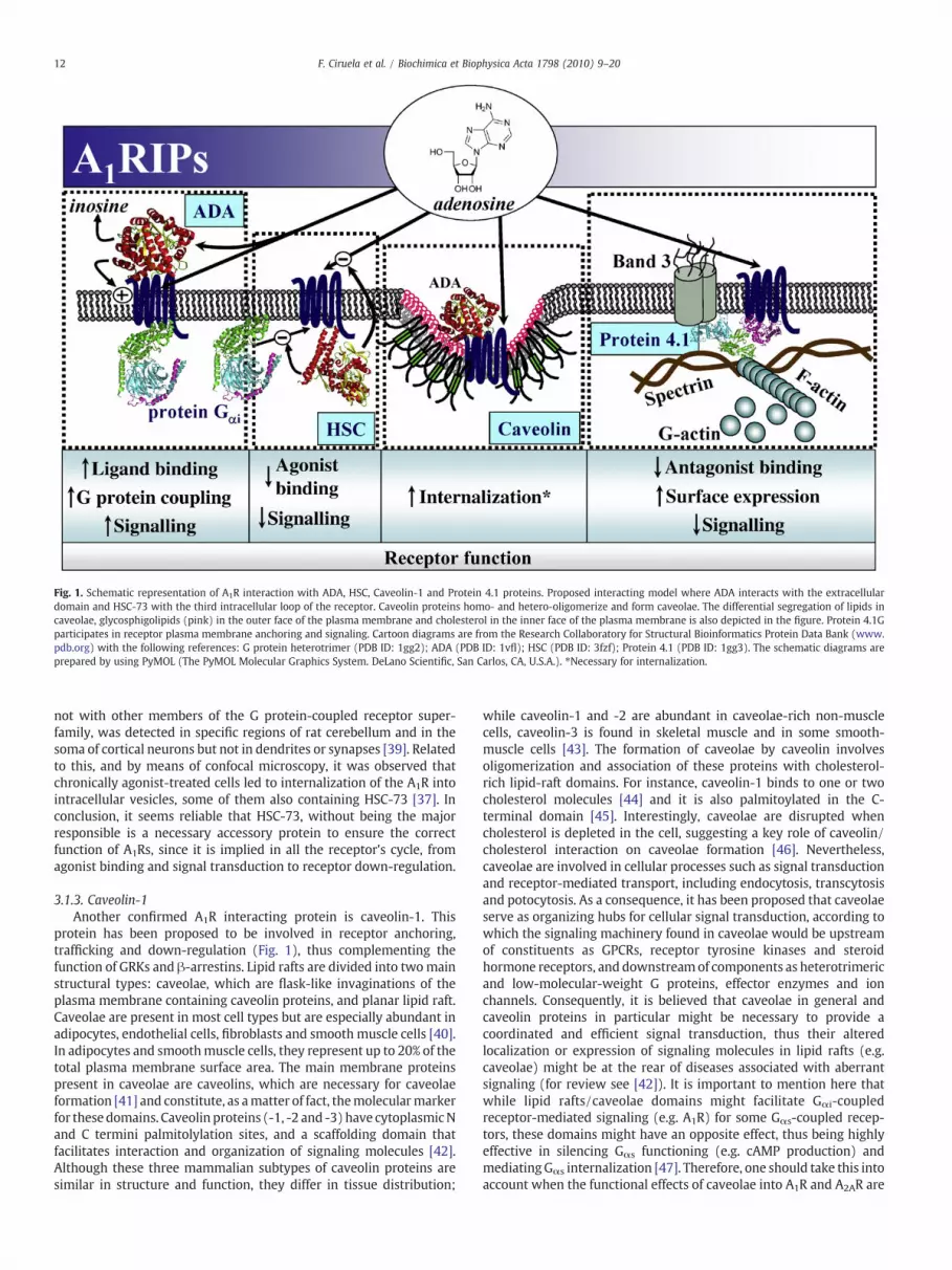
Fig. 1. Schematic representation of A1R interaction with ADA, HSC, Caveolin-1 and Protein 4.1 proteins. Proposed interacting model where ADA interacts with the extracellulardomain and HSC-73 with the third intracellular loop of the receptor. Caveolin proteins homo- and hetero-oligomerize and form caveolae. The differential segregation of lipids incaveolae, glycosphigolipids (pink) in the outer face of the plasma membrane and cholesterol in the inner face of the plasma membrane is also depicted in the figure. Protein 4.1Gparticipates in receptor plasma membrane anchoring and signaling. Cartoon diagrams are from the Research Collaboratory for Structural Bioinformatics Protein Data Bank (www.pdb.org) with the following references: G protein heterotrimer (PDB ID: 1gg2); ADA (PDB ID: 1vfl); HSC (PDB ID: 3fzf); Protein 4.1 (PDB ID: 1gg3). The schematic diagrams areprepared by using PyMOL (The PyMOL Molecular Graphics System. DeLano Scientific, San Carlos, CA, U.S.A.). ⁎Necessary for internalization.
12 F. Ciruela et al. / Biochimica et Biophysica Acta 1798 (2010) 9–20
not with other members of the G protein-coupled receptor super-family, was detected in specific regions of rat cerebellum and in thesoma of cortical neurons but not in dendrites or synapses [39]. Relatedto this, and by means of confocal microscopy, it was observed thatchronically agonist-treated cells led to internalization of the A1R intointracellular vesicles, some of them also containing HSC-73 [37]. Inconclusion, it seems reliable that HSC-73, without being the majorresponsible is a necessary accessory protein to ensure the correctfunction of A1Rs, since it is implied in all the receptor's cycle, fromagonist binding and signal transduction to receptor down-regulation.
3.1.3. Caveolin-1Another confirmed A1R interacting protein is caveolin-1. This
protein has been proposed to be involved in receptor anchoring,trafficking and down-regulation (Fig. 1), thus complementing thefunction of GRKs and β-arrestins. Lipid rafts are divided into twomainstructural types: caveolae, which are flask-like invaginations of theplasma membrane containing caveolin proteins, and planar lipid raft.Caveolae are present in most cell types but are especially abundant inadipocytes, endothelial cells, fibroblasts and smooth muscle cells [40].In adipocytes and smoothmuscle cells, they represent up to 20% of thetotal plasma membrane surface area. The main membrane proteinspresent in caveolae are caveolins, which are necessary for caveolaeformation [41] and constitute, as amatter of fact, themolecularmarkerfor these domains. Caveolin proteins (-1, -2 and -3) have cytoplasmicNand C termini palmitolylation sites, and a scaffolding domain thatfacilitates interaction and organization of signaling molecules [42].Although these three mammalian subtypes of caveolin proteins aresimilar in structure and function, they differ in tissue distribution;
while caveolin-1 and -2 are abundant in caveolae-rich non-musclecells, caveolin-3 is found in skeletal muscle and in some smooth-muscle cells [43]. The formation of caveolae by caveolin involvesoligomerization and association of these proteins with cholesterol-rich lipid-raft domains. For instance, caveolin-1 binds to one or twocholesterol molecules [44] and it is also palmitoylated in the C-terminal domain [45]. Interestingly, caveolae are disrupted whencholesterol is depleted in the cell, suggesting a key role of caveolin/cholesterol interaction on caveolae formation [46]. Nevertheless,caveolae are involved in cellular processes such as signal transductionand receptor-mediated transport, including endocytosis, transcytosisand potocytosis. As a consequence, it has been proposed that caveolaeserve as organizing hubs for cellular signal transduction, according towhich the signaling machinery found in caveolae would be upstreamof constituents as GPCRs, receptor tyrosine kinases and steroidhormone receptors, and downstreamof components as heterotrimericand low-molecular-weight G proteins, effector enzymes and ionchannels. Consequently, it is believed that caveolae in general andcaveolin proteins in particular might be necessary to provide acoordinated and efficient signal transduction, thus their alteredlocalization or expression of signaling molecules in lipid rafts (e.g.caveolae) might be at the rear of diseases associated with aberrantsignaling (for review see [42]). It is important to mention here thatwhile lipid rafts/caveolae domains might facilitate Gαi-coupledreceptor-mediated signaling (e.g. A1R) for some Gαs-coupled recep-tors, these domains might have an opposite effect, thus being highlyeffective in silencing Gαs functioning (e.g. cAMP production) andmediatingGαs internalization [47]. Therefore, one should take this intoaccount when the functional effects of caveolae into A1R and A2AR are

13F. Ciruela et al. / Biochimica et Biophysica Acta 1798 (2010) 9–20
studied as these receptors are coupled to Gαi and Gαs, respectively(Table 1). Interestingly, a direct interaction between caveolin-1 andthe C-terminal domain of A1R was demonstrated by means of pulldown experiments and co-localization assays [37,48,49] (Fig. 1).Moreover, it was reported that agonist-induced internalization of thestable complex ADA/A1R was mediated by clathrin-independentendocytosis. Thus, detailed electron microscopy in DDT1MF-2 cellsrevealed that ADA/A1R complexes internalized through caveolae andthat these complexes were differentially sorted in intracellularendosomes which were recycled back to the plasma membraneseparately [49]. Overall, these results indicate that desensitization andinternalization of A1R are modulated by ADA and both proteinsinternalize after agonist challenge via non-coated vesicles, namelycaveolae, due to the caveolin-1/A1R interaction (Fig. 1).
3.1.4. Protein 4.1GThe last described adenosine A1R interacting protein is a member
of the 4.1 family of cytoskeletal-associated proteins, the protein 4.1G.The protein 4.1 superfamily is classified into two main groupsaccording to their cellular expression pattern: the red blood cellswhich express an erythroid form, also called 4.1R, and the non-erythroid cells that present the 4.1G (general type), 4.1B (brain type)and 4.1N (neuron type) forms. Despite being encoded by differentgenes, proteins 4.1R, 4.1G, 4.1N and 4.1B all share similar structuralfeatures, such as the presence of three highly conserved domains: a N-terminal conserved FERM (Four.1 protein, Ezrin, Radixin, Moesin)domain that links this cytoskeletal scaffold to the plasma membranevia interactions with band 3 and glycophorin C, a spectrin–actin-binding domain (SABD) that potentiates the interactions of spectrintetramers with F-actin and a C-terminal domain (CTD) that bindsFKBP13 (13 kDa FK506-binding protein) [50]. These multifunctionalproteins are critical components of the spectrin/actin cytoskeletonand provide attachments between cytoskeleton and cell membranes,thus playing an important structural and regulatory role in thestabilization and assembly of these membranes.
Protein 4.1G binds to the third intracellular loop of A1R (Fig. 1), andis involved in the trafficking and down-regulation of the receptor [51].As the spectrin–actin-binding domain of protein 4.1 G can form acomplexwith actin and spectrin, it is believed that protein 4.1 Gwouldanchor associated molecules, such as intramembranous proteins (e.g.A1R), to the cytoskeleton. Lu and collaborators showed that protein4.1G, by interacting with A1R (Fig. 1), reduced receptor-mediatedinhibition of cAMP accumulation and intracellular calcium release,thus interferingwith the receptor signal transduction [51]. In addition,protein 4.1G also altered the cell-surface expression of the receptor.Interestingly, by means of immunostaining techniques, it wasdemonstrated that in the CNS protein 4.1 G co-localized with markersfor microglia, such as CD45, OX-42 and ionized calcium-bindingadapter molecule 1 (Iba1), but not with markers for neuronal or otherglial cells [52]. In the samework, a co-localization between protein 4.1G and A1R in mouse cerebrum was also shown [52], leading to thebelief that a further understanding of the protein 4.1 G distribution inthe CNS might help to clarify some of the functions of A1Rs.
3.2. Adenosine A2A receptor interacting proteins (A2ARIPs)
Quite the opposite to A1Rs, the adenosine A2 receptors subtype hasbeen extensively shown to have an excitatory action [53]. Thestimulation of A2ARs results in a calcium-dependent release ofglutamate and acetylcholine, by means of a mechanism that mayinvolve P-type channels [54] (Table 1). However, due to the fact thatA1Rs are more abundant and show higher affinity for adenosine(Table 1), the A1Rs effects (e.g. reduced excitotoxicity by reducingglutamate release [54]) prevail under most of circumstances.Nevertheless, some evidences indicate that these two receptors,which are coupled to apparently opposite signaling pathways, when
co-expressed in the same cells such as in glutamatergic neurons of thehippocampus [55] and striatum [56] might establish a molecular and/or functional cross-talk [57,58]. Indeed, it has been shown that A2ARand A1R oligomerize when co-expressed in the same cell (e.g. striatalneurons) [56], thus suggesting that these two receptors might formpart of a shared molecular transduction complex or signalosome.Therefore, the A2AR/A1R heterodimer will allow the A2AR to overcomethe A1R functional dominance by means of an intramembranereceptor–receptor communication phenomenon where the A2ARactivation will promote a transinhibition of A1R function [56]. Withinthis line of inquiry, it is laudable to hypothesize that A1R and A2ARinteracting proteins might potentially impinge in this intramembranereceptor–receptor communication. Thus, these accessory proteins, bydriving receptor cell surface expression and plasma membraneanchoring, might participate directly in the regulation of thistransconformational-transinhibitory switch, a phenomenon that willonly occur in specific subcellular domains were these two receptorsconcur. Indeed, A1R and A2AR interact with F-actin binding proteins(e.g. Protein 4.1 and α-actinin, respectively) that anchor receptors tothe cell surface by using the F-actin cytoskeleton. Conversely, it is alsofeasible that the stimulation of a protomer within the A2AR/A1Rheteromer can in theory modulate not only the interaction with itsown accessory proteins but also to impinge in the counterpartprotomer's ability to interact with other proteins. Overall, the in vivoexistence of an A2AR/A1R heteromer would allow adenosine to exert afine-tuning modulation of glutamatergic neurotransmission, provid-ing a switch mechanism by which low and high concentrations ofadenosine would inhibit and stimulate, respectively, glutamaterelease [59]. Thus, the role of A2AR/A1R interacting proteins in thisswitch mechanism will constitute by sure an issue to be study in thefuture.
Whereas A2BR is widely spread within the brain, the A2AR isprimarily located in the dopamine-rich areas of the basal ganglia, aswell as in the nucleus accumbens and olfactory bulb [60]. In these brainareas, the A2ARs are involved in processes that include neuronalplasticity and development. Interestingly, A2AR antagonists have beenshown to possess strong neuroprotective effects as well as to preventapoptosis. Consequently, in recent years this subtype of adenosinereceptor has been the centre of a growing number of studies becauseof its potential therapeutic use [2,3].
The intracellular carboxyl-terminal tail of the A2AR is unusuallylong when compared to the C-terminal tail of the other adenosinereceptors: 122 amino acids in contrast to the 34 residues of the A1R,the 39 of the A2BR or the 34 of the A3R. Thus, by taking advantage ofthe yeast two-hybrid (YTH) screening methodology and by using thislong A2AR C-terminal tail as bait, several binding partners for thereceptor have been found. Accordingly, the A2AR C-terminal tail hasbeen shown to function as a binding site for several proteins, namelyα-actinin, Arf nucleotide site opener (ARNO)/cytohesin-2, ubiquiting-specific processing protease (USP4), translin-associated protein-X(TRAX) and Neuronal Calcium-Binding Protein 2 (NECAB2) (Fig. 2).
3.2.1. α-actininIt has been recently demonstrated that A2AR anchors to the actin
cytoskeleton through a direct interaction of the receptor C-terminaltail with α-actinin (Fig. 2) [61]. α-actinin, discovered over more than40 years ago as a component of the skeletal muscle [62], is a major F-actin cross-linking protein composed of two identical anti-parallelrod shaped peptides that bind actin filaments in a parallel fashion(Fig. 2), thus playing a key role in bundling actin filaments inmultiple cell-type and cytoskeleton frameworks. Each α-actininmonomer has three functionally distinct domains: the N-terminus,containing two calponin homology domains (CH) that mediate theinteraction with actin, a central region composed of four spectrin-like motifs (SPEC) and the C-terminus which contains EF-handdomains (EFH), thus making the protein sensible to calcium ions (for

Fig. 2. Schematic representation of A2AR interaction with a-actinin, ARNO, TRAX, USP and NECAB. The schematic cartoon of the A2AR structure parallel to the plasma membrane isadapted from [128] (PDB ID: 3eml). Other cartoon diagrams from the RCSB are: Actinin (PDB ID: 1sjj); ARNO (PDB ID: 1r8s); USP/UBP (PDB ID: 1nbf); USP/UBP (PDB ID: 1nbf). Theschematic diagrams are prepared has described in Fig. 1. §Promote A2AR long lasting ERK1/2 activation.
14 F. Ciruela et al. / Biochimica et Biophysica Acta 1798 (2010) 9–20
review see [63]). It is important to mention here that while non-muscle isoforms (α-actinin-1 and α-actinin-4) can bind calciumions, the muscle isoforms (α-actinin-2 and α-actinin-3) do not [64].Thus, the only described functional difference between these α-actinin isoforms is the fact that the binding of the non-muscleisoforms to F-actin is calcium sensible whereas the muscle isoformsare calcium insensitive [65]. Interestingly, α-actinin has emerged as amajor multivalent platform for a large number of protein–proteininteractions, thus apart from its interaction with actin filaments ithas been described to interact with several cytoskeletal andregulatory proteins [64,66]. On the other hand, while the A2AR wasthe first GPCR shown to bind α-actinin [61], other receptors and ionchannels have also been found to interact with α-actinin, namelyKv1-type potassium channels [67], ATP-gated ion channels P2X7 [68],glutamate NMDA receptors [69], L-type calcium channel [70],policystin-2 [71] and metabotropic glutamate receptor type 5 [72].Thus, although the precise regulatory role of these interactionsremains to be established, it is tempting to assume that the existenceof such protein complexes is involved either in the anchoring ofthose transmembrane proteins to specific subcellular locations or inthe targeting of α-actinin to specific functional plasma membranedomains. For instance, it has been described that under resting cellconditions, α-actinin is bound to NMDA receptor causing a decreaseof the receptor channel shut time, which results in an increased openprobability (Popen). When the intracellular calcium concentrationincreases during neuronal excitation, α-actinin dissociates andcalmodulin binds to the receptor, ultimately resulting in a reductionin Popen [73]. Interestingly, the NMDA/α-actinin-2 interaction wasreported in rat striatum [69], precisely where the A2AR is particularlyconcentrated. In these rat striatal neurons, the stimulation of A2ARproduced an inhibition of NMDA currents, and since the treatment
with an actin depolymerizing agent (e.g. cytochalasin B) precludedthis A2AR-mediated inhibitory effect [74], it could be concluded thatthis mechanism of action was dependent of an intact α-actinin/actincytoskeleton. Hence, it is likely that in the rat striatum the actincytoskeleton in general and α-actinin particularly play a key role inthe A2AR-mediated functional regulation of NMDA action. Futurework will be needed to elucidate the potential role of these protein–protein interactions in specific pathological conditions were thesetwo receptors might be involved, for instance in Parkinson's disease.Furthermore, the A2AR structural dependence of actin cytoskeletonwas also corroborated by the fact that a truncated version of thereceptor, lacking the α-actinin binding site, was unable to eitherinternalize or to cluster upon agonist challenge, an effect that wasreproduced when the actin cytoskeleton was depolymerized byincubating the cells with cytochalasin D [61]. From here, we canassume that the α-actinin-mediated A2AR attachment to the actincytoskeleton is a prerequisite for the agonist-induced plasmamembrane clustering and β-arrestin-mediated internalization. Over-all, these data underlie the importance of this physical interaction forthe A2AR trafficking and function.
3.2.2. Arf nucleotide site opener (ARNO)/cytohesin-2Another confirmed A2AR interacting protein, through the C-
terminal tail at the inner leaflet of the plasma membrane, is ARNO(Arf nucleotide site opener)/cytohesin-2, a nucleotide exchangefactor for the small (monomeric) G proteins of the Arf (ADP-ribosylation factor) family (Fig. 2) [75]. ARNO/cytohesin-2 is thoughtto act as the guanine nucleotide exchange factor of Arf6, a member ofthe Arf family required for the internalization of many GPCRs [76] anddistinct from other members of this family because of its locationprimarily at the cell membrane (Fig. 2). Here, Arf6 regulates

15F. Ciruela et al. / Biochimica et Biophysica Acta 1798 (2010) 9–20
endocytosis through both the clathrin/dynamin-dependent and a lesswell-understood clathrin/caveolae-independent pathway. Interest-ingly, it was shown that the A2AR-mediated adenylyl cyclaseactivation was neither affected by ARNO nor by its dominant-negative[75]. These results suggest that ARNO/cytohesin-2 is neither involvedin the Gs-mediated cAMP increase induced after A2AR stimulation norin the agonist-mediated receptor desensitization mechanism. How-ever, as previously mentioned, the A2AR can also signal through theERK/MAP kinase pathway in a Gs-independent fashion, in whichactivation of A2ARs promotes an ERK1/2 biphasic phosphorylation.Firstly, there is an initial fast and robust phosphorylation, that is nextfollowed by a swift decline that rapidly rises again, in a slower andslight phosphorylation with a longer time-span [25]. Interestingly, itwas demonstrated by means of the co-expression of A2AR with anARNO dominant-negative mutant, that ARNO/cytohesin-2 wasessential in the A2AR-mediated ERK1/2 phosphorylation secondphase [75]. Consequently, the binding of ARNO/cytohesin-2 to theproximal portion of the C-terminal tail of the receptor might be aprerequisite that allows the A2AR to signal in a Gs-independent mode.Overall, it can be concluded that different mechanisms must beinvolved in the A2AR-mediated biphasic phosphorylation of ERK1/2,which brings a greater diversity regarding the signaling features ofthis adenosine receptor subtype and presumably of other GPCRs.Another fact worth mentioning is that although the C-terminal tail ofthe A2AR is relatively long, both α-actinin and ARNO/cytohesin-2interact with a similar amino acidic region of the receptor: α-actininbinds specifically to a region between amino acids 293 and 321 [61],while ARNO/cytohesin-2 interacts with the region comprised be-tween amino acids 290 and 311 [75]. Thus, the close vicinity showedby both interacting molecules opens up the question of the existenceof a fine-tuning regulatory mechanism driving the binding of α-actinin and ARNO/cytohesin-2 to the C-terminal tail of A2AR. Furtherefforts will be needed to find out the basis of those regulatorymechanisms and we envisage that this interacting dichotomy mightbe behind the regulation of the actin cytoskeleton remodeling undercertain circumstances. Indeed, ARNO/cytohesin-2 is recruited to theplasma membrane by ARL4D where it efficiently activates Arf6. As aconsequence, Arf6-GTP induces membrane ruffling by decreasing thenumber of actin stress fibers, thus modulating actin remodeling [77].On the other hand, α-actinin is necessary for F-actin bundleformation, hence having the opposite effect than ARNO/cytohesin-2.Therefore, given that both α-actinin and ARNO/cytohesin-2 proteinscompete for the interaction with the A2AR, it would seem adequate toprospect if this receptor plays a role in such fine-tuning regulation.
3.2.3. Ubiquitin-specific protease 4 (USP4)In a different way from the previously described A2AR-binding
proteins, the ubiquitin-specific protease 4 (USP4) has been shown tobind to this receptor within the last 50 amino acids of the C-terminaltail (Fig. 2) [78], thus it does not compete with α-actinin nor withARNO/cytohesin-2 for the C-terminal domain of the receptor.
Ubiquitination plays a key role in the quality control mechanism ofcells and ensures a correct folding of newly synthesized membraneproteins before they leave the endoplasmic reticulum (ER). Mem-brane proteins have to be inserted cotranslationally into theendoplasmic reticulum via the translocon, a channel formed by theSec61 subunit [79]. Incorrect folding, detected by chaperons in the ER,leads to the activation of ubiquitinating enzymes and the consequentretrotranslocation of the misfolded protein and degradation by the26S proteasome [80]. Deubiquitinating enzymes are divided intothree different subfamilies: the smaller ubiquitin C-terminal hydro-lase (UCH) subfamily, the larger ubiquitin-specific processing prote-ase (USP/UBP) subfamily and the subfamily of OUT-domaincontaining proteins. Thus, USP/UBPs are large proteins and representthe vast majority of deubiquitinating enzymes [81]. It was Milojevicand collaborators that identified the ubiquitin-specific protease USP4
as a protein partner that played a pivotal role in controlling cellsurface trafficking of newly synthesized A2ARs (Fig. 2). In such way,several evidences for the specificity of the interaction between USP4and A2ARs were provided. Namely, when compared to the wild-typereceptor, the overexpression of USP4 failed to promote the cell surfaceexpression of a C-terminal deleted A2AR version which lacked theUSP4 interacting domain. On the other hand, another protein from theUSP family, namely the USP14, failed to mimic the USP4 effects (e.g. torescue A2AR to the cell surface). And finally, USP4 did not have anyeffect on cell surface expression of another GPCR, as the metabotropicglutamate receptor type 5, thus confirming the specificity of the A2AR/USP4 interaction [78]. Collectively, all the existing data indicate thatby its interaction with USP4, the A2AR relaxes its ER quality control,thus favoring its ER exit and the subsequent plasma membranetrafficking (Fig. 2). For instance, an increment in the expression ofUSP4 may increase the subsequent deubiquitination of A2ARs, thusleading to an increase in A2AR cell surface expression (Fig. 2).Interestingly, there are several human pathologies that are the resultof an exaggerated intervention of these ER quality control mechan-ismswhich are designated to guaranty that misfolded proteins are notdelivered to the cell surface, even in the case where they may still befunctional [80,82]; for instance, such is the case of cystic fibrosis,typically caused by a mutation on the CFTR transporter [83], anddiabetes insipidus, where there is a defect of the V2-vasopressinreceptor [84]. Thus, in a similar manner, this might have specialrelevance in the pathofisiological conditions where A2ARs have beenproved to be involved (e.g. Parkinson's disease), and where its hiper-or hipofunction might be controlled by the USP4-mediated receptorcell surface expression.
3.2.4. Translin-associated protein X (TRAX)TRAX (Translin-associated protein X) is another protein that binds
to the C-terminal tail of the A2AR (Fig. 2). This interaction wasidentified in a yeast two-hybrid screen and later confirmed by pull-down assays, co-immunoprecipitation and co-localization experi-ments in the brain [85]. TRAX is a 33-kDa cytoplasmatic protein thatwas first described as an interacting partner for Translin [86]. Translinis an RNA and single-stranded DNA-binding protein, involved in DNArearrangement and repair, mitotic cell division, mRNA transport andtranslational regulation [87]. For instance, one of the TRAX-mediatedeffects on Translin consists in reducing the binding affinity for RNAbut not to DNA [88]. The TRAX/Translin protein complexes are locatedin centrosomes, where they play an important role in cell cycle controland proliferation [89], and in neuronal dendrites, where a role in RNAprocessing is the most plausible function [90]. TRAX may beimplicated as well in DNA repair via binding to the nuclear matrixprotein, C1D, an activator of the DNA-dependent protein kinaseessential for DNA double-strand repair and V(D)J recombination [91].Also, it was recently shown that TRAX regulated GAP-43 transcriptionand regeneration-promoting effects during the postnatal maturationperiod [92]. Nevertheless, very little is known about TRAX biologicalfunctions, even when, in addition, four other TRAX interactingproteins with a cytosolic location have been described: snaxip1,MEA-2, Akap9, and Sun-1 [93].
Interestingly, it was recently proved that the ability of A2AR toregulate proliferation and neurite outgrowth was TRAX-dependent(Fig. 2) [85]: first, a truncated version of the receptor missing its C-terminal tail, where the potential binding site of TRAX is located,registered a decline of the rescue effect; and secondly, a markedlyreduction was achieved with the transfection of an antisenseconstruct of TRAX in PC-12 cells. Moreover, TRAX overexpression inthese cells rescued by itself the reduced neurite outgrowth caused bya p53 impairment. Overall, under the light of all these evidences, theinteraction between the A2AR and TRAX might represent a new GPCRsignal transduction pathway where extracellular signals are trans-mitted to the nucleus or to the translational machinery.

16 F. Ciruela et al. / Biochimica et Biophysica Acta 1798 (2010) 9–20
3.2.5. Neuronal Calcium-Binding Protein 2 (NECAB2)So far, the last protein shown to interact with the C-terminal tail of
the A2AR is NECAB2 (Neuronal Calcium-Binding Protein 2) [94].NECAB2 belongs to a family of neuronal Ca2+-binding proteins thatshare common structural features. Briefly, at the N-terminus, NECABproteins contain an EF-hand domain with a single Ca2+-binding sitewhich is responsible for the calcium binding, thus differing frommanyother neuronal EF-hand Ca2+-binding proteins [95]. Next to the EF-hand domain, there is a central unique and highly conserved regionalso called NHR (NECAB homology region) domain which ischaracterized by the presence of a coiled-coil domain. Finally, at theC-terminal part of the NECAB molecule a DUF176 or ABM motif isfound, a bacterial domain of unknown function in mammalians thatwas previously found in monooxygenases involved in the biosynthe-sis of several antibiotics in Streptomyces species. NECAB's family iscomposed by threemembers that are expressed in rat either primarilyin brain (NECAB1 and 2) or in both brain and muscle (NECAB3) [96].The NECAB protein function still remains largely unknown. RegardingNECAB1, which is also called EFCBP1, it was isolated using affinitychromatography on the C2A domain of synaptotagmin 1 (Syt 1) [97].This subtype of NECAB protein is mainly expressed in the temporaland fontal lobes, the occipital pole and the putamen in the brain [98],thus making feasible the potential implication of this protein in thesensory processing and cognitive functions, although the mechanismsbehind its function remain unravelled. About NECAB3, also calledXB51/NIP1, it was isolated as an interacting partner of the neuron-specific X11-like protein (X11L) [99] and it has been suggested that itmay play an important role in the regulatory system of amyloidprecursor protein (APP) metabolism and β-amyloid generation[99,100]. Also, it has been shown that NECAB3 is a potential substrateof themammalian protein kinase Nek2, a protein that share homologywith the Aspergillus NIMA kinase which plays a key role in controllingentrance into mitosis and required for centrosome assembly andmaintenance [101]. Concerning NECAB2, it was demonstrated bothheterologous in systems and in native tissue the existence of a shortisoform, NECAB2S, generated by the presence of an extra putativetranslation initiation codon within the cDNA sequence of NECAB2,thus generating a shorter protein (39 kDa) when compared to the fulllength NECAB2 protein (43 kDa) [94,96]. Interestingly, both long andshort isoforms of NECAB2were recently identified as binding partnersfor the C-terminal domain of A2AR [94]. By means of immunoelectronmicroscopy detection of NECAB2 and A2AR in the rat striatopallidalstructures, it was shown that both proteins were co-distributed in thesame glutamatergic nerve terminals, thus this interaction might bephysiologically relevant in vivo [94]. Indeed, besides NECAB2 reduced(∼25%) cell surface expression of A2AR, the agonist-promotedsignaling of the receptor was enhanced (Fig. 2) [94]. In addition,calcium ions markedly inhibited in a dose-dependent manner thebinding of NECAB2 to A2AR in vitro, suggesting that the degree ofinteraction between these two proteins was determined by thephysiological concentration of intracellular calcium ions. Therefore,signals that raise the intracellular calcium concentration will, intheory, promote dissociation of NECAB2 from A2AR, thus affecting thereceptor-mediated signal transduction and cell surface expression ofthe receptor. NECAB2 and α-actinin share in common the fact thatthey interact with A2AR and that they both contain EF-hand domains,thus making the interaction with A2AR potentially dependent ofintracellular calcium ions. In contrast, they differ in their ability tomodulate cell surface expression of A2AR.
Overall, it is important to mention here that although the A2AR C-terminal tail may exist in an extended conformation [102], henceproviding room enough for direct protein–protein interactions, thesize of the individual binding partners (see Fig. 2) makes it unlikelythat the C terminus can accommodate several partners simultaneous-ly. These interactions probably occur in a more subtle way, since thebinding site for some of the interacting molecules is pretty close or
even overlap (e.g. α-actinin and ARNO). Thus, individual interactionsare likely to be only transient, a feature that will allow the A2AR C-terminal tail to become a signal integrator where interacting proteinswill associate or dissociate depending on cellular inputs.
3.3. Adenosine A2B receptor interacting proteins (A2BRIPs)
The knowledge of A2BRs molecular pharmacology and physiolog-ical relevance has been always one step behind the other adenosinereceptor subtypes. Several reasons might account for this issue butclassically the lack of selective drugs together with the fact that A2BRshave lower affinity for adenosine when compared to A1R and A2AR(Table 1), has led to the false assumption that these receptors are oflesser physiological relevance [26]. Interestingly, this adenosinereceptor subtype couples to different intracellular pathways fromthose described for A2AR, a fact that may account for their distinctphysiological role. Thus, A2BR stimulation leads to an increase in eithercAMP or IP3 levels, so it has been proposed that it can activate both Gs,through which they couple to calcium channels [26], and Gq proteins(Table 1). As far as the intracellular pathways are concerned, thesereceptors have as much in common with A1R (activation ofphospholipase C) as with A2AR receptors (activation of adenylylcyclase).
From the pharmacological point of view, A2BRs have beenhistorically orphans of selective agonists and its affinity for nonselec-tive agonists (e.g. 5′-Nethylcarboxamidoadenosine, NECA) is notablylower when compared to the other adenosine receptors. It was notuntil very recently that high affinity agonists and antagonists for A2BRwere developed (for review see [103]). Interestingly, A2BR wereimplicated in mast cell activation and asthma, vasodilation, regulationof cell growth, intestinal function and modulation of neurosecretion[26]. As a consequence, this subtype of adenosine receptors wouldhave a broad potential of therapeutic action; in fact, as we speak, someA2BR agonists are under preclinical scrutiny for potential treatment ofcardiac ischemia [2,104].
Also, when compared with the previously described adenosinereceptor subtypes, the search for A2BR interacting partners has beenalways one step behind, thus the use of massive proteomic (e.g. pull-down or protein–protein affinity chromatography) and genomic (e.g.yeast two-hybrid screening) experimental approaches has not beencommonly applied in the study of this receptor subtype. Therefore,only sporadic descriptions of interacting proteins have been shown,namely adenosine deaminase (ADA), SNARE complex and NHERF-2.Also, another particular interaction involving A2BR and the deletedcolorectal cancer protein (DCC) has been described, but it have beenmet with some controversy. Briefly, DCC, which has been postulatedto be a netrin receptor, was found to be a binding partner of A2BR in ayeast two-hybrid screen [105]. These authors found that A2BR was anetrin-1 receptor and induced cAMP accumulation on binding netrin-1, thus proposing that the growth-promoting function of netrin-1would require a receptor complex containing DCC and A2BR [105]. Onthe other hand, Stein and collaborators demonstrated that DCC, apartto bind directly netrin-1, played a central role in netrin signaling ofaxon growth and guidance and this was independent of A2BRactivation [106]. Thus, the A2BR activation, which was initiallyproposed to contribute to netrin effects on axons, was not requiredfor rat commissural axon outgrowth or Xenopus spinal axon attractionto netrin-1 [106]. Therefore, the initially described A2BR/DCCinteraction might have not relevance in vivo.
Trafficking of the A2BR involves different processes and interactingproteins which are necessary for triggering and sustaining receptormovement, as it is generally described for GPCRs. Interestingly, it wasobserved that, in cells with reduced levels of arrestin or when itsexpression was depleted, endogenous A2BR resensitization wasaffected, while overexpression of either arrestin-2 or arrestin-3reversed this impairment. Furthermore, agonist activation of A2BRs

17F. Ciruela et al. / Biochimica et Biophysica Acta 1798 (2010) 9–20
promoted arrestin-2GFP translocation from cytosol to cell membraneand subsequently after long agonist treatment both proteins, A2ARand arrestin-2GFP, underwent internalization to compartments whichco-localized with the endosomal markers transferrin and rab-5 [107].Also, it was demonstrated that the A2BR internalized in an arrestin-and dynamin-sensitive fashion [108]. It has been extensivelydescribed that arrestins bind to GPCRs upon GRK-mediated receptorphosphorylation [109]; for A2BR, it was described that GRK2 was thekinase involved in the process [110] and a serine residue (Ser329) closeto the end of the COOH terminus of A2BR was critical for rapid agonist-induced desensitization and internalization of the receptor [111].Overall, it is generally accepted that A2BR undergoes desensitizationand internalization in a GRK- and arrestin-dependent manner.Interestingly, it was demonstrated that after transient (5 min) A2BRagonist challenge the receptor co-immunoprecipitated as part of amultiprotein complex containing SNAP-23 and VAMP-2 [112]. Inpolarized epithelial cells, it was proposed that under restingconditions A2BR accumulated in intracellular compartments, thusafter apical or basolateral agonist challenge the receptor was recruitedto the apical membrane where the SNARE proteins, VAMP-2 andSNAP-23, were in charge of this recruitment, therefore the SNARE-mediated A2BR-plasma membrane recruitment might be required forreceptor signaling (Fig. 3) [112].
Once the receptor is recruited to the plasma membrane by theSNARE proteins, then it is anchored there with its signaling complexvia its interaction with NHERF-2 and ezrin proteins (Fig. 3) [113].Ezrin is known to act as a protein kinase A (PKA) anchoring protein[114] in addition to associate with the actin cytoskeleton (Fig. 3)
Fig.3. Schematic representation of A2BR interaction with ADA, NHERF-2 and SNARE proteinNHERF-2 with the intracellular domains of the receptor. Ezrin (PDB ID: 1ni2), NHERF-2 (PDBare prepared has described in Fig. 1.
(reviewed in [115]). The interaction between NHERF, ezrin, and PKAhas been shown to be critical for the functional response oftransporters including CFTR [114] and NHE-3 [116]. Interestingly, itwas shown that A2BR co-immunoprecipitated with both ezrin andNHERF-2 proteins [113], a fact that seemed to indicate that thiscomplexwould act as a stabilizing scaffold thatmay enhance signalingafter the receptor is stimulated.
As it has been described for A1R, adenosine deaminase (ADA) alsointeracts with the A2BR subtype. ADA has been shown to be expressedin the surface of lymphocytes where it interacts with the T-cellactivation antigen CD26. Interestingly, the interaction of ADA withA2BR in CHO and Jurkat cells, an immortalized line of T lymphocytecells, increased the affinity of the receptor for the non-selectiveagonist NECA and also potentiated receptor-mediated cAMP produc-tion [117]. Therefore, cell surface ADA, apart from degradingextracellular adenosine, would regulate the adenosine actions thatare mediated via A2BR subtype, in lymphocytes. Finally, the interac-tion between A2BR and the enzyme was also observed in cultureddendritic cells [118]. Dendritic cells are the most potent antigen-presenting cells (APC) specialized in the initiation of immuneresponses by directing the activation and differentiation of naïve Tlymphocytes [119]. In this context, ADA anchored to the surface ofdendritic cells by means of an A2BR-mediated attachment wouldinteract with the CD26which is expressed on the surface of the T cells,thus triggering costimulation. This costimulatory signal wouldpromote an augmented T cell activation with a Th1 pattern andproinflammatory cytokine production, therefore enabling an en-hanced immune response [118].
s. Proposed interacting model where ADA interacts with the extracellular domain andID: 2ocs), PKA (PDB ID: 2uzt), SNARE complex (PDB ID: 1kil). The schematic diagrams

18 F. Ciruela et al. / Biochimica et Biophysica Acta 1798 (2010) 9–20
3.4. Adenosine A3 receptor interacting proteins (A3RIPs)
The A3R was the last adenosine receptor subtype cloned [120], andit was quickly demonstrated that this adenosine receptor subtypeinhibited cAMP accumulation after agonist challenge [121]. Therefore,A3R is coupled to Gi2,3 and Gq/11 proteins, leading to adenylyl cyclaseinhibition and stimulation of phospholipase C (PLC), thus reducingcAMP levels and increasing IP3, intracellular calcium and DAG(Table 1). These main signaling pathways, together with othersecondary routes, regulate several functions, such as neuroprotectionand neurodegeneration, cardioprotection, inflammatory signals,immunostimulation and tumorigenic effects [122]. For instance,adenosine released during cardiac ischemia exerts a potent, protectiveeffect in the heart, an effect that might be mediated via the activationof A3R, which is expressed on cardiac ventricular cells. Furthermore,A3R has been shown to be involved in the inhibition of neutrophildegranulation in neutrophil-mediated tissue injury. Also, it has bothneuroprotective and neurodegenerative effects, and it may alsomediate both cell proliferation and cell death. In consequence, giventhat the A3R has emerged as a new adenosine-based therapeutictarget, the pharmacology around this receptor has quickly grown; forinstance, the A3R selective agonist CP-532,903 has been shown toprotect against myocardial ischemia and reperfusion injury in amouse model of infarction [123], and CF-101, also an A3R agonist, is inclinical trials for the treatment of rheumatoid arthritis [124]. Overall,nowadays it can be speculated that A3R receptor selective ligandsmight show utility in the treatment of ischemic conditions, glaucoma,asthma, arthritis, cancer and other disorders in which inflammation isa feature [122].
It is important to mention here that when compared with theother adenosine receptor subtype members in general and with A1Rand A2AR in particular, the A3R does not have any specific proteindescribed to interact with, apart from the hypothesized for GPCRsfunctioning (e.g. G proteins, etc.). Thus, it was shown that GRK2 wasinvolved in the receptor desensitization, although no direct interac-tionwas proven [125]. Furthermore, this trafficking did not seem to bemediated by arrestins, since they did not co-localize with A3R uponagonist stimulation in neither RBL-2H3 nor CHO cell lines [126,127].Thus, when compared to the other three adenosine receptor subtypesvery little is known about the surrounding A3R multiproteinenvironment which might be critical to understand the receptorfunction under normal and pathological conditions. Therefore, morework providing information about A3R interacting partners is needed.
4. Concluding remarks
GPCRs in general, and adenosine receptors in particular, have beenshown to interact with a large array of accessory proteins differentfrom receptors that in one way or another impinge into receptorfunction. The ARIPs described here have been discovered by means ofgenomic and proteomic approaches and validated by using typicalbiochemical and also fluorescence-based methods (e.g. fluorescence/bioluminescence resonance energy transfer approaches). Surprising-ly, despite of the relatively small extracellular/intracellular receptordomains, they have been shown to interact prolifically with a largenumber and variety of proteins. Therefore, one would expect thatthese interactions, some of them targeting the same receptor domain,should be regulated in a subtle way in order to allow a fine-tunemodulation of receptor function. Now, the challenge would be tofunctionally/molecularly characterize these interactions in nativetissue and to determine how they orchestrate to regulate receptorfunctioning. At this point, several issues should be taken into accountto fully understand the role of these interactions in both normal andpathological conditions: the differential spatio-temporal expressionpattern of each independent protein versus the receptor involved, thestoichiometry and relative affinity of these accessory proteins for the
receptors, and the functional and molecular cross-talk between thedifferent ARIPs. Overall, the in vivo know-how of these issues will bemandatory before the design of any tentative adenosine receptorinteractome-based therapeutic strategy.
Acknowledgements
This work was supported by grants SAF2008-01462 and Con-solider-Ingenio CSD2008-00005 from Ministerio de Ciencia e Innova-ción. The authors belong to the “Neuropharmacology and Pain”accredited research group (Generalitat de Catalunya, 2009 SGR 232).
References
[1] A.N. Drury, A. Szent-Gyorgyi, The physiological activity of adenine compoundswith especial reference to their action upon the mammalian heart, J. Physiol. 68(1929) 213–237.
[2] K.A. Jacobson, Z.G. Gao, Adenosine receptors as therapeutic targets, Nat. Rev.,Drug Discov. 5 (2006) 247–264.
[3] M.P. Abbracchio, G. Burnstock, A. Verkhratsky, H. Zimmermann, Purinergicsignalling in the nervous system: an overview, Trends Neurosci. 32 (2009)19–29.
[4] A.C. Newby, Adenosine and the concept of “retaliatory metabolites”, TrendsBiochem. Sci. 9 (1984) 42–44.
[5] I. Pull, H. McIlwain, Adenine derivatives as neurohumoral agents in the brain.The quantities liberated on excitation of superfused cerebral tissues, Biochem. J.130 (1972) 975–981.
[6] A. Sattin, T.W. Rall, The effect of adenosine and adenine nucleotides on the cyclicadenosine 3′, 5′-phosphate content of guinea pig cerebral cortex slices, Mol.Pharmacol. 6 (1970) 13–23.
[7] T. Degubareff, W. Sleator Jr., Effects of caffeine on mammalian atrial muscle, andits interaction with adenosine and calcium, J. Pharmacol. Exp. Ther. 148 (1965)202–214.
[8] T. Trost, K. Stock, Effects of adenosine derivatives on cAMP accumulation andlipolysis in rat adipocytes and on adenylate cyclase in adipocyte plasmamembranes, Naunyn Schmiedebergs Arch. Pharmacol. 299 (1977) 33–40.
[9] C. Londos, D.M. Cooper, J. Wolff, Subclasses of external adenosine receptors, Proc.Natl. Acad. Sci. U. S. A. 77 (1980) 2551–2554.
[10] D. van Calker, M. Muller, B. Hamprecht, Adenosine regulates via two differenttypes of receptors, the accumulation of cyclic AMP in cultured brain cells, J.Neurochem. 33 (1979) 999–1005.
[11] A.M. Sebastiao, J.A. Ribeiro, Fine-tuning neuromodulation by adenosine, TrendsPharmacol. Sci. 21 (2000) 341–346.
[12] S. Latini, F. Pedata, Adenosine in the central nervous system: releasemechanismsand extracellular concentrations, J. Neurochem. 79 (2001) 463–484.
[13] S.H. Snyder, Adenosine as a neuromodulator, Annu. Rev. Neurosci. 8 (1985)103–124.
[14] S. Ferre, K. Fuxe, Adenosine as a volume transmission signal. A feedback detectorof neuronal activation, Prog. Brain Res. 125 (2000) 353–361.
[15] B.B. Fredholm, Purinoceptors in the nervous system, Pharmacol. Toxicol. 76(1995) 228–239.
[16] M.E. Olah, G.L. Stiles, Adenosine receptor subtypes: characterization andtherapeutic regulation, Annu. Rev. Pharmacol. Toxicol. 35 (1995) 581–606.
[17] J. Bockaert, J.P. Pin, Molecular tinkering of G protein-coupled receptors: anevolutionary success, EMBO J. 18 (1999) 1723–1729.
[18] R. Fredriksson, M.C. Lagerstrom, L.G. Lundin, H.B. Schioth, The G-protein-coupledreceptors in the human genome form five main families. Phylogenetic analysis,paralogon groups, and fingerprints, Mol. Pharmacol. 63 (2003) 1256–1272.
[19] L.F. Kolakowski Jr., GCRDb: a G-protein-coupled receptor database, Recept.Channels 2 (1994) 1–7.
[20] O. Fritze, S. Filipek, V. Kuksa, K. Palczewski, K.P. Hofmann, O.P. Ernst, Role of theconserved NPxxY(x)5,6F motif in the rhodopsin ground state and duringactivation, Proc. Natl. Acad. Sci. U. S. A. 100 (2003) 2290–2295.
[21] G.E. Rovati, V. Capra, R.R. Neubig, The highly conserved DRY motif of class A Gprotein-coupled receptors: beyond the ground state, Mol. Pharmacol. 71 (2007)959–964.
[22] T.V. Dunwiddie, S.A. Masino, The role and regulation of adenosine in the centralnervous system, Annu. Rev. Neurosci. 24 (2001) 31–55.
[23] T.M. Palmer, G.L. Stiles, Adenosine receptors, Neuropharmacology 34 (1995)683–694.
[24] R.B. Marala, S.J. Mustafa, Direct evidence for the coupling of A2-adenosinereceptor to stimulatory guanine nucleotide-binding-protein in bovine brainstriatum, J. Pharmacol. Exp. Ther. 266 (1993) 294–300.
[25] S. Ferre, M. Karcz-Kubicha, B.T. Hope, P. Popoli, J. Burgueno, M.A. Gutierrez, V.Casado, K. Fuxe, S.R. Goldberg, C. Lluis, R. Franco, F. Ciruela, Synergisticinteraction between adenosine A2A and glutamate mGlu5 receptors: implica-tions for striatal neuronal function, Proc. Natl. Acad. Sci. U. S. A. 99 (2002)11940–11945.
[26] I. Feoktistov, I. Biaggioni, Adenosine A2B receptors, Pharmacol. Rev. 49 (1997)381–402.

19F. Ciruela et al. / Biochimica et Biophysica Acta 1798 (2010) 9–20
[27] F. Ciruela, R. Lujan, Molecular Aspects of G Protein-Coupled Receptors:Interacting Proteins and Function, Nova Science Publishers, Inc., Hauppauge,New York, USA., 2007
[28] M. Kimura, N. Saitoh, T. Takahashi, Adenosine A(1) receptor-mediatedpresynaptic inhibition at the calyx of Held of immature rats, J. Physiol. 553(2003) 415–426.
[29] K.A. Moore, R.A. Nicoll, D. Schmitz, Adenosine gates synaptic plasticity athippocampal mossy fiber synapses, Proc. Natl. Acad. Sci. U. S. A. 100 (2003)14397–14402.
[30] C. Lluis, R. Franco, O. Cordero, Ecto-ADA in the development of the immunesystem, Immunol. Today 19 (1998) 533–534.
[31] R. Franco, V. Casado, F. Ciruela, C. Saura, J. Mallol, E.I. Canela, C. Lluis, Cell surfaceadenosine deaminase: much more than an ectoenzyme, Prog. Neurobiol. 52(1997) 283–294.
[32] F. Ciruela, C. Saura, E.I. Canela, J. Mallol, C. Lluis, R. Franco, Adenosine deaminaseaffects ligand-induced signalling by interacting with cell surface adenosinereceptors, FEBS Lett. 380 (1996) 219–223.
[33] C. Saura, F. Ciruela, V. Casado, E.I. Canela, J. Mallol, C. Lluis, R. Franco, Adenosinedeaminase interacts with A1 adenosine receptors in pig brain corticalmembranes, J. Neurochem. 66 (1996) 1675–1682.
[34] M.A. Ruiz, M. Escriche, C. Lluis, R. Franco, M. Martin, A. Andres, M. Ros, AdenosineA(1) receptor in cultured neurons from rat cerebral cortex: colocalization withadenosine deaminase, J. Neurochem. 75 (2000) 656–664.
[35] M. Torvinen, S. Gines, J. Hillion, S. Latini, M. Canals, F. Ciruela, F. Bordoni, W.Staines, F. Pedata, L.F. Agnati, C. Lluis, R. Franco, S. Ferre, K. Fuxe, Interactionsamong adenosine deaminase, adenosine A(1) receptors and dopamine D(1)receptors in stably cotransfected fibroblast cells and neurons, Neuroscience 113(2002) 709–719.
[36] E. Gracia, A. Cortes, J.J. Meana, J. Garcia-Sevilla, M.S. Herhsfield, E.I. Canela, J.Mallol, C. Lluis, R. Franco, V. Casado, Human adenosine deaminase as an allostericmodulator of human A(1) adenosine receptor: abolishment of negativecooperativity for [H](R)-pia binding to the caudate nucleus, J. Neurochem. 107(2008) 161–170.
[37] C.A. Saura, J. Mallol, E.I. Canela, C. Lluis, R. Franco, Adenosine deaminase and A1adenosine receptors internalize together following agonist-induced receptordesensitization, J. Biol. Chem. 273 (1998) 17610–17617.
[38] Z. Li, P. Srivastava, Heat-shock proteins, Curr. Protoc. Immunol. Appen. 1 (2004)Appendix 1T.
[39] S. Sarrio, V. Casado, M. Escriche, F. Ciruela, J. Mallol, E.I. Canela, C. Lluis, R. Franco,The heat shock cognate protein hsc73 assembles with A(1) adenosine receptorsto form functional modules in the cell membrane, Mol. Cell. Biol. 20 (2000)5164–5174.
[40] N.J. Severs, Caveolae: static inpocketings of the plasma membrane, dynamicvesicles or plain artifact? J. Cell. Sci. 90 (Pt. 3) (1988) 341–348.
[41] R.G. Parton, K. Simons, The multiple faces of caveolae, Nat. Rev., Mol. Cell Biol.8 (2007) 185–194.
[42] H.H. Patel, F. Murray, P.A. Insel, Caveolae as organizers of pharmacologicallyrelevant signal transduction molecules, Annu. Rev. Pharmacol. Toxicol. 48(2008) 359–391.
[43] Z. Tang, P.E. Scherer, T. Okamoto, K. Song, C. Chu, D.S. Kohtz, I. Nishimoto, H.F.Lodish, M.P. Lisanti, Molecular cloning of caveolin-3, a novel member of thecaveolin gene family expressed predominantly in muscle, J. Biol. Chem. 271(1996) 2255–2261.
[44] M. Murata, J. Peranen, R. Schreiner, F. Wieland, T.V. Kurzchalia, K. Simons, VIP21/caveolin is a cholesterol-binding protein, Proc. Natl. Acad. Sci. U. S. A. 92 (1995)10339–10343.
[45] D.J. Dietzen, W.R. Hastings, D.M. Lublin, Caveolin is palmitoylated on multiplecysteine residues. Palmitoylation is not necessary for localization of caveolin tocaveolae, J. Biol. Chem. 270 (1995) 6838–6842.
[46] K.G. Rothberg, J.E. Heuser, W.C. Donzell, Y.S. Ying, J.R. Glenney, R.G. Anderson,Caveolin, a protein component of caveolaemembrane coats, Cell 68 (1992) 673–682.
[47] J.A. Allen, J.Z. Yu, R.J. Donati, M.M. Rasenick, Beta-adrenergic receptor stimulationpromotes G alpha s internalization through lipid rafts: a study in living cells, Mol.Pharmacol. 67 (2005) 1493–1504.
[48] S. Gines, F. Ciruela, J. Burgueno, V. Casado, E.I. Canela, J. Mallol, C. Lluis, R. Franco,Involvement of caveolin in ligand-induced recruitment and internalization of A(1) adenosine receptor and adenosine deaminase in an epithelial cell line, Mol.Pharmacol. 59 (2001) 1314–1323.
[49] M. Escriche, J. Burgueno, F. Ciruela, E.I. Canela, J. Mallol, C. Enrich, C. Lluis, R.Franco, Ligand-induced caveolae-mediated internalization of A1 adenosinereceptors: morphological evidence of endosomal sorting and receptor recycling,Exp. Cell Res. 285 (2003) 72–90.
[50] K.B. Hoover, P.J. Bryant, The genetics of the protein 4.1 family: organizers of themembrane and cytoskeleton, Curr. Opin. Cell Biol. 12 (2000) 229–234.
[51] D. Lu, H. Yan, T. Othman, C.P. Turner, T. Woolf, S.A. Rivkees, Cytoskeletal protein4.1G binds to the third intracellular loop of the A1 adenosine receptor andinhibits receptor action, Biochem. J. 377 (2004) 51–59.
[52] N. Ohno, N. Terada, J. Tanaka, A. Yokoyama, H. Yamakawa, Y. Fujii, T. Baba, O.Ohara, S. Ohno, Protein 4.1 G localizes in rodent microglia, Histochem. Cell Biol.124 (2005) 477–486.
[53] A.M. Sebastiao, J.A. Ribeiro, Adenosine A2 receptor-mediated excitatory actionson the nervous system, Prog. Neurobiol. 48 (1996) 167–189.
[54] D.K. von Lubitz, Adenosine and cerebral ischemia: therapeutic future or death ofa brave concept? Eur. J. Pharmacol. 371 (1999) 85–102.
[55] N. Rebola, R.J. Rodrigues, L.V. Lopes, P.J. Richardson, C.R. Oliveira, R.A. Cunha,Adenosine A1 and A2A receptors are co-expressed in pyramidal neurons and co-
localized in glutamatergic nerve terminals of the rat hippocampus, Neuroscience133 (2005) 79–83.
[56] F. Ciruela, V. Casado, R.J. Rodrigues, R. Lujan, J. Burgueno, M. Canals, J. Borycz, N.Rebola, S.R. Goldberg, J. Mallol, A. Cortes, E.I. Canela, J.F. Lopez-Gimenez, G.Milligan, C. Lluis, R.A. Cunha, S. Ferre, R. Franco, Presynaptic control of striatalglutamatergic neurotransmission by adenosine A1-A2A receptor heteromers, J.Neurosci. 26 (2006) 2080–2087.
[57] L.V. Lopes, R.A. Cunha, J.A. Ribeiro, Cross talk between A(1) and A(2A) adenosinereceptors in the hippocampus and cortex of young adult and old rats, J.Neurophysiol. 82 (1999) 3196–3203.
[58] L.V. Lopes, R.A. Cunha, B. Kull, B.B. Fredholm, J.A. Ribeiro, Adenosine A(2A)receptor facilitation of hippocampal synaptic transmission is dependent on tonicA(1) receptor inhibition, Neuroscience 112 (2002) 319–329.
[59] F. Ciruela, S. Ferre, V. Casado, A. Cortes, R.A. Cunha, C. Lluis, R. Franco,Heterodimeric adenosine receptors: a device to regulate neurotransmitterrelease, Cell. Mol. Life Sci. 63 (2006) 2427–2431.
[60] C. Missale, S.R. Nash, S.W. Robinson, M. Jaber, M.G. Caron, Dopamine receptors:from structure to function, Physiol. Rev. 78 (1998) 189–225.
[61] J. Burgueno, D.J. Blake, M.A. Benson, C.L. Tinsley, C.T. Esapa, E.I. Canela, P. Penela,J. Mallol, F. Mayor Jr., C. Lluis, R. Franco, F. Ciruela, The adenosine A2A receptorinteracts with the actin-binding protein alpha-actinin, J. Biol. Chem. 278 (2003)37545–37552.
[62] K. Maruyama, S. Ebashi, Alpha-actinin, a new structural protein from striatedmuscle. II. Action on actin, J. Biochem. 58 (1965) 13–19.
[63] B. Sjoblom, A. Salmazo, K. Djinovic-Carugo, Alpha-actinin structure andregulation, Cell. Mol. Life Sci. 65 (2008) 2688–2701.
[64] C.A. Otey, O. Carpen, Alpha-actinin revisited: a fresh look at an old player, CellMotil. Cytoskelet. 58 (2004) 104–111.
[65] F. Landon, Y. Gache, H. Touitou, A. Olomucki, Properties of two isoforms ofhuman blood platelet alpha-actinin, Eur. J. Biochem. 153 (1985) 231–237.
[66] K. Djinovic-Carugo, M. Gautel, J. Ylanne, P. Young, The spectrin repeat: astructural platform for cytoskeletal protein assemblies, FEBS Lett. 513 (2002)119–123.
[67] D. Cukovic, G.W. Lu, B. Wible, D.F. Steele, D. Fedida, A discrete amino terminaldomain of Kv1.5 and Kv1.4 potassium channels interacts with the spectrinrepeats of alpha-actinin-2, FEBS Lett. 498 (2001) 87–92.
[68] M. Kim, L.H. Jiang, H.L. Wilson, R.A. North, A. Surprenant, Proteomic andfunctional evidence for a P2X7 receptor signalling complex, EMBO J. 20 (2001)6347–6358.
[69] A.W. Dunah, M. Wyszynski, D.M. Martin, M. Sheng, D.G. Standaert, Alpha-actinin-2 in rat striatum: localization and interaction with NMDA glutamatereceptor subunits, Brain Res. Mol. Brain Res. 79 (2000) 77–87.
[70] A. Sadeghi, A.D. Doyle, B.D. Johnson, Regulation of the cardiac L-type Ca2+channel by the actin-binding proteins alpha-actinin and dystrophin, Am. J.Physiol., Cell., Physiol. 282 (2002) C1502–1511.
[71] Q. Li, N. Montalbetti, P.Y. Shen, X.Q. Dai, C.I. Cheeseman, E. Karpinski, G. Wu, H.F.Cantiello, X.Z. Chen, Alpha-actinin associates with polycystin-2 and regulates itschannel activity, Hum. Mol. Genet. 14 (2005) 1587–1603.
[72] N. Cabello, R. Remelli, L. Canela, A. Soriguera, J. Mallol, E.I. Canela, M.J. Robbins, C.Lluis, R. Franco, R.A. McIlhinney, F. Ciruela, Actin-binding protein alpha-actinin-1interacts with the metabotropic glutamate receptor type 5b and modulates thecell surface expression and function of the receptor, J. Biol. Chem. 282 (2007)12143–12153.
[73] B.K. Rycroft, A.J. Gibb, Regulation of single NMDA receptor channel activity byalpha-actinin and calmodulin in rat hippocampal granule cells, J. Physiol. 557(2004) 795–808.
[74] K.Wirkner, H. Assmann, L. Koles, Z. Gerevich, H. Franke,W. Norenberg, R. Boehm,P. Illes, Inhibition by adenosine A(2A) receptors of NMDA but not AMPA currentsin rat neostriatal neurons, Br. J. Pharmacol. 130 (2000) 259–269.
[75] I. Gsandtner, C. Charalambous, E. Stefan, E. Ogris, M. Freissmuth, J. Zezula,Heterotrimeric G protein-independent signaling of a G protein-coupled receptor.Direct binding of ARNO/cytohesin-2 to the carboxyl terminus of the A2Aadenosine receptor is necessary for sustained activation of the ERK/MAP kinasepathway, J. Biol. Chem. 280 (2005) 31898–31905.
[76] T. Houndolo, P.L. Boulay, A. Claing, G protein-coupled receptor endocytosis inADP-ribosylation factor 6-depleted cells, J. Biol. Chem. 280 (2005) 5598–5604.
[77] C.C. Li, T.C. Chiang, T.S.Wu, G. Pacheco-Rodriguez, J. Moss, F.J. Lee, ARL4D recruitscytohesin-2/ARNO to modulate actin remodeling, Mol. Biol. Cell 18 (2007)4420–4437.
[78] T. Milojevic, V. Reiterer, E. Stefan, V.M. Korkhov, M.M. Dorostkar, E. Ducza, E.Ogris, S. Boehm, M. Freissmuth, C. Nanoff, The ubiquitin-specific protease Usp4regulates the cell surface level of the A2A receptor, Mol. Pharmacol. 69 (2006)1083–1094.
[79] K. Mitra, J. Frank, A. Driessen, Co- and post-translational translocation throughthe protein-conducting channel: analogous mechanisms at work? Nat. Struct.Mol. Biol. 13 (2006) 957–964.
[80] Z. Kostova, D.H. Wolf, For whom the bell tolls: protein quality control of theendoplasmic reticulum and the ubiquitin-proteasome connection, EMBO J. 22(2003) 2309–2317.
[81] S.M. Nijman, M.P. Luna-Vargas, A. Velds, T.R. Brummelkamp, A.M. Dirac, T.K.Sixma, R. Bernards, A genomic and functional inventory of deubiquitinatingenzymes, Cell 123 (2005) 773–786.
[82] Y. Ye, Y. Shibata, M. Kikkert, S. van Voorden, E. Wiertz, T.A. Rapoport, Inauguralarticle: recruitment of the p97 ATPase and ubiquitin ligases to the site ofretrotranslocation at the endoplasmic reticulummembrane, Proc. Natl. Acad. Sci.U. S. A. 102 (2005) 14132–14138.

20 F. Ciruela et al. / Biochimica et Biophysica Acta 1798 (2010) 9–20
[83] R.K. Rowntree, A. Harris, The phenotypic consequences of CFTR mutations, Ann.Hum. Genet. 67 (2003) 471–485.
[84] A. Oksche, W. Rosenthal, The molecular basis of nephrogenic diabetes insipidus,J. Mol. Med. 76 (1998) 326–337.
[85] C.N. Sun, H.C. Cheng, J.L. Chou, S.Y. Lee, Y.W. Lin, H.L. Lai, H.M. Chen, Y. Chern,Rescue of p53 blockage by the A(2A) adenosine receptor via a novel interactingprotein, translin-associated protein X, Mol. Pharmacol. 70 (2006) 454–466.
[86] K. Aoki, R. Ishida, M. Kasai, Isolation and characterization of a cDNA encoding aTranslin-like protein, TRAX, FEBS Lett. 401 (1997) 109–112.
[87] M. Claussen, R. Koch, Z.Y. Jin, B. Suter, Functional characterization of DrosophilaTranslin and Trax, Genetics 174 (2006) 1337–1347.
[88] V.M. Chennathukuzhi, Y. Kurihara, J.D. Bray, N.B. Hecht, Trax (translin-associatedfactor X), a primarily cytoplasmic protein, inhibits the binding of TB-RBP(translin) to RNA, J. Biol. Chem. 276 (2001) 13256–13263.
[89] R. Ishida, H. Okado, H. Sato, C. Shionoiri, K. Aoki, M. Kasai, A role for the octamericring protein, Translin, in mitotic cell division, FEBS Lett. 525 (2002) 105–110.
[90] P.M. Finkenstadt, W.S. Kang, M. Jeon, E. Taira, W. Tang, J.M. Baraban,Somatodendritic localization of Translin, a component of the Translin/TraxRNA binding complex, J. Neurochem. 75 (2000) 1754–1762.
[91] T. Erdemir, B. Bilican, D. Oncel, C.R. Goding, U. Yavuzer, DNA damage-dependentinteraction of the nuclear matrix protein C1D with Translin-associated factor X(TRAX), J. Cell. Sci. 115 (2002) 207–216.
[92] U. Schroer, G.F. Volk, T. Liedtke, S. Thanos, Translin-associated factor-X (Trax) is amolecular switch of growth-associated protein (GAP)-43 that controls axonalregeneration, Eur. J. Neurosci. 26 (2007) 2169–2178.
[93] J.D. Bray, V.M. Chennathukuzhi, N.B. Hecht, Identification and characterization ofcDNAs encoding four novel proteins that interact with translin associated factor-X, Genomics 79 (2002) 799–808.
[94] L. Canela, R. Lujan, C. Lluis, J. Burgueno, J. Mallol, E.I. Canela, R. Franco, F. Ciruela,The neuronal Ca(2+)-binding protein 2 (NECAB2) interacts with the adenosineA(2A) receptor and modulates the cell surface expression and function of thereceptor, Mol. Cell. Neurosci. 36 (2007) 1–12.
[95] R.D. Burgoyne, D.W. O'Callaghan, B. Hasdemir, L.P. Haynes, A.V. Tepikin,Neuronal Ca2+-sensor proteins: multitalented regulators of neuronal function,Trends Neurosci. 27 (2004) 203–209.
[96] S. Sugita, A. Ho, T.C. Sudhof, NECABs: a family of neuronal Ca(2+)-bindingproteins with an unusual domain structure and a restricted expression pattern,Neuroscience 112 (2002) 51–63.
[97] S. Sugita, T.C. Sudhof, Specificity of Ca2+-dependent protein interactionsmediatedby the C2A domains of synaptotagmins, Biochemistry 39 (2000) 2940–2949.
[98] H. Wu, D. Li, Y. Shan, B. Wan, S. Hexige, J. Guo, C. Wu, L. Yu, EFCBP1/NECAB1, abrain-specifically expressed gene with highest abundance in temporal lobe,encodes a protein containing EF-hand and antibiotic biosynthesis monoox-ygenase domains, DNA Seq. 18 (2007) 73–79.
[99] D.S. Lee, S. Tomita, Y. Kirino, T. Suzuki, Regulation of X11L-dependent amyloidprecursor protein metabolism by XB51, a novel X11L-binding protein, J. Biol.Chem. 275 (2000) 23134–23138.
[100] A. Sumioka, S. Imoto, R.N. Martins, Y. Kirino, T. Suzuki, XB51 isoforms mediateAlzheimer's beta-amyloid peptide production by X11L (X11-like protein)-dependent and -independent mechanisms, Biochem. J. 374 (2003) 261–268.
[101] J.C. Yoo, J.R. Chang, S.H. Kim, S.K. Jang, D.J. Wolgemuth, K. Kim, K. Rhee, NIP1/XB51/NECAB3 is a potential substrate of Nek2, suggesting specific roles of Nek2in Golgi, Exp. Cell Res. 292 (2004) 393–402.
[102] J. Zezula, M. Freissmuth, The A(2A)-adenosine receptor: a GPCR with uniquefeatures, Br. J. Pharmacol. 153 (Suppl. 1) (2008) S184–190.
[103] M.W. Beukers, I. Meurs, A.P. Ijzerman, Structure–affinity relationships ofadenosine A2B receptor ligands, Med. Res. Rev. 26 (2006) 667–698.
[104] Z.G. Gao, K.A. Jacobson, Emerging adenosine receptor agonists, Expert Opin.Emerg. Drugs 12 (2007) 479–492.
[105] V. Corset, K.T. Nguyen-Ba-Charvet, C. Forcet, E. Moyse, A. Chedotal, P. Mehlen,Netrin-1-mediated axon outgrowth and cAMP production requires interactionwith adenosine A2b receptor, Nature 407 (2000) 747–750.
[106] E. Stein, Y. Zou, M. Poo, M. Tessier-Lavigne, Binding of DCC by netrin-1 tomediateaxon guidance independent of adenosine A2B receptor activation, Science 291(2001) 1976–1982.
[107] S.J. Mundell, A.L. Matharu, E. Kelly, J.L. Benovic, Arrestin isoforms dictatedifferential kinetics of A2B adenosine receptor trafficking, Biochemistry 39(2000) 12828–12836.
[108] A. Claing, S.J. Perry, M. Achiriloaie, J.K. Walker, J.P. Albanesi, R.J. Lefkowitz, R.T.Premont, Multiple endocytic pathways of G protein-coupled receptors delineat-ed by GIT1 sensitivity, Proc. Natl. Acad. Sci. U. S. A. 97 (2000) 1119–1124.
[109] R.T. Premont, R.R. Gainetdinov, Physiological roles of G protein-coupled receptorkinases and arrestins, Annu. Rev. Physiol. 69 (2007) 511–534.
[110] S.J. Mundell, J.S. Luty, J. Willets, J.L. Benovic, E. Kelly, Enhanced expression of Gprotein-coupled receptor kinase 2 selectively increases the sensitivity of A2Aadenosine receptors to agonist-induced desensitization, Br. J. Pharmacol. 125(1998) 347–356.
[111] A.L. Matharu, S.J. Mundell, J.L. Benovic, E. Kelly, Rapid agonist-induced desensiti-zation and internalization of the A(2B) adenosine receptor is mediated by a serineresidue close to the COOH terminus, J. Biol. Chem. 276 (2001) 30199–30207.
[112] L. Wang, V. Kolachala, B. Walia, S. Balasubramanian, R.A. Hall, D. Merlin, S.V.Sitaraman, Agonist-induced polarized trafficking and surface expression of theadenosine 2b receptor in intestinal epithelial cells: role of SNARE proteins, Am. J.Physiol., Gastrointest. Liver Physiol. 287 (2004) G1100–1107.
[113] S.V. Sitaraman, L.Wang, M.Wong, M. Bruewer, M. Hobert, C.H. Yun, D. Merlin, J.L.Madara, The adenosine 2b receptor is recruited to the plasma membrane andassociates with E3KARP and Ezrin upon agonist stimulation, J. Biol. Chem. 277(2002) 33188–33195.
[114] F. Sun, M.J. Hug, C.M. Lewarchik, C.H. Yun, N.A. Bradbury, R.A. Frizzell, E3KARPmediates the association of ezrin and protein kinase A with the cystic fibrosistransmembrane conductance regulator in airway cells, J. Biol. Chem. 275 (2000)29539–29546.
[115] A. Bretscher, D. Chambers, R. Nguyen, D. Reczek, ERM-Merlin and EBP50 proteinfamilies in plasma membrane organization and function, Annu. Rev. Cell Dev.Biol. 16 (2000) 113–143.
[116] C.H. Yun, G. Lamprecht, D.V. Forster, A. Sidor, NHE3 kinase A regulatory proteinE3KARP binds the epithelial brush border Na+/H+ exchanger NHE3 and thecytoskeletal protein ezrin, J. Biol. Chem. 273 (1998) 25856–25863.
[117] C. Herrera, V. Casado, F. Ciruela, P. Schofield, J. Mallol, C. Lluis, R. Franco,Adenosine A2B receptors behave as an alternative anchoring protein for cellsurface adenosine deaminase in lymphocytes and cultured cells, Mol. Pharmacol.59 (2001) 127–134.
[118] R. Pacheco, J.M. Martinez-Navio, M. Lejeune, N. Climent, H. Oliva, J.M. Gatell, T.Gallart, J. Mallol, C. Lluis, R. Franco, CD26, adenosine deaminase, and adenosinereceptors mediate costimulatory signals in the immunological synapse, Proc.Natl. Acad. Sci. U. S. A. 102 (2005) 9583–9588.
[119] A. Lanzavecchia, F. Sallusto, Dynamics of T lymphocyte responses: intermediates,effectors, and memory cells, Science 290 (2000) 92–97.
[120] Q.Y. Zhou, C. Li, M.E. Olah, R.A. Johnson, G.L. Stiles, O. Civelli, Molecular cloningand characterization of an adenosine receptor: the A3 adenosine receptor, Proc.Natl. Acad. Sci. U. S. A. 89 (1992) 7432–7436.
[121] C.A. Salvatore, M.A. Jacobson, H.E. Taylor, J. Linden, R.G. Johnson, Molecularcloning and characterization of the human A3 adenosine receptor, Proc. Natl.Acad. Sci. U. S. A. 90 (1993) 10365–10369.
[122] S. Gessi, S. Merighi, K. Varani, E. Leung, S. Mac Lennan, P.A. Borea, The A3adenosine receptor: an enigmatic player in cell biology, Pharmacol. Ther. 117(2008) 123–140.
[123] T.C. Wan, Z.D. Ge, A. Tampo, Y. Mio, M.W. Bienengraeber, W.R. Tracey, G.J.Gross, W.M. Kwok, J.A. Auchampach, The A3 adenosine receptor agonist CP-532,903 [N6-(2,5-dichlorobenzyl)-3′-aminoadenosine-5′-N-methylcarboxa-mide] protects against myocardial ischemia/reperfusion injury via thesarcolemmal ATP-sensitive potassium channel, J. Pharmacol. Exp. Ther. 324(2008) 234–243.
[124] M.H. Silverman, V. Strand, D. Markovits, M. Nahir, T. Reitblat, Y. Molad, I. Rosner,M. Rozenbaum, R.Mader, M. Adawi, D. Caspi, M. Tishler, P. Langevitz, A. Rubinow,J. Friedman, L. Green, A. Tanay, A. Ochaion, S. Cohen, W.D. Kerns, I. Cohn, S.Fishman-Furman, M. Farbstein, S.B. Yehuda, P. Fishman, Clinical evidence forutilization of the A3 adenosine receptor as a target to treat rheumatoid arthritis:data from a phase II clinical trial, J. Rheumatol. 35 (2008) 41–48.
[125] M.L. Trincavelli, D. Tuscano, M. Marroni, K.N. Klotz, A. Lucacchini, C. Martini,Involvement of mitogen protein kinase cascade in agonist-mediated human A(3)adenosine receptor regulation, Biochim. Biophys. Acta 1591 (2002) 55–62.
[126] F. Santini, R.B. Penn, A.W. Gagnon, J.L. Benovic, J.H. Keen, Selective recruitment ofarrestin-3 to clathrin coated pits upon stimulation of G protein-coupledreceptors, J. Cell. Sci. 113 (Pt. 13) (2000) 2463–2470.
[127] G. Ferguson, K.R. Watterson, T.M. Palmer, Subtype-specific regulation of receptorinternalization and recycling by the carboxyl-terminal domains of the human A1and rat A3 adenosine receptors: consequences for agonist-stimulated translo-cation of arrestin3, Biochemistry 41 (2002) 14748–14761.
[128] V.P. Jaakola, M.T. Griffith, M.A. Hanson, V. Cherezov, E.Y. Chien, J.R. Lane, A.P.Ijzerman, R.C. Stevens, The 2.6 angstrom crystal structure of a human A2Aadenosine receptor bound to an antagonist, Science 322 (2008) 1211–1217.