Embed Size (px)
Citation preview

Colloids and Surfaces B: Biointerfaces 48 (2006) 128–137
Adsorption of lysozyme to phospholipid and meibomianlipid monolayer films
Poonam Mudgil, Margaux Torres, Thomas J. Millar ∗School of Natural Sciences, Parramatta Campus, Science Building, University of Western Sydney, Locked Bag 1797,
South Penrith Distribution Centre, Sydney, NSW 1797, Australia
Received 10 October 2005; received in revised form 27 January 2006; accepted 27 January 2006
Abstract
It is believed that a lipid layer forms the outer layer of the pre-ocular tear film and this layer helps maintain tear film stability by lowering itssurface tension. Proteins of the aqueous layer of the tear film (beneath the lipid layer) may also contribute to reducing surface tension by adsorbingto, or penetrating the lipid layer. The purpose of this study was to compare the penetration of lysozyme, a tear protein, into films of meibomianlipids and phospholipids held at different surface pressures to determine if lysozyme were part of the surface layer of the tear film. Films ofmatppElt©
K
1
yaT(ttaofstl
0d
eibomian lipids or phospholipids were spread onto the surface of a buffered aqueous subphase. Films were compressed to particular pressuresnd lysozyme was injected into the subphase. Changes in surface pressure were monitored to determine adsorption or penetration of lysozyme intohe surface film. Lysozyme penetrated a meibomian lipid film at all pressures tested (max = 20 mN/m). It also penetrated phosphatidylglycerol,hosphatidylserine or phosphatidylethanolamine lipid films up to a pressure of 20 mN/m. It was not able to penetrate a phosphatidylcholine film atressures ≥10 mN/m irrespective of the temperature being at 20 or 37 ◦C. However, it was able to penetrate it at very low pressures (<10 mN/m).pifluorescence microscopy showed that the protein either adsorbs to or penetrates the lipid layer and the pattern of mixing depended upon the
ipid at the surface. These results indicate that lysozyme is present at the surface of the tear film where it contributes to decreasing the surfaceension by adsorbing and penetrating the meibomian lipids. Thus it helps to stabilize the tear film.
2006 Elsevier B.V. All rights reserved.
eywords: Tear film; Langmuir; Surface pressure; Dry eye; Fluorescence microscopy; Lysozyme
. Introduction
A model of the pre-ocular tear film, first proposed over 30ears ago, has a lipid layer at the air–tear interface which coversn aqueous layer that is in contact with the ocular surface [1].he aqueous layer contains salts, more than 60 different proteins
including lysozyme) and mucins [2,3]. The mucins are believedo be in higher concentrations close to the ocular surface. Athe ocular surface, some of the mucins are membrane-boundnd coat the epithelial cells [3,4]. The lipid layer is a mixturef waxes, cholesterol esters, phospholipids, sphingolipids andatty acids (known collectively as meibomian lipids) which areecreted by meibomian glands in the eyelids [5,6]. A low surfaceension is essential for a functional tear film. The meibomianipid layer is thought to be responsible for lowering the surface
∗ Corresponding author. Tel.: +61 2 96859901; fax: +61 2 96859915.E-mail address: [email protected] (T.J. Millar).
tension of the tear film at the air–water interface [7]. With highsurface tensions, there is a tendency to have a clinical conditioncalled “dry-eye” [8,9].
The concept of the air–tear interface comprising only mei-bomian lipids is now being challenged by the idea that someproteins such as lipocalin, lysozyme and lactoferrin might alsobe present in this layer [7,10–12]. A mixture of proteins andlipids at the air–liquid interface is similar to models of lungsurfactant, which, like tears, also serves to lower surface ten-sion. Lung surfactant is made of phospholipids, cholesterol andsurfactant proteins (SPA, SPB, SPC and SPD). SPB and SPCare lipophilic and facilitate rapid insertion of lipids into, andfolding of the lipid monolayer as the surface area expands andcontracts [13]. How the proteins of tears might interact with themeibomian lipids is not known.
Interactions of proteins with lipids, whether it be for transport,e.g. serum lipoproteins; lipid hydrolysis; metabolism or cell sig-nalling, e.g. receptors and channels, is a rapidly expanding fieldof research. In general, these proteins have structural domains
927-7765/$ – see front matter © 2006 Elsevier B.V. All rights reserved.oi:10.1016/j.colsurfb.2006.01.017

P. Mudgil et al. / Colloids and Surfaces B: Biointerfaces 48 (2006) 128–137 129
that enable them to be recognized as predominantly lipophilic.There is some evidence that what are typically regarded ashydrophilic proteins may also have obscure functions when theyassociate with lipids. One such protein is lysozyme. This isa major bactericidal protein in tears [14]. It primarily catal-yses the hydrolysis of �1 → 4 glycosidic bonds between N-acetylglucosamine and N-acetylmuramic acid in bacterial walls.It has also been associated with membrane fusion, particularly atlow pH. Although the mechanism for this is not clear, evidencesuggests that it might be by an initial association with negativelycharged phospholipids, followed by reorganization of both thelipids and the protein [15] allowing penetration of lysozyme intothe lipid layer.
Lysozyme has also been extensively studied in terms of itsadsorption and structure at liquid–air and liquid–solid interfaces[16–20]. For this reason, and that it is a major tear protein, it waschosen from the other tear proteins for these initial penetrationstudies. In general, it has been found that higher concentrationsin the bulk lead to greater adsorption at the air–liquid interface.Once adsorbed, lysozyme unfolds resulting in a conformationalchange where �-sheet secondary structure predominates over�-helices which are typical of an aqueous phase protein [20].Both ionic strength and pH of the subphase also affect adsorp-tion and preferred conformation of lysozyme at the air–liquidinterface. At low ionic strengths, maximum adsorption occurs atthe isoelectric point of lysozyme (pH 11), whereas at high ionicsiamNptilpbletellwpb
2
2
SoN3w
working concentration was 40 �g/mL or less because 1 mLwas the maximum volume injected into the ∼80 mL subphase.The main phospholipids used were: phosphatidylcholine (1,2-dipalmitoyl-sn-glycero-3-phosphocholine); phosphatidylglyc-erol (PG; l-�-phosphatidyl-dl-glycerol (1,2-diacyl-sn-glycero-3-phospho-[1-rac-glycerol]; with 32% palmitic acid, 43%linoleic acid); phosphatidylserine (l-�-phosphatidyl-l-serine(1,2-diacyl-sn-glycero-3-phospho-l-serine; with 40% stearicacid, 29% oleic acid) and phosphatidylethanolamine (PE; l-�-phosphatidylethanolamine (1,2-diacyl-sn-glycero-3-phospho-ethanolamine; from bovine brain). In some experimentsdiarachidoyl phosphatidylcholine (DAPC; 1,2-diarachidoyl-sn-glycero-3-phosphocholine) and distearoyl phosphatidylethano-lamine (DSPE; 1,2-distearoyl-sn-glycero-3-phosphoethanola-mine) were used. The purity of the phospholipids was atleast 99% by TLC. Phospholipid solutions were made upat 0.5 mg/mL in chloroform. Bovine meibomian lipids wereobtained from slaughterhouse material. Eyelids were squeezedusing forceps and the extracted meibomian lipids were dissolvedin chloroform. They were then dried, weighed and reconstitutedat 1 mg/mL in chloroform. The water used in all experimentswas purified by ion exchange and had a resistance of 18.2 M�
(Millipore, Milli-Q).
2.2. Penetration of the lipid films by lysozyme
(aPtpC(slatroliimshnow
2
b9la
trengths, the adsorption is independent of pH due to salt screen-ng of the charges on lysozyme [19]. However, the presence oflipid monolayer at the air–water interface prevents the confor-ational changes as lysozyme adsorbs beneath the lipid layer.evertheless lysozyme is able to penetrate the lipid layer at tem-eratures above 25 ◦C [21]. In terms of the tear film, the normalemperature is about 36 ◦C, pH 6.9, and it has a relatively highonic strength. The meibomian lipid layer is composed of mixedipids, predominantly waxes [5,6], but the amphiphatic lipids,articularly the anionic lipids, e.g. phosphatidylserine (PS), areelieved to give a more stable tear film compared with neutralipids, e.g. phosphatidylcholine (PC) [22]. Some of these param-ters of tears favour lysozyme penetration, e.g. relatively highemperature, where others inhibit penetration onto the surface,.g. relatively low pH compared with the isoelectric point ofysozyme, and the presence of a lipid layer. Since penetration ofysozyme is likely to lower the surface tension of the tear film,e have investigated whether penetration is likely and whicharameters are important in favouring penetration into the mei-omian lipid layer.
. Materials and methods
.1. Materials
Lysozyme and the phospholipids were purchased fromigma Chemical Co., Australia. Lysozyme at a concentrationf 3.2 mg/mL in 10 mM phosphate buffered saline (PBS; 0.9%aCl, pH 7.4) was always made fresh and was used within0 min of making the solution. The concentration of lysozymeas based on that reported in human tears [2]. However, the
Lipids dissolved in chloroform were applied drop-wise10 �L for phospholipids and 20 �L for meibomian lipids) frommicrosyringe (Hamilton Co., Switzerland) onto an air-10 mMBS buffer (pH 7.4) interface of a double-barrier Langmuir
rough (NIMA, 102 M, Nima Technology Ltd., UK). Surfaceressure (Π) was monitored using a Wilhelmy plate (Whatman,hr 1 filter paper) and Π was set to near 10, 15 or 20 mN/m
Π init) by reducing the surface area. Once the film had beenet to near the desired pressure, the area was kept constant,ysozyme was injected into the subphase outside the barriers,nd Π was monitored until it became constant. This was deemedo be the equilibrium pressure (Πeq). Each experiment wasepeated at least three times. In some experiments, a low amountf lysozyme was injected into the subphase and allowed to equi-ibrate into the lipid layer, and then more lysozyme was injectednto the subphase to determine if penetration into the air–liquidnterface were still possible. The temperature of the trough was
aintained at 20 or 37 ◦C by a water jacket. The trough and pres-ure transducer were calibrated using stearic acid. The relativeumidity in the laboratory ranged from 35 to 55%, but there waso noticeable difference between the same experiments carriedut at a different relative humidity. Lipids or lysozyme aloneere used as control experiments.
.3. Fluorescence microscopy and photography of the films
For microscopic studies of surface films and their penetrationy lysozyme, a mixture of 1% fluorescently tagged lipid with9% of the parent unlabelled lipid was used. The fluorescentlyabelled lipids (Avanti Polar Lipids Inc., OR, USA) were: 1-cyl-2-[12-[(7-nitro-2-1,3-benzoxadiazol-4-yl)amino dodeca-

130 P. Mudgil et al. / Colloids and Surfaces B: Biointerfaces 48 (2006) 128–137
noyl]-sn-glycero-3-phospho-choline; 1-acyl-2-[12-[(7-nitro-2-1,3-benzoxadiaz-ol-4-yl)amino]dodecanoyl]-sn-glycero-3-phospho-ethanolamine; 1-acyl-2-[12-[(7-nitro-2-1,3-benz-oxa-diazol-4-yl)amino]dodecanoyl]-sn-glycero-3-[phospho-rac-(1-glycerol)] and 1-palmitoyl-2-[12-[(7-nitro-2-1,3-benzoxadia-zol-4-yl)amino] dodecanoyl]-sn-glycero-3-phospho-l-serine.When meibomian lipids were used, they were doped with1% labelled PC. Texas red labelled lysozyme was preparedaccording to the method of Brinkley [23]. The purity wasconfirmed using SDS-PAGE and it was used at a ratio of5% labelled to 95% unlabelled lysozyme. The results of thepenetration experiments with or without the fluorescent tagswere almost identical, which was similar to previous findings[24].
The doped lipid mixtures were spread onto the buffer surfacebetween the barriers in the Langmuir trough and the Texas reddoped lysozyme mixture was injected into the subphase outsidethe barriers as described above. The trough was placed under aLeica epifluorescence microscope equipped with an excitationband pass filter of 450–490 nm, a dichroic mirror with a reflec-tion short pass of 510 nm and a barrier filter with a line pass of515 nm for labelled lipids (fluorescing green), and an excitationband pass filter of 535–550 nm, dichroic mirror with a reflec-tion short pass of 590 nm and a barrier filter with a band pass of610–675 nm for Texas red labelled lysozyme (fluorescing red).The filters could be swapped using a manual slide. The mono-
layer was then observed using a 40× objective giving a totalmagnification of 400×. Digital images were recorded using anAndor Ixon back illuminated DV887ECS-BV camera at a shut-ter speed of 0.01 s.
3. Results
3.1. Adsorption of lysozyme to monolayers ofphospholipids and meibomian lipids
As soon as the predetermined Π init had been reached witha particular phospholipid or meibomian lipids on the surface,the barriers were stopped and lysozyme was injected into thesubphase. The results from application of 50 �L of lysozymeinto the subphase with different phospholipids on the surfaceare compared first. There was an initial fall in Π as the sur-face film relaxed (Fig. 1). This was followed by an increasein pressure, which indicated adsorption of lysozyme to the sur-face film (Fig. 1). For meibomian lipids, with Π init = 10 mN/m at20 ◦C, the Π–T profiles showed three distinct slopes as lysozymeadsorbed to the film (Fig. 1). These phases during lysozymeadsorption are represented by more continuous transitions inthe PG, PS and PE films. The first slope has been interpreted asrepresenting initial adsorption (Π increases as more moleculesare added to the same surface area) [16]. The second slope repre-sented a combination of new lysozyme molecules adsorbing to
F2app
ig. 1. Π–T curves for penetration of 50 �L of lysozyme at 20 ◦C into a film of dif0 mN/m ( ). For meibomian lipids with Π init = 10 mN/m, three distinct phases oll lipids except DPPC, as Π init increased, penetration still occurred, but the initialosition to 1. Increasing the temperature to 37 ◦C for Π init = 10 mN/m (- - - -) sharphosphatidylcholine. At 37 ◦C, the degree of penetration was unchanged as illustrate
ferent lipids set at various Π init: Π init = 10 mN/m (—), 15 mN/m ( ) andf penetration (1–3) can be seen and are less apparent for the other lipids. For
rate of penetration decreased illustrated by the decrease in slope at equivalently increased the rate of penetration of lysozyme into all lipid layers except ford by the pressure asymptote.
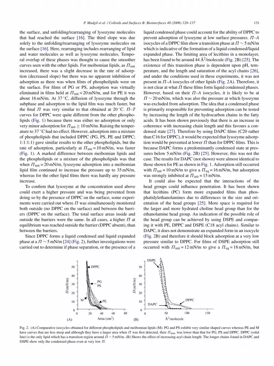
P. Mudgil et al. / Colloids and Surfaces B: Biointerfaces 48 (2006) 128–137 131
the surface, and unfolding/rearranging of lysozyme moleculesthat had reached the surface [16]. The third slope was duesolely to the unfolding/rearranging of lysozyme molecules onthe surface [16]. Here, rearranging includes rearranging of lipidand water molecules as well as lysozyme molecules. Tempo-ral overlap of these phases was thought to cause the smoothercurves seen with the other lipids. For meibomian lipids, as Π initincreased, there was a slight decrease in the rate of adsorp-tion (decreased slope) but there was no apparent inhibition ofadsorption as there was when films of phospholipids were onthe surface. For films of PG or PS, adsorption was virtuallyeliminated in films held at Π init = 20 mN/m, and for PE it wasabout 16 mN/m. At 37 ◦C, diffusion of lysozyme through thesubphase and adsorption to the lipid film was much faster, butthe final Π was very similar to that obtained at 20 ◦C. Π–Tcurves for DPPC were quite different from the other phospho-lipids (Fig. 1) because there was either no adsorption or onlyvery minor adsorption for Π init ≥ 10 mN/m. Raising the temper-ature to 37 ◦C had no effect. However, adsorption into a mixtureof phospholipids that included DPPC (PG, PS, PE and DPPC;1:1:1:1) gave similar results to the other phospholipids, but therate of adsorption, particularly at Π init = 10 mN/m, was faster(Fig. 1). A marked difference between meibomian lipids andthe phospholipids or a mixture of the phospholipids was thatwhen Π init = 20 mN/m, lysozyme adsorption into a meibomianlipid film continued to increase the pressure up to 35 mN/m,wi
cdmbeoeb
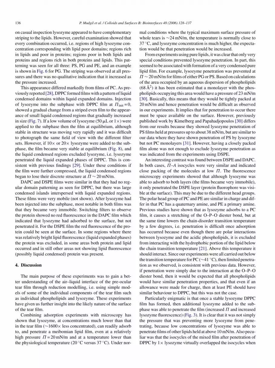
pc
liquid condensed phase could account for the ability of DPPC toprevent adsorption of lysozyme at low surface pressures. Π–Aisocycles of a DPPC film show a transition phase at Π ∼ 5 mN/mwhich is indicative of the formation of a liquid condensed/liquidexpanded phase. The limiting area of lecithins in a monolayer,has been found to be around 44 A2/molecule (Fig. 2B) [25]. Theexistence of this transition phase is dependent upon pH, tem-perature, and the length and saturation of the acyl chains [26],and under the conditions used in these experiments, it was notpresent in Π–A isocycles of other lipids (Fig. 2A). Therefore, itis not clear at what Π these films form liquid condensed phases.However, based on their Π–A isocycles, it is likely to be atΠ ∼ 20 mN/m, which was also the pressure at which lysozymewas excluded from adsorption. The idea that a condensed phaseis primarily responsible for preventing adsorption can be testedby increasing the length of the hydrocarbon chains in the fattyacids. It has been shown previously that there is an increase incoherence with increasing chain length and this favours a con-densed state [27]. Therefore by using DAPC films (C20 ratherthan C16 for DPPC), it would be expected that lysozyme adsorp-tion would be prevented at lower Π than for DPPC films. This isbecause DAPC forms a predominantly condensed state at pres-sures below 5 mN/m (Fig. 2B) [25]. However, this was not thecase. The results for DAPC (not shown) were almost identical tothose shown for PE as shown in Fig. 1. Adsorption still occurredwith Π = 10 mN/m to give a Π = 16 mN/m, but adsorptionw
htpetetiD(po
F bomiah s firstl ows thD
hereas for the other lipid films there was hardly any pressurencrease.
To confirm that lysozyme at the concentration used aboveould exert a higher pressure and was being prevented fromoing so by the presence of DPPC on the surface, some experi-ents were carried out where Π was simultaneously monitored
oth outside (no DPPC on the surface) and between the barri-rs (DPPC on the surface). The total surface areas inside andutside the barriers were the same. In all cases, a higher Π atquilibrium was reached outside the barrier (DPPC absent), thanetween the barriers.
Since DPPC forms a liquid condensed and liquid expandedhase at a Π ∼ 5 mN/m [24] (Fig. 2), further investigations werearried out to determine if phase separation, or the presence of a
ig. 2. (A) Comparative isocycles obtained for different phospholipids and meiave curves that are less steep and although they have a larger area when Π waine) is the only lipid which has a transition region around Π ∼ 5 mN/m. (B) ShSPE show only the condensed phase even at very low Π.
init eqas strongly inhibited at Π init = 15 mN/m.It could also be expected that the interactions of the
ead groups could influence penetration. It has been shownhat lecithins (PC) form more expanded films than phos-hatidylethanolamines due to differences in the size and ori-ntation of the head groups [25]. More space is required forhe larger and more hydrated choline head group than for thethanolamine head group. An indication of the possible role ofhe head group can be achieved by using DSPE and compar-ng it with PE, DPPC and DSPE (C18 acyl chains). Similar toAPC, it does not demonstrate an expanded form in an isocycle
Fig. 2B) and therefore it should block adsorption at a very lowressure similar to DPPC. For films of DSPE adsorption stillccurred with Π init = 12 mN/m to give a Πeq = 18 mN/m, but
n lipids (M). PG and PS exhibit very similar shaped curves whereas PE and Mdetected, their Πmax was lower than that for PG, PS and DPPC. DPPC (solide effect of increasing acyl chain length. The longer chains found in DAPC and

132 P. Mudgil et al. / Colloids and Surfaces B: Biointerfaces 48 (2006) 128–137
Fig. 3. (A) A series of curves showing the penetration of 50 �L of lysozyme into a DPPC film with Π init set to 0, 2, 5, 8 or 10 mN/m (0–10). With Π init = 10 mN/mhigher amounts of lysozyme (10× lysozyme = 500 �L and 20× lysozyme = 1000 �L) applied to the subphase increase Πeq. (B) After 50 �L of lysozyme was allowedto penetrate into a DPPC film with Π init set to 0 and allowed to reach equilibrium, a further 950 �L added to the subphase (arrow), showed that lysozyme was ableto still penetrate the film. (C) Equilibrium isocycles virtually overlapped irrespective of whether it was the equilibrium when 50 �L of lysozyme was allowed topenetrate into a DPPC film with Π init set to 0 (unbroken line), or the final equilibrium of the experiment shown in B (broken line).
adsorption was strongly inhibited at Π init = 16 mN/m (data notshown).
Since the concentration of lysozyme used to test penetrationwas about 1600 times less than that found in tears, it was ofinterest to determine if increasing the concentration of lysozymein the subphase resulted in increased adsorption. If Π init wereset at 10 mN/m, there was no change in pressure when 50 �L oflysozyme solution was injected into the subphase, but there was
a substantial change in pressure if higher volumes were applied.The equilibrium pressure attained was concentration dependentwith 1000 �L giving a Πeq of ∼20 mN/m (Fig. 3A).
It was also important to know if lysozyme could adsorb tothe surface once an apparently stable lysozyme/DPPC film hadformed. For these experiments, DPPC was applied to the surfacewith Π init = 0 mN/m, 50 �L of lysozyme solution was injectedinto the subphase, and the pressure was allowed to reach equi-
F ysozy1 re bota = 10 m
ig. 4. Micrographs showing the appearance of Texas red labelled lysozyme:l2 h later (D and E) and 12 h after applying it into the subphase (F). A and B werea was 40 cm2. C was at Π = 24 mN/m (maximum compression), D was at Π
me (5%:95%) applied to the air–buffer interface just after application (A–C),h when Π = 0: A was when the surface area was 80 cm2 and B when the surface
N/m, E was at Π = 24 mN/m and F at Π = 16 mN/m.

P. Mudgil et al. / Colloids and Surfaces B: Biointerfaces 48 (2006) 128–137 133
Fig. 5. Micrographs showing a pure meibomian lipid film (left column) at different pressures, and the effects of lysozyme penetration on a meibomian lipid filmset at Π init = 10 mN/m (middle and right column). Middle and right columns are matched fields showing the lipids fluorescence (middle column) and lysozymefluorescence (right column) at different pressures as penetration progressed. Penetration caused dispersion of lipids at all pressures and initially there were lipid richzones that excluded lysozyme (arrows). These then disappeared as the pressure increased and corresponding dark zones appeared in both films, e.g. both zone typescan be seen at Π = 12 mN/m (arrows). At the highest pressure there were bright lipid zones distributed throughout the film which had no corresponding regions inthe lysozyme film.

134 P. Mudgil et al. / Colloids and Surfaces B: Biointerfaces 48 (2006) 128–137
Fig. 6. Micrographs showing penetration of lysozyme into a PG film set at Π init = 10 mN/m after 4 h. The same region of the film is shown using different filters;left, PG fluorescence; right, lysozyme fluorescence. The PG film (left) is relatively amorphous with some striping. Lysozyme penetration shows a similar pattern(right). The dots are the same points on the surface. In some cases the dark and bright stripes correspond in both images. In other regions the bright stripe in lysozymecorresponds with a dark region in the PG film. Note also that the right hand side of this figure where there is strong staining with PG but little fluorescence withlysozyme.
librium. A further 950 �L of lysozyme was injected into thesubphase. The result was that the lysozyme/PC film was not abarrier and there was an increase in pressure to a new Πeq of20 mN/m (Fig. 3B). Unexpectedly, if isocycles were performedat the end of either the first equilibrium or the second equilib-rium, then they were almost identical, despite there presumablybeing more protein in the surface film after application of addi-tional protein (Fig. 3C). Note that isocycles were not performedat the first equilibrium in experiments where a higher concen-tration of lysozyme was to be added to the subphase.
3.2. Fluorescence microscopy of surface films
To provide a base, the microscopic appearance of pure filmswas examined initially, and then compared with films at differ-
ent stages of adsorption. For lysozyme, there was a differencewhen it was applied to the surface as compared to when it wasapplied into the subphase. Lysozyme applied to the surface didnot immediately disappear into the subphase but formed clumps,which gradually coalesced as the surface area of the trough wasreduced and Π increased (Fig. 4). With time, the clumping dis-appeared and lysozyme was more evenly dispersed and formeda striped patterning on the surface at maximum Π (24 mN/m)(Fig. 4E). When lysozyme was injected into the subphase andthe surface was examined between the barriers, it also showed adispersed striped patterning (Fig. 4F).
Meibomian lipids formed a film with dark irregular zones atlow Π. These gradually became much smaller as Π increased.At the highest pressure obtainable with meibomian lipids(Π = 20 mN/m), the dark zones were smaller and grey rather than
F lly ser he timw
ig. 7. Micrograph showing penetration of lysozyme into a DPPC film originaow lysozyme fluorescence. The numbers represent the pressure of the film at tay to small liquid condensed (dark) regions that gradually increased in size.
t with Π init = 0 mN/m. The top row shows DPPC fluorescence and the bottome of the micrograph. Initially there was a striped pattern which gradually gave

P. Mudgil et al. / Colloids and Surfaces B: Biointerfaces 48 (2006) 128–137 135
black (Fig. 5). When lysozyme was injected into the subphase,the meibomian lipid layer had a much more dispersed appear-ance at all pressures. At the beginning of adsorption, there wereareas which showed both red (lysozyme) and green (lipids) flu-orescence and areas with more intense green fluorescence (highlipid density) devoid of red fluorescence. Since the Texas redfluorescence is seen only when the fluorophore is sticking outfrom the surface, this indicated that lysozyme had actually pen-etrated the lipid layer except in some regions of higher lipiddensity. As Π increased due to penetration, some dark regionsappeared which were deficient of both lipid and lysozyme flu-orescence. These dark regions became more pronounced as thepressure increased and the fluorescence outside these areas was
very even. At Πeq (24 mN/m), lysozyme fluorescence appearedto be evenly dispersed throughout the film whereas the lipids hadmore intense patches. The dark regions became small circularareas in both the lipid and lysozyme views.
Microscopic examination of fluorophore doped films for PG,PS or PE at all Π > 0 showed a very even fluorescence with-out any substructural details (not shown). When lysozyme wasinjected into the subphase with the lipid film at Π init = 10 mN/m,as the pressure increased (representing penetration) an amor-phous red fluorescence of the protein could be detected whichindicated that the protein was distributing evenly (mixed) withthe lipid film. In addition to this general appearance, there wereoccasional areas where weak striping appeared in the film and
Faflp
ig. 8. Micrographs showing a DPPC film after penetration with 10× lysozyme whichnd the right column lysozyme fluorescence. The first row shows the appearance ourther 12 h and the bottom row the same film which had been further compressed to 3ocation), and liquid condensed areas are generally devoid of protein. However, theressure, the overall organization of liquid condensed and liquid expanded regions is
was originally set with Π init = 0 mN/m. Left column shows DPPC fluorescencef the film once it had reached equilibrium (15 mN/m), the middle row after a4 mN/m. The film is very stable in all cases (dots in bottom row show the samedark striped region in the first row has protein, and in the bottom row at highmuch disrupted.
136 P. Mudgil et al. / Colloids and Surfaces B: Biointerfaces 48 (2006) 128–137
on casual inspection lysozyme appeared to have complementarystriping to the lipids. However, careful examination showed thatevery combination occurred, i.e. regions of high lysozyme con-centration corresponding with lipid poor domains; regions richin lipids and poor in proteins; regions poor in both lipids andproteins and regions rich in both proteins and lipids. This pat-terning was seen for all three: PS, PG and PE, and an exampleis shown in Fig. 6 for PG. The striping was observed at all pres-sures and there was no qualitative indication that it increased asthe pressure increased.
This appearance differed markedly from films of PC. As pre-viously reported [28], DPPC formed films with a pattern of liquidcondensed domains within liquid expanded domains. Injectionof lysozyme into the subphase with DPPC film at Π init = 0,showed a gradual change from a striped even film to the appear-ance of small liquid condensed regions that gradually increasedin size (Fig. 7). If a low volume of lysozyme (50 �L or 1×) wereapplied to the subphase then the film at equilibrium, althoughstable in structure was moving very rapidly and it was difficultto photograph the same field of view with the different filtersets. However, if 10× or 20× lysozyme were added to the sub-phase, the film became very stable at equilibrium (Fig. 8), andthe liquid condensed regions excluded lysozyme, i.e. lysozymepenetrated the liquid expanded phases of DPPC. This is con-sistent with previous findings [29]. Under these conditions ifthe film were further compressed, the liquid condensed regionsb
ucTbttiptwto(
4
tteaho
sitht
mal conditions where the typical maximum surface pressure ofwhole tears is ∼24 mN/m, the temperature is normally close to37 ◦C, and lysozyme concentration is much higher, the expecta-tion would be that penetration would be increased.
From experiments using pure lipids, it was clear that only veryspecial conditions prevented lysozyme penetration. In part, thisseemed to be associated with formation of a very condensed purelipid film. For example, lysozyme penetration was prevented atΠ ∼ 20 mN/m for films of either PG or PS. Based on calculationsof the area occupied by an aqueous dispersion of phospholipids(68 A2) it has been estimated that a monolayer with the phos-pholipids occupying this area would have a pressure of 23 mN/m[30]. Basically, this means that they would be tightly packed at20 mN/m and hence penetration would be difficult as observedin our experiments. It implies that for penetration to occur theremust be space available on the surface. However, previouslypublished work by Kimelberg and Papahadjopoulos [30] differsfrom our results because they showed lysozyme penetration ofPS films held at pressures up to about 38 mN/m, but are similar toour data where they have shown penetration of PS by lysozymebut not PC monolayers [31]. However, having a closely packedfilm alone was not enough to exclude lysozyme penetration aswas indicated from the experiments using DSPE.
An interesting contrast was found between DSPE and DAPC.In both cases, Π–A isocycles were very similar and indicatedclose packing of the molecules at low Π. The fluorescencemaibTfPfitbhbftsttidwas
fiplttplD
egan to lose their discrete structure at Π ∼ 20 mN/m.DAPC and DSPE films were similar in that they had no reg-
lar domain patterning as seen for DPPC, but there was largeondensed islands interspersed with liquid expanded regions.hese films were very mobile (not shown). After lysozyme hadeen injected into the subphase, most notable in both films washat they became very immobile. Switching filters to observehe protein showed no red fluorescence in the DAPC film whichndicated that lysozyme had adsorbed to the surface, but notenetrated it. For the DSPE film the red fluorescence of the pro-ein could be seen at the surface. In some regions where thereas relatively bright lipid fluorescence (liquid expanded phases)
he protein was excluded, in some areas both protein and lipidccurred and in still other areas not showing lipid fluorescencepossibly liquid condensed) protein was present.
. Discussion
The main purpose of these experiments was to gain a bet-er understanding of the air–liquid interface of the pre-ocularear film through reduction modelling, i.e. using simple mod-ls of some of the individual components of the tear film suchs individual phospholipids and lysozyme. These experimentsave given us further insight into the likely nature of the surfacef the tear film.
Combining adsorption experiments with microscopy hashown that lysozyme, at concentrations much lower than thatn the tear film (∼1600× less concentrated), can readily adsorbo, and penetrate a meibomian lipid film, even at a relativelyigh pressure Π = 20 mN/m and at a temperature lower thanhe physiological temperature (20 ◦C versus 37 ◦C). Under nor-
icroscopy experiments showed that although lysozyme wasble to adsorb to both layers (the films became very immobile),t only penetrated the DSPE layer (protein fluorophore was visi-le at the surface). This may be due to the different head groups.he polar head group of PC and PE are similar in charge and dif-
er in that PC has a quaternary amine, and PE a primary amine.revious studies have shown that as lysozyme adsorbs to a PClm, it causes a stretching of the O–P–O diester bond, but at
he same time lowers the chain-disorder transition temperaturey a few degrees, i.e. penetration is difficult once adsorptionas occurred because even though there are polar interactionsetween lysozyme and the acidic phospholipids, it is excludedrom interacting with the hydrophobic portion of the lipid belowhe chain transition temperature [21]. Above this temperature ithould interact. Since our experiments were all carried out belowhe transition temperature for PC (∼41 ◦C), then limited penetra-ion as we observed, is consistent with previous data. However,f penetration were simply due to the interaction at the O–P–Oiester bond, then it would be expected that all phospholipidsould have similar penetration properties, and that even if an
llowance were made for charge, then at least PE should haveimilar behaviour to DPPC, but this was not the case.
Particularly enigmatic is that once a stable lysozyme DPPClm has formed, then additional lysozyme added to the sub-hase was able to penetrate the film (increased Π and increasedysozyme fluorescence) (Fig. 3). It is clear that it was not simplyhe pressure that was preventing more lysozyme from pene-rating, because low concentrations of lysozyme was able toenetrate films of other lipids held at above 10 mN/m. Also pecu-iar was that the isocycles of the mixed film after penetration ofPPC by 1× lysozyme virtually overlapped the isocycles when

P. Mudgil et al. / Colloids and Surfaces B: Biointerfaces 48 (2006) 128–137 137
20× lysozyme was used (Fig. 3C). This is strange because inthe case of 20× lysozyme there were more molecules on thesurface (indicated by the higher surface pressure at equilibrium;Fig. 3B).
By comparing all of our data on the different lipids leads usto conclude that DPPC behaves in an exceptional way. Hence,it should not be treated as a representative phospholipid as ithas been in many previous studies. In addition, how the quater-nary amine encourages adsorption of lysozyme but prevents itspenetration needs further investigation.
It was interesting from a clinical perspective that PS and PGfilms were most easily penetrated by lysozyme. It has been previ-ously shown that anionic phospholipids increase the stability ofthe tear film [22], although the exact reason for this is not known.Our data suggest that it could be due to these lipids having anenabling effect on proteins penetrating the lipid layer and thatwhen proteins are present, the surface film becomes much moregel like (stable). Lysozyme has a net positive charge at pH 7.4,and hence its increased ability to penetrate anionic lipid filmscould have an ionic component. The negatively charged phos-pholipids would be at a lower free energy in the presence of thepositively charged lysozyme. Such ionic interactions betweenthe lipids and the proteins could also contribute to increasedstability of the tear film.
Given these observations, and that meibomian lipids are amixture and also include some unsaturated fatty acids, it isuiats[sbmlttlael
5
tia
the surface it contributes to the decrease in surface tension likethe lipids and helps in the stabilization of the meibomian lipidlayer and the tear film.
References
[1] F.J. Holly, M.A. Lemp, Contact Lens Soc. Am. 5 (1971) 12.[2] R.J. Fullard, C. Snyder, Invest. Ophthalmol. Vis. Sci. 31 (1990) 1119.[3] I.K. Gipson, Y. Hori, P. Argueso, Ocular Surf. 2 (2004) 131.[4] P. Argueso, I.K. Gipson, Exp. Eye Res. 73 (2001) 281.[5] N. Nicolaides, J.K. Kaitaranta, T.N. Rawdah, J.I. Macy, F.M. Boswell
III, R.E. Smith, Invest. Ophthalmol. Vis. Sci. 20 (1981) 522.[6] J.P. McCulley, W.E. Shine, Adv. Exp. Med. Biol. 438 (1998) 319.[7] B. Nagyova, J.M. Tiffany, Curr. Eye Res. 19 (1999) 4.[8] J. Zhao, P. Wollmer, Acta Ophthalmol. Scand. 76 (1998) 438.[9] J. Zhao, R. Manthorpe, P. Wollmer, Clin. Physiol. Funct. Imag. 22 (2002)
24.[10] J.M. Tiffany, B. Nagyova, Adv. Exp. Med. Biol. 506 (2002) 581.[11] F. Miano, M. Calcara, F. Giuliano, T.J. Millar, V. Enea, J. Phys.: Con-
dens. Matter 16 (2004) S0953.[12] S.T. Tragoulias, P.J. Anderton, G.R. Dennis, F. Miano, T.J. Millar,
Cornea. 24 (2005) 189.[13] S. Krol, M. Ross, M. Sieber, S. Kunneke, H.J. Galla, A. Janshoff, Bio-
phys. J. 79 (2000) 904.[14] A. Kijlstra, A. Kuizenga, Adv. Exp. Med. Biol. 350 (1994) 299.[15] K. Arnold, D. Hoekstra, S. Ohki, Biochim. Biophys. Acta 1124 (1992)
88.[16] D.E. Graham, M.C. Phillips, J. Colloid Interface Sci. 70 (1979) 403.[17] W. van der Vegt, W. Norde, H.C. van der Mei, H.J. Busscher, J. Colloid
Interface Sci. 179 (1996) 57.[
[[
[
[
[[
[[
[
[
[
[
[[
[
nlikely to form close packing, it is most probable that lysozymes present at the surface of the tear film, and not simply in thequeous phase as current models suggests [3,7,32]. Assuminghat lysozyme is present at the surface, then it is of interest topeculate on its possible role. Lysozyme denatures at the surface33] and hence it is unlikely to have an anti-bacterial role at theurface. By the fluorescent micrographs, lysozyme appears toe acting as a dispersant of the meibomian lipids. This wouldean that the surface of the tear film would be more akin to
ung surfactant [13] rather than a mixed lipid layer. It is notablehat the proteins involved in lung surfactant, surfactant pro-eins B and C, are strongly hydrophobic rather than hydrophilicike lysozyme. However, the tears also contain lipocalins thatre hydrophobic in nature, and it would be of interest toxamine their penetration into the surface film of meibomianipids.
. Conclusion
Taken together these data indicate that lysozyme is one ofhe important surface-active proteins present in the tear film andt has ability to move from the bulk solution to the surface andbsorb to and penetrate the lipid layer present at the surface. At
18] T.J. Su, J.R. Lu, R.K. Thomas, Z.F. Cui, J. Penfold, J. Colloid InterfaceSci. 203 (1998) 419.
19] J.R. Lu, T.J. Su, B.J. Howlin, J. Phys. Chem. B 103 (1999) 5903.20] C. Postel, O. Abillion, B. Desbat, J. Colloid Interface Sci. 266 (2003)
74.21] J.L. Lippert, R.M. Lindsay, R. Schultz, Biochim. Biophys. Acta 599
(1980) 32.22] D.R. Korb, J.V. Greiner, T. Glonek, Adv. Exp. Med. Biol. 506 (2002)
495.23] M. Brinkley, Bioconj. Chem. 3 (1992) 2.24] A. Bredlow, H.J. Galla, L.D. Bergelson, Chem. Phys. Lipids 62 (1992)
293.25] M.C. Phillips, D. Chapman, Biochim. Biophys. Acta 163 (1968) 301.26] A.D. Williams, J.M. Wilkin, R.A. Dluhy, Colloids Surf. A 102 (1995)
231.27] I. Kuzmenko, H. Rapaport, K. Kjaer, J. Als-Nielsen, I. Weissbuch, M.
Lahav, L. Leiserowitz, Chem. Rev. 101 (2001) 1659.28] B. Piknova, W.R. Schief, V. Vogel, B.M. Discher, S.B. Hall, Biophys.
J. 81 (2001) 2172.29] P. Kruger, J.E. Baatz, R.A. Dluhy, M. Losche, Biophys. Chem. 99 (2002)
209.30] H.K. Kimelberg, D. Papahadjopoulos, Biochim. Biophys. Acta 233
(1971) 805.31] H.K. Kimelberg, D. Papahadjopoulos, J. Biol. Chem. 246 (1971) 1142.32] J.C. Pandit, B. Nagyova, A.J. Bron, J.M. Tiffany, Exp. Eye Res. 68
(1999) 247.33] F. Miano, M. Calcara, T.J. Millar, V. Enea, Colloids Surf. B 44 (2005)
49.


