Embed Size (px)
Citation preview
Affordable Dermascope for Resource-Poor Settings
Jenna Johnstone, Brian Bacik, Shannon Burke, Rachel Dzombak, Khanjan Mehta, Peter Butler Humanitarian Engineering and Social Entrepreneurship (HESE) Program
The Pennsylvania State University University Park, PA 16802
Correspondence: [email protected]
Abstract – Skin diseases in developing countries receive little attention when compared with well-known killers such as HIV/AIDS, pneumonia and tuberculosis. In communities with few doctors and even fewer dermatological specialists (if any), Community Health Workers (CHWs) are charged with meeting the needs of these at-risk populations. However, skin diseases can be difficult to assess by CHWs and often signify larger underlying problems. Teledermatology is being increasingly employed as a means of remotely assessing and diagnosing skin ailments. Typical commercially-available dermascopes cannot withstand the harsh conditions of developing countries and there is a need for inexpensive and ruggedized dermascopes for resource-constrained settings. This paper presents the design and field-testing results for a dermascope intended for use in rural Kenya as a part of an operational telemedicine system. Though the design faced challenges with ambient light, material availability, and camera mobility, the final prototype produced images deemed acceptable for diagnosis by Kenyan clinicians. Index Terms—telemedicine, teledermatology, skin diseases, dermascope
I. INTRODUCTION Skin diseases are often considered low priority in the
landscape of global disease burdens compared to the “big three killer” diseases that cause significant mortality: HIV/AIDS, tuberculosis, and pneumonia [1]. However, skin diseases also cause significant mortality in sub-Saharan Africa with about 20,000 people dying each year from skin-related afflictions [2]. These rates are similar to the mortality rates of hepatitis B, meningitis, obstructed labor and rheumatic heart disease [3]. Published data suggest the prevalence of skin diseases in developing countries ranges from 20% to 80%, depending on geographic location [4]. It is estimated that skin diseases in sub-Saharan Africa account for 896,000 disability-adjusted life years (DALYs), on par with endocrine and anxiety disorders, gout, and war-related injuries [3]. A study conducted in western Ethiopia revealed that while nearly 50% of rural community members claimed to have skin disease, out of those denied the presence of skin issues, over 67 percent were found to have treatable skin conditions [3]. Pyoderma, a generic term describing any variant of a superficial bacterial skin infection, was found to be 35% prevalent in less developed areas [5]. Skin disease is particularly common amongst those living with diabetes whose associated symptoms are not properly managed [6]. Diabetes is becoming increasingly prevalent in sub-Saharan Africa with numbers expected to reach 23.6 million affected by 2030 [7]. Also, about 22.5 million people, or 68% of the global population, living with HIV/AIDS resides in sub-
Saharan Africa [8]. Close to 90% of individuals with HIV/AIDS are diagnosed with skin disease at some point during the course of their illness [9]. Skin diseases are often considered a reflection of a patient’s underlying immune condition and hence carry a negative social stigma [10]. The high percentages of people in Sub-Saharan Africa suffering from HIV/AIDS and diabetes manifest the criticalness of affordable and accessible dermatological care.
Though economic impact of alleviating the burden of skin diseases can be significant, literature investigating associated costs in underdeveloped countries is scarce. In a retrospective study, 102 cases of Buruli ulcers were found at a district hospital in Ghana. Treatment for this ulcer costs an average of $701.06 per patient with additional indirect costs (loss of productivity, food, etc.) incurred due to hospitalization adding up to $549.49 per patient [11]. Over 54% of Ghana’s population lives below US $2 per day, making these treatment costs fundamentally out of reach for individual patients [12]. As such, much of the affected population does not seek health care until a problem grows critical because of financial constraints [11]. In studies of patients with onchocerciasis, a parasitic disease that causes severe skin irritation, the disease seemed to occur more often in patients with less income, less personal wealth, smaller farm sizes and more days of leave as compared to control groups [13] [14]. These studies shed light on the impact that dire economic conditions can have on individuals and why low-income communities are often the most at-risk for skin diseases.
Despite low mortality rates, effective diagnosis and treatment of skin diseases is needed. Some diseases cause disfigurement and others are transmissible from person to person. Most of these diseases are treatable if recognized and diagnosed. In addition, screening the skin for abnormalities can be used as a clinical tool for diagnosis of other diseases, including leprosy and HIV. However, clinical skills are often lacking in primary care settings in the developing world. In some parts of sub-Saharan Africa, there is one doctor for 10,000 patients [15]. The numbers of specialists are significantly lower and are concentrated in urban areas. South Africa, one of the most developed African economies, has a ratio of one dermatologist per 3 or 4 million people [16]. These factors result in an alarming lack of access to quality clinical care to patients with skin abnormalities.
The lack of trained dermatologists and physicians in the developing world gives validity to telemedicine and specifically, teledermatology as a potential solution to the burden of skin disease. Teledermatology is the use of
978-1-4799-2402-8/13/$31.00 ©2013 IEEE 314 IEEE 2013 Global Humanitarian Technology Conference
technology to send and receive medical information between patient and doctor, in this case, photographs of skin abnormalities for use in clinical diagnosis. In the past few years, technology improvements have facilitated the emergence of several teledermatology projects. One such venture, the African Teledermatology Project, uses store-and-forward technology to connect patients in Africa with doctors around the world [17]. Preliminary results support the reliability and efficacy of teledermatology, though in some cases poor image quality prevented the pictures from providing any clinical information. During another pilot in South Africa, medical diagnosis was possible in 99% of the cases and an improvement in patient treatment was made in 55% of consultations [18]. Finally, teledermatology was shown as an effective means to diagnose tinea in Kenyan schoolchildren [19]. Collectively, these studies validate that when operators are trained in taking appropriate photos of skin abnormalities and additional medical history is provided, teledermatology can be effectively and efficiently used to diagnose skin conditions. In rural settings, such operators are most likely to be community health workers (CHWs).
Community Health Workers (CHWs) are community members who volunteer to track the health of specific households in their vicinity through monthly visits. Through their active health monitoring, CHWs act as a first line of defense against infectious diseases and chronic conditions. They educate community members about family planning mechanisms, nutritious diets, prenatal care, and other relevant topics [20]. Any biomedical equipment provided to CHWs, must be designed to survive in the field. For biomedical devices, this can often prove challenging. Over 95% of biomedical devices found in public hospitals in developing countries are imported [21], and 96% of such devices fail within 5 years [22]. Devices fail due to a disconnect between the context that they were designed in, and the reality of where they are used. CHWs travel long distances in dusty, humid or rainy weather conditions, to reach their assigned households. They are challenged by an unreliable power infrastructure, poor transportation, and a general lack of resources for device upkeep. To meet the needs of CHWs and their communities, engineers must create biomedical devices that are affordable, easy to use, and ruggedized, that is, they are specifically designed for the developing country context.
The Humanitarian Engineering and Social Entrepreneurship (HESE) Program at the Pennsylvania State University has developed and currently operates a telemedicine system called Mashavu: Networked Health Solution [23]. This entrepreneurial venture combats the lack of access to pre-primary medical care caused by low doctor-to-patients ratios. Mashavu Kiosk Operators (MKOs) gather patients’ health history and vital statistics (height, weight, blood pressure, and BMI) for a small fee. This information is entered into and securely stored on a web-based portal using either a web-enabled smartphone or, when stationed at a cyber café, desktop computers. This information is then viewed by a medical professional, typically a nurse, who responds with tailored recommendations within 20 minutes. The goal of Mashavu is
to encourage community members to take an active interest in their health and to create opportunities through which otherwise unpaid CHWs can generate livelihoods. Established in 2009, Mashavu has been used by over a 100,000 individuals throughout Kenya and Tanzania. Over the course of field-testing efforts, the Mashavu team realized the need for an imaging component. Images can help medical professionals working with Mashavu to assess patients’ issues and make diagnoses. When patients report skin abnormalities, images grow increasingly critical if affective recommendations are to be prescribed. This need manifested itself, when in 2010, the team notices that a significant portion of children at a Mashavu kiosk showed signs of ringworm infections. Subsequently, an IRB-approved study was conducted to assess the feasibility of diagnosing ringworm through pictures. Schoolchildren were tested for ringworm and other skin diseases and a low-cost camera was used to capture children’s skin lesions. After sharing the images with Kenyan clinicians, it was determined that the described operations provided sufficient information to accurately detect tinea [19].
The intervention approach and trial results garnered positive feedback from Kenyan doctors and nurses, thus encouraging the Mashavu team to create a dermascope suitable for use in east Africa in conjunction with the telemedicine system. This paper presents a low-cost dermascope for resource-constrained settings. This device was designed for integration into a telemedicine system in rural Kenya. The article starts with a discussion of current dermascope technologies and the context of use in Kenya. Design decisions, and their rationale, are discussed in details with a concluding section on field-testing results and lessons learned.
II. DERMASCOPE TECHNOLOGIES Most teledermatology ventures, including the ones cited in
the introduction, utilize cameras that are not designed for the harsh operating environments in resource-constrained contexts. They are too expensive for widespread use by CHWs during their health visits. The costs and features of a few commonly used dermascopes are summarized in Table I.
TABLE I. COMMERCIAL DERMASCOPES
Dermascope Cost Zoom Lighting USB
Compatibility Dermlite II Hybrid $925 10x 24 LEDs Yes USB Video Dermascope $850
12x-100x
Built-in LED light No, SD card
Dino-Lite Pro AM413T $400 10x-70x 8 LEDs Yes Handyscope for iPhone $650 20x
Polarized LEDs Yes
3GEN DermLite $1,075 8x 24 LEDs No PSU Dernascope $56.25
Fixed Focus 6 LEDs Yes
While these dermascopes may be affordable and
sustainable in Western contexts, they are not suitable for widespread use in developing countries. Inexpensive and durable cameras with good image quality are needed to fill this
clinical void. Specifically, a dermascope must have a simple design to enable easy construction, maintenance and repairs. The product costs must remain low to ensure widespread affordability and accessibility by healthcare systems, especially at the CHW level. The device should be ruggedized and able to withstand the harsh environment. It is imperative that quality of the images is standardized and usable for effective clinical diagnosis. The functionality of biomedical devices in such contexts is constrained by many factors including capital, operational costs, environmental elements and operator education levels [24]. In order to build a durable and affordable dermascope for use in the east African context, these constraints would have to be met through a series of rigorous design decisions. The design team aimed to create a dermascope that captured accurate images, could withstand dust, was easy to use, and met contextual anthropometric and socio-cultural requirements.
III. HARDWARE DESIGN FOR EAST AFRICAN CONTEXT
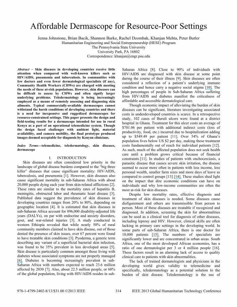
This section describes the design of the low-cost dermascope and the rationale for the design decisions. For the scope to be successful in East Africa, ruggedized housing was needed to protect the camera and lighting systems appropriately while still allowing for adjustment of the camera (Figure 1). The housing system had to meet anthropometric requirements since ease of use in the field is important for both the user and the patient. Another pertinent consideration for the design was image standardization.
Standardization of the images captured by the scope was essential for proper assessment and patient diagnosis. Placement and type of LED lights used influence glare, particularly in the case of pigmented skin. While incorporating multiple slits down the length of the PVC pipe seemed to aid camera movement, the slits combined with a lack of cap on the top of the pipe permitted ambient light interference in the scope images. In the Kenyan context, lighting is extremely variable, depending primarily on where the camera is used (dispensary, kiosk, hospital, outdoors, etc). The prototype utilized a web camera and 6 LED lights. The light intensity of each LED could be adjusted using a potentiometer integrated into the USB cord. The camera was mounted in a 20 cm long, 7.6 cm diameter PVC sewer pipe housing. The mounting system was composed of a zinc-plated steel bolt which was tightened onto a matching nut attached to the web camera. In order to allow the user to adjust the height of the camera, a short slit along the length of the PVC pipe was provided. After attaching a nut to the camera, a bolt was tightened into the stationary nut and a tension pin, and washers and a spring were used to apply constant tension. The entire bolt support could then move freely along the slit.
A primary goal of the dermascope design was determining how to prevent ambient light from interfering with image quality. By utilizing the minimum number of slits, ambient light was greatly decreased. The dermascope (Figure 1) features two slits, one for the bolt and camera construct and another shorter slit to allow the USB cord of the camera to exit from the PVC pipe. A 7.6 cm diameter PVC pipe cap was then
added to the top of the pipe, preventing ambient light from entering. With the pipe cap secured on top of the housing, the majority of the USB cord slit was covered, except for a small opening for the actual cord. This allowed for the camera to be easily removed from the pipe if necessary and for the cord to escape from the pipe without allowing ambient light to enter.
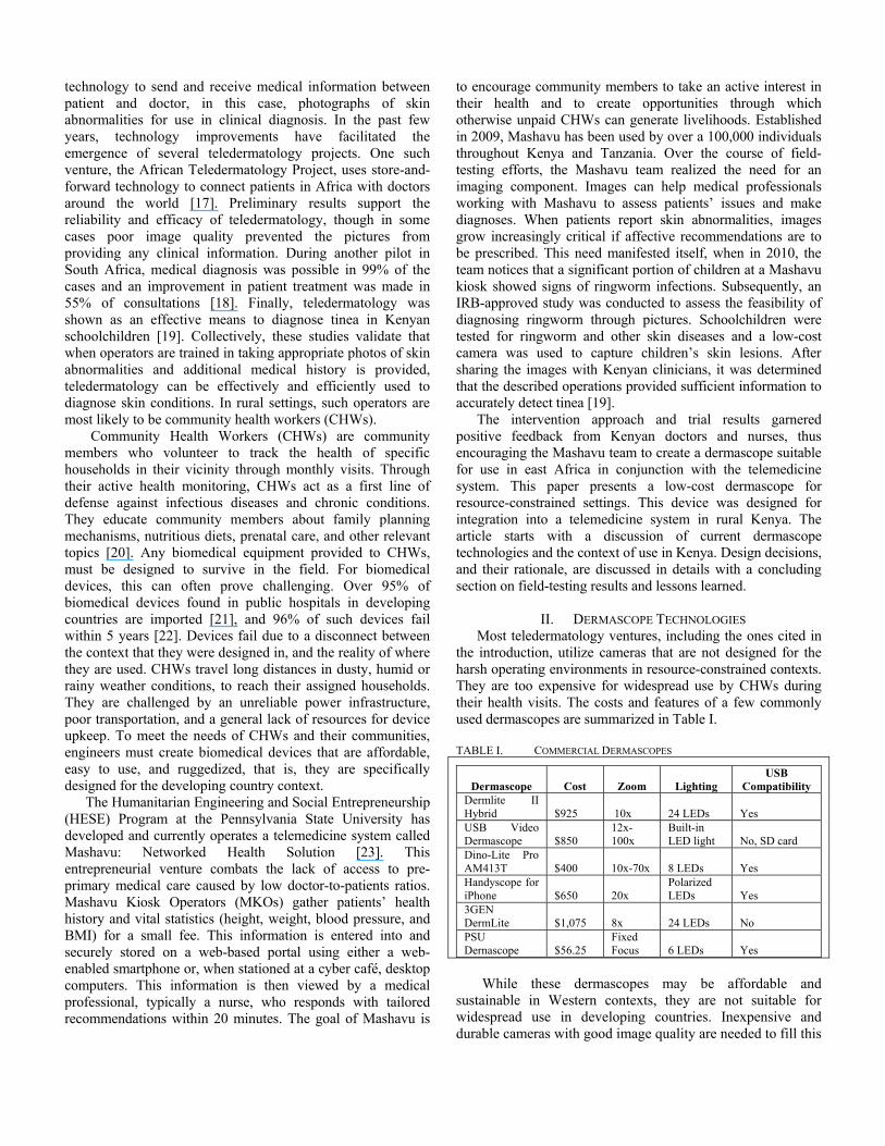
A. Solidworks Model Once ambient light was eliminated, artificial light had to be
introduced to capture standardized images. Through the use of finite element analysis and physical testing of the device, it was determined that a ring of six lights around the perimeter of the PVC housing and the camera provided the best lighting conditions inside the scope. Solidworks was used to design a “horseshoe” ring on which six white LED lights were mounted (Figure 2). The “horseshoe” design was constructed out of a heat-fusable polymer using a Rapid Prototyping machine. It was designed so that the same bolt, nut, spring, and tension pin mechanism used with the camera could be attached to allow the horseshoe to move vertically along a slit in the PVC pipe. The LEDs were connected to a board which was then reinforced with super glue and enclosed in an insulating polymer before being fixed to the inside of the cap. The LEDs could be adjusted via the original potentiometer and the entire circuit board was safely enclosed within the housing. The shape and placement of the horseshoe as well as the type of LED lights allowed for lighting without glare.
Figure 1: Dermascope Prototype
Physical testing and COMSOL modeling showed that
optimal imaging conditions occurred when the horseshoe and the camera were placed adjacent to each other inside the pipe. It was also more difficult to operate when these two components were separate, so the final design consisted of the horseshoe and camera fused together, allowing them to move within the tube as one connected piece.
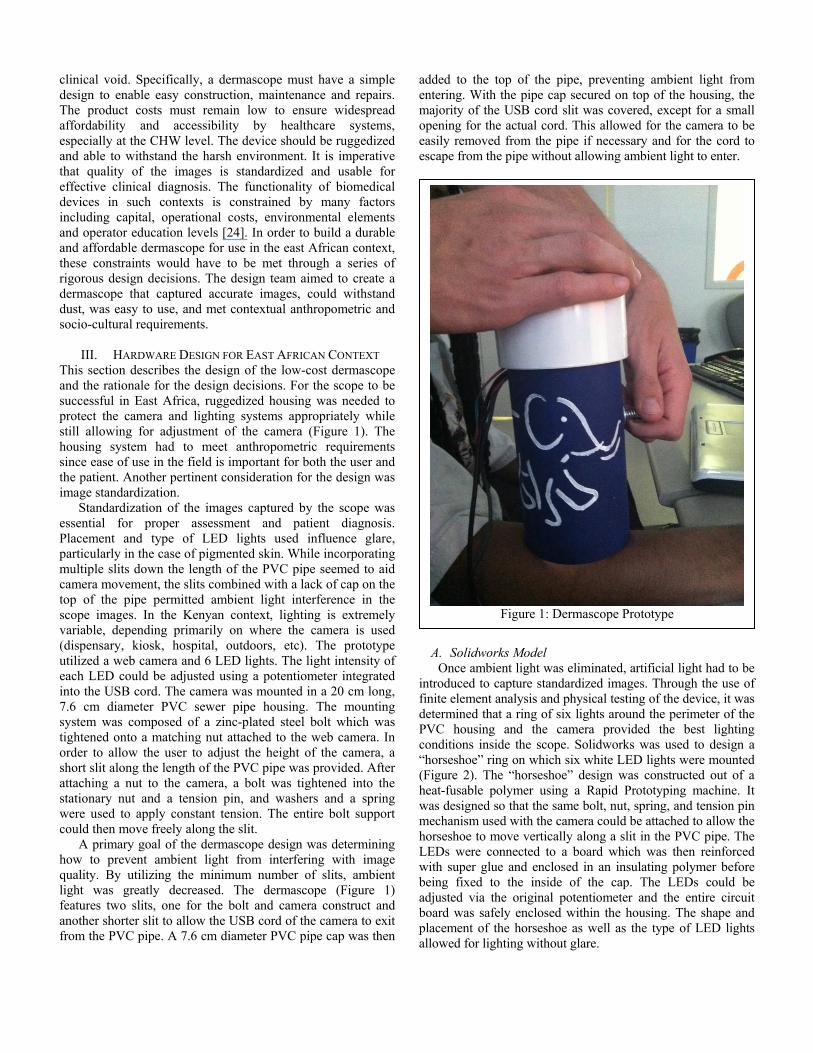
B. COMSOL Model COMSOL Multiphysics software was used to model the
proper number and placement of light sources in order to maximize light consistency at the surface of the skin. The simulation was executed in 2-D Electromagnetic Wave Physics in the frequency domain. The original model consisted of a rectangle with identical dimensions to the physical device and one light source located at the middle of the top boundary. By altering various parameters related to the light source(s), the model was able to simulate different lighting arrangements. This analysis showed that placing multiple lights along the walls of the PVC tube gave the most consistent lighting conditions at the surface of the skin, while minimizing glare. Further, the model showed that the best lighting conditions occurred approximately 10 to 15 cm from the skin. Hence, slits to limit the camera and light fixture’s range of operation were provided between 11 and 14 cm from the skin (Figure 3 & 4).
Figure 3: Light intensity at skin surface when the lights are 10 cm away. Inset: Light intensity profile through the PVC tube.
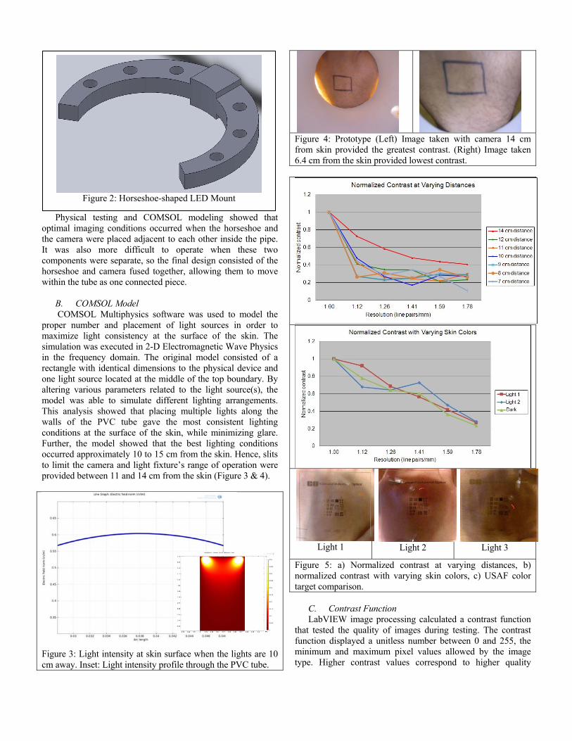
Figure 4: Prototype (Left) Image taken with camera 14 cm from skin provided the greatest contrast. (Right) Image taken 6.4 cm from the skin provided lowest contrast.
Light 1 Light 2 Light 3
Figure 5: a) Normalized contrast at varying distances, b) normalized contrast with varying skin colors, c) USAF color target comparison.
C. Contrast Function LabVIEW image processing calculated a contrast function
that tested the quality of images during testing. The contrast function displayed a unitless number between 0 and 255, the minimum and maximum pixel values allowed by the image type. Higher contrast values correspond to higher quality
Figure 2: Horseshoe-shaped LED Mount
images. This was a valuable tool to quantify image results and determine conditions for optimal image quality during experimental trials. Design decisions such as the maximum length of the camera mounting slit and the permanent position of the horseshoe with respect to the camera were made based on data obtained from this function. The contrast function along with the COMSOL model were used to determine the depth of field of the Logitech C500 camera from a patient’s skin. Since the camera has a fixed focus system, the distance of the camera from the skin was a crucial factor for obtaining quality images. To find the depth of field, images were taken at various distances from the patient’s skin and the contrast of these images was compared. It was found that maximum contrast occurred between 11 cm and 14 cm from the skin. Contrast decreased rapidly when the camera was closer than 11 cm from the skin and the images became noticeably blurry (Figure 4). The images were reviewed by a registered nurse and a physician, who concluded that all the images greater than 10 cm from the skin were of adequate quality for diagnosis.
Another analysis compared the contrast of images of a USAF resolution target (figure). These images were analyzed with the ImageJ image processing software. The first test was done by keeping background color constant and varying the height of the camera. The highest level of contrast, and thus the highest quality image was obtained at the furthest distance between the camera and the target. The second test was done by holding distance constant and varying background color. Three different skin tones were imaged, and the results showed that darker complexions provided the least favorable contrast (Figure 5). As a result of this testing and consultation with the physician, a slit was cut in the PVC pipe that would limit the movement of the camera to between 11 and 14 cm from the patient’s skin. This arrangement gave the operator some freedom to adjust the camera when necessary while ensuring that the captured images would be of the highest possible quality. When the LED plate was behind the camera, the camera would create a shadow on the skin. When the plate was too far in front of the camera, the lights would obstruct the view of the skin. As a result of this testing, it was decided to attach the camera to the LED plate so that the lights were located just in front of the camera. This configuration provided the most consistent lighting, the highest quality images, and made operation of the device simpler.
IV. SOFTWARE INTERFACE
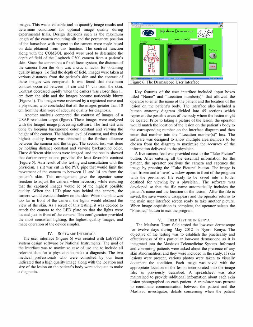
The user interface (Figure 6) was created with LabVIEW system design software by National Instruments. The goal of the interface was to maximize ease of use and to include all relevant data for a physician to make a diagnosis. The two medical professionals who were consulted by our team indicated that a high quality image along with the location and size of the lesion on the patient’s body were adequate to make a diagnosis.
Figure 6: The Dermascope User Interface
Key features of the user interface included input boxes
titled “Name” and “Location number(s)” that allowed the operator to enter the name of the patient and the location of the lesion on the patient’s body. The interface also included a human anatomy diagram divided into 45 sections which represent the possible areas of the body where the lesion might be located. Prior to taking a picture of the lesion, the operator would match the location of the lesion on the patient’s body to the corresponding number on the interface diagram and then enter that number into the “Location number(s)” box. The software was designed to allow multiple area numbers to be chosen from the diagram to maximize the accuracy of the information delivered to the physician.
A live camera feed was provided next to the “Take Picture” button. After entering all the essential information for the patient, the operator positions the camera and captures the image by pressing the “Take Picture” button. The image is then frozen and a ‘save’ window opens in front of the program with the pre-named file ready to be saved into a folder destined for viewing by a physician.. The software was developed so that the file name automatically includes the patient’s name and the location of the lesion. After the file is saved, the save window disappears and the operator returns to the main user interface screen ready to take another picture. When image acquisition is complete, the operator selects the “Finished” button to exit the program.
V. FIELD TESTING IN KENYA The Mashavu Team field tested the low-cost dermascope
for twelve days during May 2012 in Nyeri, Kenya. The objective of the testing was to establish the practicality and effectiveness of this particular low-cost dermascope as it is integrated into the Mashavu Telemedicine System. Informed and consenting patients were asked about the presence of any skin abnormalities, and they were included in the study. If skin lesions were present, various photos were taken to visually document the condition. Each image was saved with the appropriate location of the lesion incorporated into the image file, as previously described. A spreadsheet was also maintained to provide additional information about each skin lesion photographed on each patient. A translator was present to coordinate communication between the patient and the Mashavu investigator; details concerning when the patient
noticed the lesion, symptoms associated with the lesion, and other relevant background information was thus obtained. Photos and spreadsheets for each day of the clinics were stored on laptop computers and backed up with flash drives and email. The images were reviewed by a physician and were found to be useful in almost all cases. While our design team worked closely with, and received feedback from, Kenyan physicians, nurses and community health workers (CHWs), the images were not used to diagnose diseases for the patients. Doing so would have categorized our efforts as a clinical trial and that would necessitate time-consuming and expensive formal processes that were beyond our immediate reach. The key lessons, related to the design of the dermascope, learned during field testing are summarized in this section.
A. CAMERAS The camera used in the dermascope was a Logitech C500.
This camera features a 30 fps frame rate, a 1.3 MP photo capture resolution and fixed-length focus. After speaking to the medical professionals that tested the dermascope, it was found that the images taken less than 11 cm from the skin were too indistinct for diagnosis via telemedicine. This result was due to the fixed focus feature of the camera. In a fixed focus camera, the focus is set during the assembly of the camera to a model standard. Therefore the camera has a fix depth of field for which the images are sufficiently clear for capture. Through the experimental testing conducted on the scope before its pilot in Kenya, we found that the depth of field of the camera was between 11 cm and 14 cm for diagnosable images. The medical professionals that advised our team mentioned that although this depth of field was sufficient for diagnosis via teledermascopy in most cases, it would be ideal to expand that depth of field, to further augment the versatility of the dermascope in Kenya.
Alternative options to focusing systems in web cameras include an autofocus or manual focus system. Autofocus cameras contain a sensor or motor that automatically focuses the camera lens at different distances from the selected object or area. These cameras are typically expensive. If the autofocusing feature were to fail in the field, a feasible repair solution would be difficult to find. Another option is a manual focus system which requires the user to focus the camera by hand. In the cramped space of the camera housing, a manual focus would be difficult to maneuver. Due to the price and time limitations during the construction of the dermascope, a fixed focus camera was chosen. Future improvement should target on the camera and focusing system utilized in the dermascope. Alternative fixed focusing cameras and software improvements could be designed to improve the quality of the captured photographs. The image quality still remained the most unsatisfactory aspect of the dermascope although the images generally sufficed for teledermascopy.
B. LIGHTING
The horseshoe was originally designed to move in parallel with the camera within the PVC housing. Upon experimental testing, it was found that the free movement was not only
unnecessary, but also allowed for user errors. If placed too far behind the camera, the horseshoe provided uneven lighting. If positioned too far in front of the camera, the lighting created a glare on the image subject. From these results, the design decision was made to mount the horseshoe onto the camera in a strategic position to optimize lighting. The inner radius of the horseshoe had to be reduced to mount the camera properly, and this weakened the entire piece. The ribbon wire was fed through the original fixture point of the bolt on the horseshoe, which forced it against the inner wall of the PVC pipe. With every movement of the camera and light fixture, the ribbon wire rubbed against the inner radius of the PVC pipe; degrading its insulation and weakening its integrity. Though the last minute alterations to the lighting system did withstand the field testing of the dermascope, there is still much room for improvement.
The curvature and radius of the horseshoe could be adjusted specifically to mount onto the camera. The ribbon wire could also be fed through the polymer of the horseshoe to reinforce and protect it from rubbing against the inner wall of the housing. Ribbon wire is flexible, yet consequently weak. The entire lighting system could be improved by simply integrating a potentiometer without the circuit board interface. The board ultimately served as an interface between the lights in the horseshoe and the potentiometer buried in the reinforced camera-USB cord. The entire board could be bypassed and the lights connected directly to a USB cord containing a potentiometer for altering the intensity of the LED lights. This would eliminate the weak points where the ribbon wire and USB cord were soldered to the board. While this is difficult to do when prototyping, it can be incorporated into the mass-manufactured design with relative ease.
C. CONTEXTUAL APPROPRIATENESS
Epoxy was used for the vast majority of the connections to provide a sturdy and rugged device. There were no issues with connection breakages during field testing of the device in Kenya, demonstrating the durability of the design. The circuit board and wire connections for the LED light system and webcam were all enclosed within shrink wrap in order to provide protection and further increase the durability of the dermascope. The device was designed with strain relief in mind, with various features included to reduce the strain on wires.
Additionally, images were taken by placing the end of the PVC pipe slightly above the surface of the skin. The original intent was to place the device flush against the skin in order to minimize the effect of ambient light and produce a consistent image. However, due to the need for sterilization between uses and the impracticality of a disposable barrier between the device and the patient, eliminating any contact between skin and the device was deemed most appropriate. This did not, however, impact the quality of the image adversely.
VI. CONCLUSION
The design of an affordable and durable dermascope capable of capturing high-quality skin images was a successful
but challenging endeavor. While the device typically produced acceptable images that could be used for accurate diagnosis, image standardization was particularly difficult. Ambient light was the biggest issue, especially since physical contact between the dermascope and skin was not appropriate due to hygiene reasons. The next iteration of the dermascope should use a webcam with a higher resolution in order to capture clearer and more detailed images. A high-resolution image also facilitates the post-processing of images to extract features of interest.
The dermascope was well-received by the CHWs as well as community members during field-testing in Kenya. Fungal infections, most notably ringworm, seemed to be the most common visibly obvious skin problem. In a separately conducted rigorous study, we have clinically validated that a webcam can be used by physicians to diagnose ringworm (tinea) remotely [19]. In the next phase, the dermascope will be integrated into the Mashavu telemedicine system and a comprehensive clinical study will be conducted.
REFERENCES
[1] [2]
A. Fenwick, "The Global Burden of Neglected Tropical Diseases," Public Health, vol. 126, no. 3, pp 233-236, 2012. C. D. Mathers, A. D. Lopez and C. J. Murray, "The Burden of Disease and Mortality by Condition: Data, Methods, and Results for 2001," in Global Burden of Disease and Risk Factors, Washington (DC), World Bank, 2006.
[3] R. Hay, S. Bendeck, S. Chen, R. Estrada, A. Haddix, T. McLeod and A. Mahé, "Skin Diseases," in Disease Control Priorities in Developing Countries, Washington (DC), World Bank, 2006.
[4] K. P. Msyamboza, L. R. Mawaya, H. W. Kubwalo, D. Ng'oma, M. Liabunya, S. Manjolo, P. P. Msiska and W. W. Somba, "Burden of leprosy in Malawi: community camp-based cross-sectional study," BMC International Health and Human Rights, vol. 12, no. 1, p. 12, 2012.
[5] Word Health Organization, "Epidemiology and Management of Common Skin Diseases in Children in Developing," Geneva, 2005.
[6] N. T. Foss, D. P. Polon, M. H. Takada, M. C. Foss-Freitas and M. C. Foss, "Skin Lesions in Diabetic Patients," Revista de Saúde Pública, vol. 39, no. 4, pp. 677-682, 2005.
[7] V. Hall, R. W. Thomsen, O. Henrikson and N. Lohse, "Diabetes in Sub Saharan Africa 1999-2011: Epidemiology and Public Health Implication. A Systematic Review," BMC Public Health, vol. 11, no. 1, p. 564, 2011.
[8] Joint United Nations Programme on HIV/AIDS , "Global report: UNAIDS report on the global AIDS epidemic," 2010.
[9] J. Hu, K. McKoy, A. Papier, S. Klaus, T. Ryan, H. Grossman, E. J. Masenga, A. Sethi and N. Craft, "Dermatology and HIV/AIDS in Africa," Journal of Global Infectious Diseases, vol. 3, no. 3, p. 275, 2011.
[10] P. V. Raju, G. Raghurama Rao, T. V. Ramani and S. Vandana, "Skin disease: clinical indicator of immune status in human immunodeficiency virus (HIV) infection," International Journal of Dermatology, vol. 44, no. 8, pp. 646-649, 2011.
[11] World Health Organization, "Buruli Ulcer: Mycobacterium Ulcerans Infection," 2000.
[12] C. Haub and T. Kaneda, "2011 World Population Data Sheet," 2011. [Online]. Available: www.prb.org. [Accessed March 2013].
[13] W. Workneh, M. Fletcher and G. Olwit, "Onchocerciasis in Field Workers at Baya Farm, Teppi Coffee Planation Project, Southwestern Ethiopia: Prevalence and Impact on Productivity," Acta Tropica, vol. 54, no. 1, pp. 89-97, 1993.
[14] O. Olapedo, W. R. Brieger, S. Otusanya, O. O. Kale, S. Offiong and M. Titiloye, "Farm Land Size and
Onchocerciasis Status of Peasant Farmers in Southwestern Nigeria," Tropical Medicine & International Health, vol. 2, no. 4, pp. 334-340, 1997.
[15] World Health Organization, "World Health Statistics," 2012. [Online]. Available: http://www.who.int/gho/publications/world_health_statistics/2012/en/index.html. [Accessed March 2013].
[16] K. McKoy, "The Importance of Dermatology in Public Health," Harvard Medical School of Dermatology, 2012.
[17] J. Weinberg, S. Kaddu, G. Gabler and C. Kovarik, "The African Teledermatology Project: Providing Access to Dermatologic Care and Education in sub-Saharan Africa," The Pan African Medical Journal, vol. 3, no. 16, 2009.
[18] K. Brauchli, D. O'Mahony, L. Banach and M. Oberholzer, "iPath- A Telemedicine Platform to Support Health Providers in Low-Resource Settings," Studies in Health Technology and Informatics , vol. 114, pp. 11-17, 2005.
[19] S. E. Smith, J. T. Ludwig, V. M. Chinchilli, K. Mehta and J. A. Stoute, "Use of Telemedicine to Diagnose Tinea in Kenyan Schoolchildren," Telemedicine and e-Health, vol. 19, no. 3, pp. 166-168, 2013.
[20] B. Buehler, R. Ruggiero and K. Mehta, "Empowering Community Health Workers with Technology Solutions," IEEE Technology and Society Magazine, vol. 32, no. 1, pp. 44-52, 2013.
[21] R. A. Malkin, "Design of Health Care Technologies for the Developing World," Annual Revview of Biomedical Engineering, vol. 9, pp. 567-587, 2007.
[22] J. Pena-Mohr, "Distributing and Transferring Medical Technology." International journal of technology assessment in health care 3.2 (1987): 281-291.," International Journal of Technology Assessment in Health Care, vol. 3, no. 2, pp. 281-291, 1987.
[23] R. Qin, R. Dzombak, R. Amin and K. Mehta, “Reliability of a Telemedicine System Designed for Rural Kenya,” Journal of Primary Care & Community Health, vol. 4, no. 3, pp. 177-181, 2013.
[24] S. Schopman, K. Kalchthaler, R. Mathern, K. Mehta and P. Butler, "Ruggedizing Biomedical Devices for Field-Testing in Resource-Constrained Environments: Context, Issues and Solutions," Journal of Humanitarian Engineering, vol. 2, no. 1, pp. 237-251, 2013


