Embed Size (px)
Citation preview
ANALYZING TRANSFORMANTS OF GP198 AND GP270 OF JUMBO
BACTERIOPHAGE RELATED TAIL FIBER GENES AND ITS
UTILIZATION AS A SERIAL POSTER
THESIS
By:
Afifatur Rofiqoh
NIM 160210103090
Supervisor : Erlia Narulita S.Pd., M.Si., Ph.D.
Co supervisor : Mochammad Iqbal S.Pd., M.Pd.
STUDY PROGRAM OF BIOLOGY EDUCATION
DEPARTMENT OF MATHEMATICS AND SCIENCE EDUCATION
FACULTY OF TEACHER TRAINING AND EDUCATION
UNIVERSITY OF JEMBER
2020
Digital Repository Universitas JemberDigital Repository Universitas Jember
i
ANALYZING TRANSFORMANTS OF GP198 AND GP270 OF JUMBO
BACTERIOPHAGE RELATED TAIL FIBER GENES AND ITS
UTILIZATION AS A SERIAL POSTER
THESIS
Compose to Fulfill One of the Requirements to Obtain the Degree of S1 at the
Biology Education Program, Mathematics And Science Department,
The Faculty of Teacher Training and Education,
Jember University
By:
Afifatur Rofiqoh
NIM 160210103090
Supervisor:
Supervisor : Erlia Narulita S.Pd., M.Si., Ph.D.
Co supervisor : Mochammad Iqbal S.Pd., M.Pd.
STUDY PROGRAM OF BIOLOGY EDUCATION
DEPARTMENT OF MATHEMATICS AND SCIENCE EDUCATION
FACULTY OF TEACHER TRAINING AND EDUCATION
UNIVERSITY OF JEMBER
2020
Digital Repository Universitas JemberDigital Repository Universitas Jember
ii
ACKNOWLEDGMENT
Praise the researchers for the presence of Allah SWT, who always gives grace,
guidance, and His gifts. Prayers and greetings may ever say to the great prophet
Muhammad SAW who became a role model for all Muslims. I present this thesis
to:
1. Beloved parents May all the sacrifices and labors of father and mother be
blessed;
2. My dear brother M.Irfan Fauzi who always taught me about how to live qona'ah
and ta'dim, gave me enthusiasm in helping life and never succeeding in
overcoming problems.
3. Thesis supervisor, Erlia Narulita S.Pd., M.Sc., Ph.D., and Mochammad Iqbal
S.Pd., M.Pd. Who always guides, directs, and facilitates wholeheartedly in
completing this thesis;
4. Friends of the "Bacteriophage 2016 Team" Riska, Wida, Zidna, Tania, Fitri,
Icha, and Brevy who have helped struggle in grief during the thesis research
process and research;
5. Friends of boarding Java 4c no.8 virtual, lilik, Salma, anastasya, and mirza who
always makes me smile in every situation and provide enthusiasm and
cheerfulness in every conversation;
6. Almamater of the Teacher Training and Education Faculty that I am still proud
of, notably the Biology Education Study Program, Jember University.
Digital Repository Universitas JemberDigital Repository Universitas Jember
iii
MOTTO
Whoever does the slightest good, surely he will see his reply
(Translate Q.S Al-Zalzalah: 7)1)
We will never know the real answer before we try2)
We will never know the actual results before we decide. Never be bored to keep on
trying and don't give up despite finding failure on your way before achieving
perfect results
1)
Kementrian Agama RI.2020. Al-Quran dan Terjemahannya 2)
www.brilio.net
Digital Repository Universitas JemberDigital Repository Universitas Jember

iv
STATEMENT OF THESIS AUTHENTICITY
Full name : Afifatur Rofiqoh
Identity Number : 160210103090
With the identity written above, certify that this thesis is an original and
authentic piece of work by the author. All materials incorporated from secondary
sources have been fully acknowledged and referenced.
I am stating indeed that the thesis entitled "Analyzing Transformants of GP198
and GP270 of Jumbo Bacteriophage Related Tail Fiber Genes and Its Utilization as
a Serial Poster". The argument is the work itself, the substance mentioned in the
citation of the source, and this thesis has never been submitted to any institution
and is not plagiarize work. I am responsible for the errors and correctness of their
contents by the scientific attitude that must uphold.
Thus I make this statement in truth without any pressure and coercion from any
party and is willing to get academic sanctions if it turns out later this statement is
not valid.
Jember 28th
April 2020
The writer
Afifatur Rofiqoh
NIM. 160210103090
Digital Repository Universitas JemberDigital Repository Universitas Jember
v
THESIS
ANALYZING TRANSFORMANTS OF GP198 AND GP270 OF JUMBO
BACTERIOPHAGE RELATED TAIL FIBER GENES AND ITS
UTILIZATION AS A SERIAL POSTER
By:
Afifatur Rofiqoh
NIM 160210103036
Supervisor : Erlia Narulita S.Pd., M.Si., Ph.D.
Co supervisor : Mochammad Iqbal S.Pd., M.Pd.
Digital Repository Universitas JemberDigital Repository Universitas Jember
vi
SUPERVISOR'S APPROVAL
ANALYZING TRANSFORMANTS OF GP198 AND GP270 OF JUMBO
BACTERIOPHAGE RELATED TAIL FIBER GENES AND ITS
UTILIZATION AS A SERIAL POSTER
THESIS
Compose to Fulfill One of the Requirements to Obtain the Degree of S1 at the
Biology Education Program, Mathematics And Science Department,
The Faculty of Teacher Training and Education,
Jember University
Name : Afifatur Rofiqoh
Identification Number : 160210103090
Department : Mathematics And Science Department
Study Program : Biology Education Program
Generation : 2016
Place and Date of Birth : Banyuwangi, August 15, 1998
Approved by:
Supervisor
Erlia Narulita S.Pd.,M.Si.,Ph.D.
NIP.198007052006042004
Digital Repository Universitas JemberDigital Repository Universitas Jember
vii
APPROVAL OF THE EXAMINATION COMMITTEE
The thesis entitled "Analyzing Transformants of GP198 and GP270 of Jumbo
Bacteriophage Related Tail Fiber Genes and Its Utilization as a Serial Poster" is
approved and accepted by the Faculty of Teacher Training and Education,
University of Jember on:
Day : Monday
Date : 28th
April 2019
Place : by the Faculty of Teacher Training and Education, University of
Jember
The Examiner Team:
The Chairperson,
Erlia Narulita S.Pd., M.Si., Ph.D. NIP.198007052006042004
Member I
Dr. Slamet Hariyadi, M.Si.
NIP. 19680101 199203 1 007
Member II
Siti Murdiyah, S.Pd., M.Pd
NIP. 19790503 200604 2 001
The Dean of Faculty of Teacher
Training and Education, Jember
University
Prof. Drs. Dafik, M.Sc., Ph.D.
NIP. 19680802199303100
Digital Repository Universitas JemberDigital Repository Universitas Jember
viii
SUMMARY
Analyzing Transformants of GP198 and GP270 of Jumbo Bacteriophage
Related Tail Fiber Genes and Its Utilization as a Serial Poster; Afifatur
Rofiqoh, 160210103090; 2020; 75 of the pages; Biology Education Study Program;
Department of Mathematics and Natural Sciences, Faculty of Teacher Training and
Education, University of Jember.
Bacteriophage based therapy is an effective solution to overcome multidrug
resistance. However, specific Bacteriophage can only infect one type of bacteria.
Bacteria also can increase self-defense against bacteriophage infections. To
overcome this problem, it needs to use jumbo bacteriophage. Jumbo Bacteriophage
can reduce dependence on host bacteria, thereby reducing the chance of
bacteriophage resistance. Besides, jumbo Bacteriophage has a wide range of hosts,
so it is an excellent candidate to be used as a biocontrol pathogenic bacterial. In the
process of bacteriophage infection to bacterial cells, the first step is attachment to
recognize receptors on the bacterial cell membrane and adsorption to start the
injection. In this process, the tail fiber of Bacteriophage has a significant role in the
efficiency of the infection process. Jumbo Bacteriophage has many structural genes
with unknown functions. The purpose of this research is to know the first cloning
result to insert the tail fiber gene into the plasmid, the second cloning result to
replace the tail fiber gene with the GFP gene, and to know the role of both gene
GP198 and GP270 in adsorption process to the bacteria cell.
The research method was carried out by PCR, electrophoresis, and
adsorbant assay. The samples used were the first cloned plasmid to pass the tail
fiber gene insert, the second cloned plasmid to replace the tile fiber gene with the
GFP gene, Ecs1 Bacteriophage (wild type), and mutants from the Ecs1
Bacteriophage. PCR performed with DNA polymerase in the form of KODone,
using primary primers, secondary primers, and primary GFP. With a pre-
denaturation stage of 94°C 2 minutes, denaturation of 98°C 10 seconds, annealing
60°C 30 seconds, extension 68°C 30 seconds and final extension 12°C with a cycle
Digital Repository Universitas JemberDigital Repository Universitas Jember
ix
of 30 replications. Analysis of PCR results using electrophoresis with agarose gel
1%, λ Stay 1 as a marker with a voltage of 100 V for 60 minutes. Adsorption assay
and bacteriophage propagation are using LB double layer media with a
concentration of 0.35% top agar. The results of PCR showed that the first cloning
process to insert tail fiber gene in bacteriophages and the second cloning to replace
the position of the tail fiber gene with the GFP gene is a success for GP198 and not
a success for GP270 proven by the size of the band that appeared. The adsorbent
assay test results indicated that mutants (without tail fiber gene) had lower
adsorption ability than Ecs1 (wild type) bacteriophages. In summary, GP198 (short
tail fiber) and GP2270 (the proximal part of the long tail fiber) have an essential
role in the adsorption process in jumbo bacteriophage Ecs1.
Digital Repository Universitas JemberDigital Repository Universitas Jember
x
ACKNOWLEDGMENT
Praise the author's pray to Allah SWT for His favor so that the author can
complete a thesis entitled " Analyzing Transformants of GP198 and GP270 of
Jumbo Bacteriophage Related Tail Fiber Genes and Its Utilization as a Serial Poster
". This thesis was prepared to complete the final project and fulfill one of the
requirements to complete education and achieve a Bachelor of Education (S1)
degree in the Biology Education Study Program, Department of Mathematics and
Natural Sciences, Faculty of Teacher Training and Education, University of
Jember.
The preparation of this thesis is inseparable from the help of various parties.
Therefore, the author would like to express thanks to:
1. Prof. Drs. Dafik, M.Sc., Ph.D. as Dean of the Teaching and Education
Faculty of Jember University;
2. Dr. Hj. Dwi Wahyuni, M.Kes., as Chairperson of the Department of
Mathematics and Natural Sciences Faculty of Teacher Training and
Education, University of Jember;
3. Dr. Iis Nur Asyiah, S.P., M.P., as Coordinator of Study Program of Biology
Education, Faculty of Teacher Training and Education, University of
Jember,
4. Erlia Narulita S.Pd., M.Si., Ph.D. and Mochammad Iqbal, S.Pd., M.Pd. As
the supervisor who has been willing to provide advice, care, and motivation
in writing this thesis;
5. Dr. Slamet Hariyadi, M.Si. And Siti Murdiyah, S.Pd., M.Pd. As Examiner
who give criticism and suggestions for improvement of this thesis;
6. Aisyah and Mukhson, as parents who always provide prayer, motivation,
and support in the form of time, energy, thoughts, materials and morals;
7. Makoto Fujie dan Takeru Kawasaki, as a supervisor at the Graduate School
of Integrated Sciences for Life, Hiroshima University Japan, who has
guided and facilitated this thesis research.
Digital Repository Universitas JemberDigital Repository Universitas Jember
xi
8. Alaaeldin Mohamed Saad, as a mentor at the Graduate School of Integrated
Sciences for Life, Hiroshima University Japan, who patiently guides during
the research process.
9. Zulhaj, Harino, Hida sensei, Tajima sensei, Mujaard, Sugisaki, Hayama,
Yoneda, Nakayama, Nakano, Ojiro, Yoshioka, Kita, Nagata, Disa, Miho,
Kamogi, Hori, Kawasaki, Luo, and all laboratory member who always
provide support, throughout the research process.
10. All Biology Education lecturers, for all the knowledge that given while
being a Biology Education student;
11. Mahbubatur Rohmah, Enki Dani Nugroho, Fendi and all laboratory
technicians in the Biology Education Study Program who have helped in
learning laboratory techniques;
12. Large families that always offer support and encouragement;
13. Fiqih Ramadhan and Ria Yulian who have provided support, accompanied
and provided much assistance in laboratory research;
14. BEE SQUAD and Bacteriophage Team 2016 (Riska Ayu Febrianti,
Widayanti Risqiyah, Tania Puji Anggraini, Zidna Amalia Firdausy, Icha
Khairunnisa, Fitri Nur Amaliyah, and Brevy Qonita) who have provided
support, accompanied and provided much assistance.
15. Boarding-mates of Jawa 4C no. 8 Nur Ummayatul Choiroh, Lilik Indrayani,
Salma Khatami Alhadi, Anastasya Febrina Enggarwati, and Mirza Ayu
Rosada thank you for your help and assistance for the completion of this
thesis;
16. Friends of Biology in 2016 who have provided spirit and memories that are
very memorable and unforgettable;
17. All parties cannot mention one by one.
The writer also accepts all criticism and suggestions from all parties for the
perfection of this thesis. The author hopes that this thesis can be useful.
Jember March 2020
The writer
Digital Repository Universitas JemberDigital Repository Universitas Jember
xii
TABLE OF CONTENT
COVER ......................................................................................................................
ACKNOWLEDGMENT ...........................................................................................
MOTTO .....................................................................................................................
STATEMENT OF THESIS AUTHENTICITY........................................................
TITLE.......................................................................................................................
SUPERVISOR'S APPROVAL................................................................................
APPROVAL OF THE EXAMINATION COMMITTEE........................................
SUMMARY..............................................................................................................
ACKNOWLEDGMENT...........................................................................................
TABLE OF CONTENTS..........................................................................................
TABLE OF FIGURE. ................................................................................................
TABLE OF TABLE ..................................................................................................
TABLE OF ATTACHMENT ....................................................................................
CHAPTER 1. INTRODUCTION .............................................................................
1.1 Background of Study .............................................................................
1.2 Problems ................................................................................................
1.3 Scope Of Problems ................................................................................
1.4 Purpose ..................................................................................................
1.5 Benefits of research ...............................................................................
CHAPTER 2. LITERATURE REVIEW ...................................................................
2.1 Bacteriophage ........................................................................................
2.1.1 Bacteriophage Therapy ..................................................................
2.1.2 Jumbo Bacteriophage .....................................................................
2.1.3 Ф EcS1 ...........................................................................................
2.2 Tail Fiber of Bacteriofag .......................................................................
2.3 Framework of thinking .........................................................................
CHAPTER 3. RESEARCH METHODOLOGY .......................................................
3.1 Types of Research .................................................................................
3.2 Research Place and Time.......................................................................
i
ii
iii
iv
v
vi
vii
viii
x
xii
xv
xvi
xvii
1
4
4
4
4
5
6
6
6
7
10
10
13
14
14
14
Digital Repository Universitas JemberDigital Repository Universitas Jember
xiii
3.3 Operational Definitions .........................................................................
3.4 Research Tools and Materials ...............................................................
3.4.1 Tools ..............................................................................................
3.4.2 Ingredients .....................................................................................
3.5 Research Procedure ...............................................................................
3.5.1 Preparation of LB medium ............................................................
3.5.2 Preparation of 0.35% TOP agar ....................................................
3.5.3 Preparation of agarose gel ..............................................................
3.5.4 Propagation of ɸEcs1 and ɸEcs1 Mutant ......................................
3.5.5 Checking the presence of tail fiber gene in the transforman .........
3.5.6 Digest with retraction enzymes......................................................
3.5.7 Checking the presence of the GFP gene in the second plasmid....
3.5.8 Confirmation the presense of tail fiber gene of transforman .........
3.5.9 EM Observation............................................................................
3.5.10 Host Range Test of ɸEcs1 and ɸEcs1 Mutant .............................
3.5.11 Poster Preparation .......................................................................
3.5.12 Poster Validation Analysis..........................................................
3.6 Research Flow ......................................................................................
CHAPTER 4. RESULT AND DISCUSSION..........................................................
4.1 Result....................................................................................................
4.1.1 The presence of tail fiber gene in the transformants of primary
plasmid............................................................................................
4.1.2 Confirmation by digest with restriction enzyme...........................
4.1.3 The presence of the GFP gene in the secondary plasmid ...........
4.1.4 EM Observation of ΦEcs1 and Mutan of ΦEcs1..........................
4.1.5 Poster Validation Analysis............................................................
4.2 Discussion........................................................................................
CHAPTER 5 CONCLUSIONS AND SUGGESTIONS..........................................
5.1 Conclusion...........................................................................................
5.2 Suggestions..........................................................................................
REFERENCES.........................................................................................................
14
14
14
15
15
15
16
16
16
17
18
18
18
19
19
20
21
21
23
23
23
23
24
25
26
26
39
39
39
40
Digital Repository Universitas JemberDigital Repository Universitas Jember
xiv
APPENDIX............................................................................................................... 49
Digital Repository Universitas JemberDigital Repository Universitas Jember
xv
TABLE OF FIGURE
Figure 2.4 Chart of Thinking Framework......................................................
Figure 3.6 Chart of Research Flow................................................................
Figure 4.1 The presence of tail fiber gene in the transformants of primary
Plasmid.........................................................................................
Figure 4.2a Confirmation by digest with restriction enzyme.........................
Figure 4.2b Confirmation by digest with restriction enzyme.........................
Figure 4.3 The presence of the GFP gene in the secondary
plasmid.........................................................................................
Figure 4.4. EM Observation of Escherichia Bacteriophages (ΦEcs1) and Φmutant
Ecs1.......................................................................................................
Figure 4.5 The Curve of adsorption assay ΦEcs1 and mutant of ΦEcs1.................
Figure 4.6 Structural components of the T4 genome particle........................
Figure 4.7 The mapping of vector plasmid SK+ pBlueScript.......................
Figure 4.8 The primary plasmid construct.....................................................
Figure 4.9 The secondary plasmid construct.................................................
13
22
23
24
24
25
25
26
28
30
32
33
Digital Repository Universitas JemberDigital Repository Universitas Jember
xvi
LIST OF TABLE
Table 3.6 Poster Validation Criteria..................................................................
Table 4.1 The validation results of the serial poster entitled Analyzing
Transformants of GP198 and GP270 of Jumbo Bacteriophage
Related Tail Fiber Genes..................................................................
21
26
Digital Repository Universitas JemberDigital Repository Universitas Jember
xvii
APPENDIX
A. RESEARCH MATRIX..................................................................
B. RESEARCH DOCUMENTATION...............................................
C. POSTER VALIDATION QUESTIONNAIRE SHEET.................
C.1 VALIDATION OF POSTER MEDIA EXPERT.........................
C.2 VALIDATION POSTER OF MATTER EXPERTS...................
C.3 POSTER USER RESPONSE.......................................................
D. POSTER DESIGN.........................................................................
E. LETTER OF RESEARCH.............................................................
F. CONSULTATION SHEET FOR THESIS PREPARATION........
49
51
54
54
59
63
75
79
80
Digital Repository Universitas JemberDigital Repository Universitas Jember
1
CHAPTER 1. INTRODUCTION
1.1 Background of Study
Infectious diseases are count as the primary conditions that cause morbidity
and mortality in the world (Konoralma, 2019). Based on the WHO (World
Health Organization), one-third of the 25 million deaths in the world, caused by
infectious diseases (Radji, 2011). The most common bacteria group cause of
contagious diseases are Enterobacteriaceae, such as Escherichia coli,
Salmonella sp., and Shigella sp. (Arens, 2018). The example of infectious
diseases is Salmonellosis and Shigellosis. Based on WHO data in 2018,
Salmonella was the highest cause of food poisoning in the world. Salmonella
generally infects humans through the consumption of contaminated food mainly
from meat, eggs, poultry, and milk. Salmonella infection is a very significant
health problem (Nurhamidah, 2018). Salmonella sp. infects humans by
salmonellosis disease. Some kind of salmonellosis diseases such as typhoid
fever, paratyphoid, and non-typhoid or gastroenteritis (Velina, 2019). Symptoms
caused by salmonellosis are gastroenteritis, diarrhea, abdominal pain, fever,
septicemia, and total infection (Velina, 2019).
Outbreaks of food poisoning in Indonesia in 2000-2015 were 61,119 cases
out of 715,579 populations at risk (8.5% attack rate) and case fatality rate of
0.4% (291 people). Shigellosis is an infectious disease caused by enteric
bacteria, especially Shigella sp. This bacterium is considered a fairly serious
health problem in the world. Based on the results of the World Health
Organization report, Shigella spp. Cause of about 165 million shigellosis and 1
million deaths per year (Tang et al., 2019). Infectious diseases are increasingly
difficult to overcome because of the presence of multidrug-resistant (MDR)
(Shahin et al., 2019). The other solution to overcome MDR is to use a virus that
can infect infectious diseases bacteria specifically, namely Bacteriophage. It is a
good solution for a candidate as a biocontrol to overcome the multidrug-resistant
(Santos et al., 2018).
Digital Repository Universitas JemberDigital Repository Universitas Jember
2
In the last few decades, bacteriophage therapy is increasing. Such as
bacteriophage use to kill Pseudomonas aeruginosa in the Murine Lung and on
Cystic Fibrosis Lung Airway Cells (Alemayehu, 2012). Elbreki et al. (2014) used
Bacteriophage as biotherapeutic agents in disease prevention and treatment.
Buttimer et al. (2017) applied Bacteriophage to control bacterial plant diseases.
Some bacteriophage is isolated and characterized by Narulita et al. (2018) to
control Escherichia coli in Jember. Most bacteriophages have specific hosts and
are limited to strains within the same species. Thus, to fight against various
bacteria, several Bacteriophage types called 'cocktail Bacteriophage' are
combined. However, the Bacteriophage cocktail application has some
disadvantages, such as cannot use for medical and commercial formulations on an
ongoing basis. Also, manufacturing cocktails with many Bacteriophage-type
components require high production costs (Saad et al., 2018).
To deal with these problems, some researchers suggested using jumbo
Bacteriophages. Jumbo Bacteriophage has been applied, such as Bacteriophage, to
control phytopathogen (Yamada et al., 2010). The positive and negative gram
bacterio-phage has isolated, the Bacillus -jumbo bacteriophages isolated by Yuan,
et al., (2016). The Escherichia jumbo bacteriophage ФEcs1 isolated by Saad et
al., (2018). Jumbo Bacteriophages are tailed bacteriophages that have a genome >
200 kbp. It is not like the common Bacteriophage that specific on one kind of
bacteria and sometimes can be resistant; jumbo bacteriophages can infect a variety
of bacteria. They can sustain infections in the long run, and their putative genes
can reduce the dependence of bacteriophage jumbo on its host (Saad et al., 2018).
The wide range of hosts makes jumbo Bacteriophage as the right candidate for
biocontrol of pathogenic bacteria (Saad et al., 2018). These structural genes of
jumbo bacteriophages are similar to common-size Bacteriophages. But there are
more other less conserved genes inserted in the structural genes of jumbo
Bacteriophage. The additional genes have no match with any other genes or match
only with putative genes in the database of GeneBank. The unknown gene of
jumbo Bacteriophage are supporting jumbo Bacteriophage to make infections
more efficiently (Hendrix 2009).
Digital Repository Universitas JemberDigital Repository Universitas Jember
3
The first step of bacteriophage infection to bacteria is attachment and
adsorption. Tail fibers of Bacteriophage are vital organs that support the
adsorption process. Tail in T-even bacteriophages is the organ that directly
involved in the adsorption process. Tail constitutes a very highly complex
structure (Anderson, 1953). The previous experiment showed that the long tail
fibers of Bacteriophage are not only had an important role in adsorption organelle,
but they also have an essential part in process control of tail contraction. There is
a reversible and irreversible binding process between Bacteriophage and bacteria.
For T4-like Bacteriophages, the reversible step is attachments of long-tail fibers to
specific receptors. The baseplate changes its shape. Then the six short fibers
extend then irreversibly bind to the lipopolysaccharides (LPS) core (Le, 2013).
Saad (2018) inserted the tail fiber gene GP 270, which is responsible for the distal
subunit of long-tail fiber and GP 198, which is responsible for short tail fibers of
jumbo ФEcs1 and cloned the mutant by primary plasmid (7D and 2C).
It's crucial to analyze the transformant of both GP198 and GP270 tail fiber-
related genes, to know the successful removal and to examine the function of
these genes. In this research, we continued to insert GFP (Green Fluorosense
Protein) gene to the primary plasmid to replace the tail fiber gene by infusion
cloning and confirm by PCR (Polymerase Chain Reaction), electrophoresis,
digest by a restriction enzyme, adsorption assay, and EM observation. Both clones
and confirmation were to prove the function/role of the tail fiber-related gene of
jumbo Bacteriophage.
Many media can use to publish the results of this research. One of them is
a scientific poster. Scientific posters are efficient and effective media for
disseminating and communicating research results to the public. The poster can be
seen at any time and a long time so that it can be read often and can be seen by
audiences in different fields of research (Darmalaksana, 2017). The printed
posters or those made of paper have the disadvantages of not being durable if not
correctly stored (Nursalam, 2017). The Global Education Census concluded that
Indonesian students are the most up-to-date technology users in the world. Digital
technology is becoming more integrated and becomes part of modern society
Digital Repository Universitas JemberDigital Repository Universitas Jember
4
(Sumardani et al., 2019). Thus the authors use posters in an electronic form called
e-posters. E-poster will be more durable and have a broader and faster publication
capacity through the internet.
1.2 Problems
1.2.1 What is the first cloning result in the insert of the tail fiber GP270 and
GP198?
1.2.2 What is the second cloning result in the replacement of the tail fiber
GP270 and GP198 with the GFP gene?
1.2.3 How is the adsorption ability of ФEcs1 ФMutant Ecs1?
1.2.4 How is the feasibility of the serial poster in the study of analyzing
transformants of GP198 and GP270 tail fiber-related genes of jumbo
Bacteriophage?
1.3 Scope Of Problems
1.3.1 Sample of Escherichia coli that used in this research was BL21 that comes
from the previous experiment by Saad (2018).
1.3.2 Sample of Bacteriophage that used are ФEcs1 and ФMutant Ecs1 that
come from the previous experiment by Saad (2018) that can
infect Escherichia coli, Salmonella spp.,dan Shigella spp.
1.3.3 Sample of the vector that used are primary plasmid (7D dan 2C) and
Secondary Plasmid (7E dan 2B) from the previous experiment by Saad
(2018)
1.3.4 Restriction Enzyme that applies in this research are Eco Rv, Bgl II, dan
Hind III
1.3.5 The success of cloning process observed by appearing or disappear the
band
Digital Repository Universitas JemberDigital Repository Universitas Jember
5
1.4 Purpose
1.4.1 To know the first cloning result in the inserting of the tail fiber GP270 and
GP198
1.4.2 To understand the second cloning result in the replacement of the fiber tail
GP270 and GP198 with the GFP gene.
1.4.3 To know the adsorption ability of ФMutant Ecs1
1.4.4 To measure the validity of a serial poster as a result of the study on
analyzing transformants of GP198 and GP270 tail fiber-related genes of
jumbo Bacteriophage
1.5 Benefits of research
1.4.1 For researchers, to get more knowledge and experiences regarding the
analysis of the tail fiber gene of jumbo Bacteriophage
1.4.2 For other researchers, provide sources of information regarding the study of
the tail fiber gene of jumbo Bacteriophage
1.4.3 For the community, giving insight as a useful alternative for handling
infectious diseases.
Digital Repository Universitas JemberDigital Repository Universitas Jember
14
CHAPTER 2. LITERATURE REVIEW
2.1 Bacteriophage
2.1.1 Bacteriophage Therapy
Therapy using bacteriophages is a treatment using viruses that infect
bacteria that act as antimicrobials; this alternative treatment is more promising
than treatment with conventional methods of antibiotics because the continuous
treatment with antibiotics can cause resistance to bacteria. However, the use of a
single bacteriophage and bacteriophage-cocktail also has the potential for
resistance. Therefore, one way is to use a single phytic lytic specific for
pathogens that cause infection. Compared to other viruses, Bacteriophage has a
more complex structure and has several different parts that are complete. All
viruses have nucleic acids, the carriers of the genes needed to collect copies of
the virus in living cells. Also, lytic bacteria and viruses have shown to accelerate
the rate of molecular evolution of species that live together even under
laboratory conditions (Ormala and Jalasvuori, 2015).
Bacteriophage therapy is an effective solution to overcome multidrug-
resistant. The success of Bacteriophage therapy methods requires an appropriate
framework or regulation and an effective strategy of choosing the right
Bacteriophage and the proper protocol. It aims to overcome the Shigella spp.
Resistant to various drugs and antibiotics (Tang et al., 2019). Bacteriophage
therapy has applied in several Eastern regions, European countries with proper
safety without causing significant side effects (Kutter, 2009). Bacteriophage
therapy was successful in treating Russian soldiers during and after World War
II (Kutter et al., 2010). Based on Slopek et al. (1987), the success of
bacteriophage therapy methods reaches 91-100% in the case of treating diseases
of the digestive system in Poland (Tang et al., 2019). The results of a clinical
trial called 'Phagoburn' show Bacteriophage therapy can treat burns as well as in
patients infected with Pseudomonas aeruginosa and Escherichia coli This
clinical trial based on Good Manufacturing and Good Clinical practices. As for
bacteriophage therapy, conducted at the University of California San Diego,
Faculty of Medicine, the US Naval Medical Research Center and Texas A&M
6
Digital Repository Universitas JemberDigital Repository Universitas Jember
7
University successfully treat patients infected with Acinetobacter who are
resistant to several types of drugs that deal with Acinetobacter baumannii
infection (LaFee & Buschman, 2017 ) (Tang et al., 2019).
2.1.2 Jumbo Bacteriophage
Bacteriophages are a virus, especially that infect bacteria. The lysis of
bacteria's cell because of Bacteriophage gives impacts for environments by the
release of dissolved organic carbon, micronutrients directly and also indirectly by
modulation of microbial communities (Srinivasiah et al., 2008). Bacteriophages
are the primary opponent of bacteria. Coevolution by bacteria-Bacteriophage has
significantly increased diversity and create an evolution (Koskella and
Brockhurst, 2014). Bacteria and Bacteriophages genes are essential to thrive in
their environments. The variety of Bacteriophage genes coupled and unknown
protein's function (Hatfull, 2015). Bacteriophage-derived enzyme has reemerged
(Santos et al., 2018).
Bacteriophages that infect the Enterobacteriaceae are interested in
analyzing because this bacterial family contains a lot of pathogenic bacteria.
Frederick Twort and Felix d'Herelle isolate bacteriophages since 1915. Nowadays,
bacteriophages are the biggest population on earth with significant contributions
to infect different bacterial strains and transfer their genetic materials effectively.
Bacteriophages already a success in infecting several pathogenic bacterial strains,
such as E. coli, C. diphtheriae, and V. cholerae strains. Because of the ability to
infect specific bacteria, it used as a diagnostic and also therapeutic agent (Arens,
2018).
When the process of infection, Bacteriophages can transfer foreign DNA
to their host includes virulence factors. It will be integrated with the host's
genome, or also kill and make the cell lysis (Chen and Novick, 2009). A large
number of bacteriophages make them be an essential thing for understanding the
evolution and ecology of bacteria, including pathogenic bacteria (Bollback and
Huelsenbeck, 2009; Boyd, 2012). their specificity, rapid multiplication, and
genomic plasticity make them be the potential one to treat the bacterial infection
(Sharma,2019). Caudovirales is an order of tailed Bacteriophage that consists of 3
Digital Repository Universitas JemberDigital Repository Universitas Jember
8
families (Buttimer, 2018). 25% of Caudovirales are Myoviridae Bacteriophages
(Ackermann, 2007) and have a contractile tail that similar to a syringe to infect
the bacteria (Browning et al., 2012). a new class of Myoviridae viruses that
carrying a large size of the genome (more than 200 kbp) called as ''jumbo
Bacteriophages'' (Effantin, 2013).
Jumbo Bacteriophages are Bacteriophage, which has genome sizes for
about from 208 to 497 kb. They have complex morphology and also complex
virion structure (Yuan and Gao, 2017). They encode more than 60 structural
proteins and with more than five protein complex head structure composed
(Effantin et al., 2013) or sometimes they have a long, curly, and wavy tail fibers
(Yuan and Gao, 2016). Jumbo bacteriophages have many unique putative
hypothetical proteins with a significance function. It makes jumbo bacteriophages
have an evolutionary advantage in a specific ecological niche (Attai, 2018).
Jumbo Bacteriophages found with more than 11 clusters and 5 singletons from 52
complete jumbo Bacteriophage genomes analyzed. Many proteins of jumbo
Bacteriophage uncharacterized (Yuan and Gao, 2017). Jumbo Bacteriophage
families that already characterized, sequence analysis, and also EM observation
(Sharma,2019).
Jumbo Bacteriophages, or sometimes named giant Bacteriophages. They
isolated from environments that are water, plants, soil, and animal tissues (Yuan
et al., 2017). A subset of 120 of them already sequenced to date (May 2019).
95,4% of jumbo bacteriophages are infecting Gram-negative bacteria and 11
jumbo bacteriophages infecting Gram-positive bacteria. The gram-positive
Bacteriophage is mostly from the Bacillus strain. The characteristic plaque of
jumbo Bacteriophage is tiny plaques, and they typically are smaller than 0.5 mm
on 0.7% soft agar. It easily overlooks when standard propagation procedures.
Their large virion also can be lead to their loss when filtration procedures that
standard in a lot of Bacteriophage isolation procedure (Sewer et al., 2007). Jumbo
bacteriophages are very interesting to study because of the structure and have
something special during infection processes (Fokine et al., 2007; 2005; Wu et al.,
2012). The understanding of their function of genetics and genome organization
Digital Repository Universitas JemberDigital Repository Universitas Jember
9
are o remained limited to their results of in silico prediction genomes assembled
from short-read sequencers, for example, Illumina machines (Lood, 2019).
Most jumbo Bacteriophages are from the family Myoviridae also have
many genes that contain DNA polymerase and RNA polymerase. Jumbo
bacteriophages can infect a variety of bacteria and can support long-term
infections. Jumbo Bacteriophage gene expression does not depend on RNAP and
can be activated only by the RNAP Phase. In addition, the jumbo Bacteriophage is
easier to infect its host because of the help of several enzymes such as chitinase,
endolysin, and glycoside hydrolase. Many other genes from jumbo
Bacteriophages work to reduce bacteriophage dependence on their host. Because
one of the characteristics of jumbo, bacteriophages have a wide variety of hosts.
Since the first discovery at the beginning of the 20th century, bacteriophages use
for clinical applications. An important factor that supports the effectiveness of
bacteriophages as biocontrol agents are their host range, as well as the ability to
install over a long period (Saad, 2018).
Digital Repository Universitas JemberDigital Repository Universitas Jember
10
2.1.3 Ф EcS1
Ф EcS1 or "Escherichia Bacteriophage EcS1" is one lytic Bacteriophage that
has a wide range isolated by (Saad. et al., 2018). (Saad, 2018) isolated this lytic
Bacteriophage from waste samples collected from the sewage treatment plant in
Higashi-Hiroshima, Japan, using the host bacterium E.coli BL21. Bacteriophages
can infect a variety of bacterial strains, including Shigella sonnei SH05001,
Shigella boydii SH00007, S. flexneri SH00006, and Salmonella enterica serovar
Enteritidis (SAL 01078). The lithic nature of EcS1 can see from the stable plaque
consisting of each host strain. 4 nm, n = 10) and tails with a length of 110 ± 5.5
nm, n = 10 (Supplementary Image. S1) This morphological characteristic shows
that EcS1 belongs to the T4 Bacteriophage (genus T4virus) in the Myoviridae
family (Saad, 2018).
There is 11 Escherichia Bacteriophage isolated by (Saad 2018). that has
successfully infected 11 germ bacteria, named E1-E11. Escherichia
bacteriophages have a large capsid and long contractile tails. They include the
myoviridae family, wich. The results showed genome sizes of around 200 kbp for
phases E1, E2, E5, E6, E7, E9 and E10, and about 450 kbp for stages of E3, E4,
E8, and E11. Bacteriophages that contain a wide variety of hosts, E9 is effective
against the bacteria Shigella sonnei SH05001, Shigella by SH00007, Shigella
flexneri SH00006, Salmonella enterica serovar Enteritidis SAL01078 and
Escherichia coli C3000 (K-12 tour), and in Korean. The wide range of hosts
makes jumbo bacteriophages transplanted to be the right candidate for use as a
biocontrol of pathogenic bacteria (Saad, 2018).
2.2 Tail Fiber of Bacteriofag
Bacteriophage T4 will extend its tail fibers for adsorption. In this condition,
Bacteriophage needs the tryptophan to continue its tail fiber. Without tryptophan,
Bacteriophage can not extend its tail fiber, although in pH 7. Bacteriophage with a
mutation in gene 37 results is only having a few tail fibers, and the ability of
Bacteriophage to absorb decreased. Tail in T-even bacteriophages is the organ that
directly involved in the adsorption process. The result of the investigation shows
Digital Repository Universitas JemberDigital Repository Universitas Jember
11
that the tail constitutes a very highly complex structure (Anderson, 1953).
Composed with a central tube surrounded by a contractile sheath and also a
complex of the base plate, which tail fibers are attached (Kellenberger, 1965).
The ability of tail fibers' role in adsorption was first alluded to by Williams
and Fraser (1956), who observed that the free tail fiber could adsorb specifically
into host or bacteria. Wildy and Anderson (1964) also show that tail fibers are
capable of agglutinating the bacteria. Based on the experiment of Brenner et al.
(1962) show that the release of tail fibers is significant for adsorption, and the
adsorption of T2 Bacteriophage to Escherichia coli depends on pH (Puck and
Tolmaeh, 1954). Lauffer and Bender (1962) suggest that the change of structure is
responsible for sedimentation and extension, which has coloration with retraction
and extension of tail fibers. The extension of tail fibers with slow sedimentation is
a necessary condition to do adsorption. The tail fibers of Bacteriophage will
bound back, and will not be able to do adsorption to the host (bacteria). It happens
when the sedimentation process (Kellenberger, 1965). Adsorption of
Bacteriophage is dependent on either tryptophan or pH, proven by tail fibers
found extended in all cases where the Bacteriophage will adsorb in pH 7 and or
presence of tryptophan for cofactor dependent T4, 38. And the tail Fibers are can't
be seen under the condition which the virus can't adsorb in low pH or absence of
tryptophan. Mutants with lacking fibers can't do the adsorption. Tail fibers can
agglutinate the bacteria (Wildy and Anderson,1964) (Kellenberger,1965).
Level, the adsorption of fix Bacteriophages, is biphasic, with the bipartite
population. Tryptophan makes slow sedimentation of T4 Bacteriophage. For
bacteriophage T2, where the pH 7 sedimentation is slow, but at pH 5 it is faster.
Furthermore, in the absence of tryptophan, the sedimentation velocity of the
tryptophan requiring T4 is independent of pH (Kellenberger, 1965). T4
Bacteriophage contracts their directly after attaching to bacteria cell and inject the
DNA into the bacterial cell. The final step was a contraction of the tail sheath,
which is pull up the baseplate along with the central tail core. At this time, short
tail fibers connect the baseplate to the host's cell wall. The long tail fibers also
have a participate in the mechanism of contraction. The previous experiment
shows the result that the long tail fibers of Bacteriophage are not only had an
Digital Repository Universitas JemberDigital Repository Universitas Jember
12
important role in adsorption organelle, but they also have an essential part in
process control of tail contraction.
Six genes require for assembly of tail fiber. Four of this gene are 34,
35,36, and 37 (Revel., et al., 1967). Short tail fibers are encoded by gene 12 (Kells
& Haselkorn, 1974). It has a fibrous that are very thin. Based on the previous
experiment, tell us that heat denaturation P12's carboxyl terminus of strongly
inhibit the tail contraction, and the facts of some researches show that the
translocation of P12 within the tail initiate the process of contraction. The
denaturation of P12's carboxyl-terminal region blocks the translocation
(Yamamoto, 1975). There is independent evidence of the importance of tail fibers
in the adsorption process that is Tryptophan activation is need by T4.38 to be
associated with tail-fiber extension (Franklin, 1961).
Digital Repository Universitas JemberDigital Repository Universitas Jember
13
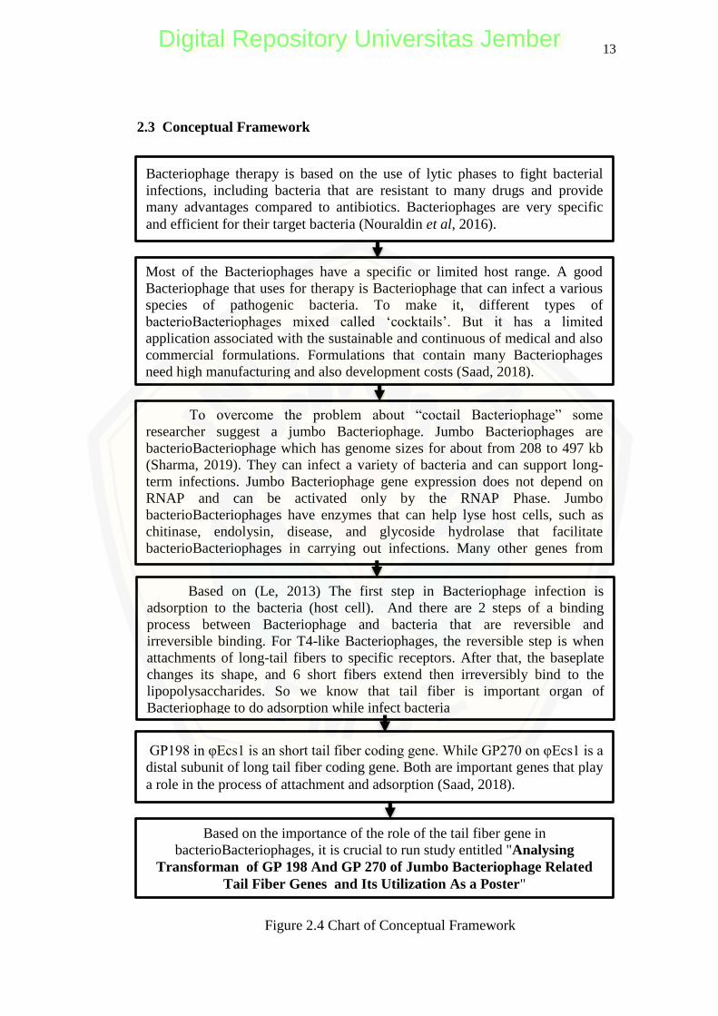
2.3 Conceptual Framework
BAB 3. METODE PENELITIAN
Bacteriophage therapy is based on the use of lytic phases to fight bacterial
infections, including bacteria that are resistant to many drugs and provide
many advantages compared to antibiotics. Bacteriophages are very specific
and efficient for their target bacteria (Nouraldin et al, 2016).
Figure 2.4 Chart of Conceptual Framework
To overcome the problem about ―coctail Bacteriophage‖ some
researcher suggest a jumbo Bacteriophage. Jumbo Bacteriophages are
bacterioBacteriophage which has genome sizes for about from 208 to 497 kb
(Sharma, 2019). They can infect a variety of bacteria and can support long-
term infections. Jumbo Bacteriophage gene expression does not depend on
RNAP and can be activated only by the RNAP Phase. Jumbo
bacterioBacteriophages have enzymes that can help lyse host cells, such as
chitinase, endolysin, disease, and glycoside hydrolase that facilitate
bacterioBacteriophages in carrying out infections. Many other genes from
jumbo Bacteriophages work to reduce bacterioBacteriophage dependence on
Most of the Bacteriophages have a specific or limited host range. A good
Bacteriophage that uses for therapy is Bacteriophage that can infect a various
species of pathogenic bacteria. To make it, different types of
bacterioBacteriophages mixed called ‗cocktails‘. But it has a limited
application associated with the sustainable and continuous of medical and also
commercial formulations. Formulations that contain many Bacteriophages
need high manufacturing and also development costs (Saad, 2018).
Based on (Le, 2013) The first step in Bacteriophage infection is
adsorption to the bacteria (host cell). And there are 2 steps of a binding
process between Bacteriophage and bacteria that are reversible and
irreversible binding. For T4-like Bacteriophages, the reversible step is when
attachments of long-tail fibers to specific receptors. After that, the baseplate
changes its shape, and 6 short fibers extend then irreversibly bind to the
lipopolysaccharides. So we know that tail fiber is important organ of
Bacteriophage to do adsorption while infect bacteria
Based on the importance of the role of the tail fiber gene in
bacterioBacteriophages, it is crucial to run study entitled "Analysing
Transforman of GP 198 And GP 270 of Jumbo Bacteriophage Related
Tail Fiber Genes and Its Utilization As a Poster"
GP198 in φEcs1 is an short tail fiber coding gene. While GP270 on φEcs1 is a
distal subunit of long tail fiber coding gene. Both are important genes that play
a role in the process of attachment and adsorption (Saad, 2018).
Digital Repository Universitas JemberDigital Repository Universitas Jember
14
CHAPTER 3. RESEARCH METHODOLOGY
3.1 Types of Research
This research is an experimental study conducted to confirm the success of
cloning carried out after mutations in the GP198 fand GP 270 of tail fiber. The
data generated in the form of a description of the success of the mutation and
cloning process carried out previously
3.2 Research Place and Time
The study conducted at the Graduate School of Integrated Sciences for Life,
Hiroshima University, Japan, in September 2019- March 2020.
3.3 Operational Definitions
a. Transformant is living creatures as a result of the transformation process (a
movement of foreign genes isolated from plants, animals, viruses, or bacteria
into the genomes of other living things). Transforman that will use in this
study is cloned plasmids that are primary plasmid (7D and 2C) and secondary
plasmid (7E and 2B).
b. Jumbo Bacteriophages are bacterial infectious viruses that have a size of
genome >/= 200 kb with a broad host range. Bacteriophage isolates that will
use bacteriophages that infect Salmonella spp., Shigella spp., And
Escherichia coli including φEcs1 and Ecs1 Mutant
c. The tail fiber gene of Bacteriophage is a gene that encodes the formation of
the tail fiber of bacteriophages. The tail fiber gene used in this study is tail
fiber GP198 (short tail fiber encoder) and GP270 (encoding the proximal part
of the long tail fiber) of φEcs1.
d. GFP (Green Fluorescent Protein) is a gene from jellyfish (Aequoreavictoria),
which is as a reporter gene in genetic transformation, by the luminescence
with luminous green color.
e. Adsorption is the process of imbibing Bacteriophage to bacterial cells during
the infection process
Digital Repository Universitas JemberDigital Repository Universitas Jember
15
3.4 Research Tools and Materials
3.4.1 Tools
The tools used in this study are Petri dishes, ose, bunsen, incubator,
autoclave, oven, stove, spectrophotometer, electrophoresis, Centrifuge, Laminar
Air Flow (LAF), thermometer, stir bar, refrigerator, micropipette, measuring cup
1000 ml, 500 ml measuring cup, UV transilluminator, doc gel, Erlenmeyer 1000
ml, Erlenmeyer 500 ml shaker, thermal cycler, tweezers, microwave,
ultracentrifuge, shiring, filtration, 500 ml glass beaker, 1000 ml glass beaker, aret,
analytical balance, pans, term shaker, stopwatch, 70% alcohol, glove.
3.4.2 Ingredients
The materials used in this study are LB (Hypopolypeptone, bacto yeast
extract, sodium chloride, agar), Escherichia coli bacteria isolate, bacteriophage
φEcs1, Mutant of φEcs1, primary plasmid (7D AND 2C), secondary plasmid (2B
and 7E), TE (Tris-EDTA) buffer, 1X TAE buffer, loading dye, KOD one,
restriction enzyme (Bgl II, Eco RV, Hind III), parafilm paper, SM buffer, λ stay 1
marker, aqua dest, ddh2o, agarose gel, seal, marker.
3.5 Research Procedure
The researcher does this study by five steps of the research's procedure. The
first step is to prepare the media to propagate the bacteria and Bacteriophage. The
second is cloning to insert the GFP gene into the plasmid. The third is
confirmation of the presence of a gene with PCR (Polymerase Chain Reaction)
and Electrophoresis. The fouth is an adsorbant assay to prove the function of the
tail fiber gene, and the last step is to design the poster and poster validation
analysis:
3.5.1 Preparation of LB medium
Preparation of LB medium used for propagating the bacteriophage φEcs1,
and Mutant of φEcs1, and for the cloning media. LB (Luria Bertani) medium
prepared by mix homogenously hypopolypeptone 35 gr, sodium chloride 35 gr,
bacto yeast extract 17.5 gr and 3500 ml of distilled water in an Erlenmeyer glass.
Digital Repository Universitas JemberDigital Repository Universitas Jember
16
Set the Ph to 7 by added NaOH. A 3000 ml medium will combine to make LB
plate agar. 3000 ml medium divided into 2 of 1000 ml Erlenmeyer tubes and 2 of
500 ml Erlenmeyer tubes. Poured out 800 ml of media into every 1000 ml
Erlenmeyer tube and added by 12 grams of agar powder. 400 ml into each 500 ml
Erlenmeyer and added by 6 grams of agar powder. It covered by a sponge and
aluminum foil. While the 500 ml one without agar powder to make LB broth. 500
ml of the medium was divided into 4.5 ml each test tube and covered it with a
sponge. I placed the tube in the tube rack and covered it with aluminum foil.
Cooked and sterilized all media into the autoclave for 20 minutes at 121 atm.
Then be awaited until the slight temperature decrease, after that poured out into
the petri dish and wait until solid. After it is solid, stored in a plastic bag and
placed in a 4°C cooling room for stock. Wait for the media to cool and ready for
use (Saad, 2018).
3.5.2 Preparation of 0.35% TOP agar
Preparation of 0.35% TOP agar used for top layer LB media for
propagating φEcs1, and Mutant of φEcs1. 0.35% TOP agar prepared by mix
homogenously 1.6 grams of hypopolipeptone, 1.6 grams of sodium chloride, 1.6
grams of agar, and 0.8 grams of bacto yeast extract with 500 ml of distilled water.
Adjust the pH to 7 by added NaOH. Boil it in the microwave and poured out into
each 5 ml canister. Covered the tube and wrapped it in aluminum foil, then
autoclave for 20 minutes at 121 atm after the autoclave top was ready to use.
Whereas for the next use TOP so that those who have compacted boiling first
(Saad, 2018).
3.5.3 Preparation of agarose gel
Preparation 1% agarose gel used for electrophoresis φEcs1, Mutant of φEcs1,
and plasmid. 1% agarose gel prepared by mix homogenously 2 grams of agarose
powder and dissolved it in 200 ml of TAX buffer 1 X. Then boiled in the
microwave, be awaited until the temperature decreased slightly then poured out it
into the agar mold and place the comb in the pattern. Wait for it to be solid, and
agarose gel, 1% was ready to use (Saad, 2018).
Digital Repository Universitas JemberDigital Repository Universitas Jember
17
3.5.4 Propagation of ɸEcs1 and ɸEcs1 Mutant
Propagation of ɸEcs1 and ɸEcs1 Mutant used for work culture Performed
inoculation of Escherichia coli (BL21) as the primary host of ɸEcs1 into 4.5 ml
LB broth and incubated in a 37 °C shaker incubator for about 5 hours. Next,
calculate the bacterial OD (Optical Density) and set it to reach the standard OD
for the plaque assay that is equal to 0.1. Then prepared a 250 µl mixture plaque
assay to consist of bacterial suspension and LB broth with OD 0.1 to reach 240 µl
volume and 10 µl bacteriophage to be propagated (ɸEcs1 or utanEcs1 Mutant)
with a concentration of 106 for ɸEcs1 and 10
4 for Ecs1 Mutant. Prepared 12 LB
plates, 6 for ɸEcs1, and 6 for ɸEcs1 Mutant. Boil 12 TOP agar tubes. Wait for
TOP so that the temperature dropped slightly (warm), then mix it with 250 µl
mixture and poured out it into the LB plate. Wait until it's solid and incubate it in
an incubator of 28°C for 1 night. The next day continues to add 5 ml of SM buffer
to each plate, seal with parafilm paper. Put in into the shaker 4°C cooling room
for 1 night. The next day harvest (plate lysate) by taking SM buffer from all plates
with 5 ml syringe and filled in into a 5 ml tube then centrifuged with a speed of
15000 g or 10,200 rpm for 1 hour (1 to 3 times). After that, throw the supernatant
and added 500 µl BC buffer then mixed well with the pellet. Moved into the
Eppendorf tube, and the bacteriophage stock was ready (Saad, 2018).
3.5.5 Insert the GFP gene to Primary Plasmid by infusion Cloning
The cloning process is done by infusion cloning to insert the GFP gene in
the plasmid. Before cloning, it is necessary to prepare LB media with a
composition of 1% hypopolypeptone, 0.5% yeast extract, 1% NaCl, and 1% agar.
Before pouring the medium into the plate, the LB medium was waited until warm,
then added X-gal 1.25 ml, IPTG 0.75 ml, and Ampicillin 90 µl in every 400 ml
LB. It also needs to be prepared 2XYT media for the transformation process with
a component of 1.6% hypopolypeptone, 1% yeast extract, 0.5% NaCl. Next, make
for the manufacture of mixture infusion cloning with ingredients including 5x HD
E.premix infusion, linearized vector, purified PCR fragment, and ddh2o.
Furthermore, the transformation process does by using a competent cell in the
Digital Repository Universitas JemberDigital Repository Universitas Jember
18
form of Escherichia coli Xl10 gold bacteria 100 µl. Put the competent cells in the
icebox until liquid. Mix the 100 µl competent cell with infusion HD en.mixture 5
µl. Put in the icebox wait until 15 minutes. Set in a water bath 47°C for 30
seconds. Mix with 2XYT 900 µl. Then incubate at 37°C for 1 hour. Centrifuge
6000 g for 5 minutes. Remove 970 µl supernatant. Mix well the pellet with 30 µl
of 2XYT media broth. The last step, pour in the LBAX media plate and incubate
in incubator 37°C. wait 24 hours, then observe the colonies that appear. Breed
successful colonies into LB media then do a plasmid extraction with a plasmid
extraction kit (Saad, 2018).
3.5.6 Check the presence of the tail fiber gene in the transforman of primary
plasmid
This PCR (Polymerase Chain Reaction) used for amplifying the tail fiber
gene (GP198 and GP270). The first confirmation did by PCR using primary
plasmids. The primary plasmid is plasmid inserted by the tail fiber gene (plasmid
7D and 2C). Plasmid 7D is a plasmid that added with tail fiber gene 198. Plasmid
2C is a plasmid that has inserted with tail fiber gene 270. The PCR runs with
primary primers that designed to detect the presence of inserted fiber tails). The
primary primers number 2 is a primer to detect the tail fiber gene 270. The
primary primers number 7 is a primer for detected the tail fiber gene 198 and
checked by secondary primers (primers that detect the reverse part of plasmids).
As an alternative control, a GFP gene PCR performers with a primary tail fiber
gene detection. Furthermore, electrophoresis with a DNA polymerase enzyme that
used was KODone (Saad, 2018).
The first is to prepare an icebox to place the Eppendorf containing KODone
until KODone becomes liquid. Furthermore, made a PCR mixture with a
formulation of 12.5 μl KODone, 9.5 μl ddH2O, 1 μl plasmid sample, 1 μl forward
primer, and 1 μl reverse primer. Resuspend at the end of making the mixture and
given by a name to the Eppendorf PCR. Inserted mixture into thermal cycler with
a pre-denaturation temperature of 94°C for 2 minutes, denaturation with a
temperature of 98°C for 10 seconds, annealing 60°C for 30 seconds, extension
with the heat of 68°C for 30 seconds, and final extension with temperature 12°C.
Digital Repository Universitas JemberDigital Repository Universitas Jember
19
cycles performed as many as 30 times. After completed PCR, the electrophoresis
was tested by λ stay 1 as a 6 µl marker, and 1 µl sample mixed with 1 µl loading
dye. The running of electrophoresis for 60 minutes with 100 V of power. The
staining process using EtBr (Ethidium Bromide) and the visualization of the band
by UV transilluminator (Saad, 2018).
3.5.7 Digest with retraction enzymes
Digest with retraction enzymes used to prove that the plasmid and ɸEcs1
have the same sequence and to confirm the cloning result. The first step did by
prepared an icebox for the buffer and the enzyme retention used. It started by mix
homogenously 23 µl ddH2O components, 3 µl buffer retraction enzymes, 3 µl
DNA samples (original 2C plasmids and Ecs1), 1 µl retraction enzymes (Eco Rv,
Hind III, Bgl II). After the mixture finish, it is resuspended and incubated at 37 °C
for 3 hours. Furthermore, electrophoresis carries out using the λ stay marker. Run
of electrophoresis was carried out for 60 minutes with a power of 100 V.
Furthermore, stain the gel was did by used EtBr for 30 minutes and observed the
band with a UV transilluminator. Checked is did together with sample (2C and
Ecs1 Ori) without digest as a negative control (Saad, 2018).
3.5.8 Checking the presence of the GFP gene in the secondary plasmid
PCR (Polymerase Chain Reaction) test used to amplify the reverse plasmid
portion. This PCR prepare by an took an icebox to put KODone, then amplified
by PCR secondary plasmid (7E and 2B), which is a primary plasmid that has been
removed from its tail fiber gene and then replaced with the GFP gene. The PCR
process carries out using secondary primers (primers that detect the reverse
plasmid portion) and primary GFP (primers that detect the GFP gene). The
mixture formulations used were 12.5 μl KODone, 9.5 μl ddH2O, 1 μl forward
primer, 1 μl reverse primer, and 1 μl DNA sample (7E and 2B). PCR carries out
with a pre-denaturation temperature of 94°C for 2 minutes, denaturation with a
temperature of 98°C for 30 seconds, annealing with a temperature of 60°C for 30
seconds, extension with a temperature of 68°C for 30 seconds and final extension
with a heat of 12°C. Repeat the cycle 30 times. Furthermore, I checked the band
Digital Repository Universitas JemberDigital Repository Universitas Jember
20
by electrophoresis with the λ stay 1 as a marker for 60 minutes, with a power of
100 V. The staining process is by soaking the agarose gel in EtBr liquid for 30
minutes. Followed by observation of the band with a UV transilluminator (Saad,
2018).
3.5.9 Electron Microscopy (EM) observation of ɸEcs1 and ɸEcs1 Mutant
Electron Microscopy (EM) observation used to know the morphology of
ɸEcs1 and ɸEcs1 was carried out by preparing bacteriophage samples with high
concentrations then negative staining with 1% of PTA.
3.5.10 Adsorption assay of ɸEcs1 and ɸEcs1 Mutant by Plaque Assay
Plaque assay based adsorption is to know the adsorption ability of each
Bacteriophage. The Escherichia coli BL21 bacterial isolate was grown in a 4.5 ml
LB broth tube, incubated at 37°C. After 5 to 7 hours, OD600 is calculating with a
spectrophotometer. BL21 bacterial culture dilutes until it reached OD600 = 1 in
4.5 ml LB volume into a 5 ml Eppendorf tube. (diluted into 2 Eppendorf tubes
measuring 5 ml). Then the ɸEcs1 10 µl bacteriophage was added to the Eppendorf
1 and ɸEcs1 Mutant 10 µl tubes into the Eppendorf 2 tube. Then every 1 minute, 5
minutes, 10 minutes, 20 minutes, and 30 minutes a 500 µl mixture of BL21 and
take the Bacteriophage, then put into a mixture of Eppendorf 2. in a 1.5 ml
Eppendorf tube. Then centrifuged for 1 minute at 15000 rpm. The supernatant is
taken by 0.5 ml of the syringe and filtered by a membrane filter; the filtration
results put into a 1.5 ml Eppendorf tube. Followed by a plaque assay. Making a
mixture of LB broth media and BL21 culture with OD600 = 1 in a 240 µl volume
and 10 µl bacteriophage suspension add beforehand. Mix the solution mixture
with 5 ml of TOP so that 0.35%, which has boiled before. Wait until the
temperature decreases slightly. Then poured the final mixture into the LB plate,
wait until it is solid and incubated at 28°C overnight. Next, count the number of
plaques that appear. Adsorbent assay and plaque assay perform several times, and
data on the amount of plaque obtained processed in the form of curves (Saad,
2018).
Digital Repository Universitas JemberDigital Repository Universitas Jember
21
3.5.11 Poster Preparation
The researcher designed a poster by the steps based on Darmalaksana,
(2017) with a little modification as follows:
Poster made by graphics processing applications, such as Corel Draw, in printed
form totaling 1 (one) sheet of height × width are 70 cm × 70 cm mounted
vertically. The poster layout design paid attention to the principle of formal and
informal balance, which includes symmetrical and asymmetrical terms, the
principle of unity of arrangement of pictures, color, background and motion
elements, and able to direct the reader's eyes to the entire poster area. The poster
contains the top part considered of the title, the name of the implementer, and the
university logo. The middle section (the content part) included the background
(introduction or abstract), Methods, Main Research Results (text and pictures or
photography or schematics), Conclusions, and References (additional); and at the
bottom, inserted the sponsor or agency logo, contact details, date and time of
research.
3.5.12 Poster Validation Analysis
Poster validation analysis obtained from the validator is in the form of
quantitative data that is processed with a formula as follow
N (Score) =
Digital Repository Universitas JemberDigital Repository Universitas Jember
22
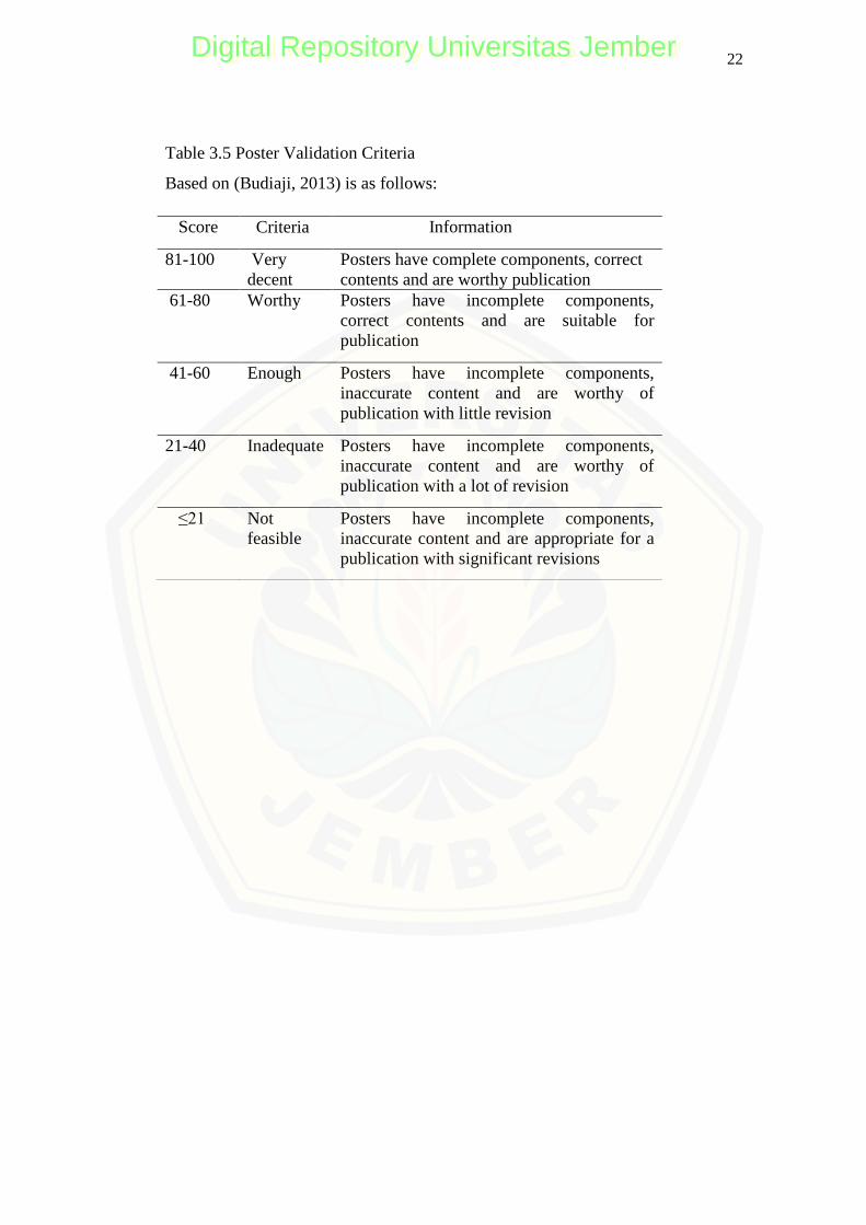
Table 3.5 Poster Validation Criteria
Based on (Budiaji, 2013) is as follows:
Score Criteria Information
81-100 Very
decent
Posters have complete components, correct
contents and are worthy publication
61-80 Worthy Posters have incomplete components,
correct contents and are suitable for
publication
41-60
Enough
Posters have incomplete components,
inaccurate content and are worthy of
publication with little revision
21-40 Inadequate Posters have incomplete components,
inaccurate content and are worthy of
publication with a lot of revision
≤21 Not
feasible
Posters have incomplete components,
inaccurate content and are appropriate for a
publication with significant revisions
Digital Repository Universitas JemberDigital Repository Universitas Jember
23
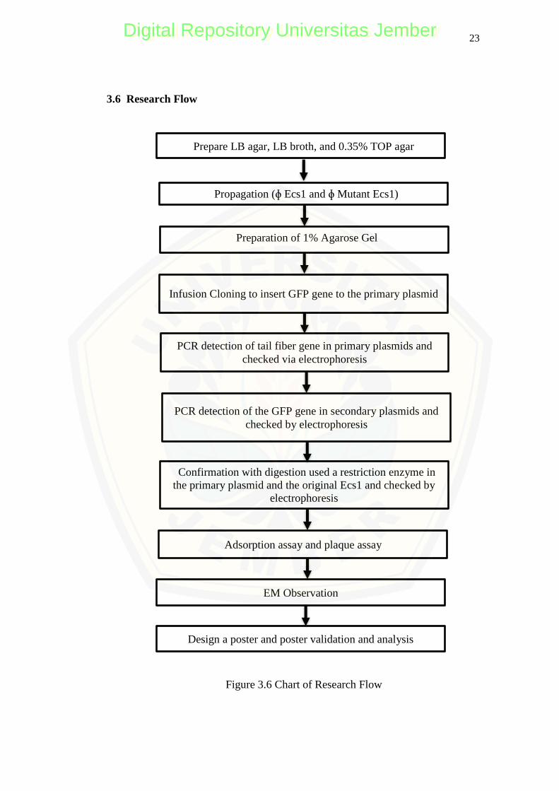
3.6 Research Flow
Prepare LB agar, LB broth, and 0.35% TOP agar
Propagation (ɸ Ecs1 and ɸ Mutant Ecs1)
Preparation of 1% Agarose Gel
PCR detection of tail fiber gene in primary plasmids and
checked via electrophoresis
Confirmation with digestion used a restriction enzyme in
the primary plasmid and the original Ecs1 and checked by
electrophoresis
Figure 3.6 Chart of Research Flow
PCR detection of the GFP gene in secondary plasmids and
checked by electrophoresis
EM Observation
Adsorption assay and plaque assay
Design a poster and poster validation and analysis
Infusion Cloning to insert GFP gene to the primary plasmid
Digital Repository Universitas JemberDigital Repository Universitas Jember
39
CHAPTER 5 CONCLUSIONS AND SUGGESTIONS
5.3 Conclusion
5.3.1 The first cloning process of inserting the tail fiber gene on the primary
plasmid is success indicate by plasmid 7D with primary primer number 7
(PP7) appears a band with the size around three kbp and with secondary
primer number 7 appears 5 Kbp of the band. Primary plasmid 2C with
primary primer number 2 shows 5 Kbp and with secondary primer show 5
Kbp. That all confirmed with a restriction enzyme.
5.3.2 The second cloning process of replacement the tail fiber GP198 with the GFP
gene is a success. It is proof with appearing 1 kbp band electrophoresis
secondary plasmid 7E using GFP primer, 5 Kbp band with secondary primer,
and EM observation of mutant without short tail fiber. The second cloning
process of replacement the tail fiber GP270 with GFP gene was not
successful proven by appearing 1 kbp band electrophoresis secondary
plasmid 2B using GFP primer and show band less than 5 Kbps with
secondary primer. Also, EM observation shows the long tail fiber still present
on mutant Bacteriophage.
5.3.3 The adsorption ability of wild type and Ecs1 Bacteriophage is higher than the
mutant indicate by the number of plaque. That was because the gene that
encodes tail fiber GP198 of Ecs1 Bacteriophage was removed.
5.3.4 The validation of the poster eligibility criteria showed average eligibility of
86,36% which was included in the category of very feasible to be published
5.2 Suggestions
Most of the genes in jumbo Bacteriophage are unknown. Further research needs
to analyze the function of these genes to support the optimal application of
Bacteriophage.
39
Digital Repository Universitas JemberDigital Repository Universitas Jember
40
REFERENCES
Ackermann, H W. (2015). The first Bacteriophage electron micrographs.
Bacteriophages. 1(4) :225-227.
Alemayehu, Debebe.,. Casey, Pat G., McAuliffe, Olivia., Guinane, Caitriona M., G.
Martin, James., Shanahan, Fergus., Coffey, Aidan., Ross, R. Paul., and Hill,
Colin. 2012. Bacteriophages MR299-2 and NH-4 Can Eliminate
Pseudomonas aeruginosa in the Murine Lung and on Cystic Fibrosis Lung
Airway Cells. Journal of Viruses, 3(2).
Amin, Astuti dan Ulfah, Maria. (2017). Sintesis Dan Karakterisasi Komposit
Hidroksiapatit dari Tulang Ikan Lamuru (Sardilnella Longiceps)-Kitosan
Sebagai Bone Filler. JF FIK. 5(1), 10.
Anderson, W. (1953), Cold Spring Harbor Symp. Quant. Biol. 18, 197.
Ardiansyah., Erina., Harris, Abdul. (2018). Pengaruh Efektivitas Ekstrak Daun
Patikan Kebo (Euphorbia hirta) Terhadap Pertumbuhan Bakteri Salmonella
Sp. JIMVET, 2(3), 380-387
Arens, Daniel K.., Brady, T. Scott., L. Carter, John., A. Pape, Jenny., Robinson,
David M.., Russell, Kerri A., A. Staley, Lyndsay., M. Stettler, Jason.,
Tateoka, Olivia B., Townsend, Michelle H., Whitley, Kiara V.., Wienclaw,
Trevor M.., Williamson, Taryn L.., Johnson, Steven M.., Grose, Julianne H.
(2018). Characterization of two related Erwinia myoviruses that are distant
relatives of the PhiKZ-like Jumbo Bacteriophages. PLOS ONE, 1-19.
https://doi.org/10.1371/journal.pone.0200202.
Arisanti, Risalia Reni., Indriani, Citra., Wilopo, Siswanto Agus. (2018). Kontribusi
agen dan faktor penyebab kejadian luar biasa keracunan pangan di
Indonesia: kajian sistematis. BKM Journal of Community Medicine and
Public Health, 34(3), 99-106.
Astuti, Tri Dyah., Hadi, Wahid Syamsul. (2018). Potensi ekstrak daun Carica
pubescens sebagai alternatif antidiare Bakteri Vibrio cholerae dan Shigella
dysentriae. Jurnal Teknologi Laboratorium, 7(2), 61–69.
Attai, Hedieh., Boon, Maarte.., Phillips, Kenya., Noben, Jean-Paul., Lavigne, Rob.,
and Brown, Pamela. (2018). Larger Than Life: Isolation and Genomic
Characterization of a Jumbo Bacteriophage That Infects the Bacterial Plant
Pathogen, Agrobacterium tumefacien. Frontiers in Microbiology, 9(1861),
1-14. 10.3389/fmicb.2018.01861.
Bhattacharya, Debanjan & Van Meir, Erwin G. 2018. A simple genotyping method
to detect small CRISPR-Cas9 induced indels by agarose gel electrophoresis.
Scientific Report. 10(1038): 2
40
Digital Repository Universitas JemberDigital Repository Universitas Jember
41
Baylis, Chris., Uyttendaele, Mieke., Joosten, Han, and Davies, Andy. (2011). The
Enterobacteriaceae And Their Significance To The Food Industry. ILSI
EUROPE Emerging Microbiological Issues Task Force, 1-52.
Benderska, N., Chakilam, S., Hugle, M., Ivanovska, J., Gandesiri, M. Dan Schulze
L.J. (2012). Apoptosis Signalling Activated By Tnf In The Lower
Gastrointestinal Tract-Review. Current Pharmaceutical Biotechnology,
2248-2258.
Beyene, G., & Tasew, H. Ahn, J., Kim, S., Jung, L.-S., & Biswas, D. (2013). In vitro
assessment of the susceptibility of planktonic and attached cells of
foodborne pathogens to Bacteriophage p22-mediated salmonella lysates.
Journal of Food Protection, 76(12), 2057–62.
Buana, E.O. dan A.K Wardani. (2014). Isolasi bakteriofag litik sebagai agen
biosanitasi pada proses pelisisan bakteri pembentuk biofilm. Jurnal Pangan
dan Agroindustri, 2(2), 36-34.
Budiaji, Weksi. (2013). Skala Pengukuran Dan Jumlah Respon Skala Likert. Jurnal
Ilmu Pertanian Dan Perikanan, 2(2), 127-133.
Buttimer, Colin., Born, Yannick., Lucid, Alan., J. Loessner, Martin., Fieseler, Lars.,
Coffey, Aidan. (2018). Erwinia amylovora Bacteriophage vB_EamM_Y3
represents another lineage of hairy Myoviridae. Research in Microbiology,
(169), 505-514.
Buttimer, Colin., McAuliffe, Olivia., Ross, R. P., Hill, Colin., O‘Mahony, Jim and
Coffey, Aidan. (2017). Bacteriophages and Bacterial Plant
Diseases. Journal Review, 8(34).
Cahyaningrum, Sari Edi., Herdyastuti, Nuniek., Rahmi, Asri., Firdausa, Amanah dan
Dini, Lisa. (2015). Uji Aktivitas Isoniazid Terenkapsulasi Kitosan Alginat
Tween 80 Pada Mycobacterium tuberculosisis. Prosiding Seminar
Nasional Kimia, 5(8), 134-137.
Casali N, Andrew P, Preston A. (2003). E. coli plasmid vectors: methods and
applications. Humana Press 12:19-25
Damayanti, Rahayu., Jannah, Siti Nur., Wijanarka, Rahaju, Sri Hartin. (2016). Isolasi
Bakteriofag Salmonella spp. Dari Biofilm pada Sistem Air Minum Isi
Ulang. Jurnal Biologi, 5(2), 59-67.
Darmalaksana, Wahyudin. (2017). Lomba Poster Ilmiah Pada Expo Hasil Penelitian
Uin Sunan Gunung Djati Bandung. Jurnal Informasi Riset dan Inovasi, 1-7.
Digital Repository Universitas JemberDigital Repository Universitas Jember
42
Dhany, N. R., Addy, H. S., & Wahyuni, W. S. (2013). Penggunaan Bakteriofag
untuk Kit Detektor Patogen Hawar Bakteri Kedelai. Jurnal Fitopatologi
Indonesia, 9(4), 116-122.
Dharmayanti, Ni Made Dwi., Arjita, I Putu Dedy. Uji Daya Hambat Ekstrak Batang
Kayu Secang (Caesalpinia Sappan L.) Terhadap Bakteri Escherichia Coli,
685-693.
Dunne, Matthew., Denyes, Jenna M., Arndt, Helena., Loessner, Martin J., Leiman,
Petr G., and Klumpp, Jochen. (2018). Salmonella Phage S16 Tail Fiber
Adhesin Features a Rare Polyglycine Rich Domain for Host Recognition.
Structural Biology and Molecular Biophysics.
doi.org/10.1016/j.str.2018.07.017
Effantin, Gre´gory., Hamasaki, Ryosuke., Kawasaki, Takeru., Bacia, Maria.,
Moriscot, Christine., Weissenhorn, Winfried., Yamada, Takashi and
Schoehn, Guy. (2013). Cryo-Electron Microscopy Three-Dimensional
Structure of the Jumbo Bacteriophage FRSL1 Infecting the Phytopathogen
Ralstonia solanacearum. Elsevier, (21) 298–305.
Elbreki, Mohamed., Ross, R. Paul., Hill, Colin., O‘Mahony, Jim.,
McAuliffe, Olivia., and Coffey, Aidan. (2014). Bacteriophages and Their
Derivatives as Biotherapeutic Agents in Disease Prevention and Treatment.
Journal of Viruses..
Etchepare, M. d., Raddatz, C. G., Flores, E. M., & Zepka, Q. L. (2016). Effect Of
Resistant Starch And Chitosan On Survival Of Lactobacillus acidophilus
Microencapsulated With Sodium Alginate. LWT - Food Science and
Technology, 511-517.
Fleetwood, Janet., Rahman, Shamim., Holland, Darren., Millson, David., Thomson,
Laura., Poppy, Guy. (2019). As clean as they look? Food hygiene inspection
scores, microbiological contamination, and foodborne illness. Food Control.
96, 76–86.
Franklin, Naomi C. (1961). Serological Study of Tail Structure and Function in
ColiBacteriophages T2 and T4‘. Virology, (14), 417-429.
Gitaswari, D A Inda., Budayanti, H Sri. (2019). Identifikasi Subtipe Enterotoxigenic
Escherichia Coli dan Enteroaggregative Escherichia Coli dari Spesimen
Usap Dubur Penjamah Makanan di Denpasar Menggunakan Polymerase
Chain Reactio. E-Jurnal Medika, 8(1), 7-8.
Hakim, L., Utama, C. S. dan Sumarsih, S. (2014). Keberadaan Bakteri Escherichia
Coli dan Salmonella Sp Pada Limbah Pabrik Pakan Unggas (Pakan
Ceceran) yang Difermentasi Dengan Starter Fungsional. Animal Agriculture
Journal, 3(3), 457-462.
Digital Repository Universitas JemberDigital Repository Universitas Jember
43
Hasanah, Rizka., Daningsih, Entin., Titin. (2016). Kelayakan Poster Kandungan Gizi
Musa Paradisiaca Pada Sub Materi Zat Makanan Kelas XI: Pendidikan
Biologi, Fkip Universitas TanjunGPura.
Hayati, Zikra., S Jannah, Siti Nur. Suprihadi., Agung. (2016). Isolasi Bakteriofag
Spesifik Pseudomonas sp. dari Biofilm Pada Sistem Pengisian Air Minum
Isi Ulang. Jurnal Biologi, 5(3), 29-35.
Hendrix., R.Lavigne, And P-J Ceyssens. (2012). Virus Taxonomy: Ninth Report Of
The International Committee On Taxonomy Of Viruses. Elsevier: 46-62.
Hendrix, R. W. (2009). Jumbo Bacteriophages. Current Topics In Microbiology And
Immunology. Pittsburgh Bacteriophage Institute and Department of
Biological Sciences, University Of Pittsburgh: 229-328
Hua, Bo., Margolin, William., Molineux, Ian J., and Liua, Jun. (2015). Structural
remodeling of bacteriophage T4 and host membranes during infection
initiation. Cell and Molecular Biology.
Hua B.S., Jianfei. (2016). Capsid Structure And DNA Packing In Jumbo
Bacteriophages. University Of Science And Technology Of China:
University Of Pittsburgh Kenneth P. Dietrich School Of Arts & Sciences
Husni, A., Subaryono, Pranoto, Y., Tazwir, & Ustadi. (2012). Pengembangan
Metode Ekstraksi Alginat Dari Rumput Laut Sargassum sp. Sebagai Bahan
Pengental. Jurnal AGRITECH, 32(1), 1-8.
Junaidi, Ahmad. Junaidi, Ahmad. (2018). Uji Viabilitas Mikroenkapsulasi
Lactobacillus Acidophilus Menggunakan Polimer Natrium Alginat-Kitosan
Terhadap Simulasi Cairan Asam Lambung. Medan: Universitas Sumatra
Utara
Kellen Berger, E., Bolle A., La Tour, E. Boyde., Epstein, R. H., Frankli N, N. C..,
Jernen. K., Scafati, A. Reale. (1965). Functions and Properties Related to
the Tail Fibers of Bacteriophage T4. Viroology26, 419-440.
Kitajima, Masaaki., Ishii, Satoshi., Takagi, Tatsuma., Okabe, Satoshi. (2019).
Complete Genome Sequence of a Novel Myoviridae Bacteriophage, Sf01,
Infecting Shigella spp. American Society For Microbiology, 8(23), 1-2.
Konoralma, Ketrina. (2019). Identifikasi Bakteri Penyebab Infeksi Nosokomial di
Rumah Sakit Umum Gmim Pancaran Kasih Manado. Jurnal Kesmas. 8(1), 23-
35
Kozlov, Alexander G., Weiland, Elizabeth., Mittal, Anuradha., Waldman, Vince.,
Antony, Edwin., Fazio1, Nicole., Pappu, Rohit V., and M. Lohman,
Timothy. 2015. Intrinsically Disordered C-terminal Tails of E. coli Single-
Digital Repository Universitas JemberDigital Repository Universitas Jember
44
Stranded DNA Binding Protein Regulate Cooperative Binding to Single-
Stranded DNA. J Mol Biol. 427(4): 763–774.
Kurnia, Ashfar. (2011). Dasar Teknologi Dna Rekombinan: Departemen Farmasi,
Universitas Indonesia, Depok, Indonesia.
Le S, He X, Tan Y, Huang G, Zhang L, Lux R, et al. (2013) Mapping the Tail Fiber
as the Receptor Binding Protein Responsible for Differential Host
Specificity of Pseudomonas a. Bacteriophages PaP1 and JG004. PloS ONE,
8(7). e68562.https://doi.org/10.1371/journal.pone.0068562.
Leiman, P. G., Kanamaru, S., A, Mesyanzhinov, V. V., Arisaka, F., And Rossmann,
M. G. (2003). Structure And Morphogenesis Of Bacteriophage T4. Cellular
And Molecular Life Sciences. 60: 2356–2370. DOI 10.1007/S00018-003-
3072-1
Lomonaco, Sara., Lascols, Christine., Crawford, Matthew A., Anderson, Kevin.,
Hodge, David R.., Fisher, Debra J.., P. Pillai, Segaran., A. Morse,
Stephen., Khan, Eru., A. Hughes, Molly., W. Allard, Marc., K. Sharma,
Shashi. (2019). Draft Genome Sequences of Antimicrobial-Resistant
Shigella Clinical Isolates from Pakistan. American Society For
Microbiology, 8(30), 1-3.
Lord, Cédric., Wlodarczyk, Katarzyna Danis., Blasdel, Bob G., Jang, Ho Bin.,
Vandenheuvel, Dieter., Briers, Yves., Noben, Jean-Paul., Noort, Vera van.,
Drulis-Kawa, Zuzanna., Lavigne, Rob. (2019). Integrative omics analysis of
Pseudomonas aeruginosa virus PA5oct highlights the molecular complexity
of jumbo Bacteriophages. bioRxiv preprint. DOI:
http://dx.doi.org/10.1101/679506. 1-40.
Maharani, A. A., Husni, A., & Ekantari, N. (2017). Karakteristik Natrium Alginat
Rumput Laut Cokelat Sargassum fluitans Dengan Metode Ekstraksi Yang
Berbeda. Jurnal Pengolahan Hasil Perikanan Indonesia, 20(3), 478-487.
Mandal, S., Puniya, A.K, and Singh, K. (2005). Effect Of Alginate Concentrations
On Survival Of Microencapsulated Lactobacillus casei NCDC-298.
International Dairy Journal, 16, 1990-1995.
Maritsa, Hasnaul., Aini, Fitratul., Nurhakim, Desri S., Sihombing, Greace M.,
Saputra, Ardiansyah. (2017). Isolasi dan Identifikasi Cemaran Bakteri
Salmonella sp. Pada Daging Ayam dan Ikan Mentah. Bio-sit. 3(2), 47-70.
Marti, Roger., Zurfluh, Katrin., Hagens, Steven., Pianezzi, Jasmin., Klumpp, Jochen.,
And Loessner, Martin J. (2013). Long Tail Fibres of The Novel Broad-Host-
Range T-Evenbacteriophage S16, Specifically Recognizesalmonellaompc.
Molecular Microbiology. 87(4): 818–834
Digital Repository Universitas JemberDigital Repository Universitas Jember
45
Mesyanzhinov, V.V., Leiman, P.G., Kostyuchenko, V.A., Kurochkina, L.P.,
Miroshnikov, K.A., Sykilinda, N. N., and Shneider, M. M. (2004).
Molecular Architecture of Bacteriophage T4. Biochemistry. 69(11): 1190-
1202. Doi: 0006-2979/04/6911-1190
Miller, Eric S., Kutter, Elizabeth., Mosig, Gisela., Arisaka, Fumio., Kunisawa,
Takashi., and Ruger, Wolfgang. (2003). Bacteriophage T4 genome.
Microbiology And Molecular Biology Reviews. 67(1): 86-156.
Mokkaram, R. R. (2009). The Influence Of Multi-Stage Alginate Coating On
Survivability Of Potential Probiotic Bacteria In Simulated Gastric And
Intestinal Juice. Food Research International, 1040-1045.
Morise, H. Shimomura, F. H. Johnson, and J. Winant. (1974) Biochemistry. 14,
26562662
Muhsinin, Soni., Sulastri, Maria Martina dan Supriadi, Dadih. (2018). Deteksi Cepat
Gen InvA pada Salmonella spp. Dengan Metode PCRm. Jurnal Sains
Farmasi & Klini, 5(3), 191-200.
Murwani, Sri., Qosimah, Dahliatul., Amri, Indah. (2017). Penyaki Bakterial
pada Ternak Hewan Besar dan Unggas, Malang: UB press.
Narulita, Erlia., Sulistyorini, Ifa., Aji, Gerda Permata., Iqbal, Mochammad., And
Murdiyah, Siti. (2018). Isolation And Characterization Of Bacteriophage In
controlling Escherichia Coli In Jember Area, Indonesia. Asian Jr. Of
Microbiol. Biotech. Env. Sc. 20(2): 439-444
Nolan, Tania., A. Bustin, Stephen. 2013. PCR Technology Current Innovations. New
York: CRC Press
Nouraldin, A. A., M. B., Harfoush, R. A. dan Essa, S. A. (2016). Bacteriophage-
Antibiotic Synergism to Control Planktonic and Biofilm Producing Clinical
Isolates of Pseudomonas aeruginosa. Alexandria Journal of Medicine, 5(2),
99-105.
Nurhamidah M, Sitti., Djide, Natsir., Arfiansyah, Rudi., Nainu, Firzan. (2018).
Deteksi Salmonella Enterica Serovar Typhimurium Dalam Produk Pangan
Siap Saji Menggunakan Metode Pcr, Melt Curve, Dan Analisis Hrm.
Original Article MFF, 22(1), 20-26.
Orlova, E V. (2012). Bacteriophages. Croatia: In Tech
Phumkhacorn, P., and Pongsak, R. (2010). Isolation and partial characterization of a
bacteriophage infecting the shrimp pathogen Vibrio harveyi. African
Journal Of Microbiology Research, 4(16), 1794-1800.
Ploeg, van der., Claudia A., Viñas, María R., Raquel, Terragno., Susana B., Bruno,
Norma Binsztein. (2010). Laboratory Protocol: “Serotyping of Shigella
spp.‖WHO Global Foodborne Infections Network, 1-24.
Digital Repository Universitas JemberDigital Repository Universitas Jember
46
Poeloengan, M., Komala, I dan Susan M. Noor. (2005). Bahaya Salmonella terhadap
kesehatan. Prosiding Lokakarya Nasional Penyakit Zoonosis: Bogor, 216-
223.
Poeloengan, Masniari., Komala, Iyep dan Noor, Susan M. (2016). Bahaya
Salmonella Terhadap Kesehatan. Lokakarya Nasional Penyakit Zoonosis,
30(1), 216-224.
Primrose SB, Twyman RM. (2006). Principles of Gene Manipulation and Genomics.
Ed.Ke-7. Oxford: Blackwell Publishing
Radji, M. (2011). Rekayasa Genetika; Pengantar Untuk Profesi Kesehatan. Sagung
Seto:Jakarta
Revel, Helen R.., Herrmann, Richard And Bishop, Robert J. (1976). Genetic
Analysis Of T4 Tail Fiber Assembly Ii. Virology, 72, 255-265.
Saad, Alaaeldin Mohamed., Soliman, Ahmed Mahrous., Kawasaki, Takeru., Fujie,
Makoto., Nariya, Hirofumi., Shimamoto, Tadashi and Yamada, Takashi.
(2018). Systemic method to isolate jumbo bacteriophages for use in
biocontrol of a wide range of pathogenic bacteria. Elsevier, 1-6.
Saad, Alaaeldin Mohamed., Askora, Ahmed., Kawasaki, Takeru., Fujie, Makoto.,
Yamada, Takashi. (2018). Full genome sequence of a polyvalent
bacteriophage infecting strains of Shigella, Salmonella, and Escherichia.
Archives of Virology, 2.
Santos, S. B., Costa, A. R., Carvalho, C., Nóbrega, F. L., & Azeredo, J. (2018).
Exploiting Bacteriophage Proteomes: The Hidden Biotechnological Potential.
Trends in Biotechnology.https://doi.org/10.1016/j.tibtech.2018.04.006.
Sharma, Ruchira., Pielstick, Brittany A., Bell, Kimberly AI., Nieman, Tanner B.,
Stubbs, Olivia A., Yeates, Edward L., Baltrus, David A., and Grose,
Julianne H. (2019). A Novel, Highly Related Jumbo Family of
Bacteriophages That Were Isolated Against Erwinia. Original Research,
10(1533). DOI: 10.3389/fmicb.2019.01533.
Shahin, Khashayar., Bouzari, Majid., Wang, Ran., Khorasgani, Mohammad Rabbani.
(2019). Distribution of antibiotic resistance genes and integrons among
Shigella spp. Isolated from water sources. Manuscript, 1-33.
Sinurat, E., & Marliani, R. (2017). Karakteristik Na-Alginat Dari Rumput Laut
Cokelat Sargassum crassifolium Dengan Perbedaan Alat Penyaring. Jurnal
Pengolahan Hasil Perikanan Indonesia, 20(2), 351-361.
Digital Repository Universitas JemberDigital Repository Universitas Jember
47
Siyam, Nur., Cahyati, Widya Hary. (2018). Peningkatan Kapasitas Penghuni Pondok
Pesantren Dalam Pencegahan Food Borne Diseases Dengan Metode Peer
Education. Jurnal Kesehatan Masyarakat. 17(8), 136-147.
Soeharsono. (2002). Zoonis Penyakit Menular dari Hewan ke Manusia. Yogyakarta:
Kanisius.
Srigede, G.L. (2015). Studi Identifikasi Bakteri (Salmonella sp.) pada Jajanan Cilok
yang Dijual di Lingkungan SD Kelurahan Kekalik Kecamatan Sekarbela
Kota Mataram. Media Bina Ilmiah, 9(7), 28-32.
Tang, Swee-Seong., Kumar Biswas, Sudhangshu., Siang Tan, Wen.,
Kumar Saha, Ananda, and Leo, Bey-Fen. (2019). Efficacy and potential of
Bacteriophage therapy against multidrug-resistant Shigella spp. PeerJ, 1-31.
DOI 10.7717/peerj.6225.
Thaha, Aminah Hajah. (2016). Gambaran Klinis dan Prevalensi Salmonellosis Pada
Ayam Ras Petelur Di Desa Tanete Kec. Maritenggae Kabupaten Sidrap.
Jurnal Ilmu dan Industri Perternakan, 3(1), 160-68.
The Author(S). (2018). Analisis Personal Hygiene Penjamah dan Sanitasi Makanan
Jajanan di Sekolah Dasar Kecamatan Gading Cempaka Kota Bengkulu.
JNPH, 6(2), 1-5.
Thomas, Achsah K.., preetha, sruthy., omanakuttan, Anjana., Vidyullata,
Lakkaraju.,Ashokan, Anjaly., Rajachandran, Vyshakh & Chattopadhyay,
sujay. (2019). Mutational convergence acts as a major player in the adaptive
parallel evolution of Shigella spp. Scientific Report, 9(3252), 1-9.
UK Standards for Microbiology Investigations. (2013). Identification of
Enterobacteriaceae Issued by the Standards Unit, Microbiology Services,
PHE Bacteriology – Identification. (16), 1-14. | Issue no: dp+ | Issue date:
dd.mm.yy <tab+enter> |.
Umawiranda, Pupuh Findia dan Cahyaningrum, Sari Edi. (2014). Enkapsulasi
Pirazinamid Menggunakan Alginat dan Kitosan. UNESA Journal of
Chemistry, 3(3), 1-8.
Velina, Yessy., Budiman, Haris., Puspitawati, Langen. (2019). Salmonella Spp:
Identifikasinya Pada Telur Ayam di Kabupaten Pringsewu Provinsi
Lampung. Jurnal Tadris Biologi, 10(1), 29-37.
Villegas, Andre., She, Yi Min., Kropinski, Andrew., Lingohr, Erika., Mazzoco,
Armanda., Ojha, Shivani., Waddell, Thomas E., Ackerman, Hans W.,
Moyles, Dianne M., Ahmed, Rafiq., And Johnson, Roger. , Dianne M.,
Ahmed, Rafiq., And Johnson, Roger. (2009). 6the Genom And Proteome Of
A Virulent Escherichia Coli O157: H7 Bacteriophage Closely Resembling
Salmonella Bacteriophage Felix OI. Virology Journal, 6(41), 1-5.
Digital Repository Universitas JemberDigital Repository Universitas Jember
48
Westermeier, Reiner. (2016). Electrophoresis in Practice. Germany: Wiley VCH (7-
36).
Yamada, Takashi., Satoh, Souichi., Ishikawa, Hiroki., Fujiwara, Akiko., Kawasaki,
Takeru., Fujie, Makoto., Ogata, Hiroyuki. (2010). A jumbo Bacteriophage
infecting the phytopathogen Ralstonia solanacearum defines a new lineage
of the Myoviridae family. Virology, 398, 135–147.
Yamamoto, Masayuki, and Uchida, Hisao. (1975). Organization and Function of the
Tail of Bacteriophage T4. Mol. Biol, 92, 207-223.
Yuan, Yihui, and Gao, Meiying. (2016). Proteomic Analysis of a Novel
Bacillus Jumbo Bacteriophage Revealing Glycoside Hydrolase As
Structural Component. Original Research, 7, 745.
Yunita, Della Mahda., Waluyo, Joko., Murdiyah, Siti. (2019). The Effect of Cocoa
Leaf Extract (Dendrophthoe pentandra (L.) Miq.) on The Growth of
Shigella dysenteriae and Salmonella typhi. Bioedukasi, 17(1), 45-50.
Yunus, R., Mongan, R., & Rosnani. (2017). Cemaran Bakteri Gram Negatif Pada
Jajanan Siomay di Kota Kendari. Medical Laboratory Technology
Digital Repository Universitas JemberDigital Repository Universitas Jember
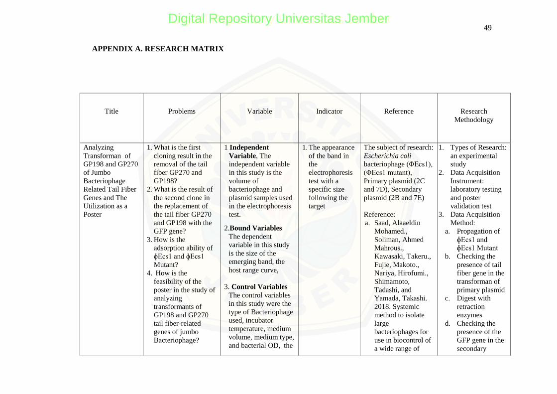
49
Title
Problems
Variable
Indicator
Reference
Research
Methodology
Analyzing
Transforman of
GP198 and GP270
of Jumbo
Bacteriophage
Related Tail Fiber
Genes and The
Utilization as a
Poster
1. What is the first
cloning result in the
removal of the tail
fiber GP270 and
GP198?
2. What is the result of
the second clone in
the replacement of
the tail fiber GP270
and GP198 with the
GFP gene?
3. How is the
adsorption ability of
ɸEcs1 and ɸEcs1
Mutant?
4. How is the
feasibility of the
poster in the study of
analyzing
transformants of
GP198 and GP270
tail fiber-related
genes of jumbo
Bacteriophage?
1 Independent
Variable, The
independent variable
in this study is the
volume of
bacteriophage and
plasmid samples used
in the electrophoresis
test.
2.Bound Variables
The dependent
variable in this study
is the size of the
emerging band, the
host range curve,
3. Control Variables
The control variables
in this study were the
type of Bacteriophage
used, incubator
temperature, medium
volume, medium type,
and bacterial OD, the
1. The appearance
of the band in
the
electrophoresis
test with a
specific size
following the
target
The subject of research:
Escherichia coli
bacteriophage (ΦEcs1),
(ΦEcs1 mutant),
Primary plasmid (2C
and 7D), Secondary
plasmid (2B and 7E)
Reference:
a. Saad, Alaaeldin
Mohamed.,
Soliman, Ahmed
Mahrous.,
Kawasaki, Takeru.,
Fujie, Makoto.,
Nariya, Hirofumi.,
Shimamoto,
Tadashi, and
Yamada, Takashi.
2018. Systemic
method to isolate
large
bacteriophages for
use in biocontrol of
a wide range of
1. Types of Research:
an experimental
study
2. Data Acquisition
Instrument:
laboratory testing
and poster
validation test
3. Data Acquisition
Method:
a. Propagation of
ɸEcs1 and
ɸEcs1 Mutant
b. Checking the
presence of tail
fiber gene in the
transforman of
primary plasmid
c. Digest with
retraction
enzymes
d. Checking the
presence of the
GFP gene in the
secondary
APPENDIX A. RESEARCH MATRIX
Digital Repository Universitas JemberDigital Repository Universitas Jember
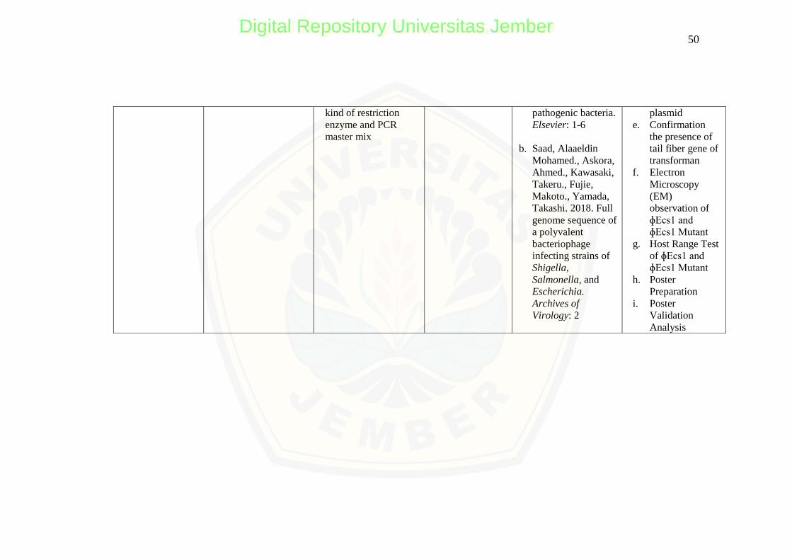
50
kind of restriction
enzyme and PCR
master mix
pathogenic bacteria.
Elsevier: 1-6
b. Saad, Alaaeldin
Mohamed., Askora,
Ahmed., Kawasaki,
Takeru., Fujie,
Makoto., Yamada,
Takashi. 2018. Full
genome sequence of
a polyvalent
bacteriophage
infecting strains of
Shigella,
Salmonella, and
Escherichia.
Archives of
Virology: 2
plasmid
e. Confirmation
the presence of
tail fiber gene of
transforman
f. Electron
Microscopy
(EM)
observation of
ɸEcs1 and
ɸEcs1 Mutant
g. Host Range Test
of ɸEcs1 and
ɸEcs1 Mutant
h. Poster
Preparation
i. Poster
Validation
Analysis
Digital Repository Universitas JemberDigital Repository Universitas Jember
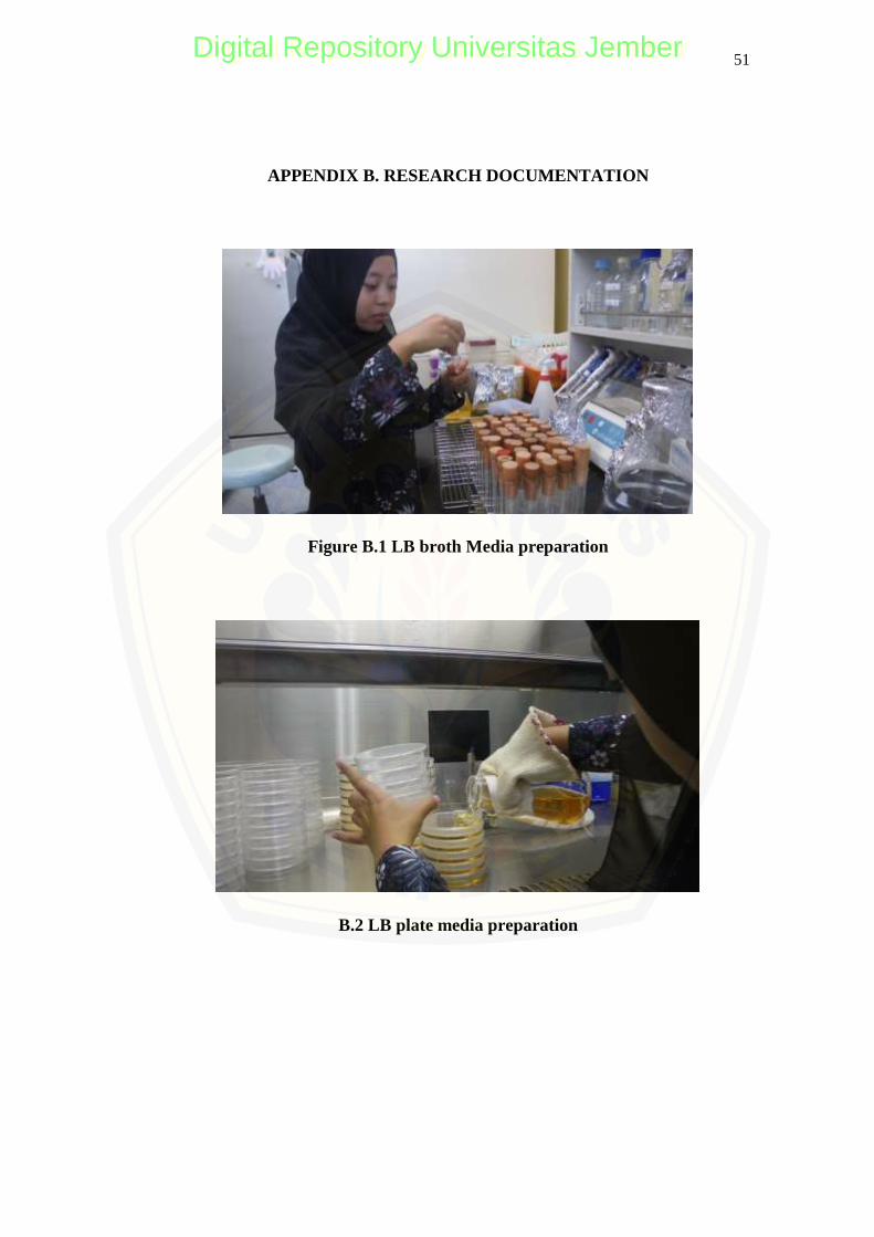
51
APPENDIX B. RESEARCH DOCUMENTATION
Figure B.1 LB broth Media preparation
B.2 LB plate media preparation
Digital Repository Universitas JemberDigital Repository Universitas Jember
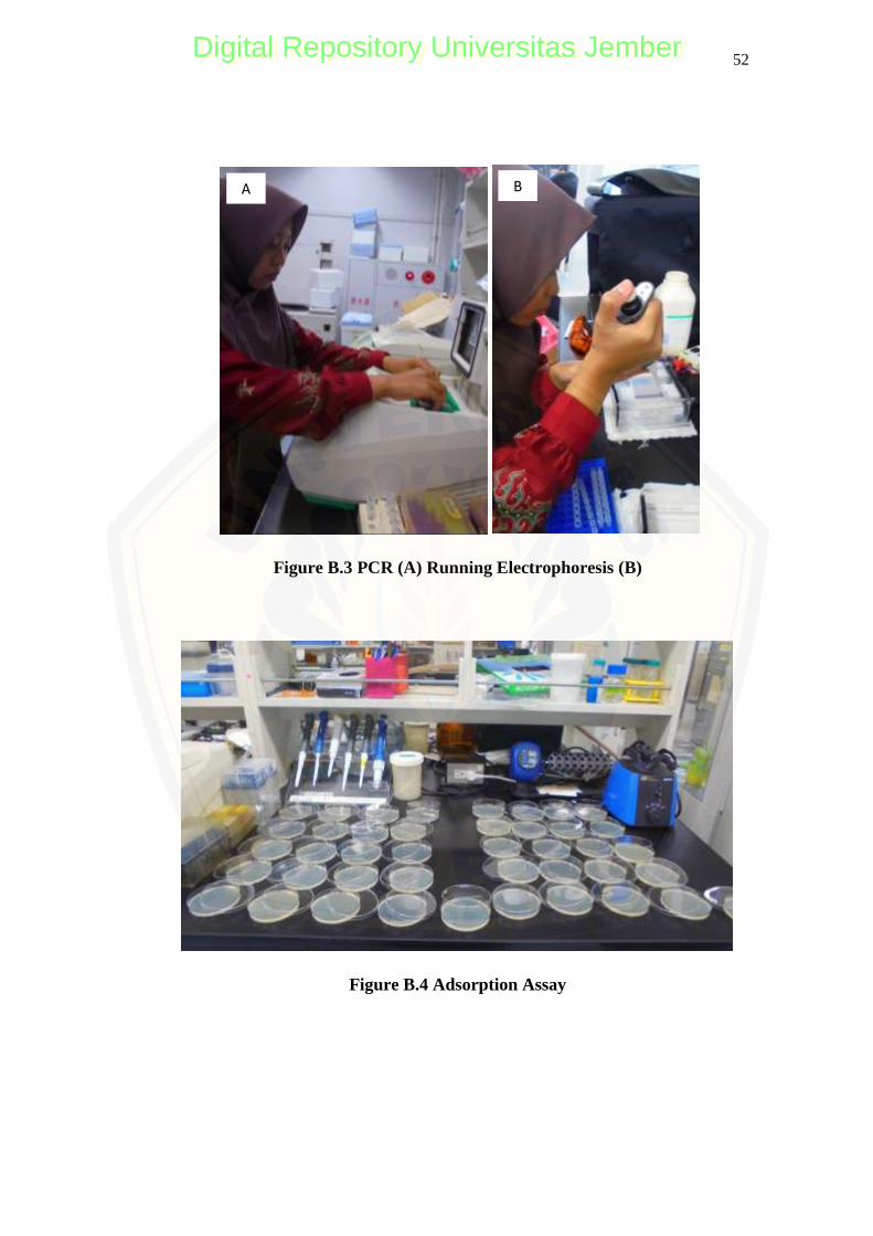
52
Figure B.3 PCR (A) Running Electrophoresis (B)
Figure B.4 Adsorption Assay
A B
Digital Repository Universitas JemberDigital Repository Universitas Jember
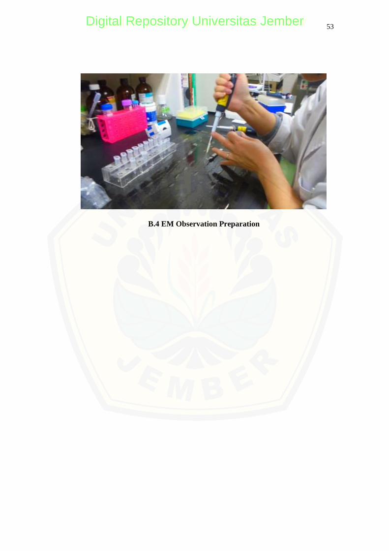
53
B.4 EM Observation Preparation
Digital Repository Universitas JemberDigital Repository Universitas Jember

54
APPENDIX C. POSTER VALIDATION QUESTIONNAIRE SHEET
C.1 VALIDATION OF POSTER MEDIA EXPERT
QUESTIONNAIRE SHEET
POSTER PRODUCT TEST
I. Researcher's Identity
Name : Afifatur Rofiqoh
NIM : 160210103090
Department /Study Program : Mathematics and Natural Sciences / Biology
Education Faculty of Teacher Training and
II. Introduction
The undersigned, a college student of Bachelor of Biology Education Study
Program, the Faculty of Education and Science University of Jember, is currently
processing a thesis for entitled ―ANALYSING TRANSFORMAN OF GP198
AND GP270 OF JUMBO BACTERIOPHAGE RELATED TAIL FIBER
GENES AND ITS UTILIZATION AS A SERIAL POSTER.‖
In connection with this, the researcher needs assistance concerning the
validation of her questionnaires necessary for the study. Knowing that you are most
fit and capable of providing such, the undersigned would like to ask your approval to
be the evaluator.
I am profoundly grateful for your kind approval.
Thank you very much, and may Allah bless you.
Sincerely yours,
AFIFATUR ROFIQOH
Student – Researcher
Digital Repository Universitas JemberDigital Repository Universitas Jember
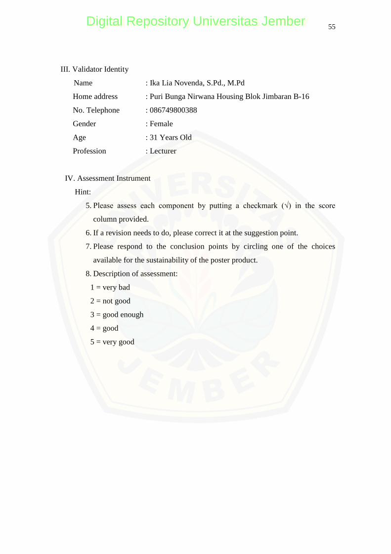
55
III. Validator Identity
Name : Ika Lia Novenda, S.Pd., M.Pd
Home address : Puri Bunga Nirwana Housing Blok Jimbaran B-16
No. Telephone : 086749800388
Gender : Female
Age : 31 Years Old
Profession : Lecturer
IV. Assessment Instrument
Hint:
5. Please assess each component by putting a checkmark (√) in the score
column provided.
6. If a revision needs to do, please correct it at the suggestion point.
7. Please respond to the conclusion points by circling one of the choices
available for the sustainability of the poster product.
8. Description of assessment:
1 = very bad
2 = not good
3 = good enough
4 = good
5 = very good
Digital Repository Universitas JemberDigital Repository Universitas Jember

56
Sub
Component Assessment criteria
Score Final
score 1 2 3 4 5
1. Components of the Eligibility of Graphics
A. Artistic and
Aesthetic
1. The layout of poster
components
√
24
2. Background color selection √
3. Font type selection √
4. Color harmony on the poster √
5. Image compatibility with
font types
√
6. Accuracy inflow and overall
harmony of poster
components
√
B.Poster
Function
7. Readability (visible)
provides clarity of
information for the reader
√
15
8. Posters possible enhanced
the reader's knowledge of the
material contained
√
9. Poster raises the attraction
for readers to know the
contents of the poster
√
10. the ability to influence the
reader to act (to act)
√
2. Presentation Components
A. Presentati-
on
Techniques
11. The flow of the poster
presentation is adjusted
√
32
12. Structured of the poster‘s
contents
√
13. Complete sentences
presented
√
Digital Repository Universitas JemberDigital Repository Universitas Jember
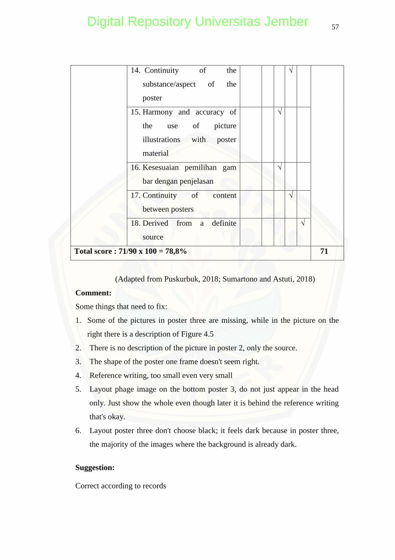
57
14. Continuity of the
substance/aspect of the
poster
√
15. Harmony and accuracy of
the use of picture
illustrations with poster
material
√
16. Kesesuaian pemilihan gam
bar dengan penjelasan
√
17. Continuity of content
between posters
√
18. Derived from a definite
source
√
Total score : 71/90 x 100 = 78,8% 71
(Adapted from Puskurbuk, 2018; Sumartono and Astuti, 2018)
Comment:
Some things that need to fix:
1. Some of the pictures in poster three are missing, while in the picture on the
right there is a description of Figure 4.5
2. There is no description of the picture in poster 2, only the source.
3. The shape of the poster one frame doesn't seem right.
4. Reference writing, too small even very small
5. Layout phage image on the bottom poster 3, do not just appear in the head
only. Just show the whole even though later it is behind the reference writing
that's okay.
6. Layout poster three don't choose black; it feels dark because in poster three,
the majority of the images where the background is already dark.
Suggestion:
Correct according to records
Digital Repository Universitas JemberDigital Repository Universitas Jember
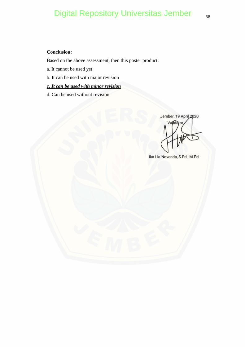
58
Conclusion:
Based on the above assessment, then this poster product:
a. It cannot be used yet
b. It can be used with major revision
c. It can be used with minor revision
d. Can be used without revision
Digital Repository Universitas JemberDigital Repository Universitas Jember
59
C.2 VALIDATION POSTER OF MATTER EXPERTS
QUESTIONNAIRE SHEET
POSTER PRODUCT TEST
I. Researcher's Identity
Name : Afifatur Rofiqoh
NIM : 160210103090
Department /Study Program : Mathematics and Natural Sciences / Biology
Education Faculty of Teacher Training and
II. Introduction
The undersigned, a college student, taking up Bachelor of Primary Education in
the Biology Education Study Program of the Faculty of Education and Science
University of Jember, is currently processing a thesis for this semester entitled
―ANALYSING TRANSFORMAN OF GP198 AND GP270 OF JUMBO
BACTERIOPHAGE RELATED TAIL FIBER GENES AND ITS
UTILIZATION AS A SERIAL POSTER.‖
In connection with this, the researcher will need assistance concerning the
validation of his questionnaires necessary for the study. Knowing that you are most
fit and capable of providing such, the undersigned has chosen and would like to ask
approval from your right office to be the evaluator.
I am highly anticipating your kind approval regarding this matter.
Thank you very much, and God bless you.
Sincerely yours,
AFIFATUR ROFIQOH
Student – Researcher
Digital Repository Universitas JemberDigital Repository Universitas Jember
60
III. Validator Identity
Name : Aditya Kurniawan, S.Si., M.Biomed
Home address : Semeru Utama A4 Jember
No. Telephone : 081 334 155 366
Gender : Male
Age : 27
Profession : Lecturer
IV. Assessment Instrument
Hint:
1. Please assess each component by putting a checkmark (√) in the score
column provided.
2. If a revision needs to do, please correct it at the suggestion point.
3. Please respond to the conclusion points by circling one of the choices
available for the sustainability of the poster product.
4. Description of assessment:
1 = very bad
2 = not good
3 = good enough
4 = good
5 = very good
Digital Repository Universitas JemberDigital Repository Universitas Jember
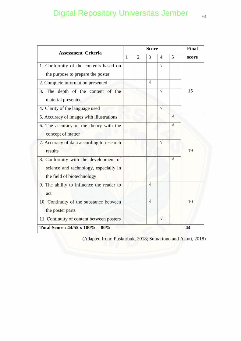
61
Assessment Criteria Score Final
score 1 2 3 4 5
1. Conformity of the contents based on
the purpose to prepare the poster
√
15
2. Complete information presented √
3. The depth of the content of the
material presented
√
4. Clarity of the language used √
5. Accuracy of images with illustrations √
19
6. The accuracy of the theory with the
concept of matter
√
7. Accuracy of data according to research
results
√
8. Conformity with the development of
science and technology, especially in
the field of biotechnology
√
9. The ability to influence the reader to
act
√
10 10. Continuity of the substance between
the poster parts
√
11. Continuity of content between posters √
Total Score : 44/55 x 100% = 80% 44
(Adapted from: Puskurbuk, 2018; Sumartono and Astuti, 2018)
Digital Repository Universitas JemberDigital Repository Universitas Jember
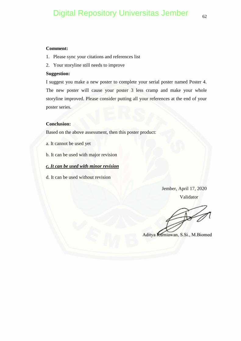
62
Comment:
1. Please sync your citations and references list
2. Your storyline still needs to improve
Suggestion:
I suggest you make a new poster to complete your serial poster named Poster 4.
The new poster will cause your poster 3 less cramp and make your whole
storyline improved. Please consider putting all your references at the end of your
poster series.
Conclusion:
Based on the above assessment, then this poster product:
a. It cannot be used yet
b. It can be used with major revision
c. It can be used with minor revision
d. It can be used without revision
Jember, April 17, 2020
Validator
Digital Repository Universitas JemberDigital Repository Universitas Jember
63
C.3 POSTER USER RESPONSE
QUESTIONNAIRE SHEET
POSTER PRODUCT TEST
I. Researcher's Identity
Name : Afifatur Rofiqoh
NIM : 160210103090
Department /Study Program : Mathematics and Natural Sciences / Biology
Education Faculty of Teacher Training and
II. Introduction
The undersigned, a college student, taking up Bachelor of Primary Education
in the Biology Education Study Program of the Faculty of Education and
Science University of Jember, is currently processing a thesis for this semester
entitled ―ANALYSING TRANSFORMAN OF GP198 AND GP270 OF
JUMBO BACTERIOPHAGE RELATED TAIL FIBER GENES AND
ITS UTILIZATION AS A SERIAL POSTER.‖
In connection with this, the researcher will need assistance concerning the
validation of his questionnaires necessary for the study. Knowing that you are
most fit and capable of providing such, the undersigned has chosen and would
like to ask approval from your right office to be the evaluator.
I am highly anticipating your kind approval regarding this matter.
Thank you very much, and God bless you.
Sincerely yours,
AFIFATUR ROFIQOH
Student – Researcher
Digital Repository Universitas JemberDigital Repository Universitas Jember
64
III. Validator Identity
Name : Ria Yulian S.Pd
Home address : Probolinggo
No. Telephone : 082132315469
Gender : Female
Age : 23 years old
Profession : Lecture assistant
IV. Assessment Instrument
Hint:
1. Please assess each component by putting a checkmark (√) in the score column
provided.
2. If a revision needs to do, please correct it at the suggestion point.
3. Please respond to the conclusion points by circling one of the choices available
for the sustainability of the poster product.
4. Description of assessment:
1 = very bad
2 = not good
3 = good enough
4 = good
5 = very good
Digital Repository Universitas JemberDigital Repository Universitas Jember
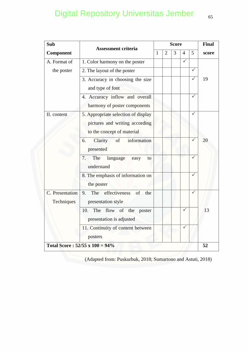
65
Sub
Component Assessment criteria
Score Final
score 1 2 3 4 5
A. Format of
the poster
1. Color harmony on the poster
19
2. The layout of the poster
3. Accuracy in choosing the size
and type of font
4. Accuracy inflow and overall
harmony of poster components
II. content 5. Appropriate selection of display
pictures and writing according
to the concept of material
20 6. Clarity of information
presented
7. The language easy to
understand
8. The emphasis of information on
the poster
C. Presentation
Techniques
9. The effectiveness of the
presentation style
13 10. The flow of the poster
presentation is adjusted
11. Continuity of content between
posters
Total Score : 52/55 x 100 = 94% 52
(Adapted from: Puskurbuk, 2018; Sumartono and Astuti, 2018)
Digital Repository Universitas JemberDigital Repository Universitas Jember
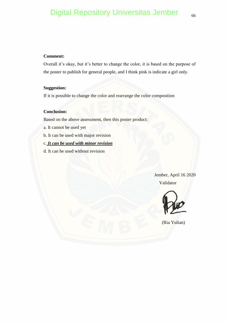
66
Comment:
Overall it‘s okay, but it‘s better to change the color, it is based on the purpose of
the poster to publish for general people, and I think pink is indicate a girl only.
Suggestion:
If it is possible to change the color and rearrange the color composition
Conclusion:
Based on the above assessment, then this poster product:
a. It cannot be used yet
b. It can be used with major revision
c. It can be used with minor revision
d. It can be used without revision
Jember, April 16 2020
Validator
(Ria Yulian)
Digital Repository Universitas JemberDigital Repository Universitas Jember
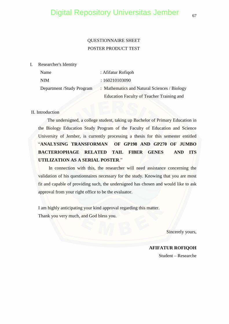
67
QUESTIONNAIRE SHEET
POSTER PRODUCT TEST
I. Researcher's Identity
Name : Afifatur Rofiqoh
NIM : 160210103090
Department /Study Program : Mathematics and Natural Sciences / Biology
Education Faculty of Teacher Training and
II. Introduction
The undersigned, a college student, taking up Bachelor of Primary Education in
the Biology Education Study Program of the Faculty of Education and Science
University of Jember, is currently processing a thesis for this semester entitled
―ANALYSING TRANSFORMAN OF GP198 AND GP270 OF JUMBO
BACTERIOPHAGE RELATED TAIL FIBER GENES AND ITS
UTILIZATION AS A SERIAL POSTER.‖
In connection with this, the researcher will need assistance concerning the
validation of his questionnaires necessary for the study. Knowing that you are most
fit and capable of providing such, the undersigned has chosen and would like to ask
approval from your right office to be the evaluator.
I am highly anticipating your kind approval regarding this matter.
Thank you very much, and God bless you.
Sincerely yours,
AFIFATUR ROFIQOH
Student – Researche
Digital Repository Universitas JemberDigital Repository Universitas Jember
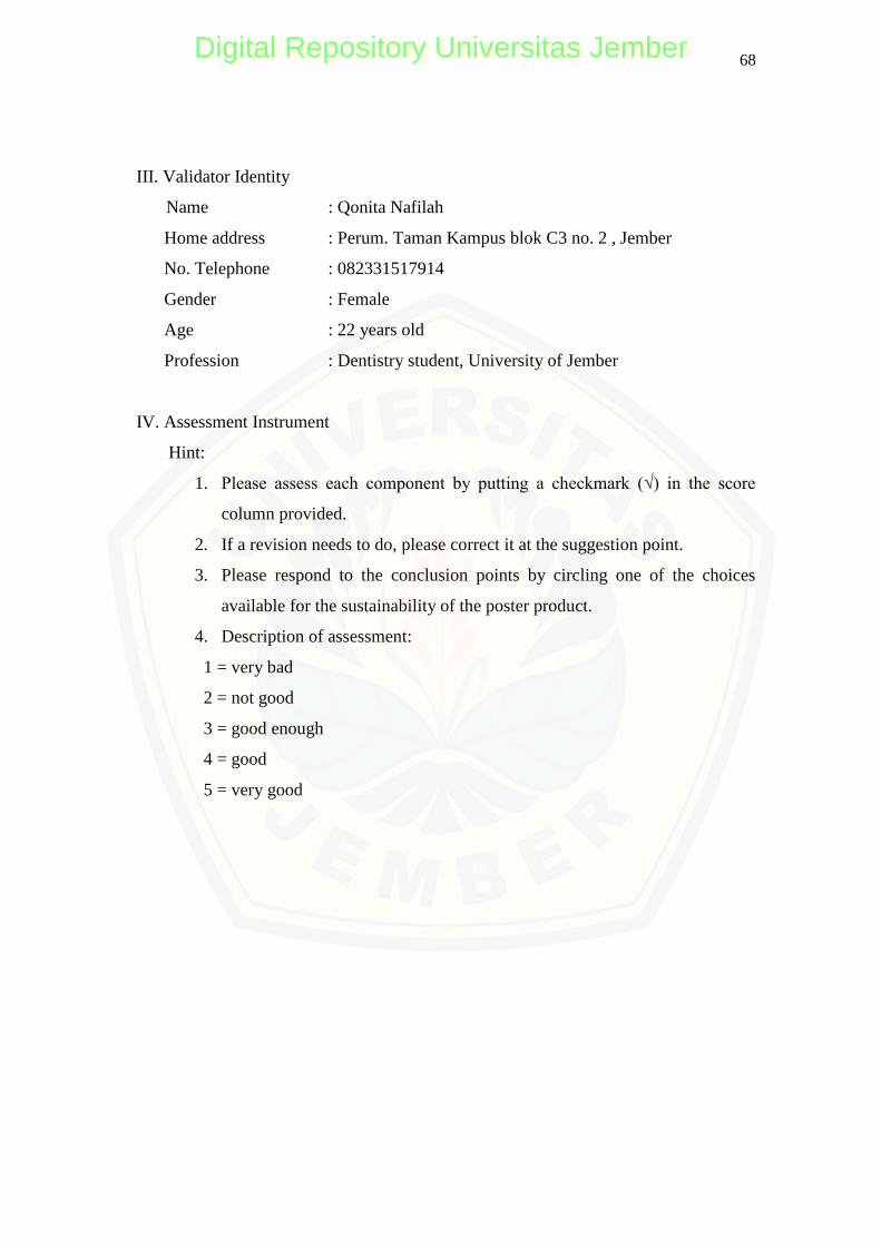
68
III. Validator Identity
Name : Qonita Nafilah
Home address : Perum. Taman Kampus blok C3 no. 2 , Jember
No. Telephone : 082331517914
Gender : Female
Age : 22 years old
Profession : Dentistry student, University of Jember
IV. Assessment Instrument
Hint:
1. Please assess each component by putting a checkmark (√) in the score
column provided.
2. If a revision needs to do, please correct it at the suggestion point.
3. Please respond to the conclusion points by circling one of the choices
available for the sustainability of the poster product.
4. Description of assessment:
1 = very bad
2 = not good
3 = good enough
4 = good
5 = very good
Digital Repository Universitas JemberDigital Repository Universitas Jember
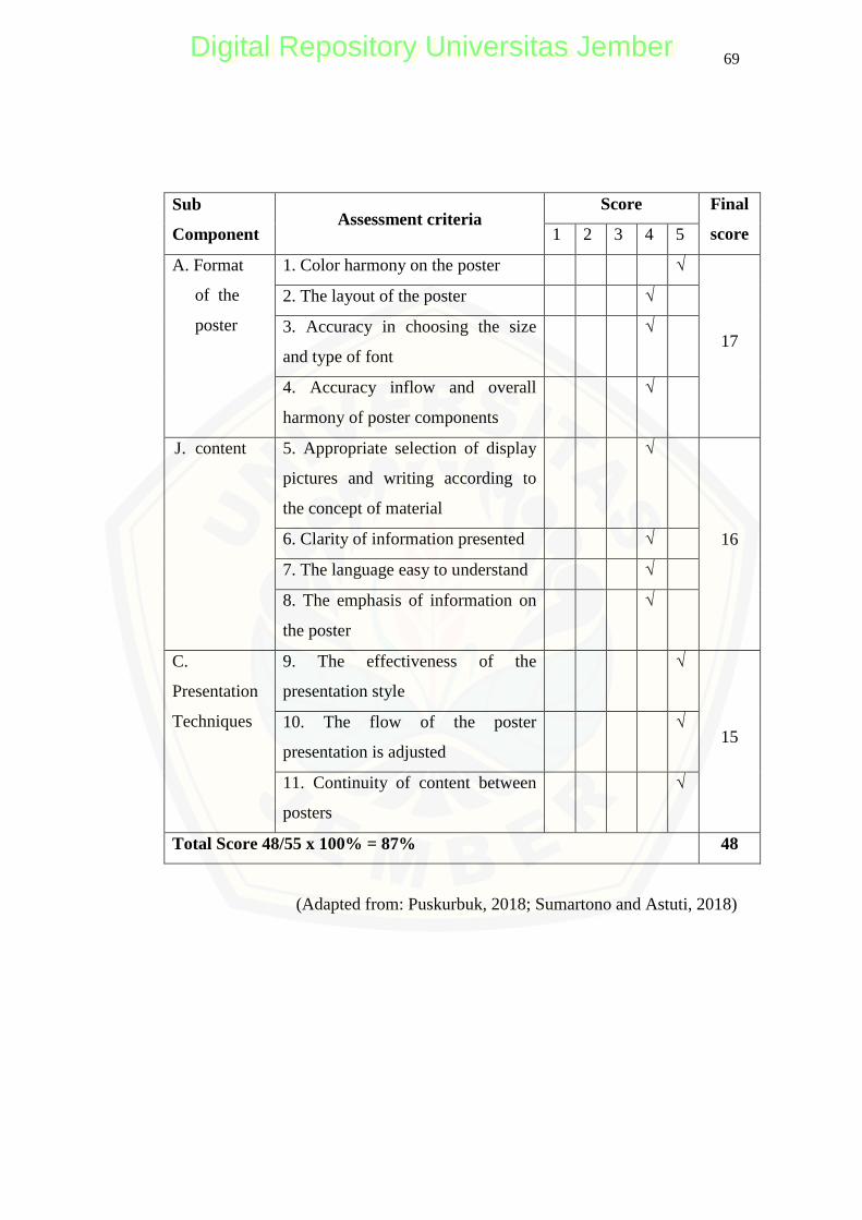
69
Sub
Component Assessment criteria
Score Final
score 1 2 3 4 5
A. Format
of the
poster
1. Color harmony on the poster √
17
2. The layout of the poster √
3. Accuracy in choosing the size
and type of font
√
4. Accuracy inflow and overall
harmony of poster components
√
J. content 5. Appropriate selection of display
pictures and writing according to
the concept of material
√
16 6. Clarity of information presented √
7. The language easy to understand √
8. The emphasis of information on
the poster
√
C.
Presentation
Techniques
9. The effectiveness of the
presentation style
√
15 10. The flow of the poster
presentation is adjusted
√
11. Continuity of content between
posters
√
Total Score 48/55 x 100% = 87% 48
(Adapted from: Puskurbuk, 2018; Sumartono and Astuti, 2018)
Digital Repository Universitas JemberDigital Repository Universitas Jember
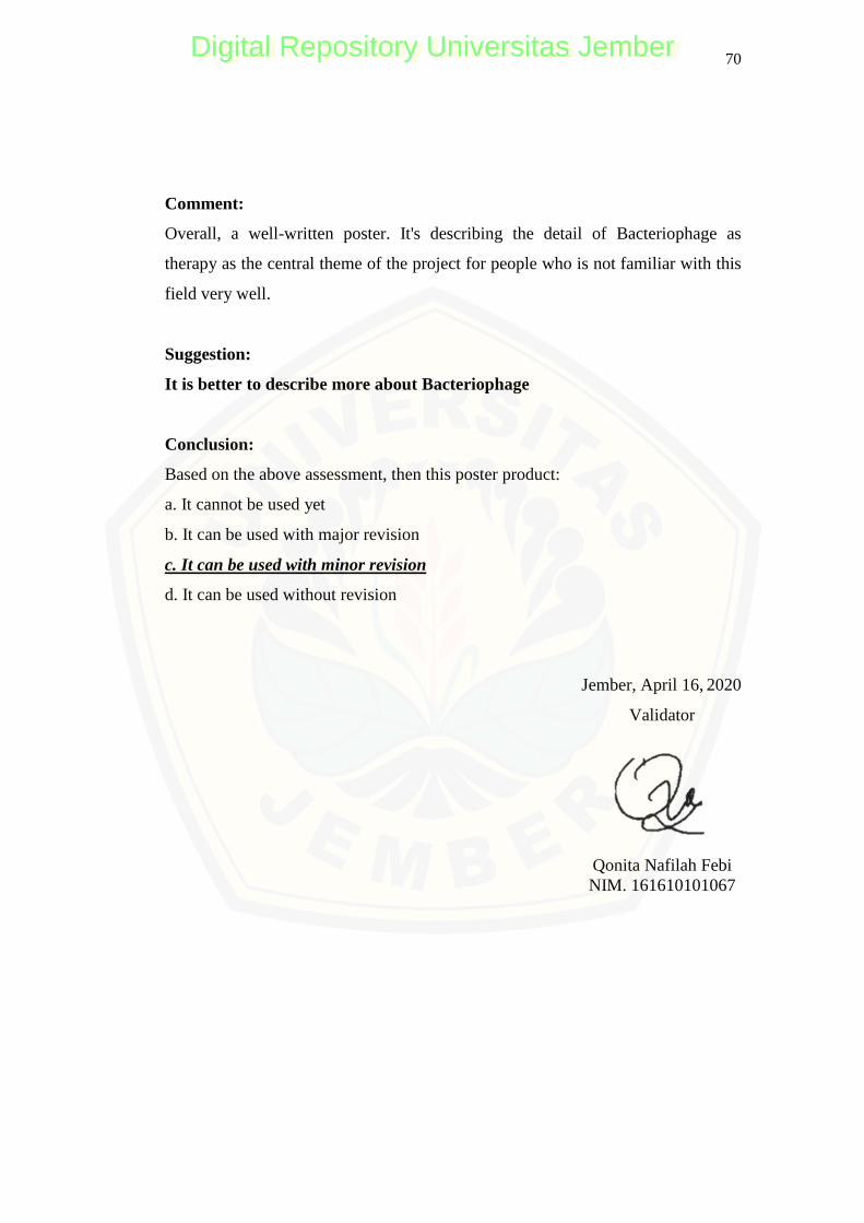
70
Comment:
Overall, a well-written poster. It's describing the detail of Bacteriophage as
therapy as the central theme of the project for people who is not familiar with this
field very well.
Suggestion:
It is better to describe more about Bacteriophage
Conclusion:
Based on the above assessment, then this poster product:
a. It cannot be used yet
b. It can be used with major revision
c. It can be used with minor revision
d. It can be used without revision
Jember, April 16, 2020
Validator
Qonita Nafilah Febi NIM. 161610101067
Digital Repository Universitas JemberDigital Repository Universitas Jember
71
QUESTIONNAIRE SHEET
POSTER PRODUCT TEST
I. Researcher's Identity
Name : Afifatur Rofiqoh
NIM : 160210103090
Department /Study Program : Mathematics and Natural Sciences / Biology
Education Faculty of Teacher Training and
II. Introduction
The undersigned, a college student, taking up Bachelor of Primary Education
in the Biology Education Study Program of the Faculty of Education and
Science University of Jember, is currently processing a thesis for this semester
entitled ―ANALYSING TRANSFORMAN OF GP198 AND GP270 OF
JUMBO BACTERIOPHAGE RELATED TAIL FIBER GENES AND
ITS UTILIZATION AS A SERIAL POSTER.‖
In connection with this, the researcher will need assistance concerning the
validation of his questionnaires necessary for the study. Knowing that you are
most fit and capable of providing such, the undersigned has chosen and would
like to ask approval from your right office to be the evaluator.
I am highly anticipating your kind approval regarding this matter.
Thank you very much, and God bless you.
Sincerely yours,
AFIFATUR ROFIQOH
Student – Researcher
Digital Repository Universitas JemberDigital Repository Universitas Jember
72
III. Validator Identity
Name : Fifi Lanna Fauziah
Home address : Banyuwangi
No. Telephone : 082132315469
Gender : Female
Age : 22 years old
Profession : Nursing student, University of Jember
IV. Assessment Instrument
Hint:
5. Please assess each component by putting a checkmark (√) in the score column
provided.
6. If a revision needs to do, please correct it at the suggestion point.
7. Please respond to the conclusion points by circling one of the choices available
for the sustainability of the poster product.
8. Description of assessment:
1 = very bad
2 = not good
3 = good enough
4 = good
5 = very good
Digital Repository Universitas JemberDigital Repository Universitas Jember
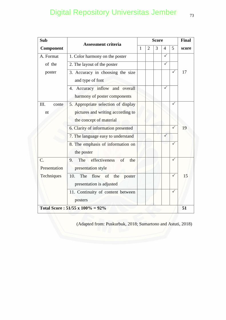
73
Sub
Component Assessment criteria
Score Final
score 1 2 3 4 5
A. Format
of the
poster
1. Color harmony on the poster
17
2. The layout of the poster
3. Accuracy in choosing the size
and type of font
4. Accuracy inflow and overall
harmony of poster components
III. conte
nt
5. Appropriate selection of display
pictures and writing according to
the concept of material
19 6. Clarity of information presented
7. The language easy to understand
8. The emphasis of information on
the poster
C.
Presentation
Techniques
9. The effectiveness of the
presentation style
15 10. The flow of the poster
presentation is adjusted
11. Continuity of content between
posters
Total Score : 51/55 x 100% = 92% 51
(Adapted from: Puskurbuk, 2018; Sumartono and Astuti, 2018)
Digital Repository Universitas JemberDigital Repository Universitas Jember
74
Comment:
Poster 1:
The title of the poster should be capital at the beginning
Figures 3 and 4 labels as Figures 1 and 2
Poster 2:
There is excess space after the range brackets.
The picture is a bit unclear
Poster 3:
The graphic color in figure 4.5 whose number of mutants must be clarified
Suggestion:
It's excellent overall, from the color selection, is cute. Tips for considering my
comments before. Hopefully, it will be more comfortable in the future. I think
your research is marvelous
Conclusion:
Based on the above assessment, then this poster product:
a. It cannot be used yet
b. It can be used with major revision
c. It can be used with minor revision
d. It can be used without revision
Jember, April 16, 2020
Validator
(Fifi Lanna Fauziah)
NIM. 162310101265
Digital Repository Universitas JemberDigital Repository Universitas Jember
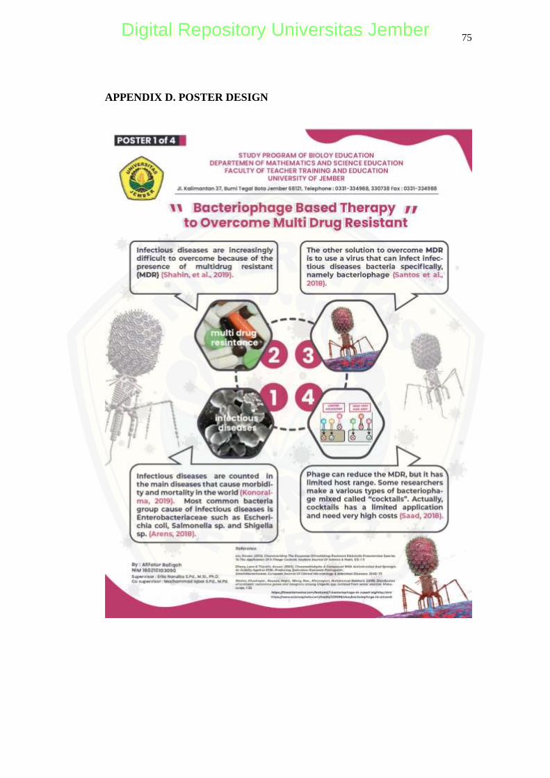
75
APPENDIX D. POSTER DESIGN
Digital Repository Universitas JemberDigital Repository Universitas Jember
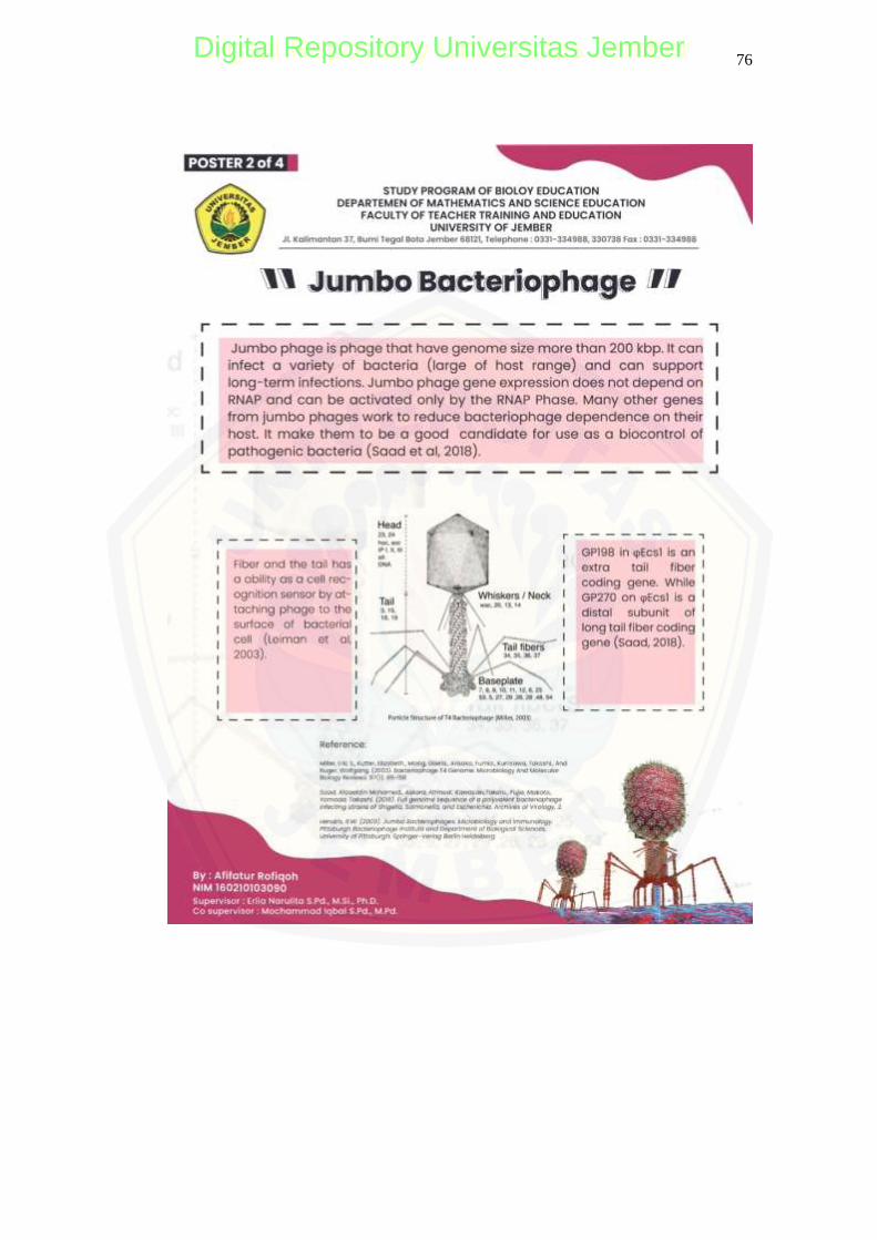
76
Digital Repository Universitas JemberDigital Repository Universitas Jember
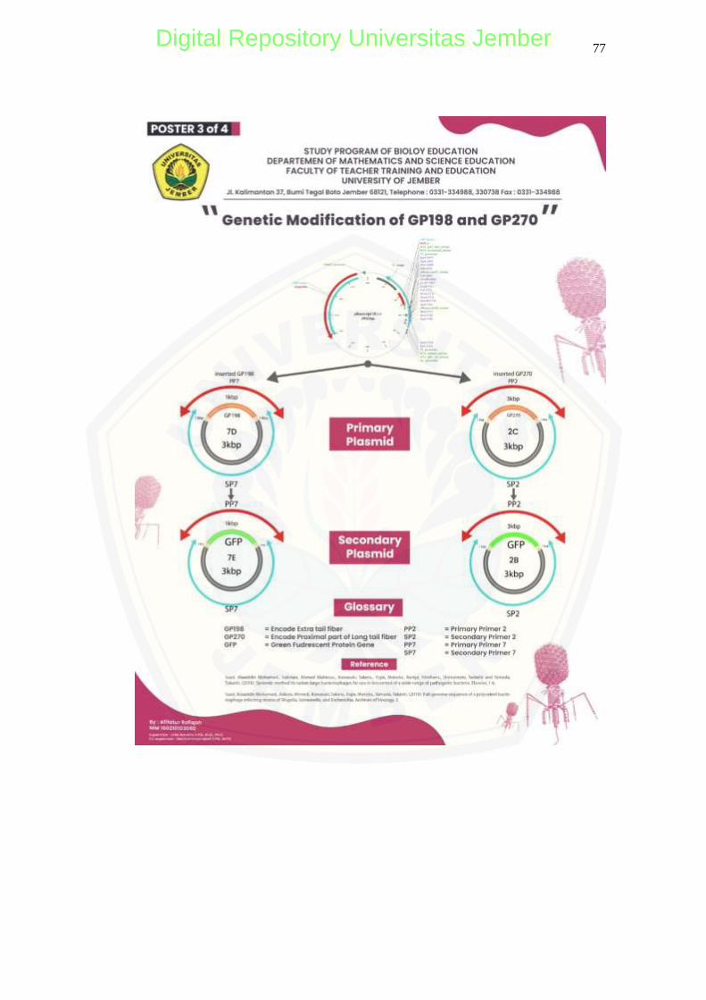
77
Digital Repository Universitas JemberDigital Repository Universitas Jember
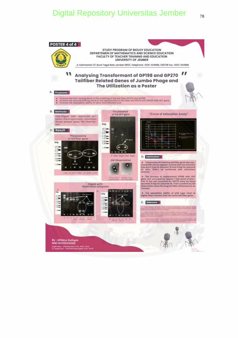
78
Digital Repository Universitas JemberDigital Repository Universitas Jember
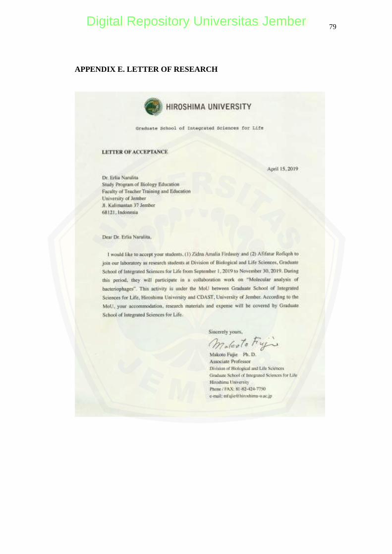
79
APPENDIX E. LETTER OF RESEARCH
Digital Repository Universitas JemberDigital Repository Universitas Jember
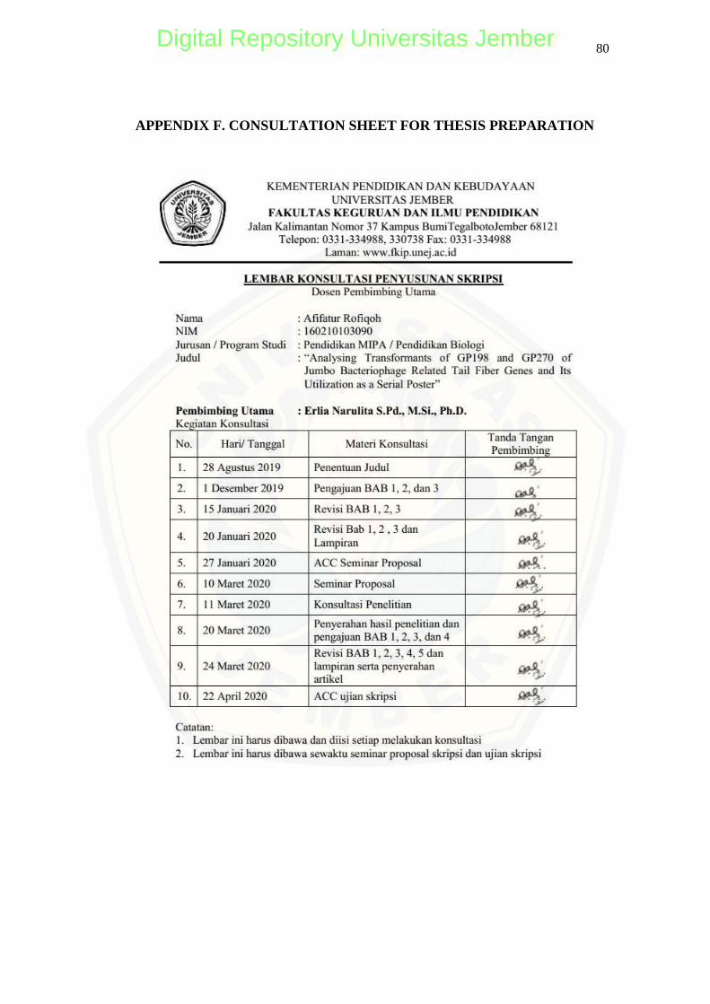
80
APPENDIX F. CONSULTATION SHEET FOR THESIS PREPARATION
Digital Repository Universitas JemberDigital Repository Universitas Jember
81
Digital Repository Universitas JemberDigital Repository Universitas Jember