Embed Size (px)
Citation preview

Agrin-induced Reorganization of Extracellular Matrix Components on Cultured Myotubes: Relationship to AChR Aggregation Ralph M. Ni tk in a n d Tova C. Rothschi ld
Department of Biological Sciences, Rutgers University, Newark, New Jersey 07102
Abstract. Agrin, an extracellular matrix-associated protein extracted from synapse-rich tissues, induces the accumulation of acetylcholine receptors (AChRs) and other synaptic components into discrete patches on cultured myotubes. The appearance of agrin-like molecules at neuromuscular junctions suggests that it may direct synaptic organization in vivo. In the pres- ent study we examined the role of extracellular matrix components in agrin-induced differentiation. We used immunohistochemical techniques to visualize the spa- tial and temporal distribution of laminin, a heparan sulfate proteoglycan (HSPG), fibronectin, and type IV collagen on cultured chick myotubes during agrin- induced aggregation of AChRs.
Myotubes displayed significant amounts of laminin and HSPG, lesser amounts of type IV collagen, and
little, if any, fibronectin. Agrin treatment caused cell surface laminin and HSPG to patch, while collagen and fibronectin distributions were generally unaffected. Many of the agrin-induced laminin and HSPG patches colocalized with AChR patches, raising the possibility of a causal relationship between matrix patching and AChR accumulations. However, patching of AChRs (complete within a few hours) preceded that of laminm or HSPG (not complete until 15-20 h), making it un- likely that matrix accumulations initiate AChR patch- ing at agrin-induced sites. Conversely, when AChR patching was blocked by treatment with anti-AChR an- tibody mAb 35, agrin was still able to effect patching of laminin and HSPG. Taken together, these findings suggest that agrin-induced accumulations of AChR and laminin/HSPG are not mechanistically linked.
F UNCTIONAL transmission across the neuromuscular junction requires the precise localization of cell sur- face, cytoskeletal, and extracellular components at
the point of nerve-muscle contact (reviewed by Peng, 1987; Schuetze and Role, 1987; Steinbach and Bloch, 1986; Rubin and Barald, 1983; Dennis, 1981; Fambrough, 1979). It is quite remarkable that these components accumulate in such a discrete region, which represents only ~,0.1% of the myofiber surface. Acetylcholine receptors (AChRs) ~, for example, are packed into the synaptic membrane at densities approaching 15-20,000 per square micron, hut are virtually absent elsewhere on the myofiber surface (reviewed in Sal- peter and Loring, 1985). To understand the mechanisms in- volved in the formation and maintenance of synaptic struc- ture, we have treated cultured myotubes with agrin, a synaptic-organizing molecule that appears to be related to factors that function at synapses in vivo.
Studies by McMahan and colleagues demonstrated that in vivo matrix-associated factors can play a significant role in the regeneration of neuromuscular structure (Sanes et al.,
Dr. Nitkin's present address is NICHD-MRDDB, Bldg. EPN, Rm. 631, 6130 Executive Blvd., Bethesda, MD 20892. Address reprint requests to him.
1. Abbreviations used in this paper: AChR, acetyicholine receptor; HSPG, heparan sulfate proteoglycan.
1978; Burden et al., 1979; McMahan and Slater, 1984; An- glister and McMahan, 1985; see also Bader, 1981). This led to the screening of matrix-enriched fractions of Torpedo electric organ for factors that affect AChR distribution (Rubin and McMahan, 1982; Nitldn et al., 1983;~ Godfrey et al., 1984). In this manner a proteinaceous factor, termed agrin, was identified and characterized (Nitldn et al., 1987). Agrin induces on cultured myotubes dense accumulations of AChRs, which are also associated with acetylcholinesterase (Wallace et al., 1985; Wallace, 1986) and other synaptic components (Wallace, 1989). mAbs against the active com- ponent recognize a related series of polypeptides with mo- lecular masses of 70, 95, 135, and 150 kD (Nitldn et al., 1987; see also Godfrey et al., 1988a). These mAbs were also used to demonstrate that agrin-like molecules are concen- trated at neuromuscular junctions in vivo (Fallon et al., 1985; Reist et al., 1987; Godfrey et al., 1988a). Therefore, not only is agrin a useful means of inducing "synaptic" differentiation on cultured myotubes, but it appears to be related to factors that direct synaptic development in vivo.
Several observations suggest that agrin-induced AChR patching occurs through a specific, physiological cellular mechanism. Agrin effects myotubes in a dose-dependent manner, although at higher agrin levels the number of AChR patches per myotube plateaus off (Godfrey et al., 1984; Wal- lace, 1989), suggesting that AChR patching may be limited
© The Rockefeller University Press, 0021-9525/90/09/1161/10 $2.00 The Journal of Cell Biology, Volume 11 I, September 1990 1161-1170 1161
on Septem
ber 8, 2015jcb.rupress.org
Dow
nloaded from
Published September 1, 1990

by cellular constraints. The accumulation of AChRs into dis- crete, high density patches takes a few hours (Godfrey et al., 1984), which argues against a mere cross-linking of AChRs by agrin (by contrast, antibodies and other multivalent ligands that induce reorganization of surface components operate within minutes, to patch, cap, and rapidly internalize bound receptors). Furthermore, the AChR accumulations induced by agrin are coordinated with accumulations of synapse-specific cytoplasmic, membrane, and extraceUular components (Wallace et al., 1985; Wallace, 1986; Wallace, 1989). Agrin-induced AChR aggregation appears to involve calcium, metabolic energy, and possibly phosphorylation (Wallace, 1988).
AChR accumulations can be triggered by a variety of fac- tors including neural extracts (reviewed in Peng, 1987; Schuetze and Role, 1987), basal lamina components (Peng, 1987; Schuetze and Role, 1987), interaction with the tissue culture substratum (Bloch et al., 1985), positively charged latex beads (Peng and Cheng, 1982), and electric fields (Orida and Poe, 1978). In contrast to agrin, some of these treatments significantly increase AChR synthesis which could indirectly impact on AChR distribution. The AChR patches induced by brain extract and latex beads appear to be morphologically similar to those induced by agrin; the patches are likewise associated with esterase and basal lam- ina components.
As a first step toward understanding how a myotube coor- dinates the accumulation of cell surface components at "syn- aptiC sites, we examined the possibility that specific ex- tracellular matrix components play a role in postsynaptic differentiation. Matrix components are present on develop- ing myotubes from the earliest stages of synapse formation (reviewed by Sanes, 1989). In some developing tissues, com- ponents of the extracellular matrix have been shown to inter- act with specific receptors to effect cellular differentiation (Buck and Horwitz, 1987).
Embryonic myotubes have only wisps of organized basal lamina (Kelly and Zacks, 1969; Kullberg et al., 1977; Jacob and Lentz, 1979). This resembles the appearance of cultured myotubes (Burrage and Lentz, 1981; Bayne et al., 1984; Chiu and Sanes, 1984). As myotubes are innervated, matrix material accumulates at sites of nerve terminal contact, along with AChRs and other synaptic components (Weldon and Cohen, 1979; Nakajima et al., 1980; Anderson and Fambrough, 1983; Bayne et al., 1984; Buchanan et al., 1989). Likewise, matrix material and AChRs accumulate at sites induced by neuronal extracts and even latex beads (Sal- peter et al., 1982; Daniels et al., 1984; Sanes et al., 1984; Olek et al., 1986; Peng and Cheng, 1982). This suggests that matrix accumulation and synaptic differentiation are part of a common developmental pathway which can be triggered by a variety of stimuli. Our studies with agrin focus on the mechanistic link between matrix accumulation and AChR clustering.
Immunohistochemical techniques were used to examine the distribution of laminin, fibronectin, type IV collagen, and a heparan sulfate proteoglycan (HSPG) on cultured myo- tubes. We found that agrin caused the aggregation of laminin and HSPG, but had little effect on fibronectin or type IV col- lagen distribution. Under these conditions, many of the lami- nin and HSPG accumulations colocalized with AChR patches.
To address the causal relationship between matrix ac- cumulation and AChR patching, we compared the time courses of these agrin-induced events. AChR patching (as detected by fluorescence microscopy) was completed well ahead of laminin and HSPG patching, suggesting that matrix accumulations do not serve as precursors for AChR patch- ing. In other experiments, AChR aggregation was prevented by treatment with anti-AChR mAbs. Under these conditions, agrin was still able to aggregate laminin and HSPG. These results suggest that agrin-induced patching of AChRs is not causally related to laminin/HSPG patching. Thus, agrin does not appear to use extracellular matrix organization to drive synaptic differentiation.
Brief accounts of this work have appeared elsewhere (Nit- kin, R. M., and T. C. Rothschild. 1988. Soc. Neurosci. Abstr. 14:514.).
Materials and Methods
Chick Myotube Cultures
Myotubes from hindlimb muscles of ll-12-d-old White Leghorn chick em- bryos (Avian Services, Frenchtown, NI) were cultured on 35-ram tissue cul- ture dishes coated with calf skin collagen (Calbiochem-Behring Corp., La ]ella, CA) and maintained in MEM-based medium (Gibco Laboratories, Grand Island, NY) supplemented with 10% horse serum (Gibco Laborato- ries) and 2% chick embryo extract (Fischbach, 1972, with minor modifica- tions described in Godfrey et al., 1984). The experiments described here were performed on 5-9-d-old cultures.
Agrin Extracts
Agrin was partially purified from electric organ of Torpedo californica as previously described (Nitkin et al., 1987) except that the detergent extrac- tion steps were omitted. To achieve maximal AChR aggregation on myo- tubes, 3-10 U of cibacron pool extract (sp act 5-30 U/#g) were used.
Localization of Extracellular Matrix Components and AChRs
To visualize the distribution of extracellular matrix components, cells were incubated with primary antibodies (concentrations listed below) for 1-2 h at 37°C, washed three times in culture medium, then incubated for an addi- tional I h in fluorescein-conjugated goat anti-mouse or anti-rabbit Ig (Cap- pel division, Organon Teknika, Malvern, PA) diluted 1:200 in culture medium. Cultures were washed three to five times in Puck's saline (Puck ct al., 1958), then fixed for 10 rain at -20°C in reagent alcohol (etha- nol/methanol/isopropanol, 18:1:1). After removal of alcohol, cultures were allowed to air dry, and coverslips were affixed with a drop of 50% (vol/vol) glycerol in Puck's saline, with 1/~g/rul phenylenediamene as an antibleach- ing agent. Incubation of cultures in fluorescent secondary antibodies alone yielded only faint uniform labeling of cells, which was well below the levels of specific labeling presented here.
For double labeling experiments, AChRs were labeled by including 2 × 10 -s M rhodamine-Bgtx (Ravdin and Axelrod, 1977) in both primary and secondary antibody solutions.
Cultures were examined through epifluorescence on an Orthoplan 2 mi- croscope equipped with I3 fluorescein and N2.1 rhodamine filters, and 63 × Planapo 1.4 NA oil immersion objective (E. Leitz, Rocldeigh, N/) at a total magnification of 504 x, and photographed using TMax 400 film (Eastman Kodak Co., Rochester, NY).
Laminin was localized using mAb 31, which was raised against chick muscle laminin (Bayne et al., 1984). Optimal labeling was achieved with a primary antibody concentration of 3.7 ~g/ml. In other experiments (not shown here) cells were labeled with a rabbit antiserum (50 #g/ml) raised against laminin from the Engelbreth-Holm-Swann (EHS) mouse tumor and affinity purified against the P1 fragment (Yurchenco et al., 1985) of laminin; this antiserum showed no detectable cross reactivity to collagen (type I or IV) or heparin when tested in a competitive ELISA.
HSPG was localized with mAb 33, which was raised against a chick mus-
The Journal of Cell Biology, Volume 111, 1990 1162
on Septem
ber 8, 2015jcb.rupress.org
Dow
nloaded from
Published September 1, 1990
cle proteoglycan (Bayne et al., 1984). Optimal labeling was achieved with a primary antibody concentration of 8.6/zg/ml.
Type IV collagen was localized with a rabbit antiserum raised against type IV collagen purified from the EHS tumor (Yurcheneo and Ruben, 1987). Cells were labeled at a 1:750 dilution of primary antibody.
Fibronectin was localized with mAb B3, which was raised against avian fibroneetin (Gardner and Fambrough, 1983). Optimal labeling of cultures was achieved with a primary antibody concentration of 0.1/~g/ml. In other experiments, cells were labeled with a rabbit antiserum (1:20,000) raised against human plasma fibroneetin (Bethesda Research Laboratories, Bethesda, MD).
Quantitation of AChR, Laminin, and HSPG Patches
For each assay condition, patches were counted in 10-12 evenly spaced mi- croscopic fields (0.4 mm diana) from each of triplicate cultures (Godfrey et ai., 1984). A patch was defined as a distinct, intense island of fluores- cence ,o2-10 ~m across (e.g., Figs. 1 b, 2 b, 3 b, 4 b, 5, a-d). Control cul- tures had a small number of fluorescent patches as well (e.g., Fig. 1 a). These "control ~ patches had less distinct boundaries and tended to be less intensely fluorescent than the majority of those appearing in agrin-treated cultures. While surveying large numbers of fields, it was not practical to distinguish control patches from agrin-induced patches; thus, both types were included in all our counts. It is our feeling that similar numbers of con- trol patches are present in agrin-treated and control cultures. Small "micro- clusters" (<0.5/~m dium) that appeared in some myotube platings (see Wal- lace, 1988) were not included in any of our counts.
Anti-AChR Antibody Treatment to Block AChR Aggregation
Studies reported here utilized mAb 35, a rat mAb raised against Electropho- rus AChR (Tzartos et al., 1981), to modulate AChR number. Similar results (not shown here) were achieved with a rat antiserum raised against Torpedo AChR (provided by Jon Lindstrom, Saik Institute).
The ability of mAb 35 to remove AChRs from the myotube surface was examined. Antibody-treated myotubes were incubated in 2 × 10 -s M t251- Bgtx (DuPont-New England Nuclear, Boston, MA) for 1 h at 37"C. Cul- tures were washed three times in Puck's saline to remove unbound t25I- Bgtx, then harvested in 0.5 ml 1 N NaOH. Cell-associated radioactivity was measured in a gamma counter. Nonspecific 125I-Bgtx binding, determined in the presence of a 100-fold excess of unlabeled Bgtx, was subtracted where appropriate.
The effects of mAbs 35 on agrin-induced patching of AChRs, laminln, and HSPG were examined by exposing antibody-treated myotubes to agrin overnight, then labeling cells to visualize AChRs and laminin or AChRs and HSPG.
Results
Distribution of Extracellular Matrix Components on Cultured Myotubes
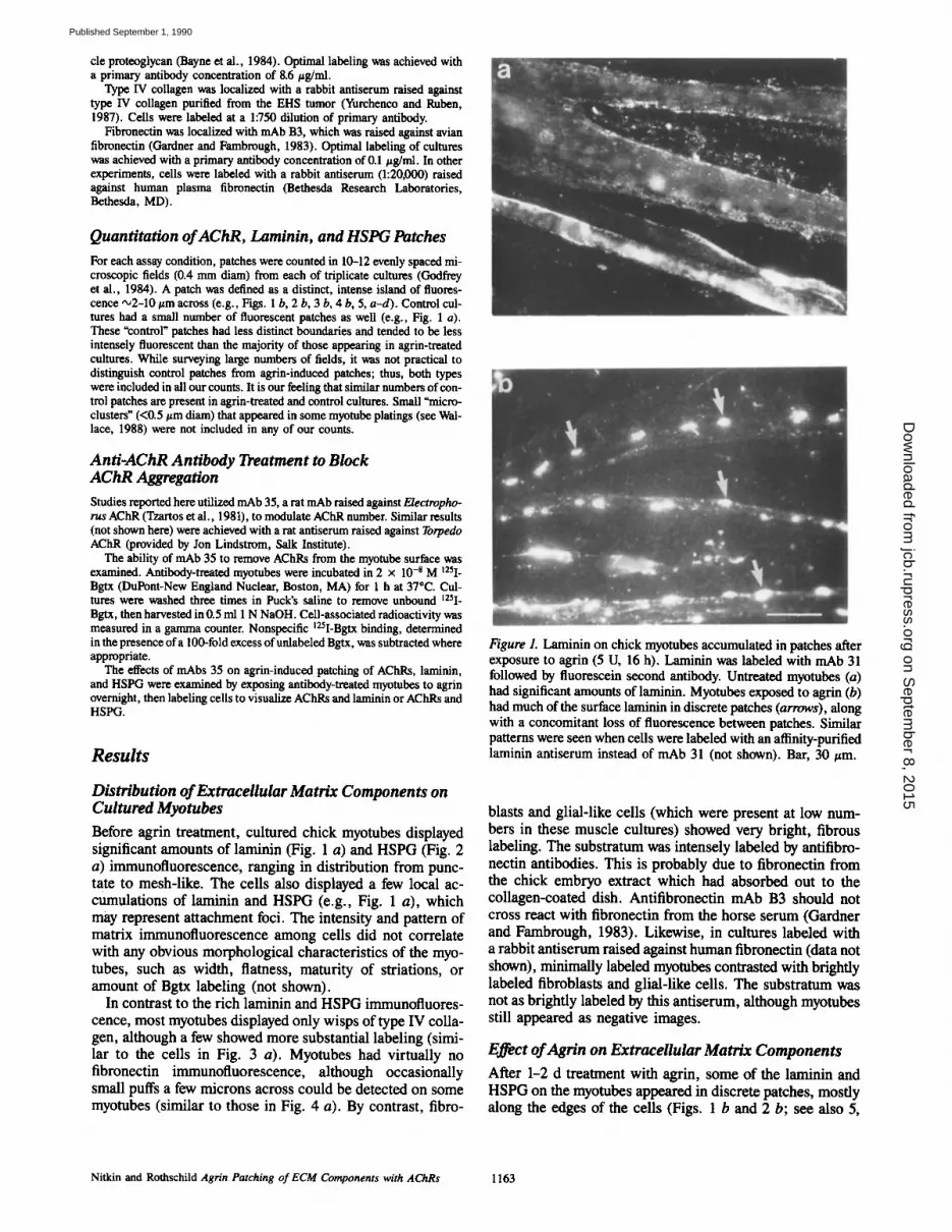
Before agrin treatment, cultured chick myotubes displayed significant amounts of laminin (Fig. 1 a) and HSPG (Fig. 2 a) immunofluorescence, ranging in distribution from punc- tare to mesh-like. The cells also displayed a few local ac- cumulations of laminin and HSPG (e.g., Fig. 1 a), which may represent attachment foci. The intensity and pattern of matrix immunofluorescence among cells did not correlate with any obvious morphological characteristics of the myo- tubes, such as width, flatness, maturity of striations, or amount of Bgtx labeling (not shown).
In contrast to the rich laminin and HSPG immunofiuores- cence, most myotubes displayed only wisps of type IV colla- gen, although a few showed more substantial labeling (simi- lar to the cells in Fig. 3 a). Myotubes had virtually no fibronectin immunofluorescence, although occasionally small puffs a few microns across could be detected on some myotubes (similar to those in Fig. 4 a). By contrast, fibro-
Figure L Laminin on chick myotubes accumulated in patches after exposure to agrin (5 U, 16 h). Laminin was labeled with mAb 31 followed by fluorescein second antibody. Untreated myotubes (a) had significant amounts of laminin. Myotubes exposed to agrin (b) had much of the surface laminin in discrete patches (arrows), along with a concomitant loss of fluorescence between patches. Similar patterns were seen when cells were labeled with an affinity-purified laminin antiserum instead of mAb 31 (not shown). Bar, 30 t~m.
blasts and glial-like cells (which were present at low num- bers in these muscle cultures) showed very bright, fibrous labeling. The substratum was intensely labeled by antifibro- nectin antibodies. This is probably due to fibronectin from the chick embryo extract which had absorbed out to the collagen-coated dish. Antifibronectin mAb B3 should not cross react with fibronectin from the horse serum (Gardner and Farnbrough, 1983). Likewise, in cultures labeled with a rabbit antiserum raised against human fibronectin (data not shown), minimally labeled myotubes contrasted with brightly labeled fibroblasts and glial-like cells. The substratum was not as brightly labeled by this antiserum, although myotubes still appeared as negative images.
Effect of Agrin on ExtraceUular Matrix Components
After 1-2 d treatment with agrin, some of the laminin and HSPG on the myotubes appeared in discrete patches, mostly along the edges of the cells (Figs. 1 b and 2 b; see also 5,
Nitkin and Rothschild Agrin Patching of ECM Components with AChRs 1163
on Septem
ber 8, 2015jcb.rupress.org
Dow
nloaded from
Published September 1, 1990

tially purified extracts using antiagrin mAb 11132 (Nitkin et al., 1987). The immunodepleted extract was not able to cause AChRs, laminin, or HSPG to cluster, while an extract passed over a column of control mouse serum retained clustering activity (data not shown).
Most of the agrin-induced AChR patches coincided with patches of laminin (Fig. 5, a and b) and HSPG (Fig. 5, c and d). The high level of correlation between AChRs and lami- nin/HSPG (63-74 %) did not improve with an additional day of agrin treatment (Table I). At some sites accumulations of laminin and HSPG were slightly more widespread than AChRs, although overall their distributions were remarkably congruent.
Agrin appears to have a selective effect on laminin and HSPG; it did not affect the distribution of type IV collagen
Figure 2. HSPG on chick myotubes accumulated in patches after exposure to agrin (5 U, 16 h). HSPG was labeled with mAb 33 fol- lowed by fluorescein second antibody. Untreated myotubes (a) had significant amounts of HSPG. Myotubes exposed to agrin (b) had much of the surface HSPG in discrete patches along the edge of the cell membrane (arrows), with a concomitant loss of fluorescence between patches. Bar, 20/zm.
a and c). The patching of laminin and HSPG appeared to be at the expense of other regions of the cell; that is, fluores- cence in away from patched regions was below the levels typically seen on untreated myotubes. Nonetheless, it was our impression that the total amounts of myotube-associated laminin and HSPG immunofluorescence were not signifi- cantly increased by this treatment. Thus, patching of matrix in response to agrin appears to involve redistribution rather than increased local synthesis or accumulation.
The morphology of the laminin and HSPG patches resem- bled that of the agrin-induced AChR patches (bright ag- gregates a few microns in diameter, with distinct edges). This allowed us to quantitate laminin and HSPG patching in much the same manner as AChR patching. Occasionally, cultures were found in which laminin and HSPG did not patch in response to agrin treatment, although AChR patch- ing appeared normal.
To demonstrate that laminin and HSPG accumulations were caused by agrin per se and not some other factor in the Torpedo extracts, agrin was specifically removed from par-
Figure 3. Agrin did not affect the distribution of type IV collagen on cultured myotubes. Myotubes treated with agrin (5 U, 48 h) were simultaneously labeled with antiserum specific for type IV collagen (followed by fluorescein second antibody) and rhodamine-Bgtx, as described in Materials and Methods. The same field was viewed through fluorescein filters to show type IV collagen (a) and rhoda- mine filters to show AChR patches (b). The distribution of collagen on myotubes, which varied from wispy to more dense (as shown here), showed no correspondence with AChR aggregations. A simi- lar range of collagen distributions was found on control myotubes (no agrin), or those exposed to agrin for only 16 h (not shown). Bar, 40 ~tm.
The Journal of Cell Biology, Volume 11 I, 1990 1164
on Septem
ber 8, 2015jcb.rupress.org
Dow
nloaded from
Published September 1, 1990

Figure 4. Myotubes had minimal amounts of fibronectin, which was largely unaffected by agrin treatment. Myotubes treated with agrin (5 U, 48 h) were simultaneously labeled with antifibronectin mAb B3 (followed by fluorescein second antibody) and rhodamine-Bgtx, as described in Materials and Methods. The same field was viewed under fluorescein optics to show the distribution of fibronectin (a) and under rhodamine optics to show agrin-patched AChRs (b). Myotubes had virtually no surface fibronectin, which caused them to appear as negative images contrasted against the brightly labeled substratum. The immunofluorescence of the substratum was proba- bly due to fibronectin from the medium that had absorbed to the dish, as discussed in the text. Generally there was no association of fibronectin with agrin-induced AChR patches; on rare occasions (as shown here) faint patches of fibronectin could be correlated with AChRs (arrows). Bar, 30/~m.
and had little, if any, effect on fibronectin. Even after 48 h of agrin treatment, collagen was still found in wispy patterns which bore no relation to the patched AChRs (Fig. 3, a and b). The amount of myotube-associated fibronectin remained minimal after agrin treatment, and generally did not corre- late with AChR accumulations (Fig. 4, a and b). Occasion- ally, small puffs of fibronectin could be correlated with AChR patches (Fig. 4 a, arrows), but such instances were r a r e .
Temporal Relationship of AChR and Laminin/HSPG Patching The colocalization of laminin and HSPG with AChR patches
raised the possibility of a causal relationship between matrix accumulation and AChR patching. This was explored by comparing the rate of AChR patching with that of laminin and HSPG (Fig. 6). After treatment with agrin, increased numbers of AChR patches could be detected after only 2-3 h, reaching maximal levels by '~6-8 h (see also Godfrey et al., 1984). However, the increase in the number of visible lami- nin and HSPG patches occurred much more slowly. By the time AChR patching had reached maximal levels, the num- bers of laminin and HSPG patches were only about halfway complete; they required ~20 h to reach maximal plateau lev- els. While these observations do not focus on the earliest stages of agrin-induced AChR and matrix accumulation, which may be below the level of visual detection, they show that AChR patching is completed hours before that of either laminin or HSPG. This temporal relationship makes it un- likely that extracellular matrix directs AChR patching at agrin-induced sites.
Dissociation of AChR Patching from that of Laminin and HSPG To determine if accumulation of AChRs is necessary for agrin-induced patching of laminin and HSPG, we used the anti-AChR antibody, mAb 35, to block AChR patching. In other studies, AChR antibodies have been shown to decrease both the number and mobility of surface AChRs (Heinemann et al., 1977; Appel et al., 1977; Kao and Drachman, 1977). Direct binding tests (not shown) on alcohol-fixed myotubes (data not shown) demonstrated that mAb 35 does not prevent Bgtx from binding to available AChRs (also Tzartos et al., 1981).
The number of surface AChRs remaining after incubation in various concentrations of mAb 35 was quantitated with J25I-Bgtx (Table /I). All three concentrations of mAb 35 were effective at reducing the number of AChRs. 39 aM anti- body was sufficient to achieve maximal effects (only 39 % of the AChRs remained). Higher concentrations of antibody (130 nM) were no more effective (38% remained). In the presence of agrin, mAb 35 was still able to reduce the num- ber of surface AChRs, although the effect was slightly less (45-54 % of the AChRs remained). A similar percentage of AChRs were removed when the myotubes were treated with a rat antiserum raised against Torpedo AChR (not shown).
To examine the effect of agrin on the population of AChRs remaining after antibody treatment, myotubes were exposed to mAb 35 and agrin, then labeled with rhodamine-Bgtx. While antibody treatment reduced total AChR fluorescence (as expected due to the partial internalization of surface AChRs) most of the AChRs remaining were not patched by agrin treatment (Table II). The ability of mAb 35 to block agrin-induced patching was dose dependent. [3 nM mAb was sufficient to block the appearance of the majority of agrin-induced patches, while higher concentrations (39 and 130 nM) reduced patching to <5%. Based on these experi- ments, it appears that mAb 35 concentrations of 39 nM or more would be sufficient to block virtually all visually de- tectable agrin-induced AChR patching.
With AChR patching blocked by antibody treatment, we examined whether agrin could still induce patching of lami- nin and HSPG. Myotubes were incubated with 78 nM mAb 35 and/or agrin, then labeled to visualize AChRs and laminin or AChRs and HSPG (Table III).
Nitkin and Rothschild Agrin Patching of ECM Components with AChRs 1165
on Septem
ber 8, 2015jcb.rupress.org
Dow
nloaded from
Published September 1, 1990

Figure 5. Laminin and HSPG accumulated at agrin-induced AChR patches. Agrin-treated (5 U, 24 h) myotubes were simultaneously labeled with mAbs against larninin or HSPG (followed by fluorescein second antibody) and rhodamine-Bgtx. In the upper pair, the same field was viewed through fluorescein and rhodamine filters to highlight the coincidence of laminin (a) and AChR (b) accumulations (arrows) on myotubes. In the lower pair, a field from another culture shows the coincidence of HSPG (c) and AChR (d) accumulations (arrows). Bar, 25 pro.
As expected, agrin treatment alone induced significant patching of all three components; this maximal response was defined as 100%. Cells with no additions had few patches of these components; this level of patching (which probably
Table L Appearance of Laminin and HSPG at Agrin-induced AChR Patches
Length of agrin AChR patches AChR patches exposure with laminin with HSPG
th) 20 - 223/302 (74%) 41 423/617 (69%) 64/101 (63%) 65 294/404 (73%)
Cultures were exposed to agrin (5-10 13) for the times indicated, then simul- taneously labeled for AChRs and laminin or AChRs and HSPG. Individual fields were viewed repeatedly, first under rhodamine optics to identify AChR patches (up to three at a time), then under fluorescein optics to determine whether laminin or HSPG had accumulated at these sites. Data are presented as fractions: denominator indicates total number of AChR patches examined and numerator indicates those that had coaccumulations of laminin or HSPG.
includes attachment foci) was defined as 0%. Cultures treated with mAb 35 alone (no agrin) had even fewer AChR patches than control (<0%), suggesting that antibody treat- ment even prevented AChRs from accumulating at attach- ment foci. Anti-AChR antibody had no significant effect on the amount or distribution of laminin or HSPG immuno- fluorescence in nonagrin-treated cultures. In the presence of both mAb 35 and agrin, virtually no AChR patches could be seen, but significant numbers of laminin (91%) and HSPG (64%) patches were still detectable (Table llI).
Long exposure photographs (10-15 s) of these cultures re- vealed a slightly different story (Fig. 7). The increased sensi- tivity of this technique showed that in mAb 35/agrin-treated cultures, low density accumulations of AChRs did occur at some laminin and HSPG patches; other patches were devoid of AChRs even at this level of scrutiny. This indicates that while mAb 35 treatment severely reduced AChR patching (so that it was not evident through simple visual inspection), it did not prevent AChR patching completely. Agrin-induced patching of laminin and HSPG was largely unaffected by the
The Journal of Cell Biology, Volume 1 ! 1, 1990 1166
on Septem
ber 8, 2015jcb.rupress.org
Dow
nloaded from
Published September 1, 1990

40 M.
m G
.o 211 m a.
i -
o o 2'0 ,o 8'0
Hours of Agrin Treatment 8 0
m Q
u. 30
0 r -
a.
~" 10
e-
c
E 0 .,..1
B
0 2'0 40 6'0 8 0 Hours of Agrin Treatment
Figure 6. Agrin-induced accumulations of AChRs occurred much more rapidly than those of laminin or HSPG. Muscle cultures were exposed to agrin (5 U) for various amounts of time indicated on the abscissa, then labeled to visualize the distributions of either AChRs (a, circles~solid line), laminin (b, squares~solid line) or HSPG (b, triangles~dotted line). Each time point is the mean number of patches per microscopic field, derived from 8-12 fields per dish, using triplicate dishes; vertical error bars show SEM among those dishes. In each experiment, the addition of agrin was staggered so that all cultures were labeled and counted together; thus, all deter- minations were made on the same age cultures. The upper and lower graphs were derived from experiments on two different mus- cle platings, resulting in different plateau levels for AChR and lami- nin/HSPG patching.
antibody treatment (Table HI), suggesting that matrix ac- cumulations do not require the presence of dense AChR patches.
Discussion
The experiments described here focus on the role of extracel- lular matrix components in synaptic differentiation. We used immunohistochemical techniques to follow the distribution of laminin, HSPG, fibronectin, and type IV collagen on cul- tured chick myotubes. In agreement with several other im- munohistochemical studies (e.g., Kuhl et al., 1982; Ander- son and Fambrough, 1983; Gardner and Fambrough, 1983; Daniels et al., 1984), we found that cultured myotubes have on their surface laminin, HSPG, and type IV collagen, but little if any fibronectin.
We found that matrix accumulation at agrin-induced sites was selective. Laminin and HSPG colocalized with AChRs but fibronectin and type IV collagen distributions were not
Table II. Anti-AChR Treatment Removes Surface AChRs and Prevents Agrin-induced AChR Patching
Number of AChRs AChR patches Treatment (fmol/culture) (per field)
No additions 77 + 0.4 100% 2.3 + 0.8 0% 13 nM mAb 35 45 + 3.3 58 - - 39 nM mAb 35 30 + 1.1 39 - - 130 nM mAb 35 29 + 2.0 38 - -
Agrin alone 79 + 3.0 103 20.2 + 2.8 100 Agrin + 13nMmAb35 42 +2 .1 55 7.9 +4 .0 27 Agrin + 39nMmAb35 42 + 0 . 4 54 2.9 4- 1.2 3 Agrin + 130nMmAb35 35 + 0 . 7 45 3.3 +0 .9 5
Myotubes were incubated for 48 h with agrin (5 U) and/or mAb 35 (at concen- trations indicated), ml-Bgtx binding was used to determine the number of AChRs on the myotube surface (nonspecific binding has been subtracted); cul- tures with no additions were defined as 100%. In sister cultures, cells were la- beled with rhodamine-Bgtx so that AChR patches could be counted; cultures with no additions were defined as 09~, while cultures treated with agrin alone were defined as 100%. Each entry is the mean + SEM (n = 3) of triplicate cultures.
affected (agrin-induced HSPG aggregations have also been reported by Wallace, 1989), At this time we cannot deter- mine whether laminin and HSPG patching represent lateral migration of matrix molecules already present on the cell surface or selective placement of newly synthesized or newly bound material. It appears that agrin does not significantly alter the total amount of surface laminin or HSPG, as judged by immunofluorescence.
Molecules like agrin could be responsible for coordinating the localization of extracellular matrix material with synap- tic components at sites in vivo, much as it does in culture. Agrin-like molecules have been detected at neuromuscular junctions from the earliest stages of development (Godfrey et al., 1988b; Fallon and Gelfman, 1989). Furthermore, ini- tial patches of AChRs are associated with laminin, HSPG, and type IV collagen, but not fibronectin (Anderson and Fambrough, 1983; Chiu and Sanes, 1984; Bayne et al., 1984).
Table IlL Although AChR Patching Is Prevented by Antibody Treatment, Agrin Can Still Cause Laminin and HSPG to Patch
AChR Laminin HSPG Treatment patching patching patching
(%) (%) (%)
No additions 0 0 0 Agrin alone 100 100 100 mAb 35 alone (<0) 17 + 9 11 + 5 Agrin + mAb 35 10 4- 10 91 + 20 64 + 18
Myotubes were incubated for 48 h with 78 nM anti-AChR mAb 35 and/or agrin (3 U), then labeled to visualize the distribution of AChRs, laminin, and HSPG. Because each muscle plating bad inherently different levels of response, the numbers of AChR, laminin, and HSPG patches in different experiments could not be averaged directly. Cultures (treated in quadruplicate) were normalized against controls with no additions (0%) and cultures exposed to agrin (100%) to derive percent response. Percentages pooled from five experiments are presented above (mean + SEM, n = 5). In the case of "mAb 35 alone," anti- body treatment eliminated some of the spontaneous (background) AChR patches, resulting in even less AChR patches than control (<0%). In the pres- ence of'Agrin + mAb 35," significant patching of laminin and HSPG occurred despite the loss of AChRs. (Long exposure photographs revealed that low den- sity accumulations of AChRs were present at some laminin and HSPG patches, see Fig. 7.)
Nitidn and Rothschild Agrin Patching of ECM Components with AChRs 1167
on Septem
ber 8, 2015jcb.rupress.org
Dow
nloaded from
Published September 1, 1990

Figure 7. Laminin and HSPG accumulated at agrin-induced patches even though AChRs were blocked with antibodies. Myotubes were incubated for 48 h with 78 nM anti-AChR mAb 35 (to remove or immobilize AChRs) and agrin (3 13). The cultures were simultaneously labeled to visualize laminin or HSPG, and AChRs. In the upper pair, the same field was viewed through fluorescein and rhodamine filters to contrast the distribution of laminin (a) and AChRs (b), respectively. In the lower pair, a field from another culture contrasts HSPG (c) with AChRs (d). Because of the rnAb 35 treatment, only minimal accumulations of AChRs appeared at laminin or HSPG patches (ar- rows). To achieve adequate contrast in b and d, film exposures were about two times longer compared to other rhodamine-labeled cultures (e.g., Figs. 3 b, 4 b, 5 b, and d); adjustments in the developing process were also made. While surveying large numbers of fields by direct visual observations alone (e.g., Table liD, faint AChR patches such as those shown above (b and d) lack sufficient contrast to be detected. Bar, 40/~m.
In this study, AChR accumulations and matrix differentia- tion were followed by fluorescence microscopy. While this technique is effective for monitoring relative distributions of components, it provides only a qualitative description of ac- tual site density. Indirect immunofluorescence makes quan- titative comparisons even more difficult, although we have tried to optimize binding of primary and fluorescein second- ary antibodies. The earliest stages of AChR/matrix accumu- lations could go undetected by these techniques. Agrin itself appears to function catalytically in the sense that only a few hundred molecules are sufficient to induce a patch contain- ing tens of thousands of AChRs (Nitkin et al., 1987).
On agrin-treated myotubes, AChR patching was com- pleted quite a few hours ahead of laminin/HSPG patching (Fig. 6). While it is possible that small amounts of laminin and/or HSPG actually precede AChRs at some agrin- induced sites, our results indicate that the distribution of AChRs matures into dense discrete patches hours before that of either laminin or HSPG. Thus, unless matrix accumula- tion is a multi-step process, it is unlikely that it directs the placement of AChRs.
Conversely, we considered the possibility that agrin-in- duced patching of AChRs is necessary to attract extracellular matrix components (AChRs do have extensive extracellular domains which could potentially interact with matrix mate- rial). We developed a means of specifically blocking agrin- induced AChR aggregation using the anti-AChR antibody, mAb 35. The reduction in AChR number is probably due to accelerated removal of AChRs from the myotube surface through antibody-mediated internalization. This process in- volves cross-linking of mobile surface receptors by a mul- tivalent ligand which rapidly leads to patching, capping, and ultimately internalization of bound receptors, mAb 35 was able to internalize AChRs without secondary antibodies; this may be due to the fact that each of the two alpha subunits of the AChR offers a potential site to allow for antibody cross-linking (Tzartos et al., 1981). AChRs were likewise in- ternalized by treatment with an antiserum raised against Torpedo AChR (not shown).
The AChRs that remain on the myotube surface after anti- body treatment, ~40% in our studies (Table II), may repre- sent a population of less mobile receptors. Even on myotubes
The Journal of Cell Biology, Volume 111, 1990 1168
on Septem
ber 8, 2015jcb.rupress.org
Dow
nloaded from
Published September 1, 1990

not exposed to antibodies, a significant number of AChRs did not participate in agrin-induced patching (note diffuse rhoda- mine fluorescence between AChR patches, especially in Figs. 3 b and 4 b). The presence of a population of less mo- bile AChRs has been suggested by photobleaching studies (e.g. Axelrod et al., 1976). Thus, it is not surprising that the population of AChRs remaining after antibody treatment was not readily patched by agrin treatment (Table II).
Long exposure photographs (Fig. 7, b and d) revealed that while AChR patching is significantly reduced by antibody treatment, a small number of AChRs still appear at agrin- induced sites. Over the 48-h antibody/agrin treatment, it is possible that some AChRs were not bound by mAb 35 or bound in a configuration that did not allow for cross-linking. These AChRs would then avoid immobilization/internaliza- tion and would be able to migrate into patches. We have not investigated whether AChRs that reach agrin-induced patches are immune to subsequent mAb treatment.
Although mAb 35 had dramatic effects on AChR patching, it had much less effect on the number of agrin-induced ma- trix patches (Table I/I). The data pooled from five experi- ments indicate that laminin and HSPG were decreased only 9 and 36 %, respectively, with significant variation among in- dividual experiments. In some experiments, the full comple- ment of agrin-induced matrix patches were present despite the virtual elimination of dense AChR patches by mAb35. The reason for this variability is unclear; it could be that prolonged exposure to antibodies (48 h) and subsequent AChR patching and capping begins to affect the distribution of other membrane receptors. Alternatively, it could suggest that there is some minimal direct connection between AChRs and laminin/HSPG localization. In vivo, AChR accumula- tions can be uncoupled from matrix differentiation. As mus- cles get innervated, AChRs are recruited away from matrix- associated regions to new sites beneath developing nerve terminals (Weinberg et al., 1981; Chiu and Sanes, 1984). Similarly, in culture AChRs that are initially associated with HSPG (and probably other matrix components) migrate to developing neuromuscular junctions, leaving much of the HSPG behind (Anderson et al., 1984; Anderson, 1986).
Redistribution of extracellular matrix components could be brought about by a variety of mechanisms. Our results suggest that the accumulation of laminin and HSPG at agrin- induced sites is not stoichiometrically linked to the accumu- lation of AChRs (Table I1/). It is possible that matrix compo- nents accumulate in response to the recruitment of specific cell surface matrix receptors (reviewed by Buck and Hor- witz, 1987). For example, the myotube could localize inte- grins or other laminin receptors, causing extracellular laminin to follow. HSPG could be attracted to these sites because of specific proteoglycan-binding domains on laminin (Sakashita et al., 1980). This would account for the striking similarity between the time courses of laminin and HSPG accumula- tions (Fig. 6 b).
ACILR patching is completed before that of laminin or HSPG, suggesting that matrix accumulation does not initiate %ynaptic" differentiation. Nonetheless, the matrix may have an important role in the maintenance of synaptic structure (Nitkin et al., 1987). The accumulation of matrix material at developing synapses could attract additional material (in- cluding more agrin), which would further enhance synaptic differentiation in that region. Such a reinforcing mechanism
could be used to stabilize developing synapses as well as strengthen especially active synapses. This could have im- portant implications for the process of polyneuronal synapse elimination. Furthermore, if analogous mechanisms operate in the central nervous system, it could serve to reinforce specific pathways during learning.
We would like to thank Drs. Edward Bonder and Peter Yurchenco for many helpful discussions and encouragement. We also thank Lorraine Sopko for technical assistance in several aspects of this work, and Harbans Kaur for preparing myotube cultures. In addition, we are grateful to Drs. Douglas Fambrough, John Hassell, Hynda Klcinman, Jon Lindstrom, and Peter Yurchenco for sharing antibodies and other reagents.
This work was supported by National Science Foundation Grant BNS 87-07530-02 and Biomedical Research Support Grant PHS RR 07059-22.
Received for publication 20 November 1989 and in revised form 27 April 1990.
References
Anderson, M. J. 1986. Nerve-induced remodeling of muscle basal lamina dur- ing synaptogenesis. J. Cell Biol. 102:863-877.
Anderson, M. J., and D. M. Fambrough. 1983. Aggregates of acetylcholine receptors are associated with plaques of basal lamina heparan sulfate pro- teoglycan on the surface of skeletal muscle fibers. J. Cell Biol. 97: 1396-1411.
Anderson, M. J., F. G. Klier, and K. E. Tanguay. 1984. Acetylcholine receptor aggregation parallels the deposition of a basal lamina proteoglycan during development of the neuromuscular junction. J. Cell Biol. 99:1769-1784.
Anglister, L., and U. J. McMahan. 1985. Basal lamina directs acetyl- cholinesterase accumulation at synaptic sites in regenerating muscle. J. Cell Biol. 101:735-743.
Appel, S. H., R. Anwyi, M. W. McAdams, and S. Elias. 1977. Accelerated degradation of acetylcholine receptors from cultured rat myotobes with my- asthenia gravis sera and globulins. Proc. Natl. Acad. Sci. USA. 74:2130- 2134.
Axelrod, D., P. Ravdin, D. E. Koppel, J. Schlessinger, W. W. Webb, E. L. Elson, and T. R. Podleski. 1976. Lateral motion of fluorescently labeled ace- tylcholine receptors in membranes of developing muscle fibers. Proc. Natl. Acad. Sci. USA. 75:2035-2039.
Bader, D. 1981. Density and distribution of alpha-bungarotoxin binding sites in postsynaptic structures of regenerated rat skeletal muscle. J. Cell Biol. 88:338-345.
Bayne, E. K., M. J. Anderson, and D. M. Fambrough. 1984. Extracellular ma- trix organization in developing muscle: correlation with acetylcholine recep- tor aggregates. J. Cell Biol. 99:1486-1501.
Bloch, R. J., D. W. Pumplin, and M. Baetscher. 1985. Acetylcholine receptor clustering and the formation of the neuromuscular junction. In Cellular and Molecular Control of Direct Cell Interactions. H.-J. Morthy, editor. Plenum Publishing Corp., New York. 239-258.
Buchanan, J., Y.-A. Sun, and M.-M. Poo. 1989. Studies of nerve-muscle inter- actions in Xenopus cell culture: fine structure of early functional contacts. J. Nearosci. 9(5):1540-1554.
Buck, C. A., and A. F. Horwitz. 1987. Cell surface receptors for extracellular matrix molecules. Annu. Rev. Cell Biol. 3:179-205.
Burden, S. J., P. B. Sargent, and U. J. McMahan. 1979. Acetylcholine recep- tors in regenerating muscle accumulate at original synaptic sites in the ab- sence of nerve. J. Cell Biol. 82:412-425.
Burrage, T. G., and T. Lentz. 1981. Ultrastructural characterization of surface specializations containing high-density acetylcholine receptors on embryonic chick myotubes in vivo and in vitro. Dev. Biol. 85:267-286.
Chiu, A. Y., and J. R. Sanes. 1984. Development of basal lamina in synaptic and extrasynaptic portions of embryonic rat muscle. Dev. Biol. 103:456- 467.
Daniels, M. P., M. Vigny, P. Sonderegger, H. C. Bauer, and Z. Vogel. 1984. Association of laminin and other basement membrane components with regions of high acetylcholine receptor density on cultured myotubes. Int. J. Dev. Neurosci. 2:87-99.
Dennis, M. J. 1981. Development of the neuromuscular junction: inductive in- teractions between cells. Annu. Rev. Neurosci. 4:43--68.
Fallon, L R., and C. E. Gelfman. 1989. Agrin-related molecules are concen- trated at acetylcholine receptor clusters in normal and anearal developing muscle. J. Cell Biol. 108:1527-1535.
Fallun, L R., R. M. Nitkin, N. E. Reist, B. G. Wallace, and U..I. McMahan. 1985. Acetylcholine receptor-aggregating factor is similar to molecules con- centrated at neuromuscular junctions. Nature (Lond.). 315:571-574.
Fambrough, D. M. 1979. Control of acetylcholine receptors in skeletal muscle. Physiol. Rev. 59:165-227.
Fischbach, G. D. 1972. Synapse formation between dissociated nerve and mus-
Nitkin and Rothschild Agrin Patching of ECM Components with AChRs 1169
on Septem
ber 8, 2015jcb.rupress.org
Dow
nloaded from
Published September 1, 1990

cle cells in low density cultures. Dev. BioL 28:407-429. Gardner, J. M., and D. M. Fambrough. 1983. Fibronectin expression during
myogenesis. J. Cell Biol. 96:474-485. Godfrey, E. W., R. M. Nitkin, B. G. Wallace, L. L. Rubin, and U. J. McMa-
hun. 1984. Components of Torpedo electric organ and muscle that cause aggregation of acetylcholine receptors on cultured muscle cells. J. Cell Biol. 99:615-627.
Godfrey, E. W., M. E. Dietz, A. L. Morstad, P. A. Wallskog, and D. Yorde. 1988a. Acetylcholine receptor-aggregating proteins are associated with the extracellular matrix of many tissues in Torpedo. J. Cell BioL 106:1263- 1272.
Godfrey, E. W., R. E. Siebenlist, P. A. Wallskog, L. M. Waiters, D. L. Bolender, and D. E. Yorde. 1988b. Basal lamina components are concen- trated in premuscle masses and at early acetylcholine receptor clusters in chick embryo hindlimb muscles. Dev. Biol. 130:471-486.
Heinemann, S., S. Bevan, R. Kullberg, J. Lindstrom, and J. Rice. 1977. Modu- lation of the acetylcholine receptor by anti-receptor antibody. Proc. Natl. Acad. Sci. USA. 74:3090-3094.
Jacob, M., and T. Lentz. 1979. Localization of acetylcholine receptors by means of horseradish peroxidase-bungarotoxin during formation and devel- opment of neuromuscular junctions in the chick embryo. J. Cell BioL 82:195-211.
Kao, I., and D. B. Drachman. 1977. Myasthenic immunoglobulin accelerates acetylcholine receptor turnover. Science (Wash. DC). 196:527-529.
Kelly, A. M., and S. I. Zacks. 1969. The fine structure of motor endplate mor- phogenesis. J. Cell Biol. 42:154-169.
Kuhl, U., R. Timpl, and K. yon der Mark. 1982. Synthesis of type IV collagen and laminin in cultures of skeletal muscle cells and their assembly on the sur- face of myotubes. Dev. Biol. 93:344-354.
Kullberg, R. W., T. L. Lentz, and M. W. Cohen. 1977. Development of myo- tomal neuromuscular junction in Xenopus laevis: an electrophysiological and fine structural study. Dev. Biol. 60:101-129.
McMahan, U. J., and C. R. Slater. 1984. The influence of basai lamina on the accumulation of acetylcholine receptors at synaptic sites on regenerating muscles. J. Cell Biol. 98:1453-1473.
Nakajima, Y., Y. Kidokoro, and F. G. Klier. 1980. The development of func- tionai neuromuscular junctions in vitro: an ultrastructural and physiological study. Dev. Biol. 77:52-72.
Nitkin, R. M., B. G. Wallace, M. E. Spira, E. W. Godfrey, and U. J. McMa- hun. 1983. Molecular components of the synaptic basal lamina that direct differentiation of regenerating neuromuscular junctions. CoMSpring Harbor Syrup. Quant. Biol. 48:653-665.
Nitkin, R. M., M. A. Smith, C. MagiU, J. R. Fallon, Y.-M. M. Yao, B. G. Wallace, and U. J. McMahan. 1987. Identification of agrin, a synaptic or- ganizing protein from Torpedo electric organ. J. Cell Biol. 105:2471-2478.
Olek, A. J., A. Ling, and M. P. Daniels. 1986. Development of ultrastructural specializations during formation of acetylcholine receptor aggregates on cul- tured myotubes. J. Neurosci. 6:487-497.
Orida, N., and M.-M. Poo. 1978. Electrophoretic movement and localisation of acetylcholine receptors in embryonic muscle cell membrane. Nature (Lond.). 275:31-35.
Peng, H. B. 1987. Development of the neuromuscular junction in culture. CRC Crit. Rev. Anat. Sci. 1:91-131.
Peng, H. B., and P. C. Cheng. 1982. Formation of postsynaptic specializations induced by latex beads in cultured muscle cells. J. Neurosci. 2:1760-1774.
Puck, T. T., S. J. Ciecura, and A. Robinson. 1958. Genetics of somatic mam- malian cells. IU. Long-term cultivation of euploid cells from human and ani- mal species. J. Exp. Med. 108:945-955.
Ravdin, P., and D. Axelrod. 1977. Fluorescent tetramethyl rhodamine deriva- tives of alpha-bungarotoxin: preparation, separation and characterization.
Anal. Biochem. 80:585-592. Reist, N. E., C. Magill, and U. J. McMahan. 1987. Agrin-like molecules at
synaptic sites in normal, denervated and damaged skeletal muscles. J. Cell Biol. 105:2457-2469.
Rubin, L. L., and U. J. McMahan. 1982. Regeneration of the neuromuscular junction: steps toward defining the molecular basis of the interaction between nerve and muscle. In Disorders of the Motor Unit. D. L. Schotland, editor. John Wiley & Sons, New York. 187-196.
Rubin, L. L., and K. F. Barald. 1983. Neuromuscular development in tissue culture. In Somatic and Autonomic Nerve-Muscle Interactions. G. Bum- stock, R. O'Brien, and G. Vrbova, editors. Elsevier Science Publishing Co., Inc., North Holland. 109-151.
Sakashita, S., E. Engvall, and E. Ruoslahti. 1980. Basement membrane glyco- protein laminin binds to heparin. FEBS (Fed. Fur. Biochem. Soc.) Lett. 116:243-246.
Salpeter, M. M., and R. H. Loring. 1985. Nicotinic acetylcholine receptors in vertebrate muscle: properties, distribution and neural control. Prog. Neu- robiol. (NY) 25:297-325.
Salpeter, M. M., S. Spanton, K. Holley, and T. R. Podleski. 1982. Brain ex- tract causes acetylcholine receptor redistribution which mimics some early events at developing neuromuscular junctions. J. Cell Biol. 93:417-425.
Sanes, J. R. 1989. Extracellular matrix molecules that influence neural develop- ment. Anna. Rev. Cell Biol. 12:491-516.
Sanes, J. R., L. M. Marshall, and U. J. McMahan. 1978. Reinnervation of mus- cle fiber basal lamina after removal of myofibers. J. Cell Biol. 78:176-198.
Sanes, J. R., D. H. Feldman, J. M. Cheney, and J. C. Lawrence. 1984. Brain extract induces synaptic characteristics in the basal lamina of cultured myo- tubes. J. Neurosci. 4:464-473.
Schuetze, S. M., and L. W. Role. 1987. Developmental regulation of nicotinic acetylcholine receptors. Anna. Rev. Neurosci. 10:403-457.
Steinbach, J. H., and R. J. Bloch. 1986. The distribution of acetylcholine recep- tors on vertebrate skeletal muscle cells. In Receptors in Cellular Recognition and Developmental Processes. R. Gorczynski, editor. Academic Press, New York. 183-213.
Tzartos, S. J., D. E. Rand, B. L. Einarson, andJ. M. Lindstrom. 1981. Map- ping of surface structures of Electrophorus acetylcholine receptor using monoclonal antibodies. J. Biol. Chem. 256:8635-8645.
Wallace, B. G. 1986. Aggregating factor from Torpedo electric organ induces patches containing acetylcholine receptors, acetylcholinesterase, and butyrylcholinesterase on cultured myotubes. J. Cell Biol. 102:783-794.
Wallace, B. G. 1988. Regulation of agrin-induced acetylcholine receptor aggre- gation by Ca ++ and phorbol ester. J. Cell Biol. 107:267-278.
Wallace, B. G. 1989. Agrin-induced specializations contain cytoplasmic, mem- brane, and extraeellular matrix-associated components of the postsynaptic apparatus. J. Neurosci. 9:1294-1302.
Wallace, B. G., R. M. Nitkin, N. E. Reist, J. R. Failon, N. N. Moayeri, and U. J. McMahan. 1985. Aggregates of acetylcholinesterase induced by ace- tylcholine receptor-aggregating factor. Nature (Lond.). 315:574-577.
Weinberg, C. B., J. R. Sanes, and Z. W. Hail. 1981. Formation of neuromuscu- lar junctions in adult rats: accumulation of acetylcholine receptors, acetyl- cholinesterase and components of synaptic basal lamina. Dee. Biol. 84: 255-266.
Weldon, P. R., and M. W. Cohen. 1979. Development of synaptic ultrastruc- ture at neuromuscular contacts in an amphibian cell culture system. J. New rocytol. 8:239-259.
Yurchenco, P. D., and G. C. Ruben. 1987. Basement membrane structure in situ: evidence for lateral associations in the type IV collagen network. J. Cell Biol. 105:2559-2568.
Yurehenco, P. D., E. C. Tsilibary, A. S. Charonis, and H. Furthmayr. 1985. Laminin polymerization in vitro. J. Biol. Chem. 260:7636-7644.
The Journal of Cell Biology, Volume 111, 1990 1170
on Septem
ber 8, 2015jcb.rupress.org
Dow
nloaded from
Published September 1, 1990


