Embed Size (px)
Citation preview
Allosteric MEK1/2 InhibitorRefametinib (BAY 86-9766)in Combination withSorafenib Exhibits AntitumorActivity in PreclinicalMurine and Rat Models ofHepatocellular Carcinoma1,2
Roberta Schmieder*,3, Florian Puehler*,3,Roland Neuhaus*, Maria Kissel*, Alex A. Adjei†,Jeffrey N. Miner‡, Dominik Mumberg*,Karl Ziegelbauer* and Arne Scholz*
*Global Drug Discovery, Bayer HealthCare, Berlin, Germany;†Roswell Park Cancer Institute, Buffalo, NY;‡Ardea Biosciences, Inc, San Diego, CA
AbstractOBJECTIVE: The objectives of the study were to evaluate the allosteric mitogen-activated protein kinase kinase (MEK)inhibitor BAY 86-9766 in monotherapy and in combination with sorafenib in orthotopic and subcutaneous hepato-cellular carcinoma (HCC) models with different underlying etiologies in two species. DESIGN: Antiproliferative poten-tial of BAY 86-9766 and synergistic effects with sorafenib were studied in several HCC cell lines. Relevant pathwaysignaling was studied in MH3924a cells. For in vivo testing, the HCC cells were implanted subcutaneously or ortho-topically. Survival and mode of action (MoA) were analyzed. RESULTS: BAY 86-9766 exhibited potent antiproliferativeactivity in HCC cell lines with half-maximal inhibitory concentration values ranging from 33 to 762 nM. BAY 86-9766was strongly synergistic with sorafenib in suppressing tumor cell proliferation and inhibiting phosphorylation of theextracellular signal–regulated kinase (ERK). BAY 86-9766 prolonged survival in Hep3B xenografts, murine Hepa129allografts, and MH3924A rat allografts. Additionally, tumor growth, ascites formation, and serum alpha-fetoproteinlevels were reduced. Synergistic effects in combination with sorafenib were shown in Huh-7, Hep3B xenografts,and MH3924A allografts. On the signaling pathway level, the combination of BAY 86-9766 and sorafenib led to inhib-ition of the upregulatory feedback loop toward MEK phosphorylation observed after BAY 86-9766 monotreatment.With regard to the underlying MoA, inhibition of ERK phosphorylation, tumor cell proliferation, and microvessel den-sity was observed in vivo. CONCLUSION: BAY 86-9766 shows potent single-agent antitumor activity and acts syner-gistically in combination with sorafenib in preclinical HCC models. These results support the ongoing clinicaldevelopment of BAY 86-9766 and sorafenib in advanced HCC.
Neoplasia (2013) 15, 1161–1171
Abbreviations: HCC, hepatocellular carcinoma; HBV, hepatitis B virus; HCV, hepatitis C virus; VEGFRs, vascular endothelial growth factor receptors; OS, overall survival;AFP, alpha-fetoproteinAddress all correspondence to: Florian Puehler, PhD, Bayer Pharma AG, Muellerstr. 178, 13353 Berlin, Germany. E-mail: [email protected] work was supported by Bayer HealthCare (Berlin, Germany) and Ardea Biosciences (San Diego, CA). Writing assistance was provided by Synapse Medical Commu-nications (New York, NY). R.S., F.P., R.N., M.K., D.M., K.Z., and A.S. are stockholders and/or employees of Bayer Pharma AG. Prior presentations: Part of this work waspresented at the American Association for Cancer Research–National Cancer Institute–European Organisation for Research and Treatment of Cancer meeting in San Francisco onNovember 12 to 16, 2011.2This article refers to supplementary materials, which are designated by Figures W1 to W3 and are available online at www.neoplasia.com.3These authors contributed equally to this work.Received 22 April 2013; Revised 9 September 2013; Accepted 9 September 2013
Copyright © 2013 Neoplasia Press, Inc. All rights reserved 1522-8002/13/$25.00DOI 10.1593/neo.13812
www.neoplasia.com
Volume 15 Number 10 October 2013 pp. 1161–1171 1161
IntroductionHepatocellular carcinoma (HCC) is the major histologic subtype of pri-mary liver cancer, accounting for 70% to 85% of cases inmost countries[1]. In 2008, liver cancer was diagnosed in an estimated 748,300 peopleand was responsible for an estimated 695,900 deaths, with the highestrates found in eastern and southeastern Asia and in central and westernAfrica [2]. Although liver cancer is less common in Europe than in partsof Asia and Africa, the incidence in the West is increasing [2,3].High 5-year survival rates can be achieved in selected patients with
preserved liver function by using partial hepatectomy in early-stageHCC, ablative therapy in locoregional disease, and liver transplantationin unresectable disease [4]. However, HCC constitutes a significantunmet medical need because of the high proportion of patients diag-nosed with advanced cancer, as well as the high rates of disease progres-sion following locoregional therapy. Adding to the complexity, HCCtypically occurs in patients having one of several underlying liver dis-eases, most commonly chronic infection with hepatitis B virus (HBV)or hepatitis C virus (HCV).The mitogen-activated protein kinase pathway—also known as the
RAS/RAF/MEK/extracellular signal-regulated kinase (ERK) pathway[MAP kinase (MAPK) pathway]—is a ubiquitous intracellular cascadethat transduces signals from cell surface receptors to regulate numerouscytoplasmic and nuclear proteins involved in cellular proliferation,survival, differentiation, migration, and angiogenesis [5–7]. Unlike othersolid tumors, mutations in the RAS and RAF genes are rarely found inHCC [8,9]. Instead, overexpression of RAS, down-regulation of thenatural inhibitors of the MAPK pathway, and overexpression of MEKand ERK are the mechanisms of MAPK pathway activation in HCC[10–14].Moreover, ERK overexpression has been correlated with diseaseprogression in HCC [15].HBV and HCV may increase risk of HCC through activation of
the MAPK pathway. After integration of HBV DNA into the humangenome, two viral transcription factors (HBx and PreS2 activator)are expressed, which stimulate MAPK pathway signaling, leading tocellular proliferation and transformation [16].Early aspects of alcohol-induced liver damage appear associated
with activation of key signaling pathways, including ERK1/2, whichin turn drives increased expression of various transduction factors,such as sterol regulatory element binding proteins and early growthfactor response 1 [17]. Physiologically relevant concentrations ofethanol, corresponding to blood alcohol levels of 0.05 to 0.18 mg/dl,increase transforming growth factor α levels, leading to activation ofMEK and ERK signaling, cell cycle progression, and cell proliferationin human HCC cell lines but not in normal human hepatocytes [18].A recent study has analyzed the phospho-ERK1/2 (pERK1/2) levels
in tumor tissue from patients with HCC related to HBV, HCV, andchronic alcohol abuse. In patients with HCV infection, 51.9% (27 of52) showed high pERK levels, 20% (8 of 40) of patients with HBVhad elevated pERK expression levels, and 37.9% (11 of 29) of patientswith ethanol abuse exhibited increased pERK1/2 staining [19].Sorafenib is a multikinase inhibitor that targets receptor tyrosine
kinases; vascular endothelial growth factor receptors (VEGFRs) 1, 2,and 3; platelet-derived growth factor β; Fms-related tyrosine kinase 3(FLT-3); Ret; c-Kit; and serine/threonine kinases RAF-1 and B-RAF[20,21]. Due to its antiproliferative, antiangiogenic, and proapoptoticeffects, sorafenib is a compound with a potent antitumoral efficacy.In the phase III Sorafenib Hepatocellular Carcinoma Assessment
Randomized Protocol trial, sorafenib significantly improved overall sur-vival (OS) and time to progression compared with placebo in patients
with advanced HCC [22]. Sorafenib also improved OS and time toprogression in another phase III trial that enrolled only Asian patientswith advanced HCC [23]. On the basis of these trials, sorafenib iscurrently the standard of care for HCC patients with Child-Pughclass A liver function who have metastatic disease or unresectable dis-ease not amenable to liver transplantation [24].BAY 86-9766 (Refametinib; previously RDEA119) is a highly
selective and potent orally available inhibitor of MEK 1 and MEK 2.The structural, pharmacologic, and pharmacokinetic properties ofBAY 869766 are described in detail in the paper by Iverson et al.[25]. In a panel of more than 200 tested kinases, only the enzymesMEK1/2 were inhibited significantly, through the allosteric bindingof BAY 86-9766. The MEK inhibitor BAY 86-9766 displays potentin vitro and in vivo antitumor activity in multiple preclinical cancermodels, including melanoma, colorectal cancer, non–small cell lungcancer, epidermal carcinoma, and HCC [21,25], and is currently inphase I and II clinical trials for various solid tumors.In the present report, BAY 86-9766 was evaluated as monotherapy
and in combination with sorafenib in four different HCC models. Toreproduce the biology of human cancer as closely as possible, we chosethree orthotopic models. In contrast to commonly used subcutaneousxenografts, orthotopic models reflect the importance of the tumormicroenvironment. They allow the interaction of organ-specific factors(for example, fibroblasts, endothelial cells, and inflammatory cells) withtumor cells [26,27]. These models show metastatic spread and a higherdegree of vascularity compared with ectopic tumors. All of these mecha-nisms, which are also important in human HCC development, can betested only in orthotopic models. In addition, the immune systemmight also play a role in response to HCC therapy. To evaluate possibleimmunologic effects, two of the four models are syngeneic: the murineHepa129 model and the MH3924A rat model.The human Hep3B model, which is HBV driven, was chosen in
recognition of the fact that three fourths of all liver cancer deaths areattributed to hepatitis B infection worldwide [28].Furthermore, we have characterized on the molecular level differ-
ences in the MAPK pathway inhibition profile for sorafenib and BAY86-9766 in monotreatment and combination treatment.
Materials and Methods
Cell Proliferation AssaysAll cell lines were plated in 96-well plates and incubated overnight
at 37°C. For the time zero determination, CellTiter-Glo reagent(Promega Corporation, Madison, WI) was added to the wells on asister plate. The plates were analyzed on a VICTOR3 plate reader(PerkinElmer Inc, Waltham, MA). Twenty-four hours after cell seed-ing, test compounds, which were evaluated in serial dilutions at concen-trations ranging from 300 pM to 10 μM, were added to the wells. Cellswere incubated for 72 hours at 37°C before the CellTiter-Glo lumines-cence signal was determined on a VICTOR X3 plate reader. Thepercentage change in cell growth was calculated by normalizing themeasurements to the extinctions of the time zero point plate (=0%)and the untreated (0 μM) cells (=100%). The half-maximal inhibitoryconcentrations (IC50) were determined by means of a four-parameterfit using MTS Software (Bayer Software, Berlin, Germany).
Analysis of Combination EffectsThe combination effects of BAY 86-9766 and sorafenib in the
72-hour cell proliferation assay were evaluated using combination
1162 MEK Inhibitor BAY 86-9766 in HCC Animal Models Schmieder et al. Neoplasia Vol. 15, No. 10, 2013
index isobologram analysis. Briefly, cells were plated onto a 384-wellplate with 25 μl of medium. After a 24-hour incubation at 37°C, 5 μlof medium containing BAY 86-9766 (D1), sorafenib (D2), or thecombination of both compounds at different ratios (0.9 × D1 +0.1 × D2, 0.7 × D1 + 0.3 × D2, 0.5 × D1 + 0.5 × D2, 0.3 × D1 +0.7 × D2, and 0.1 × D1 + 0.9 × D2) was used to make serial three-fold dilutions to generate seven concentration-response curves. Eachexperiment was conducted in triplicate.The mapping of the ratio of half-maximal effective concentration to
half-maximal inhibitory concentration (EC50/IC50) was determined bymeans of a four-parameter fit using MTS Software (Bayer Software).The corresponding component doses of BAY 86-9766 and sorafenibat the EC50/IC50 were calculated and used for plotting isobolograms.Multiple-drug effect was analyzed as described by Chou [29], and thecombination index was calculated using the following formula: Com-bination index = [D1x]/D1′ + [D2x]/D2′, where D1x and D2x referto the drug 1 and drug 2 concentration at the EC50/IC50 in combi-nation, and D1′ and D2′ refer to the EC50/IC50 values of D1 and D2,respectively, as a single agent. In this analysis, combination indexes of0 to 0.3, 0.3 to 0.6, and 0.6 to 0.9 were defined as very strong synergy,strong synergy, and synergy, respectively.
Orthotopic Hep3B HCC XenograftHuman HBV-driven Hep3B HCC cells were implanted orthotopi-
cally onto female Naval Medical Research Institute (NMRI) nudemice. Treatment was started on day 8 following implantation with oncedaily single-agent BAY 86-9766 (5 or 25 mg/kg), once daily sorafenib(maximum tolerated dose of 30 mg/kg), or combination of BAY86-9766 and sorafenib at same doses. Each regimen was administeredto 12 or 13 mice. A control group received vehicle. Levels of HCCserum marker alpha-fetoprotein (AFP) were determined on days 26and 42 postimplantation when all animals were still alive. The primaryend point in this model was median survival, which was determined byKaplan-Meier analysis.
Allograft Models
Orthotopic rat MH3924A allografts. Female August CopenhagenIrish (ACI) rats were housed and maintained in accordance with BayerInstitutional Animal Care. For induction of orthotopic HCC tumors,rat MH3924A cells (3 × 10⁶) in 50% BD Matrigel (BD Biosciences,San Diego, CA) were injected into the liver under total body anesthe-sia. The rats were randomly divided into four treatment groups, eachcontaining 18 animals. Treatment was started 10 days after tumor cellinjection, at which time the animals showed distinct tumor growth.BAY 86-9766 (3 mg/kg) and sorafenib (6 mg/kg), or their cor-responding vehicles, were administered orally once daily at least 4 hoursapart. After the first animal in the vehicle group died, six rats from eachgroup were killed and dissected to evaluate primary tumor weight andmetastatic spread and to collect tumor samples for immunohisto-chemical staining. The remaining 12 rats in each group were followedto determine median survival.For pharmacokinetic (PK) analysis, sorafenib was administered
5.75 hours after BAY 86-9766, and blood samples for analysis ofplasma BAY 86-9766 and sorafenib concentrations were collectedbefore and 0.5, 1, 2, 5, 8, and 24 hours after dosing of BAY 86-9766 on day 14. Samples were collected from a venous catheter intotubes containing lithium heparin and then centrifuged to isolate theplasma, which was stored at −20°C until analysis. Samples were
analyzed after precipitation with acetonitrile (1/5, vol/vol) using anAgilent 1200 HPLC with liquid chromatography/mass spectrometry/mass spectrometry (LC/MS/MS) detection. Drug concentrations werequantified by calibration with a standard curve and an internal standard.
Orthotopic Hepa129 HCC allografts. Murine Hepa129 HCCcells (5 × 10⁵) were implanted orthotopically into the liver femaleC3H/He mice. Treatment with BAY 86-9766 (25 mg/kg, once daily)or vehicle was started 4 days after implantation. Each group contained10 mice. Starting on day 18, the daily dose of BAY 86-9766 wasreduced to 12.5 mg/kg due to slight body weight loss. The mice werefollowed to determine median survival by Kaplan-Meier analysis.
Statistical MethodsStatistical analyses were performed using GraphPad Prism 5. In the
Kaplan-Meier curves, fractional survival (Y) was plotted as a functionof time (X) and groups were compared using the log-rank test usinga significance level of .05. In case of multiple comparisons, a P levelcorrection was carried out using the Bonferroni method.For the comparison of immunohistochemistry (IHC) results, tumor
and liver weight, Bonforroni multiple comparison test was used, a veryconservative but proper method in case of comparing only a few groups.
Western Blot Analysis and Immunohistochemistry fromMH3924A Cells and AllograftSee Supplementary Materials and Methods section.
Results
Antiproliferative Activity of BAY 86-9766 as Single AgentBAY 86-9766was tested as a single agent inHCC cell lines of different
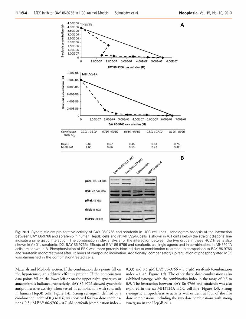
etiologies (Table 1). In each cell line, BAY 86-9766 inhibited cell prolifer-ation in vitro in a concentration-dependent manner with greater potencyagainst human HepG2 cells with NRAS mutations (IC50 = 33 nM)than against human Hep3B and PLC/PRF/5 cells with underlyingHBV infection (IC50 = 366 and 762 nM, respectively). In comparison,BAY 86-9766 was equipotent with another MEK inhibitor (AZD6244)in the HepG2 cells but three to four times more potent in the othercell lines. Besides the human HCC lines, BAY 86-9766 exhibited aconcentration-dependent antiproliferative activity in MH3924A andHepa129 cells, with IC50 values of 540 and 502 nM, respectively.
Strong Synergism between BAY 86-9766 and Sorafenib inTerms of Antiproliferative Effects and Feedback RegulationAs a method of choice to measure in vitro synergy, the combina-
tion index isobologram analysis has been performed as described in
Table 1. Antiproliferative Activity of the MEK Inhibitors BAY 86-9766 and AZD6244 in HCCCell Lines.
Cell Line Species Etiology/Genetic Mutation IC50 (nM)
BAY 86-9766 AZD6244
PLC/PRF/5 Human HBV infection 762 3060Hep3B Human HBV infection 366 1093HepG2 Human NRAS mutation 33 32MH3924A Rat Not determined 540 4595Hepa129 Mouse Not determined 502 2260
Neoplasia Vol. 15, No. 10, 2013 MEK Inhibitor BAY 86-9766 in HCC Animal Models Schmieder et al. 1163
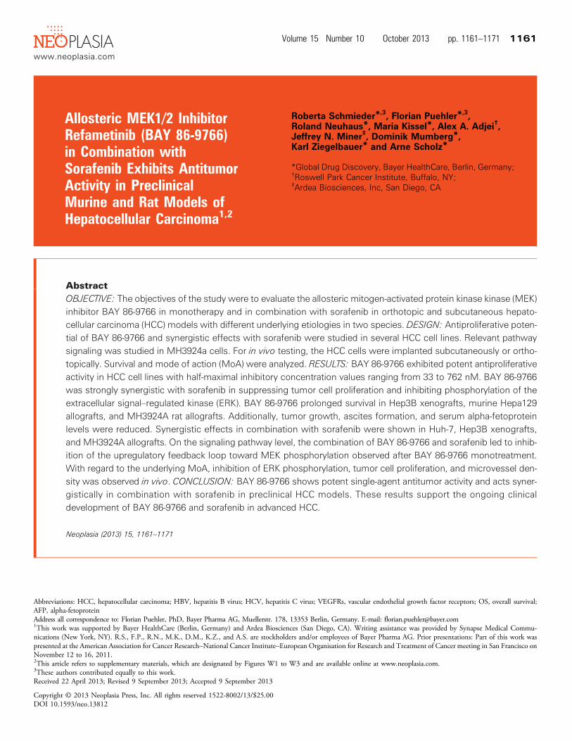
Materials and Methods section. If the combination data points fall onthe hypotenuse, an additive effect is present. If the combinationdata points fall on the lower left or on the upper right, synergism orantagonism is indicated, respectively. BAY 86-9766 showed synergisticantiproliferative activity when tested in combination with sorafenibin human Hep3B cells (Figure 1A). Strong synergism, defined by acombination index of 0.3 to 0.6, was observed for two dose combina-tions: 0.3 μMBAY 86-9766 + 0.7 μM sorafenib (combination index =
0.33) and 0.5 μM BAY 86-9766 + 0.5 μM sorafenib (combinationindex = 0.45; Figure 1A). The other three dose combinations alsoexhibited synergy, with the combination index in the range of 0.6 to0.9. The interaction between BAY 86-9766 and sorafenib was alsoexplored in the rat MH3924A HCC cell line (Figure 1A). Strongsynergistic antiproliferative activity was evident at four of the fivedose combinations, including the two dose combinations with strongsynergism in the Hep3B cells.
Figure 1. Synergistic antiproliferative activity of BAY 86-9766 and sorafenib in HCC cell lines. Isobologram analysis of the interactionbetween BAY 86-9766 and sorafenib in human Hep3B cells and rat MH3924A cells is shown in A. Points below the straight diagonal lineindicate a synergistic interaction. The combination index analysis for the interaction between the two drugs in these HCC lines is alsoshown in A (D1, sorafenib; D2, BAY 86-9766). Effects of BAY 86-9766 and sorafenib, as single agents and in combination, in MH3924Acells are shown in B. Phosphorylation of ERK was more potently blocked due to combination treatment in comparison to BAY 86-9766and sorafenib monotreatment after 12 hours of compound incubation. Additionally, compensatory up-regulation of phosphorylated MEKwas diminished in the combination-treated cells.
1164 MEK Inhibitor BAY 86-9766 in HCC Animal Models Schmieder et al. Neoplasia Vol. 15, No. 10, 2013
ERK and MEK phosphorylation was examined in MH3924A cellsand visualized by Western blot analysis. Due to combination treat-ment with BAY 86-9766 and sorafenib, phosphorylation of ERK wascompletely blocked after 12 hours in comparison to monotherapy. Inaddition, compensatory up-regulation of phosphorylated MEK signalafter BAY 86-9766 monotreatment was diminished in cells treated withcombination of both agents (Figure 1B).
Efficacy of BAY 86-9766 as Monotherapy and in Combinationwith Sorafenib in HCC Xenograft ModelsSingle-agent BAY 86-9766 (20 mg/kg) caused a small inhibition
in the growth of subcutaneous human Huh-7 HCC xenografts,which was nearly identical to the growth inhibition observed withsorafenib (50 mg/kg). In contrast, substantial tumor growth inhibi-tion was observed when BAY 86-9766 was administered in combi-nation with sorafenib. At the last assessment, the combinationreduced tumor volume by 70% compared with vehicle-treated con-
trol animals, whereas the two drugs individually reduced tumor vol-ume by approximately 20%. These results indicate that thecombination of BAY 86-9766 and sorafenib produces synergistictumor growth inhibition in Huh-7 xenografts (data not shown).In the orthotopic HBV-driven human Hep3B xenograft model,
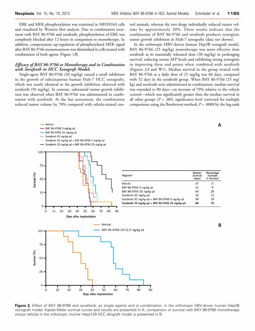
BAY 86-9766 (25 mg/kg) monotherapy was more effective thansorafenib at its maximally tolerated dose (30 mg/kg) in prolongingsurvival, reducing serum AFP levels and exhibiting strong synergismin improving these end points when combined with sorafenib(Figures 2A and W1). Median survival in the group treated withBAY 86-9766 at a daily dose of 25 mg/kg was 60 days, comparedwith 52 days in the sorafenib group. When BAY 86-9766 (25 mg/kg) and sorafenib were administered in combination, median survivalwas extended to 80 days—an increase of 70% relative to the vehiclecontrol—which was significantly greater than the median survival inall other groups (P = .003, significance level corrected for multiplecomparisons using the Bonferroni method; P = .0008 by the log-rank
Figure 2. Effect of BAY 86-9766 and sorafenib, as single agents and in combination, in the orthotopic HBV-driven human Hep3Bxenograft model. Kaplan-Meier survival curves and results are presented in A; comparison of survival with BAY 86-9766 monotherapyversus vehicle in the orthotopic murine Hepa129 HCC allograft model is presented in B.
Neoplasia Vol. 15, No. 10, 2013 MEK Inhibitor BAY 86-9766 in HCC Animal Models Schmieder et al. 1165
test). Of note, long-term treatment (up to 55 days) with the combi-nation was well tolerated. Body weight loss in the group was below10%, demonstrating long-term tolerability with treatment.Serum AFP measured on day 26 mirrored the survival results, with
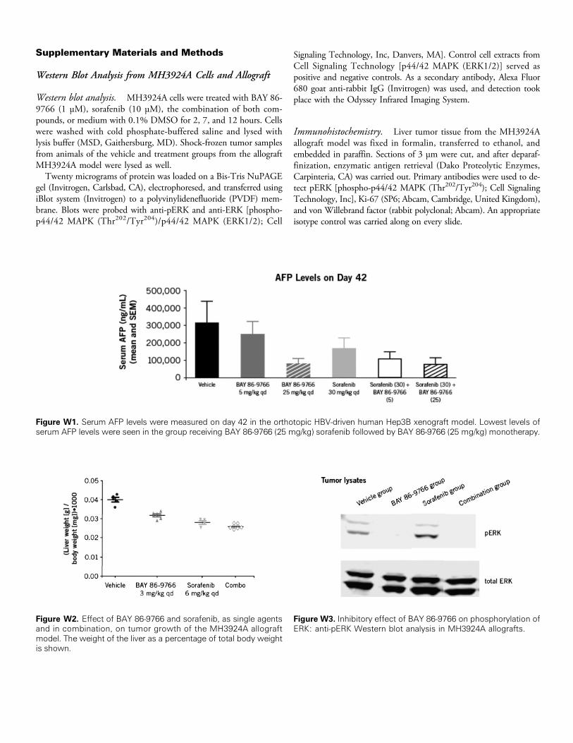
the lowest levels seen in the group receiving BAY 86-9766 (25 mg/kg)plus sorafenib followed by the group receiving the same dose ofBAY 86-9766 as monotherapy (data not shown). These two groups werealso the ones with the lowest serum AFP levels on day 42 (Figure W1).However, significant between-group differences in AFP levels onday 26 or 42 were not seen in multiple comparison statistical tests.
Efficacy of BAY 86-9766 as Monotherapy and in Combinationwith Sorafenib in HCC Allograft ModelsSingle-agent BAY 86-9766 significantly prolonged median survival
in the orthotopic murine Hepa129 HCC allograft model. Treatmentwas started on day 4 following implantation at a dose of 25 mg/kg,once daily, but the dose was reduced to 12.5 mg/kg, once daily, start-ing on day 18 after borderline weight loss was observed. With thisregimen, median survival was 58 days in the BAY 86-9766 groupcompared with 30 days for the vehicle-treated animals (P < .0001 bylog-rank test; Figure 2B).
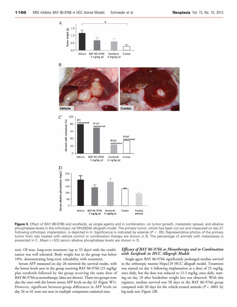
Figure 3. Effect of BAY 86-9766 and sorafenib, as single agents and in combination, on tumor growth, metastatic spread, and alkalinephosphatase levels in the orthotopic rat MH3924A allograft model. The primary tumor, which has been cut out and measured on day 21following orthotopic implantation, is depicted in A. Significance is indicated by asterisk (P < .05). Representative photos of the primarytumor from rats treated with vehicle control or combination therapy are shown in B. The percentage of animals with metastases ispresented in C. Mean (+SD) serum alkaline phosphatase levels are shown in D.
1166 MEK Inhibitor BAY 86-9766 in HCC Animal Models Schmieder et al. Neoplasia Vol. 15, No. 10, 2013
Single-agent BAY86-9766 [3mg/kg;maximal tolerated dose (MTD)determined in the rat model] and single-agent sorafenib (6 mg/kg;MTD determined in the rat model) reduced primary tumor weightcompared with vehicle control in the orthotopic ratMH3924A allograftmodel (not reaching statistical significance; Figure 3A). In comparison,combination therapy with BAY 86-9766 and sorafenib produced sig-nificant tumor growth inhibition compared with vehicle control (meanprimary tumor weight: 0.24 vs 1.14 g; P < .05 by Bonferroni multiplecomparison test). All animals in the control group had tumors ≥0.8 g,whereas 33%, 25%, and 60% of the animals had primary tumors≤0.2 g in the BAY 86-9766, sorafenib, and combination groups, re-spectively. The reductions in primary tumor growth were mirrored bysignificant decreases in liver weight (Figure W2). Median liver weightexpressed as a percentage of body weight was 3.2% in the BAY 86-
9766 group, 2.8% in the sorafenib group, and 2.5% in the combinationgroup compared with 4.0% in the vehicle control group (P < .05 for allcomparisons vs control by Bonferroni multiple comparison test).In the MH3924A allograft model, most animals in the control and
single-agent BAY 86-9766 groups had evidence of tumor metastases,whereas the lowest rates of metastatic spread were seen in the sorafeniband combination therapy groups (Figure 3C ). A similar profile wasevident when the number of metastases per animal was analyzed:Animals in the control group had a mean of three metastatic sites,whereas a mean of less than one site per animal was found in thesorafenib and combination therapy groups. However, neither the in-cidence of tumor metastases nor the number of metastases per animaldiffered significantly across treatment groups. Ascites was detected in60% of the control animals but in none of the animals in the other
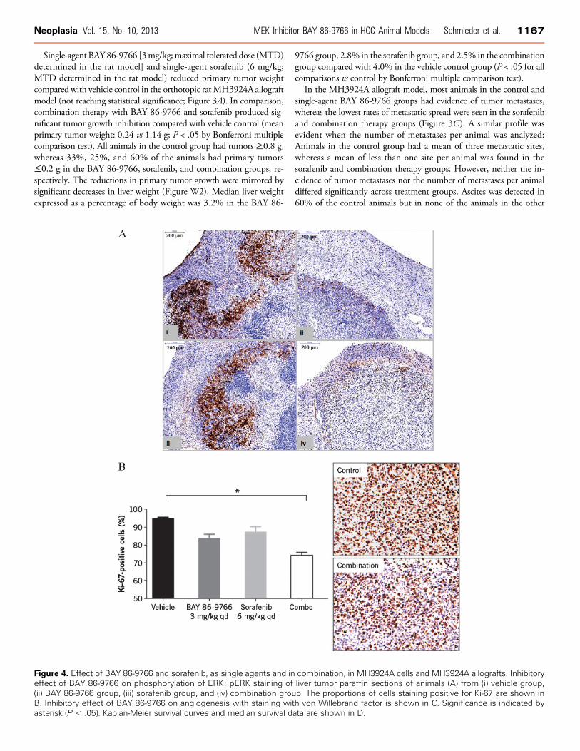
Figure 4. Effect of BAY 86-9766 and sorafenib, as single agents and in combination, in MH3924A cells and MH3924A allografts. Inhibitoryeffect of BAY 86-9766 on phosphorylation of ERK: pERK staining of liver tumor paraffin sections of animals (A) from (i) vehicle group,(ii) BAY 86-9766 group, (iii) sorafenib group, and (iv) combination group. The proportions of cells staining positive for Ki-67 are shown inB. Inhibitory effect of BAY 86-9766 on angiogenesis with staining with von Willebrand factor is shown in C. Significance is indicated byasterisk (P < .05). Kaplan-Meier survival curves and median survival data are shown in D.
Neoplasia Vol. 15, No. 10, 2013 MEK Inhibitor BAY 86-9766 in HCC Animal Models Schmieder et al. 1167
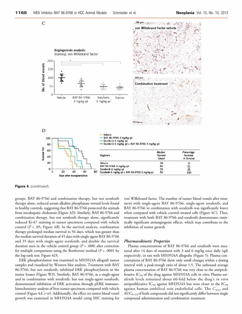
groups. BAY 86-9766 and combination therapy, but not sorafenibtherapy alone, reduced serum alkaline phosphatase toward levels foundin healthy controls, suggesting that BAY 86-9766 protected the animalsfrom intrahepatic cholestasis (Figure 3D). Similarly, BAY 86-9766 andcombination therapy, but not sorafenib therapy alone, significantlyreduced Ki-67 staining in tumor specimens compared with vehiclecontrol (P < .05; Figure 4B). In the survival analysis, combinationtherapy prolonged median survival to 56 days, which was greater thanthe median survival duration of 45 days with single-agent BAY 86-9766and 35 days with single-agent sorafenib, and double the survivalduration seen in the vehicle control group (P = .008) after correctionfor multiple comparisons using the Bonferroni method (P < .0001 bythe log-rank test; Figure 4D).ERK phosphorylation was examined in MH3924A allograft tumor
samples and visualized by Western blot analysis. Treatment with BAY86-9766, but not sorafenib, inhibited ERK phosphorylation in thetumor lysates (Figure W3). Similarly, BAY 86-9766, as a single-agentand in combination with sorafenib, but not single-agent sorafenib,demonstrated inhibition of ERK activation through pERK immuno-histochemistry analysis of liver tumor specimens compared with vehiclecontrol (Figure 4A, i–iv). Additionally, the effect on tumor blood vesselgrowth was examined in MH3924A model using IHC staining for
von Willebrand factor. The number of tumor blood vessels after treat-ment with single-agent BAY 86-9766, single-agent sorafenib, andBAY 86-9766 in combination with sorafenib was significantly lowerwhen compared with vehicle control–treated cells (Figure 4C). Thus,treatment with both BAY 86-9766 and sorafenib demonstrates statis-tically significant antiangiogenic effects, which may contribute to theinhibition of tumor growth.
Pharmacokinetic PropertiesPlasma concentrations of BAY 86-9766 and sorafenib were mea-
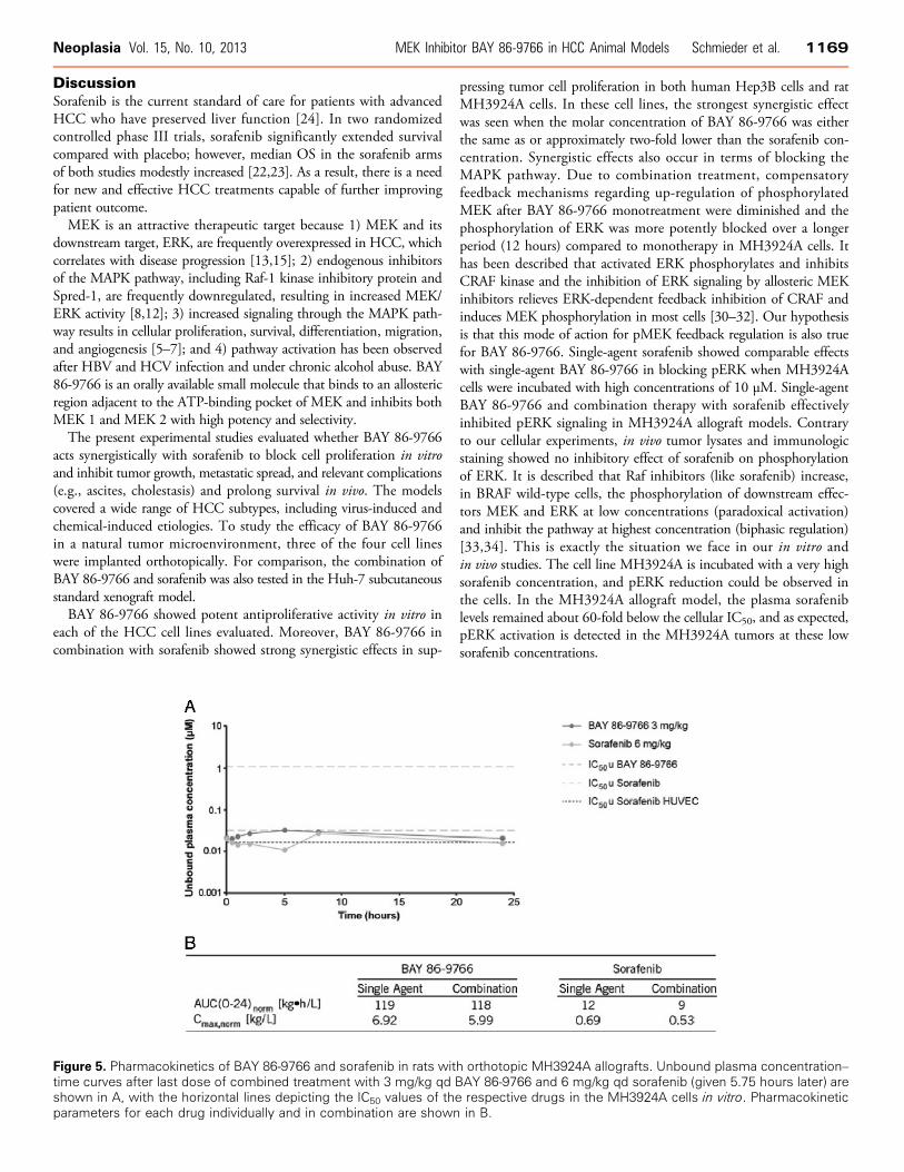
sured after 14 days of treatment with 3 and 6 mg/kg once daily (qd)respectively, in rats with MH3924A allografts (Figure 5). Plasma con-centrations of BAY 86-9766 show only small changes within a dosinginterval with a peak-trough ratio of about 1.5. The unbound averageplasma concentration of BAY 86-9766 was very close to the antiproli-ferative IC50 of the drug against MH3924A cells in vitro. Plasma sor-afenib levels remained about 60-fold below the drug’s in vitroantiproliferative IC50 against MH3924A but were closer to the IC50against human umbilical vein endothelial cells. The Cmax andAUC0–24 of both compounds did not significantly differ between singlecompound administration and combination treatment.
Figure 4. (continued).
1168 MEK Inhibitor BAY 86-9766 in HCC Animal Models Schmieder et al. Neoplasia Vol. 15, No. 10, 2013
DiscussionSorafenib is the current standard of care for patients with advancedHCC who have preserved liver function [24]. In two randomizedcontrolled phase III trials, sorafenib significantly extended survivalcompared with placebo; however, median OS in the sorafenib armsof both studies modestly increased [22,23]. As a result, there is a needfor new and effective HCC treatments capable of further improvingpatient outcome.MEK is an attractive therapeutic target because 1) MEK and its
downstream target, ERK, are frequently overexpressed in HCC, whichcorrelates with disease progression [13,15]; 2) endogenous inhibitorsof the MAPK pathway, including Raf-1 kinase inhibitory protein andSpred-1, are frequently downregulated, resulting in increased MEK/ERK activity [8,12]; 3) increased signaling through the MAPK path-way results in cellular proliferation, survival, differentiation, migration,and angiogenesis [5–7]; and 4) pathway activation has been observedafter HBV and HCV infection and under chronic alcohol abuse. BAY86-9766 is an orally available small molecule that binds to an allostericregion adjacent to the ATP-binding pocket of MEK and inhibits bothMEK 1 and MEK 2 with high potency and selectivity.The present experimental studies evaluated whether BAY 86-9766
acts synergistically with sorafenib to block cell proliferation in vitroand inhibit tumor growth, metastatic spread, and relevant complications(e.g., ascites, cholestasis) and prolong survival in vivo. The modelscovered a wide range of HCC subtypes, including virus-induced andchemical-induced etiologies. To study the efficacy of BAY 86-9766in a natural tumor microenvironment, three of the four cell lineswere implanted orthotopically. For comparison, the combination ofBAY 86-9766 and sorafenib was also tested in the Huh-7 subcutaneousstandard xenograft model.BAY 86-9766 showed potent antiproliferative activity in vitro in
each of the HCC cell lines evaluated. Moreover, BAY 86-9766 incombination with sorafenib showed strong synergistic effects in sup-
pressing tumor cell proliferation in both human Hep3B cells and ratMH3924A cells. In these cell lines, the strongest synergistic effectwas seen when the molar concentration of BAY 86-9766 was eitherthe same as or approximately two-fold lower than the sorafenib con-centration. Synergistic effects also occur in terms of blocking theMAPK pathway. Due to combination treatment, compensatoryfeedback mechanisms regarding up-regulation of phosphorylatedMEK after BAY 86-9766 monotreatment were diminished and thephosphorylation of ERK was more potently blocked over a longerperiod (12 hours) compared to monotherapy in MH3924A cells. Ithas been described that activated ERK phosphorylates and inhibitsCRAF kinase and the inhibition of ERK signaling by allosteric MEKinhibitors relieves ERK-dependent feedback inhibition of CRAF andinduces MEK phosphorylation in most cells [30–32]. Our hypothesisis that this mode of action for pMEK feedback regulation is also truefor BAY 86-9766. Single-agent sorafenib showed comparable effectswith single-agent BAY 86-9766 in blocking pERK when MH3924Acells were incubated with high concentrations of 10 μM. Single-agentBAY 86-9766 and combination therapy with sorafenib effectivelyinhibited pERK signaling in MH3924A allograft models. Contraryto our cellular experiments, in vivo tumor lysates and immunologicstaining showed no inhibitory effect of sorafenib on phosphorylationof ERK. It is described that Raf inhibitors (like sorafenib) increase,in BRAF wild-type cells, the phosphorylation of downstream effec-tors MEK and ERK at low concentrations (paradoxical activation)and inhibit the pathway at highest concentration (biphasic regulation)[33,34]. This is exactly the situation we face in our in vitro andin vivo studies. The cell line MH3924A is incubated with a very highsorafenib concentration, and pERK reduction could be observed inthe cells. In the MH3924A allograft model, the plasma sorafeniblevels remained about 60-fold below the cellular IC50, and as expected,pERK activation is detected in the MH3924A tumors at these lowsorafenib concentrations.
Figure 5. Pharmacokinetics of BAY 86-9766 and sorafenib in rats with orthotopic MH3924A allografts. Unbound plasma concentration–time curves after last dose of combined treatment with 3 mg/kg qd BAY 86-9766 and 6 mg/kg qd sorafenib (given 5.75 hours later) areshown in A, with the horizontal lines depicting the IC50 values of the respective drugs in the MH3924A cells in vitro. Pharmacokineticparameters for each drug individually and in combination are shown in B.
Neoplasia Vol. 15, No. 10, 2013 MEK Inhibitor BAY 86-9766 in HCC Animal Models Schmieder et al. 1169
BAY 86-9766 also demonstrated potent antitumor activity in thexenograft and allograft models. As a single agent, BAY 86-9766inhibited tumor growth in the human Huh-7 HCC xenograft model,prolonged survival and reduced serum AFP levels in the human Hep3BHCC xenograft model, and prolonged survival in the murine Hepa129allograft model. In the rat MH3924A allograft model, BAY 86-9766monotherapy reduced tumor growth and ascites formation, protectedagainst cholestasis, and prolonged survival. Positive effects on metastaticspread could be achieved through sorafenibmonotherapy and combina-tion therapy. When given in combination, BAY 86-9766 and sorafenibacted synergistically in reducing tumor growth and prolonging survivalin multiple models, including the human Hep3B HCC xenograft andthe rat MH3924A allograft.Combination of BAY 86-9766 with sorafenib may achieve syn-
ergistic activity in two ways, namely, 1) blockade of the MAPKpathway at two different points (RAF with sorafenib and MEK withBAY 86-9766) or 2) blockade of parallel signaling pathways (block-ing the MAPK pathway with BAY 86-9766 and blocking VEGFR-mediated signaling pathways with sorafenib). Evidence favoringthe first possibility has been reported in melanoma cells where thecombination of a BRAF inhibitor and MEK inhibitor enhancedapoptosis and prevented the onset of resistance [35]. Additionally,our findings demonstrated that both BAY 86-9766 and sorafenibmonotherapies, as well as BAY 86-9766 + sorafenib combinationtherapy, had significant antiangiogenic effects in the MH3924AHCC model.Tumor blood vessel formation was inhibited by single-agent BAY
86-9766, single-agent sorafenib, and BAY 86-9766 in combinationwith sorafenib. BAY 86-9766 monotherapy also effectively inhibitedpERK signaling. Together, these data provide evidence that sorafeniband BAY 86-9766 are acting synergistically by blocking parallelsignal pathways; sorafenib is primarily blocking VEGFR-mediatedsignaling, while BAY 86-9766 acts directly on the MAPK pathwayin vitro and in vivo.The rat MH3924A allograft model may shed some light on the
mechanism for in vivo synergism between BAY 86-9766 and sorafenib.Throughout the 24-hour dosing level, plasma BAY 86-9766 con-centrations remained close to the drug’s antiproliferative IC50 againstMH3924A cells. These findings suggest that the efficacy of BAY 86-9766 results from a direct effect on the tumor cells. Although plasmasorafenib concentrations remained below its antiproliferative IC50against tumor cells, it was close to its IC50 against endothelial cells,thereby suggesting that the efficacy of sorafenib may be due to anindirect effect. Taken together, the antiproliferative effect of BAY 86-9766 and the antiangiogenic properties of sorafenib might combinein the MH3924A in vivo model to produce a synergistic antitumoraleffect. Nevertheless, our in vitro combination experiments also indicatea direct synergistic antiproliferative effect between BAY 86-9766 andsorafenib in MH3924A tumor cells.In summary, the models used in these investigations cover mul-
tiple HCC subtypes, including virus-induced and chemical-inducedetiologies. Even in tumor models that show less potent antiprolifera-tive IC50 values in vitro than the NRAS-mutated HepG2 cell line,BAY 86-9766 showed great in vivo potency, which emphasizes theeffectiveness of the MEK inhibitor.The role of the tumor stroma and immunologic interactions
was addressed by the orthotopical transplantation of the allograftcells in the liver and the inclusion of two models with immuno-competent animals.
Antitumor efficacy of BAY 86-9766, particularly when used incombination with sorafenib, was observed in each model, suggestingthat this novel MEK inhibitor has potential for broad usage acrossmultiple HCC subtypes. BAY 86-9766 exhibited significant single-agent antitumor activity, but MEK inhibitor monotherapy may notbe sufficient for clinical efficacy in a variety of clinical settings. Ina recent phase II trial, a different MEK inhibitor, selumetinib (AZD6244), did not produce radiographic responses in patients with ad-vanced HCC even though there was evidence of ERK inhibition[36]. The potent synergism observed between BAY 86-9766 andsorafenib suggests that a combination therapy approach may be amore promising addition to treatment of HCC. Along these lines,the results of a phase II clinical trial (NCT01204177) [37] investigatingBAY 86-9766 (50 mg, twice a day) in combination with sorafenib(400 mg, twice a day) as first-line treatment for patients with advancedHCC will soon be submitted for publication [37,38].
References[1] Venook AP, Papandreou C, Furuse J, and de Guevara LL (2010). The incidence
and epidemiology of hepatocellular carcinoma: a global and regional perspective.Oncologist 15(suppl 4), 5–13.
[2] Jemal A, Bray F, Center MM, Ferlay J, Ward E, and Forman D (2011). Globalcancer statistics. CA Cancer J Clin 61, 69–90.
[3] Ma YT and Palmer DH (2012). Impact of restricting access to high-cost medica-tions for hepatocellular carcinoma. Expert Rev Pharmacoecon Outcomes Res 12,465–473.
[4] Olsen SK, Brown RS, and Siegel AB (2010). Hepatocellular carcinoma: reviewof current treatment with a focus on targeted molecular therapies. Therap AdvGastroenterol 3, 55–66.
[5] Roberts PJ and Der CJ (2007). Targeting the Raf-MEK-ERK mitogen-activatedprotein kinase cascade for the treatment of cancer. Oncogene 26, 3291–3310.
[6] Friday BB and Adjei AA (2008). Advances in targeting the Ras/Raf/MEK/Erkmitogen-activated protein kinase cascade with MEK inhibitors for cancer therapy.Clin Cancer Res 14, 342–346.
[7] Fremin C and Meloche S (2010). From basic research to clinical developmentof MEK1/2 inhibitors for cancer therapy. J Hematol Oncol 3, 8.
[8] Wong CM and Ng IO (2008). Molecular pathogenesis of hepatocellularcarcinoma. Liver Int 28, 160–174.
[9] Tannapfel A, Sommerer F, Benicke M, Katalinic A, Uhlmann D, Witzigmann H,Hauss J, and Wittekind C (2003). Mutations of the BRAF gene in cholangio-carcinoma but not in hepatocellular carcinoma. Gut 52, 706–712.
[10] Nonomura A, Ohta G, Hayashi M, Izumi R, Watanabe K, Takayanagi N,Mizukami Y, and Matsubara F (1987). Immunohistochemical detection of rasoncogene p21 product in liver cirrhosis and hepatocellular carcinoma. Am JGastroenterol 82, 512–518.
[11] Jagirdar J, Nonomura A, Patil J, Thor A, and Paronetto F (1989). ras oncogenep21 expression in hepatocellular carcinoma. J Exp Pathol 4, 37–46.
[12] Schuierer MM, Bataille F, Weiss TS, Hellerbrand C, and Bosserhoff AK (2006).Raf kinase inhibitor protein is downregulated in hepatocellular carcinoma. OncolRep 16, 451–456.
[13] Schmidt CM, McKillop IH, Cahill PA, and Sitzmann JV (1997). IncreasedMAPK expression and activity in primary human hepatocellular carcinoma.Biochem Biophys Res Commun 236, 54–58.
[14] Tsuboi Y, Ichida T, Sugitani S, Genda T, Inayoshi J, Takamura M, Matsuda Y,Nomoto M, and Aoyagi Y (2004). Overexpression of extracellular signal-regulatedprotein kinase and its correlation with proliferation in human hepatocellularcarcinoma. Liver Int 24, 432–436.
[15] Ito Y, Sasaki Y, Horimoto M, Wada S, Tanaka Y, Kasahara A, Ueki T, HiranoT, Yamamoto H, Fujimoto J, et al. (1998). Activation of mitogen-activatedprotein kinases/extracellular signal-regulated kinases in human hepatocellularcarcinoma. Hepatology 27, 951–958.
[16] Lupberger J and Hildt E (2007). Hepatitis B virus-induced oncogenesis.World JGastroenterol 13, 74–81.
[17] Nagy LE (2004). Molecular aspects of alcohol metabolism: transcriptionfactors involved in early ethanol-induced liver injury. Annu Rev Nutr 24,55–78.
1170 MEK Inhibitor BAY 86-9766 in HCC Animal Models Schmieder et al. Neoplasia Vol. 15, No. 10, 2013
[18] Hennig M, Yip-Schneider MT, Klein P, Wentz S, Matos JM, Doyle C, Choi J,Wu H, O’Mara A, Menze A, et al. (2009). Ethanol-TGFα-MEK signalingpromotes growth of human hepatocellular carcinoma. J Surg Res 154, 187–195.
[19] Schmitz KJ, Wohlschlaeger J, Lang H, Sotiropoulos GC, Malago M, StevelingK, Reis H, Cicinnati VR, Schmid KW, and Baba HA (2008). Activation of theERK and AKT signalling pathway predicts poor prognosis in hepatocellularcarcinoma and ERK activation in cancer tissue is associated with hepatitis C virusinfection. J Hepatol 48, 83–90.
[20] Wilhelm SM, Carter C, Tang L, Wilkie D, McNabola A, Rong H, Chen C,Zhang X, Vincent P, McHugh M, et al. (2004). BAY 43-9006 exhibits broadspectrum oral antitumor activity and targets the RAF/MEK/ERK pathway andreceptor tyrosine kinases involved in tumor progression and angiogenesis. CancerRes 64, 7099–7109.
[21] Liu PC, Liu X, Li Y, Covington M, Wynn R, Huber R, Hillman M, Yang G,Ellis D, Marando C, et al. (2006). Identification of ADAM10 as a major sourceof HER2 ectodomain sheddase activity in HER2 overexpressing breast cancercells. Cancer Biol Ther 5, 657–664.
[22] Llovet JM, Ricci S, Mazzaferro V, Hilgard P, Gane E, Blanc JF, de Oliveira AC,Santoro A, Raoul JL, Forner A, et al. (2008). Sorafenib in advanced hepato-cellular carcinoma. N Engl J Med 359, 378–390.
[23] Cheng AL, Kang YK, Chen Z, Tsao CJ, Qin S, Kim JS, Luo R, Feng J, Ye S,Yang TS, et al. (2009). Efficacy and safety of sorafenib in patients in the Asia-Pacific region with advanced hepatocellular carcinoma: a phase III randomised,double-blind, placebo-controlled trial. Lancet Oncol 10, 25–34.
[24] National Comprehensive Cancer Network (NCCN). NCCN Clinical PracticeGuidelines in Oncology/Hepatobiliary Cancers v.2004. Available at: http://www.nccn.org/.
[25] Iverson C, Larson G, Lai C, Yeh LT, Dadson C, Weingarten P, Appleby T, VoT, Maderna A, Vernier JM, et al. (2009). RDEA119/BAY 869766: a potent,selective, allosteric inhibitor of MEK1/2 for the treatment of cancer. Cancer Res69, 6839–6847.
[26] Jonkers J and Berns A (2002). Conditional mouse models of sporadic cancer.Nat Rev Cancer 2, 251–265.
[27] Killion JJ, Radinsky R, and Fidler IJ (1998). Orthotopic models are necessary topredict therapy of transplantable tumors inmice.CancerMetastasis Rev 17, 279–284.
[28] Mohandas K (2004). Hepatitis B associated hepatocellular carcinoma: epidemi-ology, diagnosis and treatment. Hep B Annual 1, 140–152.
[29] Chou TC (2006). Theoretical basis, experimental design, and computerizedsimulation of synergism and antagonism in drug combination studies. PharmacolRev 58, 621–681.
[30] Ishii N, Harada N, Joseph EW, Ohara K, Miura T, Sakamoto H, Matsuda Y,Tomii Y, Tachibana-Kondo Y, Iikura H, et al. (2013). Enhanced inhibitionof ERK signaling by a novel allosteric MEK inhibitor, CH5126766, that sup-presses feedback reactivation of RAF activity. Cancer Res 73, 4050–4060.
[31] Wang Y, Van Becelaere K, Jiang P, Przybranowski S, Omer C, and Sebolt-Leopold J (2005). A role for K-ras in conferring resistance to the MEK inhibitor,CI-1040. Neoplasia 7, 336–347.
[32] Huynh H, Soo KC, Chow PK, and Tran E (2007). Targeted inhibition of theextracellular signal-regulated kinase kinase pathway with AZD6244 (ARRY-142886) in the treatment of hepatocellular carcinoma. Mol Cancer Ther 6,138–146.
[33] Poulikakos PI, Zhang C, Bollag G, Shokat KM, and Rosen N (2010). RAFinhibitors transactivate RAF dimers and ERK signalling in cells with wild-typeBRAF. Nature 18, 427–430.
[34] Holderfield M, Merritt H, Chan J, Wallroth M, Tandeske L, Zhai H, Tellew J,Hardy S, Hekmat-Nejad M, Stuart DD, et al. (2013). RAF inhibitors activate theMAPK pathway by relieving inhibitory autophosphorylation. Cancer Cell 13,594–602.
[35] Paraiso KH, Fedorenko IV, Cantini LP, Munko AC, Hall M, Sondak VK,Messina JL, Flaherty KT, and Smalley KS (2010). Recovery of phospho-ERKactivity allows melanoma cells to escape from BRAF inhibitor therapy. Br JCancer 102, 1724–1730.
[36] O’Neil BH, Goff LW, Kauh JS, Strosberg JR, Bekaii-Saab TS, Lee RM, Kazi A,Moore DT, Learoyd M, Lush RM, et al. (2011). Phase II study of the mitogen-activated protein kinase 1/2 inhibitor selumetinib in patients with advancedhepatocellular carcinoma. J Clin Oncol 29, 2350–2356.
[37] U.S. National Institutes of Health. Assessing BAY86-9766 Plus Sorafenib forthe Treatment of Liver Cancer (BASIL), 2010. Available at: http://www.clinicaltrials.gov/ct2/show/NCT01204177?term=NCT01204177&rank=1.Accessed October 01, 2013.
[38] Weekes CD, Von Hoff DD, Adjei AA, Leffingwell DP, Eckhardt SG, Gore L,Lewis KD, Weiss GJ, Ramanathan RK, Dy GK, et al. (2013). Multicenterphase I trial of the mitogen-activated protein kinase 1/2 inhibitor BAY 86-9766in patients with advanced cancer. Clin Cancer Res 19, 1232–1243.
Neoplasia Vol. 15, No. 10, 2013 MEK Inhibitor BAY 86-9766 in HCC Animal Models Schmieder et al. 1171
Supplementary Materials and Methods
Western Blot Analysis from MH3924A Cells and Allograft
Western blot analysis. MH3924A cells were treated with BAY 86-9766 (1 μM), sorafenib (10 μM), the combination of both com-pounds, or medium with 0.1% DMSO for 2, 7, and 12 hours. Cellswere washed with cold phosphate-buffered saline and lysed withlysis buffer (MSD, Gaithersburg, MD). Shock-frozen tumor samplesfrom animals of the vehicle and treatment groups from the allograftMH3924A model were lysed as well.Twenty micrograms of protein was loaded on a Bis-Tris NuPAGE
gel (Invitrogen, Carlsbad, CA), electrophoresed, and transferred usingiBlot system (Invitrogen) to a polyvinylidenefluoride (PVDF) mem-brane. Blots were probed with anti-pERK and anti-ERK [phospho-p44/42 MAPK (Thr202/Tyr204)/p44/42 MAPK (ERK1/2); Cell
Signaling Technology, Inc, Danvers, MA]. Control cell extracts fromCell Signaling Technology [p44/42 MAPK (ERK1/2)] served aspositive and negative controls. As a secondary antibody, Alexa Fluor680 goat anti-rabbit IgG (Invitrogen) was used, and detection tookplace with the Odyssey Infrared Imaging System.
Immunohistochemistry. Liver tumor tissue from the MH3924Aallograft model was fixed in formalin, transferred to ethanol, andembedded in paraffin. Sections of 3 μm were cut, and after deparaf-finization, enzymatic antigen retrieval (Dako Proteolytic Enzymes,Carpinteria, CA) was carried out. Primary antibodies were used to de-tect pERK [phospho-p44/42 MAPK (Thr202/Tyr204); Cell SignalingTechnology, Inc], Ki-67 (SP6; Abcam, Cambridge, United Kingdom),and von Willebrand factor (rabbit polyclonal; Abcam). An appropriateisotype control was carried along on every slide.
Figure W1. Serum AFP levels were measured on day 42 in the orthotopic HBV-driven human Hep3B xenograft model. Lowest levels ofserum AFP levels were seen in the group receiving BAY 86-9766 (25 mg/kg) sorafenib followed by BAY 86-9766 (25 mg/kg) monotherapy.
Figure W2. Effect of BAY 86-9766 and sorafenib, as single agentsand in combination, on tumor growth of the MH3924A allograftmodel. The weight of the liver as a percentage of total body weightis shown.
Figure W3. Inhibitory effect of BAY 86-9766 on phosphorylation ofERK: anti-pERK Western blot analysis in MH3924A allografts.


