Embed Size (px)
Citation preview

J Physiol 586.6 (2008) pp 1649–1667 1649
α-Adrenergic inhibition increases collateral circuitconductance in rats following acute occlusion of thefemoral artery
Jessica C. Taylor1, Zeyi Li1, H. T. Yang1, M. Harold Laughlin1,2,3 and Ronald L. Terjung1,2,3
1Department of Biomedical Sciences, College of Veterinary Medicine, 2Department of Medical Pharmacology and Physiology, College of Medicine and3Dalton Cardiovascular Research Center, University of Missouri, Columbia, MO 65211, USA
This study evaluated whether α-adrenergic activation contributes to collateral circuit vascular
resistance in the hindlimb following acute unilateral occlusion of the femoral artery in rats.
Blood pressures (BPs) were measured above (caudal artery) and below (distal femoral artery)
the collateral circuit. Arterial BPs were reduced (15–35 mmHg) with individual (prazosin,
rauwolscine) or combined (phentolamine) α-receptor inhibition. Blood flows (BFs) were
measured using microspheres before and afterα inhibition during the same treadmill speed. α1
inhibition increased blood flow by ∼40% to active muscles that were not affected by femoral
occlusion, whereas collateral-dependent BFs to the calf muscles were reduced by 29 ± 8.4%
(P < 0.05), due to a decrease in muscle conductance with no change in collateral circuit
conductance. α2 inhibition decreased both collateral circuit (39 ± 6.0%; P < 0.05) and calf
muscle conductance (36 ± 7.3%; P < 0.05), probably due to residual α1 activation, since renal
BF was markedly reduced with rauwolscine. Most importantly, inhibiting α2 receptors in the
presence of α1 inhibition increased (43 ± 12%; P < 0.05) collateral circuit conductance.
Similarly, non-selective α inhibition with phentolamine increased collateral conductance
(242 ± 59%; P < 0.05). We interpret these findings to indicate that both α1- and α2-receptor
activation can influence collateral circuit resistance in vivo during the high flow demands caused
by exercise. Furthermore, we observed a reduced maximal conductances of active muscles that
were ischaemic. Our findings imply that in the presence of excessive sympathetic activation,
which can occur in the condition of intermittent claudication during exertion, an exaggerated
vasoconstriction of the existing collateral circuit and active muscle will occur.
(Resubmitted 8 December 2007; accepted after revision 17 January 2008; first published online 24 January 2008)
Corresponding author R. L. Terjung: Department of Biomedical Sciences, E102 Vet. Medical Bldg, University of
Missouri, Columbia, MO 65211, USA. Email: [email protected]
Local blood flow to a given tissue is typically establishedby a balance among extrinsic influences (e.g. sympatheticinnervation, circulating factors) and the inherentvasomotor tone established by the vasculature and localtissue conditions. Sympathetic innervation can provide apowerful vasoconstriction influence through α-receptoractivation which is governed by two major receptorsubtypes, α1 and α2. Both α1- and α2-receptors areresponsive to noradrenaline (norepinephrine) (Langer& Hicks, 1984) resulting in a robust vasoconstriction(Smiesko et al. 1989). α1-Receptors are located in bothrelatively large arteries and resistance vessels, whereasα2-Receptors reside primarily in the smaller resistancevessels, particularly third- and fourth-order arterioles(Ping & Faber, 1993). On the other hand, local factorsinfluencing blood flow become more dominant under
tissue conditions that obligate a greater blood flow. Forexample, with the increased metabolic demands suchas occur during exercise, local dilatory influences areaugmented to increase blood flow to the active muscles.However, coupled with the enhanced sympathetic outflowduring exercise, there is a sustained sympathetic vaso-constrictor influence to active muscles (Buckwalter et al.1997; Hamann et al. 2002) that serves to temper theelevation in blood flow. This is thought to contribute tovascular regulation wherein blood flow to active musclesincreases, essentially in proportion to the intensity ofaerobic-type exercise. Nonetheless, dilatory conditionswithin active muscle can be significantly blunted toreduce blood flow, if the vasoconstrictive drive becomesexaggerated due to central sympathetic system activation(Secher et al. 1977; Harms et al. 1998; Volianitis & Secher,
C© 2008 The Authors. Journal compilation C© 2008 The Physiological Society DOI: 10.1113/jphysiol.2007.149567
1650 J. C. Taylor and others J Physiol 586.6
2002). Thus, the fine balance between vasoconstrictoryand dilatory influences becomes important in the controlof blood flow to active muscle.
Occlusion of a primary supply artery, as can occurin conditions of peripheral arterial insufficiency (PAI),creates a situation in which pre-existing vascularanastomoses (Yang et al. 1996; Buschmann & Schaper,1999, 2000) are recruited to form a collateral circuit thatcircumvents the obstruction to deliver blood flow to thedistal tissues. Changes in pressure gradients and vascularcontrol mechanisms occur immediately in response toocclusion (Rosenthal & Guyton, 1968; Paskins-Hurlburt& Hollenberg, 1992). The lower distal pressure, inducedby occlusion, can increase over time, but typically is notfully restored. Therefore, a pressure drop remains acrossthis added resistance of the collateral circuit (Rosenthal &Guyton, 1968; Unthank et al. 1995, 1996). Furthermore, asignificant increase in collateral resistance typically strips alarge fraction of the flow reserve to the distal muscles (Yang& Terjung, 1993; Yang et al. 1995, 1996). The new roleof these anastomoses, to serve as thoroughfare vessels todeliver flow downstream, raises some interesting prospectsfor vascular control. On one hand, the vascular circuithas a limited structure of vessels which restricts peakconductance; on the other hand, the vessels are beingused in a different manner, as thoroughfare vessels, ascompared to their normal role of flow delivery to thelocal tissue. This raises the scenario that these newlyfunctioning collateral vessels could be subject to vaso-constriction influences, typical of local tissue that wouldnot be optimal for delivery of flow downstream. Thus,downstream collateral-dependent blood flow could beconstrained by both the structure and vasomotor functionof the collateral circuit.
The function of the collateral circuit is probably complexin the condition of peripheral arterial insufficiency, whichoften manifests itself as intermittent claudication. Inthis condition, a limited flow capacity to the lowerextremities results in exertion-induced changes in gaitthat are typically relieved upon rest (Dormandy &Rutherford, 2000). As blood flow demands increase atthe onset of physical activity there is a concomitantincrease in sympathetic outflow that restrains blood flow(Peterson et al. 1988). This could aggravate the alreadylimiting collateral circuit resistance, since local conditionssurrounding the upstream collateral vessels may not favourvessel dilatation (Ito et al. 1997; Buschmann & Schaper,1999; Yang et al. 2000b, 2002). Further, it is probablethat the stress of locomotion and/or ischaemic pain mayaugment up-regulation of sympathetic outflow (Bakkeet al. 2007). This could significantly detract from theability of an existing collateral circuit to support distal flowneeds. Therefore, the purpose of this study was to examinefactors that could contribute to collateral circuit resistance.Specifically, we hypothesized that α-adrenergic receptor
activation contributes to the resistance of collateral vessels.This could restrict blood flow to the collateral-dependenttissues, especially active muscles during walking, beyondthat already established by the limited structure of thepre-existing collateral circuit.
Methods
Ethical approval
The care and treatment of all animals and experimentalprocedures were conducted in accordance with the Guidefor the Care and Use of Laboratory Animals published bythe US National Institutes of Health (NIH Publication no.85–23, revised 1996) and approved by the Animal Careand Use Committee of the University of Missouri.
Experimental design
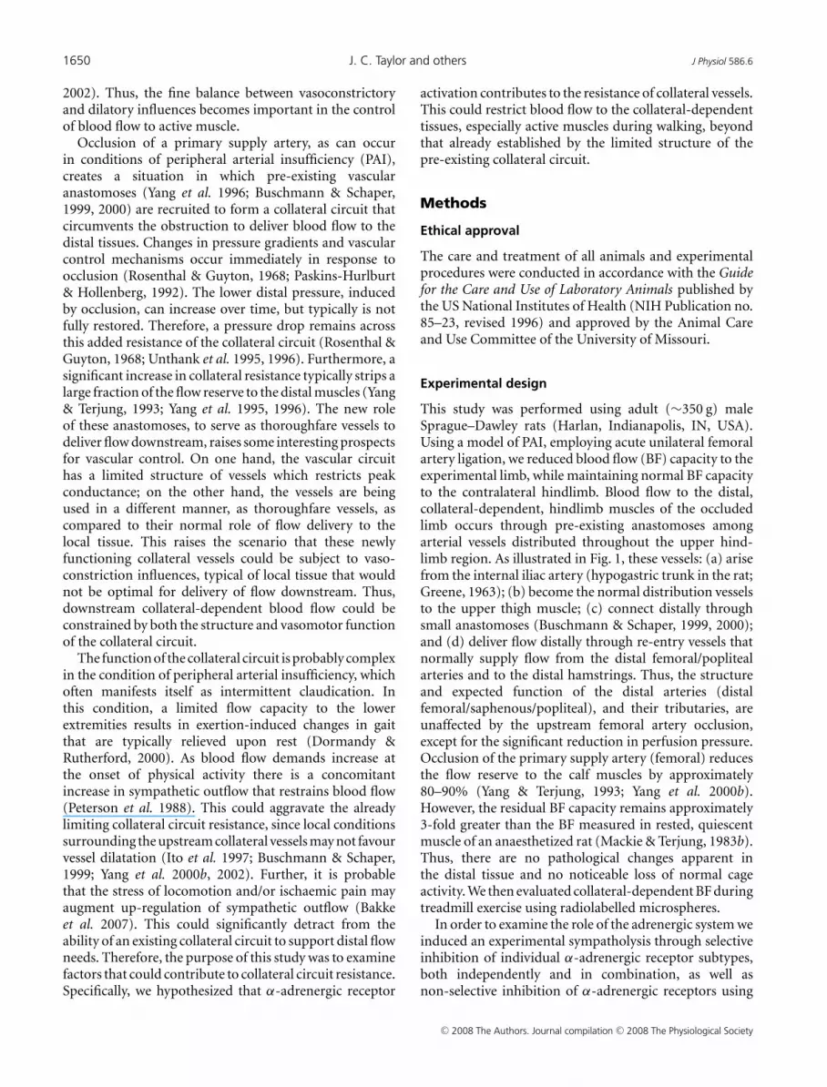
This study was performed using adult (∼350 g) maleSprague–Dawley rats (Harlan, Indianapolis, IN, USA).Using a model of PAI, employing acute unilateral femoralartery ligation, we reduced blood flow (BF) capacity to theexperimental limb, while maintaining normal BF capacityto the contralateral hindlimb. Blood flow to the distal,collateral-dependent, hindlimb muscles of the occludedlimb occurs through pre-existing anastomoses amongarterial vessels distributed throughout the upper hind-limb region. As illustrated in Fig. 1, these vessels: (a) arisefrom the internal iliac artery (hypogastric trunk in the rat;Greene, 1963); (b) become the normal distribution vesselsto the upper thigh muscle; (c) connect distally throughsmall anastomoses (Buschmann & Schaper, 1999, 2000);and (d) deliver flow distally through re-entry vessels thatnormally supply flow from the distal femoral/poplitealarteries and to the distal hamstrings. Thus, the structureand expected function of the distal arteries (distalfemoral/saphenous/popliteal), and their tributaries, areunaffected by the upstream femoral artery occlusion,except for the significant reduction in perfusion pressure.Occlusion of the primary supply artery (femoral) reducesthe flow reserve to the calf muscles by approximately80–90% (Yang & Terjung, 1993; Yang et al. 2000b).However, the residual BF capacity remains approximately3-fold greater than the BF measured in rested, quiescentmuscle of an anaesthetized rat (Mackie & Terjung, 1983b).Thus, there are no pathological changes apparent inthe distal tissue and no noticeable loss of normal cageactivity. We then evaluated collateral-dependent BF duringtreadmill exercise using radiolabelled microspheres.
In order to examine the role of the adrenergic system weinduced an experimental sympatholysis through selectiveinhibition of individual α-adrenergic receptor subtypes,both independently and in combination, as well asnon-selective inhibition of α-adrenergic receptors using
C© 2008 The Authors. Journal compilation C© 2008 The Physiological Society

J Physiol 586.6 α-Adrenergic control of collateral vasculature 1651
phentolamine. Our experiments appropriately evaluatedcollateral circuit conductance, since exercise conditionswere selected (20 m min−1 15% gradient) to elicit maximalcollateral-dependent blood flow (Yang & Terjung, 1993;Yang et al. 2002). However, these exercise conditions arewell within the capacity of the animal, as they can sustainthis speed well beyond the brief duration (∼2.5 min)needed for each blood flow determination.
Rats were divided into four groups to evaluate whetherα-adrenergic receptor activation contributes to collateralcircuit resistance following acute femoral occlusion:group 1 (n = 11), prazosin inhibition of α1-adrenergicreceptors; group 2 (n = 14), rauwolscine inhibition ofα2-adrenergic receptors; group 3 (n = 6), combinedinhibition of α1- and α2-adrenergic receptors; and group4 (n = 11), non-selective α-adrenergic receptor inhibitionwith phentolamine. In groups 1, 2 and 4 each animalserved as its own control with blood flow measurementsobtained pre- and postdrug infusion, approximately 5 min
Pdistalfemoral a.
collateral-dependent
muscle
X
Qt = Qthigh + Qdistal limb
Gcollateral = distal limbQ
caudal a.P - Pdistalfemoral a.
( (
Qdistal limb = caudal a.P Pzero-( (
collateralR + Rdistallimb
( (
Gdistal limb = distal limbQ
-Pdistalfemoral a.
( zeroP (
P distalfemoral a.
commoniliac a.
distal legarteries
R distallimb
R thighmuscle
P zero=
caudal a.P
internaliliac a.
Rcollentry
R collre-entry
femoral a.occlusion
Figure 1. Arterial vessels of the hindlimb and diagram illustrating the collateral circuit in the hindlimbOcclusion of the femoral artery causes the distal limb muscles to become collateral-dependent tissues. Note thatblood flow to the distal limb tissues must circumvent the obstruction of the femoral artery through the upperlimb vasculature (as defined by the thick lines and resistances). The X in the X-ray shows the site of femoral arteryocclusion, whereas the black arrow shows the internal iliac vessel complex (hypogastric trunk; Greene, 1963) fromwhich collateral flow emanates. The white arrow on the X-ray illustrates re-entry vessels which deliver collateralblood flow into the ‘normal’ distal limb arteries (e.g. distal femoral, popliteal, saphenous). The formulas definehow collateral circuit and distal limb tissue conductances were calculated. G is vascular conductance; R vascularresistance; Qt total blood flow; P (pressure) = zero is the pressure of the femoral vein, assumed to be 0 mmHg.Note that Rcoll entry (resistance from the origin of the internal iliac to entry of the collateral vessels) can havevascular elements in common that contribute to the total resistance of the thigh muscles.
apart. In group 3, blood flow measurements were obtainedwith α1 inhibition only and then α1 and α2-inhibitiontogether, effectively examining the interaction betweenthe two receptor subtypes. An additional group of rats(n = 9), with bilateral femoral artery occlusion, weregiven phentolamine to assess the validity of employingthe unilaterally ligated animals of the previous groups.Collateral vessels were isolated from additional animals(n = 6), not used for BF determinations, to evaluate theinfluence of luminal pressure on their vasoresponsivenessto acetylcholine (ACh) and phenylephrine (PE) in vitro.
Infusion of the α1-antagonist prazosin (50 μg) wasadministered between the two exercise bouts (20 m min−1
15% gradient). This dose was sufficient to inhibit aphenylephrine-induced elevation in blood pressure (BP)by 64.1 ± 6.7% (n = 8). In group 2 the α2-adrenergicantagonist rauwolscine (150 μg) was infused between twosimilar exercise bouts. This dose was sufficient to inhibita medetomidine-induced elevation in BP by 50.3 ± 9.1%
C© 2008 The Authors. Journal compilation C© 2008 The Physiological Society

1652 J. C. Taylor and others J Physiol 586.6
(n = 6). In group 3 the same dose of prazosin wasadministered prior to the first exercise bout, followed byrauwolscine infusion and a second similar exercise bout.In group 4, infusion of the non-selective α-antagonistphentolamine (2.6 mg) was administered between thetwo exercise bouts. This dose was sufficient to inhibit aphenylephrine-induced elevation in distal femoral arteryBP by 66.4 ± 9.7% (n = 6). Rats were familiarized withthe treadmill by running 5–6 min day−1 for 4–5 days priorto experimentation. This conditioning to the treadmillestablishes excellent, willing exercise performance for theblood flow determination, but does not induce adaptationstypical of exercise training (Yang et al. 1995). Blood flowmeasurements were obtained during exercise in order tominimize the resistance of the distal limb tissue, which isnecessary to determine the conductance capacity of theupstream collateral circuit (Yang & Terjung, 1993; Yanget al. 2002).
Animal care
Rats were housed two per cage in a temperature-controlled(20 ± 1◦C) animal room with a 12 : 12 h light–dark cycle.Rats were fed a standard rat chow diet and water adlibitum. All animals were killed under deep anaesthesiaby a pneumothorax.
Surgical procedures
Details of the femoral artery ligation procedure havebeen previously described (Yang & Terjung, 1993). Briefly,animals were brought to the laboratory the morning ofthe experiment and anaesthetized with a combination ofketamine (100 mg kg−1) and acepromazine (0.5 mg kg−1).For unilateral artery ligation employed for groups 1, 2, 3and 4, the left femoral artery was exposed distal to theinguinal ligament and the surrounding connective tissuewas dissected away from the artery. Following femoralligation, a catheter (polyethylene-50 tubing) was insertedinto the femoral artery toward the foot, distal to thesite of occlusion for the purposes of drug infusion andmeasurement of distal femoral artery pressure (Fig. 1). Asecond catheter was placed in the left carotid artery andadvanced to the aortic arch for the infusion of micro-spheres. A third catheter was inserted into the caudal(tail) artery for withdrawal of the reference blood sampleand for monitoring heart rate and blood pressure. Caudalartery pressures were taken to represent mean arterialpressure perfusing the hindlimb proximal to the occlusion(Fig. 1). Catheters were filled with heparinized saline(100 i.u. ml−1), tunnelled under the skin, and exteriorizedat the base of the neck (Mathien & Terjung, 1990).The rats are alert from the anaesthesia in ∼45 min, arefully recovered soon thereafter, and can easily walk on
the treadmill after ∼4 h. This surgical procedure andexperimental protocol has been routinely used in priorstudies (Yang & Terjung, 1993; Yang et al. 1996, 2000b,2002; Yang & Feng, 2000). Animals that received bilateralfemoral artery ligation were prepared surgically as above,except that both limbs were rendered ischaemic duringexercise.
Blood flow determination
Blood flow was determined using radiolabelled micro-spheres (85Sr and 141Ce, 15 ± 0.1 μm diameter, NewEngland Nuclear; Boston, MA, USA) infused during thesecond minute of treadmill exercise (Laughlin et al. 1982;Mathien & Terjung, 1990). A well-mixed suspension ofmicrospheres was infused into the arch of the aorta,via the carotid catheter, followed by a saline flush overapproximately 20 s. Ten seconds prior to the infusion ofmicrospheres, withdrawal of the reference blood samplebegan at a rate of 500 μl min−1 via the caudal arterycatheter. Adequate mixing of the microspheres in thecirculation was verified by comparing flows from the leftand right kidney and the left and right abdominal andpsoas muscles. After completion of the second exercisebout, rats were killed with an overdose of pentobarbitalsodium (i.a.) followed by a pneumothorax. Tissue sampleswere taken comprising both hindlimbs, the middle thirdof the kidneys, the diaphragm, and portions of theabdominal muscles; these samples were counted to a 1%error (Wallac Wizard 1480 Autogamma Counter, Turku,Finland) and corrected for ‘spillover’ between isotopecounting windows. Blood flows (ml min−1 (100 g)−1) werecalculated as:
Blood flow = (0.5 ml × min−1 × CPM −1
RBS
)× (
CPMtissue
× (tissue wt
)−1) × 100
where RBS is reference blood sample and CPM is countsper minute. Blood flows from individual tissue sectionswere combined to determine blood flow to the whole hind-limb and the proximal and distal portions of the hind-limb.
Blood pressure determinations and conductancecalculations
BPs were determined using the PowerLab 4/S system(ADInstruments), connected to a PowerMac G3, withthe pressure transducers positioned at the height of eachrespective measurement site (i.e. caudal artery and distalfemoral artery) while the rat was on the treadmill. Vascularconductances were calculated by dividing the measuredBFs (ml min−1 (100 g)−1), as described above, by theappropriate pressure change (mmHg) across the tissueor circuit of interest. Collateral BF is that flow delivered,
C© 2008 The Authors. Journal compilation C© 2008 The Physiological Society

J Physiol 586.6 α-Adrenergic control of collateral vasculature 1653
Table 1. Blood pressures (mmHg) in caudal and distal femoral arteries
Caudal artery Distal femoral artery
Pre-exercise Exercise Pre-exercise Exercise
Prazosin (n = 11)a
Pre- 133 ± 2.5 138 ± 3.1 38 ± 2.4 24 ± 0.7∗∗
Post- 116 ± 6.6∗ 113 ± 3.2∗ 38 ± 6.8 25 ± 2.4∗∗
Rauwolscine (n = 14)Pre- 128 ± 2.7 126 ± 3.8 34 ± 3.0 26 ± 1.6∗∗
Post- 119 ± 4.3∗ 110 ± 4.8∗ 26 ± 2.5∗ 22 ± 2.0Combined (n = 6)
Prazosin alone 122 ± 4.5 134 ± 7.8 37 ± 1.4 26 ± 2.8∗∗
Praz + rauwolscine 86 ± 5.8∗ 84 ± 4.9∗ 26 ± 4.8∗ 18 ± 3.6∗,∗∗
Phentolamine (unilateral occlusion; n = 11)b
Pre- 128 ± 3.3 128 ± 4.0 33 ± 4.6 31 ± 4.0Post- 88 ± 5.4∗ 90 ± 4.7∗ 23 ± 2.4∗ 26 ± 4.7
Phentolamine (bilateral occlusion; n = 9)Pre- 130 ± 6.1 129 ± 7.2 43 ± 3.3 32 ± 5.0Post- 78 ± 6.2∗ 106 ± 6.8∗,∗∗ 24 ± 3.4∗ 24 ± 4.9
Values are mean ± S.E.M.; ∗decrease due to α1, α2, or combined inhibition (P < 0.05); ∗∗decrease/increase dueto exercise (P < 0.05); an = 5 for distal femoral artery pressures; bn = 9 for distal femoral artery pressures.The n given for each group represents the number of individual animals used for statistical analyses.
through the upper thigh collateral circuit, to the distallimb tissues (Fig. 1). Collateral conductance is calculatedby dividing this flow by the pressure change across theupper thigh (caudal pressure minus distal femoral arterypressure). Likewise, specific tissue conductances of thedistal limb muscles (e.g. soleus, red and white gastro-cnemius, plantaris) were calculated by dividing theirrespective measured BFs by the pressure change across thedistal tissues (distal femoral artery pressure minus venouspressure, taken as zero).
Isolation of collateral arteries
Additional animals, not used for BF determinations,were anaesthetized with ketamine and acepromazine(100 mg/0.05 mg (kg body wt)−1, respectively) and killedby a pneumothorax. The hindlimbs were removed andplaced in 4◦C Krebs buffer solution for isolation ofthe perforating artery (Greene, 1963), a vessel shownto function as a collateral artery (Prior et al. 2004).With the use of a dissecting microscope approximately4–5 mm of the distal portion of perforating arterywas isolated, just prior to its re-entry into the down-stream distal femoral/popliteal artery. This section is there-entry portion of an arterial anastomosis connecting thedistal femoral/popliteal to the internal iliac artery in theproximal hindlimb, as observed radiographically (Fig. 1;Yang et al. 1996; Prior et al. 2004). The isolated perforatingarteries were transferred to a Lucite chamber containingfiltered physiological saline solution (PSS) containing145 mm NaCl, 4.7 mm KCl, 1.2 mm NaH2PO4, 1.17 mm
MgSO4, 2.0 mm CaCl2, 5.0 mm glucose, 2.0 mm pyruvate,
0.02 mm EDTA, 3.0 mm Mops, and 1 g (100 ml)−1 bovineserum albumin, pH 7.4. Each end of the vessel wascannulated with resistance-matched glass micropipettesfilled with filtered PSS–albumin solution and tied securelyto the pipette with 10-0 nylon ophthalmic suture. Aftercannulation, the vessel chamber was transferred to thestage of an inverted microscope (Olympus) coupled toa video camera (Panasonic), videomicrometer (Micro-circulation Research Institute, Texas A & M University),videotape recorder (Panasonic), and data acquisitionsystem (Macintosh/MacLab) for on-line recording ofintraluminal diameter. To maintain constant intraluminalpressure, the micropipettes cannulating each arterywere connected to two independent hydrostatic pressurereservoirs. Arteries that were free from leaks were allowedto equilibrate for approximately 1 h at 37◦C to developspontaneous tone. The bathing medium was changedevery 15 min during the equilibration period. Intraluminaldiameter was measured continuously throughout theexperiment by use of the videomicrometer.
In vitro studies
Vasoresponsiveness of the cannulated arteries wasevaluated first at low pressure (45 cmH2O), characteristicof the distal femoral artery pressure measured afterocclusion of the femoral artery (Table 1), and then at ahigh pressure (120 cmH2O) more characteristic of arterialpressure in the absence of femoral artery occlusion.Baseline tone was determined before each dose–responseprocedure. To assess endothelium-dependent dilatation,a dose–response relationship to the cumulative addition
C© 2008 The Authors. Journal compilation C© 2008 The Physiological Society

1654 J. C. Taylor and others J Physiol 586.6
of ACh (1 × 10⁻⁹ to 1 × 10⁻⁴) was determined. Thevessels were washed with bathing medium and allowedto equilibrate for 20 min following the completion ofan entire dose–response measurement. Then, followingwashout of the bathing medium and re-equilibrationfor 20 min, the vasoconstrictor response to cumulativeaddition of phenylephrine (PE, 1 × 10⁻⁹ to 1 × 10⁻⁴)was evaluated. Again, following washout of the bathingmedium and raising the luminal pressure to 120 cmH2O,the dose–response sequence to ACh and PE were repeated.This permitted us to evaluate the influence of acutemanagement of intraluminal pressure on the vaso-responsiveness of the collateral artery. At the conclusionof each experiment, maximal passive diameter wasdetermined at 120 cmH2O after a 60 min incubationperiod in calcium-free PSS (i.e. containing 2 mm EDTAand CaCl2 was replaced with 2.0 mm NaCl).
Vasodilatation to ACh is expressed as the percentagepossible, calculated as: Change from baseline foreach dose/(maximal diameter (at 120 cmH2O) − base-line) × 100. Vasoconstriction to PE is expressed as:Absolute diameter for each dose/maximal diameter (at120 cmH2O) × 100.
Statistics
Values are presented as mean ± s.e.m. The data wereevaluated by analyses of variance (repeated measures,
Con
duct
ance
Kidney Abdominal PsoasDiaphragm
(ml m
in-1
100
g-1m
m H
g-1)
*
*
*
0.0
0.5
1.0
1.5
2.0
2.5
3.0
3.5
4.0
0
50
100
150
200
250
300
350
400
450
Blo
od F
low
Pre-PrazosinPrazosin
(ml m
in-1
100
g-1
) *
*
*
Figure 2. Blood flows and conductances in the kidney andnon-ischaemic, non-hindlimb muscles following prazosininfusion∗Significantly increased with prazosin (P < 0.01).
combined independent with nested repeated measures)followed by Fisher’s least significant difference for posthoc analyses. Collateral circuit and renal blood flows andconductances were assessed by paired and unpaired t tests.A P < 0.05 was considered statistically significant.
Results
α1-Adrenergic receptor inhibition
Blood pressure in the caudal and distal femoralarteries with prazosin. The administration of prazosinsignificantly lowered blood pressure (P < 0.05) in thecaudal artery (Table 1) both prior to exercise (13%) andduring exercise (18%). However, blood pressure in thedistal femoral artery was unaffected by prazosin infusion.The onset of treadmill exercise alone did not changeBP in the caudal artery, but did decrease distal pressureboth pre-prazosin (38%) and post-prazosin (35%). Heartrates, obtained from the arterial pressure wave form,were relatively high (approximately 475–500 beats min−1)characteristic of anticipating exercise, increased modestlywith exercise, and unchanged by drug treatment (data notshown).
Blood flow and conductance to non-ischaemic tissuesincreased with α1-adrenergic antagonism. Blood flowsand conductances increased (Fig. 2) in the abdominal,psoas and diaphragm muscles with the addition ofprazosin, illustrating that tonic α1-mediated constrictionnormally exists during this treadmill running. Blood flowand conductance to the kidneys were not changed withprazosin.
α1-Adrenergic antagonism increases conductance toactive normal, perfused tissues, but decreases conductanceto the distal muscles of the ischaemic hindlimb. Asillustrated in Fig. 3, prazosin increased conductances in theproximal, distal and calf muscles of the normally perfusedhindlimb. Individual muscle sections (Fig. 4) within thisactive calf muscle also exhibited significant increases(P < 0.05) in vascular conductance with prazosin.Similarly, conductance of the proximal muscles of theoccluded limb, that are not collateral-dependent, increasedwith prazosin. In contrast, blood flows and conductancesof the collateral-dependent distal limb and calf muscles ofthe occluded hindlimb decreased with prazosin (Fig. 3).Consistently, individual muscle sections (Fig. 4) thatcomprise the active, ischaemic calf muscle exhibitedsignificant decreases (P < 0.05) in vascular conductancewith prazosin.
Collateral conductance is not altered by prazosin. Whileblood flow to the distal limb decreased with prazosin(Fig. 3), it was due to the increase in resistanceof the distal muscles and not due to a change in
C© 2008 The Authors. Journal compilation C© 2008 The Physiological Society

J Physiol 586.6 α-Adrenergic control of collateral vasculature 1655
resistance of the collateral circuit. Thus, prazosin did notsignificantly change conductance of the collateral circuit(0.156 ± 0.015 versus 0.136 ± 0.017 ml min−1 (100 g)−1
mmHg−1; n = 11).
α2-Adrenergic receptor inhibition
Blood pressure is lowered with rauwolscine. Theadministration of rauwolscine significantly lowered bloodpressure (P < 0.05) in the caudal artery (Table 1) priorto (7%) and during exercise (12%). Further, rauwolscinesignificantly lowered blood pressure (P < 0.05) in the distalfemoral artery (Table 1) prior to exercise (20%). Whereasexercise reduced blood pressure in the distal femoral arteryprior to rauwolscine (24%), there was no further declineduring exercise with rauwolscine.
Blood flow and conductance to the kidney are loweredwith α2-adrenergic antagonism. As illustrated in Fig. 5,renal blood flows and conductances were significantlyreduced, 47% and 38%, respectively, with α2-receptorantagonism.
Blood flows and conductances to non-ischaemic tissuesare not systematically changed with α2-adrenergicantagonism. In contrast to that observed with
(ml m
in-1
100
g-1
)
150
0
50
100
200
250 Pre-PrazosinPrazosin
Blo
od F
low
Non-Occluded Hindlimb
0.00
0.25
0.50
0.75
1.00
1.25
Proximal CalfDistal
(ml m
in-1
100
g-1m
m H
g-1)
Con
duct
ance
*
*
*
(ml m
in-1
100
g-1m
m H
g-1)
0.0
0.5
1.0
1.5
2.0
2.5
Proximal CalfDistal
Con
duct
ance *
*
*
0
25
50
75
100Pre-PrazosinPrazosin
(ml m
in-1
100
g-1
)
Blo
od F
low
Occluded Hindlimb
* *
Figure 3. Blood flows and conductances in the proximal, distal and calf muscle regions of the occludedand non-occluded hindlimbs before and after prazosin infusionNote the differences in y-axes between the non-occluded and occluded hindlimbs. ∗Significantly increased ordecreased with prazosin (P < 0.05).
α1-adrenergic antagonism, rauwolscine did notsystematically alter blood flows and conductancesof the non-ischaemic muscles (abdominal, psoas; Fig. 5)and muscles of the normally perfused hindlimb (Figs 6and 7).
Blood flows and conductances to the calf musclesof the occluded hindlimb are decreased by α2-receptorantagonism. As observed with prazosin, blood flows andconductances of the distal hindlimb and calf muscles(Fig. 6), and their component muscle sections (Fig. 7) ofthe occluded limb significantly decreased (P < 0.01) withα2 inhibition.
Collateral circuit blood flow and conductance are reducedby rauwolscine. Blood flow to the distal limb tissueswas reduced by rauwolscine (Fig. 6) due to significantincreases in resistance of the distal limb muscles and in thecollateral circuit. Thus, in contrast to that observed withprazosin, rauwolscine significantly reduced (38.6 ± 6.0%;P < 0.005) the conductance of the collateral circuit.
Sequential α1- and α2-adrenergic inhibition
Double α-adrenergic inhibition lowers caudal and distalfemoral blood pressures. As shown in Table 1, additionof rauwolscine to animals already treated with prazosin
C© 2008 The Authors. Journal compilation C© 2008 The Physiological Society

1656 J. C. Taylor and others J Physiol 586.6
lowered caudal artery blood pressure both prior to andduring exercise. As in the previous group of animals,rauwolscine significantly lowered (P < 0.05) distal femoralartery blood pressure. Exercise further reduced distalfemoral artery pressure (P < 0.05) both with prazosin onlyand with double α-adrenergic inhibition.
Renal vascular conductance with dual α-adrenergicinhibition. Prior prazosin treatment eliminated thedecrease in renal conductance observed with α2 inhibitiononly, as renal conductances were unchanged by theaddition of rauwolscine (3.36 ± 0.42 versus 2.92 ± 0.55 mlmin−1 (100 g)−1 mmHg−1).
Conductances increase in non-ischaemic muscles withdual α1 and α2 inhibition. Vascular conductancesincreased (P < 0.01) to the proximal (91%), thedistal (104%) and calf (85%) muscle groups of thenon-ischaemic limb with the addition of rauwolscineto animals already given prazosin (Fig. 8). Further,the addition of rauwolscine significantly increased
Occluded Hindlimb
0
10
20
30
40
Blo
od F
low
Pre-Prazosin
Prazosin
(ml m
in-1
100
g-1
)
Non-Occluded Hindlimb
Blo
od F
low
(ml m
in-1
100
g-1
)
0
100
200
300
500
400
Pre-Prazosin
Prazosin
0.0
1.0
2.0
3.0
5.0
4.0
Con
duct
ance
0.0Soleus White
(ml m
in-1
100
g-1m
m H
g-1)
Red Plantaris
*
*
**
Con
duct
ance
Soleus White
(ml m
in-1
100
g-1m
m H
g-1)
Red Plantaris
*
* *
*
0.0
0.5
1.0
1.5
2.5
2.0
Figure 4. Blood flows and conductances in the muscle fibre sections of the occluded and non-occludedhindlimbs before and after prazosin infusionSoleus: slow-twitch red; Red: fast-twitch red; White (white gastrocnemius): fast-twitch white; and Plantaris: mixedfibre. Note the differences in y-axes between the non-occluded and occluded hindlimbs. ∗Significantly increasedor decreased with prazosin (P < 0.05).
conductances (P < 0.01) in the individual fibre sectionsthat comprise the calf muscles (Fig. 9). Interestingly, thisinfluence of addedα2-inhibition (P < 0.01; Fig. 8) was alsoobserved in the proximal limb muscle of the occludedlimb.
Blood flows and conductances to the calf musclesof the occluded hindlimb are unaffected by dual α1
and α2 inhibition. As shown in Fig. 8, blood flows andconductances to the distal hindlimb and calf muscle ofthe ischaemic hindlimb were unchanged by rauwolscinein the presence of prazosin. Similarly, blood flows andconductances to the individual muscle sections (Fig. 9)that comprise the calf muscles were unchanged.
Collateral circuit conductance is increased by prazosinplus rauwolscine. The addition of rauwolscine to animalsalready given prazosin significantly increased collateralcircuit conductance by 43.2 ± 12.2% (P < 0.02). Thus, thecombination of the two adrenergic inhibitors eliminatedthe decrease in collateral conductance observed with α1
inhibition aloe.
C© 2008 The Authors. Journal compilation C© 2008 The Physiological Society

J Physiol 586.6 α-Adrenergic control of collateral vasculature 1657
Blo
od F
low
(ml m
in-1
100
g-1
)Pre-RauwolscineRauwolscine
*
0
50
100
150
200
250
300
350
400
450
*
Con
duct
ance
0.0
0.5
1.0
1.5
2.0
2.5
3.0
3.5
4.0
Kidney Abdominal PsoasDiaphragm
(ml m
in-1
100
g-1m
m H
g-1)
*
Figure 5. Blood flows and conductances in the kidney andnon-ischaemic, non-hindlimb muscles following rauwolscineinfusion∗Significantly increased or decreased with rauwolscine (P < 0.025).
0.0
0.5
1.0
1.5
2.0
2.5
Proximal CalfDistal
(ml m
in-1
100
g-1m
m H
g-1)
Con
duct
ance
150
0
50
100
200
250 Pre-RauwolscineRauwolscine
(ml m
in-1
100
g-1
)
Blo
od F
low
Non-Occluded Hindlimb
Pre-RauwolscineRauwolscine
(ml m
in-1
100
g-1
)
Blo
od F
low
Occluded Hindlimb
* *
0
25
50
75
100
0.00
0.25
0.50
0.75
1.00
1.25
Proximal CalfDistal
(ml m
in-1
100
g-1m
m H
g-1)
Con
duct
ance
* *
Figure 6. Blood flows and conductances in the proximal, distal and calf muscle regions of the occludedand non-occluded hindlimbs before and after rauwolscine infusionNote the differences in y-axes between the non-occluded and occluded hindlimbs. ∗Significantly decreased withrauwolscine (P < 0.001).
Non-selective α-adrenergic inhibition withphentolamine
Non-selective α-adrenergic inhibition lowers caudaland distal femoral artery blood pressures. As shownin Table 1, phentolamine reduced caudal pressure(∼50 mmHg; P < 0.001), similar to that observed withcombined α1 and α2 inhibition. Further, distal femoralartery pressure decreased post-phentolamine (P < 0.01),but remained unchanged with exercise (P < 0.10).
Renal blood flow and vascular conductance are reduced,but conductances to the non-ischaemic muscles areincreased with non-selective α-adrenergic inhibition.As shown in Fig. 10, phentolamine reduced renal BF(P < 0.01) and conductance (P < 0.05). In contrast,conductances of muscles not subject to ischaemia, inducedby femoral artery occlusion (diaphragm, abdominal andpsoas muscles), increased (P < 0.05) with non-selectiveα-receptor inhibition.
Blood flows and conductances to the hindlimband specific muscle fibre sections are increased withnon-selective α-adrenergic inhibition. Blood flows tothe hindlimb segments of both limbs were increased byphentolamine (Fig. 11); however, BFs to the calf muscleof the occluded hindlimb remained markedly ischaemic
C© 2008 The Authors. Journal compilation C© 2008 The Physiological Society

1658 J. C. Taylor and others J Physiol 586.6
with flows less than 10% of those observed in the normalcontralateral limb. Similarly, vascular conductances of theindividual muscle fibre sections were significantly elevatedwith phentolamine (Fig. 12; P < 0.001). However, theconductances of the muscle fibre sections of the ischaemiclimb were significantly lower (P < 0.001) than those of thecorresponding muscle fibre sections of the normal limb,even in the presence of phentolamine treatment (Fig. 12).
Collateral circuit conductance is increased byphentolamine. Collateral circuit conductance increased(P < 0.01) in the presence of non-selective α inhibitionwith phentolamine. This increase was observed in bothunilaterally (n = 11) and bilaterally (n = 9) occludedgroups; thus, the results have been combined forillustration in Fig. 14.
Non-Occluded Hindlimb
Blo
od F
low
(ml m
in-1
100
g-1
)
0
100
200
300
500
400
Pre-Rauwolscine
Rauwolscine
Occluded Hindlimb
0
10
20
30
40
Blo
od F
low
(ml m
in-1
100
g-1
)
Pre-Rauwolscine
Rauwolscine
**
**
Con
duct
ance
Soleus White
(ml m
in-1
100
g-1m
m H
g-1)
Red Plantaris0.0
1.0
2.0
3.0
5.0
4.0
Con
duct
ance
(ml m
in-1
100
g-1m
m H
g-1)
Soleus WhiteRed Plantaris
* *
**
0.0
0.5
1.0
1.5
2.5
2.0
Figure 7. Blood flows and conductances in the muscle fibre sections of the occluded and non-occludedhindlimbs before and after rauwolscine infusionSoleus: slow-twitch red; Red: fast-twitch red; White (white gastrocnemius): fast-twitch white; and Plantaris: mixedfibre. Note the differences in y-axes between the non-occluded and occluded hindlimbs. ∗Significant decrease withrauwolscine (P < 0.05).
Bilateral occlusion of the femoral arteries
As given in Table 1, caudal and distal femoral artery BPchanges, in animals with bilateral occlusion of the femoralarteries given phentolamine, was the same as observedwith unilateral femoral occlusion. The exception was anincrease in caudal artery pressure upon exercise in thepresence of phentolamine.
The biological responses of blood flow and conductanceboth pre- and post-phentolamine were not alteredin the presence of bilateral femoral artery occlusion,as compared to unilateral. For example, renal BFdecreased 20 ± 7.9%, whereas diaphragm (52 ± 21%),abdominal (139 ± 29%) and psoas (40 ± 19%) musclesincreased. Further, increases were observed in the proximal(59 ± 14%) and distal (108 ± 21%) hindlimb segments.Thus, the illustrations provided for the kidney (Fig. 10),non-ischaemic muscles (Fig. 10), and hindlimb segments(Fig. 11) for the phentolamine response in unilaterally
C© 2008 The Authors. Journal compilation C© 2008 The Physiological Society

J Physiol 586.6 α-Adrenergic control of collateral vasculature 1659
occluded animals are applicable to the bilaterally occludedgroup. The similarity of response is further illustratedby the individual muscle fibre conductances given inFig. 12. Note that there were no differences in either thesoleus, red and white gastrocnemius, or plantaris musclesin the response during running prior to phentolamineadministration. However, it is clear that conductances ofthe high vascular capacity muscles (soleus and red gastro-cnemius), in both unilateral and bilateral groups, werewell below those observed in the non-occluded limb wherenormal perfusion can occur. Thus, the experimental modelthat employed unilateral occlusion of the femoral arteryhas not obfuscated the findings. Following non-selectiveα inhibition with phentolamine, however, the increasein conductance was greater in the bilaterally occludedgroup, as compared to the unilaterally occluded group.The comparisons among the groups for the white gastro-cnemius muscle section are confounded by the relativeabsence of recruitment of this type of motor unit duringthis easy treatmill running, in the absence of ischaemia.
Vascular responsiveness of isolated vessels
As illustrated in Fig. 13, the collateral vessels (initial toneof 23 ± 4.1%) were less responsive to ACh at low pressure
(ml m
in-1
100
g-1
)
150
0
50
100
200
250PrazosinPrazosin+ Rauwolscine
Blo
od F
low
Non-Occluded Hindlimb
*
(ml m
in-1
100
g-1m
m H
g-1)
0.00
0.25
0.50
0.75
1.00
1.25
Proximal CalfDistal
Con
duct
ance
(ml m
in-1
100
g-1m
m H
g-1)
0.0
0.5
1.0
1.5
2.0
2.5
Proximal CalfDistal
Con
duct
ance
*
**
(ml m
in-1
100
g-1
)
0
25
50
75
100
Blo
od F
low
Occluded Hindlimb
PrazosinPrazosin+ Rauwolscine
Figure 8. Blood flows and conductances in the proximal, distal and calf muscle regions of the occludedand non-occluded hindlimbs with prazosin and after the addition of rauwolscineNote the differences in y-axes between the non-occluded and occluded hindlimbs. ∗Significant increase with theaddition of rauwolscine (P < 0.01).
(P < 0.05), reaching a peak of ∼25% dilatation of thatpossible at the normal higher pressure of 120 cmH2O.Dilatory response to ACh was greater (P < 0.05) at thehigher pressure (∼60%). Thus, following occlusion of thefemoral artery, this vessel would not achieve the sameACh-mediated dilatation in vessel diameter, as normallypossible at the more stretched higher pressure. In contrast,vasoconstriction to PE was just as robust at the lowpressure as the high pressure. The only difference wasthe initial starting diameter, being larger at the higherpressure.
Discussion
The primary findings of this study demonstrate thatthe inherent collateral circuit, apparent in the rat hind-limb upon acute occlusion of the femoral artery, is sub-ject to α-adrenergic control during flow demands causedby exercise. Further, vascular control within ischaemicmuscle, caused by occlusion of the femoral artery, appearsless able to reduce resistance to create an optimal vascularconductance during muscle contractions. These findingsare, to our knowledge, novel in demonstrating vascularresponses that could exacerbate the acute decrease in bloodflow to collateral-dependent muscles in the periphery.
C© 2008 The Authors. Journal compilation C© 2008 The Physiological Society

1660 J. C. Taylor and others J Physiol 586.6
In the present study, we examined the influence ofselective and non-selective α1- and α2-receptor inhibitionon collateral circuit resistance during treadmill exercisefollowing acute ligation of the femoral artery. Exerciseconditions were carefully selected that, as we previouslydemonstrated, elicit maximal collateral-dependent bloodflow (Yang et al. 2000a, b, 2002). In order to measure bloodflow and conductance of the collateral circuit, it is essentialthat the collateral vessels exert the dominant resistancein the vascular circuit. Running on the treadmill, as wasdone in this study, minimized downstream resistance of thedistal muscles such that the upstream collateral resistanceexerted 75–85% of the total resistance of the circuit(Yang et al. 2002). Thus, treatments that significantlymodify vasomotor activity of the collateral vessels wouldchange collateral-dependent blood flow and/or distal limbpressures.
α1-specific inhibition
While administration of prazosin, an α1-specificantagonist (Cambridge et al. 1977), decreased arterial
Non-Occluded Hindlimb
Blo
od F
low
(ml m
in-1
100
g-1
)
0
100
200
300
500
400
PrazosinPrazosin+ Rauwolscine
0.0
0.5
1.0
1.5
2.5
2.0
Con
duct
ance
(ml m
in-1
100
g-1m
m H
g-1)
Soleus WhiteRed Plantaris
Occluded Hindlimb
0
10
20
30
40
Blo
od F
low
(ml m
in-1
100
g-1
)
PrazosinPrazosin+ Rauwolscine
*
**
*
Con
duct
ance
Soleus White
(ml m
in-1
100
g-1m
m H
g-1)
Red Plantaris0.0
1.0
2.0
3.0
6.0
4.0
Figure 9. Blood flows and conductances in the muscle fibre sections of the occluded and non-occludedhindlimbs with prazosin and the addition of rauwolscineSoleus: slow-twitch red; Red: fast-twitch red; White (white gastrocnemius): fast-twitch white; and Plantaris: mixedfibre. Note the differences in y-axes between the non-occluded and occluded hindlimbs. ∗Significantly increasedwith rauwolscine (P < 0.001).
pressure during exercise (18%), conductances ofthe non-ischaemic muscles of the normal hindlimb(Fig. 2) were significantly increased. These increases inconductances cannot easily be attributed to an alterationin muscle recruitment caused by a shift in weight bearingacross the hindlimbs, as there was no discernable changein gait of the rat during running. More importantly,the increase in flows were also evident in the trunkmuscles (abdominal and psoas) and the diaphragm,tissues not affected by femoral artery occlusion. Thus,in the absence of prazosin, sympathetically mediatedα1-receptor activation is present to temper bloodflow to normal, active muscle. The removal of thisα1-receptor activation with prazosin clearly demonstratesexperimental sympatholysis (Buckwalter et al. 2005).Nonetheless, the conductance of the collateral circuit wasnot changed by α1-receptor inhibition (Fig. 14). Prazosindid reduce collateral-dependent blood flow, due in part tothe reduction in systemic pressure and to a reduction inconductances within the active muscles of the distal limb.Indeed, the calf muscle (Fig. 3) and its component musclefibre sections (Fig. 4) did not dilate to the extent observed
C© 2008 The Authors. Journal compilation C© 2008 The Physiological Society

J Physiol 586.6 α-Adrenergic control of collateral vasculature 1661
Blo
od F
low
(ml m
in-1
100
g-1
)Pre-PhentolaminePhentolamine
*
0
50
100
150
200
250
300
350
400
450
*
*
Con
duct
ance
0.0
0.5
1.0
1.5
2.0
2.5
3.0
3.5
4.0
Kidney Abdominal PsoasDiaphragm
(ml m
in-1
100
g-1m
m H
g-1)
*
*
*
Figure 10. Blood flows and conductances in the kidney andnon-ischaemic, non hindlimb muscles following phentolamineinfusion∗Significantly increased or decreased with phentolamine (P < 0.05).
0.0
0.5
1.0
1.5
2.0
2.5
Proximal CalfDistal
(ml m
in-1
100
g-1m
m H
g-1)
Con
duct
ance
**
*
Pre-PhentolaminePhentolamine
0
25
50
75
100
(ml m
in-1
100
g-1
)
Blo
od F
low
Occluded Hindlimb
**
0.00
0.25
0.50
0.75
1.00
1.25
Proximal CalfDistal
(ml m
in-1
100
g-1m
m H
g-1)
Con
duct
ance
**
*
Non-Occluded Hindlimb
Pre-PhentolaminePhentolamine
150
0
50
100
200
250
(ml m
in-1
100
g-1
)
Blo
od F
low
* *
*
Figure 11. Blood flows and conductances in the proximal, distal and calf muscle regions of the occludedand non-occluded hindlimbs before and after phentolamine infusionNote the differences in y-axes between the non-occluded and occluded hindlimbs. ∗Significantly increased withphentolamine (P < 0.005).
in normally perfused muscles of the contralateral limb.The absence of an increase in conductance of the collateralcircuit with prazosin implies that α1-receptors, in thenascent collateral circuit, are not important during exerciseconditions requiring maximal collateral-dependent bloodflow to the calf muscles. However, it is important toappreciate that any inhibition with prazosin is specificto α1-receptors, as it does not significantly modify thevasomotor function of the α2-receptors of the sympatheticsystem (Cambridge et al. 1977). Thus, an appreciation ofthe subsequent experiments places these findings in a morecomplete context.
α2-specific inhibition
Administration of rauwolscine, an α2-specific antagonist(Perry & U’Prichard, 1981), significantly reducedcollateral-dependent blood flow to the distal limb. Thiswas due both to a decrease in conductance of the collateralcircuit (Fig. 14) and to a decrease in conductance of thedistal limb muscles (Fig. 6) and their component musclefibre sections (Fig. 7). As with prazosin, the decreasedconductances of the distal muscles that were ischaemicare again indicative of a lesser dilatory response of theseactive muscles that were perfused at a low pressure. Most
C© 2008 The Authors. Journal compilation C© 2008 The Physiological Society

1662 J. C. Taylor and others J Physiol 586.6
0.0
1.0
2.0
3.0
4.0
5.0(m
l min
-110
0 g-1
mm
Hg-1
)
Con
duct
ance
*
*
Soleus
†
†
†
0.0
1.0
2.0
3.0
4.0
5.0
(ml m
in-1
100
g-1m
m H
g-1)
Con
duct
ance
*
Unilat Non-occlBilatPre-Phentolamine
Unilat Non-occlBilatPhentolamine
WhiteGastrocnemius
†
0.0
1.0
2.0
3.0
4.0
5.0
(ml m
in-1
100
g-1m
m H
g-1)
Con
duct
ance
*
*RedGastrocnemius
†
†
†
0.0
1.0
2.0
3.0
4.0
5.0
(ml m
in-1
100
g-1m
m H
g-1)
Con
duct
ance
*
*
Unilat Non-occlBilatPre-Phentolamine
Unilat Non-occlBilatPhentolamine
Plantaris
†
†
†
Figure 12. Influence of bilateral versus unilateral occlusion of the femoral artery on the conductancesin the muscle fibre sections before and after phentolamine infusionSoleus: slow-twitch red; Red: fast-twitch red; White (white gastrocnemius): fast-twitch white; and Plantaris:mixed fibre. ∗Significantly greater than occluded (unilateral or bilateral; P < 0.001). †Significantly greater thancorresponding group, Pre-Phentolamine (P < 0.001).
important to the purpose of this study, however, is thesignificant decrease in conductance of the collateral circuit.On the surface this appears confounding, since inhibitionof α2-receptors might be expected to increase collateral
Dil
atat
ion
(% o
f m
axim
um)
45 cmH2O
120 cmH2O
101010101010 -4-5-6-7-8-90
10
20
30
40
50
60
70
80
ACh Dose (M)101010101010 -4-5-6-7-8-9
Phenylephrine Dose (M)
0
10
20
30
40
50
60
80
100
70
90
Con
stri
ctio
n (%
of
max
dia
met
er)
45 cmH2O
120 cmH2O
Figure 13. Influence of luminal pressure on the vasoresponsiveness of isolated collateral vessels,obtained from the distal perforating artery of the rat hindlimbAcetylcholine-induced dilatation was significantly less (P < 0.05) at the low pressure, typical in the artery followingfemoral artery occlusion, whereas vasoconstriction to phenylephrine was not different across pressures.
circuit conductance. However, it is again importantto recognize that rauwolscine inhibition is specific toα2-receptors (Perry & U’Prichard, 1981), leaving theα1-receptors of the adrenergic system effectively intact.
C© 2008 The Authors. Journal compilation C© 2008 The Physiological Society

J Physiol 586.6 α-Adrenergic control of collateral vasculature 1663
Thus, it is possible that residual α1-receptor activationwas elevated, probably related to baroreceptor activationin response to rauwolscine and exercise. Interestingly,the decrease in renal blood flow and conductancemakes this highly probable, since renal cortical vascularresistance is most sensitive to α1-receptor activation(Eppel et al. 2004). Thus, it is reasonable to interpretthe rauwolscine experiment to indicate that the collateralcircuit vasculature is responsive to α1-receptor activation.This is not surprising, since α1-receptors are known toexist on the larger vessels (Heusch et al. 1984; Langer &Hicks, 1984) that would comprise the entry and re-entryportions of the collateral circuit. In view of the interactionsamong α1-receptor and α2-receptor activation in thepresence of inhibition of either one alone, it becameimportant to assess the response of the collateral circuitusing dual inhibition.
Combined α1 and α2 inhibition
If the above interpretation is correct and α1-receptors arepresent and active, then the observed absence of dilatationof the collateral circuit upon α1-receptor inhibition withprazosin (Fig. 14) implies that α2-receptor activationmay have been increased. Therefore, the application ofrauwolscine with prior inhibition of α1-receptors withprazosin should lead to dilatation of the collateral circuit.Indeed, the combined administration of prazosin plusrauwolscine completely reversed the 39% decrease incollateral conductance observed with rauwolscine alone(Fig. 14). Thus, the collateral circuit appears responsiveto α2-receptor activation. This outcome is also notsurprising since the nascent collateral circuit is comprisedof pre-existing arteriolar anastomoses (Yang et al. 1996;Buschmann & Schaper, 1999, 2000) that are a primary site
Col
late
ral C
ircu
it C
ondu
ctan
ce
*
Rau-wolscine
(n=14)
Prazosin(n=17)
Pre-drug(n=51)
(mlm
in-1
100
g-1m
mH
g-1)
†
Prazosin+ Rau-
wolscine(n=6)
0.00
0.10
0.30
0.20
0.40 ‡
Phentol-amine(n=20)
Figure 14. Collateral circuit conductance prior toand following pharmacological inhibition ofα-receptorsPre-drug, prazosin, and phentolamine values are thecombined responses across treatment groups for eachof these respective measurements. ∗Significantreduction in collateral conductance from pre-drug,observed with α2-receptor inhibition (rauwolscine);† was removed by the prior inhibition of α1-receptorswith prazosin (different from rauwolscine only;P < 0.05); whereas ‡ combined inhibition of α1- andα2-receptors with non-selective phentolaminesignificantly increased collateral circuit conductance(above pre-drug; P < 0.01).
for the α2-receptors (Ping & Faber, 1993). We interpretthe findings of these studies to indicate that the inherentcollateral circuit, apparent following occlusion of thefemoral artery, is responsive to both α1- and α2-receptoragonists, similar to that reported for coronary collateralsin some (Weinheimer et al. 1983; Maruoka et al. 1987),but not all, studies (Harrison et al. 1986). This raises thepotential that the resistance of the collateral circuit can befar greater than that determined by the limiting structure,if peripheral sympathetic activation occurs. This outcomewould exacerbate the reduction in collateral-dependentblood flow to the distal limb.
The above studies only imply that α1- and α2-receptorsare involved in the control of collateral circuit resistanceduring exercise, since complete blockade of these receptorswas not achieved in our experiments. Greater than50–65% inhibition of these receptors was not possiblewhile still permitting the animals to maintain BPcontrol during exercise. Thus, it became importantto provide more definitive evidence for the criticalrole of α-adrenergic activation in the control of thenascent collateral circuit. This came from experimentsutilizing combined non-selective inhibition of α1-and α2-receptors with phentolamine. Collateral circuitconductance increased appreciably (Fig. 14) even thoughα-receptor blockade remained incomplete at 65%inhibition with phentolamine. We believe that this releaseof α-receptor activation was sufficient and definitive, sinceit is likely that the increase in collateral circuit conductancewould have been even greater, in the presence of completeα-receptor blockade. Thus, α-adrenergic control ofthe collateral circuit occurs following acute occlusion ofthe femoral artery. On the other hand, the control of localvascular resistance is more complicated than being due tosimple modulation in α1- and/or α2-receptor activation.
C© 2008 The Authors. Journal compilation C© 2008 The Physiological Society

1664 J. C. Taylor and others J Physiol 586.6
For example, neuropeptide Y (NPY), a potent (Pernowet al. 1987) non-adrenergic vasoconstrictor which activatesNPY Y1 receptors, has been shown to regulate vascularconductance of the rat hindlimb at rest (Jackson et al.2004). NPY is co-released from sympathetic nerves withnoradrenaline and observed to increase in plasma duringstrenuous exercise (Lundberg et al. 1985) or following astressful event such as vascular occlusion (Lee et al. 2003).Since NPY is not responsive to α-adrenergic inhibition, itis possible that the collateral circuit may have experiencedthis potentially important stimulus for vasoconstrictionduring the stress of exercise. Alternatively, it is possible thatflow-mediated dilatation of resistance arterioles (Laughlinet al. 1996) may be inadequate in the nascent collateralvessels. Evidence illustrated in Fig. 13 demonstrates thatendothelial-mediated dilatation to ACh is tempered invessels subjected to the lower luminal pressure measuredfollowing acute occlusion of the femoral artery. Whilethis tempered dilatory response is reasonably attributedto the lower radial wall tension, it could nonetheless affectresistance of the collateral vessels if this carries over toflow-mediated dilatation in vivo. The above-mentionedfactors could have also contributed to the absence of flowelevation with combined α1- and α2-receptor inhibition,where sequential delivery of rauwolscine and prazosinover time could have permitted vascular adjustments topre-empt the flow increase.
Suboptimal conductances in activecollateral-dependent muscle
A second important finding of this study was the surprisingabsence of complete dilatation within the active calf muscleof the ischaemic hindlimb. The powerful stimulus forvasodilatation of muscle contractions increases bloodflow in regions within the muscle where fibre recruitmentoccurs during exercise. Thus, muscle blood flowsincrease as a function of exercise intensity, for examplethose created by increasing treadmill speed (Laughlin& Armstrong, 1982). While running at the treadmillspeed used in this study is demanding for ischaemicmuscle, it is not for normally perfused muscle; thus, theincreases in blood flows in muscles of the non-occludedhindlimb, in the absence of pharmacologicalintervention, did not obligate maximal conductances.However, α-receptor inhibition, particularly withcombined α1- and α2-receptor inhibition (Figs 9 and12), resulted in exceptionally high vascular conductancesthat are similar to the maxima observed during maximaltreadmill running in vivo (Laughlin & Armstrong,1982) or under optimal contraction conditions in situ(Mackie & Terjung, 1983a). Differences among thesehigh conductances within muscle regions of the normallimb reflect the well recognized variations in inherentvascular capacity among muscle fibre types (Laughlin &
Armstrong, 1982; Mackie & Terjung, 1983a). Curiously,vascular conductances of the corresponding muscleregions of the ischaemic limb remained significantlybelow those observed in the normal limb (compareconductances across limbs in Figs 4, 7, 9 and 12). Thiswas observed even though the BFs of the occludedhindlimb were profoundly ischaemic with flows onlyapproximately 10% of the normal non-occluded hind-limb. This observation cannot be dismissed as an artifactof unilateral occlusion where the effort of locomotion istransferred to the non-occluded hindlimb. Note that thepre-phentolamine conductances of the muscle sectionsin the bilaterally occluded animals were not differentfrom those observed in the unilaterally occluded animals(Fig. 12). Even the high conductances of the bilaterallyoccluded animals in the presence of phentolamine, thatmight be expected with a greater ischaemic pressorresponse, were well below the conductances of thecorresponding muscle sections of the non-occluded limb.Thus, the vasculature of the ischaemic muscles did notappear to optimally dilate as would be expected, sincehypoxia potentiates increases in blood flow during musclecontractions (Rowell et al. 1986). While the reasonsfor these apparent hyper-constriction and/or dulleddilatation response(s) are not known, some possibilitiescan be suggested. For example, our findings on thevasoresponsiveness of vessels at low luminal pressure,characteristic of that present in the distal hindlimbfollowing acute occlusion of the femoral artery areinstructive. The vasoconstriction response to PE was justas robust at the low pressure as at the higher normalpressure (Fig. 13). This implies that during exercisethe same, or higher sympathetic activity as noted withvascular insufficiency (Huber et al. 1991), would beas effective at vasoconstriction in the ischaemic as inthe non-ischaemic hindlimb. Interestingly, Sapienzaet al. (1996) observed an exaggerated vasoconstrictionattributed to an upregulation of both α1- and α2-receptorsin the distal femoral artery following proximal occlusionand reperfusion of the artery. This is somewhat analogousto the retained α2-dependent vasoconstriction observedin the coronary circulation in the presence of an upstreamstenosis (Heusch & Deussen, 1983). Thus, if sympatheticactivation occurred in the smaller resistance vessels withinmuscle, it could contribute to our findings. Similarly,NPY vasoconstriction could be involved, as it exhibits apreferential action at the level of the small resistancearterioles (Pernow et al. 1987; Ekelund & Erlinge, 1997)where NPY-immunoreactive nerves predominate (Pernowet al. 1987). NPY’s influence is also plausible, since it canvasoconstrict dynamically exercising skeletal muscle underhigh blood flow conditions in a manner independent ofα-mediated vasoconstriction (Buckwalter et al. 2005).On the other hand, a tempered endothelial-mediatedvasodilatation could be induced by the acute onset of the
C© 2008 The Authors. Journal compilation C© 2008 The Physiological Society

J Physiol 586.6 α-Adrenergic control of collateral vasculature 1665
lower transluminal pressures within the vasculature ofthe active muscle. Again, greater insights into this processwill be possible with further experimentation.
Potential implications for patients
The findings of this study could have significantimplications for patients with peripheral vascular disease.While this experiment, utilizing acute onset ischaemiacaused by occlusion of the femoral artery in animalswith normal vessels, is far too simplistic to characterizethe complexities in peripheral arterial insufficiency inhumans, there are some similarities. Any significantvascular obstruction in patients causes a loss of flowreserve and reduces pressure, distal to the site ofobstruction. The consequences become more globalwhen the lesion is more proximal. While the importantsubstrates for collateral circuit development, the smallarteriolar anastomoses (Buschmann & Schaper, 1999,2000), are present in humans, their abundance canvary (Buschmann & Schaper, 2000). The developmentof these collateral channels into a functioning collateralcircuit can be significant (Hall et al. 1982); however,evidence for their function is not common amongstudies. Even in the presence of a structurally significantcollateral circuit, there may be an undue resistance ofthe collateral vessels created by α-receptor activation thatcould limit the effectiveness of the circuit. Further, ifthe distal resistance vessels exhibit aberrant dilatationor constriction responses, the utility of the collateralcircuit would be in jeopardy. These untoward responsesare plausible, since patients with peripheral arterialinsufficiency typically have abnormal vessels complicatedby atherogenesis, exaggerated responses of distal vessels tonoradrenaline (Coats & Hillier, 2000; Jarajapu et al. 2001),and heightened sympathetic discharge at rest (Huber et al.1991) and on exertion, as evident by hypertension (Yang& Feng, 2000; Bakke et al. 2007). Whether these factorsexacerbate the consequences of an already limited bloodflow in patients with peripheral arterial insufficiency orwhether removal of this exaggerated sympathetic dischargeimproves exercise capacity, remains to be determined.Interestingly, many of the vascular adaptations inducedby exercise training in normal individuals (Laughlin et al.1994, 1996) would counteract these complications and,if observed in active patients, could contribute to theuniformly observed benefit of physical activity in patientsexhibiting intermittent claudication (Stewart et al. 2002).
References
Bakke EF, Hisdal J, Jorgensen JJ, Kroese A & Stranden E (2007).Blood pressure in patients with intermittent claudicationincreases continuously during walking. Eur J Vasc EndovascSurg 33, 2–25.
Buckwalter JB, Hamann JJ & Clifford PS (2005). NeuropeptideY1 receptor vasoconstriction in exercising canine skeletalmuscles. J Appl Physiol 99, 2115–2120.
Buckwalter JB, Mueller PJ & Clifford PS (1997). Sympatheticvasoconstriction in active skeletal muscles during dynamicexercise. J Appl Physiol 83, 1575–1580.
Buschmann I & Schaper W (1999). Arteriogenesis versusangiogenesis: two mechanisms of vessel growth. News PhysiolSci 14, 121–125.
Buschmann I & Schaper W (2000). The pathophysiology of thecollateral circulation (arteriogenesis). J Pathol 190, 338–342.
Cambridge D, Davey MJ & Massingham R (1977). Prazosin, aselective antagonist of post-synaptic α-adrenoceptors.Br J Pharmacol 59, 514P–515P.
Coats P & Hillier C (2000). Differential responses in humansubcutaneous and skeletal muscle vascular beds to criticallimb ischaemia. Eur J Vasc Endovasc Surg 19, 387–395.
Dormandy JA & Rutherford RB (2000). Management ofperipheral arterial disease (PAD). TASC Working Group.TransAtlantic Inter-Society Consensus (TASC). J Vasc Surg31, S1–S296.
Ekelund U & Erlinge D (1997). In vivo receptor characterizationof neuropeptide Y-induced effects in consecutive vascularsections of cat skeletal muscle. Br J Pharmacol 120, 387–392.
Eppel GA, Lee LL & Evans RG (2004). α-Adrenoceptorsubtypes mediating regional kidney blood flow responses torenal nerve stimulation. Auton Neurosci 112, 15–24.
Greene EC (1963). Anatomy of the Rat . (Reprinted). HafnerPublishing, New York.
Hall JA, Dixon GH, Barnard RJ & Pritikin N (1982). Effect ofdiet and exercise on peripheral vascular disease. PhysSportsmed 10, 90–101.
Hamann JJ, Buckwalter JB, Valic Z & Clifford PS (2002).Sympathetic restraint of muscle blood flow at the onset ofdynamic exercise. J Appl Physiol 92, 2452–2456.
Harms CA, Wetter TJ, McClaran SR, Pegelow DF, Nickele GA,Nelson WB et al. (1998). Effects of respiratory muscle workon cardiac output and its distribution during maximalexercise. J Appl Physiol 85, 609–618.
Harrison DG, Chilian WM & Marcus ML (1986). Absence offunctioning α-adrenergic receptors in mature caninecoronary collaterals. Circ Res 59, 133–142.
Heusch G & Deussen A (1983). The effects of cardiacsympathetic nerve stimulation on perfusion of stenoticcoronary arteries in the dog. Circ Res 53, 8–15.
Heusch G, Deussen A, Schipke J & Thamer V (1984). Alpha 1-and alpha 2-adrenoceptor-mediated vasoconstriction oflarge and small canine coronary arteries in vivo. J CardiovascPharmacol 6, 961–968.
Huber KH, Rexroth W, Werle E, Koeth T, Weicker H & Hild R(1991). Sympathetic neuronal activity in diabetic andnon-diabetic subjects with peripheral arterial occlusivedisease. Klinische Wochenschrift 69, 233–238.
Ito WD, Arras M, Scholz D, Winkler B, Htun P & Schaper W(1997). Angiogenesis but not collateral growth is associatedwith ischemia after femoral artery occlusion. Am J PhysiolHeart Circ Physiol 273, H1255–H1265.
Jackson DN, Noble EG & Shoemaker JK (2004). Y1- andα1-receptor control of basal hindlimb vascular tone.Am J Physiol Regul Integr Comp Physiol 287, R228–R233.
C© 2008 The Authors. Journal compilation C© 2008 The Physiological Society

1666 J. C. Taylor and others J Physiol 586.6
Jarajapu YP, Coats P, McGrath JC, MacDonald A & Hillier C(2001). Increased α1- and α2-adrenoceptor-mediatedcontractile responses of human skeletal muscle resistancearteries in chronic limb ischemia. Cardiovasc Res 49,218–225.
Langer SZ & Hicks PE (1984). α-Adrenoreceptor subtypes inblood vessels: physiology and pharmacology. J CardiovascPharmacol 6, S547–S558.
Laughlin MH & Armstrong RB (1982). Muscular blood flowdistribution patterns as a function of running speed in rats.Am J Physiol Heart Circ Physiol 243, H296–H306.
Laughlin MH, Armstrong RB, White J & Rouk K (1982). Amethod for using microspheres to measure muscle bloodflow in exercising rats. J Appl Physiol 52, 1629–1635.
Laughlin MH, Korthuis RJ, Duncker DJ & Bache RJ (1996).Control of blood flow to cardiac and skeletal muscle duringexercise. Handbook of Physiology, section 12, Integration ofMotor, Circulatory, Respiratory and Metabolic Control DuringExercise, chap. 16, pp. 705–769. American PhysiologicalSociety, USA.
Laughlin MH, McAllister RM & Delp MD (1994). Physicalactivity and the microcirculation in cardiac and skeletalmuscle. Physical Activity, Fitness, and Health InternationalProceedings and Consensus Statements, chap. 18, pp. 302–319.Human Kinetics Publishers, Champaign, IL, USA.
Lee EW, Michalkiewicz M, Kitlinska J, Kalezic I, Switalska H,Yoo P et al. (2003). Neuropeptide Y induces ischemicangiogenesis and restores function of ischemic skeletalmuscles. J Clin Invest 111, 1853–1862.
Lundberg JM, Martinsson A, Hemsen A, Theodorsson-Norheim E, Svedenhag J, Ekblom B et al. (1985). Co-releaseof neuropeptide Y and catecholamines during physicalexercise in man. Biochem Biophys Res Commun 133,30–36.
Mackie BG & Terjung RL (1983a). Blood flow to differentskeletal muscle fiber types during contraction. Am J PhysiolHeart Circ Physiol 245, H265–H275.
Mackie BG & Terjung RL (1983b). The influence of training onblood flow to the different skeletal muscle fiber types in therat. J Appl Physiol 55, 1072–1078.
Maruoka Y, McKirnan MD, Engler RL & Longhurst JC (1987).Functional significance of α-adrenergic receptors in maturecoronary collateral circulation of dogs. Am J Physiol HeartCirc Physiol 253, H582–H590.
Mathien GM & Terjung RL (1990). Muscle blood flow intrained rats with peripheral arterial insufficiency. Am JPhysiol Heart Circ Physiol 258, H759–H765.
Paskins-Hurlburt AJ & Hollenberg NK (1992). ‘Tissue need’and limb collateral arterial growth. Skeletal contractile powerand perfusion during collateral development in the rat. CircRes 70, 546–553.
Pernow J, Ohlen A, Hokfelt T, Nilsson O & Lundberg JM(1987). Neuropeptide Y: presence in perivascularnoradrenergic neurons and vasoconstrictor effects onskeletal muscle blood vessels in experimental animals andman. Regul Pept 19, 313–324.
Perry BD & U’Prichard DC (1981). [3H]Rauwolscine(α-yohimbine): a specific antagonist radioligand for brainα2-adrenergic receptors. Eur J Pharmacol 76, 461–464.
Peterson DF, Armstrong RB & Laughlin MH (1988).Sympathetic neural influences on muscle blood flowin rats during submaximal exercise. J Appl Physiol 65,434–440.
Ping P & Faber JE (1993). Characterization of α-adrenoceptorgene expression in arterial and venous smooth muscle. Am JPhysiol Heart Circ Physiol 265, H1501–H1509.
Prior BM, Lloyd PG, Ren J, Li H, Yang HT, Laughlin MH &Terjung RL (2004). Time course of changes in collateralblood flow and isolated vesssel size and gene expression afterfemoral artery occlusion in rats. Am J Physiol Heart CircPhysiol 287, H2434–H2447.
Rosenthal SL & Guyton AC (1968). Hemodynamics ofcollateral vasodilatation following femoral artery occlusionin anesthetized dogs. Circ Res 23, 239–248.
Rowell LB, Saltin B, Kiens B & Christensen NJ (1986). Is peakquadriceps blood flow in humans even higher duringexercise with hypoxemia? Am J Physiol Heart Circ Physiol251, H1038–H1044.
Sapienza P, Edwards JD, Mingoli A, McGregor PE, Cavallari N& Agrawal DK (1996). Ischemia-induced peripheral arterialvasospasm role of α1- and α2-adrenoceptors. J Surg Res 62,192–196.
Secher NH, Clausen JP, Klausen K, Noer I & Trap-Jensen J(1977). Central and regional circulatory effects of addingarm exercise to leg exercise. Acta Physiol Scand 100,288–297.
Smiesko V, Lang DJ & Johnson PC (1989). Dilator response ofrat mesenteric arcading arterioles to increased blood flowvelocity. Am J Physiol Heart Circ Physiol 257, H1958–H1965.
Stewart KJ, Hiatt WR, Regensteiner JG & Hirsch AT (2002).Exercise training for claudication. New Eng J Med 347,1941–1951.
Unthank JL, Nixon JC, Burkhart HM, Fath SW & Dalsing MC(1996). Early collateral and microvascular adaptations tointestinal artery occlusion in rat. Am J Physiol Heart CircPhysiol 271, H914–H923.
Unthank JL, Nixon JC & Lash JM (1995). Early adaptations incollateral and microvascular resistances after ligation of therat femoral artery. J Appl Physiol 79, 73–82.
Volianitis S & Secher NH (2002). Arm blood flow andmetabolism during arm and combined arm and leg exercisein humans. J Physiol 544, 977–984.
Weinheimer G, Golenhofen K & Mandrek K (1983).Comparison of the inhibitory effects of nifedipine,nitroglycerine, and nitroprusside sodium on different typesof activation in canine coronary arteries, with comparativestudies in human coronary arteries. Cardiovasc Pharmacol 5,989–997.
Yang HT, Deschenes MR, Ogilvie RW & Terjung RL (1996).Basic fibroblast growth factor increases collateral blood flowin rats with femoral arterial ligation. Circ Res 79, 62–69.
Yang HT & Feng Y (2000). bFGF increases collateral blood flowin aged rats with femoral artery ligation. Am J Physiol HeartCirc Physiol 278, H85–H93.
Yang HT, Feng Y, Allen LA, Protter A & Terjung RL (2000a).Efficacy and specificity of bFGF increased collateral flow inexperimental peripheral arterial insufficiency. Am J PhysiolHeart Circ Physiol 278, H1966–H1973.
C© 2008 The Authors. Journal compilation C© 2008 The Physiological Society

J Physiol 586.6 α-Adrenergic control of collateral vasculature 1667
Yang HT, Laughlin MH & Terjung RL (2000b). Prior exercisetraining increases collateral-dependent blood flow in ratsafter acute femoral artery occlusion. Am J Physiol Heart CircPhysiol 279, H1890–H1897.
Yang HT, Ogilvie RW & Terjung RL (1995). Training increasescollateral-dependent muscle blood flow in aged rats. Am JPhysiol Heart Circ Physiol 268, H1174–H1180.
Yang HT, Ren J, Laughlin MH & Terjung RL (2002). Priorexercise training produces NO-dependent increases incollateral blood flow after acute arterial occlusion. Am JPhysiol Heart Circ Physiol 282, H301–H310.
Yang HT & Terjung RL (1993). Angiotensin-converting enzymeinhibition increases collateral-dependent muscle blood flow.J Appl Physiol 75, 452–457.
Acknowledgements
We gratefully acknowledge the technical assistance of Jane Chen
and Hong Song. This study was supported by NIH grants
T32-AR48523, RO1-HL37387, and RO1-HL36088.
C© 2008 The Authors. Journal compilation C© 2008 The Physiological Society


