Embed Size (px)
Citation preview

1
Altered expression of methylenetetrahydrofolate reductase modifies response to
methotrexate in mice
Basak Celtikci1, Daniel Leclerc1, Andrea K. Lawrance1, Liyuan Deng1, Hana C. Friedman4,
Natalia I. Krupenko2, Sergey A. Krupenko2, Stepan Melnyk3, S. Jill James3, Alan C. Peterson4
and Rima Rozen1,*
1 Departments of Human Genetics and Pediatrics, McGill University Health Centre, Montreal
Children’s Hospital Research Institute, Montreal, Quebec, H3Z 2Z3, Canada 2 Department of Biochemistry and Molecular Biology, Medical University of South Carolina,
Charleston, SC 29425, USA 3 Department of Pediatrics, University of Arkansas for Medical Sciences, Arkansas Children’s
Hospital Research Institute, Little Rock, AR, USA 4 Departments of Neurology and Neurosurgery, McGill University Health Centre, Montreal,
Quebec, H3A 1A1, Canada
Running Head: MTHFR and response to methotrexate
Support: This work was supported by the Canadian Institutes of Health Research (RR) and by
NIH DK54388 (SAK). BC was the recipient of a Turkish High Education Council-Hacettepe
University Hospital scholarship and Cole Foundation Award. AKL was the recipient of a
Studentship Award from the Montreal Children’s Hospital Research Institute.
* For correspondence: Dr. Rima Rozen
Montreal Children’s Hospital Research Institute
4060 Ste. Catherine West #200
Montreal, Canada H3Z 2Z3
Tel: 514-412-4358
Fax: 514-412-4331
e-mail: [email protected]

2
ABSTRACT
Objective: Folates provide one-carbon units for nucleotide synthesis and methylation reactions.
A common polymorphism (677C→T) in methylenetetrahydrofolate reductase (MTHFR) encodes
an enzyme with reduced activity. Response to the anti-folate methotrexate (MTX) may be
modified in 677TT individuals since MTHFR converts nonmethylated folates, utilized for
thymidine and purine synthesis, to 5-methyltetrahydrofolate, utilized in homocysteine
remethylation to methionine. To study potential interactions between MTHFR activity and
MTX, we examined the impact of decreased and increased MTHFR expression on MTX
response in mice.
Methods: Mthfr-deficient (Mthfr+/− and Mthfr−/− ) and wild-type (Mthfr+/+) mice were injected
with MTX or saline and assessed for hematological parameters (hematocrit, hemoglobin, red and
white blood cell numbers), plasma homocysteine, nephrotoxicity, hepatotoxicity and splenic
dUTP/dTTP ratios. MTHFR-overexpressing transgenic mice (MTHFR-Tg) were generated,
metabolites and folate distributions were measured, and response to MTX was assessed.
Results: MTX-treated Mthfr+/− and Mthfr−/− mice displayed hyperhomocysteinemia and
decreased hematocrit, hemoglobin and red blood cell numbers compared to wild-type animals.
Mthfr−/− mice also showed increased nephrotoxicity and hepatotoxicity. MTHFR-Tg mice were
generated and confirmed to have increased expression of MTHFR with altered distributions of
folate and thiols in a tissue-specific manner. Following MTX treatment, MTHFR-Tg mice
exhibited the same decreases in hematological parameters as Mthfr-deficient mice, and
significantly decreased thymidine synthesis (higher dUTP/dTTP ratios) compared to wild-type
mice, but they were protected from MTX-induced hyperhomocysteinemia.
Conclusions: Under- and over- expression of MTHFR increase MTX-induced
myelosuppression but have distinct effects on plasma homocysteine and nephrotoxicity.
Pharmacogenetic analysis of polymorphisms in folate-dependent enzymes may be useful in
optimization of MTX therapy.
Keywords: methylenetetrahydrofolate reductase, methotrexate, polymorphism, homocysteine,
hematocrit, hemoglobin, red blood cells, thymidylate

3
INTRODUCTION
Folate, an essential vitamin for synthesis, repair and methylation of DNA, helps to
maintain genome integrity. Methylenetetrahydrofolate reductase (MTHFR) may play a key role
in regulating distribution of one-carbon moieties between nucleotide synthesis and methylation
reactions (Figure 1). MTHFR catalyzes the irreversible reduction of 5,10-
methylenetetrahydrofolate (5,10-methyleneTHF) to 5-methyltetrahydrofolate (5-methylTHF).
The substrate of MTHFR provides carbon units for conversion of dUMP to dTMP by
thymidylate synthase (TS), or is converted to 10-formyltetrahydrofolate (10-formylTHF) for
purine synthesis. The product of MTHFR serves as the methyl donor for remethylation of
homocysteine to methionine; methionine is converted to S-adenosylmethionine (SAM) for use in
numerous methylation reactions. S-adenosylhomocysteine (SAH), the by-product of
methylation reactions, is subsequently hydrolyzed, thus regenerating homocysteine, a potentially
cytotoxic amino acid. In addition to remethylation, homocysteine may undergo transsulfuration
to generate cysteine and glutathione.
Homozygosity for a polymorphism in MTHFR (677CT) results in the production of a
thermolabile enzyme with reduced activity [1]. Individuals with mild MTHFR deficiency, which
affects 10 – 15% of many Caucasian populations, have lower levels of methylTHF [2], elevated
plasma Hcy [1] and global DNA hypomethylation [3]. 677TT individuals are also at higher risk
for neural tube defects, vascular disease and pregnancy complications [4] and are at lower risk
for certain forms of leukemia and colorectal cancer [5,6]. Furthermore, since mild MTHFR
deficiency disrupts the flux of one-carbon moieties, there is recent interest in examining the ways
in which 677TT individuals respond to drugs that target folate metabolism, such as methotrexate.
Methotrexate (MTX) is widely used in treatment of leukemia, solid tumors, and
rheumatoid arthritis. It is an inhibitor of dihydrofolate reductase (DHFR), the enzyme required
to maintain the active tetrahydrofolate (THF) pool. As a consequence of this inhibition, 5,10-
methyleneTHF and 10-formylTHF, the carbon donors in biosynthesis of thymidylate and
purines, respectively, are depleted resulting in inhibition of DNA and RNA synthesis, cell cycle
arrest and apoptosis [7]. 5-MethylTHF pools are also affected by MTX and their depletion
results in hyperhomocysteinemia and DNA hypomethylation [8,9].
Response to MTX can be influenced by polymorphisms in genes encoding folate-
dependent enzymes [10]. The most common MTX side effect is myelosuppression,

4
characterized by anemia (reduced hematocrit, hemoglobin and number of red blood cells),
leucopoenia (reduced number of white blood cells) and thrombocytopenia (reduced number of
platelets) [11]. Other MTX-related complications include neurotoxicity, renal dysfunction,
hepatotoxicity, and gastrointestinal toxicity [12]. Recent studies have suggested an association
between the MTHFR polymorphism and increased MTX toxicity or decreased efficacy [11,13-
16]. Among bone marrow transplant patients treated with MTX, 677TT patients had a higher
risk of oral mucositis and delayed platelet recovery compared to 677CC patients [11]. 677TT
patients experienced severe myelotoxicity following cyclophosphamide, MTX, and 5-
fluorouracil treatment for early breast cancer [13], and this genotype group with acute
lymphoblastic leukemia or acute promyelocytic leukemia was more likely to display
myelosuppression and hepatotoxicity following MTX treatment [17].
In this study, we investigated the effects of altered MTHFR activity on MTX response in
vivo, using mouse models. In earlier work, we had generated Mthfr-deficient mice and showed
that Mthfr−/− and Mthfr+/− mice have altered distribution of folates, hyperhomocysteinemia and
decreased DNA methylation [18,19]; these findings are similar to those observed in 677TT
individuals. Since MTX interferes with DNA synthesis and increases homocysteine levels, we
hypothesized that Mthfr deficiency might enhance MTX-dependent cytotoxicity on
hematopoietic cells and enhance the increase in homocysteine. To further test the impact of
MTHFR expression on MTX response, we generated an MTHFR-overexpressing transgenic
mouse line (MTHFR-Tg). We hypothesized that, in response to MTX, MTHFR-Tg mice would
have reduced availability of 5,10-methyleneTHF for DNA synthesis and therefore an adverse
hematopoietic profile. We found that MTX-treated Mthfr- deficient and MTHFR-Tg mice were
both adversely affected in terms of proliferation of the hematopoietic system, compared to their
wild-type littermates, although they responded differently with respect to homocysteine elevation
and nephrotoxicity.

5
MATERIALS AND METHODS
Methotrexate response
Mice
Experimentation received approval from the Animal Care Committee of the Montreal
Children’s Hospital according to the recommendations of the Canadian Council on Animal Care.
Mice were fed standard mouse chow (Laboratory diet 5001, Agribrands Purina).
Adult male mice were injected intraperitoneally once every three days for two weeks
(four injections total) with either saline or MTX (Faulding). The doses of MTX , either 10
mg/kg (LD10) or 20 mg/kg (LD20), have been demonstrated to produce toxicity without death
[20]. Weight loss and general appearance were evaluated after each injection. Twenty-four
hours after the last injection, mice were sacrificed for cardiac blood and tissues.
The first study examined the effect of Mthfr deficiency on response to MTX. Adult male
Mthfr−/−, Mthfr+/− and Mthfr+/+ mice were used; these mice were the result of 15 generations of
backcrossing to C57Bl/6 from BALB/c, the original background strain [18]. C57Bl/6 Mthfr−/−
mice have a higher survival rate compared with BALB/c Mthfr−/− mice (unpublished data).
The second study examined the effect of MTHFR overexpression on response to MTX.
MTHFR-Tg, generated as described below, and wild-type littermates were the result of at least 6
generations backcrossing to C57Bl/6 from their original mixed background (129/Sv and
C57Bl/6).
Hematological parameters
Cardiac blood was collected in potassium–EDTA tubes. Complete blood cell counts
were obtained through the Animal Resource Center of McGill University.
Measurements of plasma total homocysteine (tHcy)
Cardiac blood was collected in potassium–EDTA tubes. Plasma was collected after
centrifugation (6000xg, 6 minutes, 4°C) and stored at -70°C. Measurements were performed by
HPLC [21].
Serum aspartate aminotransferase and blood urea nitrogen levels
Cardiac blood was collected in serum gel tubes. Serum was collected after centrifugation
(4000xg,10 minutes, room temperature) and analyzed on the day of collection by a clinical
chemistry automatic analyzer at the Animal Resource Center of McGill University.
Evaluation of nucleotide pools

6
Free deoxyribonucleotides were separated by HPLC as previously described [22] with
slight modifications. Briefly, frozen spleen was ground to powder, treated with 0.6 M
trichloroacetic acid, neutralized with trioctylamine and injected onto an Econosphere C18 column
(particle size 5 µm, length 250 x 4.6mm, Waters instrument Part No. 70071, Mandel, Montreal,
Canada). Separation of dUTP and dTTP was achieved by isocratic elution with 100% Buffer A
(0.1 M NH4H2PO4, 0.33 M KCl, 0.25% methanol, pH 5.35) for 12 minutes followed by a linear
gradient to 25% Buffer B (0.1 M NH4H2PO4, 0.4 M KCl, 20% methanol, pH 5) for 18 minutes
followed by a linear gradient to 80% Buffer B for 10 minutes, then 10 minutes of 80% Buffer B,
followed by re-equilibration with 100% Buffer A for 20 minutes. Flow rate for the entire run was
0.8 mL/min. UV detection monitored peaks at wavelengths of 254 nm and 280 nm. Peaks were
assigned by co-elution with known standards.
MTHFR-overexpressing transgenic mice
Generation
An HPRT targeting vector was constructed in which a MTHFR cDNA regulated by the
CMV promoter was inserted by Gateway technology (Invitrogen)[23]. The parent plasmid,
pIRES-MTHFR (MTHFR coding sequence inserted into the pIRES vector), was digested with
NsiI and BsrGI into 2 segments: 6 kb (mouse CMV promoter, MTHFR cDNA, polyadenylation
site and ampicillin resistance) and 1.3 kb (non-MTHFR sequences). The 6 kb fragment was self-
ligated and transferred into E. coli Top 10 F’. Positive ampicillin-resistant colonies were selected
by restriction digestion and sequencing of junction areas. A positive clone, designated
pCMVMR, was digested with NruI and XhoI to isolate a 4.9 kb segment (CMV promoter,
MTHFR cDNA and polyadenylation site), which was ligated into pENTR 1A (Gateway entry
vector). pENTR1A carries the attL1 and attL2 recombination sites for efficient recombination
with a Gateway-based Hprt targeting vector which carries attR1 and attR2 recombination sites
[23]. The ligation product was transformed into E. coli Top 10 F’. Positive kanamycin-resistant
colonies were selected by restriction digestion and sequencing of junction areas. The resulting
construct was pENTRMR. Site-specific recombination of pENTRMR with the HPRT
destination vector [23], at the att sites, generated the final construct pHPRTMR, which was
transferred into E. coli Top 10 F’. Ampicillin-resistant colonies were selected and analyzed by
restriction digestion and sequencing of junction areas. To increase homologous recombination
efficiency, pHPRTMR was linearized with PvuI before electroporation into Hprt- ES cells.

7
Homologous recombination simultaneously restored the deleted HPRT locus in Hprt- ES cells
and inserted a single copy of the MTHFR cDNA into the HPRT 5' flanking region. Restoration
of HPRT expression was confirmed by HAT selection of ES clones. Six ES clones were further
analyzed by PCR amplification of the 2 kb segment covering the MTHFR coding sequence,
using the two primers: 5’-GAG GGC AGT GCC AGC AGT GG-3’ and 5’-GGA GGA GGA
AGG CGG GAC AGG-3’. The integrity of functional sequences upstream and downstream of
the MTHFR cDNA was also tested by PCR. Amplification of segments upstream of the MTHFR
cDNA segment (in the CMV promoter) was performed with all possible combinations of 4
primers (2 sense, 2 antisense): 5’-GGG CCA GAT ATA CGC GTT GAC AT-3’, 5’-GAT TAT
TGA CTA GTT ATT AAT AGT AAT CAA-3’, 5’-ATC GGG TAT ATA CCT CAA GGC G -
3’ and 5’-ATG TAT TGA ATG CCA TTT ACC GGG-3’. PCR amplification of a segment
downstream of MTHFR cDNA, and encompassing the polyadenylation site, was done using the 2
primers: 5’-CGT CCA TCT GGT CAG AAA AGA CAA T -3’ and 5’-AAG GAT TAT TTT
ACT CCT TTA ACG TAG -3’. Of the 6 clones selected, all demonstrated MTHFR
overexpression by enzymatic assays [24]. Three of these positive ES clones were injected into
C57Bl/6 blastocysts and two chimeric male mice derived from one clone successfully
transmitted the MTHFR transgene to the next generation (germline female mice). Germline
female mice were mated with male C57Bl/6 mice and offspring were backcrossed onto the
C57Bl/6 background for at least 5 generations.
PCR-based genotyping
For identification of mice with a transgene, mice were genotyped by PCR using a human
MTHFR cDNA [25] exon 1 (GenBank GI:4336807) sense primer 5’-GGA ATC TGG TGA CAA
GTG GTT-3’ and an antisense primer 5’-CTG CCA TCC GGT CAA ACC TT-3’ encompassing
the last 3 bp of exon 1 and the first 17 bp of exon 2 (GenBank GI:4336808). With these primers,
the transgene as well as the mouse Mthfr-pseudogene (Mthfr-ps) [26] are amplified (95 bp).
However, only the transgene is digested with EcoRI to yield bands of 66 and 29 bp (Figure S1A,
left panel).
To distinguish between heterozygosity and homozygosity for the transgene in females, a
PCR-based method with the following 3 primers was used: a sense sequence 5’-GGG CCC TGA
GGC GCG GGA T-3’ in human HPRT exon 1 (GenBank GI:54288828) that is present in the
transgene, a sense sequence 5’-TTG AAG GTC AAC TGC ATA GTG AGT T-3’ in mouse Hprt

8
intron 1 (GenBank GI:38348687) that is absent in the transgene and an antisense sequence 5’-
CCA AAC TCC AAA TCA TAA AAG CCA TT-3’ in mouse Hprt intron 1 (GenBank
GI:3834868), that is present in the transgene and in the endogenous Hprt gene. The expected
sizes of PCR fragments are 151 bp, 151 + 95 bp, and 95 bp for wild-type, heterozygous and
homozygous transgenic females, respectively, as shown in Figure S1A, right panel.
Southern blotting
Genomic DNA was digested with DraIII and ScaI, ApaLI and ScaI, or PshAI, separated
by agarose electrophoresis, and blotted. Blots were probed with an 847 bp DIG-labeled DNA
segment specific for a portion of the CMV promoter and MTHFR exons 1 and 2. The DNA
probe was generated using primers 5’-CCT ACT TGG CAG TAC ATC TAC GTA t -3’ and 5’-
GCT TAG CTT TGT GCA GAT GGC CC- 3’, with the PCR DIG Probe synthesis kit (Roche).
Images were generated by exposure to lumi-film chemiluminescent detection film (Roche)
(Figure S1B).
RT-PCR, Western blotting and MTHFR enzyme activity assays
After RNA isolation using the Trizol procedure (Invitrogen) and treatment with DNase
(Invitrogen), RT was performed with SuperScript II Reverse Transcriptase (Invitrogen) and RT-
PCR was performed using primers in exon 2 and exon 5; mouse Gapdh served as the internal
control [18]. Intensity of the Mthfr band relative to Gapdh was used to estimate mRNA levels.
Protein extracts from cells and tissues were prepared as described [18]. For Western
blotting, 50-100 µg protein were run on SDS–polyacrylamide gels. Proteins were transferred to
nitrocellulose membranes at 70 V, 2 hours at 4°C. Nonspecific binding sites were blocked with
2% skim milk in Tris-buffered saline/Tween buffer overnight at 4°C. Blots were incubated with
rabbit antiserum against purified MTHFR [1] or β-Actin (control). Intensity of the MTHFR
band relative to β-Actin was used to estimate protein levels. An activity assay, which assays
MTHFR in the physiologically reverse direction, was performed using radiolabeled 14C- 5-
methylTHF monoglutamate as substrate and menadione as electron acceptor, as previously
described [24].
Evaluation of global DNA methylation
HPLC with UV detection [27] or TLC (Thin Layer Chromatography) [28] was used to
calculate the ratio of methylated cytosines/ (methylated cytosines + unmethylated cytosines). For
HPLC, 10 µg of RNA-free genomic DNA was treated with nuclease P1 and calf intestinal

9
alkaline phosphatase (CIAP). Bases were separated on a column using buffers containing 0.01M
NH4H2PO4 and either 2.5% (pH 5.3) or 10% (pH 5.1) methanol, allowing quantification of
relative amounts of each base.
TLC was performed as previously described [28]. 5 µg of RNA-free genomic DNA was
digested with Msp I (a methyl-cytosine insensitive enzyme), treated with CIAP, end-labeled with
[γ-32P]ATP, hydrolyzed to the nucleotide bases by nuclease P1, spotted on a cellulose TLC plate
and developed in isobutyric-acid-water-ammonium hydroxide (66:33:1). Images were quantified
by phosphorimagery.
Measurement of folate-related metabolites
Levels of SAM, SAM/SAH, adenosine, methionine, homocysteine, GSH,
cysteinylglycine and cysteine in liver, brain and duodenum were measured by HPLC using
electrochemical (coulometric) detection [29].
Measurement of folate derivatives
Levels of 5,10-methyleneTHF, 5-methylTHF, 10-formylTHF, dihydrofolate and total
folate in plasma, liver, brain and duodenum were measured by a ternary complex assay [30].
Statistical analysis
Two-factor analysis of variance (ANOVA) and independent-sample t-tests were
performed using SPSS for WINDOWS software, version 11.0. P-values <0.05 were considered
significant.

10
RESULTS
Effects of MTHFR deficiency on MTX treatment
Plasma thiols
Mice treated with MTX (10 mg/kg) had higher plasma homocysteine and
homocysteine/cysteine ratios, compared to mice injected with saline (Figure 2A, 2C). Mthfr+/−
and Mthfr−/− mice had elevated homocysteine compared to Mthfr+/+ mice, as previously described
[18]. A genotype effect was also evident in cysteine levels, specifically between Mthfr−/− and
Mthfr+/+ in both treatment groups (Figure 2B).
Hematological parameters
This experiment was carried out using 10 mg/kg and 20 mg/kg MTX, with similar trends.
However, genotype differences were more pronounced at 20 mg/kg. Hematocrit, hemoglobin
and RBC number in MTX-treated mice were significantly lower compared to those in saline-
treated mice (Figure 3, data shown for MTX 20 mg/kg). Compared to saline-treated animals,
MTX treatment significantly decreased hematocrit, hemoglobin and RBC number in Mthfr−/− and
Mthfr+/− mice, but not in Mthfr+/+ mice. In addition, MTX-treated Mthfr−/− mice had lower
hematocrit, hemoglobin, RBC number and WBC number compared to Mthfr+/+ mice.
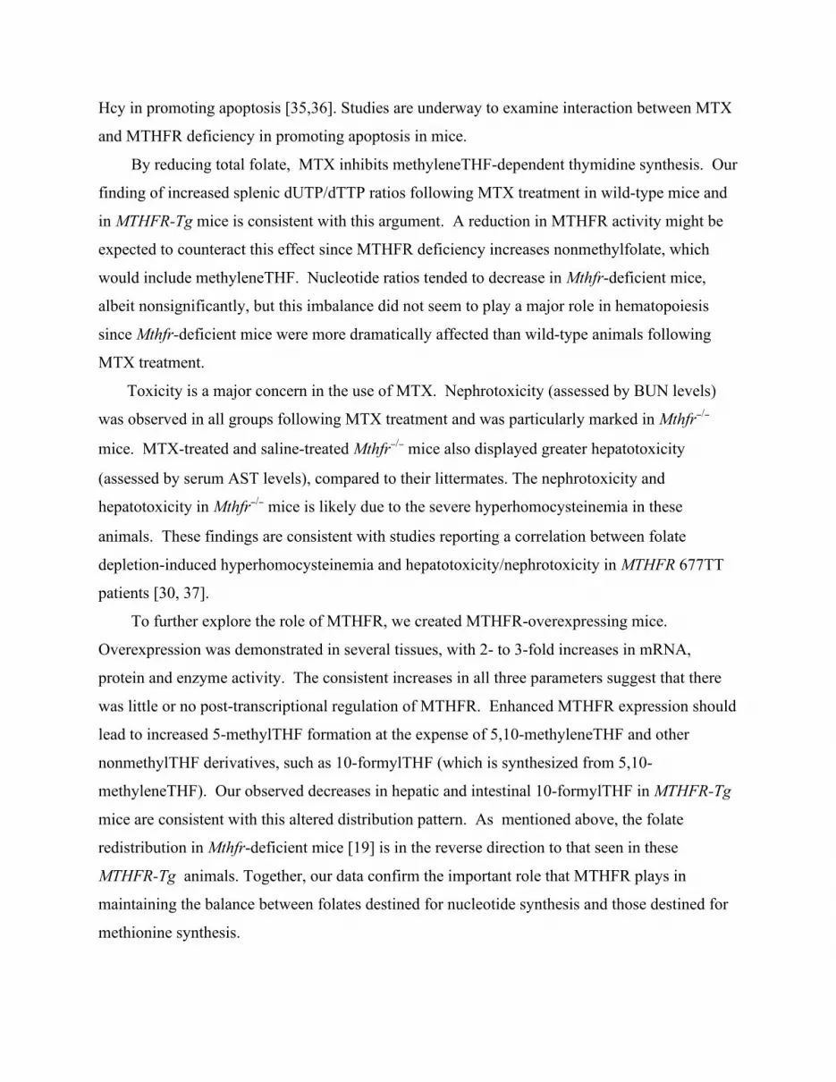
dUTP/dTTP ratios
Since hematopoietic cells are formed and destroyed in spleen, we examined this tissue for
nucleotide levels to determine if imbalances in nucleotide pools, due to altered distributions of
folate derivatives, could have contributed to reduced proliferation and decreased numbers of
hematopoietic cells. MTX-treated Mthfr+/+ mice had significantly higher dUTP/dTTP ratios,
compared to saline-treated Mthfr+/+ mice (Figure 4); this finding suggests that MTX reduced the
levels of 5,10-methyleneTHF that is required for dUMP conversion to dTMP. Mthfr+/− and
Mthfr−/− mice had nonsignificantly lower dUTP/dTTP ratios compared to their wild-type
littermates. Although this result was not statistically significant, it is consistent with the reported
increase in nonmethyl folates, which include the MTHFR substrate 5,10-methyleneTHF, in
MTHFR-deficient humans and mice [2,19].
Toxicity
MTX can cause significant toxicity to kidney and liver. Serum blood urea nitrogen
(BUN) and aspartate aminotransferase (AST) levels were measured as markers of nephrotoxicity
and hepatotoxicity, respectively. Compared to saline controls, MTX-treated mice had

11
significantly higher serum levels of BUN (Figure 5A) and borderline significant increases in
AST (Figure 5B; p=0.08). Mthfr−/− mice had the highest levels of BUN and AST levels,
especially following MTX treatment. Significant differences between Mthfr−/− mice and their
MTHFR-expressing littermates were observed for BUN and AST.
Generation and characterization of MTHFR-overexpressing transgenic mice
Generation
To overexpress MTHFR, we constructed an insertion targeting vector, in which the
human MTHFR cDNA, driven by the ubiquitous CMV promoter, was inserted into the Hprt
locus [23]. Intermediate constructs (pIRES-MTHFR, pCMVMR or pENTRMR) were
transfected into cultured cells (N2a and RAW264.7 cell lines) to confirm MTHFR
overexpression by Western blotting and enzyme assays (data not shown). The final construct
pHPRTMR was transfected into Hprt- embryonic stem (ES) cells and multiple HAT- resistant
clones were recovered. Six clones were further analyzed and all contained the transfected
construct as demonstrated by amplification of a 2 kb segment that covered the MTHFR coding
sequence, segments upstream of the MTHFR cDNA (in the CMV promoter) and a segment
encompassing the polyadenylation site. These 6 clones were also shown to have a 2-fold
increase in enzyme activity. Three clones were injected into blastocysts and 2 chimeric male
mice generated from one cell line transmitted the MTHFR transgene, giving rise to germline
females.
To identify transgene-bearing mice, we used a PCR method in which the MTHFR
transgene and the Mthfr pseudogene (Mthfr-ps), a retrotransposon with high homology to a
mouse Mthfr cDNA segment [26], are both amplified (95 bp). PCR products were incubated
with EcoRI that cuts the transgene (66 bp+29 bp), but not the pseudogene (Figure S1A, left
panel). With this method, the Mthfr-ps provides a positive control for amplification and the
EcoRI- digested fragments indicate the presence of the transgene. The endogenous gene does
not amplify under these conditions, because the 3’ end of the antisense primer encompasses
sequences in exon 1 and the rest of the oligonucleotide binds to exon 2. Distinction between
heterozygosity and homozygosity for the transgene is performed with a 3-primer PCR (Figure
S1A, right panel).
We also performed Southern blotting of DNA from homozygous transgenic female mice
to confirm that the expected recombination event had occurred at Hprt and to identify positive

12
animals in subsequent generations (Figure S1, B and C). Bands of the expected sizes were
observed for transgenic animals and not in wild-type mice.
Confirmation of overexpression
Germline female mice were mated with C57Bl/6 mice and in male progeny, MTHFR
overexpression was confirmed in various tissues (Figure S2). All studies were performed on
male mice, to avoid confounding by X inactivation, since the transgene is inserted into the X-
linked Hprt locus. MTHFR overexpression was first confirmed by semi-quantitative RT-PCR in
liver and kidney (Figure S2A). There was an approximate 2.5-fold increase in MTHFR mRNA,
normalized to Gapdh, in liver and a 1.4-fold increase in kidney. Western blotting demonstrated
that immunoreactive MTHFR protein levels, relative to β-actin, were higher in tested tissues of
MTHFR-Tg mice compared to those in wild-type mice. Increases were 1.8-fold in liver, 4.5-fold
in kidney, 1.9-fold in intestine, 2.5-fold in brain and 2.5-fold in heart (Figure S2B).
Immunohistochemistry was also performed for liver, kidney and intestine of wild-type and
transgenic mice, and relative signal intensities were consistent with the results from Western
blotting (data not shown).
MTHFR enzyme activity was increased 2.8-fold in liver, 2-fold in kidney, 1.3-fold in
intestine, 1.7-fold in brain and 2.4-fold in heart (Figure S2C). Results for enzyme activity were
consistent with the increases in protein levels on Western blots. Although there was some
variability, transgenic mice appeared to have a 2- to 3-fold average increase in MTHFR
expression, with somewhat lower increases in intestine.
Phenotype
Both heterozygous and homozygous MTHFR-Tg mice were viable, healthy and grew
normally. Mating of Tg/+ females with C57Bl/6 mice produced offspring with the expected 1:1
proportions of wild-type: transgenic mice (n=133:133). Wild-type: transgenic ratios for male
and female offspring were similar (n= 65:65 for males and n= 68:68 for females). These data
suggest that there was no significant fetal loss of MTHFR-Tg animals. Matings of heterozygotes
produced viable homozygous MTHFR-Tg females in expected proportions.
There were no gross pathological changes in 3, 12, 18 month-old male MTHFR-Tg mice
following total necropsy and examination of over 30 tissues. Complete blood count and routine
biochemical assessments were similar to those of wild-type littermates (3 mice per genotype in
each age group).

13
Metabolite levels
Measurements of reduced folate pools (Table 1) and of relevant tissue metabolites
(Table 2) in MTHFR-Tg and wild-type littermates provided evidence for altered fluxes of one-
carbon units. Transgenic mice did not show changes in folate distributions in plasma, where
most of the folate is in the form of 5-methylTHF, the MTHFR product (Table 1). Brain, which
has low quantities of total folate compared to liver and intestine, also did not display any
genotype-specific differences. However, in liver and duodenum, MTHFR-Tg mice had
significantly lower levels of 10-formylTHF. Evaluation of metabolites in the homocysteine
remethylation and transsulfuration pathways revealed some genotype-specific and tissue-specific
differences (Table 2). MTHFR-Tg mice had an increase of methionine in brain, an increase of
glutathione in liver, and a decrease of cysteine in duodenum.
The decrease in 10-formylTHF is consistent with enhanced conversion of nonmethylTHF
to methylTHF by MTHFR. Increased availability of methylTHF would drive methionine
synthesis by homocysteine remethylation, as shown by the significant increase of methionine in
brain and a nonsignificant increase in liver.
MTHFR-Tg and wild-type littermates were also examined for deoxyribonucleotide pools
(dUTP/dTTP ratios) in liver, duodenum and brain. Global DNA methylation was assessed in
liver, duodenum and brain by 2 methods: HPLC and TLC. There were no significant genotype
differences in these parameters (data not shown, n= 5 per genotype).
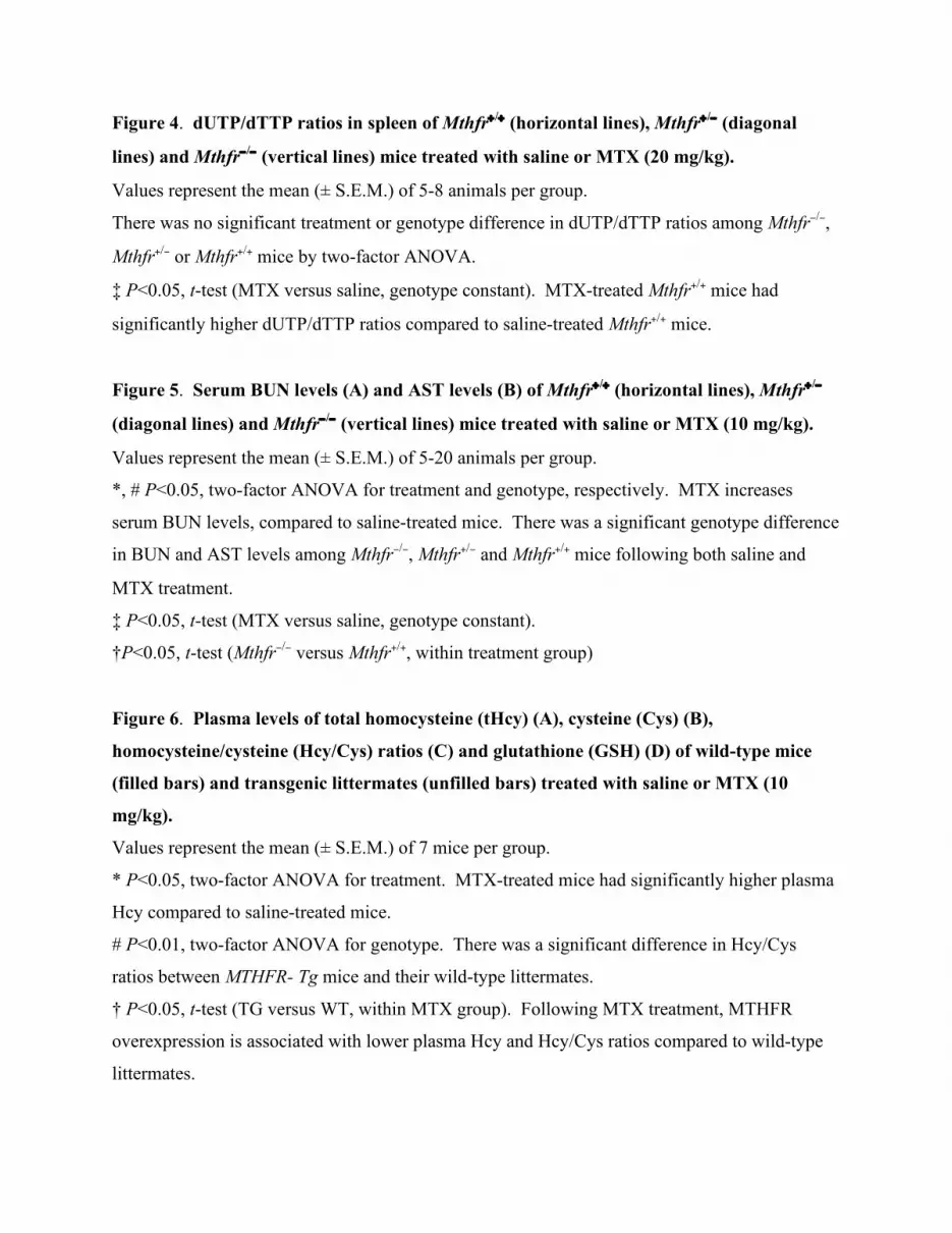
Effects of MTHFR overexpression on MTX treatment
Plasma thiols
Mice treated with MTX (10 mg/kg) had higher plasma homocysteine compared to mice
injected with saline (Figure 6). However, mice with MTHFR overexpression had lower plasma
homocysteine levels and lower homocysteine/cysteine ratios following MTX treatment
compared to their MTX-treated wild-type littermates.
Hematological parameters
Hematocrit, hemoglobin and RBC number in MTX (10 mg/kg or 20 mg/kg)-treated mice
were significantly lower compared to those in saline-treated mice (Figure 7, data shown for
MTX 10 mg/kg). Compared to saline-treated mice, these three parameters were significantly
reduced in MTX-treated MTHFR-Tg mice, but unaffected in MTX-treated wild-type mice.

14
Furthermore, in MTX-treated mice, MTHFR overexpression was associated with lower
hematocrit, hemoglobin and RBC number compared to wild-type littermates.
dUTP/dTTP ratios
MTX treatment significantly increased dUTP/dTTP ratios in spleen of both genotype
groups, suggesting that there was decreased folate-dependent conversion of dUMP to dTMP due
to the drug (Figure 8). This observation is similar to the trend discussed above for MTX-treated
Mthfr-deficient mice. However, these ratios were significantly higher in MTHFR-Tg mice
compared to wild-type littermates, in both the saline- and MTX-treated groups, suggesting that
MTHFR overexpression also compromises the conversion of dUMP to dTMP. This observation
is consistent with enhanced conversion of 5,10-methyleneTHF to 5-methylTHF through MTHFR
overexpression, which decreases availability of 5,10-methyleneTHF for thymidine synthesis.
Toxicity
There were no significant treatment or genotype differences in serum BUN or AST levels
in the experiments on MTHFR-Tg mice and their wild-type littermates (data not shown).

15
DISCUSSION
Genetic disruptions in folate metabolism may influence the efficacy or toxicity of
chemotherapeutics that target enzymes in folate metabolism. To understand the regulation of
one-carbon metabolism and the interaction between MTHFR activity and MTX, we used a well-
characterized mouse model deficient in MTHFR (Mthfr−/− and Mthfr+/−) and generated a new
model that overexpresses MTHFR (MTHFR-Tg).
By inhibiting DHFR, MTX limits the reduction of dihydrofolate to THF, thereby
disrupting all THF-dependent reactions [7]. Two of these reactions, thymidine and purine
synthesis, are reduced due to decreased availability of 5,10-methyleneTHF and 10-formylTHF,
respectively, and result in an inhibition of DNA synthesis. MTHFR activity may affect MTX
response since it regulates the distribution of one-carbon groups between thymidylate synthesis
and methylation reactions. Several studies have suggested that MTX-induced myelosuppression
may be exacerbated in individuals carrying the MTHFR 677 polymorphism [11,13,16,17]. In
contrast, a study of this polymorphism and MTX response in breast cells in vitro concluded that
the reduction in MTHFR activity was associated with reduced sensitivity to MTX [15]. Our
results are consistent with the former findings since MTX-treated Mthfr−/− and Mthfr+/− mice
exhibited significant myelosuppression (reduction in hematocrit, hemoglobin and RBC number
compared with saline-treated mice), whereas Mthfr+/+ mice did not show this response. Since the
product of the MTHFR reaction, 5-methylTHF, is the primary circulatory form of folate, the low
circulating folate levels in 677TT patients or in Mthfr-deficient mice may be compounded by the
additional folate-lowering effect of MTX treatment. This folate deficiency may cause significant
DNA damage or reduce DNA synthesis, particularly in rapidly proliferating hematopoietic cells.
We have previously reported that Mthfr−/− mice have lower plasma total folate levels (25% of
wild-type) and a lower proportion of 5-methylTHF in plasma, liver and brain [19].
An alternate mechanism for enhanced myelosuppression in Mthfr-deficient mice is increased
apoptosis of hematopoietic cells. Apoptosis may be triggered by the high circulating
homocysteine levels in Mthfr−/− and Mthfr+/− mice. Several studies have demonstrated a pro-
apoptotic effect of homocysteine in cortical neurons and cultured lymphocytes, possibly through
an increase in oxidative stress [31,32] and we have directly demonstrated increased apoptosis in
brain of Mthfr−/− mice [33]. Furthermore, a metabolite of Hcy, Hcy thiolactone, has been shown
to be elevated in hyperhomocysteinemic humans and mice [34], and may be more efficient than
16
Hcy in promoting apoptosis [35,36]. Studies are underway to examine interaction between MTX
and MTHFR deficiency in promoting apoptosis in mice.
By reducing total folate, MTX inhibits methyleneTHF-dependent thymidine synthesis. Our
finding of increased splenic dUTP/dTTP ratios following MTX treatment in wild-type mice and
in MTHFR-Tg mice is consistent with this argument. A reduction in MTHFR activity might be
expected to counteract this effect since MTHFR deficiency increases nonmethylfolate, which
would include methyleneTHF. Nucleotide ratios tended to decrease in Mthfr-deficient mice,
albeit nonsignificantly, but this imbalance did not seem to play a major role in hematopoiesis
since Mthfr-deficient mice were more dramatically affected than wild-type animals following
MTX treatment.
Toxicity is a major concern in the use of MTX. Nephrotoxicity (assessed by BUN levels)
was observed in all groups following MTX treatment and was particularly marked in Mthfr−/−
mice. MTX-treated and saline-treated Mthfr−/− mice also displayed greater hepatotoxicity
(assessed by serum AST levels), compared to their littermates. The nephrotoxicity and
hepatotoxicity in Mthfr−/− mice is likely due to the severe hyperhomocysteinemia in these
animals. These findings are consistent with studies reporting a correlation between folate
depletion-induced hyperhomocysteinemia and hepatotoxicity/nephrotoxicity in MTHFR 677TT
patients [30, 37].
To further explore the role of MTHFR, we created MTHFR-overexpressing mice.
Overexpression was demonstrated in several tissues, with 2- to 3-fold increases in mRNA,
protein and enzyme activity. The consistent increases in all three parameters suggest that there
was little or no post-transcriptional regulation of MTHFR. Enhanced MTHFR expression should
lead to increased 5-methylTHF formation at the expense of 5,10-methyleneTHF and other
nonmethylTHF derivatives, such as 10-formylTHF (which is synthesized from 5,10-
methyleneTHF). Our observed decreases in hepatic and intestinal 10-formylTHF in MTHFR-Tg
mice are consistent with this altered distribution pattern. As mentioned above, the folate
redistribution in Mthfr-deficient mice [19] is in the reverse direction to that seen in these
MTHFR-Tg animals. Together, our data confirm the important role that MTHFR plays in
maintaining the balance between folates destined for nucleotide synthesis and those destined for
methionine synthesis.

17
Methionine is formed through homocysteine remethylation, a process which utilizes 5-
methylTHF. Enhanced homocysteine remethylation was observed in the MTHFR-Tg mice, as
demonstrated by elevated brain methionine. Although methionine elevations were not observed
in liver or intestine, there was evidence of enhanced homocysteine transsulfuration to glutathione
in liver, a process which is regulated by SAM. Since SAM activates cystathionine-β-synthase,
the first enzyme in the transsulfuration pathway [38], increased production of methionine and
SAM in these tissues should enhance homocysteine transsulfuration to generate cysteine and
glutathione. Rodents preferentially convert cysteine to glutathione; methionine-enriched diets in
mice resulted in increased plasma glutathione levels without changes in cysteine [39]. The
observations in that study are consistent with our observation of increased glutathione in liver.
The decrease in cysteine in the intestine is consistent with enhanced conversion of cysteine to
glutathione although we did not observe a change in glutathione levels in this tissue. Brain does
not have an active transsulfuration pathway [40], as indicated by the relatively low baseline
levels of cysteine in brain compared to other tissues, and the absence of a change in
transsulfuration metabolites in the transgenic mice. In our earlier study of mice with decreased
MTHFR expression (Mthfr−/− and Mthfr+/−), we observed the reverse pattern i.e., a decrease in
methionine and glutathione with an increase in cysteine in liver [41].
We did not observe significant decreases in 5,10-methyleneTHF levels in transgenic
mice. Nonetheless, there were two manifestations of methyleneTHF reductions: the decrease in
10-formylTHF, as mentioned above, and the increase in the dUTP/dTTP ratio in spleen,
indicating compromised activity of thymidylate synthase presumably due to the decrease in its
substrate 5,10-methyleneTHF. The altered nucleotide ratios in spleen of transgenic mice were
observed in untreated and MTX-treated mice.
MTX treatment resulted in the expected decreases in several hematopoietic parameters
(hematocrit, hemoglobin and RBC number) in both MTHFR-Tg and wild-type mice. MTHFR-Tg
mice responded similarly to Mthfr−/− mice, with greater reductions in these parameters compared
to wild-type littermates. This finding, as well as the pronounced MTX-induced dUTP/dTTP
ratio increase, may be due to the inherent reduction in nonmethylTHF in MTHFR-
overexpressing mice. These decreases in thymidine synthesis and in proliferation of cells in
transgenic mice suggest that increased MTHFR expression may enhance chemosensitivity to
MTX.

18
A major side effect of MTX, hyperhomocysteinemia, is a risk factor for coronary artery
disease, stroke and neurologic impairment [42, 43]. In our study, MTHFR overexpression was
protective against MTX-induced homocysteine increases; MTX treatment increased plasma
homocysteine levels in wild-type mice but not in MTHFR-Tg littermates. In addition, MTHFR-
Tg mice did not have elevations in markers of nephrotoxicity or hepatotoxicity, in contrast to our
observations in Mthfr−/− mice.
There were no differences in homocysteine levels or in hematopoietic parameters
between untreated wild-type and transgenic mice. This finding suggests that the 2- to 3-fold
increase in MTHFR activity alone was not enough to disturb these functions. However, the
combined stress of genetic disruption and drug treatment led to more significant effects on
homocysteine remethylation and to a greater inhibition of proliferation. In summary, an increase
in MTHFR expression during MTX treatment may enhance chemosensitivity of hematopoietic
cells to MTX, through an increase in dUTP/dTTP ratios, without enhancing nephrotoxicity or
hyperhomocysteinemia. A decrease in MTHFR expression during MTX treatment has similar
effects on chemosensitivity but is associated with enhanced toxicity and hyperhomocysteinemia.
The enhanced chemosensitivity in this situation may be due to homocysteine-mediated apoptosis.
There is significant inter-patient variability in response to MTX treatment, and MTX
toxicity is often the reason for discontinuation of therapy. Pharmacogenetic analysis of
polymorphisms in folate-metabolizing enzymes may provide a useful tool to predict MTX
efficacy or toxicity and thereby maximize the benefit : risk ratio. Our studies illustrate the critical
role of MTHFR in MTX chemosensitivity and the importance of pharmacogenetic testing for the
MTHFR polymorphism and possibly other polymorphisms in folate metabolism.

19
REFERENCES
1. Frosst P, Blom HJ, Milos R, Goyette P, Sheppard CA, Matthews RG, et al. A candidate
genetic risk factor for vascular disease: A common mutation in methylenetetrahydrofolate
reductase. Nat Genet 1995; 10: 111-113.
2. Bagley PJ, Selhub J. A common mutation in the methylenetetrahydrofolate reductase
gene is associated with an accumulation of formylated tetrahydrofolates in red blood cells. Proc
Natl Acad Sci USA 1998; 95: 13217-13220.
3. Friso S, Choi SW, Girelli D, Mason JB, Dolnikowski GG, Bagley PJ, et al. A common
mutation in the 5,10-methylenetetrahydrofolate reductase gene affects genomic DNA
methylation through an interaction with folate status. Proc Natl Acad Sci USA 2002; 99: 5606-
5611.
4. Schwahn B, Rozen R. Polymorphisms in the methylenetetrahydrofolate reductase gene:
Clinical consequences. Am J Pharmacogenomics 2001; 1: 189-201.
5. Ma J, Stampfer MJ, Giovannucci E, Artigas C, Hunter DJ, Fuchs C, et al.
Methylenetetrahydrofolate reductase polymorphism, dietary interactions, and risk of colorectal
cancer. Cancer Res 1997; 57: 1098-1102.
6. Skibola CF, Smith MT, Kane E, Roman E, Rollinson S, Cartwright RA, et al.
Polymorphisms in the methylenetetrahydrofolate reductase gene are associated with
susceptibility to acute leukemia in adults. Proc Natl Acad Sci USA 1999; 96: 12810-12815.
7. Longo-Sorbello GS, Bertino JR. Current understanding of methotrexate pharmacology
and efficacy in acute leukemias. Use of newer antifolates in clinical trials. Haematologica 2001;
86: 121-127.
8. Fiskerstrand T, Ueland PM, Refsum H. Folate depletion induced by methotrexate affects
methionine synthase activity and its susceptibility to inactivation by nitrous oxide. J Pharmacol
Exp Ther 1997; 282: 1305-1311.
9. Winter-Vann AM, Kamen BA, Bergo MO, Young SG, Melnyk S, James SJ, et al.
Targeting ras signaling through inhibition of carboxyl methylation: An unexpected property of
methotrexate. Proc Natl Acad Sci USA 2003; 100: 6529-6534.
10. Kremer JM. Methotrexate pharmacogenomics. Ann Rheum Dis 2006; 65: 1121-1123.

20
11. Ulrich CM, Yasui Y, Storb R, Schubert MM, Wagner JL, Bigler J, et al.
Pharmacogenetics of methotrexate: Toxicity among marrow transplantation patients varies with
the methylenetetrahydrofolate reductase c677t polymorphism. Blood 2001; 98: 231-234.
12. Ranganathan P, McLeod HL. Methotrexate pharmacogenetics: The first step toward
individualized therapy in rheumatoid arthritis. Arthritis Rheum 2006; 54: 1366-1377.
13. Toffoli G, Veronesi A, Boiocchi M, Crivellari D. Mthfr gene polymorphism and severe
toxicity during adjuvant treatment of early breast cancer with cyclophosphamide, methotrexate,
and fluorouracil (cmf). Ann Oncol 2000; 11: 373-374.
14. Toffoli G, Cecchin E. Uridine diphosphoglucuronosyl transferase and
methylenetetrahydrofolate reductase polymorphisms as genomic predictors of toxicity and
response to irinotecan-, antifolate- and fluoropyrimidine-based chemotherapy. J Chemother
2004; 16 Suppl 4: 31-35.
15. Sohn KJ, Croxford R, Yates Z, Lucock M, Kim YI. Effect of the
methylenetetrahydrofolate reductase c677t polymorphism on chemosensitivity of colon and
breast cancer cells to 5-fluorouracil and methotrexate. J Natl Cancer Inst 2004; 96: 134-144.
16. Toffoli G, Russo A, Innocenti F, Corona G, Tumolo S, Sartor F, et al. Effect of
methylenetetrahydrofolate reductase 677c-->t polymorphism on toxicity and homocysteine
plasma level after chronic methotrexate treatment of ovarian cancer patients. Int J Cancer 2003;
103: 294-299.
17. Chiusolo P, Reddiconto G, Casorelli I, Laurenti L, Sora F, Mele L, et al. Preponderance
of methylenetetrahydrofolate reductase c677t homozygosity among leukemia patients intolerant
to methotrexate. Ann Oncol 2002; 13: 1915-1918.
18. Chen Z, Karaplis AC, Ackerman SL, Pogribny IP, Melnyk S, Lussier-Cacan S, et al.
Mice deficient in methylenetetrahydrofolate reductase exhibit hyperhomocysteinemia and
decreased methylation capacity, with neuropathology and aortic lipid deposition. Hum Mol
Genet 2001; 10: 433-443.
19. Ghandour H, Chen Z, Selhub J, Rozen R. Mice deficient in methylenetetrahydrofolate
reductase exhibit tissue-specific distribution of folates. J Nutr 2004; 134: 2975-2978.
20. Margolis S, Philips FS, Sternberg SS. The cytotoxicity of methotrexate in mouse small
intestine in relation to inhibition of folic acid reductase and of DNA synthesis. Cancer Res 1971;
31: 2037-2046.

21
21. Durand P, Fortin LJ, Lussier-Cacan S, Davignon J, Blache D. Hyperhomocysteinemia
induced by folic acid deficiency and methionine load--applications of a modified hplc method.
Clin Chim Acta 1996; 252: 83-93.
22. Cross DR, Miller BJ, James SJ. A simplified hplc method for simultaneously quantifying
ribonucleotides and deoxyribonucleotides in cell extracts or frozen tissues. Cell Prolif 1993; 26:
327-336.
23. Denarier E, Forghani R, Farhadi HF, Dib S, Dionne N, Friedman HC, et al. Functional
organization of a schwann cell enhancer. J Neurosci 2005; 25: 11210-11217.
24. Goyette P, Christensen B, Rosenblatt DS, Rozen R. Severe and mild mutations in cis for
the methylenetetrahydrofolate reductase (mthfr) gene, and description of five novel mutations in
mthfr. Am J Hum Genet 1996; 59: 1268-1275.
25. Goyette P, Pai A, Milos R, Frosst P, Tran P, Chen Z, et al. Gene structure of human and
mouse methylenetetrahydrofolate reductase (mthfr). Mamm Genome 1998; 9: 652-656.
26. Leclerc D, Darwich-Codore H, Rozen R. Characterization of a pseudogene for murine
methylenetetrahydrofolate reductase. Mol Cell Biochem 2003; 252: 391-395.
27. Kuo KC, McCune RA, Gehrke CW, Midgett R, Ehrlich M. Quantitative reversed-phase
high performance liquid chromatographic determination of major and modified
deoxyribonucleosides in DNA. Nucleic Acids Res 1980; 8: 4763-4776.
28. Trasler J, Deng L, Melnyk S, Pogribny I, Hiou-Tim F, Sibani S, et al. Impact of dnmt1
deficiency, with and without low folate diets, on tumor numbers and DNA methylation in min
mice. Carcinogenesis 2003; 24: 39-45.
29. Melnyk S, Pogribna M, Pogribny IP, Yi P, James SJ. Measurement of plasma and
intracellular s-adenosylmethionine and s-adenosylhomocysteine utilizing coulometric
electrochemical detection: Alterations with plasma homocysteine and pyridoxal 5'-phosphate
concentrations. Clin Chem 2000; 46: 265-272.
30. Schmitz JC, Grindey GB, Schultz RM, Priest DG. Impact of dietary folic acid on reduced
folates in mouse plasma and tissues. Relationship to dideazatetrahydrofolate sensitivity. Biochem
Pharmacol 1994; 48: 319-325.
31. Kruman, II, Culmsee C, Chan SL, Kruman Y, Guo Z, Penix L, et al. Homocysteine elicits
a DNA damage response in neurons that promotes apoptosis and hypersensitivity to
excitotoxicity. J Neurosci 2000; 20: 6920-6926.

22
32. Mangiagalli A, Samuele A, Armentero MT, Bazzini E, Nappi G, Blandini F. Effects of
homocysteine on apoptosis-related proteins and anti-oxidant systems in isolated human
lymphocytes. European journal of biochemistry / FEBS 2004; 271: 1671-1676.
33. Chen Z, Schwahn BC, Wu Q, He X, Rozen R. Postnatal cerebellar defects in mice
deficient in methylenetetrahydrofolate reductase. Int J Dev Neurosci 2005; 23: 465-474.
34. Chwatko G, Boers GH, Strauss KA, Shih DM, Jakubowski H. Mutations in
methylenetetrahydrofolate reductase or cystathionine beta-synthase gene, or a high-methionine
diet, increase homocysteine thiolactone levels in humans and mice. FASEB J 2007;21:1707-
1713.
35. Kerkeni M, Tnani M, Chuniaud L, Miled A, Maaroufi K, Trivin F. Comparative study on
in vitro effects of homocysteine thiolactone and homocysteine on huvec cells: Evidence for a
stronger proapoptotic and proinflammative homocysteine thiolactone. Molec Cell Biochem
2006;291:119-126.
36. Jakubowski H. The molecular basis of homocysteine thiolactone-mediated vascular
disease. Clin Chem Lab Med 2007;45:1704-1716.
37. van Ede AE, Laan RF, Blom HJ, Huizinga TW, Haagsma CJ, Giesendorf BA, et al. The
c677t mutation in the methylenetetrahydrofolate reductase gene: A genetic risk factor for
methotrexate-related elevation of liver enzymes in rheumatoid arthritis patients. Arthritis Rheum
2001; 44: 2525-2530.
38. Prudova A, Bauman Z, Braun A, Vitvitsky V, Lu SC, Banerjee R. S-adenosylmethionine
stabilizes cystathionine beta-synthase and modulates redox capacity. Proc Natl Acad Sci USA
2006; 103: 6489-6494.
39. Likogianni V, Janel N, Ledru A, Beaune P, Paul JL, Demuth K. Thiol compounds
metabolism in mice, rats and humans: Comparative study and potential explanation of rodents
protection against vascular diseases. Clin Chim Acta 2006; 372: 140-146.
40. Mudd SH, Finkelstein JD, Irreverre F, Laster L. Transsulfuration in mammals.
Microassays and tissue distributions of three enzymes of the pathway. J Biol Chem 1965; 240:
4382-4392.
41. Schwahn BC, Laryea MD, Chen Z, Melnyk S, Pogribny I, Garrow T, et al. Betaine rescue
of an animal model with methylenetetrahydrofolate reductase deficiency. Biochem J 2004; 382:
831-840.

23
42. Hoekstra M, Haagsma CJ, Doelman CJ, van de Laar MA. Intermittent rises in plasma
homocysteine in patients with rheumatoid arthritis treated with higher dose methotrexate. Ann
Rheum Dis 2005; 64: 141-143.
43. Kishi S, Griener J, Cheng C, Das S, Cook EH, Pei D, et al. Homocysteine,
pharmacogenetics, and neurotoxicity in children with leukemia. J Clin Oncol 2003; 21: 3084-
3091.
44. Tran P, Leclerc D, Chan M, Pai A, Hiou-Tim F, Wu Q, et al. Multiple transcription start
sites and alternative splicing in the methylenetetrahydrofolate reductase gene result in two
enzyme isoforms. Mamm Genome 2002; 13: 483-492.
Table 1. Folate derivatives in plasma, liver, duodenum and brain of wild-type (Wt) and
transgenic (Tg) mice
Plasma Liver Duodenum Brain
Folate derivative Wt Tg Wt Tg Wt Tg Wt Tg 5,10-methyleneTHF +THF 2.6
(0.5)
2.0
(0.3)
90.1
(4.6)
89.7
(7.7)
75.0
(23.3)
51.9
(10.8)
4.2
(0.1)
4.9
(0.3)
5-methylTHF 27.6
(1.0)
28.7
(1.7)
36.5
(1.9)
40.2
(2.7)
25.5
(7)
13.0
(2.3)
1.8
(0.2)
2.0
(0.2)
10-formylTHF 1.1
(0.2)
1.3
(0.4)
16.9
(1.7)
10.3*
(1.5)
14.0
(4.2)
4.4*
(0.6)
0.04
(0.02)
0.06
(0.06)
DHF+F 3.6
(0.9)
3.7
(0.7)
3.8
(1.5)
6.8
(1.9)
0.4
(0.2)
0.7
(0.2)
0.10
(0.05)
0.20
(0.06)
Total folate 34.9
(0.8)
35.6
(2.4)
147.2
(5.0)
147.0
(9.5)
114.9
(34.0)
70.0
(12.9)
6.1
(0.3)
7.2
(0.5)
Values are mean of 5 mice per genotype, with standard deviation in parentheses. Plasma units,
pmol/ml; tissue units, pmol/mg protein.
*P<0.05 for comparison between Tg and Wt in the same tissue.
10-FormylTHF levels in liver and duodenum were significantly decreased in Tg compared to Wt
littermates.

24
THF, Tetrahydrofolate; DHF, Dihydrofolate; F, Folate.

25
Table 2. Metabolites in liver, duodenum and brain of wild-type (Wt) and transgenic
(Tg) mice.
Liver Duodenum Brain
Metabolite Wt Tg
Wt Tg Wt Tg
SAH (pmol/mg protein) 219.2
(20.1)
263.3
(26.6)
209.5
(29.2)
254.7
(43)
110.8
(7.2)
140.6
(12.7)
SAM (pmol/mg protein) 492.9
(98.7)
531.8
(76.6)
630.2
(151.7)
687.0
(126.4)
472.6
(40.5)
459.0
(107.7)
SAM/SAH 2.4
(0.6)
2.1
(0.3)
2.1
(0.3)
3.0
(0.7)
4.4
(0.5)
2.9
(0.7)
Adenosine (pmol/mg
protein)
872.0
(83.5)
869.9
(77.9)
903.8
(70.6)
898.6
(78.2)
1629.6
(51.0)
1662.0
(45.9)
Methionine (nmol/mg
protein)
35.2
(4.4)
45.9
(10.4)
83.3
(8.8)
73.0
(10.8)
45.3
(6.8)
66.3*
(5.6)
Homocysteine (nmol/mg
protein)
0.60
(0.03)
0.70
(0.06)
2.40
(0.30)
2.10
(0.30)
0.30
(0.04)
0.40
(0.03)
GSH (nmol/mg protein) 39.1
(2.8)
46.9*
(1.1)
28.7
(6.3)
25.6
(2.5)
22.6
(1.1)
20.9
(0.6)
CysGly (nmol/mg
protein)
4.1
(0.5)
4.2
(0.4)
13.4
(1.0)
12.5
(1.7)
19.6
(0.9)
16.8
(1.0)
Cysteine (nmol/mg
protein)
12.6
(1.2)
12.3
(1.3)
50.6
(7.6)
26.0*
(1.2)
3.9
(0.2)
5.1
(0.9)

26
Values are mean of 5 mice per genotype with standard deviation is in parentheses.
*P<0.05 for comparison between Tg and Wt in the same tissue.
Methionine levels in brain were increased, glutathione levels in liver were increased, and
cysteine levels in duodenum were decreased in Tg compared to Wt littermates.
SAH, S-adenosylhomocysteine; SAM, S-adenosylmethionine; GSH, Glutathione; CysGly,
Cysteinylglycine.

27
FIGURE CAPTIONS
Figure 1. Folate metabolic pathway. MTHFR, methylenetetrahydrofolate reductase; DHFR,
dihydrofolate reductase; TS, thymidylate synthase; THF, tetrahydrofolate; DHF, dihydrofolate;
SAM, S-adenosylmethionine; SAH, S-adenosylhomocysteine.
Figure 2. Plasma levels of total homocysteine (tHcy) (A), cysteine (Cys) (B),
homocysteine/cysteine (Hcy/Cys) ratios (C) and glutathione (GSH) (D) of Mthfr+/+
(horizontal lines), Mthfr+/− (diagonal lines) and Mthfr−/− (vertical lines) mice treated with
saline or MTX (10 mg/kg).
Values represent the mean (± S.E.M.) of 5-20 mice per group.
*, # P<0.05, two-factor ANOVA for treatment and genotype, respectively. MTX-treated mice
had significantly higher plasma Hcy and Hcy/Cys ratios compared to saline-treated mice.
There was a significant difference in plasma Hcy and cysteine levels, and Hcy/Cys ratios among
Mthfr−/−, Mthfr+/− and Mthfr+/+ mice.
†P<0.05, t-test (Mthfr−/− versus Mthfr+/+ , within treatment group)
∫ P<0.05, t-test (Mthfr+/− versus Mthfr+/+, within treatment group)
Figure 3. Hematocrit (A), hemoglobin (B), number of red blood cells (RBC) (C) and
number of white blood cells (WBC) (D) of Mthfr+/+ (horizontal lines), Mthfr+/− (diagonal
lines) and Mthfr−/− (vertical lines) mice treated with saline or MTX (20 mg/kg).
Values represent the mean (± S.E.M.) of 5-8 mice per group.
*, ¶ P<0.01, two-factor ANOVA for treatment and interaction between treatment and genotype,
respectively. Hematocrit, hemoglobin, and RBC number were significantly lower in MTX-
treated mice compared to saline-treated mice.
‡P<0.005, t-test (MTX versus saline, genotype constant). Mthfr−/− and Mthfr+/− mice are affected
by MTX; Mthfr+/+ mice are not.
†P<0.05, t-test (Mthfr−/− versus Mthfr+/+, within treatment group)
∫ P<0.05, t-test (Mthfr+/− versus Mthfr+/+, within treatment group)
28
Figure 4. dUTP/dTTP ratios in spleen of Mthfr+/+ (horizontal lines), Mthfr+/− (diagonal
lines) and Mthfr−/− (vertical lines) mice treated with saline or MTX (20 mg/kg).
Values represent the mean (± S.E.M.) of 5-8 animals per group.
There was no significant treatment or genotype difference in dUTP/dTTP ratios among Mthfr−/−,
Mthfr+/− or Mthfr+/+ mice by two-factor ANOVA.
‡ P<0.05, t-test (MTX versus saline, genotype constant). MTX-treated Mthfr+/+ mice had
significantly higher dUTP/dTTP ratios compared to saline-treated Mthfr+/+ mice.
Figure 5. Serum BUN levels (A) and AST levels (B) of Mthfr+/+ (horizontal lines), Mthfr+/−
(diagonal lines) and Mthfr−/− (vertical lines) mice treated with saline or MTX (10 mg/kg).
Values represent the mean (± S.E.M.) of 5-20 animals per group.
*, # P<0.05, two-factor ANOVA for treatment and genotype, respectively. MTX increases
serum BUN levels, compared to saline-treated mice. There was a significant genotype difference
in BUN and AST levels among Mthfr−/−, Mthfr+/− and Mthfr+/+ mice following both saline and
MTX treatment.
‡ P<0.05, t-test (MTX versus saline, genotype constant).
†P<0.05, t-test (Mthfr−/− versus Mthfr+/+, within treatment group)
Figure 6. Plasma levels of total homocysteine (tHcy) (A), cysteine (Cys) (B),
homocysteine/cysteine (Hcy/Cys) ratios (C) and glutathione (GSH) (D) of wild-type mice
(filled bars) and transgenic littermates (unfilled bars) treated with saline or MTX (10
mg/kg).
Values represent the mean (± S.E.M.) of 7 mice per group.
* P<0.05, two-factor ANOVA for treatment. MTX-treated mice had significantly higher plasma
Hcy compared to saline-treated mice.
# P<0.01, two-factor ANOVA for genotype. There was a significant difference in Hcy/Cys
ratios between MTHFR- Tg mice and their wild-type littermates.
† P<0.05, t-test (TG versus WT, within MTX group). Following MTX treatment, MTHFR
overexpression is associated with lower plasma Hcy and Hcy/Cys ratios compared to wild-type
littermates.

29
Figure 7. Hematocrit (A), hemoglobin (B), number of red blood cells (RBC) (C) and
number of white blood cells (WBC) (D) of wild-type mice (filled bars) and transgenic
littermates (unfilled bars) treated with saline or MTX (10 mg/kg).
Values represent the mean (± S.E.M.) of 7 mice per group.
* P<0.01, two-factor ANOVA for treatment. Hematocrit, hemoglobin, and number of RBCs
were significantly lower in MTX-treated mice compared to saline-treated mice.
‡P<0.001, t-test (MTX versus saline treatment, genotype constant). Hematocrit, hemoglobin and
RBC number in MTX-treated MTHFR-Tg mice were adversely affected compared to their saline-
treated littermates.
† P<0.05, t-test (TG versus WT, within MTX group). Following MTX treatment, MTHFR-Tg
mice have lower hematocrit, hemoglobin and RBC number than MTHFR-Wt mice.
Figure 8. dUTP/dTTP ratios in spleen of wild-type mice (filled bars) and transgenic
littermates (unfilled bars) treated with saline or MTX (10 mg/kg).
Values represent the mean (± S.E.M.) of 5 animals per group.
*, # P<0.05, two-factor ANOVA for treatment and genotype.
MTX increases dUTP/dTTP ratios, compared to saline-treated mice. dUTP/dTTP ratios in
MTHFR-Tg are significantly increased compared to wild-type littermates in both saline-treated
and MTX-treated groups.
Figure S1. Methodologies for determination of genotypes. (A) PCR-based genotyping. In the
left panel, genomic DNA from male transgenic (Tg) and wild-type (Wt) mice was amplified
using 2 primers that generate a band of the same size (95 bp) in the MTHFR transgene and in the
Mthfr-ps pseudogene. EcoR1 cuts the transgene amplicon (66 bp+29 bp), but not the pseudogene.
The expected bands were obtained for Wt (95 bp) and Tg (66 bp+29 bp). In the right panel, the
expected bands were obtained for Wt (151 bp), heterozygous (Tg/+) (151 bp+95 bp) and
homozygous female transgenic mice (Tg/Tg) (95 bp). For both panels, controls (C) were
obtained from PCR reactions performed without genomic DNA. (B) Southern blot analysis of
the MTHFR cDNA insertion into the Hprt locus. Digestion of genomic DNA with DraIII + ScaI
generated restriction fragments of 7.1 kb, 3.5 kb and 4.0 kb. ApaLI + ScaI digestion generated
bands of 7.5 kb, 3.9 kb + 3.5 kb, and 3.8 kb representing Tg, Wt, and ps alleles, respectively.

30
Bands of 7.2 kb and 12.4 kb + 1.9 kb, after PshAI digestion, represent Tg and Wt alleles,
respectively. The expected sizes of bands are indicated on the right; molecular weight markers
are shown on the left. (C) Schematic representations of the expected bands from the transgene
on the X chromosome, the endogenous Mthfr gene on chromosome 4 and Mthfr-ps on
chromosome 5. Positions of the probe used for hybridization are shown for each chromosomal
segment. On the X chromosome, the striped region of the transgene indicates the region of
homology with the targeting vector.
Figure S2. Confirmation of MTHFR overexpression. (A) Semi-quantitative RT-PCR analysis
of MTHFR expression in liver and kidney of MTHFR-Tg mice and wild-type littermates. RT-
PCR was performed using primers spanning MTHFR exons 2 – 5, with mouse Gapdh as the
internal control. Pictures shown for liver and kidney were generated from different gels. (B)
Western blot analysis of MTHFR expression in various tissues of MTHFR-Tg mice and wild-
type littermates. Equal amounts of protein (50 µg) were loaded in each lane, and rabbit
antiserum against purified porcine liver MTHFR was used to probe MTHFR, with β-actin as the
internal control. There are 2 isoforms of MTHFR (77 kDa and 70 kDa) in some tissues, as
previously reported [41]. The 2.2 kb human cDNA encodes the polypeptide of 70 kDa.
Autoradiographs from liver, kidney and heart were generated from one gel, and autoradiographs
from intestine and brain were generated from another gel. (C) The MTHFR enzyme assay,
which assays MTHFR in the physiologically reverse direction, was performed using radiolabeled
5-methylTHF monoglutamate as substrate and menadione as electron acceptor. Specific
MTHFR activity was calculated for each sample; values are mean of 3 – 5 mice ± S.E.M. Filled
bars depict values for wild-type mice; unfilled bars, Tg littermates.


