Embed Size (px)
Citation preview
B R A I N R E S E A R C H 1 3 8 8 ( 2 0 1 1 ) 1 6 7 – 1 7 7
ava i l ab l e a t www.sc i enced i r ec t . com
www.e l sev i e r . com/ loca te /b ra i n res
Research Report
Altered gray matter morphometry and resting-state functionaland structural connectivity in social anxiety disorder☆
Wei Liaoa,1, Qiang Xua,1, Dante Mantinib, Jurong Dinga, João Paulo Machado-de-Sousac,Jaime E.C. Hallakc, Clarissa Trzesniakc, Changjian Qiud, Ling Zenga, Wei Zhangd,⁎⁎,José Alexandre S. Crippac, Qiyong Gonge, Huafu Chena,⁎aKey Laboratory for NeuroInformation of Ministry of Education, School of Life Science and Technology,University of Electronic Science and Technology of China, Chengdu 610054, PR ChinabLaboratory of Neuro-psychophysiology, K. U. Leuven Medical School, Leuven, BelgiumcDepartment of Neuroscience and Behavior, Ribeirão Preto Medical School,University of São Paulo and National Institute for Translational Medicine (INCT-TM, CNPq), São Paulo, BrazildMental Health Center, Department of Radiology, West China Hospital of Sichuan University, Chengdu 610041, PR ChinaeHuaxi MR Research Center (HMRRC), Department of Radiology, West China Hospital of Sichuan University, Chengdu 610041, PR China
A R T I C L E I N F O
☆ The authors declare no competing intere⁎ Correspondence to: H. Chen, Key Laborato
University of Electronic Science and Technol⁎⁎ Correspondence to:W. Zhang, Mental Healt
85582944.E-mail addresses: [email protected] (H
1 These authors contributed equally to this
0006-8993/$ – see front matter © 2011 Elsevidoi:10.1016/j.brainres.2011.03.018
A B S T R A C T
Article history:Accepted 7 March 2011Available online 12 March 2011
In social anxiety disorder (SAD), impairments in limbic/paralimbic structures are associatedwith emotional dysregulation and inhibition of the medial prefrontal cortex (MPFC). Little isknown, however, about alterations in limbic and frontal regions associatedwith the integratedmorphometric, functional, and structural architecture of SAD. Whether altered gray mattervolume is associated with altered functional and structural connectivity in SAD. Threetechniques were used with 18 SAD patients and 18 healthy controls: voxel-basedmorphometry; resting-state functional connectivity analysis; and diffusion tensor imagingtractography. SAD patients exhibited significantly decreased graymatter volumes in the rightposterior inferior temporal gyrus (ITG) and right parahippocampal/hippocampal gyrus (PHG/HIP). Graymatter volumes in these two regions negatively correlatedwith the fear factor of theLiebowitz Social Anxiety Scale. In addition, we found increased functional connectivity in SADpatients between the right posterior ITG and the left inferior occipital gyrus, and between theright PHF/HIP and left middle temporal gyrus. SAD patients had increased right MPFC volume,along with enhanced structural connectivity in the genu of the corpus callosum. Reducedlimbic/paralimbic volume, together with increased resting-state functional connectivity,suggests the existence of a compensatory mechanism in SAD. Increased MPFC volume,consonant with enhanced structural connectivity, suggests a long-time overgeneralization ofstructural connectivity and a role of this area in themediation of clinical severity. Overall, our
Keywords:Social anxiety disorderGray matter morphometryResting stateFunctional connectivityStructural connectivity
sts.ry for NeuroInformation of Ministry of Education, School of Life Science and Technology,ogy of China, Chengdu 610054, PR China. Fax: +86 28 83208238.h Center, West China Hospital of Sichuan University, Chengdu 610041, PR China. Fax: +86 28
. Chen), [email protected] (W. Zhang).work.
er B.V. All rights reserved.

168 B R A I N R E S E A R C H 1 3 8 8 ( 2 0 1 1 ) 1 6 7 – 1 7 7
results may provide a valuable basis for future studies combining morphometric, functionaland anatomical data in the search for a comprehensive understanding of the neural circuitryunderlying SAD.
© 2011 Elsevier B.V. All rights reserved.
1. Introduction
Social anxiety disorder (SAD) generally refers to the excessivefear and avoidance of a wide array of social situations (Filhoet al., 2010; Stein and Stein, 2008). There is increasing evidencethat altered brain functional patterns underlie the processing ofemotional stimuli under conditions of social fear and anxiety(Etkin and Wager, 2007; Freitas-Ferrari et al., 2010; Phan et al.,2006; Stein et al., 2002; Straube et al., 2004; Tillfors et al., 2001).Specifically, the most consistent findings from functionalneuroimaging studies in SAD patients indicate increasedactivity in the limbic/paralimbic system (amygdala, insula,parahippocampus and hippocampus, and dorsal anterior cin-gulate cortex) and altered activity in the prefrontal cortex (Blairet al., 2008a,b; Gentili et al., 2008; Lorberbaum et al., 2004; Steinet al., 2002; Straube et al., 2004). In addition, there is significantand consistent evidenceof hyperactivationof limbic/paralimbicstructures associated with emotional dysregulation during theprocessing of facial emotionand inclassical exposure situations(Mathew et al., 2001; Phan et al., 2006; Stein et al., 2002).Furthermore, themedialprefrontal cortex (MPFC) playsapivotalrole in themodulationand inhibitionof excessive limbicactivity(Etkin et al., 2006; Tillfors et al., 2001, 2002). This is a strongindication that anxiety might be associated with an imbalancein limbic-cortical connectivity (Bishop, 2007; Warwick et al.,2008). Together, these results suggest that brain functionalchanges in SAD involve impaired long-range communicationduring sensory and emotional processing (Danti et al., 2010) andat rest (Liao et al., 2010b).
Brainmorphometry analysis is complementary to functionalinvestigations, as it is a powerful tool to detect brain abnormal-ities that may vary progressively during the course of illness(Ferrari et al., 2008). Potts and colleagues found no significantdifferences between the volumes of the caudate, putamen,thalamus, andwhole brainof SADpatients andhealthy controls(Potts et al., 1994). More recently, Irle and coworkers found thatgeneralized SAD patients had reduced amygdalar and hippo-campal size (Irle et al., 2010). It is worth noting that both studiesuseda regionof interest (ROI) to investigate brainmorphometry.This may potentially bias the detection of positive findings(Ferrari et al., 2008; Freitas-Ferrari et al., 2010).More importantly,this approach is not adequate for the study of large neuralsystems. Accordingly, it is still unclear whether gray mattervolume alterations in SAD are related to altered functionalconnectivity within brain networks.
The analysis of task-independent, resting-state functionalconnectivity may allow for a better understanding of SADmechanisms (Warwick et al., 2008). One pioneering resting-state functional magnetic resonance imaging (fMRI) study ongeneralized anxiety disorder (GAD) has demonstrated thataberrant brain networks selectively interact with the basolat-eral and centromedial subregions of the amygdala (Etkinet al., 2009). More recently, our group found diffuse changes
on widely distributed brain networks in SAD patients at rest(Liao et al., 2010a). However, functional connectivity analysis isbased on low-frequency spontaneous fluctuations of the bloodoxygen level-dependent (BOLD) signal between distant brainregions. It is, therefore, unable to reveal whether they areanatomically connected or not (Honey et al., 2009). Conversely,diffusion tensor imaging (DTI) fiber tractography allows thestudy of human structural connectivity (Catani et al., 2002;Johansen-Berg and Rushworth, 2009). Recently, a DTI study onSAD documented reduced fractional anisotropy (FA) in theright uncinate fasciculus, underlying aberrant limbic-prefron-tal interactions (Phan et al., 2009). In addition, increased graymatter volume is likely to reflect exuberant synaptic connec-tivity (Supekar et al., 2010). These studies suggested theimportance of studying changes in structural connectivity toimprove the understanding of the neural underpinnings ofSAD.
Themain objective of this study was to test whether alteredgray matter volume is associated with altered functional andstructural connectivity in SAD. For the first time, three imagingmodalities (voxel-based morphometry [VBM], resting-statefunctional connectivity, and DTI tractography) were combinedin order to perform a comprehensive evaluation of the neuralcircuitry underlying SAD.
2. Results
2.1. Clinical and demographic data
The group's demographics and clinical scores are shown inTable 1. Compared with healthy controls (HC), SAD patientshad significantly higher scores (total, fear factor, and avoid-ance factor) in the Liebowitz Social Anxiety Scale (LSAS), theHamilton Anxiety Rating Scale (HAMA), and the HamiltonDepression Rating Scale (HAMD). They also had higher levelsof anxiety as assessedwith the Spielberger State-Trait AnxietyInventory (STAI). The remaining characteristics did not differacross groups.
2.2. Morphometry analysis
Compared with HC, SAD patients had significantly decreasedgray matter volumes in the right posterior inferior temporalgyrus (ITG), the parahippocampal gyrus (PHG), and the lefthippocampal gyrus (HIP). Increased gray matter volume inSAD patients was found in the right MPFC (Fig. 1, Table S1).Significant negative correlations (p<0.05, Bonferroni corrected)were observed between the fear factor of the LSAS and graymatter volumes in the right posterior ITG (r=−0.69, p=0.004) andleft PHG (r=−0.69, p=0.004) in the SAD group. There were nosignificant correlations in the HC group between gray mattervolume in those brain regions and LSAS scores (Table S1).

Table 1 – Clinical and demographic data.
SAD(n=18) a
HC(n=18)
SAD vs. HC
T value p value
Gender(male/female)
12/6 13/5 – 0.73b
Age (y) 22.67±3.77 21.89±3.69 0.63 0.54Education (y) 14.11±1.53 14.17±2.04 −0.09 0.93Duration (m) 49.22±40.17 – – –LSASTotal score 54.39±11.96 19.11±7.89 10.45 <0.0001Fear factor 28.50±6.20 8.50±5.07 10.59 <0.0001Avoidance factor 25.89±7.32 10.61±4.86 7.38 <0.0001
HAMD 7.33±6.15 1.06±1.59 4.19 <0.001HAMA 6.39±4.97 0.94±1.55 4.43 <0.0001STAISTAI-T 48.33±7.40 32.67±4.97 7.46 <0.0001STAI-SPre-scan 41.11±8.66 31.28±4.75 4.22 <0.001Post-scan 37.11±9.50 32.94±7.12 1.49 0.15
Head MotionTranslation (mm) 0.03±0.01 0.04±0.02 −0.61 0.55Rotation (°) 0.03±0.01 0.03±0.02 −0.20 0.84
Abbreviations: SAD=social anxiety disorder; HC=healthy controls;LSAS=Liebowitz SocialAnxiety Scale;HAMA=HamiltonAnxietyRatingScale; HAMD=Hamilton Depression Rating Scale; STAI=SpielbergerState-Trait Anxiety Inventory.a Questionnaire data are given as means±standard deviation (SD).b p Value was obtained by the Kruskal–Wallis test. Other p valueswere obtained by two-sample, two-tailed t-tests.
169B R A I N R E S E A R C H 1 3 8 8 ( 2 0 1 1 ) 1 6 7 – 1 7 7
2.3. Functional connectivity analysis
The right posterior ITGwas found to be functionally connectedin both groups with the bilateral dorsolateral prefrontal, theposterior parietal, and the middle temporal cortex; negativecorrelationsmainly involving the inferior andmiddle occipitalcortices, and the bilateral precentral gyrus (Fig. 2A). Comparedwith HC, SAD patients had increased functional connectivity(weak negative connection) in the left inferior occipital gyrus(IOG) (Fig. 3A). Significant positive correlations (p<0.05,Bonferroni corrected) were observed between the degree offunctional connectivity in the left IOG and the total (r=0.58,p=0.012) and avoidance (r=0.60, p=0.008) scores in the LSAS(Fig. 3A and Table S2).
The left PHG was functionally connected with the bilateralanterior superior temporal gyrus, the hippocampal gyrus, andthe entorhinal cortex (Kahn et al., 2008) in both groups.Significant negative connectivities included mainly a rightfronto-parietal network (Fig. 2B). Compared with HC, SADpatients had increased functional connectivity in the leftmiddle temporal gyrus (MTG) (Fig. 3B). We observed positivecorrelations (p<0.05, uncorrected) between the fear factor ofthe LSAS (r=0.54, p=0.039) and the degree of functionalconnectivity in the left MTG for the SAD group. This analogouscorrelation was not significant in the HC group (r=−0.19,p=0.456) (Table S2).
The right MPFC showed significantly positive functionalconnectivity in the posterior cingulate cortex/precuneus, thebilateral inferior parietal, gyrus, middle temporal, and supe-rior frontal gyrus. These regions are considered to be part of
the default mode network (DMN) (Raichle et al., 2001). Bothgroups had significantly negative functional connectivityregions involving executive control network (ECN) and thedorsal attention network (DAN) (Corbetta and Shulman, 2002)(Fig. 2C). There were no significant differences in functionalconnectivity between SAD patients and HC when the rightMPFC was used as seed.
2.4. Structural connectivity analysis
Structural connections of the right posterior ITG included thearcuate fasciculus fiber bundle (Fig. 4A). This is a lateralassociative bundle composed of long and short fibers connect-ing the perisylvian cortex with the frontal, parietal, andtemporal lobes. A seed ROI located in the left PHG showed theinferior fronto-occipital fasciculus, a ventral associative bundlearising from the ventral occipital lobe and going toward theorbitofrontal cortex (Fig. 4B). When the seed ROI was located inthe right MPFC, fiber tracts passed through the genu (anteriorportion) of the corpus callosum to interconnect left and rightprefrontal cortical regions (Fig. 4C). ComparingSADpatients andHC, the fiber density (weight) and mean FA showed significantincrease (p<0.05, Bonferroni corrected) only in fibers passingthrough the genu of the corpus callosum. In addition, both thefiber density and mean FA of the three seed ROI showed nosignificant correlations with the LSAS.
3. Discussion
To the best of our knowledge, the present cross-sectional studyis the first to combine structural MRI, resting-state functionalconnectivity, and diffusion tensor imaging tractography toinvestigate alterations in limbic and frontal regions in SAD.Patients had significantly decreased graymatter volumes in theright ITG and right limbic/paralimbic structures (PHG and HIP)that are compatible with increased functional connectivity. Inaddition, increased gray matter volume was observed in theright MPFC, along with enhanced structural connectivity.
Only two earlier studies using brain morphometry analysesin SAD are available to date. The first indicated no difference instriatal structures (caudate, putamen, and thalamus) (Pottset al., 1994), while the second found reduced amygdalar andhippocampal volumes (Irle et al., 2010). Since these studiesperformed ROI-based morphometry comparisons, the possibil-ity of both false positive and false-negative findings should beconsidered (Freitas-Ferrari et al., 2010). As opposed to thoseprevious studies, we have now used an automatedwhole-brainVBM-based technique for the investigationof SAD,whichallowsassessing areas not previously investigated by the ROI-basedmethod. Decreased right posterior ITG volume was found inSAD patients compared to HC. Functionally, the ITG is involvedinvisual perception (Ishai et al., 1999) and is linked to theventralvisual pathway (Baddeley et al., 1997). Furthermore, the ITG hasreciprocal connectionswith the amygdala and the orbitofrontalcortex, which are predominantly involved in the affectiveprocessing of negative expressions (Iidaka et al., 2001). It hasalso been reported that the processing of external social cues insocial phobia is characterized by a negative bias (Arrais et al.,2010; Clark and Wells, 1995; Machado-de-Sousa et al., 2010).
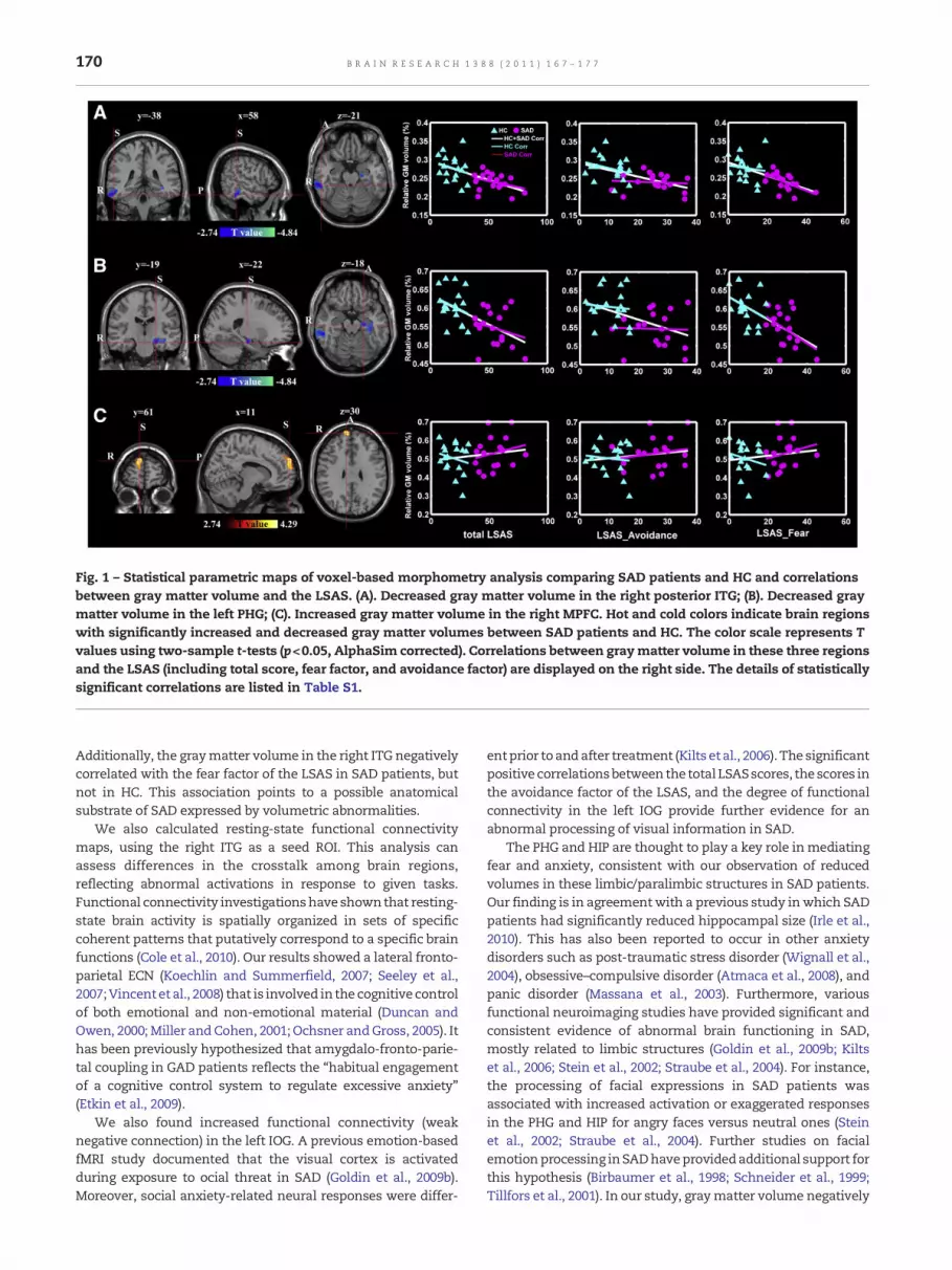
Fig. 1 – Statistical parametric maps of voxel-based morphometry analysis comparing SAD patients and HC and correlationsbetween gray matter volume and the LSAS. (A). Decreased gray matter volume in the right posterior ITG; (B). Decreased graymatter volume in the left PHG; (C). Increased gray matter volume in the right MPFC. Hot and cold colors indicate brain regionswith significantly increased and decreased gray matter volumes between SAD patients and HC. The color scale represents Tvalues using two-sample t-tests (p<0.05, AlphaSim corrected). Correlations between graymatter volume in these three regionsand the LSAS (including total score, fear factor, and avoidance factor) are displayed on the right side. The details of statisticallysignificant correlations are listed in Table S1.
170 B R A I N R E S E A R C H 1 3 8 8 ( 2 0 1 1 ) 1 6 7 – 1 7 7
Additionally, the graymatter volume in the right ITG negativelycorrelated with the fear factor of the LSAS in SAD patients, butnot in HC. This association points to a possible anatomicalsubstrate of SAD expressed by volumetric abnormalities.
We also calculated resting-state functional connectivitymaps, using the right ITG as a seed ROI. This analysis canassess differences in the crosstalk among brain regions,reflecting abnormal activations in response to given tasks.Functional connectivity investigationshave shownthat resting-state brain activity is spatially organized in sets of specificcoherent patterns that putatively correspond to a specific brainfunctions (Cole et al., 2010). Our results showed a lateral fronto-parietal ECN (Koechlin and Summerfield, 2007; Seeley et al.,2007;Vincent et al., 2008) that is involved in the cognitive controlof both emotional and non-emotional material (Duncan andOwen, 2000; Miller and Cohen, 2001; Ochsner andGross, 2005). Ithas been previously hypothesized that amygdalo-fronto-parie-tal coupling in GAD patients reflects the “habitual engagementof a cognitive control system to regulate excessive anxiety”(Etkin et al., 2009).
We also found increased functional connectivity (weaknegative connection) in the left IOG. A previous emotion-basedfMRI study documented that the visual cortex is activatedduring exposure to ocial threat in SAD (Goldin et al., 2009b).Moreover, social anxiety-related neural responses were differ-
entprior toandafter treatment (Kilts etal., 2006). The significantpositive correlationsbetween the total LSASscores, the scores inthe avoidance factor of the LSAS, and the degree of functionalconnectivity in the left IOG provide further evidence for anabnormal processing of visual information in SAD.
The PHG and HIP are thought to play a key role inmediatingfear and anxiety, consistent with our observation of reducedvolumes in these limbic/paralimbic structures in SAD patients.Our finding is in agreementwith a previous study inwhich SADpatients had significantly reduced hippocampal size (Irle et al.,2010). This has also been reported to occur in other anxietydisorders such as post-traumatic stress disorder (Wignall et al.,2004), obsessive–compulsive disorder (Atmaca et al., 2008), andpanic disorder (Massana et al., 2003). Furthermore, variousfunctional neuroimaging studies have provided significant andconsistent evidence of abnormal brain functioning in SAD,mostly related to limbic structures (Goldin et al., 2009b; Kiltset al., 2006; Stein et al., 2002; Straube et al., 2004). For instance,the processing of facial expressions in SAD patients wasassociated with increased activation or exaggerated responsesin the PHG and HIP for angry faces versus neutral ones (Steinet al., 2002; Straube et al., 2004). Further studies on facialemotionprocessing inSADhaveprovidedadditional support forthis hypothesis (Birbaumer et al., 1998; Schneider et al., 1999;Tillfors et al., 2001). In our study, graymatter volume negatively

Fig. 2 – Resting-state functional connectivity maps for HC (left column) and SAD patients (right column). Seed ROI (regions withgraymatter volume reduction) located in the right posterior ITG (MNI coordinates: 58, −38, −21) (top row). Seed ROI (regionswithgraymatter volume reduction) located in the left PHG (MNI coordinates: −22, −19, −18) (middle row). Seed ROI (regions with graymatter volume increase) located in the right MPFC (MNI coordinates: 11, 61, 30) (bottom row). Hot and cold colors indicate brainregions with significant positive and negative temporal correlations with the selected seed ROI, respectively. Color scalesrepresent T values in each functional connectivity map using one-sample t-test (p<0.05, false discovery rate criterion formultiple comparisons).
171B R A I N R E S E A R C H 1 3 8 8 ( 2 0 1 1 ) 1 6 7 – 1 7 7
correlatedwith the fear factor of theLSAS in the SADgrouponly.Thus, we would argue that sustained emotional dysregulationcould lead toprogressiveatrophyof thePHGandHIP responsiblefor emotional processing and social behaviors.
The functional connectivity analysis involving the rightPHG/HIP as seed showed increased functional connectivity inthe right MTG in SAD patients compared to controls. Previousstudies observed increased neural activity related to different

Fig. 3 – Increased resting-state functional connectivity in SAD patients compared to HC. (A). Regions of increased resting-statefunctional connectivity in the left inferior occipital gyrus (MNI coordinates: −42, −75, −6; Brodmann's area 19; voxelsize=22 mm3) with the seed ROI located in the right posterior ITG; (B). Regions showing increased resting-state functionalconnectivity in the left middle temporal gyrus (MNI coordinates: 60, −21, −3; Brodmann's area 21; voxel size=25 mm3) with theseed ROI located in the left PHG. Hot colors indicate brain regionswith significantly increased functional connectivity. The colorscale represents T values using two-sample t-tests (p<0.05, AlphaSim corrected). Correlations between the degree of functionalconnectivity (Z(r) value) of these two regions and the LSAS (including total score, fear factor, and avoidance factor, respectively)are displayed on the right side. The details of statistically significant correlations are listed in Table S2.
172 B R A I N R E S E A R C H 1 3 8 8 ( 2 0 1 1 ) 1 6 7 – 1 7 7
facial emotional expressions in subjects with SAD not only inthe amygdala and the insula, but also in medial temporalstructures (Gentili et al., 2008; Stein et al., 2002). For instance,SAD patients had increased parahippocampal–hippocampalactivity and anxiety symptoms during a stressful publicspeaking task, which was reversed after treatment with theSSRI citalopram (Furmark et al., 2005). We therefore speculate
Fig. 4 – Fiber tracts of three selected ROI (one subject from the HCfasciculus passing through the right posterior ITG; (B). Fiber tractsPHG; (C). Fiber tracts in the genu (anterior portion) of the corpus cthe obtained fibers is based on the standard RGB code applied toplane, green: dorsoventral orientation, blue: rostro-caudal direction track. Vertical bars indicate estimated standard errors. ** reprcorrected.
that increased connectivity in the right MTG network iseffective in the inhibition or extinction of excessive cortico-limbic activity in anxiety disorders in general, and in SAD inparticular.
Another finding was the presence of significantly increasedright MPFC volume. TheMPFC, an important brain region in theneurocircuitry of SAD, plays a pivotal role in the modulation of
group for display purposes). (A). Fiber tracts in the arcuatein inferior fronto-occipital fasciculus passing through the leftallosum passing through the right MPFC. The color-coding ofthe vector at every segment of each fiber (red: medio-lateral
on). Bars represent mean values of fiber density andmean FAesents the statistical significance with p<0.05, Bonferroni

173B R A I N R E S E A R C H 1 3 8 8 ( 2 0 1 1 ) 1 6 7 – 1 7 7
excessive limbic activity (Etkin et al., 2006; Tillfors et al., 2001,2002; Warwick et al., 2008). For example, in two positronemission tomography studies on SAD, Tillfors and colleaguessuggested that regional cerebral blood flow (rCBF) reductions inthe frontal cortex is associated with increased rCBF in theamygdala, HIP, and ITG, all of which have been implicated inemotional dysregulation and failure to inhibit negative affect(Tillfors et al., 2001, 2002). In an fMRI study, Etkin and coworkersrevealed that increased activity in the ventromedial prefrontalcortex and decreased activity in the amygdala were involved inthe extinctionof conditioned fear (Etkin et al., 2006). Ina resting-state single photon emission computed tomography study,Warwick and colleagues demonstrated increased rCBF in thefrontal cortex of SAD patients (Warwick et al., 2008). Althoughdecreases in activity are not necessarily related to decreases involume (Ferrari et al., 2008), we argue that an increased rightMPFC volume may contribute to the progressive and sustainedmodulation and inhibition of excessive limbic activity.
The MPFC is thought to provide information from priorexperiences in the form of memories during the constructionof self-relevant mental simulation (Buckner et al., 2008).Although aberrant global coherent connectivity in the MPFCof SAD patients has been observed at rest (Liao et al., 2010a)and during listening to threat-related words (Zhao et al., 2007),we found no significant differences in functional connectivitywithin this brain region. One possible reason is that volumeincreases are not necessarily related to increases in functionalconnectivity.
Another remarkable finding was that the fiber density(weight) and mean FA on track were significantly increased inthe genu of the corpus callosumwhen the seed ROI was locatedin the right MPFC for the structural connectivity analysis. Thegenu of the corpus callosum was found to connect the left andright prefrontal cortical regions. The activity in the dorsalanterior cingulate cortex (dACC) anterior to the genu of thecorpus callosum has been consistently associated with moodstates (Blair et al., 2008a; Critchley, 2003; Smith et al., 1999) andcorrelated with volume (Boes et al., 2008). This region isactivated, among other situations, when subjects processharsh and disgusted facial expressions (Amir et al., 2005; Blairet al., 1999; Phanet al., 2006; Shin andLiberzon, 2010) or evaluateemotional information (Phan et al., 2003). Together, the dACCand the MPFC are thought to implement regulatory strategiesand to direct attentional control, in addition to reducingcognitive conflict (Kerns et al., 2004). This neural circuitrywould underlie emotional regulation problems in patientswith SAD during cognitive reappraisal (Goldin et al., 2009a).The modulating function of the MPFC and the dACC seems notto be restricted to theprocessing of facial affect, but to also occurin intrinsic structural connectivity associated with long-timeovergeneralization.
Some limitations of the study and future directions forresearch in the field deserve to bementioned. First, furthermulti-modal studies will be necessary to promote deeper insight intothe neurophysiopathology of SAD. In addition, although we re-cruited a consistent number of SAD patients who were medica-tion- and comorbidity-free, our findings should be extended tolarger SAD samples in the future, especially for VBM analysis.
The present study used a multimodal neuroimaging ap-proach comprising structural MRI, resting-state functional
connectivity, and diffusion tensor imaging tractography toinvestigate the alterations of limbic and frontal areas in SAD.Reduced limbic/paralimbic gray matter volume, together withincreased resting-state functional connectivity, suggests theexistence of a compensatory mechanism in SAD. IncreasedMPFC graymatter volume, consonantwith enhanced structuralconnectivity, suggests a long-time overgeneralization of struc-tural connectivity and a role of this structure in mediatingclinical severity. Overall, our results may provide a valuablebasis for future studies combining morphometric, functional,and anatomical data for a comprehensive understanding of theneural circuitry underlying SAD.
4. Experimental procedures
4.1. Participants
The study was approved by the local ethics committee ofHuaxi Hospital, Sichuan University, and written informedconsent was obtained from all subjects.
The SAD group was composed of 18 patients (22.67±3.77 years, all right-handed) recruited through the MentalHealth Center of Huaxi Hospital, China (Table 1). GeneralizedSAD diagnosis was determined by consensus between twoattending psychiatrists and a trained interviewer using thepatient version of the StructuredClinical Interview for theDSM-IV (SCID). Subjects who presented other psychiatric disordersaccording to the SCID were excluded from the sample.Additionally, we excluded individuals in treatment with psy-chotropic medication or in use of psychoactive substances,those presenting neurological or general medical conditions orthose who had received previous treatment for SAD (eitherpharmacological or psychotherapeutic).
The comparison group consisted of 18 age-, sex-, andeducation-matched HC (21.89±3.69 years, all right-handed).The SCID was used to confirm the absence of past or currentpsychiatric or neurological illness. HC weremedication-free atthe time of the scanning. Additionally, they were interviewedto confirm that there was no history of psychiatric illnessamong their first-degree relatives. All participants in the twogroups were evaluated with the LSAS, STAI, HAMA, andHAMD. More specifically, the STAI was administered in twoparts: the STAI-State score, which measures the level of stateanxiety at the time of completing the scale, and the STAI-Traitscore, which measures the inherent trait anxiety level of thesubject. The STAI-Trait was completed immediately beforeand after the MRI scanning (pre-scanning and post-scanning)session (Campbell et al., 2007).
4.2. Data acquisition
All subjects underwent structural, functional and DTI scanningusing a 3.0T GE-Signa MRI scanner (EXCITE, General Electric,Milwaukee, USA) at Huaxi MR Research Center. Functionalimages were acquired using a single-shot, gradient-recalledecho planar imaging sequence (TR=2000ms, TE=30ms and flipangle=90º, FOV=24 cm, in-plane matrix=64×64, voxel size=3.75×3.75×5, without inter-slice gap, and 30 transverse slices).For each subject, a total of 205 volumeswere acquired, resulting

174 B R A I N R E S E A R C H 1 3 8 8 ( 2 0 1 1 ) 1 6 7 – 1 7 7
in a total scan time of 410 s. Subjects were instructed simplyto rest with their eyes closed, not to think of anything inparticular, and not to fall asleep. Axial anatomical images wereacquiredusing a volumetric three-dimensional spoiled gradientrecalled (SPGR) sequence (TR=8.5 ms, TE=3.4 ms, flipangle=12º, field of view=240×240 mm2, matrix size=512×512,voxel size=0.47×0.47×1 mm3, and 156 axial slices). The DTIacquisition used a single-shot spin-echo planar imaging se-quence in contiguous axial planes covering the whole brain(TR=12000 ms, TE=71.4 ms, field of view=240×240 mm2, flipangle=90º,matrix size=256×256, voxel size=0.94×0.94×3mm3,50 slices). At each slice position, except for S0 (b=0 s/mm2), asingle b-value (b=1000 s/mm2) was applied to 15 non-collineargradient directions.
4.3. Voxel-based morphometry analysis
Voxel-based morphometry analysis (Ashburner, 2009) wasperformed using SPM8 (http://www.fil.ion.ucl.ac.uk/spm).First, all T1-weighted anatomical images were manuallyreoriented to place the anterior commissure at the origin ofthe three-dimensional Montreal Neurological Institute (MNI)space. The images were then segmented into gray matter,white matter, and cerebrospinal fluid (CSF) (Ashburner andFriston, 2005). A diffeomorphic non-linear registration algo-rithm (diffeomorphic anatomical registration through expo-nentiated lie algebra—DARTEL) (Ashburner, 2007) was used tospatially normalize the segmented images. This proceduregenerated a template for a group of individuals. The resultingimages were spatially normalized into the MNI space usingaffine spatial normalization. An additional processing stepconsisted of multiplying each spatially normalized graymatter image by its relative volume before and after normal-ization. This ensured that the total amount of gray matter ineach voxel was preserved. Finally, the resulting gray matterimages were smoothed with an 8-mm full-width half-maxi-mum (FWHM) isotropic Gaussian kernel.
Voxel-wise comparisons of gray matter volume betweengroups were performed using two-sample t-tests. Age andgender (Freitas-Ferrari et al., 2010), along with total gray mattervolume, were modeled as covariates of no interest. Thestatistical significance of group differences was set at p<0.05(combined height threshold p<0.005 andminimum cluster sizeof 1347 voxels), using the AlphaSim program in the REST toolkit(http://sourceforge.net/projects/resting-fmri). The averagedvalues of gray matter volume of all voxels in abnormal areas,corrected for age, gender, and total gray matter volume, werecorrelated with the LSAS (including total score, fear factor, andavoidance factor) using Pearson correlation analysis.
4.4. Functional connectivity analysis
The fMRI images (after exclusion of the first five images to ensuresteady-state longitudinal magnetization) were initially correctedfor temporal differences and head motion. No translation orrotation parameters in any given data set exceeded ±1.5 mmor ±1.5°. Group differences in respect to head translationand rotation have been evaluate elsewhere (Liao et al., 2010a).The functional images were warped into a standard stereotaxicspace at a resolution of 3×3×3mm3, using the MNI echo-planar
imaging template. At last, the imageswere spatially smoothed at8mm FWHM.
Functional connectivity was investigated using a temporalcorrelation approach (Fox et al., 2005, 2009). Brain areas showingreduced/increased gray matter volume were defined as seedROI (thewhole significant cluster). To remove possible spurioussources of variance, the time series were preprocessed asfollows: first, six headmotion parameters, the averaged signalsfrom CSF and white matter, and the global brain signal wereregressed (Foxet al., 2005, 2009); next, the timeserieswereband-pass filtered (0.01~0.08 Hz). A correlation analysis was con-ducted between the seed ROI and the remaining voxels in thebrain. The resulting r valueswere convertedusing Fisher's r-to-ztransformation to improve the Gaussianity of their distribution.
Individual Z value maps in each group were gathered usingone-sample t-test (p<0.05, with false discovery rate [FDR]corrected). To compare the functional connectivity maps be-tween groups, two-sample t-tests were used. Age, gender, andtotal graymatter volume were alsomodeled as covariates of nointerest. The significance level of group differences was set atp<0.05 (combined height threshold of p<0.001 and a minimumcluster size of 22 voxels). The averaged Z values of all voxels inareas with altered connectivity were corrected for age, gender,and total gray matter volume. Subsequently, they were corre-lated with the LSAS (including total score, fear factor andavoidance factor) using Pearson correlation analysis.
4.5. Diffusion tensor imaging tractography analysis
For DTI, head motion was removed by aligning 15 diffusion-weighted volumes to the unweighted S0 image (b=0 s/mm2).Eddy current distortions were corrected by affine registration tothe reference S0 image. Whole brain fiber tracking (an interpo-lated streamline propagation algorithm) was performed in theDTInativespaceusingTrackVis (Schmahmannetal., 2007). Pathtracing proceededuntil either the fractional anisotropy (FA)waslower than 0.15 or the angle between the current and theprevious path segment was higher than 35° (Liao et al., 2010c).As three seed ROI with morphometric alterations were derivedfrom the normalized MNI space, the inverse transformation ofthe spatial normalizationwas applied to acquire the seed ROI inthe native DTI space (Gong et al., 2009; Liao et al., 2010c). Fiberbundles passing through the three ROI were then extractedfrom the total collection of brain fibers. For each seed ROI, theconnection density (number of connections per unit surface) of
fibers can be expressed as w eð Þ = 1S
∑f∈Fe
1l fð Þ, where, S stands for
two-dimensionally intersected surfaces between the indivi-dual's white matter and ROI, Fe is a set including all fiberspassing through the ROI, and l( f ) is the length of fiber f along itstrajectory. S corrects for the slightly variable size of intersectedsurfaces (Hagmann et al., 2008). Mean FA values on the sametract were also extracted. Structural connectivity in three ROIwas compared between groups using two-sample t-tests. Ageand gender were modeled as covariates of no interest. Thesignificance of group differences in each ROI was set at p<0.05,with Bonferroni correction. Furthermore, the fiber densityand mean FA for the three ROI, after correction for age andgender, were correlated with the LSAS (including total score,fear factor, and avoidance factor) using Pearson correlationanalysis.
175B R A I N R E S E A R C H 1 3 8 8 ( 2 0 1 1 ) 1 6 7 – 1 7 7
Supplementary materials related to this article can befound online at doi:10.1016/j.brainres.2011.03.018.
Acknowledgments
This researchwas supported by theNatural Science Foundationof China (Grant nos. 61035006, 90820006 and 30625024 toHC andGQ), the 863 Program (Grant no. 2008AA02Z408 to HC and GQ),the 973 Project (Grant no. 2008CB517407 to WZ), the FlandersResearch Foundation (Grantno. A4/5-SDS15387 toDM) and JASC(1A) and JECH (2) are recipients of a CNPq (Brazil) ResearchAward.
R E F E R E N C E S
Amir, N., Klumpp, H., Elias, J., Bedwell, J.S., Yanasak, N., Miller, L.S.,2005. Increased activation of the anterior cingulate cortexduring processing of disgust faces in individuals with socialphobia. Biol. Psychiatry 57, 975–981.
Arrais, K.C., Machado-de-Sousa, J.P., Trzesniak, C., Santos Filho,A., Ferrari, M.C., Osorio, F.L., Loureiro, S.R., Nardi, A.E.,Hetem, L.A., Zuardi, A.W., Hallak, J.E., Crippa, J.A., 2010.Social anxiety disorder women easily recognize fearfull, sadand happy faces: the influence of gender. J. Psychiatr. Res. 44,535–540.
Ashburner, J., 2007. A fast diffeomorphic image registrationalgorithm. Neuroimage 38, 95–113.
Ashburner, J., 2009. Computational anatomy with the SPMsoftware. Magn. Reson. Imaging 27, 1163–1174.
Ashburner, J., Friston, K.J., 2005. Unified segmentation.Neuroimage 26, 839–851.
Atmaca, M., Yildirim, H., Ozdemir, H., Ozler, S., Kara, B., Ozler, Z.,Kanmaz, E., Mermi, O., Tezcan, E., 2008. Hippocampus andamygdalar volumes in patients with refractoryobsessive-compulsive disorder. Prog. Neuropsychopharmacol.Biol. Psychiatry 32, 1283–1286.
Baddeley, R., Abbott, L.F., Booth, M.C., Sengpiel, F., Freeman, T.,Wakeman, E.A., Rolls, E.T., 1997. Responses of neurons inprimary and inferior temporal visual cortices to natural scenes.Proc. Biol. Sci. 264, 1775–1783.
Birbaumer, N., Grodd, W., Diedrich, O., Klose, U., Erb, M., Lotze, M.,Schneider, F., Weiss, U., Flor, H., 1998. fMRI reveals amygdalaactivation to human faces in social phobics. Neuroreport 9,1223–1226.
Bishop, S.J., 2007. Neurocognitive mechanisms of anxiety: anintegrative account. Trends Cogn. Sci. 11, 307–316.
Blair, R.J., Morris, J.S., Frith, C.D., Perrett, D.I., Dolan, R.J., 1999.Dissociable neural responses to facial expressions of sadnessand anger. Brain 122 (Pt 5), 883–893.
Blair, K., Geraci, M., Devido, J., McCaffrey, D., Chen, G., Vythilingam,M., Ng, P., Hollon, N., Jones, M., Blair, R.J., Pine, D.S., 2008a.Neural response to self- and other referential praise andcriticism in generalized social phobia. Arch. Gen. Psychiatry 65,1176–1184.
Blair, K., Shaywitz, J., Smith, B.W., Rhodes, R., Geraci, M., Jones, M.,McCaffrey, D., Vythilingam, M., Finger, E., Mondillo, K., Jacobs,M., Charney, D.S., Blair, R.J., Drevets, W.C., Pine, D.S., 2008b.Response to emotional expressions in generalized socialphobia and generalized anxiety disorder: evidence for separatedisorders. Am. J. Psychiatry 165, 1193–1202.
Boes, A.D., McCormick, L.M., Coryell, W.H., Nopoulos, P., 2008.Rostral anterior cingulate cortex volume correlates withdepressed mood in normal healthy children. Biol. Psychiatry63, 391–397.
Buckner, R.L., Andrews-Hanna, J.R., Schacter, D.L., 2008. Thebrain's default network: anatomy, function, and relevance todisease. Ann. NY Acad. Sci. 1124, 1–38.
Campbell, D.W., Sareen, J., Paulus, M.P., Goldin, P.R., Stein, M.B.,Reiss, J.P., 2007. Time-varying amygdala response to emotionalfaces in generalized social phobia. Biol. Psychiatry 62, 455–463.
Catani, M., Howard, R.J., Pajevic, S., Jones, D.K., 2002. Virtual in vivointeractive dissection of white matter fasciculi in the humanbrain. Neuroimage 17, 77–94.
Clark, D.M., Wells, A., 1995. A cognitive model of social phobia. In:Heimberg, R.G., Liebowitz, M.R., Hope, D., Schneider, F. (Eds.),Social phobia: Diagnosis, assessment and treatment. Guild-ford, New York, pp. 69–93.
Cole, D.M., Smith, S.M., Beckmann, C.F., 2010. Advances andpitfalls in the analysis and interpretation of resting-state FMRIdata. Front Syst. Neurosci. 4, 8.
Corbetta, M., Shulman, G.L., 2002. Control of goal-directed andstimulus-driven attention in the brain. Nat. Rev. Neurosci. 3,201–215.
Critchley, H., 2003. Emotion and its disorders. Br. Med. Bull. 65,35–47.
Danti, S., Ricciardi, E., Gentili, C., Gobbini, M.I., Pietrini, P.,Guazzelli, M., 2010. Is social phobia a “Mis-Communication”disorder? Brain functional connectivity during face perceptiondiffers between patients with social phobia and healthy controlsubjects. Front Syst. Neurosci. 4, 152.
Duncan, J., Owen, A.M., 2000. Common regions of the humanfrontal lobe recruited by diverse cognitive demands. TrendsNeurosci. 23, 475–483.
Etkin, A., Wager, T.D., 2007. Functional neuroimaging of anxiety: ameta-analysis of emotional processing in PTSD, social anxietydisorder, and specific phobia. Am. J. Psychiatry 164, 1476–1488.
Etkin, A., Egner, T., Peraza, D.M., Kandel, E.R., Hirsch, J., 2006.Resolving emotional conflict: a role for the rostral anteriorcingulate cortex in modulating activity in the amygdala.Neuron 51, 871–882.
Etkin, A., Keller, K.E., Schatzberg, A.F., Menon, V., Greicius, M.D.,2009. Disrupted amygdalar subregion functional connectivityand evidence for a compensatory network in generalizedanxiety disorder. Arch. Gen. Psychiatry 66, 1361–1372.
Ferrari, M.C., Busatto, G.F., McGuire, P.K., Crippa, J.A., 2008.Structural magnetic resonance imaging in anxiety disorders:an update of research findings. Rev. Bras. Psiquiatr. 30, 251–264.
Filho, A.S., Hetem, L.A., Ferrari, M.C., Trzesniak, C., Martin-Santos,R., Borduqui, T., de Lima Osorio, F., Loureiro, S.R., Busatto Filho,G., Zuardi, A.W., Crippa, J.A., 2010. Social anxiety disorder: whatare we losing with the current diagnostic criteria? ActaPsychiatr. Scand. 121, 216–226.
Fox, M.D., Snyder, A.Z., Vincent, J.L., Corbetta, M., Van Essen, D.C.,Raichle, M.E., 2005. The human brain is intrinsically organizedinto dynamic, anticorrelated functional networks. Proc. NatlAcad. Sci. U.S.A. 102, 9673–9678.
Fox, M.D., Zhang, D., Snyder, A.Z., Raichle, M.E., 2009. The globalsignal and observed anticorrelated resting state brain networks.J. Neurophysiol. 101, 3270–3283.
Freitas-Ferrari, M.C., Hallak, J.E., Trzesniak, C., Filho, A.S.,Machado-de-Sousa, J.P., Chagas, M.H., Nardi, A.E., Crippa, J.A.,2010. Neuroimaging in social anxiety disorder: a systematicreview of the literature. Prog. Neuropsychopharmacol. Biol.Psychiatry 34, 565–580.
Furmark, T., Appel, L., Michelgard, A.,Wahlstedt, K., Ahs, F., Zancan,S., Jacobsson, E., Flyckt, K., Grohp, M., Bergstrom, M., Pich, E.M.,Nilsson, L.G., Bani, M., Langstrom, B., Fredrikson, M., 2005.Cerebral blood flow changes after treatment of social phobiawith the neurokinin-1 antagonist GR205171, citalopram, orplacebo. Biol. Psychiatry 58, 132–142.
Gentili, C., Gobbini, M.I., Ricciardi, E., Vanello, N., Pietrini, P.,Haxby, J.V., Guazzelli, M., 2008. Differential modulation ofneural activity throughout the distributed neural system for

176 B R A I N R E S E A R C H 1 3 8 8 ( 2 0 1 1 ) 1 6 7 – 1 7 7
face perception in patients with Social Phobia and healthysubjects. Brain Res. Bull. 77, 286–292.
Goldin, P.R., Manber-Ball, T., Werner, K., Heimberg, R., Gross, J.J.,2009a. Neural mechanisms of cognitive reappraisal of negativeself-beliefs in social anxiety disorder. Biol. Psychiatry 66,1091–1099.
Goldin, P.R., Manber, T., Hakimi, S., Canli, T., Gross, J.J., 2009b.Neural bases of social anxiety disorder: emotional reactivityand cognitive regulation during social and physical threat.Arch. Gen. Psychiatry 66, 170–180.
Gong, G., He, Y., Concha, L., Lebel, C., Gross, D.W., Evans, A.C.,Beaulieu, C., 2009. Mapping anatomical connectivity patternsof human cerebral cortex using in vivo diffusion tensorimaging tractography. Cereb. Cortex 19, 524–536.
Hagmann, P., Cammoun, L., Gigandet, X., Meuli, R., Honey, C.J.,Wedeen, V.J., Sporns, O., 2008. Mapping the structural core ofhuman cerebral cortex. PLoS Biol. 6, e159.
Honey, C.J., Sporns, O., Cammoun, L., Gigandet, X., Thiran, J.P.,Meuli, R., Hagmann, P., 2009. Predicting human resting-statefunctional connectivity from structural connectivity. Proc. NatlAcad. Sci. U.S.A. 106, 2035–2040.
Iidaka, T., Omori, M., Murata, T., Kosaka, H., Yonekura, Y., Okada,T., Sadato, N., 2001. Neural interaction of the amygdala withthe prefrontal and temporal cortices in the processing of facialexpressions as revealed by fMRI. J. Cogn. Neurosci. 13,1035–1047.
Irle, E., Ruhleder, M., Lange, C., Seidler-Brandler, U., Salzer, S.,Dechent, P., Weniger, G., Leibing, E., Leichsenring, F., 2010.Reduced amygdalar and hippocampal size in adults withgeneralized social phobia. J. Psychiatry Neurosci. 35, 126–131.
Ishai, A., Ungerleider, L.G., Martin, A., Schouten, J.L., Haxby, J.V.,1999. Distributed representation of objects in the humanventral visual pathway. Proc. Natl Acad. Sci. U.S.A. 96,9379–9384.
Johansen-Berg, H., Rushworth, M.F., 2009. Using diffusion imagingto study human connectional anatomy. Annu. Rev. Neurosci.32, 75–94.
Kahn, I., Andrews-Hanna, J.R., Vincent, J.L., Snyder, A.Z., Buckner, R.L.,2008. Distinct cortical anatomy linked to subregions of themedialtemporal lobe revealed by intrinsic functional connectivity. J.Neurophysiol. 100, 129–139.
Kerns, J.G., Cohen, J.D., MacDonald III, A.W., Cho, R.Y., Stenger, V.A.,Carter, C.S., 2004. Anterior cingulate conflict monitoring andadjustments in control. Science 303, 1023–1026.
Kilts, C.D., Kelsey, J.E., Knight, B., Ely, T.D., Bowman, F.D., Gross, R.E.,Selvig, A., Gordon, A., Newport, D.J., Nemeroff, C.B., 2006. Theneural correlates of social anxiety disorder and response topharmacotherapy. Neuropsychopharmacology 31, 2243–2253.
Koechlin, E., Summerfield, C., 2007. An information theoreticalapproach to prefrontal executive function. Trends Cogn. Sci.11, 229–235.
Liao, W., Chen, H., Feng, Y., Mantini, D., Gentili, C., Pan, Z., Ding, J.,Duan, X., Qiu, C., Lui, S., Gong, Q., Zhang, W., 2010a. Selectiveaberrant functional connectivity of resting state networks insocial anxiety disorder. Neuroimage 52, 1549–1558.
Liao, W., Qiu, C., Gentili, C., Walter, M., Pan, Z., Ding, J., Zhang, W.,Gong, Q., Chen, H., 2010b. Altered effective connectivitynetwork of the amygdala in social anxiety disorder: aresting-state FMRI study. PLoS ONE 5, e15238.
Liao, W., Zhang, Z., Pan, Z., Mantini, D., Ding, J., Duan, X., Luo, C.,Wang, Z., Tan, Q., Lu, G., Chen, H., 2010c. Default mode networkabnormalities in mesial temporal lobe epilepsy: A Studycombining fMRI and DTI. Hum. Brain Mapp., doi:10.1002/hbm.21076.
Lorberbaum, J.P., Kose, S., Johnson, M.R., Arana, G.W., Sullivan, L.K.,Hamner, M.B., Ballenger, J.C., Lydiard, R.B., Brodrick, P.S., Bohn-ing, D.E., George, M.S., 2004. Neural correlates of speechanticipatory anxiety in generalized social phobia. Neuroreport15, 2701–2705.
Machado-de-Sousa, J.P., Arrais, K.C., Alves, N.T., Chagas, M.H., deMeneses-Gaya, C., Crippa, J.A., Hallak, J.E., 2010. Facial affectprocessing in social anxiety: tasks and stimuli. J. Neurosci.Methods 193, 1–6.
Massana, G., Serra-Grabulosa, J.M., Salgado-Pineda, P., Gasto, C.,Junque, C., Massana, J., Mercader, J.M., 2003. Parahippocampalgray matter density in panic disorder: a voxel-based morpho-metric study. Am. J. Psychiatry 160, 566–568.
Mathew, S.J., Coplan, J.D., Gorman, J.M., 2001. Neurobiologicalmechanisms of social anxiety disorder. Am. J. Psychiatry 158,1558–1567.
Miller, E.K., Cohen, J.D., 2001. An integrative theory of prefrontalcortex function. Annu. Rev. Neurosci. 24, 167–202.
Ochsner, K.N., Gross, J.J., 2005. The cognitive control of emotion.Trends Cogn. Sci. 9, 242–249.
Phan, K.L., Liberzon, I., Welsh, R.C., Britton, J.C., Taylor, S.F., 2003.Habituation of rostral anterior cingulate cortex to repeatedemotionally salient pictures. Neuropsychopharmacology 28,1344–1350.
Phan, K.L., Fitzgerald, D.A., Nathan, P.J., Tancer, M.E., 2006.Association between amygdala hyperactivity to harsh facesand severity of social anxiety in generalized social phobia. Biol.Psychiatry 59, 424–429.
Phan, K.L., Orlichenko, A., Boyd, E., Angstadt, M., Coccaro, E.F.,Liberzon, I., Arfanakis, K., 2009. Preliminary evidence of whitematter abnormality in the uncinate fasciculus in generalizedsocial anxiety disorder. Biol. Psychiatry 66, 691–694.
Potts, N.L., Davidson, J.R., Krishnan, K.R., Doraiswamy, P.M., 1994.Magnetic resonance imaging in social phobia. Psychiatry Res.52, 35–42.
Raichle, M.E., MacLeod, A.M., Snyder, A.Z., Powers, W.J., Gusnard,D.A., Shulman, G.L., 2001. A default mode of brain function.Proc. Natl Acad. Sci. U.S.A. 98, 676–682.
Schmahmann, J.D., Pandya,D.N.,Wang, R.,Dai, G., D'Arceuil, H.E., deCrespigny, A.J., Wedeen, V.J., 2007. Association fibre pathways ofthe brain: parallel observations fromdiffusion spectrum imagingand autoradiography. Brain 130, 630–653.
Schneider, F., Weiss, U., Kessler, C., Muller-Gartner, H.W., Posse, S.,Salloum, J.B., Grodd,W.,Himmelmann, F., Gaebel,W., Birbaumer,N., 1999. Subcortical correlates of differential classicalconditioning of aversive emotional reactions in social phobia.Biol. Psychiatry 45, 863–871.
Seeley, W.W., Menon, V., Schatzberg, A.F., Keller, J., Glover, G.H.,Kenna, H., Reiss, A.L., Greicius, M.D., 2007. Dissociable intrinsicconnectivity networks for salience processing and executivecontrol. J. Neurosci. 27, 2349–2356.
Shin, L.M., Liberzon, I., 2010. The neurocircuitry of fear, stress, andanxiety disorders. Neuropsychopharmacology 35, 169–191.
Smith, K.A., Morris, J.S., Friston, K.J., Cowen, P.J., Dolan, R.J., 1999.Brain mechanisms associated with depressive relapse andassociated cognitive impairment following acute tryptophandepletion. Br. J. Psychiatry 174, 525–529.
Stein, M.B., Stein, D.J., 2008. Social anxiety disorder. Lancet 371,1115–1125.
Stein, M.B., Goldin, P.R., Sareen, J., Zorrilla, L.T., Brown, G.G., 2002.Increased amygdala activation to angry and contemptuousfaces in generalized social phobia. Arch. Gen. Psychiatry 59,1027–1034.
Straube, T., Kolassa, I.T., Glauer, M., Mentzel, H.J., Miltner, W.H.,2004. Effect of task conditions on brain responses tothreatening faces in social phobics: an event-related functionalmagnetic resonance imaging study. Biol. Psychiatry 56,921–930.
Supekar, K., Uddin, L.Q., Prater, K., Amin, H., Greicius, M.D.,Menon, V., 2010. Development of functional and structuralconnectivity within the default mode network in youngchildren. Neuroimage 52, 290–301.
Tillfors, M., Furmark, T., Marteinsdottir, I., Fischer, H., Pissiota, A.,Langstrom, B., Fredrikson, M., 2001. Cerebral blood flow in

177B R A I N R E S E A R C H 1 3 8 8 ( 2 0 1 1 ) 1 6 7 – 1 7 7
subjects with social phobia during stressful speaking tasks: aPET study. Am. J. Psychiatry 158, 1220–1226.
Tillfors, M., Furmark, T., Marteinsdottir, I., Fredrikson, M., 2002.Cerebral blood flow during anticipation of public speaking insocial phobia: a PET study. Biol. Psychiatry 52, 1113–1119.
Vincent, J.L., Kahn, I., Snyder, A.Z., Raichle, M.E., Buckner, R.L., 2008.Evidence for a frontoparietal control system revealed by intrinsicfunctional connectivity. J. Neurophysiol. 100, 3328–3342.
Warwick, J.M., Carey, P., Jordaan, G.P., Dupont, P., Stein, D.J., 2008.Resting brain perfusion in social anxiety disorder: a voxel-wise
whole brain comparison with healthy control subjects. Prog.Neuropsychopharmacol. Biol. Psychiatry 32, 1251–1256.
Wignall, E.L., Dickson, J.M., Vaughan, P., Farrow, T.F.,Wilkinson, I.D.,Hunter, M.D., Woodruff, P.W., 2004. Smaller hippocampalvolume in patients with recent-onset posttraumatic stressdisorder. Biol. Psychiatry 56, 832–836.
Zhao, X.H., Wang, P.J., Li, C.B., Hu, Z.H., Xi, Q., Wu, W.Y., Tang,X.W., 2007. Altered default mode network activity in patientwith anxiety disorders: an fMRI study. Eur. J. Radiol. 63,373–378.