Embed Size (px)
Citation preview

916 Journal of Lipid Research
Volume 42, 2001
Altered phospholipid-apoB-100 interactions and generation of extra membrane material in proteolysis-induced fusion of LDL particles
Markku O. Pentikäinen,* Marja T. Hyvönen,*
,†
Katariina Öörni,* Tiia Hevonoja,* Ari Korhonen,
†,§
Erno M. P. Lehtonen-Smeds,* Mika Ala-Korpela,* and Petri T. Kovanen
1,
*
Wihuri Research Institute,* FIN-00140 Helsinki, Finland; Nuclear Magnetic Resonance Research Group,
†
Department of Physical Sciences, University of Oulu, FIN-90571 Oulu, Finland; and Oulu Polytechnic Institute of Technology,
§
FIN-90250 Oulu, Finland
Abstract Lipid droplets and membrane material are pro-duced in the extracellular matrix of the arterial intima dur-ing atherogenesis. Both in vitro and in vivo experimentationsuggests that fusion of modified LDL particles leads to for-mation of such lipid droplets. Here we applied proton NMRspectroscopy to probe surface phospholipids phosphatidyl-choline (PC) and sphingomyelin (SM) of LDL particles duringproteolytic degradation of apolipoprotein B-100 (apoB-100).Initiation of apoB-100 degradation was accompanied by theabruptly increased intensity of the choline -N(CH
3
)
3
reso-nance of PC molecules, indicating disruption of their inter-actions with apoB-100. However, subsequent particle fusionwas accompanied by a steady decrease in the intensity of thecholine resonances of both PC and SM. Electron micros-copy of the proteolyzed LDL revealed irregularly shapedmultilamellar membranes attached to aggregates of fusedparticles. This suggests formation of membrane materialwith low hydration, in which some of the atomic motionsare hindered. Characterization of the behavior of the sur-face lipids of LDL particles during apoB-100 degradationand other types of LDL modification will aid in understand-ing molecular mechanisms leading to fusion and generationof multilamellar membrane material in the arterial intimaduring atherogenesis.—
Pentikäinen, M. O., M. T. Hyvönen,K. Öörni, T. Hevonoja, A. Korhonen, E. M. P. Lehtonen-Smeds, M. Ala-Korpela, and P. T. Kovanen.
Altered phos-pholipid-apoB-100 interactions and generation of extramembrane material in proteolysis-induced fusion of LDLparticles.
J. Lipid Res.
2001.
42:
916–922.
Supplementary key words
arterial lipid accumulation
•
NMR spec-troscopy
•
proteolytic LDL fusion
The initial stages of atherosclerosis are characterized byretention and accumulation of extracellular lipid in theform of lipid droplets (
�
100–400 nm in diameter), vesi-cles, and bilayer and multilamellar membrane structuresin the arterial intima, the inner layer of the arterial wall(1–6). Work from several laboratories has indicated thatfusion of native LDL particles (
�
22 nm in diameter)
would be a modification that would give rise to cholesterylester-rich particles closely resembling the lipidic particlesobserved in atherosclerotic lesions (7–10). Fusion hasalso been observed to enhance the retention of LDL parti-cles in the arterial intima by increasing the strength oftheir binding to aortic proteoglycans (11–13).
Particle fusion is induced in vitro by a variety of modifi-cations of the LDL surface. These include proteolysis of theapolipoprotein B-100 (apoB-100) (14, 15) and hydrolysis ofthe surface phospholipids with sphingomyelinase (8, 11),phospholipase A
2
(16), or phospholipase C (17, 18). Modi-fications of the LDL surface are likely to occur in vivo, be-cause a variety of extracellularly located proteases and li-polytic enzymes are present in the intima (19, 20). In ourlaboratory, we have previously characterized proteolytic fu-sion of LDL particles as an in vitro model for studying theprocesses in the arterial intima thought to be responsiblefor the formation of extracellular lipid droplets (10–12, 14,15, 21). As part of our efforts to understand lipoproteinparticle interactions, we developed a fusion assay based on
1
H NMR spectroscopy (22). In addition to monitoring par-ticle size,
1
H NMR spectroscopy can provide physicochemi-cal information about lipoprotein lipids and enable non-invasive follow-up of lipoprotein fusion (23–25).
An important finding has been that high field
1
H NMRspectroscopy permits individual detection of phosphatidyl-choline (PC) and sphingomyelin (SM) molecules at the sur-face of LDL particles via the resonance of the cholinehead group -N(CH
3
)
3
at about 3.2 ppm (25). Using thismethodology, Murphy et al. (25) found that 19% of thetotal surface phospholipids of LDL are NMR invisible, ap-
Abbreviations: apoB-100, apolipoprotein B; EM, electron micros-copy; PC, phosphatidylcholine; SM, sphingomyelin; TSP, sodium 3-tri-methylsilyl[2,2,3,3-D
4
] propionate.
1
To whom correspondence should be addressed.e-mail: Petri. [email protected]
by guest, on July 20, 2015w
ww
.jlr.orgD
ownloaded from

Pentikäinen et al.
Extra surface material in proteolytically fused LDL 917
parently because apoB-100 immobilized 27% of the PCmolecules. This and a fluorescence spectroscopic study(26) provide evidence of the preferential interaction ofapoB-100 with PC and suggest that SM resides in a bulklipid phase at the surface of the LDL particle. A model ofthe interaction between apoB-100 and PC was illustratedin a review of LDL structure (27).
Here, we modified LDL particles with proteolytic en-zymes,
�
-chymotrypsin or trypsin, and studied the molecu-lar interactions at the surface of the proteolyzed particlesby
1
H NMR, together with electron microscopy (EM) andbiochemical experimentation. The experiments revealedthat, in addition to triggering fusion of LDL particles, pro-teolysis induced time-dependent changes in the PC andSM resonances, apparently by disrupting the apoB-100-PCinteraction at the onset of proteolysis, and later, duringparticle fusion, by generating low-hydrated membranematerial. This process mimicks the extracellular forma-tion of lipid droplets and membrane material in the arte-rial intima during atherogenesis.
MATERIALS AND METHODS
Materials
[1,2-
3
H]cholesteryl linoleate, [
14
C]PC, and PD-10 and Super-ose 6 HR columns were from Amersham Pharmacia Biotech(Piscataway, NJ);
�
-chymotrypsin (from bovine pancreas), deute-rium oxide (D
2
O), deuterated chloroform (CDCl
3
) and metha-nol (CD
3
OD), and trypsin (from bovine pancreas) were fromSigma (St. Louis, MO). Purified phospholipid transfer proteinwas a kind gift from P. Somerharju (University of Helsinki, Hel-sinki, Finland).
Isolation and modifications of LDL
Human LDL (d
�
1.019–1.050 g/ml) were isolated fromplasma of healthy volunteers by sequential ultracentrifugation(28). The isolated LDL was dialyzed extensively against buffer A(150 mM NaCl, 1 mM EDTA, pH 7.4), filtered through a 0.22
�
m pore size filter, fractionated, and stored at 4
�
C. In some ex-periments, the LDL was labeled with [
3
H]cholesteryl linoleate asdescribed previously (15). LDL was labeled with [
14
C]PC by in-cubation of sonicated [
14
C]dipalmitoylphosphatidylcholine vesi-cles with LDL in the presence of purified phospholipid transferprotein for 18 h at 37
�
C followed by reisolation of the labeledLDL by size-exclusion chromatography over two Superose 6 HRcolumns connected in series.
The NMR measurements were carried out in buffer B (137 mMNaCl, 2.7 mM KCl, 10
�
M Na
2
EDTA, 10 mM phosphate, pD 7.0 inD
2
O) (25). To change the buffer, LDL was gel filtered twicethrough PD-10 columns equilibrated and eluted with buffer B. Theamounts and concentrations of LDL are expressed in terms of pro-tein, as determined by the procedure of Lowry et al. (29) with bo-vine serum albumin as standard. LDL (1 mg/ml) was proteolyzedby incubation with
�
-chymotrypsin (0.1 or 0.025 mg/ml) or trypsin(0.1 mg/ml) at 37
�
C in buffer B for the indicated times (15, 21,22). In some experiments, lipids of native and modified LDL wereextracted into deuterated chloroform–methanol 2:1 (v/v) accord-ing to Bligh and Dyer (30) and analyzed by proton NMR.
The association between surface and core lipids in proteo-lyzed particles was analyzed with LDL particles labeled with both[
3
H]cholesteryl linoleate and [
14
C]PC in density-gradient ultra-
centrifugation and in size-exclusion chromatography over Su-perose 6 HR columns. Samples were processed for thin-sectionEM exactly as described previously (21), and viewed in a JEOL(Tokyo, Japan) JEM-1200EX transmission electron microscopeat the Electron Microscopy Core Facility (Institute of Biotechnol-ogy, University of Helsinki).
1
H NMR spectroscopy
Experimental.
The
1
H NMR data were obtained at 37
�
C with aBruker (Leipzig, Germany) DRX 500 MHz spectrometer at theNMR Laboratory (University of Oulu, Oulu, Finland). A sealed co-axial insert (outer diameter of 2 mm) containing an external stan-dard, sodium 3-trimethylsilyl[2,2,3,3-D
4
] propionate (TSP, 8 mM)and MnSO
4
(0.6 mM) in 99.8% D
2
O, was placed in a 5-mm NMRtube containing 0.465 ml of sample (22). In each experiment 64flame ionization detection (FID) signals of 64K data points wereaccumulated, using a pulse repetition time of 6.6 s and 90
�
pulses. The spectral width was 6.25 kHz. LDL and LDL plus
�
-chymotrypsin or trypsin samples were incubated in the magnetfor up to 48 h and data were recorded every 10 to 60 min. Fourdifferent experiments with
�
-chymotrypsin and two with trypsin-treated LDL particles were carried out, leading to follow-up datasets, each consisting of 48 to 286 individual spectra. The measuredFID signals were Fourier transformed to the frequency domainspectra, using exponential apodization of either 0.0 or 0.3 Hz. Atthe concentrations and under the experimental conditions used,over the frequency range studied,
�
-chymotrypsin or trypsin gavea hardly detectable broad signal and thus did not disturb the in-formation about the LDL lipids (data not shown). Spectra oflipid extracts of native and proteolyzed LDL in deuteratedcholoroform–methanol 2:1 (v/v) were acquired as describedabove, except that a 20-s pulse repetition time was applied andno external standard was used.
Spectral analysis.
The terminal fatty acid methyl -CH
3
and thePC and SM choline head group
-
N(CH
3
)
3
resonances weresubjected to line-shape fitting analysis to resolve the areas andchemical shifts of the resonances. In the spectra of native andproteolyzed LDL the -CH
3
resonance (at about
�
0.8 ppm) wasfitted with four Lorentzian components. The
-
N(CH
3
)
3
reso-nance was fitted with two components, corresponding to the sig-nals from SM (
�
3.24 ppm) and PC (
�
3.25 ppm). In the spectraof the lipid extracts, nine resonance peaks from
�
0.73 to 0.62ppm were used to describe the total -CH
3
area. A linear base-line and a phase parameter were also estimated for the reso-nance regions analyzed. A total line shape-fitting algorithm ofthe PEak reseaRCH (PERCH) software (University of Kuopio,Kuopio, Finland) was applied (31).
Particle size-dependent chemical shifts.
A physical model has beendeveloped for lipoprotein particle structure to explain the size-dependent chemical shifts in the
1
H NMR resonances of lipopro-tein lipids (32). This phenomenon is attributed to the anisot-ropy of the magnetic susceptibility due to orientation of the sur-face monolayer of the lipoprotein particles. Solution of themagnetic field equations for a spherical particle with an isotro-pic core and a radially oriented surface gave a simple equationlinking the frequency of the ith NMR line,
�
i
, and the lipopro-tein particle radius, R (in SI units):
Eq. 1)
Here is the asymptotic value of
�
i
at limit R
→ ∞
,
�
0
is theoperating frequency of the spectrometer,
��
is the anisotropy ofthe magnetic volume susceptibility of the particle sur face, and
�
is the thickness of the surface. As the frequency
�
i
is an explicitfunction of the radius R, this model explains in a natural way the
�i R( ) �i0 2
3---�0��
RR �----------------ln�
�i0
by guest, on July 20, 2015w
ww
.jlr.orgD
ownloaded from

918 Journal of Lipid Research
Volume 42, 2001
systematic dependence of the frequency observable with NMRon the size of the lipoprotein particles. This theoretical formula-tion has been shown to provide a means for dynamic follow-upof lipoprotein particle fusion and also for distinguishing fusionfrom aggregation of the particles (22). Using the chemical shiftsof the fatty acid -CH
3
resonances of lipoprotein lipids, Equa-tion 1 was applied here to probe the fusion of LDL particles inthe proteolytically treated samples and to calculate and followup the average size of the LDL particles in the samples. Thethickness,
�
, and the anisotropy of the magnetic susceptibility,
��
, of the surface monolayer were kept constant for particles ofdifferent sizes, and values of 2.0 nm for
�
and of
0.223 for
��
(in SI units) were used according to previous work (22, 32).
RESULTS
LDL was proteolyzed extensively with
�
-chymotrypsinso that after 6 h no intact apoB-100 was present in SDS-PAGE and at 24 h 60% of the apoB-100 had been cleavedto trichloroacetic acid (TCA)-soluble fragments (
�
5,000kDa) (15, 22). Proteolysis was accomplished within theNMR spectrometer and thus we were able to follow the ki-netics of the proteolysis-induced fusion and the changesin the mobilities of the surface lipids on the LDL parti-cles.
Figure 1
shows the choline head group -N(CH
3
)
3
res-onances of PC and SM in the
1
H NMR spectrum of nativeLDL and in its lipid extract, illustrating how the behaviorof PC and SM choline head groups can be studied sepa-rately. Figure 1 also shows the time-dependent behavior ofthe -CH
3
resonance, which shifted toward higher frequen-cies, indicating particle fusion during
�
-chymotrypsin di-gestion (Equation 1) (22, 32).
Analysis of the areas of the choline head group reso-nances in the
1
H NMR spectra showed an increase in PCresonance, which accords with disruption of the tightapoB-100-PC interactions (
Fig. 2
). Unexpectedly, however,after 4 h of proteolysis, the area of the PC resonance
started to decrease, whereas SM resonance started to de-crease immediately. Similar behavior was observed forboth PC and SM, but at a lower rate, when trypsin or alower concentration of
�
-chymotrypsin was used (notshown). Interestingly, detailed analysis of the -CH
3
reso-nance revealed a slight shift toward lower frequencies be-fore the major shift toward higher frequencies was ob-served (Fig. 2). The area of -CH
3
groups also decreasedslightly (0–7%) during the incubations (not shown). Ab-sence of a peak corresponding to free choline groups (at3.17 ppm) suggested that the decreases in PC and SM res-onances were not due to hydrolysis of the PC and SM mol-ecules by, for instance, phospholipases (not shown).Moreover, in our previous studies we have found that dur-ing proteolytic modification the lipids of LDL are not hy-drolyzed (7). Thus, in contrast to oxidative modificationof LDL (33), the endogenous phospholipase A
2
of LDL,that is, the platelet-activating factor acylhydrolase, is notactivated during proteolytic treatment of LDL. When lip-ids were extracted from the modified particles in solventand analyzed by
1
H NMR, the areas of PC and SM (-CH
3
asa reference) were found to be similar in native LDL (0.20for PC and 0.074 for SM) and in LDL proteolyzed for 4 h(0.20 and 0.079), 12 h (0.19 and 0.071), 24 h (0.24 and0.086), and 48 h (0.17 and 0.067). This further dem-onstrates that the PC and SM molecules were intact, butthat, in the modified particles, the mobilities of their headgroups were inhibited. Taken together, it appeared thatproteolytic degradation of apoB-100 rapidly increased themobility of the head groups of PC molecules and later in-duced particle fusion, during which both PC and SM mol-ecules formed structures in which their atomic mobilitieswere restricted, especially in the head group region.
To find out whether there had been any changes in parti-cle morphology that could explain the observed behaviorof PC and SM, we studied the modified particles by EM.When samples were cast in agarose, stained, and thin sec-
Fig. 1. The 1H NMR lipid resonances in the ali-phatic region of native LDL spectrum. Left inset: PCand SM resonances in the native LDL spectrum andin the spectrum of organic lipid extract of the nativeLDL sample. Right inset: - CH3 resonance and itsshift, which is due to particle fusion during proteoly-sis of LDL by �-chymotrypsin. Note that the chemi-cal shift of the extract spectrum has been adjusted tomatch the PC and SM resonances observed in theLDL spectrum, whereas the other spectra are refer-enced to the external TSP reference at 0.0 ppm.
by guest, on July 20, 2015w
ww
.jlr.orgD
ownloaded from

Pentikäinen et al.
Extra surface material in proteolytically fused LDL 919
tions were analyzed, we observed not only fused particles,but also large amounts of membranous material (
Fig. 3
).Most of this material was associated with the fused particles,which were bound to each other (i.e., formed an aggre-gate), and many of the membranes were found to be multi-lamellar. To ascertain whether the excess of membrane ma-terial had formed vesicles, phospholipid and cholesterylester double-labeled proteolyzed LDL was analyzed by size-
exclusion chromatography and by density gradient ultra-centrifugation. The two methods showed that the distribu-tion of phospholipids and cholesteryl esters was similar(not shown), indicating that no separate vesicles had beenformed from the excess of membrane material.
DISCUSSION
The detection of PC and of SM molecules via the cho-line head group -N(CH
3
)
3
resonance allowed probing ofthe behavior of the surface lipids of LDL during digestionof an important component of the LDL surface, apoB-100. Similar to the original findings by Murphy et al. (25),we found that during LDL proteolysis, the mobility of thePC head groups increases initially, probably because of di-minishing immobilization of the PC head groups by apoB-100. This is in good agreement with the proposed penta-partite NH
3
-
�
1
-
�
1
-
�
2
-
�
2
-�3-COOH structure for apoB-100at the LDL surface (34), where the �1 and �2 clusters havebeen suggested to associate irreversibly with lipids (35,36). Molecular graphics of a model � sheet and PC in ourreview of LDL structure (27) illustrated how, at the molec-ular level, the mobility of the PC head groups could be in-hibited by apoB-100. The slow decrease rather than increasein the mobility of SM during apoB-100 proteolysis demon-strates the difference in the behavior of PC and SM in theLDL surface, and suggests that SM resides in domains thatare not tightly associated with apoB-100.
Interestingly, at the onset of LDL proteolysis, which wasassociated with the increase of PC mobility, the -CH3 reso-nance shifted toward lower frequencies. The reason for this
Fig. 2. The change in LDL diameter calculated from the shift ofthe -CH3 resonance by Equation 1 (top) and the change in the areaof PC and SM peaks in the 1H NMR spectra as a function of time forLDL incubated at 37�C in the NMR spectrometer in the presence of�-chymotrypsin (bottom).
Fig. 3. Transmission electron micrograph of native LDL and LDLproteolyzed for 48 h at 37�C by �-chymotrypsin. Note the extensiveaggregation of the proteolyzed particles and the multilamellarmembranous material protruding from the fused particles. Inset:Native-sized LDL particles.
Fig. 4. Formation of extra surface material during proteolytic fu-sion of LDL particles. Shown is the surface volume of the lipidspresent in proteolyzed LDL [assuming that 70% of the surface vol-ume is lipids (37)] and the surface volume needed to cover a fusedparticle. Inset: The diameters calculated for spherical fused LDLparticles. In the calculations the diameter of native LDL was taken as22 nm and the thickness of the surface layer was taken as 2 nm.
by guest, on July 20, 2015w
ww
.jlr.orgD
ownloaded from

920 Journal of Lipid Research Volume 42, 2001
phenomenon is not clear, but could be due to condensationof the particles on loss of surface protein before sufficientsurface defects were formed in the particles to allow their fu-sion. At a later stage of proteolysis, the kinetics of the de-crease in the mobilities of PC and SM were found to followthe kinetics of LDL fusion. We have previously found thatfusion only takes place after extensive proteolysis of apoB-100 (10, 11, 14, 15). This can be achieved with proteaseshaving broad specificity, such as �-chymotrypsin, trypsin,and pronase, which yield small (TCA-soluble) fragmentsthat are released from the particle surface (15). In contrast,specific proteases that, although they cleave apoB-100, donot release any fragments have been found not to triggerLDL particle fusion (15). These findings accord with theabove-described hypothesis, suggesting that the shift of -CH3resonance toward lower frequencies was due to loss of sur-face protein before the actual fusion process started.
Assuming that the thickness of particle surface is con-stant, in a large particle the ratio of the surface to core vol-ume is much less than in a small particle. Accordingly, asillustrated in Fig. 4, although hydrolysis of apoB-100 is ex-pected to decrease the surface volume of LDL up to�30% (37), the amount of surface lipids required tocover a fused particle is much less than is present in theproteolyzed particles. For example, when a lipid dropletwith a diameter of 100 nm is generated from fusion of�150 LDL particles, only �22% of the surface lipidspresent in the proteolyzed LDL particles is needed tocover the surface of the fused particle. The result of thecalculation indicating the formation of extra surface ma-terial from LDL is consistent with the experiments inwhich changes in the PC and SM mobilities were observedand with the observations in EM of the membranes ex-tending from the fused LDL particles. Although pro-
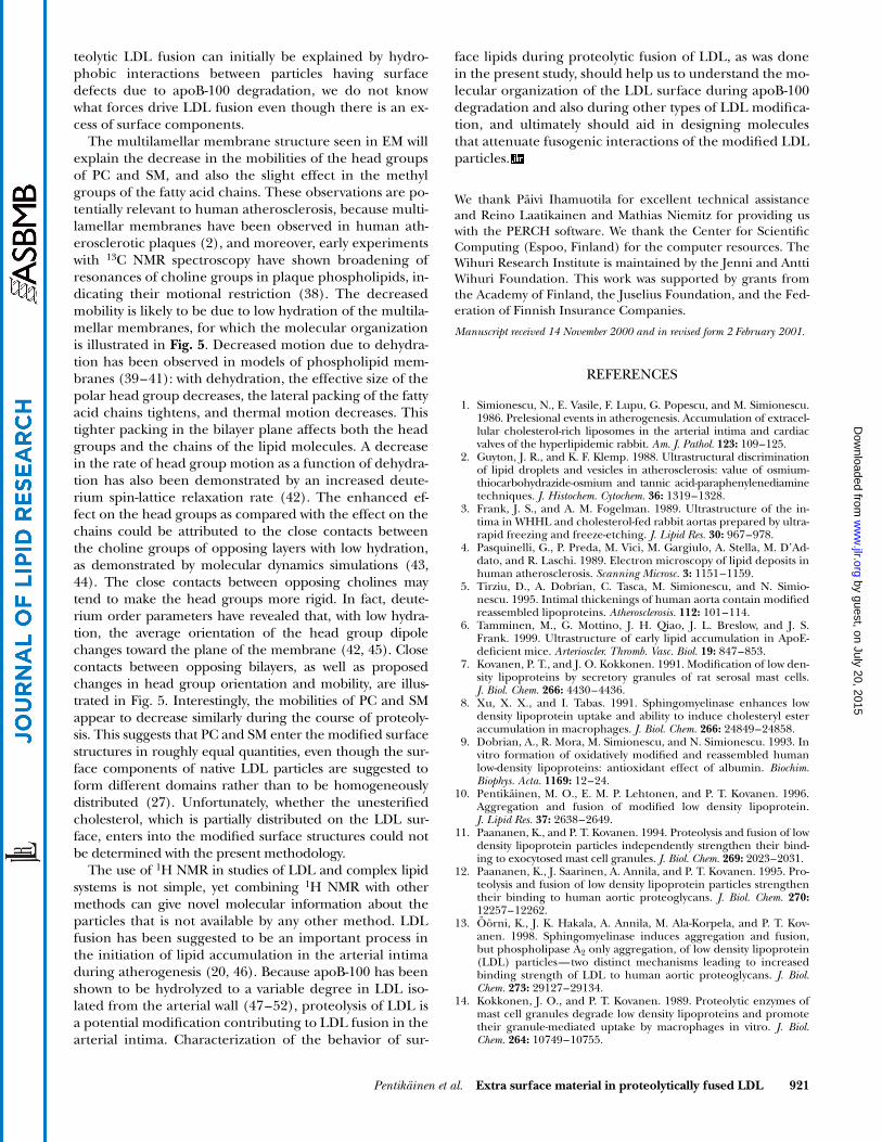
Fig. 5. Electron micrograph of multilamellar membranous material extending from a fused LDL particle (top left) together with the visu-alization of a hypothetical multilamellar structure at the molecular level (top right) and the head group regions of opposing layers at theatomic level (bottom right). The illustration at the atomic level demonstrates the close contacts between the head groups that are likely tooccur with low hydration of opposing layers. The molecular graphics were made with RasMol software (RasWin Molecular Graphics, Win-dows version 2.6) and the model was constructed by reducing the spacing between the opposing layers, which were taken as snapshots of amolecular dynamics simulation of a palmitoyl-linoleoyl-PC bilayer (53). All the atoms are represented by spheres with 1.2-Å radius. The hy-drogen atoms and water molecules were omitted for clarity. The possible changes in the head group orientation and mobility due to dehy-dration are illustrated by the phosphorus-nitrogen vectors (bottom left). The two phosphorus-bound oxygen atoms, the three nitrogen-bound carbon atoms, and all the hydrogen atoms in the PC molecules were omitted for clarity. The color coding for the bottom figures is asfollows: blue for nitrogen, yellow for phosphorus, red for oxygen, and green for carbon.
by guest, on July 20, 2015w
ww
.jlr.orgD
ownloaded from
Pentikäinen et al. Extra surface material in proteolytically fused LDL 921
teolytic LDL fusion can initially be explained by hydro-phobic interactions between particles having surfacedefects due to apoB-100 degradation, we do not knowwhat forces drive LDL fusion even though there is an ex-cess of surface components.
The multilamellar membrane structure seen in EM willexplain the decrease in the mobilities of the head groupsof PC and SM, and also the slight effect in the methylgroups of the fatty acid chains. These observations are po-tentially relevant to human atherosclerosis, because multi-lamellar membranes have been observed in human ath-erosclerotic plaques (2), and moreover, early experimentswith 13C NMR spectroscopy have shown broadening ofresonances of choline groups in plaque phospholipids, in-dicating their motional restriction (38). The decreasedmobility is likely to be due to low hydration of the multila-mellar membranes, for which the molecular organizationis illustrated in Fig. 5. Decreased motion due to dehydra-tion has been observed in models of phospholipid mem-branes (39–41): with dehydration, the effective size of thepolar head group decreases, the lateral packing of the fattyacid chains tightens, and thermal motion decreases. Thistighter packing in the bilayer plane affects both the headgroups and the chains of the lipid molecules. A decreasein the rate of head group motion as a function of dehydra-tion has also been demonstrated by an increased deute-rium spin-lattice relaxation rate (42). The enhanced ef-fect on the head groups as compared with the effect on thechains could be attributed to the close contacts betweenthe choline groups of opposing layers with low hydration,as demonstrated by molecular dynamics simulations (43,44). The close contacts between opposing cholines maytend to make the head groups more rigid. In fact, deute-rium order parameters have revealed that, with low hydra-tion, the average orientation of the head group dipolechanges toward the plane of the membrane (42, 45). Closecontacts between opposing bilayers, as well as proposedchanges in head group orientation and mobility, are illus-trated in Fig. 5. Interestingly, the mobilities of PC and SMappear to decrease similarly during the course of proteoly-sis. This suggests that PC and SM enter the modified surfacestructures in roughly equal quantities, even though the sur-face components of native LDL particles are suggested toform different domains rather than to be homogeneouslydistributed (27). Unfortunately, whether the unesterifiedcholesterol, which is partially distributed on the LDL sur-face, enters into the modified surface structures could notbe determined with the present methodology.
The use of 1H NMR in studies of LDL and complex lipidsystems is not simple, yet combining 1H NMR with othermethods can give novel molecular information about theparticles that is not available by any other method. LDLfusion has been suggested to be an important process inthe initiation of lipid accumulation in the arterial intimaduring atherogenesis (20, 46). Because apoB-100 has beenshown to be hydrolyzed to a variable degree in LDL iso-lated from the arterial wall (47–52), proteolysis of LDL isa potential modification contributing to LDL fusion in thearterial intima. Characterization of the behavior of sur-
face lipids during proteolytic fusion of LDL, as was donein the present study, should help us to understand the mo-lecular organization of the LDL surface during apoB-100degradation and also during other types of LDL modifica-tion, and ultimately should aid in designing moleculesthat attenuate fusogenic interactions of the modified LDLparticles.
We thank Päivi Ihamuotila for excellent technical assistanceand Reino Laatikainen and Mathias Niemitz for providing uswith the PERCH software. We thank the Center for ScientificComputing (Espoo, Finland) for the computer resources. TheWihuri Research Institute is maintained by the Jenni and AnttiWihuri Foundation. This work was supported by grants fromthe Academy of Finland, the Juselius Foundation, and the Fed-eration of Finnish Insurance Companies.
Manuscript received 14 November 2000 and in revised form 2 February 2001.
REFERENCES
1. Simionescu, N., E. Vasile, F. Lupu, G. Popescu, and M. Simionescu.1986. Prelesional events in atherogenesis. Accumulation of extracel-lular cholesterol-rich liposomes in the arterial intima and cardiacvalves of the hyperlipidemic rabbit. Am. J. Pathol. 123: 109–125.
2. Guyton, J. R., and K. F. Klemp. 1988. Ultrastructural discriminationof lipid droplets and vesicles in atherosclerosis: value of osmium-thiocarbohydrazide-osmium and tannic acid-paraphenylenediaminetechniques. J. Histochem. Cytochem. 36: 1319–1328.
3. Frank, J. S., and A. M. Fogelman. 1989. Ultrastructure of the in-tima in WHHL and cholesterol-fed rabbit aortas prepared by ultra-rapid freezing and freeze-etching. J. Lipid Res. 30: 967–978.
4. Pasquinelli, G., P. Preda, M. Vici, M. Gargiulo, A. Stella, M. D’Ad-dato, and R. Laschi. 1989. Electron microscopy of lipid deposits inhuman atherosclerosis. Scanning Microsc. 3: 1151–1159.
5. Tirziu, D., A. Dobrian, C. Tasca, M. Simionescu, and N. Simio-nescu. 1995. Intimal thickenings of human aorta contain modifiedreassembled lipoproteins. Atherosclerosis. 112: 101–114.
6. Tamminen, M., G. Mottino, J. H. Qiao, J. L. Breslow, and J. S.Frank. 1999. Ultrastructure of early lipid accumulation in ApoE-deficient mice. Arterioscler. Thromb. Vasc. Biol. 19: 847–853.
7. Kovanen, P. T., and J. O. Kokkonen. 1991. Modification of low den-sity lipoproteins by secretory granules of rat serosal mast cells.J. Biol. Chem. 266: 4430–4436.
8. Xu, X. X., and I. Tabas. 1991. Sphingomyelinase enhances lowdensity lipoprotein uptake and ability to induce cholesteryl esteraccumulation in macrophages. J. Biol. Chem. 266: 24849–24858.
9. Dobrian, A., R. Mora, M. Simionescu, and N. Simionescu. 1993. Invitro formation of oxidatively modified and reassembled humanlow-density lipoproteins: antioxidant effect of albumin. Biochim.Biophys. Acta. 1169: 12–24.
10. Pentikäinen, M. O., E. M. P. Lehtonen, and P. T. Kovanen. 1996.Aggregation and fusion of modified low density lipoprotein.J. Lipid Res. 37: 2638–2649.
11. Paananen, K., and P. T. Kovanen. 1994. Proteolysis and fusion of lowdensity lipoprotein particles independently strengthen their bind-ing to exocytosed mast cell granules. J. Biol. Chem. 269: 2023–2031.
12. Paananen, K., J. Saarinen, A. Annila, and P. T. Kovanen. 1995. Pro-teolysis and fusion of low density lipoprotein particles strengthentheir binding to human aortic proteoglycans. J. Biol. Chem. 270:12257–12262.
13. Öörni, K., J. K. Hakala, A. Annila, M. Ala-Korpela, and P. T. Kov-anen. 1998. Sphingomyelinase induces aggregation and fusion,but phospholipase A2 only aggregation, of low density lipoprotein(LDL) particles—two distinct mechanisms leading to increasedbinding strength of LDL to human aortic proteoglycans. J. Biol.Chem. 273: 29127–29134.
14. Kokkonen, J. O., and P. T. Kovanen. 1989. Proteolytic enzymes ofmast cell granules degrade low density lipoproteins and promotetheir granule-mediated uptake by macrophages in vitro. J. Biol.Chem. 264: 10749–10755.
by guest, on July 20, 2015w
ww
.jlr.orgD
ownloaded from

922 Journal of Lipid Research Volume 42, 2001
15. Piha, M., L. Lindstedt, and P. T. Kovanen. 1995. Fusion of proteo-lyzed LDL in the fluid phase: a novel mechanism generatingatherogenic lipoprotein particles. Biochemistry. 34: 10120–10129.
16. Hakala, J. K., K. Öörni, M. Ala-Korpela, and P. T. Kovanen. 1999.Lipolytic modification of LDL by phospholipase A 2 induces parti-cle aggregation in the absence and fusion in the presence of hepa-rin. Arterioscler. Thromb. Vasc. Biol. 19: 1276–1283.
17. Suits, A. G., A. Chait, M. Aviram, and J. W. Heinecke. 1989. Phago-cytosis of aggregated lipoprotein by macrophages: low density lipo-protein receptor-dependent foam-cell formation. Proc. Natl. Acad.Sci. USA. 86: 2713–2717.
18. Liu, H., D. G. Scraba, and R. O. Ryan. 1993. Prevention of phos-pholipase-C induced aggregation of low density lipoprotein by am-phipathic apolipoproteins. FEBS Lett. 316: 27–33.
19. Pentikäinen, M. O., K. Öörni, M. Ala-Korpela, and P. T. Kovanen.2000. Modified LDL—trigger of atherosclerosis and inflammationin the arterial intima. J. Intern. Med. 247: 359–370.
20. Öörni, K., M. O. Pentikäinen, M. Ala-Korpela, and P. T. Kovanen.2000. Aggregation, fusion, and vesicle formation of modified LDLparticles: molecular mechanisms and effects on matrix interac-tions. J. Lipid Res. 41: 1703–1714.
21. Pentikäinen, M. O., E. M. P. Lehtonen, K. Öörni, S. Lusa, P.Somerharju, M. Jauhiainen, and P. T. Kovanen. 1997. Human arte-rial proteoglycans increase the rate of proteolytic fusion of lowdensity lipoprotein particles. J. Biol. Chem. 272: 25283–25288.
22. Ala-Korpela, M., M. O. Pentikäinen, A. Korhonen, T. Hevonoja, J.Lounila, and P. T. Kovanen. 1998. Detection of low density lipo-protein particle fusion by proton nuclear magnetic resonancespectroscopy. J. Lipid Res. 39: 1705–1712.
23. Bell, J. D., M. L. Barnard, H. G. Parkes, E. L. Thomas, C. H. Brennan,S. C. Cunnane, and P. C. Dagnelie. 1996. Effects of n-3 fatty acids onthe NMR profile of plasma lipoproteins. J. Lipid Res. 37: 1664–1674.
24. Parks, J. S., and H. Hauser. 1996. Low density lipoprotein particlesize and core cholesteryl ester physical state affect the protonNMR magnetic environment of fatty acid methylene and methylnuclei. J. Lipid Res. 37: 1289–1297.
25. Murphy, H. C., M. Ala-Korpela, J. J. White, A. Raoof, J. D. Bell,M. L. Barnard, S. P. Burns, and R. A. Iles. 1997. Evidence for dis-tinct behaviour of phosphatidylcholine and sphingomyelin at thelow density lipoprotein surface. Biochem. Biophys. Res. Commun.234: 733–737.
26. Sommer, A., E. Prenner, R. Gorges, H. Stutz, H. Grillhofer, G. M.Kostner, F. Paltauf, and A. Hermetter. 1992. Organization of phos-phatidylcholine and sphingomyelin in the surface monolayer oflow density lipoprotein and lipoprotein(a) as determined by time-resolved fluorometry. J. Biol. Chem. 267: 24217–24222.
27. Hevonoja, T., M. O. Pentikäinen, M. T. Hyvönen, P. T. Kovanen,and M. Ala-Korpela. 2000. Structure of low density lipoprotein(LDL) particles. Basis for understanding molecular changes inmodified LDL. Biochim. Biophys. Acta. 1488: 189–210.
28. Havel, R. J., H. A. Eder, and J. H. Bragdon. 1955. The distributionand chemical composition of ultracentrifugally separated lipopro-teins in human serum. J. Clin. Invest. 34: 1345–1353.
29. Lowry, O. H., N. J. Rosebrough, A. L. Fall, and R. J. Randall. 1951.Protein measurement with the Folin phenol reagent. J. Biol. Chem.193: 265–275.
30. Bligh, E. G., and W. J. Dyer. 1959. A rapid method for total lipidextraction and purification. Can. J. Biochem. Physiol. 37: 911–917.
31. Laatikainen, R., M. Niemitz, W. J. Malaisse, M. Biesemans, and R.Willem. 1996. A computational strategy for the deconvolution ofNMR spectra with multiplet structures and constraints: analysis of over-lapping 13C–2H multiplets of 13C enriched metabolites from cellsuspensions incubated in deuterated media. Magn. Reson. Med. 36:359–365.
32. Lounila, J., M. Ala-Korpela, J. Jokisaari, M. J. Savolainen, and Y. A.Kesäniemi. 1994. Effects of orientational order and particle size onthe NMR line positions of lipoproteins. Phys. Rev. Lett. 72: 4049–4052.
33. Stremler, K. E., D. M. Stafforini, S. M. Prescott, G. A. Zimmerman,and T. M. McIntyre. 1989. An oxidized derivative of phosphatidyl-choline is a substrate for the platelet-activating factor acetylhydro-lase from human plasma. J. Biol. Chem. 264: 5331–5334.
34. Segrest, J. P., M. K. Jones, V. K. Mishra, G. M. Anantharamaiah,and D. W. Garber. 1994. ApoB-100 has a pentapartite structurecomposed of three amphipathic alpha-helical domains alternating
with two amphipathic beta-strand domains. Detection by the com-puter program LOCATE. Arterioscler. Thromb. 14: 1674–1685.
35. Goormaghtigh, E., V. Cabiaux, J. D. Meutter, M. Rosseneu, and J-M.Ruysschaert. 1993. Secondary structure of the particle associatingdomain of apolipoprotein B-100 in low-density lipoprotein by at-tenuated total reflection infrared spectroscopy. Biochemistry. 32:6104–6110.
36. Segrest, J. P., M. K. Jones, V. K. Mishra, V. Pierotti, S. H. Young, J.Boren, T. L. Innerarity, and N. Dashti. 1998. Apolipoprotein B-100:conservation of lipid-associating amphipathic secondary structuralmotifs in nine species of vertebrates. J. Lipid Res. 39: 85–102.
37. Schumaker, V. N., M. L. Phillips, and J. E. Chatterton. 1994. Apoli-poprotein B and low-density lipoprotein structure: implicationsfor biosynthesis of triglyceride-rich lipoproteins. Adv. Protein Chem.45: 205–248.
38. Hamilton, J. A., E. H. Cordes, and C. J. Glueck. 1979. Lipid dynam-ics in human low density lipoproteins and human aortic tissuewith fibrous plaques. A study by high field 13C NMR spectroscopy.J. Biol. Chem. 254: 5435–5441.
39. Wiener, M. C., and S. H. White. 1992. Structure of a fluid dio-leoylphosphatidylcholine bilayer determined by joint refinementof x-ray and neutron diffraction data. II. Distribution and packingof terminal methyl groups. Biophys. J. 61: 428–433.
40. Wiener, M. C., and S. H. White. 1992. Structure of a fluid dio-leoylphosphatidylcholine bilayer determined by joint refinementof x-ray and neutron diffraction data. III. Complete structure. Bio-phys. J. 61: 437–447.
41. Lehtonen, J. Y. A., and P. K. J. Kinnunen. 1995. Poly(ethylene gly-col)-induced and temperature-dependent phase separation influid binary phospholipid membranes. Biophys. J. 68: 525–535.
42. Ulrich, A. S., and A. Watts. 1994. Molecular response of the lipidhead group to bilayer hydration monitored by 2H-NMR. Biophys. J.66: 1441–1449.
43. Marrink, S-J., M. Berkowitz, and H. J. C. Bedersen. 1993. Molecu-lar dynamics simulation of a membrane/water interface: the or-dering of water and its relation to the hydration force. Langmuir. 9:3122–3131.
44. Essman, U., L. Perera, and M. Berkowitz. 1995. The origin of thehydration interaction of lipid bilayers from MD simulation of di-palmitoylphosphatidylcholine membranes in gel and liquid crys-talline phases. Langmuir. 11: 4519–4531.
45. Bechinger, B., and J. Seelig. 1991. Conformational changes of thephosphatidylcholine head group due to membrane dehydration.A 2H-NMR study. Chem. Phys. Lipids. 58: 1–5.
46. Lusis, A. J. 2000. Atherosclerosis. Nature. 407: 233–241.47. Clevidence, B. A., R. E. Morton, G. West, D. M. Dusek, and H. F.
Hoff. 1984. Cholesterol esterification in macrophages. Stimulationby lipoproteins containing apo B isolated from human aortas. Arte-riosclerosis. 4: 196–207.
48. Daugherty, A., B. S. Zwiefel, B. E. Sobel, and G. Schonfeld. 1988.Isolation of low density lipoprotein from atherosclerotic vasculartissue of Watanabe heritable hyperlipidemic rabbits. Arteriosclerosis.8: 768–777.
49. Ylä-Herttuala, S., W. Palinski, M. E. Rosenfeld, S. Parthasarathy,T. E. Carew, S. Butler, J. L. Witztum, and D. Steinberg. 1989. Evi-dence for the presence of oxidatively modified low density lipo-protein in atherosclerotic lesions of rabbit and man. J. Clin. Invest.84: 1086–1095.
50. Hoff, H. F., and J. O’Neil. 1991. Lesion-derived low density lipo-protein and oxidized low density lipoprotein share a lability for ag-gregation, leading to enhanced macrophage degradation. Arterio-scler. Thromb. 11: 1209–1222.
51. Steinbrecher, U. P., and M. Lougheed. 1992. Scavenger receptor-independent stimulation of cholesterol esterification in macro-phages by low density lipoprotein extracted from human aortic in-tima. Arterioscler. Thromb. 12: 608–625.
52. Tailleux, A., G. Torpier, B. Caron, J-C. Fruchart, and C. Fievet. 1993.Immunological properties of apoB-containing lipoprotein particles inhuman atherosclerotic arteries. J. Lipid Res. 34: 719–728.
53. Hyvönen, M. T., T. T. Rantala, and M. Ala-Korpela. 1997. Structureand dynamic properties of diunsaturated 1-palmitoyl-2-linoleoyl-sn-glycero-3-phosphatidylcholine lipid bilayer from molecular dy-namics simulation. Biophys. J. 73: 2907–2923.
by guest, on July 20, 2015w
ww
.jlr.orgD
ownloaded from


