Embed Size (px)
Citation preview
Altered Prion Protein Expression Pattern in CSF as aBiomarker for Creutzfeldt-Jakob DiseaseMauricio Torres1,2, Luis Cartier3*, Jose Manuel Matamala1,2,3, Nancy Hernandez1,2, Ute Woehlbier1,2,
Claudio Hetz1,2,4,5*
1 Neuroscience Biomedical Institute, Faculty of Medicine, University of Chile, Santiago, Chile, 2 Center for Molecular Studies of the Cell, Institute of Biomedical Sciences,
University of Chile, Santiago, Chile, 3 Department of Neurological Sciences, Faculty of Medicine, University of Chile, Santiago, Chile, 4 Neurounion Biomedical Foundation,
Santiago, Chile, 5 Harvard School of Public Health, Boston, Massachusetts, United States of America
Abstract
Creutzfeldt-Jakob disease (CJD) is the most frequent human Prion-related disorder (PrD). The detection of 14-3-3 protein inthe cerebrospinal fluid (CSF) is used as a molecular diagnostic criterion for patients clinically compatible with CJD. However,there is a pressing need for the identification of new reliable disease biomarkers. The pathological mechanisms leading toaccumulation of 14-3-3 protein in CSF are not fully understood, however neuronal loss followed by cell lysis is assumed tocause the increase in 14-3-3 levels, which also occurs in conditions such as brain ischemia. Here we investigated the relationbetween the levels of 14-3-3 protein, Lactate dehydrogenase (LDH) activity and expression of the prion protein (PrP) in CSFof sporadic and familial CJD cases. Unexpectedly, we found normal levels of LDH activity in CJD cases with moderate levelsof 14-3-3 protein. Increased LDH activity was only observed in a percentage of the CSF samples that also exhibited high 14-3-3 levels. Analysis of the PrP expression pattern in CSF revealed a reduction in PrP levels in all CJD cases, as well as markedchanges in its glycosylation pattern. PrP present in CSF of CJD cases was sensitive to proteases. The alterations in PrPexpression observed in CJD cases were not detected in other pathologies affecting the nervous system, including cases ofdementia and tropical spastic paraparesis/HTLV-1 associated myelopathy (HAM/TSP). Time course analysis in several CJDpatients revealed that 14-3-3 levels in CSF are dynamic and show a high degree of variability during the end stage of thedisease. Post-mortem analysis of brain tissue also indicated that 14-3-3 protein is upregulated in neuronal cells, suggestingthat its expression is modulated during the course of the disease. These results suggest that a combined analysis of 14-3-3and PrP expression pattern in CSF is a reliable biomarker to confirm the clinical diagnosis of CJD patients and follow diseaseprogression.
Citation: Torres M, Cartier L, Matamala JM, Hernandez N, Woehlbier U, et al. (2012) Altered Prion Protein Expression Pattern in CSF as a Biomarker for Creutzfeldt-Jakob Disease. PLoS ONE 7(4): e36159. doi:10.1371/journal.pone.0036159
Editor: Noriyuki Nishida, Nagasaki University Graduate School of Biomedical Sciences, Japan
Received October 21, 2011; Accepted March 30, 2012; Published April 27, 2012
Copyright: � 2012 Torres et al. This is an open-access article distributed under the terms of the Creative Commons Attribution License, which permitsunrestricted use, distribution, and reproduction in any medium, provided the original author and source are credited.
Funding: This work was supported primarily by the Fondo Nacional de Desarrollo Cientıfico y Tecnologico (FONDECYT grant no. 1100176); Programa Fondo deInvestigacion Avanzado en Areas Prioritarias (FONDAP grant no. 15010006); Millennium Institute no. P09-015-F (to CH) and Fondo Nacional de DesarrolloCientıfico y Tecnologico (FONDECYT grant no. 3110067 to UW). In addition the authors thank support by Alzheimer’s Association, The Michael J Fox Foundationfor Parkinson Research, ALS Therapy Alliance, North American spinal society and International Center for Genetic Engineering and Biotechnology, Italy (to CH); andComision Nacional de Investigacion Cientıfica y Tecnologica, Gobierno de Chile (CONICYT Doctoral fellowship to MT). The funders had no role in study design,data collection and analysis, decision to publish, or preparation of the manuscript.
Competing Interests: The authors have declared that no competing interests exist.
* E-mail: [email protected] (LC); [email protected] (CH)
Introduction
Protein misfolding is a common hallmark of several neurode-
generative disorders including Alzheimer’s disease, Parkinson’s
disease, amyotrophic lateral sclerosis (ALS), as well as Prion-
related disorders (PrDs), among other pathologies [1]. Although in
many diseases, the molecular events underlying neurodegenera-
tion has been elucidated, there is an urgent need for the
identification of reliable biomarkers for early diagnosis and to
follow disease progression in clinical trials. PrDs are the classic
example of a protein misfolding disorder where alterations in the
structure of the prion protein (PrP) lead to progressive degener-
ation [2,3]. The most common PrD in humans is Creutzfeldt-
Jakob disease (CJD), where the sporadic form (sCJD) accounts for
nearly 85% of all cases with an incidence of 1 case per million
individuals [4]. Familial CJDs (fCJD) are caused by mutations in
the prion gene (PRNP) and represent in average 10% of all cases,
whereas less than 1% of CJDs are infectious, highlighting new
variant CJD, which is most likely associated with the consumption
of cattle affected with bovine spongiform encephalopathy [5,6]. In
a few countries, including Chile, the incidence of CJD is
considerably higher (.2.5 increase), where around 30% of all
cases are familial associated with the E200K mutation of PRNP
[7,8]. PrDs lead to progressive fatal neurodegeneration, ultimately
resulting in the patient’s death within a few months of diagnosis
[9]. Clinical manifestations of CJD include rapid progression of
dementia, myoclonus, visual and cerebellar symptoms, pyramidial
or extrapyramidial signs and akinetic mutism [10]. A definitive
diagnosis of CJD can only be made after neuropathological
examination of brain tissue and demonstration of spongiform
degeneration of the brain, accompanied by extensive neuronal loss
and accumulation of PrPRES, a misfolded and protease-resistant
form of the normal cellular prion protein (PrPC) [11,12].
Biochemical analysis of cerebrospinal fluid (CSF) is commonly
used for clinical diagnosis of CJD. Increased levels of 14-3-3, tau
protein, S100B or neuron-specific enolase are used to guide
PLoS ONE | www.plosone.org 1 April 2012 | Volume 7 | Issue 4 | e36159
clinical diagnosis and to distinguish PrDs from other causes of
rapidly progressive dementia. The importance of 14-3-3 protein
for diagnosis has been widely discussed in the context of CJD [13–
16]. However, contradictory findings regarding 14-3-3 test
specificity and sensitivity have been reported (see examples in
[17–19]). In fact an elevation of 14-3-3 levels in the CSF is thought
to occur because of non-specific release from areas after massive
brain damage, which accompanies a variety of neurological
conditions including central nervous system infections and
inflammatory diseases, brain tumors, and other types of neurode-
generative diseases [20]. These studies reflect the need for
additional biomarkers for diagnosis and to follow disease
progression in CJD, which represents a general need for most
neurodegenerative diseases. Direct monitoring of PrPRES in blood
and CSF samples of CJD cases may represent a better indicator for
diagnosis as previously reported based on the fact that alterations
in this protein are the main cause of the disease [21–23]. Soluble
PrPC is present in high levels in CSF of healthy people, but lower
concentrations of PrPC are found in CSF of CJD patients [24,25].
However, this was also observed in patients suffering from other
unrelated neurodegenerative diseases [25].
In this study, we report the analysis of PrPC levels in CSF in a
large group of CJD patients. We found dramatic changes in the
overall quantities and glycosylation pattern of PrP, which directly
correlated with disease progression. Our data suggest that
monitoring the pattern and levels of PrPC in combination with
measurements of 14-3-3 level in CSF of CJD patients may be used
as a predictive biomarker for CJD diagnosis and progression.
Results
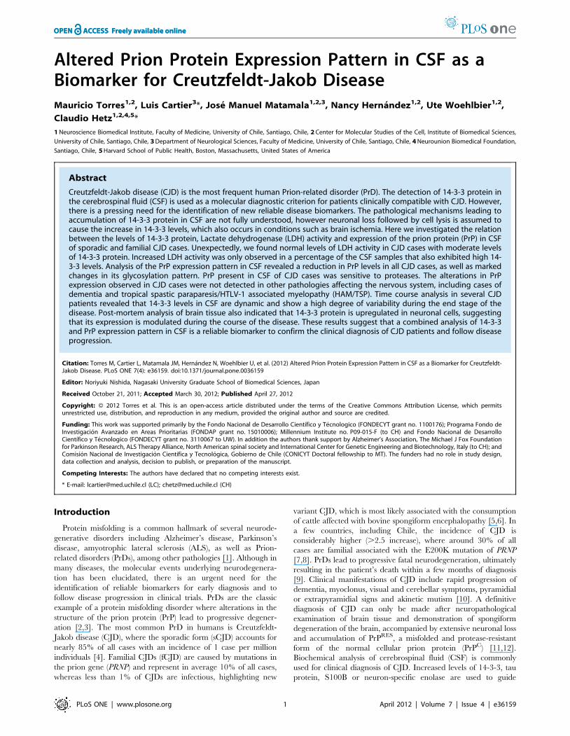
14-3-3 protein and LDH levels in CSF of CJD patients14-3-3 levels were monitored in CSF samples from CJD patients
and control subjects by Western blot analysis (Figure 1A), followed
by semi-quantitative analysis using densitometry (Figure 1B).
Increased 14-3-3 levels were found in most CSF samples analyzed
from CJD patients when compared with control subjects. We
measured LDH activity in CSF as a marker for tissue disruption
and found a moderate average increase in mean LDH activity in
CJD patients compared to the control group (Figure 1C).
However, LDH activity displayed broad variability between
different CJD patient samples, whereas this parameter was highly
homogeneous in the control group (Figure 1C). We found that
increased LDH activity correlated only in patients with high 14-3-
3 protein levels in CSF (Figure 1D). Examination of the total
protein amount in CSF samples did not show significant
differences between control subjects and CJD patients (Figure 1E).
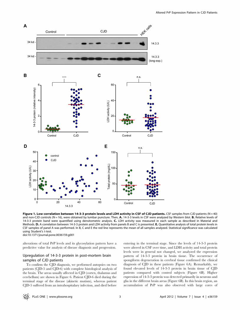
Altered PrP expression pattern in CSF of CJD casesWe then monitored the levels of PrP in CSF from CJD cases
using Western blot analysis (Figures 2A and B). In control subjects,
two main PrP bands with similar expression levels were detected
possibly reflecting different glycosylation states (see below). A
dramatic decrease in total PrPC levels was observed in CJD-
derived samples (Figure 2A). Unexpectedly, the ratio between the
two main PrP forms present in CSF was drastically altered in most
CJD patients, where the upper band was reduced, and in some
patients was not even detectable (Figures 2A and C). We also
compared the ratio between the two PrP bands in fCJD and sCJD
cases, and did not observe significant differences between both
groups of patients (Figure 2D). Finally, an inverse correlation was
observed between 14-3-3 protein and total PrP levels in CSF of
CJD patients compared to control subjects (Figure 2E). Similarly,
an inverse correlation was observed between 14-3-3 levels and the
PrP band ratio (data not shown).
We performed additional controls to determine whether
changes in 14-3-3 and PrP levels are specific for CJD pathology
or can be observed in other conditions affecting the nervous
system. We compared these parameters in CSF samples obtained
from three healthy control individuals, five CJD patients, three
non-CJD dementia patients (see exclusion criteria in Material and
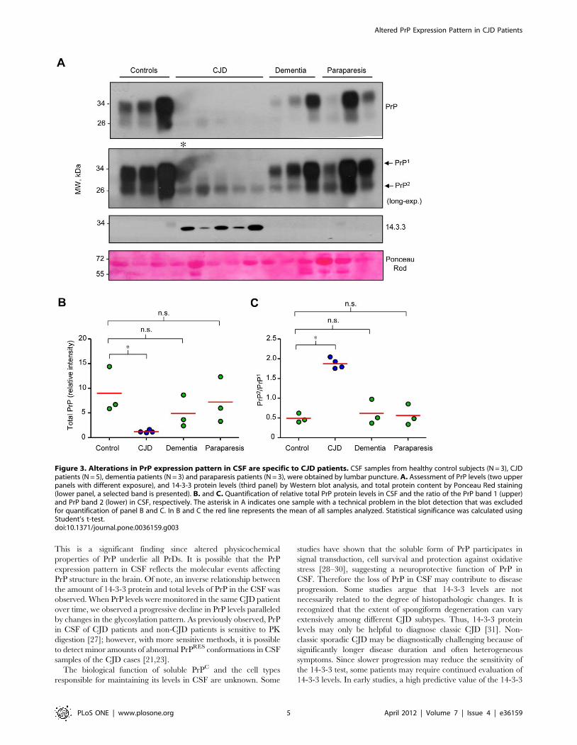
Methods) and three cases of HAM/TSP (Figure 3). A slight
decrease in total PrP levels was observed only in two dementia
patients and one HAM/TSP patient, but no increase in 14-3-3
protein was found (Figures 3A and B). The changes observed in
the PrP band ratio detected in the analysis were not observed in
this small set of non-CJD disease controls (Figure 3C).
Altered PrP glycosylation pattern in CSF of CJD casesTo address the possible cause of the changes in the ratio of the
two PrP forms observed in CSF of CJD patients, we first compared
the distribution of PrP bands in CSF with the pattern of human
brain cortex tissue from healthy controls, which is known to
contain the classical three PrP forms (mono-, di-, and non-
glycosylated). Interestingly, this comparison revealed that the two
bands present in CSF have a similar electrophoretic migration to
the mono- and di-glycosylation PrP forms (Figure 4A). Samples
were then treated with PNGase F (an unspecific glycosidase),
which revealed the presence of mostly glycosylated PrP forms in
CSF samples (Figure 4B).
A key biochemical signature of PrDs is the generation of PrPRES
in brain. We treated CSF samples from control and CJD patients
with different concentrations of proteinase K (PK). With this
method no PrPRES species were detected even with very low levels
of PK (Figure 4C). Additionally, we analyzed the sensitivity to
thermolysin, a protease that can discriminate between normal and
intermediate misfolded forms of PrP sensitive to PK [26].
Although we found a slight resistance of PrP to the treatment
with this protease in CJD cases (Figure 4D), the vast majority of
samples analyzed were sensitive to thermolysin (data not shown).
Dynamic changes of PrP and 14-3-3 protein levels duringthe progression of CJD
PrP and 14-3-3 protein levels were monitored in CSF from six
CJD patients (CJD1-CJD6) over time at different disease stages
(Figure 5). The first sample analyzed (point 0) corresponded to the
time of clinical diagnosis of CJD. Unexpectedly, the overall 14-3-3
protein and PrP levels depicted clear differences when the same
patient was studied over time, displaying distinct kinetics of
changes with the course of the disease. In the CSF of patient 6, 14-
3-3 levels peaked, then declined (Figure 5). In other CJD patients,
an elevation of 14-3-3 protein levels was observed over time
(patients 1, 2 and 5), whereas other patients showed increased
levels at time of diagnosis and a reduction in 14-3-3 levels over
time (patient 3) or were not altered over time (patient 4). Thus, the
pattern of 14-3-3 expression is highly variable over time when the
CSF of the same patient is analyzed at different time points of
disease progression.
Overall the analysis of the PrP expression pattern over time
showed robust and sustained changes (Figure 5). In contrast to 14-
3-3 levels, changes in PrP expression levels and altered
glycosylation pattern correlated with disease progression/severity
(Figure 5). Of note, in some samples an additional band between
PrP band 1 and band 2 was detected (CJD-2, CJD-5 and CJD-6),
which may represent further changes in the PrP structure and/or
post-translational modifications. Thus, these results indicate that
Altered PrP Expression Pattern in CJD Patients
PLoS ONE | www.plosone.org 2 April 2012 | Volume 7 | Issue 4 | e36159
alterations of total PrP levels and its glycosylation pattern have a
predictive value for analysis of disease diagnosis and progression.
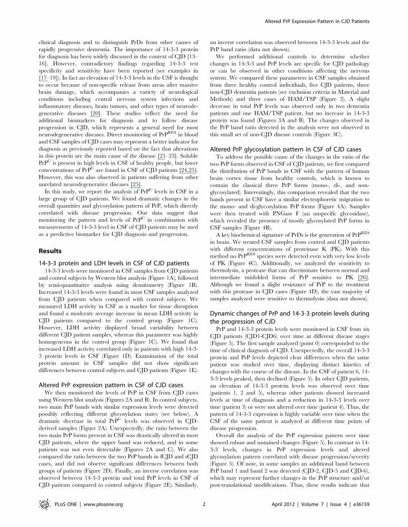
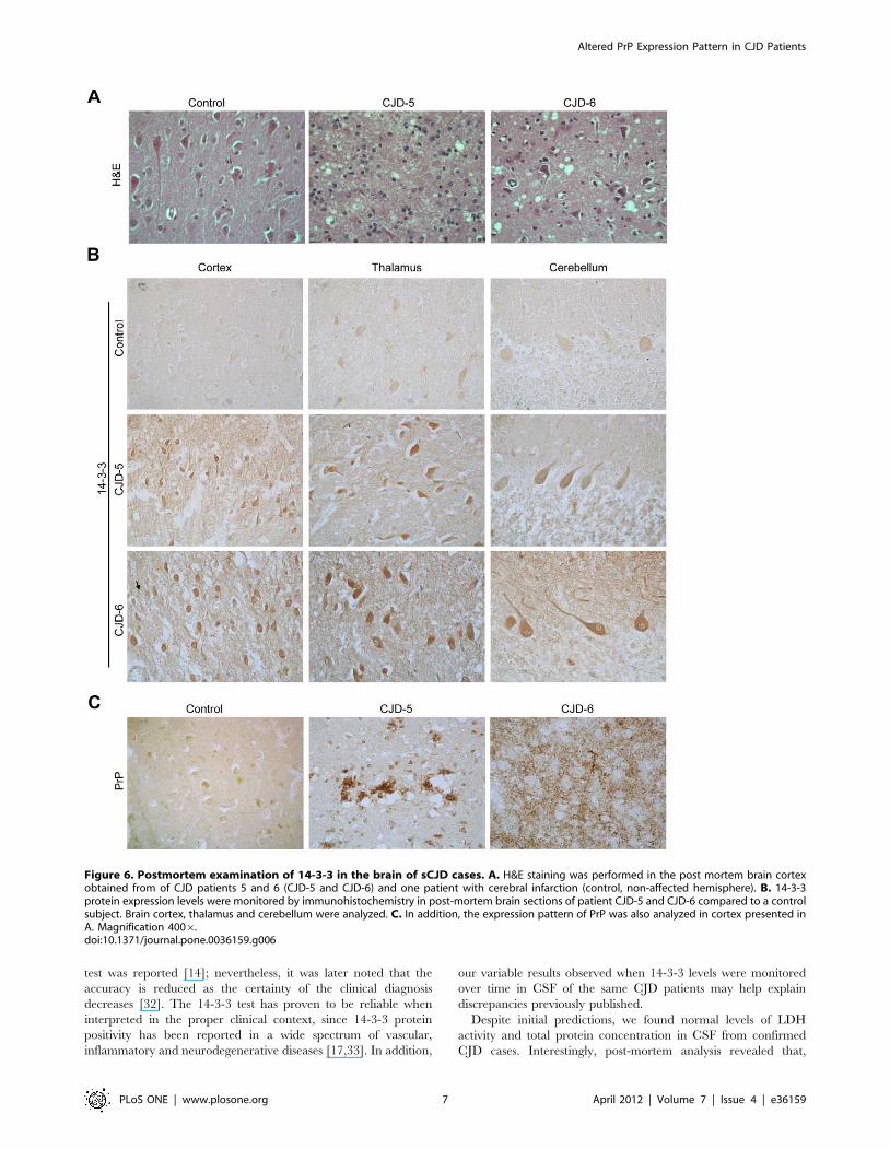
Upregulation of 14-3-3 protein in post-mortem brainsamples of CJD patients
To confirm the CJD diagnosis, we performed autopsies on two
patients (CJD-5 and CJD-6) with complete histological analysis of
the brain. The areas usually affected in CJD (cortex, thalamus and
cerebellum) are shown in Figure 6. Patient CJD-6 died during the
terminal stage of the disease (akinetic mutism), whereas patient
CJD-5 suffered from an intrahospitalary infection, and died before
entering in the terminal stage. Since the levels of 14-3-3 protein
were altered in CSF over time, and LDH activity and total protein
levels were in general not changed, we analyzed the expression
pattern of 14-3-3 protein in brain tissue. The occurrence of
spongiform degeneration in cerebral tissue confirmed the clinical
diagnosis of CJD in these patients (Figure 6A). Remarkably, we
found elevated levels of 14-3-3 protein in brain tissue of CJD
patients compared with control subjects (Figure 6B). Higher
expression of 14-3-3 protein was detected primarily in neurons and
glia in the different brain areas (Figure 6B). In this brain region, an
accumulation of PrP was also observed with large cores of
Figure 1. Low correlation between 14-3-3 protein levels and LDH activity in CSF of CJD patients. CSF samples from CJD patients (N = 40)and non-CJD controls (N = 16), were obtained by lumbar puncture. Then, A. 14-3-3 levels in CSF were analyzed by Western blot. B. Relative levels of14-3-3 protein band were quantified using densitometric analysis. C. LDH activity was measured in each sample as described in Material andMethods. D. A correlation between 14-3-3 protein and LDH activity from panels B and C is presented. E. Quantitative analysis of total protein levels inCSF samples of panel A was performed. In B, C and E the red line represents the mean of all samples analyzed. Statistical significance was calculatedusing Student’s t-test.doi:10.1371/journal.pone.0036159.g001
Altered PrP Expression Pattern in CJD Patients
PLoS ONE | www.plosone.org 3 April 2012 | Volume 7 | Issue 4 | e36159
extracellular PrP plaques surrounding areas of extensive spongi-
form change in patient CJD-5, whereas patient CJD-6 presented a
more diffuse staining pattern (Fig. 6C). Despite differential prion
patterns observed in the brain tissue of patients, we found similarly
elevated levels of 14-3-3 protein.
Discussion
This work was performed as part of a coordinated effort to
develop a centralized diagnosis and surveillance of Chilean
patients suffering from a prevalent human PrD form. Here, we
present a full study that examines potential CSF biomarkers for
CJD that can be used in combination with 14-3-3 protein. We
monitored the levels of 14-3-3 protein, LDH activity and different
aspects of PrP biology in the CSF of patients with the clinical
diagnosis of CJD. The general consensus in the field indicates that
although CSF 14-3-3 levels are not specific, in concert with
characteristic clinical features, it may be used for the diagnosis of
CJD. Here we report that total PrP levels combined with changes
in its glycosylation pattern mirrors the disease progression in CJD.
Figure 2. Altered PrP expression pattern in CSF of CJD patients. CSF samples from CJD patients (N = 40) and non-CJD controls (N = 16), wereobtained by lumbar puncture. A. PrP levels in CSF were examined by Western blot. B. and C. Quantification of relative levels of total PrP protein inCSF and the ratio of the PrP band 1 (upper) and PrP band 2 (lower) in CSF, respectively. D. Comparison between ratios of PrP band 1 and 2 in sCJDversus fCJD E. Correlation of total PrP protein levels with 14-3-3 protein in each CSF samples presented in Fig 1. In B, C and D the red line representsthe mean of all samples analyzed. Statistical significance was calculated using Student’s t-test.doi:10.1371/journal.pone.0036159.g002
Altered PrP Expression Pattern in CJD Patients
PLoS ONE | www.plosone.org 4 April 2012 | Volume 7 | Issue 4 | e36159
This is a significant finding since altered physicochemical
properties of PrP underlie all PrDs. It is possible that the PrP
expression pattern in CSF reflects the molecular events affecting
PrP structure in the brain. Of note, an inverse relationship between
the amount of 14-3-3 protein and total levels of PrP in the CSF was
observed. When PrP levels were monitored in the same CJD patient
over time, we observed a progressive decline in PrP levels paralleled
by changes in the glycosylation pattern. As previously observed, PrP
in CSF of CJD patients and non-CJD patients is sensitive to PK
digestion [27]; however, with more sensitive methods, it is possible
to detect minor amounts of abnormal PrPRES conformations in CSF
samples of the CJD cases [21,23].
The biological function of soluble PrPC and the cell types
responsible for maintaining its levels in CSF are unknown. Some
studies have shown that the soluble form of PrP participates in
signal transduction, cell survival and protection against oxidative
stress [28–30], suggesting a neuroprotective function of PrP in
CSF. Therefore the loss of PrP in CSF may contribute to disease
progression. Some studies argue that 14-3-3 levels are not
necessarily related to the degree of histopathologic changes. It is
recognized that the extent of spongiform degeneration can vary
extensively among different CJD subtypes. Thus, 14-3-3 protein
levels may only be helpful to diagnose classic CJD [31]. Non-
classic sporadic CJD may be diagnostically challenging because of
significantly longer disease duration and often heterogeneous
symptoms. Since slower progression may reduce the sensitivity of
the 14-3-3 test, some patients may require continued evaluation of
14-3-3 levels. In early studies, a high predictive value of the 14-3-3
Figure 3. Alterations in PrP expression pattern in CSF are specific to CJD patients. CSF samples from healthy control subjects (N = 3), CJDpatients (N = 5), dementia patients (N = 3) and paraparesis patients (N = 3), were obtained by lumbar puncture. A. Assessment of PrP levels (two upperpanels with different exposure), and 14-3-3 protein levels (third panel) by Western blot analysis, and total protein content by Ponceau Red staining(lower panel, a selected band is presented). B. and C. Quantification of relative total PrP protein levels in CSF and the ratio of the PrP band 1 (upper)and PrP band 2 (lower) in CSF, respectively. The asterisk in A indicates one sample with a technical problem in the blot detection that was excludedfor quantification of panel B and C. In B and C the red line represents the mean of all samples analyzed. Statistical significance was calculated usingStudent’s t-test.doi:10.1371/journal.pone.0036159.g003
Altered PrP Expression Pattern in CJD Patients
PLoS ONE | www.plosone.org 5 April 2012 | Volume 7 | Issue 4 | e36159
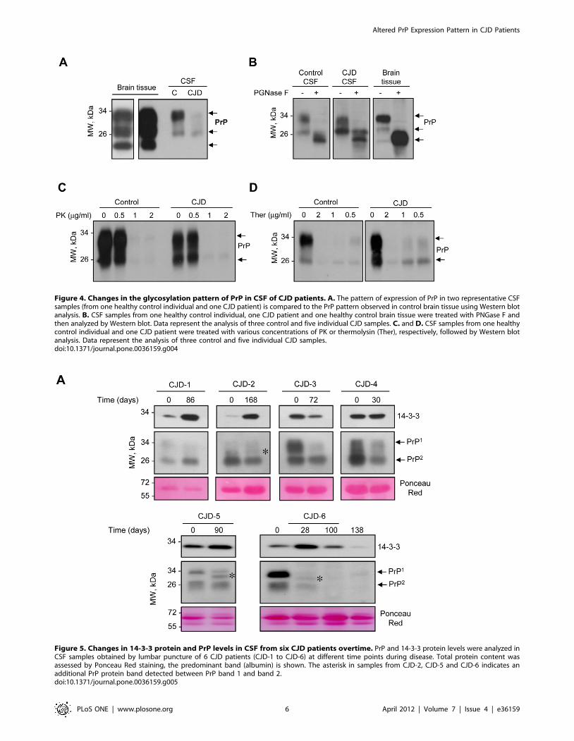
Figure 4. Changes in the glycosylation pattern of PrP in CSF of CJD patients. A. The pattern of expression of PrP in two representative CSFsamples (from one healthy control individual and one CJD patient) is compared to the PrP pattern observed in control brain tissue using Western blotanalysis. B. CSF samples from one healthy control individual, one CJD patient and one healthy control brain tissue were treated with PNGase F andthen analyzed by Western blot. Data represent the analysis of three control and five individual CJD samples. C. and D. CSF samples from one healthycontrol individual and one CJD patient were treated with various concentrations of PK or thermolysin (Ther), respectively, followed by Western blotanalysis. Data represent the analysis of three control and five individual CJD samples.doi:10.1371/journal.pone.0036159.g004
Figure 5. Changes in 14-3-3 protein and PrP levels in CSF from six CJD patients overtime. PrP and 14-3-3 protein levels were analyzed inCSF samples obtained by lumbar puncture of 6 CJD patients (CJD-1 to CJD-6) at different time points during disease. Total protein content wasassessed by Ponceau Red staining, the predominant band (albumin) is shown. The asterisk in samples from CJD-2, CJD-5 and CJD-6 indicates anadditional PrP protein band detected between PrP band 1 and band 2.doi:10.1371/journal.pone.0036159.g005
Altered PrP Expression Pattern in CJD Patients
PLoS ONE | www.plosone.org 6 April 2012 | Volume 7 | Issue 4 | e36159
test was reported [14]; nevertheless, it was later noted that the
accuracy is reduced as the certainty of the clinical diagnosis
decreases [32]. The 14-3-3 test has proven to be reliable when
interpreted in the proper clinical context, since 14-3-3 protein
positivity has been reported in a wide spectrum of vascular,
inflammatory and neurodegenerative diseases [17,33]. In addition,
our variable results observed when 14-3-3 levels were monitored
over time in CSF of the same CJD patients may help explain
discrepancies previously published.
Despite initial predictions, we found normal levels of LDH
activity and total protein concentration in CSF from confirmed
CJD cases. Interestingly, post-mortem analysis revealed that,
Figure 6. Postmortem examination of 14-3-3 in the brain of sCJD cases. A. H&E staining was performed in the post mortem brain cortexobtained from of CJD patients 5 and 6 (CJD-5 and CJD-6) and one patient with cerebral infarction (control, non-affected hemisphere). B. 14-3-3protein expression levels were monitored by immunohistochemistry in post-mortem brain sections of patient CJD-5 and CJD-6 compared to a controlsubject. Brain cortex, thalamus and cerebellum were analyzed. C. In addition, the expression pattern of PrP was also analyzed in cortex presented inA. Magnification 4006.doi:10.1371/journal.pone.0036159.g006
Altered PrP Expression Pattern in CJD Patients
PLoS ONE | www.plosone.org 7 April 2012 | Volume 7 | Issue 4 | e36159
although there was a marked reduction in neurons in affected
areas in CJD patients, 14-3-3 protein expression was significantly
upregulated in brain tissue. Taken together, our results support the
new concept that increased CSF 14-3-3 levels may reflect the
induction of a cellular response against prion rather than cell lysis.
In agreement with this hypothesis, it has been reported that some
14-3-3 protein isoforms may play a role in synaptic function [34–
36]. In addition, 14-3-3 proteins function as conformational
stabilizers of other proteins and regulate diverse signal transduc-
tion pathways related to cell survival [37]. 14-3-3 is also involved
in axonal transport of prion proteins and their transport to the cell
surface [38].
Changes in the glycosylation pattern of PrP in CSF may reflect
alterations in the protein maturation pathways during its synthesis.
PrP is generated and folded in the endoplasmic reticulum (ER)
where it is subjected to several post-translational modifications,
and then is transported to the Golgi apparatus to undergo
maturation by glycosylations to be further exported to the plasma
membrane [39]. Perturbations in the function of the ER have been
described in human post-mortem samples of sCJD and new
variant CJD [40], in addition to animal and cellular models of
scrapie [41,42]. Altered glycosylation of PrP also has been
reported in the brains of many animal models of PrDs (see
example in [43]), and all three PrP forms can reach the plasma
membrane [44]. Based on these observations, we speculate that
the changes in the PrP glycosylation pattern in CSF may reflect an
active stress response at the level of the ER and secretory pathway
of affected neurons in CJD. PrP levels may also decrease in CSF
because it may cluster in large and stable aggregates in the brain
due to PrP misfolding. Additionally, the results of Meyne et al.,
indicate that reduced PrP amounts in the CSF are observed in a
wide range of neurodegenerative diseases [25]. In agreement with
these results, a slight decrease in total PrP levels was observed in
two dementia patients and one HAM/TSP patient, however, the
changes in the ratio of PrP isoforms was observed only in the CJD
patients analyzed.
To our knowledge, this study is the first report with comparative
measurements of 14-3-3 protein, PrP levels and glycosylation
pattern in CSF of CJD patients following these changes at different
stages of disease progression. Our results suggest that analysis of
14-3-3 protein level and PrP expression pattern in CSF samples
may be a reliable biomarker for diagnosis of CJD.
Materials and Methods
Patient samples and clinical diagnosisCSF samples were collected from 46 patients diagnosed with
CJD (sCJD, N = 35 patients and fCJD, N = 11 patients) and 16
anesthesia controls of healthy individuals (Table 1). Samples were
collected, centrifuged and immediately frozen at 220uC for
transport from the hospital to the laboratory for biochemical
analysis. Then samples were stored as aliquots at 280uC. The
clinical diagnosis of CJD was defined by the following parameters
including: motor alterations, cerebellar signs, myoclonus, devel-
opment of sub-acute dementia, disturbances of alert, vision and
memory. All patients showed hyperintensity in the basal ganglia
and cortex in the magnetic resonance study, pseudo-periodic
activity in EEG and elevated 14-3-3 protein in CSF. Additionally,
CSF samples were studied from patients with other neurological
symptoms, including dementia and tropical spastic paraparesis/
HTLV-1 associated myelopathy (HAM/TSP). The dementia
patients showed a chronic and progressive decline of intellect
and behaviour, which caused a gradual restriction in their
customary daily living activities. In addition, dementia patients
showed cognitive impairment associated with systemic organic
disease not related to Alzheimer diseases or CJD. The diagnosis of
HAM/TSP was made according to the World Health Organiza-
tion guidelines [45]. Other known causes (i.e., multiple sclerosis,
spinal cord compression) of progressive spastic paraparesis were
excluded. All HAM/TSP patients were positive for the HTLV-I
viruses.
In routine practice, a CSF analysis is performed in all patients
with the suspected diagnosis of CJD. All patients gave written
informed consent to participate in the study as part of a secondary
analysis after 14-3-3 levels were tested as a diagnostic practice. The
study was conducted according to the provisions of the Helsinki
Declaration, and was designed in accordance with the relevant
Chilean legislation and was carried out with the approval of the
Ethics Committee of the El Salvador Hospital, Santiago, Chile. All
samples were manipulated in a level II biosecurity facility.
Histological analysis of human post mortem samples was approved
by the Ethics Committee of the Faculty of medicine of the
University of Chile and FONDECYT funding agency (protocol
number CBA #0323 FMUCH).
CSF protein analysisCSF samples were collected in polypropylene tubes, centrifuged
at 2500 rpm for 5 min to eliminate possible contamination with
cells or tissue, and the supernatant was stored at 280uC prior to
analysis. For this study the presence of 14-3-3 protein, PrP and
active LDH was analyzed. 14-3-3 and PrP protein levels in CSF
were measured after analysis of 20 ml of sample by Western blot
analysis using standard methods [46]. Dilutions of antibodies used:
rabbit polyclonal anti-14-3-3, 1:1000 (Santa Cruz Biotechnology,
Cat.No. sc-629) or mouse monoclonal anti-PrP (3F4), 1:3000
(COVANCE, Cat. No. SIG-39620); secondary antibodies were
horseradish peroxidase-conjugated anti-rabbit (Invitrogen) or anti-
mouse antibody (Zymed) at a dilution of 1:3000. Positive and
negative controls were included in each Western blot run. As a 14-
3-3 positive control a HEK-293 cell lysate was used. As negative
controls CSF from healthy individuals or patients with either a
clinical or pathological diagnosis of an alternative disease were
used. Membranes were developed by enhanced chemilumines-
cence assay (Amersham Biosciences, Cardiff, UK). Protein bands
were quantified by densitometry on films with a non-saturated
signal using ImageJ software. CSF LDH activity was measured
using a commercially available kit (Biovision, Cat.No. K311),
according to the manufacturer’s recommendations in combination
with a LDH standard (Cayman Chemicals, Cat.No. 10009321).
Proteinase K (PK), Thermolysin, and PNGase F treatmentsPK assays were performed using an adapted protocol we
previously described [47], 20 ml of CSF samples were treated
30 min at 37uC with different concentrations of PK (0.5, 1 and
2 mg/ml). Proteolysis was stopped by adding phenylmethylsulfonyl
fluoride followed by SDS-sample buffer and heating for 5 min at
95uC. In thermolysin assays, 20 ml of CSF samples were treated
30 min at 70uC with different concentrations of thermolysin from
Bacillus thermoproteolyticus rokko (Sigma–Aldrich). Proteolysis was
stopped by adding SDS-sample buffer and heating for 5 min at
95uC. For deglycosylation, 20 ml of CSF samples were treated with
N-glycosidase F (PNGase F) (Biolabs) following manufacturer’s
recommendations. Briefly, after addition of denaturation buffer
CSF samples were incubated for 5 min at 95uC. Samples were
cooled to 25uC, and then the reaction buffer and 10 U of N-
glycosidase F (PNGaseF) (BioLabs) was added. After 1 h at 37uC,
sample buffer was added and samples were heated for 5 min at
95uC. Samples were analyzed by Western blot. As a positive
Altered PrP Expression Pattern in CJD Patients
PLoS ONE | www.plosone.org 8 April 2012 | Volume 7 | Issue 4 | e36159

control, a human post mortem sample derived from brain cortex
of a healthy control individual was analyzed. Frozen brain tissue
was obtained from the Harvard Brain Tissue Resource Center
(Boston, USA http://www.brainbank.mclean.org/) and then
processed for biochemical analysis by homogenization of equiv-
alent amounts of tissue in PBS containing protease and
phosphatase inhibitors with further dilution in RIPA buffer.
Histology analysisTen micrometer-thick sections were obtained from formalin
fixed, paraffin embedded blocks of the brains of CJD and control
patients. The paraffin-embedded sections were deparaffinized in
xylene, followed by rehydration in a decreasing concentration of
ethanol solutions. For routine pathological examination, depar-
affinized sections from all blocks were stained with hematoxylin
and eosin stains and Luxol Fast Blue. Sections for immunohisto-
chemistry were incubated in 10 mM citrate sodium buffer
(pH 6.0) and heated in a microwave oven three times for 5 min
for antigen recovery. Washed in TBS IHC wash buffer and treated
with formic acid for 5 min and washed again. Sections were
pretreated with 0.3% H2O2 in methanol for 30 min at room
temperature to inhibit endogenous peroxidase activity. After
washing two times with TBS IHC wash buffer for 5 min, sections
were blocked with 0,3% normal horse serum for 30 min at room
temperature, followed by incubation with anti-14-3-3 (1:1000) and
mouse monoclonal anti-PrP (3F4) (1:1000) in a humidified
chamber at 4uC overnight. Negative control sections were
incubated with a negative control reagent (Dako) instead of
primary antibodies. After washing two times with TBS IHC wash
buffer for a total of 5 min, the sections were incubated with
biotinylated secondary antibody for 30 min at room temperature,
and rinsed two times with TBS IHC wash buffer for a total of
5 min, followed by incubation with an ABC kit (Vector) for
30 min at room temperature. After rinsing with, TBS IHC wash
buffer, peroxidase labeling was visualized with DAB (Impact DAB,
Vector) for 3 min at room temperature. Sections were rinsed in
tap water for 10 min, dehydrated, cleared and mounted.
Statistical analysisData was analyzed by parametric t-test (two-tailed) and
significance was expressed as follow: * p,0.05; ** p,0.01;
*** p,0.005; n.s.: non significant. For the statistical analyses
SigmaPlot and GraphPad software were employed.
Acknowledgments
This study is dedicated to all Chilean families that were affected by the loss
of close and beloved relatives because of this devastating disease (CJD). We
thank Carmen Vergara for technical assistance.
Author Contributions
Conceived and designed the experiments: MT UW CH. Performed the
experiments: MT UW NH JM. Analyzed the data: MT UW CH.
Contributed reagents/materials/analysis tools: CH LC. Wrote the paper:
MT UW CH.
References
1. Soto C (2003) Unfolding the role of protein misfolding in neurodegenerative
diseases. Nat Rev Neurosci 4: 49–60.
2. Tuite MF, Serio TR () The prion hypothesis: from biological anomaly to basic
regulatory mechanism. Nat Rev Mol Cell Biol 11: 823–833.
3. Soto C Prion hypothesis: the end of the controversy? Trends Biochem Sci 36:
151–158.
4. Safar JG, Geschwind MD, Deering C, Didorenko S, Sattavat M, et al. (2005)
Diagnosis of human prion disease. Proc Natl Acad Sci U S A 102: 3501–3506.
5. Westermark GT, Westermark P () Prion-like aggregates: infectious agents in
human disease. Trends Mol Med 16: 501–507.
6. Prusiner SB (1998) Prions. Proc Natl Acad Sci U S A 95: 13363–13383.
7. Galvez S, Cartier L (1987) [Clinical analysis of a series of 69 definitive cases of
Jakob-Creutzfeldt disease occurring in Chile between 1960 and 1985]. Rev Med
Chil 115: 1148–1154.
8. Brown P, Galvez S, Goldfarb LG, Nieto A, Cartier L, et al. (1992) Familial
Creutzfeldt-Jakob disease in Chile is associated with the codon 200 mutation of
the PRNP amyloid precursor gene on chromosome 20. J Neurol Sci 112: 65–67.
9. Prusiner SB, Hsiao KK (1994) Human prion diseases. Ann Neurol 35: 385–395.
10. Poser S, Mollenhauer B, Kraubeta A, Zerr I, Steinhoff BJ, et al. (1999) How to
improve the clinical diagnosis of Creutzfeldt-Jakob disease. Brain 122(Pt 12):
2345–2351.
11. Budka H, Aguzzi A, Brown P, Brucher JM, Bugiani O, et al. (1995)
Neuropathological diagnostic criteria for Creutzfeldt-Jakob disease (CJD) and
other human spongiform encephalopathies (prion diseases). Brain Pathol 5:
459–466.
12. Kretzschmar HA, Ironside JW, DeArmond SJ, Tateishi J (1996) Diagnostic
criteria for sporadic Creutzfeldt-Jakob disease. Arch Neurol 53: 913–920.
13. Hsich G, Kenney K, Gibbs CJ, Lee KH, Harrington MG (1996) The 14-3-3
brain protein in cerebrospinal fluid as a marker for transmissible spongiform
encephalopathies. N Engl J Med 335: 924–930.
14. Zerr I, Bodemer M, Gefeller O, Otto M, Poser S, et al. (1998) Detection of 14-3-
3 protein in the cerebrospinal fluid supports the diagnosis of Creutzfeldt-Jakob
disease. Ann Neurol 43: 32–40.
15. Beaudry P, Cohen P, Brandel JP, Delasnerie-Laupretre N, Richard S, et al.
(1999) 14-3-3 protein, neuron-specific enolase, and S-100 protein in cerebro-
spinal fluid of patients with Creutzfeldt-Jakob disease. Dement Geriatr Cogn
Disord 10: 40–46.
16. Lemstra AW, van Meegen MT, Vreyling JP, Meijerink PH, Jansen GH, et al.
(2000) 14-3-3 testing in diagnosing Creutzfeldt-Jakob disease: a prospective study
in 112 patients. Neurology 55: 514–516.
17. Burkhard PR, Sanchez JC, Landis T, Hochstrasser DF (2001) CSF detection of the
14-3-3 protein in unselected patients with dementia. Neurology 56: 1528–1533.
18. Chapman T, McKeel DW, Jr., Morris JC (2000) Misleading results with the 14-
3-3 assay for the diagnosis of Creutzfeldt-Jakob disease. Neurology 55:
1396–1397.
19. Geschwind MD, Martindale J, Miller D, DeArmond SJ, Uyehara-Lock J, et al.
(2003) Challenging the clinical utility of the 14-3-3 protein for the diagnosis of
sporadic Creutzfeldt-Jakob disease. Arch Neurol 60: 813–816.
20. Zerr I, Poser S (2002) Clinical diagnosis and differential diagnosis of CJD and
vCJD. With special emphasis on laboratory tests. APMIS 110: 88–98.
Table 1. General information for CSF analysis.
Number cases M/F Median age ± SDMedian age ± SDMale
Median age ± SDFemale
sCJD 35 11/24 59.0610.0 60.966.2 58.7611.5
fCJD 11 7/4 57.268.4 54.169.1 62.563.7
Dementia 3 1/2 51.665.5 52 51.567.7
Paraparesis 3 1/2 60.0612.5 47 66.567.7
Healthy controls 16 12/4 63.7611.0 63.8612.0 63.568.9
doi:10.1371/journal.pone.0036159.t001
Altered PrP Expression Pattern in CJD Patients
PLoS ONE | www.plosone.org 9 April 2012 | Volume 7 | Issue 4 | e36159
21. Bieschke J, Giese A, Schulz-Schaeffer W, Zerr I, Poser S, et al. (2000)
Ultrasensitive detection of pathological prion protein aggregates by dual-colorscanning for intensely fluorescent targets. Proc Natl Acad Sci U S A 97:
5468–5473.
22. Edgeworth JA, Farmer M, Sicilia A, Tavares P, Beck J, et al. (2011) Detection ofprion infection in variant Creutzfeldt-Jakob disease: a blood-based assay. Lancet
377: 487–493.23. Atarashi R, Satoh K, Sano K, Fuse T, Yamaguchi N, et al. (2011) Ultrasensitive
human prion detection in cerebrospinal fluid by real-time quaking-induced
conversion. Nat Med 17: 175–178.24. Tagliavini F, Prelli F, Porro M, Salmona M, Bugiani O, et al. (1992) A soluble
form of prion protein in human cerebrospinal fluid: implications for prion-related encephalopathies. Biochem Biophys Res Commun 184: 1398–1404.
25. Meyne F, Gloeckner SF, Ciesielczyk B, Heinemann U, Krasnianski A, et al.(2009) Total prion protein levels in the cerebrospinal fluid are reduced in
patients with various neurological disorders. J Alzheimers Dis 17: 863–873.
26. Cronier S, Gros N, Tattum MH, Jackson GS, Clarke AR, et al. (2008) Detectionand characterization of proteinase K-sensitive disease-related prion protein with
thermolysin. Biochem J 416: 297–305.27. Wong BS, Green AJ, Li R, Xie Z, Pan T, et al. (2001) Absence of protease-
resistant prion protein in the cerebrospinal fluid of Creutzfeldt-Jakob disease.
J Pathol 194: 9–14.28. Watt NT, Hooper NM (2005) Reactive oxygen species (ROS)-mediated beta-
cleavage of the prion protein in the mechanism of the cellular response tooxidative stress. Biochem Soc Trans 33: 1123–1125.
29. Chen S, Mange A, Dong L, Lehmann S, Schachner M (2003) Prion protein astrans-interacting partner for neurons is involved in neurite outgrowth and
neuronal survival. Mol Cell Neurosci 22: 227–233.
30. Mouillet-Richard S, Ermonval M, Chebassier C, Laplanche JL, Lehmann S,et al. (2000) Signal transduction through prion protein. Science 289: 1925–1928.
31. Castellani RJ, Colucci M, Xie Z, Zou W, Li C, et al. (2004) Sensitivity of 14-3-3protein test varies in subtypes of sporadic Creutzfeldt-Jakob disease. Neurology
63: 436–442.
32. Huang N, Marie SK, Livramento JA, Chammas R, Nitrini R (2003) 14-3-3protein in the CSF of patients with rapidly progressive dementia. Neurology 61:
354–357.33. Berg D, Holzmann C, Riess O (2003) 14-3-3 proteins in the nervous system. Nat
Rev Neurosci 4: 752–762.
34. Jones DH, Martin H, Madrazo J, Robinson KA, Nielsen P, et al. (1995)
Expression and structural analysis of 14-3-3 proteins. J Mol Biol 245: 375–384.
35. Baxter HC, Liu WG, Forster JL, Aitken A, Fraser JR (2002) Immunolocalisation
of 14-3-3 isoforms in normal and scrapie-infected murine brain. Neuroscience
109: 5–14.
36. Martin H, Rostas J, Patel Y, Aitken A (1994) Subcellular localisation of 14-3-3
isoforms in rat brain using specific antibodies. J Neurochem 63: 2259–2265.
37. Fantl WJ, Muslin AJ, Kikuchi A, Martin JA, MacNicol AM, et al. (1994)
Activation of Raf-1 by 14-3-3 proteins. Nature 371: 612–614.
38. Wiltfang J, Otto M, Baxter HC, Bodemer M, Steinacker P, et al. (1999) Isoform
pattern of 14-3-3 proteins in the cerebrospinal fluid of patients with Creutzfeldt-
Jakob disease. J Neurochem 73: 2485–2490.
39. Hetz CA, Soto C (2006) Stressing out the ER: a role of the unfolded protein
response in prion-related disorders. Curr Mol Med 6: 37–43.
40. Hetz C, Russelakis-Carneiro M, Maundrell K, Castilla J, Soto C (2003)
Caspase-12 and endoplasmic reticulum stress mediate neurotoxicity of
pathological prion protein. EMBO J 22: 5435–5445.
41. Hetz C, Russelakis-Carneiro M, Walchli S, Carboni S, Vial-Knecht E, et al.
(2005) The disulfide isomerase Grp58 is a protective factor against prion
neurotoxicity. J Neurosci 25: 2793–2802.
42. Torres M, Castillo K, Armisen R, Stutzin A, Soto C, et al. Prion protein
misfolding affects calcium homeostasis and sensitizes cells to endoplasmic
reticulum stress. PLoS One 5: e15658.
43. Russelakis-Carneiro M, Hetz C, Maundrell K, Soto C (2004) Prion replication
alters the distribution of synaptophysin and caveolin 1 in neuronal lipid rafts.
Am J Pathol 165: 1839–1848.
44. Stahl N, Borchelt DR, Hsiao K, Prusiner SB (1987) Scrapie prion protein
contains a phosphatidylinositol glycolipid. Cell 51: 229–240.
45. De Castro-Costa CM, Araujo AQ, Barreto MM, Takayanagui OM, Sohler MP,
et al. (2006) Proposal for diagnostic criteria of tropical spastic paraparesis/
HTLV-I-associated myelopathy (HAM/TSP). AIDS Res Hum Retroviruses 22:
931–935.
46. Hetz C, Bernasconi P, Fisher J, Lee AH, Bassik MC, et al. (2006) Proapoptotic
BAX and BAK modulate the unfolded protein response by a direct interaction
with IRE1alpha. Science 312: 572–576.
47. Hetz C, Castilla J, Soto C (2007) Perturbation of endoplasmic reticulum
homeostasis facilitates prion replication. J Biol Chem 282: 12725–12733.
Altered PrP Expression Pattern in CJD Patients
PLoS ONE | www.plosone.org 10 April 2012 | Volume 7 | Issue 4 | e36159


