Embed Size (px)
Citation preview
10.1128/IAI.71.5.2704-2715.2003.
2003, 71(5):2704. DOI:Infect. Immun. CarlierTorrentera, Patrick De Baetselier, Oberdan Leo and Yves Eric Muraille, Carl De Trez, Bernard Pajak, Fabiola Aguilar major
LeishmaniaResistant Mice Infected with and Lymph Nodes of Susceptible andPhenotype of Infected Cells from Lesions Amastigote Load and Cell Surface
http://iai.asm.org/content/71/5/2704Updated information and services can be found at:
These include:
REFERENCEShttp://iai.asm.org/content/71/5/2704#ref-list-1at:
This article cites 38 articles, 22 of which can be accessed free
CONTENT ALERTS more»articles cite this article),
Receive: RSS Feeds, eTOCs, free email alerts (when new
http://journals.asm.org/site/misc/reprints.xhtmlInformation about commercial reprint orders: http://journals.asm.org/site/subscriptions/To subscribe to to another ASM Journal go to:
on Septem
ber 6, 2014 by guesthttp://iai.asm
.org/D
ownloaded from
on S
eptember 6, 2014 by guest
http://iai.asm.org/
Dow
nloaded from
INFECTION AND IMMUNITY, May 2003, p. 2704–2715 Vol. 71, No. 50019-9567/03/$08.00�0 DOI: 10.1128/IAI.71.5.2704–2715.2003Copyright © 2003, American Society for Microbiology. All Rights Reserved.
Amastigote Load and Cell Surface Phenotype of Infected Cells fromLesions and Lymph Nodes of Susceptible and Resistant Mice
Infected with Leishmania majorEric Muraille,1 Carl De Trez,2 Bernard Pajak,2 Fabiola Aguilar Torrentera,3 Patrick De Baetselier,1
Oberdan Leo,4 and Yves Carlier1*Laboratory of Parasitology, Universite Libre de Bruxelles, Erasme,1 Laboratory of Animal Physiology, Institut de Biologie
et de Medecine Moleculaire, Universite Libre de Bruxelles, Gosselies,2 and Department of Immunology, Parasitology,and Ultrastructure, Vlaams Interuniversitair Instituut voor Biotechnologie, Vrije Universiteit Brussel,
B-1640 Sint Genesius Rode,4 Belgium, and Departamento of Inmunologia, Escuela Nacionalde Ciencias Biologicas, Instituto Politecnico Nacional, Mexico City, Mexico3
Received 3 September 2002/Returned for modification 14 November 2002/Accepted 23 January 2003
Cells of the dendritic cell (DC) lineage, by their unique ability to stimulate naive T cells, may be of crucialimportance in the development of protective immune responses to Leishmania parasites. The aim of this studywas to compare the impact of L. major infection on DCs in BALB/c (susceptible, developing Th2 responses),C57BL/6 (resistant, developing Th1 responses), and tumor necrosis factor (TNF)�/� C57BL/6 mice (suscep-tible, developing delayed and reduced Th1 responses). We analyzed by immunohistochemistry the phenotypeof infected cells in vivo. Granulocytes (GR1�) and macrophages (CD11b�) appear as the mainly infected cellsin primary lesions. In contrast, cells expressing CD11c, a DC specific marker, are the most frequently infectedcells in draining lymph nodes of all mice tested. These infected CD11c� cells harbored a particular morphologyand cell surface phenotype in infected C57BL/6 and BALB/c mice. CD11c� infected cells from C57BL/6 andTNF�/� C57BL/6 mice displayed a weak parasitic load and a dendritic morphology and frequently expressedCD11b or F4/80 myeloid differentiation markers. In contrast, some CD11c� infected cells from BALB/c micewere multinucleated giant cells. Giant cells presented a dramatic accumulation of parasites and differentiationmarkers were not detectable at their surface. In all mice, lymph node CD11c� infected cells expressed a lowmajor histocompatibility complex II level and no detectable CD86 expression. Our results suggest that infectedCD11c� DC-like cells might constitute a reservoir of parasites in lymph nodes.
Leishmania spp. are protozoan parasites belonging to theTrypanosomatidae family. They are transmitted by phleboto-mine sand flies to several mammals, including humans (re-viewed in references 8 and 31). Leishmania promastigotes, theextracellular flagellated forms of the parasite in the insectvector, gain access to host tissues during sand-fly bite, wherethey are internalized by macrophages. They then transforminto amastigotes which replicate by binary fission until they arereleased by the rupture of the host cell. Leishmania sp. inducea large spectrum of diseases in humans, from cutaneous lesionsto progressive fatal visceralizing diseases. Clinical manifesta-tions depend on the parasite species, immune response, andgenetics of the host. A lot of information on these factors hasbeen drawn from the murine models of Leishmania majorinfection. Clearance of L. major parasites in infected cellsimplicates effector mechanisms (7, 20) positively regulated bygamma interferon-producing CD4� T cells (Th1 cells) anddownregulated by interleukin-4 (IL-4)- or IL-10-producingCD4� T cells (Th2 cells) (reviewed in references 31 and 32).Most inbred mouse strains (including C3H and C57BL/6 mice)develop a protective immune Th1 response and are able tocontrol infection. The key role played by IL-12 in the Th1
differentiation has been extensively documented in Leishmaniainfection (28, 35). In contrast, BALB/c mice develop an IL-4-mediated Th2 response and a progressive fatal disease (19).
Cells of the dendritic cell (DC) lineage are professionalantigen-presenting cells. They take up antigens, generate ma-jor histocompatibility complex (MHC)-peptide complexes, mi-grate from the sites of antigen capture to secondary lymphoidorgans, and finally stimulate T cells after a physical interactionwith them (reviewed in reference 21). It has been shown thatamastigotes, but not promastigotes, efficiently infected DC invitro (37), suggesting that during the course of infection skinDC might be infected by amastigotes released from macro-phages. Recent studies suggest that DC might be critical in theinitiation and regulation of a protective immune response toLeishmania: (i) DCs have the unique ability to transport viableamastigotes from the primary lesion to the draining lymphnode (25); (ii) in vitro and in vivo experiments suggest thatDCs, rather than macrophages, might be the major source ofIL-12 during the early stages of Leishmania infection (16); (iii)antigen-pulsed epidermal DCs protect BALB/c susceptiblemice from infection with L. major (12); and (iv) pretreatmentwith the recombinant hematopoietic cytokine Flt3, that in-duces the expansion of DCs, partially protects against progres-sive leishmaniasis in susceptible mice (23).
Several works have demonstrated in vitro that L. major in-fection impairs the antigen-presenting function of macro-phages (15) and their ability to produce IL-12 (9), making
* Corresponding author. Mailing address: Laboratory of Parasitol-ogy, Faculte de Medecine, Universite Libre de Bruxelles, CP616, routede Lennik 808, B-1070 Brussels, Belgium. Phone: 32-2-555-62-55. Fax:32-2-555-61-28. E-mail: [email protected].
2704
on Septem
ber 6, 2014 by guesthttp://iai.asm
.org/D
ownloaded from
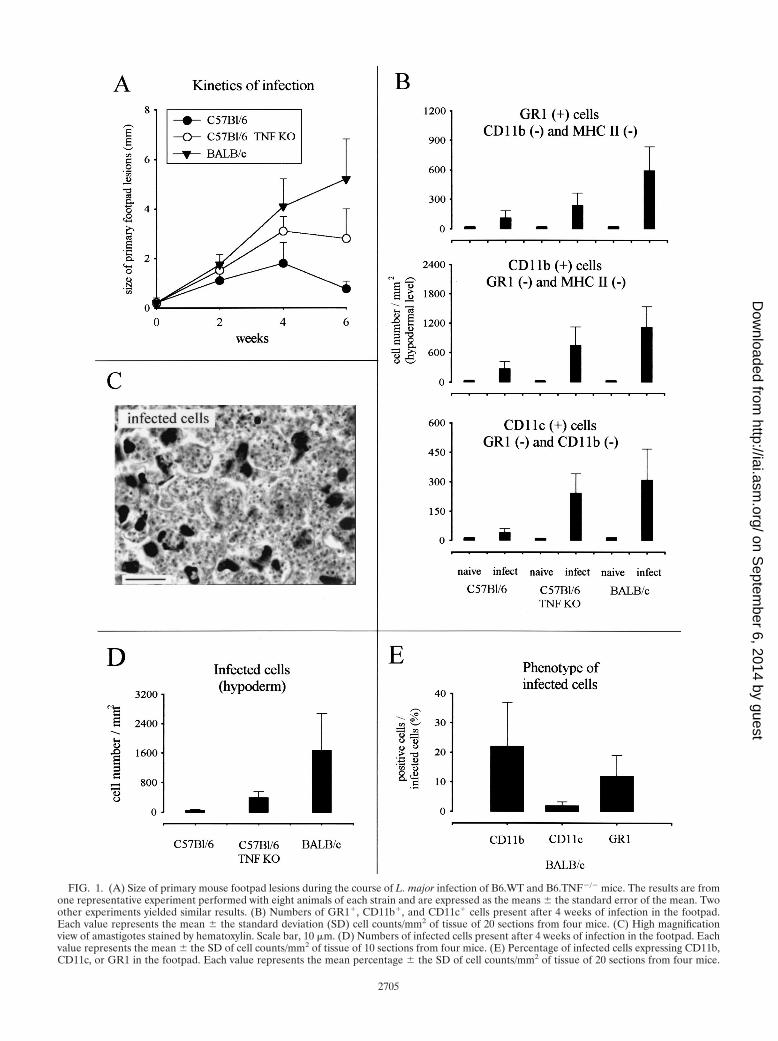
FIG. 1. (A) Size of primary mouse footpad lesions during the course of L. major infection of B6.WT and B6.TNF�/� mice. The results are fromone representative experiment performed with eight animals of each strain and are expressed as the means � the standard error of the mean. Twoother experiments yielded similar results. (B) Numbers of GR1�, CD11b�, and CD11c� cells present after 4 weeks of infection in the footpad.Each value represents the mean � the standard deviation (SD) cell counts/mm2 of tissue of 20 sections from four mice. (C) High magnificationview of amastigotes stained by hematoxylin. Scale bar, 10 �m. (D) Numbers of infected cells present after 4 weeks of infection in the footpad. Eachvalue represents the mean � the SD of cell counts/mm2 of tissue of 10 sections from four mice. (E) Percentage of infected cells expressing CD11b,CD11c, or GR1 in the footpad. Each value represents the mean percentage � the SD of cell counts/mm2 of tissue of 20 sections from four mice.
2705
on Septem
ber 6, 2014 by guesthttp://iai.asm
.org/D
ownloaded from
infected macrophages an unlikely candidate to prime naive Tcells in draining lymph nodes. Actually, the impact of L. majorinfection in vivo on DC populations is unknown. The aim ofthis study was to compare in vivo the impact of L. majorinfection on cells of the DC lineage in susceptible and resistantmice. BALB/c and C57BL/6 (B6.WT) strains of mice are clas-sically used as models of mice susceptible and resistant to L.major, respectively. However, as described below, these strainsdevelop, due to genetic difference, a very different class ofimmune response (Th2 and Th1, respectively) against L. majorparasite rending the interpretation of data complex. Thus,we used C57BL/6 mice lacking tumor necrosis factor (TNF)(B6.TNF�/�) mice as a model of susceptible mice in theC57BL/6 genetic background. In these mice susceptibility to L.major is not associated with the development of Th2 responsebut is correlated with a delayed and reduced Th1 response(38). We analyzed in situ the cell surface phenotype of infectedcells in primary footpad lesions and draining lymph nodes ofBALB/c, B6.WT, and B6.TNF�/� mice. Our results show thatcells displaying DC-like phenotype are the most frequentlyinfected cells in the draining lymph node and harbor differentparasitic loads and cell surface markers in susceptible andresistant mice.
MATERIALS AND METHODS
Mice and parasite. Six- to eight-week-old female BALB/c and C57BL/6 micewere purchased from Charles River Laboratories. C57BL/6 lacking TNF-� were
as previously described (38) and were bred in our animal facility. The mainte-nance and care of mice complied with the guidelines of the ULB Ethics Com-mittee for the humane use of laboratory animals. Promastigotes of L. major(MHOM/IR/�/173 strain) were propagated in vitro at room temperature inRPMI 1640 medium (Life Technologies, Gaithersburg, Md.), supplemented with10% fetal calf serum (Life Technologies), penicillin G (100 U/ml), and strepto-mycin (100 �g/ml). Parasites harvested in the stationary phase after 8 to 10 daysof culture were centrifuged (2,500 � g, 10 min, 4°C) and then washed three timesin RPMI before being used to inoculate animals.
Leishmania infection, lesion monitoring, and tissue processing. Mice wereinfected subcutaneously in the rear left hind footpad with 5 � 106 stationary-phase promastigotes of L. major in a final volume of 25 �l (in RPMI medium).The contralateral right footpad received an identical volume of RPMI mediumwithout parasites as an internal control. The thickness of infected and uninfectedfootpads was regularly measured with a vernier caliper, and the difference be-tween both measurements corresponded to the size of lesions. At selected timepoints, some mice were killed by cervical dislocation. Footpad lesions (or normaltissue in controls) were cut tangentially to the bone ground. Popliteal homolat-eral draining lymph nodes were collected to perform immunohistochemical stud-ies (see below). The distribution and enumeration of amastigotes and infectedcells were determined in organ sections stained with hematoxylin.
Immunohistochemistry. In the present study, we used Immunohistowax pro-cessing, a new fixation and embedding method for light microscopy, initiallydeveloped for DC identification in situ, which preserves antigen immunoreac-tivity and morphological structures (27). Briefly, primary lesions and draininglymph nodes were fixed for 3 days in Immunohistofix (Aphase, Gosselies, Bel-gium), followed by dehydratation in a graded series of ethanol solution (30, 50,70, 90, and 100%) for 30 min each at room temperature. Tissues were embeddedin Immunohistowax (Aphase) and cut into 3- to 6-�m sections, deembedded bya wash in acetone for 10 min, and transferred to phosphate-buffered saline(PBS). The tissue sections were treated for 30 min with blocking reagent (1% inPBS; BoehringerMannheim, Mannheim, Germany) to saturate the sites of non-
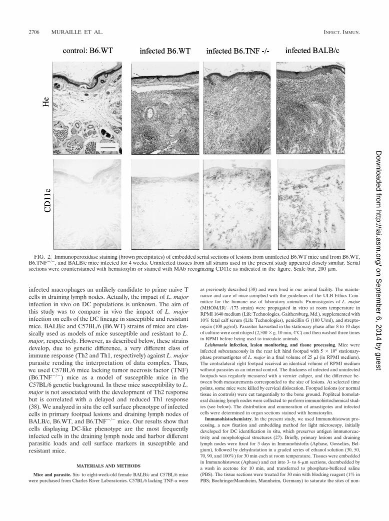
FIG. 2. Immunoperoxidase staining (brown precipitates) of embedded serial sections of lesions from uninfected B6.WT mice and from B6.WT,B6.TNF�/�, and BALB/c mice infected for 4 weeks. Uninfected tissues from all strains used in the present study appeared closely similar. Serialsections were counterstained with hematoxylin or stained with MAb recognizing CD11c as indicated in the figure. Scale bar, 200 �m.
2706 MURAILLE ET AL. INFECT. IMMUN.
on Septem
ber 6, 2014 by guesthttp://iai.asm
.org/D
ownloaded from
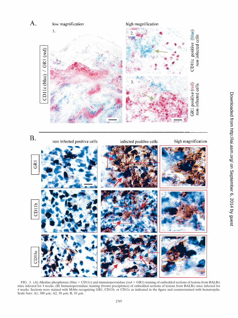
FIG. 3. (A) Alkaline phosphatase (blue � CD11c) and immunoperoxidase (red � GR1) staining of embedded sections of lesions from BALB/cmice infected for 4 weeks. (B) Immunoperoxidase staining (brown precipitates) of embedded sections of lesions from BALB/c mice infected for4 weeks. Sections were stained with MAbs recognizing GR1, CD11b, or CD11c as indicated in the figure and counterstained with hematoxylin.Scale bars: A1, 300 �m; A2, 30 �m; B, 10 �m.
2707
on Septem
ber 6, 2014 by guesthttp://iai.asm
.org/D
ownloaded from
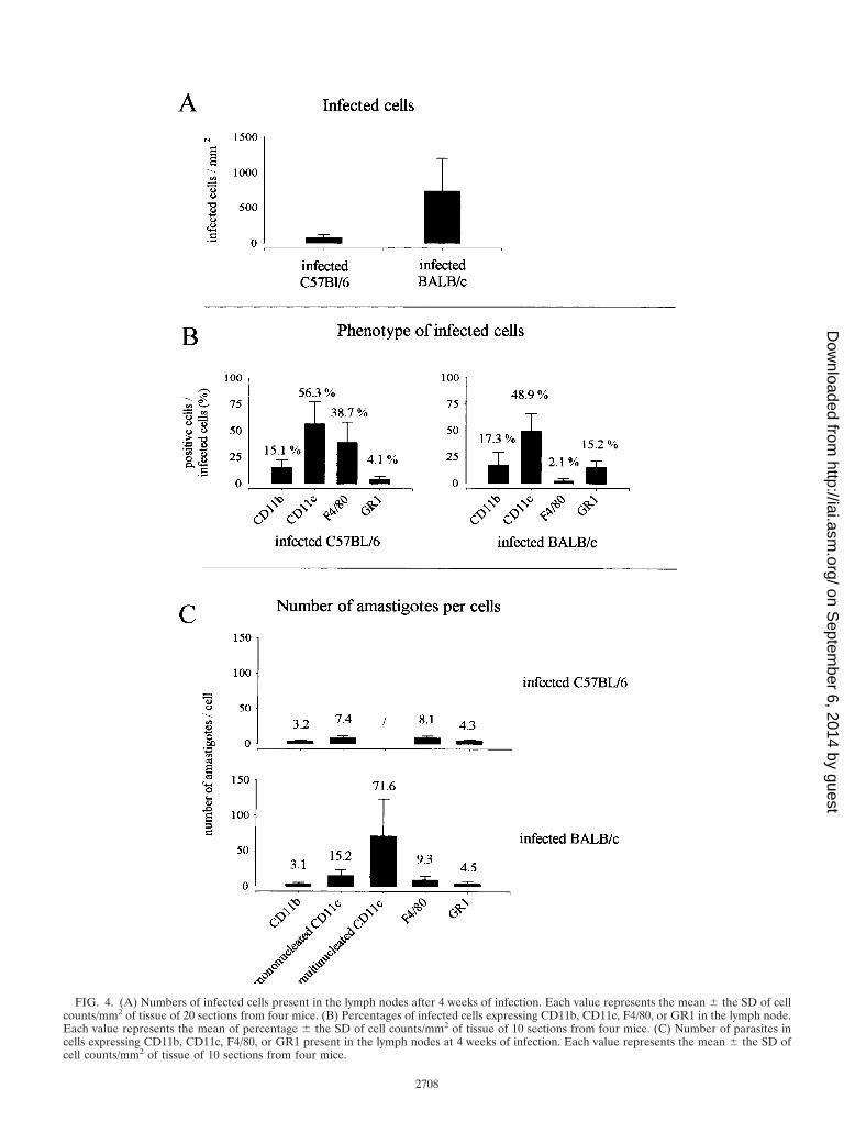
FIG. 4. (A) Numbers of infected cells present in the lymph nodes after 4 weeks of infection. Each value represents the mean � the SD of cellcounts/mm2 of tissue of 20 sections from four mice. (B) Percentages of infected cells expressing CD11b, CD11c, F4/80, or GR1 in the lymph node.Each value represents the mean of percentage � the SD of cell counts/mm2 of tissue of 10 sections from four mice. (C) Number of parasites incells expressing CD11b, CD11c, F4/80, or GR1 present in the lymph nodes at 4 weeks of infection. Each value represents the mean � the SD ofcell counts/mm2 of tissue of 10 sections from four mice.
2708
on Septem
ber 6, 2014 by guesthttp://iai.asm
.org/D
ownloaded from
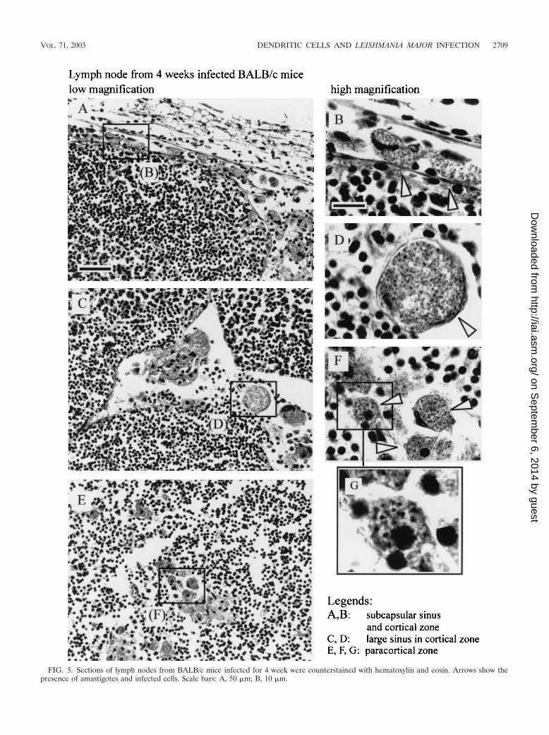
FIG. 5. Sections of lymph nodes from BALB/c mice infected for 4 week were counterstained with hematoxylin and eosin. Arrows show thepresence of amastigotes and infected cells. Scale bars: A, 50 �m; B, 10 �m.
VOL. 71, 2003 DENDRITIC CELLS AND LEISHMANIA MAJOR INFECTION 2709
on Septem
ber 6, 2014 by guesthttp://iai.asm
.org/D
ownloaded from
2710 MURAILLE ET AL. INFECT. IMMUN.
on Septem
ber 6, 2014 by guesthttp://iai.asm
.org/D
ownloaded from
specific reactions. The endogenous peroxidase activity was neutralized by 3%H2O2 in PBS for 60 min. The slides were then incubated with one of thefollowing biotinylated antibodies (10 �g/ml in 0.5% PBS-blocking reagent): 2G9(anti-I-A/I-E monoclonal antibody [MAb]; BD Pharmingen, San Diego, Calif.),C1:A3-1 (anti-F4/80 MAb; Serotec, Oxford, United Kingdom), GL1 (anti-CD86MAb; American Type Culture Collection), M1/70 (anti-CD11b MAb; BDPharmingen), HL3 (anti-CD11c MAb; BD Pharmingen), RA3-6B2 (anti-CD45R/B220 MAb; BD Pharmingen), RB6-8C5 (anti-GR1 MAb; BD Pharmin-gen), RM4-5 (anti-CD4 MAb; Pharmingen), and 53-6.7 (anti-mouse-CD8�MAb; BD Pharmingen). They were further incubated, as indicated in Results,with either (i) avidin-biotin-peroxidase complex (Vectastain ABC kit; VectorLaboratories, Burlingame, Calif.) and stained with a solution of diaminobenzi-dine tetrahydrochloride (DAB tablets, giving brown precipitates; Sigma, St.Louis, Mo.) or a solution of 3-amino-9-ethylcarbazole (AEC tablets, giving redprecipitates; Sigma) or (ii) avidin-biotin-alkaline phosphatase complex (Vec-tastain ABC kit, AK-5000; Vector Laboratories) and stained with alkaline phos-phatase substrates (SK-5300, giving blue precipitates; Vector Laboratories). Incase of double staining of cells, the excess of biotin from the first antibodies wasblocked with the Vector blocking kit (Vector) before incubation of the sectionswith a second biotinated antibody. Digitized images were captured by using acharge-coupled device color camera (Ikegami Tsushinki, Tokyo, Japan) andanalyzed by using CorelDraw 7 software (Corel, Ottawa, Ontario, Canada).
RESULTS
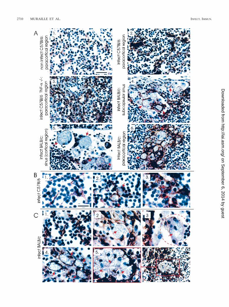
Large number of CD11c� cells are present in the footpadlesions of susceptible mice infected with L. major. In agree-ment with previous studies (26, 38), the maximal lesion size inB6.WT and B6.TNF�/� infected mice was observed aroundthe fourth week (Fig. 1A). At this time, the lesions in BALB/cinfected mice were well developed without mutilation as ob-served later in the course of infection (data not shown). Miceinfected with 5 � 106 L. major were therefore killed 4 weeksafter inoculation for immunohistochemistry analysis. Threemajor populations were observed and enumerated in lesion byusing antibodies recognizing GR1, CD11b, and CD11c (Fig.1B and 2). Using these antibodies in combinations, we did notobserve any double staining in the different control or infectedmouse strains. This result demonstrates that each marker iden-tified a distinct population. An example of such a combinationis shown Fig. 3A (GR1/CD11c combination). Accordingly toprevious studies (11, 17, 22, 33), CD11c� CD11b� GR1� cellswere identified as cells of the DC lineage (Langerhans cellsand dermal DCs), CD11c� CD11b� GR1� as macrophages,and CD11c� CD11b� GR1� cells were identified as granulo-cytes. Infected B6.TNF�/� and BALB/c susceptible mice pre-sented a dramatic infiltration and accumulation of these threecell populations in their lesions (Fig. 1B), which was mainlypresent at the hypodermal level of the skin (see Fig. 2 forCD11c distribution). In comparison, infected resistant B6.WTmice displayed only a moderate recruitment of these cells.
CD11b� cells and GR1� cells are the most frequently in-fected cells in footpad lesions. A high-magnification view ofamastigotes stained by hematoxylin in infected cells fromBALB/c lesion is shown in Fig. 1C. As expected, BALB/c micein comparison to other mice displayed higher numbers of in-
fected cells in lesions (Fig. 1D). Amastigotes were detected inCD11c�, CD11b�, and GR1� cells by using hematoxylin stain-ing. CD11b� cells and GR1� cells appeared to be the mostfrequently infected cells compared to CD11c� cells in BALB/cmice (Fig. 1E). Figure 3B show the morphology of uninfectedand infected CD11b�, CD11c�, and GR1� cells. The intenseaggregation of infected cells prevented a precise estimation ofthe number of amastigotes per cell. Nevertheless, our obser-vations suggest that they were numerous and at roughly similaramounts in CD11b�, CD11c�, and GR1� cells. Coexpressionof CD4, CD8, or CD86 markers were not detected in infectedcells (data not shown). Similar qualitative results were ob-served in infected cells from B6.WT and B6.TNF�/� mice(data not shown).
Distribution of infected cells in draining lymph node ofinfected mice. Immunohistochemical analysis showed impor-tant modifications of lymph node organization during thecourse of L. major infection. At 4 weeks after parasite inocu-lation, the paracortical region of the draining lymph nodes ofinfected mice, but not of control mice, presented multiple largelobules containing T cells (identified as CD90.2�) and B cells(identified as CD45R� and MHC-II�) separated by large sinus(data not shown). In BALB/c mice, which presented the high-est level of infected cells (Fig. 4A), cells containing amastigoteswere localized in the subcapsular region (Fig. 5A and B), in thecortical and paracortical sinus (Fig. 5C and D), and in theparacortical region (Fig. 5E to G). In B6.WT and B6.TNF�/�
mice, infected cells were observed mainly in the paracorticalregion of lymph node (data not shown).
Amastigotes are massively accumulated in CD11c� cells indraining lymph nodes of BALB/c mice. In comparison to un-infected DCs (CD11c�) displaying classical dendritic morphol-ogy (Fig. 6B1 and C1), the morphology of infected CD11c�
cells was strongly modified. In infected B6.WT mice, largeaggregates of CD11c� cells with dendritic morphology wereobserved in paracortical region (Fig. 6A2). Few parasites weredetected in these cells (Fig. 6B2 and B3). A similar but en-hanced phenomenon was observed in infected B6.TNF�/�
mice (Fig. 6A3). In contrast, in lymph nodes from infectedBALB/c, infected CD11c� cells presenting a massive accumu-lation of parasites were observed in subcapsular region (Fig.6A4), cortical and paracortical sinus (Fig. 6A5), and paracor-tical region (Fig. 6A6). It is interesting that CD11c� infectedcells present in subcapsular, cortical and paracortical sinusexpressed frequently an intermediate level of CD11c (Fig.6C3) in comparison to CD11c� infected cells of paracorticalregion (Fig. 6C4).
The analysis of the phenotype expressed by the infected cells(Fig. 4B) showed that CD11c� cells were the most frequentlyinfected in lymph nodes of C57BL/6 and BALB/c mice com-pared to GR1� and CD11b� cells. Comparison of the numberof intracellular amastigotes showed that the highest parasite
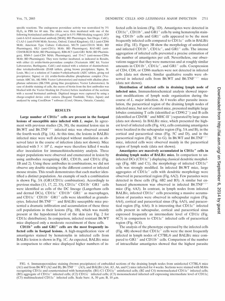
FIG. 6. Immunoperoxidase staining (brown precipitates) of embedded sections of the draining lymph nodes from uninfected C57BL/6 mice(A1) and from B6.WT (A2 and B), B6.TNF�/� (A3), and BALB/c (A4, A5, A6, and C) mice infected for 4 weeks. Sections were stained with MAbsrecognizing CD11c and counterstained with hematoxylin. (B1) C1 CD11c� uninfected cells; (B2 and C4) mononucleated CD11c� infected cells;(B3) aggregate of CD11c� infected cells; (C2) CD11c� infected cells; (C3) mononucleated infected cell expressing intermediate level of CD11c;(C5) multinucleated CD11c� infected cells. Scale bars: A, 50 �m; B, 10 �m.
VOL. 71, 2003 DENDRITIC CELLS AND LEISHMANIA MAJOR INFECTION 2711
on Septem
ber 6, 2014 by guesthttp://iai.asm
.org/D
ownloaded from
2712 MURAILLE ET AL. INFECT. IMMUN.
on Septem
ber 6, 2014 by guesthttp://iai.asm
.org/D
ownloaded from
levels were observed in mononucleated CD11c� cells fromBALB/c lymph nodes (Fig. 4C) and overall in giant multinu-cleated CD11c� cells (Fig. 4C and 6C5). These latter cells werenot present in infected B6.WT mice. In summary, CD11c�
cells appear as the most frequently infected cells in lymphnodes from infected mice. CD11c� from BALB/c mice, but notfrom other mice, displayed a more intense multiplication ofamastigotes.
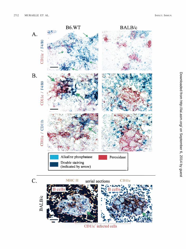
Infected CD11c� cells from lymph nodes of C57BL/6, butnot BALB/c mice, express the CD11b and F4/80 myeloid mark-ers. The existence of several subpopulations of DCs in blood(10) and lymphoid organs (4, 17, 22) based on the expressionof CD4, CD8, CD11b, CD45R (B220), CD90.2, and F4/80markers has been well established. In order to determine thephenotype of infected CD11c� cells in both strains of mice, weperformed systematic double staining with the anti-CD11c an-tibody and another antibody recognizing one of the abovelisted markers. Our results (Fig. 7A) show that some aggre-gated infected CD11c� cells from C57BL/6 mice, but nothighly infected cells from BALB/c mice, expressed CD11b orF4/80 myeloid markers. These cells did not express at a de-tectable level lymphoid markers such as CD4, CD8, CD45R,and CD90.2 (data not shown). CD11c� infected cells fromdraining lymph nodes of B6.WT and B6.TNF�/� mice dis-played similar phenotype (data not shown). The frequent co-expression of CD11c and F4/80 in infected B6.WT might ex-plain the higher number of F4/80� infected cells observed inB6.WT compared to BALB/c mice (Fig. 4B).
Infected CD11c� cells from the lymph nodes of both mousestrains express low levels of MHC-II, but no CD86 moleculeswere detected. Lymph node DCs are frequently described asexpressing high surface levels of MHC-II (33). In both strainsof mice, we observed that noninfected CD11c� cells expressedMHC-II at high levels (data not show). In contrast, the largemajority of highly infected CD11c� cells from BALB/c andaggregated infected cells from B6.WT mice express only lowlevels of surface MHC-II (inferior to that observed on B cells)(Fig. 7C). CD86 expression was not detected on infectedCD11c� cells from lymph nodes of B6.WT or BALB/c mice,whereas it was frequently detected on CD45R� cells (B cells)of lymph nodes from infected BALB/c mice (data not shown)
DISCUSSION
We analyzed here the in vivo phenotype of L. major-infectedcells in the primary site of infection and the draining lymphnode of B6.WT, B6.TNF�/� and BALB/c mice 4 weeks afterparasite inoculation. Previous works have demonstrated thatLeishmania parasites can infect macrophages (31), granulo-cytes (24), and DCs (5, 34, 37). However, as far as we know theimportance of this phenomena has not been estimated quan-titatively in vivo during the peak of infection in both resistant
and susceptible mice. Moreover, the expression of functionallyimportant markers, such as MHC-II and costimulatory mole-cules, and differentiation markers at the surface of infectedcells has not been analyzed previously.
Our results show that granulocytes and macrophages, iden-tified as CD11b� CD11c� GR1� and CD11b� CD11c�
GR1�, respectively, are the main infected cells in primarylesions. In contrast, cells expressing a high level of CD11c, aDC specific marker, are the most frequently infected in drain-ing lymph nodes of infected mice. Qualitatively similar resultshave been obtained at 6 and 9 weeks of infection (data notshown). The preferential infection of DCs by intracellularpathogens has been reported previously in other models. Den-gue virus, a flavivirus causing febrile illness, inoculated intohuman skin, like Leishmania parasites, by mosquitoes, hasbeen reported to infect 10-fold more DCs than monocytes ormacrophages (39). DCs have also been described as the majorreservoir of human immunodeficiency virus in human lymphnodes (14).
CD11c� infected cells detected in lymph nodes might (i)have migrated from lesion to draining lymph node or (ii) beinfected in the lymph node with parasites released by othercells. The presence of infected cells expressing an intermediateor high level of CD11c marker in the subcapsular sinus oflymph nodes indicates that at least a portion of infected cellsthat have migrated from the lesion to the lymph node areCD11c� cells. Recently, it has been demonstrated that all DCpopulations can be derived from only one population of DCprecursors (10). However, during inflammation, DCs can alsodifferentiate from monocytes (30). For this reason, distinguish-ing between monocytes/macrophages and DCs is difficult, par-ticularly during the course of an infection. Randolph et al. (30)have previously reported that macrophages (identified asCD11b� CD11c�) recruited in the footpad after an inflamma-tory stimulus can differentiate into “monocyte-derived DCs”(acquiring CD11c marker) and migrate into the lymph nodes.In our study, we observed that a few CD11c� cells located inlesions were infected by L. major. Thus, CD11c� infected cellsobserved in the subcapsular sinus of the draining lymph nodemight derive from both monocyte-derived DCs and DCs in-fected in the footpad.
Compared to BALB/c mice, the lymph nodes of B6.WT micecontained low number of infected cells displaying a moderateparasitic load and DC morphology. In contrast, in lymph nodesof BALB/c mice, infected CD11c� cells presented high para-sitic loads, and we frequently observed CD11c� multinucle-ated giant cells harboring a dramatic accumulation of para-sites. Indeed, multinucleated giant cells have been previouslydescribed in lymph node biopsies from Leishmania-infectedpatients, but their cell surface phenotype has not been re-ported (36). B6.TNF�/� mice, due to a reduced and delayedTh1 response, are susceptible to L. major infection (38). In
FIG. 7. (A and B) Immunoalkaline phosphatase (blue � F4/80 and CD11b as indicated) and immunoperoxidase (red � CD11c) staining ofembedded sections of lymph nodes from BALB/c and B6.WT mice infected for 4 weeks, as indicated in the figure. Arrows show the presence ofdouble staining. (C) Immunoperoxidase staining (brown precipitates) of embedded serial sections of the draining lymph nodes from BALB/c miceinfected for 4 weeks. Sections were stained with MAbs recognizing MHC-II and CD11c, as indicated in the figure, and counterstained withhematoxylin. Scale bars: A, 150 �m; B; 10 �m; C, 50 �m.
VOL. 71, 2003 DENDRITIC CELLS AND LEISHMANIA MAJOR INFECTION 2713
on Septem
ber 6, 2014 by guesthttp://iai.asm
.org/D
ownloaded from
agreement with an earlier study (38), we observed in thesemice an increased frequency of infected cells. However, lymphnode infected cells from these mice presented weak parasiticloads and no syncytium was observed. Thus, we can concludethat syncytium formation appears as a consequence of highparasitic loads associated with Th2 response in BALB/c mice.It is interesting that, in contrast to macrophages, DCs do notproduce NO in response to L. major infection (6), suggestingthat these cells are not able to eliminate amastigotes or preventtheir multiplication. Thus, these data and our study suggestthat DCs can potentially constitute a reservoir of parasites inthe case of a nonadapted Th2 immune response.
Another interesting observation was that lymph nodeCD11c� infected cells from B6.WT and B6.TNF�/�, but notfrom BALB/c mice, frequently expressed the CD11b and F4/80myeloid differentiation markers. The reason for this differenceis not known. The similar cell phenotype observed in resistantB6.WT and susceptible B6.TNF�/� mice suggests that the ge-netic background or the profile of immune response (Th1)might be involved. Whereas the phenotype of DC subsets inlymph node mice has been described in naive mice (17), thishas not been reported during the course of infection, making itdifficult to associate the phenotype of some CD11c� infected(CD4�CD8�, CD11b�, or F4/80�) cells from the lymph nodesof B6.WT mice to a classical subset of DCs. However, theabsence of detectable CD4 and CD8 expression suggests thatthese cells might be skin migrating cells (17). The expression ofthe myeloid markers CD11b and/or F4/80 also suggests thatthese cells are similar to monocyte-derived DCs, as describedby Randolph et al. (30). Interestingly, a recent work (18) hasdemonstrated that splenic myeloid DCs (CD4�, CD8�,CD11b�, and F4/80� [22]) rather than other splenic DC sub-sets are preferentially infected in vitro by L. major.
We detected low expression of MHC-II but no expression ofCD86 costimulatory molecule on CD11c� infected cells fromlymph nodes of B6.WT or BALB/c mice, suggesting that theantigen-presenting function of these cells is downregulated.This is not due to technical problems since these markers wereclearly detected at the surface of noninfected cells on the sametissue sections. Conflicting results have been reported on theability of Leishmania species to activate cells of the DC lin-eage. It has been shown in vitro that bone marrow-derived DCsdo not mature in response to L. mexicana infection (3). Simi-larly, a functional impairment of in vitro T-cell stimulatoryproperties of splenic DCs at 60 days postinfection, correlatingwith a reduced surface expression of MHC-II, has been re-ported during the course of L. donovani infection in BALB/cmice (2). In contrast, Qi et al. (29) have observed in vitro thatL. amazonensis induced the upregulation of costimulatory mol-ecules on bone marrow-derived DCs, and von Stebut and al.(37) showed that in vitro infection of fetal-skin-derived DCsfrom both BALB/c and B6.WT mice infected with L. majorinduced the upregulation of costimulatory molecules and therelease of IL-12. These contrasting results obtained in vitromight be due to the Leishmania species used and the origin ofthe DCs (bone marrow, spleen, and fetal skin). We have ob-served in situ that lymph node CD11c� infected cells fromsusceptible mice presented high numbers of parasites, whichwere clearly greater than the parasitic loads observed in cellsinfected in vitro (18). This suggests that in vivo infection might
more deeply affect the physiology of cells. Moreover, the du-ration of infection and/or environmental factors present in vivomight also influence the phenotype and the T-cell stimulatoryproperties of DCs. Thus, it is possible that a short time ofinfection in vitro with L. major activates DC maturation, asreported by von Stebut et al. (37) but that, conversely, long-term infection induces a downregulation of the typical markersof antigen presenting function on DCs, as observed with L.donovani infection (2). Moll et al. (25) reported that lymphnode DCs from B6.WT that have recovered from infectionwith L. major harbor viable parasites. These authors observedthat lymph node cells from these mice can efficiently activateLeishmania-specific T cells but that the depletion of DCs im-pairs this activation. These authors concluded that infectedDCs efficiently present Leishmania antigen to specific T cells.These results do not exclude the possibility that infected DCsrelease antigen that can be presented in vitro to T cells bynoninfected competent DCs. Due probably to their large size,isolation of CD11c� infected cells by a standard procedure hasnot been possible in our laboratory, and the subsequent anal-ysis of their antigen-presenting cell functions has not beenperformed. Thus, we cannot actually determine whether in-fected cells are functionally able to present antigen. However,the downregulation of antigen-presenting markers at their cellsurface suggests strongly that presentation is performed bynoninfected DCs. Leishmania parasites reside and multiplyinto parasitophorous vacuoles. These organelles of phagolyso-somal origin have similar properties to vacuoles where pep-tide–MHC-II molecule complexes are formed before their ex-pression at the cell surface. In vitro studies have shown thatMHC-II molecules are associated with the parasitophorousvacuoles of macrophages infected by L. major, L. amazonensis,and L. mexicana (1, 13). Some Leishmania species, such as L.amazonensis and L. mexicana, internalize and degrade MHC-IImolecules (1, 13), suggesting that Leishmania parasites mightaffect antigen presentation.
In summary, our analysis of the cell surface phenotype of L.major-infected cells show that cells presenting the CD11c DCmarker are the most frequently infected cells in the draininglymph nodes of resistant and susceptible mice. In susceptibleBALB/c mice, these cells constitute an important reservoir ofparasites and present a reduced expression of antigen presen-tation markers.
ACKNOWLEDGMENTS
We thank Dominique Le Ray (Institute of Tropical Medicine, Ant-werp, Belgium) for providing the MHOM/IR/�/173 strain of L. major.We thank Jean-Claude Antoine for critical review of the manuscriptand Alain Wathelet and Ella Omasta for diligent technical assistance.
This work was supported by grants from the Belgian Ministry ofScientific Policy (Action de Recherche Concertee), the Fonds Nationalde la Recherche Scientifique (FNRS; Credit aux Chercheurs [Bel-gium]), and the Universite Libre de Bruxelles. E.M. is supported by theFNRS, and C.D.T. is supported by the Fonds pour la Recherche dansl’Industrie et l’Agriculture (Belgium).
REFERENCES
1. Antoine, J.-C., T. Lang, E. Prina, N. Courret, and R. Hellio. 1999. H-2Mmolecules, like MHC II molecules, are targeted to parasitophorous vacuolesof Leishmania-infected macrophages and internalized by amastigotes of L.amazonensis and L. mexicana. J. Cell Sci. 112:2559–2570.
2. Basu, A., G. Chakrabarti, A. Saha, and S. Bandyopadhyay. 2000. Molulationof CD11c� splenic dendritic cell functions in murine visceral leishmaniasis:correlation with parasite replication in the spleen. Immunology 99:305–313.
2714 MURAILLE ET AL. INFECT. IMMUN.
on Septem
ber 6, 2014 by guesthttp://iai.asm
.org/D
ownloaded from
3. Bennett, C. L., A. Misslitz, L. Colledge, T. Aebischer, and C. C. Blackburn.2001. Silent infection of bone marrow-derived dendritic cells by Leishmaniamexicana amastigotes. Eur. J. Immunol. 31:876–883.
4. Bjorck, P. 2001. Isolation and characterization of plasmacytoid dendriticcells from Flt3 ligand and granulocyte-macrophage colony-stimulating fac-tor-treated mice. Blood 98:3520–3526.
5. Blank, C., H. Fuchs, K. Rappersberger, M. Rollinghoff, and H. Moll. 1993.Parasitis of epidermal Langerhans cells in experimental cutaneous leishman-iasis with Leishmania major. J. Infect. Dis. 167:418–425.
6. Blank, C., C. Bogdan, C. Bauer, K. Erb, and H. Moll. 1996. Murine epider-mal Langerhans cells do not express inducible nitric oxide synthase. Eur.J. Immunol. 26:792–796.
7. Bogdan, C., M. Rollinghoff, and A. Diefenbach. 2000. The role of nitric oxidein innate immunity. Immunol. Rev. 173:17–26.
8. Bradley, D. J. 1987. Genetics of susceptibility and resistance in the vertebratehost, p. 551–581. In W. Peters and R. Killick-Kendrick (ed.), The leishman-iases in biology and medicine. Academic Press, Ltd., London, United King-dom.
9. Carrera, I., R. T. Gazzinelli, R. Badolato, S. Hieny, W. Muller, R. Kuhn, andD. L. Sacks. 1996. Leishmania promastigotes selectively inhibit interleu-kin-12 induction in bone marrow-derived macrophages from susceptible andresistant mice. J. Exp. Med. 183:515–526.
10. del Hoyo, G. M., P. Martin, H. H. Vargas, S. Ruiz, C. F. Arias, and C.Ardavin. 2002. Characterization of a common precursor population for den-dritic cells. Nature 415:1043–1047.
11. Duraiswamy, N., Y. Tse, C. Hammerberg, S. Kang, and K. D. Cooper. 1994.Distinction of class II MHC� Langerhans cell-like interstitial dendritic an-tigen-presenting cells in murine dermis from dermal macrophages. J. Inves-tig. Dermatol. 103:678–683.
12. Flohe, S. B., C. Bauer, S. Flohe, and H. Moll. 1998. Antigen-pulsed epider-mal Langerhans cells protect susceptible mice from infection with the intra-cellular parasite Leishmania major. Eur. J. Immunol. 28:3800–3811.
13. Flohe, S., T. Lang, and H. Moll. 1997. Synthesis, stability, and subcellulardistribution of major histocompatibility complex class II molecules in Lang-erhans cells infected with Leishmania major. Infect. Immun. 65:3444–3450.
14. Frankel, S. S., B. M. Wenig, A. P. Burke, P. Mannan, L. D. Thompson, S. L.Abbondanzo, A. M. Nelson, M. Pope, and R. M. Steinman. 1996. Replicationof HIV-1 in dendritic cell-derived syncitia at the mucosal surface of theadenoid. Science 272:115–117.
15. Fruth, U., N. Solioz, and J. A. Louis. 1993. Leishmania major interferes withantigen presentation by infected macrophages. J. Immunol. 150:1857–1864.
16. Gorak, P. M. A., C. R. Engwerda, and P. M. Kaye. 1998. Dendritic cells, butnot macrophages, produce IL-12 immediately following Leishmania dono-vani infection. Eur. J. Immunol. 28:687–695.
17. Henri, S., D. Vremec, A. Kamath, J. Waithman, S. Williams, C. Benoist, K.Burnham, S. Saeland, Handman, E., and K. Shortman. 2001. The dendriticcell populations of mouse lymph nodes. J. Immunol. 167:741–748.
18. Henri, S., J. Curtis, H. Hochrein, D. Vremec, K. Shortman, and E. Hand-man. 2002. Hierarchy of susceptibility of dendritic cell subsets to infection byLeishmania major: inverse relationship to interleukin-12 production. Infect.Immun. 70:3874–3880.
19. Himmelrich, H., P. Launois, I. Maillard, T. Biedermann, F. Tacchini-Cot-tier, R. M. Locksley, M. Rocken, and J. A. Louis. 2000. In BALB/c mice, IL-4production during the initial phase of infection with Leishmania major isnecessary and sufficient to instruct Th2 cell development resulting in pro-gressive disease. J. Immunol. 164:4819–4825.
20. Huang, F. P., D. Xu, E. O. Esfandiari, W. Sands, X. Q. Wei, and F. Y. Liew.1998. Mice defective in Fas are highly susceptible to Leishmania majorinfection despite elevated IL-12 synthesis, strong Th1 responses, and en-hanced nitric oxide production. J. Immunol. 160:4143–4147.
21. Jenkins, M. K., A. Khoruts, E. Ingulli, D. L. Mueller, S. J. McSorley, R. L.Reinhardt, A. Itano, and K. A. Pape. 2001. In vivo activation of antigen-specific CD4 T cells. Annu. Rev. Immunol. 19:23–45.
22. Kamath, A. T., J. Pooley, M. A. O’Keeffe, D. Vremec, Y. Zhan, A. M. Lew, A.
D’Amico, L. Wu, D. F. Tough, and K. Shortman. 2000. The development,maturation, and turnover rate of mouse spleen dendritic cell populations.J. Immunol. 165:6762–6770.
23. Kremer, I. B., M. P. Gould, K. D. Cooper, and F. P. Heinzel. 2001. Pretreat-ment with recombinant Flt3 ligand partially protects against progressivecutaneous leishmaniasis in susceptible BALB/c mice. Infect. Immun. 69:673–680.
24. Laufs, H., K. Muller, J. Fleischer, N. Reiling, N. Jahnke, J. C. Jensenius, W.Solbach, and T. Laskay. 2002. Intracellular survival of Leishmania major inneutrophil granulocytes after uptake in the absence of heat-labile serumfactors. Infect. Immun. 70:826–835.
25. Moll, H., H. Fuchs, C. Blank, and M. Rollinghoff. 1993. Langerhans cellstransport Leishmania major from the infected skin to the draining lymphnode for presentation to antigen-specific T cells. Eur. J. Immunol. 23:1595–1601.
26. Noben-Trauth, N., W. E. Paul, and D. L. Sacks. 1999. IL-4- and IL-4 recep-tor-deficient BALB/c mice reveal differences in susceptibility to Leishmaniamajor parasite substrains. J. Immunol. 162:6132–6140.
27. Pajak, B., T. De Smedt, V. Moulin, C. De Trez, R. Maldonado-Lopez, G.Vansanten, E. Briend, J. Urbain, O. Leo, and M. Moser. 2000. Immunohis-towax processing, a new fixation and embedding method for light micros-copy, which preserves antigen immunoreactivity and morphological struc-tures: visualization of dendritic cells in peripheral organs. J. Clin. Pathol.53:518–524.
28. Park, A. Y., B. D. Hondowicz, and P. Scott. 2000. IL-12 is required tomaintain a Th1 response during Leishmania major infection. J. Immunol.165:896–902.
29. Qi, H., V. Popov, and L. Soong. 2001. Leishmania amazonensis-dendritic cellinteractions in vitro and the priming of parasite-specific CD4� T cells in vivo.J. Immunol. 167:4534–4542.
30. Randolph, G. J., K. Inaba, D. F. Robbiani, R. M. Steinman, and W. A.Muller. 1999. Differentiation of phagocytic monocytes into lymph node den-dritic cells in vivo. Immunity 11:753–761.
31. Reiner, S. L., and R. M. Locksley. 1995. The regulation of immunity toLeishmania major. Annu. Rev. Immunol. 13:151–177.
32. Scott, P., E. Pearce, A. W. Cheever, R. L. Coffman, and A. Sher. 1989. Roleof cytokines and CD4� T-cell subsets in the regulation of parasite immunityand disease. Immunol. Rev. 112:161–182.
33. Steinman, R. M., M. Pack, and K. Inaba. 1997. Dendritic cells in the T-cellareas of lymphoid organs. Immunol. Rev. 156:25–37.
34. Stenger, S., N. Donhauser, H. Thuring, M. Rollinghoff, and C. Bogdan. 1996.Reactivation of latent leishmaniasis by inhibition of inducible nitric oxidesynthase. J. Exp. Med. 183:1501–1514.
35. Stobie, L., S. Gurunathan, C. Prussin, D. L. Sacks, N. Glaichenhaus, C. Y.Wu, and R. A. Seder. 2000. The role of antigen and IL-12 in sustaining Th1memory cells in vivo: IL-12 is required to maintain memory/effector Th1 cellssufficient to mediate protection to an infectious parasite challenge. Proc.Natl. Acad. Sci. USA 97:8427–8432.
36. Tallada, N., A. Raventos, S. Martinez, C. Compano, and B. Almirante. 1993.Leishmania lymphadenitis diagnosed by fine-needle aspiration biopsy. Di-agn. Cytopathol. 9:673–676.
37. von Stebut, E., Y. Belkaid, T. Jakob, D. L. Sacks, and M. C. Udey. 1998.Uptake of Leishmania major amastigotes results in activation and interleukin12 release from murine skin-derived dendritic cells: implications for theinitiation of anti-Leishmania immunity. J. Exp. Med. 188:1547–1552.
38. Wilhelm, P., U. Ritter, S. Labbow, N. Donhauser, M. Rollinghoff, C. Bogdan,and H. Kornern. 2001. Rapidly fatal leishmaniasis in resistant C57BL/6 micelacking TNF. J. Immunol. 166:4012–4019.
39. Wu, S.-J. L., G. Grouard-Vogel, W. Sun, J. R. Mascola, E. Brachtel, R.Putvatana, M. K. Louder, L. Filgueira, M. A. Marovitch, H. K. Wong, A.Blauvelt, G. Murphy, M. L. Robb, B. L. Innes, D. L. Birx, C. G. Hayes, andS. S. Frankel. 2000. Human skin Langerhans cells are targets of dengue virusinfection. Nat. Med. 6:816–820.
Editor: J. M. Mansfield
VOL. 71, 2003 DENDRITIC CELLS AND LEISHMANIA MAJOR INFECTION 2715
on Septem
ber 6, 2014 by guesthttp://iai.asm
.org/D
ownloaded from