Embed Size (px)
Citation preview
An High-Throughput In Vivo Screening System to SelectH3K4-Specific Histone Demethylase InhibitorsCecilia Mannironi1, Marco Proietto2, Francesca Bufalieri2, Enrico Cundari1, Angela Alagia2,
Svetlana Danovska2, Teresa Rinaldi2, Valeria Famiglini3, Antonio Coluccia3, Giuseppe La Regina3,
Romano Silvestri3, Rodolfo Negri2*
1 Istituto di Biologia e Patologia Molecolari Consiglio Nazionale delle Ricerche, Rome, Italy, 2 Istituto Pasteur Fondazione Cenci Bolognetti, Dipartimento di Biologia e
Biotecnologie ‘‘C. Darwin’’, Sapienza Universita di Roma, Rome, Italy, 3 Dipartimento di Chimica e Tecnologie del Farmaco, Sapienza Universita di Roma, Rome, Italy
Abstract
Background: Histone demethylases (HDMs) have a prominent role in epigenetic regulation and are emerging as potentialtherapeutic cancer targets. The search for small molecules able to inhibit HDMs in vivo is very active but at the present fewcompounds were found to be specific for defined classes of these enzymes.
Methodology/Principal Findings: In order to discover inhibitors specific for H3K4 histone demethylation we set up ascreening system which tests the effects of candidate small molecule inhibitors on a S.cerevisiae strain which requires Jhd2demethylase activity to efficiently grow in the presence of rapamycin. In order to validate the system we screened a libraryof 45 structurally different compounds designed as competitive inhibitors of a -ketoglutarate (a-KG) cofactor of the enzyme,and found that one of them inhibited Jhd2 activity in vitro and in vivo. The same compound effectively inhibits humanJumonji AT-Rich Interactive Domain (JARID) 1B and 1D in vitro and increases H3K4 tri-methylation in HeLa cell nuclearextracts (NEs). When added in vivo to HeLa cells, the compound leads to an increase of tri-methyl-H3K4 (H3K4me3) but doesnot affect H3K9 tri-methylation. We describe the cytostatic and toxic effects of the compound on HeLa cells atconcentrations compatible with its inhibitory activity.
Conclusions/Significance: Our screening system is proved to be very useful in testing putative H3K4-specific HDMinhibitors for the capacity of acting in vivo without significantly altering the activity of other important 2-oxoglutarateoxygenases.
Citation: Mannironi C, Proietto M, Bufalieri F, Cundari E, Alagia A, et al. (2014) An High-Throughput In Vivo Screening System to Select H3K4-Specific HistoneDemethylase Inhibitors. PLoS ONE 9(1): e86002. doi:10.1371/journal.pone.0086002
Editor: Arthur J. Lustig, Tulane University Health Sciences Center, United States of America
Received June 21, 2013; Accepted December 3, 2013; Published January 29, 2014
Copyright: � 2014 Mannironi et al. This is an open-access article distributed under the terms of the Creative Commons Attribution License, which permitsunrestricted use, distribution, and reproduction in any medium, provided the original author and source are credited.
Funding: This work has been supported by: FIRB 2011–2013 (grant no. RBIN06E9Z8) ‘‘Molecular Bases of Diseases’’; PRIN 2009 ‘‘Role of S. cerevisiae GeneralRegulatory Factors in chromatin organization and dynamics’’ and Progetti di Ricerca di Ateneo (grant no. C26A1139XY) to RN and: PRIN 2010 (grantno. 2010W7YRLZ); Progetti di Ricerca di Universita, Sapienza Universita di Roma 2012 and Bando Futuro in Ricerca 2010 (grant no. RBFR10ZJQT) to RS. VLfellowship is sponsored by Regione Lazio. The funders had no role in study design, data collection and analysis, decision to publish, or preparation of themanuscript.
Competing Interests: The authors have declared that no competing interests exist.
* E-mail: [email protected]
Introduction
Chromatin structure governs several aspects of cell metabolism.
Histone N-terminal tails are subjected to several covalent
modifications which form a sophisticated combinatory code which
is read and interpreted by a plethora of regulatory protein
complexes [1,2]. Among the various modifications, Lysine (K)
methylation is particularly interesting, due to its widespread roles
in transcriptional regulation, DNA repair and epigenetic inheri-
tance [3]. In S.cerevisiae, three lysine methyl transferases, Set1, Set2
and Dot1, catalyse histone mono-, di- or tri-methylation at K4,
K36 and K79, respectively. These epigenetic marks, which are
absolutely conserved among eukaryotes, have been associated with
actively transcribed loci [4], although their roles in controlling
transcription efficiency may be distinct and strongly context-
dependent [5].
In particular, H3K4 tri-methylation is enriched at the
promoters and 59 portions of actively transcribed open reading
frames in both yeast and higher eukaryotes [6] and seems to play
multiple, variable and sometime conflicting roles in transcription
[5,7–10].
In higher eukaryotes methylation of H3K9 and K27 residues is
strictly coupled to transcriptional repression and silencing [2],
although these modifications are not observed in S.cerevisiae [1].
For many years, histone lysine methylation has been considered
irreversible and persisting through cell division. Recently, two
families of HDMs have been identified in eukaryotes: the Lysine
Specific histone Demethylase 1 (LSD1) family and the Jmjc-
domain-containing family [11–14]. The LSD1 HDMs are
monoamine oxidases that can demethylate mono- and di-
methylated H3K4 and H3K9 and require flavin adenine
dinucleotide (FAD) for their function [13–15]. On the other end,
Jumonji C domain-containing HDMs (JHDMs), 5 members in
S.cerevisiae and at least 27 members in H.sapiens, are Fe2+ and a-
KG-dependent hydroxylases, and their reported substrate residues
PLOS ONE | www.plosone.org 1 January 2014 | Volume 9 | Issue 1 | e86002
include H3K4, H3K9, H3K27, and H3K36 at all methylation
states [11,12]. The JHDM Jhd2 (encoded by YJR119c and also
called Lysine specific Demethylase 5, KDM5) was purified from
budding yeast and was shown to specifically remove H3K4 di- and
tri-methylation [16–18]. JHDMs are potential therapeutic cancer
targets [19] and among them, those capable to demethylate
specifically H3K4 (JARID1A-1D, [20] look particularly interesting.
Indeed, at least one of these enzymes is strictly associated with
human cancer: KDM5A (also known as RBP2 and JARID1A) is
over-expressed in gastric cancer, and its inhibition triggers cellular
senescence of gastric cancer cells [21]. In acute myeloid leukemia
(AML), KDM5A has been shown to form a fusion protein with a
nucleoporin 98 gene (NUP98), and over-expression of this fusion
protein alone is sufficient to induce AML in murine models.
Furthermore, genetic ablation of KDM5A decreases tumor
formation and prolongs survival in pRB-defective mice [22]. Very
recently, KDM5A was found to be a critical epigenetic factor for
the development of drug resistance in lung and breast cancer cells
[23,24]. Other JARID HDMs may be involved in cancerogenesis.
JARID1B is up-regulated in 90% of human breast cancers and
recently it has been shown to promote breast tumor cell cycle
progression through epigenetic repression of microRNA let-7e
[25]. Both JARID1A and JARID1B appear to contribute to
retinoblastoma-mediated gene silencing during cellular senescence
[26]. The search of in vivo inhibitors of JARID enzymatic activity is
therefore very active, although only one of the HDM inhibitors
which were found so far was shown to specifically inhibit H3K4
modification in vivo and in vitro [27], while all the others [28–31]
seem to have more general and pleiotropic effects. In order to
screen for specific H3K4 demethylase inhibitors, we developed an
experimental system based on S.cerevisiae, taking advantage of the
fact that this yeast contains only one H3K4-specific JHDM, i.e.
Jhd2. The stringent requirement on Jhd2 demethylase activity of a
particular strain to grow in the presence of rapamycin allowed to
detect the possible inhibitory activity of 45 compounds, selected by
a computer-driven drug design approach, by determining their
cytostatic effects on yeast cells.
Materials and Methods
Yeast strains and plasmidsAll S.cerevisiae strains and plasmids used in this work are reported
in Table 1 and Table 2, respectively.
Design and synthesis of candidate inhibitorsMolecular modeling studies were performed on a MacPro dual
2.66 GHz Xeon running Ubuntu 12. The crystal structures were
downloaded from the PDB (http://www.rcsb.org/). Hydrogen
atoms were added to the protein, using Molecular Operating
Environment (MOE) 2007.09. (http://www.chemcomp.com/).
Ligand structures were built with MOE and minimized using
the MMFF94x force field. The docking simulations were
performed using PLANTS [32] and Autodock [33]. The JARID,
Jhd2 and JMJD2 JmjC domain sequences were retrieved by the
Uniprot database (http://www.uniprot.org/). Structure alignment
and homology modeling were performed by MOE. The best score
models were then selected for docking studies.
Screening of Jhd2 inhibitors in S.cerevisiaeExponentially growing cells from the double deletion strain
SDBY1066 (Djhd2-Dnot4) transformed with pDPM2 were inocu-
lated at a cell density corresponding to 0.2 OD600 in a 96
microtiter plate wells containing 200 ml of 2% Bacto-peptone 1%
Bacto-yeast extract, 3% Glucose (YPD) plus 50 nM rapamycin.
Each test compound of the library was dissolved in dimethyl
sulfoxide (DMSO) and added at 5 or 15 mM final concentration
and OD600 was monitored at 24 h and 48 h after incubation at
30uC. The following controls were added: untreated pDPM2-
transformed SDBY1066 strain; wild type BY4741 strain with and
without 50 nM rapamycin and DMSO at the same percentage as
for the inhibitor dilutions (0.5% or 1.5%) and wild type strain
treated with each inhibitor (15 mM) in the presence of rapamycin.
Alternatively, cell growth was monitored in 50 ml liquid cultures
of YPD containing the indicated concentrations of rapamycin,
DMSO or a candidate inhibitor, inoculated with exponentially
growing cells at a cell density corresponding to 0.2 OD600 with
constant shaking. Cell growth was monitored for 8 h at 30uC.
Preparation of S.cerevisiae Cell Free Extract (CFE)Cells from pDPM2-transformed SDBY1066 strain or from
pDM4-trasformed YCVS3 strain were grown in YPD at a cell
density corresponding to 0.8 OD600 and pelleted. The cells were
subsequently washed two times and resuspended in 0.5 ml of
50 mM Hepes (pH 8); 50 mM KCl; 1 mM EDTA, 10% glycerol
and protease inhibitors (complete EDTA-free Protease Inhibitor
Table 1. Yeast strains.
Yeast Strain Genotype Reference
BY4741 MATa; his3D1; leu2D0; lys2D0; ura3D0 Euroscarf
Dnot4 BY4741; MATa; his3D1; leu2D0; lys2D0; ura3D0; YER068w::kanMX4 Euroscarf
Djhd2 BY4741; MATa; his3D1; leu2D0; lys2D0; ura3D0; YJR119c::kanMX4 Euroscarf
SDBY1066 BY4741: MATa his3D leu2D0 LYS2 met15D0 ura3D0 not4D::KanMX jhd2D::HygMX Ref.[36]
SDBY1066 with pDPM2 BY4741 MATa; his3D; leu2D0;LYS met15D0 ura3D0; not4D::KanMX; jhd2D::HygMX with pDPM2 Ref.[36]
MBY1282 with pDPM4 MATa; his3D2; ade2::hisG leu2D0; ura3D0; met15D0; trp1D63; Ty1his3Al-236; Ty1ade2Al-515; SET1-N-3XMYC; with pDPM4
Ref.[36]
YCVS3 with pDPM4 MATa; ade2-1; ura3-1; his3-11; trp1-1; leu2_3,112; can1-100; set1::ura3 Ref. [52] (with pDPM4 - this study)
doi:10.1371/journal.pone.0086002.t001
Table 2. Plasmids.
PlasmidInsertedGene Promoter Vector Source
pDPM2 JHD2-FLAG JHD2p pRS415 Ref.[36]
pDPM4 JHD2-FLAG PYK1p pDPM1/PYK1p Ref.[36]
doi:10.1371/journal.pone.0086002.t002
In Vivo Screening for JARID Inhibitors
PLOS ONE | www.plosone.org 2 January 2014 | Volume 9 | Issue 1 | e86002
Cocktail, Roche). Samples were added with equal volumes of glass
beads (Sigma G8772, diameter: 425–600 mm) and vortexed 8
times for 2 min at 4uC with 1 min intervals. Lysates were
subsequently recovered and cleared by centrifugation at 3000 g.
Preparation of HeLa NENE from HeLa cells were prepared using the Nuclear Extract
Kit (Active Motif) according to the standard protocol.
Testing demethylase activity on S.cerevisiae or HeLa cellsNE
The indicated volumes of NE were added to 10 ml of reaction
containing 5 mg of purified calf thymus histones (from Sigma
Aldrich) in 50 mM Hepes (pH 8), 1 mM a-KG, 0.1 mM Fe2SO4,
2 mM ascorbate containing protease inhibitors (Complete EDTA-
free Protease Inhibitor Cocktail, Roche). The candidate inhibitor
DMSO dissolved was tested at different concentrations as
indicated (DMSO 2.5% of reaction volume). 5-deoxy-5-
methylthioadenosine (MTA) final concentration was 100 mM.
Reactions were kept 5 h and 3 h at 37uC for yeast and HeLa cell
extracts, respectively. Reactions were stopped by 26 Laemmli
loading buffer addition and directly loaded on gels for western blot
analysis.
Testing compound 3195 inhibitor on purified JARIDenzymes
Purified human recombinant JARID1B/Plu-1 (BPS Bioscience)
(240 ng), or JARID1D (Abnova) (240 ng) or KDM6B (Sigma-
Aldrich) (80 ng) were added to 15 ml of a reaction mixture
containing 5 mg of purified calf thymus histones (Sigma Aldrich) in
50 mM Tris HCl pH 7.5); 1 mM a-KG; 0.1 mM (NH4)2Fe(SO4)2;
2 mM ascorbate; protease inhibitors (Complete EDTA-free
Protease Inhibitor Cocktail, Roche). The candidate inhibitor was
added at indicated concentrations; 2,4-pyridinedicarboxylic acid
(2,4-PDCA) (Sigma Aldrich) was at 5 mM. Reactions were
incubated for 3 h at 37uC, stopped by 26Laemmli loading buffer
addition and directly loaded on gels for western blot analysis.
Histone levels were quantified by coomassie blue stained H1
histones. Powder samples and DMSO stock solutions of 3195 were
stored at 225uC
Extraction of histones from HeLa cellsHistones were extracted from HeLa cells following the standard
acid extraction procedure [34] .
Western Blot AnalysisYeast extracts for western blot analysis were prepared using
standard glass bead disruption into a buffer A (50 mM Tris HCl at
pH 7.5, 2 M Sucrose, 5 mM MgCl2, 1 mM DTT, Complete
protease inhibitor cocktail), 45 min at 4uC. Lysed cells were
centrifuged at 3100 rpm for 15 min at 4uC and pellets were
resuspended in buffer B (20 mM HEPES pH 7.5, 1.5 mM MgCl2,
0.5 M NaCl, 0.2 mM EDTA, 20% Glycerol, 1% Triton X-100,
1 mM DTT, Complete Protease Inhibitor Cocktail (Roche).Yeast
and HeLa acid extracts were separated on 15% SDS-PAGE
polyacrilamide gels and transferred on nitrocellulose membranes
(Whatman) by TransBlot method (Bio-Rad) in 25 mM Tris,
192 mM Glycine, 20% Methanol, 1 h at100 V at 4uC.
To characterize HIF-1 expression total cell lysates were obtained
using RIPA lysis buffer (150 mM NaCl, 50 mM Tris pH8.0, 0.5%
sodium deoxycholate, 0.1% SDS, 1% Nonidet P-40) containing
Complete Protease Inhibitor Cocktail (Roche) and were separated
on 7.5% SDS-PAGE polyacrilamide gels. Membranes were
sequentially hybridized with the following antibodies: H3 (Active
Motif, rabbit polyclonal 1:1000); anti-H3K4me3 (Cell Signaling,
rabbit polyclonal, 1:1000); anti-tri-methyl-H3K36 (H3K36me3)
(Active Motif, rabbit monoclonal, 1:1000); anti-tri-methyl-H3K9
(H3K9me3) (Cell Signaling, rabbit monoclonal, 1:1000); anti-tri-
methyl-H3K27 (H3K27me3) (Cell Signaling, rabbit monoclonal,
1:1000). HIF-1 antibody was from BD Transduction Laboratories
(1:500). 1:25000 HRP-conjugated anti-rabbit and anti-mouse
(Abcam) were used as secondary antibodies. Chemiluminescence
signals intensity ratios were quantified by chemoluminescence
imaging with the ChemiDocTM XRS (Bio-Rad).
Growth and treatment of HeLa cellsHeLa cells were grown in Dulbecco modified Eagle’s minimal
essential medium (DMEM) containing 10% of fetal bovin serum
(FBS), 1% penicillin/streptomicin and 1% L-glutamine. HeLa
cells were plated at a density of 22000 cells/cm2 in 35 mm
diameter plates and grown at 37uC in 5% CO2. After 24 h
compound 3195, DMSO or desferrioxamine (DFOM) were added
at the indicated concentrations in the growth medium and cells
were grown for an additional 24 h..
Flow-cytometryFlow-cytometry analysis of DNA content was carried out using
an EPICS xl flow-cytometer (Beckman-Coulter). Control and
inhibitor treated HeLa cells were recovered by trypsinization,
cellular pellets were washed with 5 ml of PBS and finally
resuspended in PBS containing 0.1% Triton and 40 mg/ml
propidium iodide (Sigma P-4170). After 20 min incubation at
37uC the samples were analyzed. The DNA content of yeast cells
was determined by analyzing propidium iodide-associated fluo-
rescence (FL3 parameter) on a linear amplification scale for cell-
cycle distribution and on a logarithmic amplification scale for
putative apoptotic (hypo-diploid) cell determination. 10000 events
were acquired for each sample. Acquired data were analyzed using
the WinMDI software by Joe Trotter, available at http://facs.scripps.edu.
Cytoxicity assayThe cytoxicity assay was performed with Cell Counting Kit-8
(Sigma-Aldrich) according to the manifacturer’s instructions.
Results
A model system to screen for H3K4 specific HDMinhibitors
The yeast S.cerevisiae is an excellent system to screen for enzymes
inhibitors. It is often possible to characterize strains whose genetic
background requires an enzymatic activity in order to efficiently
grow in particular conditions, a situation which allows massive
screenings of libraries of potentially effective substances. This
strategy seemed really ideal in searching for inhibitors of histone
demethylases specific for H3K4 that, although existing in multiple
forms in mammalians, have a unique orthologue in S.cerevisiae.
This orthologue called Jhd2 shares a high degree of homology to
mammalian JHDMs, mainly within the catalytic domain (JmjC-
domain) (see Fig. S1). Deletion of JHD2 in S.cerevisiae neither slows
down growth rate nor provokes any evident phenotype [35]. We
discovered that deletion of JHD2, in conjunction with deletion of
NOT4, a gene coding for a protein involved JHD2 post-
transcriptional regulation [36], but also in many other cell
regulatory processes such as RNA processing, proteolysis and
transcription elongation [37], shows an evident conditional
phenotype. Indeed, Fig. 1 shows that while a strain deleted in
In Vivo Screening for JARID Inhibitors
PLOS ONE | www.plosone.org 3 January 2014 | Volume 9 | Issue 1 | e86002
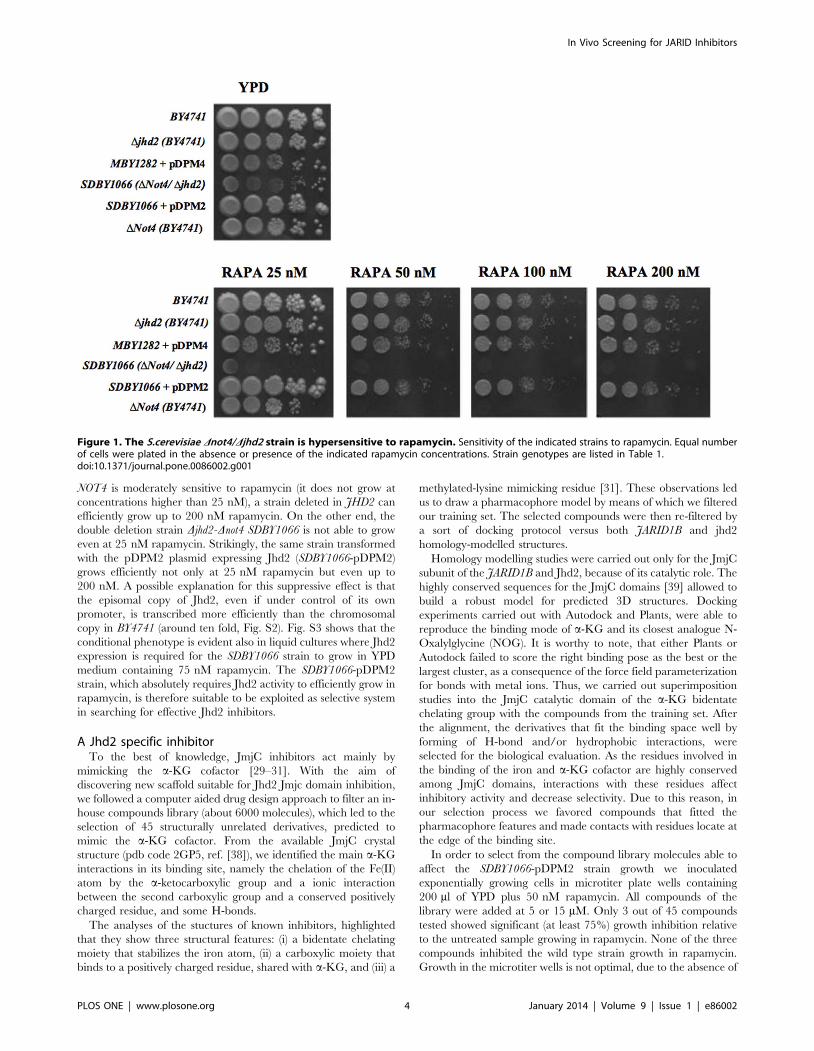
NOT4 is moderately sensitive to rapamycin (it does not grow at
concentrations higher than 25 nM), a strain deleted in JHD2 can
efficiently grow up to 200 nM rapamycin. On the other end, the
double deletion strain Djhd2-Dnot4 SDBY1066 is not able to grow
even at 25 nM rapamycin. Strikingly, the same strain transformed
with the pDPM2 plasmid expressing Jhd2 (SDBY1066-pDPM2)
grows efficiently not only at 25 nM rapamycin but even up to
200 nM. A possible explanation for this suppressive effect is that
the episomal copy of Jhd2, even if under control of its own
promoter, is transcribed more efficiently than the chromosomal
copy in BY4741 (around ten fold, Fig. S2). Fig. S3 shows that the
conditional phenotype is evident also in liquid cultures where Jhd2
expression is required for the SDBY1066 strain to grow in YPD
medium containing 75 nM rapamycin. The SDBY1066-pDPM2
strain, which absolutely requires Jhd2 activity to efficiently grow in
rapamycin, is therefore suitable to be exploited as selective system
in searching for effective Jhd2 inhibitors.
A Jhd2 specific inhibitorTo the best of knowledge, JmjC inhibitors act mainly by
mimicking the a-KG cofactor [29–31]. With the aim of
discovering new scaffold suitable for Jhd2 Jmjc domain inhibition,
we followed a computer aided drug design approach to filter an in-
house compounds library (about 6000 molecules), which led to the
selection of 45 structurally unrelated derivatives, predicted to
mimic the a-KG cofactor. From the available JmjC crystal
structure (pdb code 2GP5, ref. [38]), we identified the main a-KG
interactions in its binding site, namely the chelation of the Fe(II)
atom by the a-ketocarboxylic group and a ionic interaction
between the second carboxylic group and a conserved positively
charged residue, and some H-bonds.
The analyses of the stuctures of known inhibitors, highlighted
that they show three structural features: (i) a bidentate chelating
moiety that stabilizes the iron atom, (ii) a carboxylic moiety that
binds to a positively charged residue, shared with a-KG, and (iii) a
methylated-lysine mimicking residue [31]. These observations led
us to draw a pharmacophore model by means of which we filtered
our training set. The selected compounds were then re-filtered by
a sort of docking protocol versus both JARID1B and jhd2
homology-modelled structures.
Homology modelling studies were carried out only for the JmjC
subunit of the JARID1B and Jhd2, because of its catalytic role. The
highly conserved sequences for the JmjC domains [39] allowed to
build a robust model for predicted 3D structures. Docking
experiments carried out with Autodock and Plants, were able to
reproduce the binding mode of a-KG and its closest analogue N-
Oxalylglycine (NOG). It is worthy to note, that either Plants or
Autodock failed to score the right binding pose as the best or the
largest cluster, as a consequence of the force field parameterization
for bonds with metal ions. Thus, we carried out superimposition
studies into the JmjC catalytic domain of the a-KG bidentate
chelating group with the compounds from the training set. After
the alignment, the derivatives that fit the binding space well by
forming of H-bond and/or hydrophobic interactions, were
selected for the biological evaluation. As the residues involved in
the binding of the iron and a-KG cofactor are highly conserved
among JmjC domains, interactions with these residues affect
inhibitory activity and decrease selectivity. Due to this reason, in
our selection process we favored compounds that fitted the
pharmacophore features and made contacts with residues locate at
the edge of the binding site.
In order to select from the compound library molecules able to
affect the SDBY1066-pDPM2 strain growth we inoculated
exponentially growing cells in microtiter plate wells containing
200 ml of YPD plus 50 nM rapamycin. All compounds of the
library were added at 5 or 15 mM. Only 3 out of 45 compounds
tested showed significant (at least 75%) growth inhibition relative
to the untreated sample growing in rapamycin. None of the three
compounds inhibited the wild type strain growth in rapamycin.
Growth in the microtiter wells is not optimal, due to the absence of
Figure 1. The S.cerevisiae Dnot4/Djhd2 strain is hypersensitive to rapamycin. Sensitivity of the indicated strains to rapamycin. Equal numberof cells were plated in the absence or presence of the indicated rapamycin concentrations. Strain genotypes are listed in Table 1.doi:10.1371/journal.pone.0086002.g001
In Vivo Screening for JARID Inhibitors
PLOS ONE | www.plosone.org 4 January 2014 | Volume 9 | Issue 1 | e86002
efficient shaking. We therefore tested the three candidate
inhibitors on larger (50 ml) cultures grown with vigorous shaking.
In these conditions, only one of the inhibitors (compound 3195)
showed a reproducible effect.
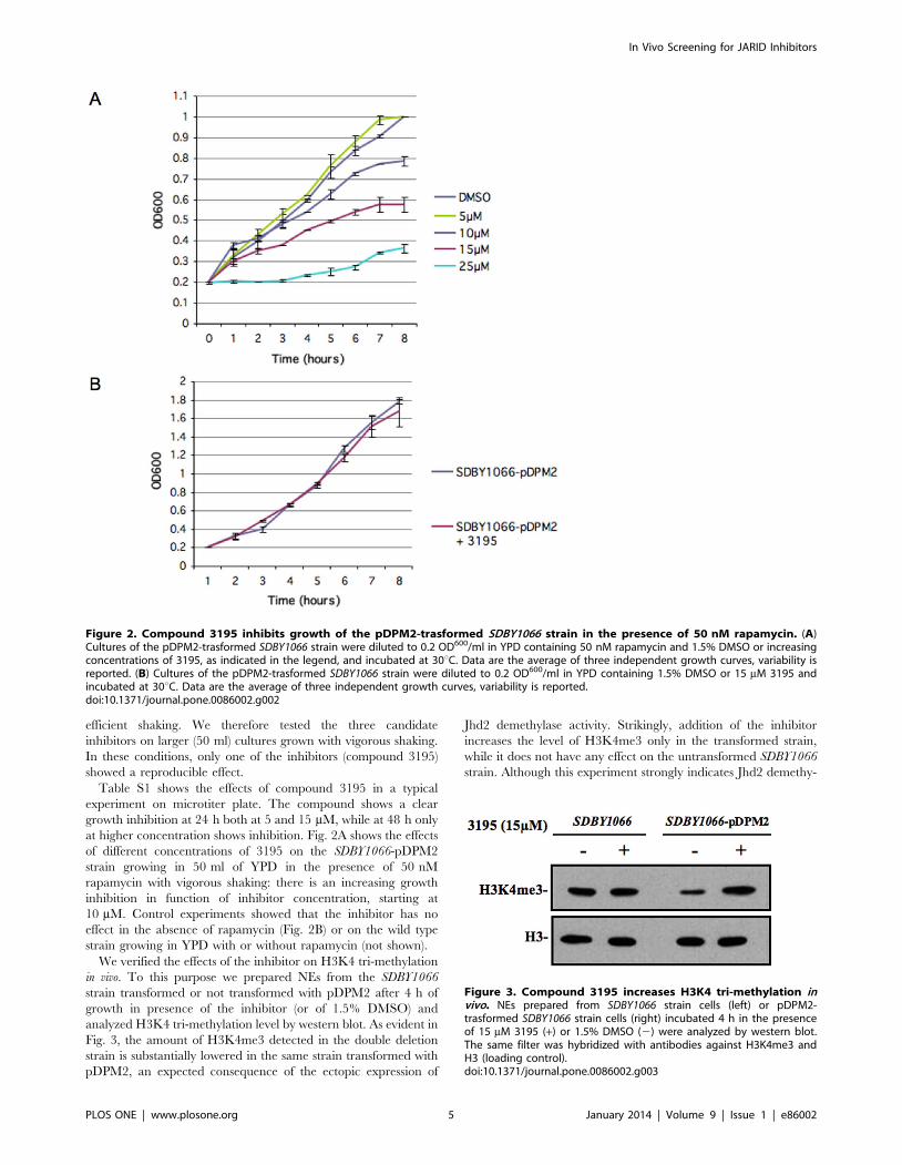
Table S1 shows the effects of compound 3195 in a typical
experiment on microtiter plate. The compound shows a clear
growth inhibition at 24 h both at 5 and 15 mM, while at 48 h only
at higher concentration shows inhibition. Fig. 2A shows the effects
of different concentrations of 3195 on the SDBY1066-pDPM2
strain growing in 50 ml of YPD in the presence of 50 nM
rapamycin with vigorous shaking: there is an increasing growth
inhibition in function of inhibitor concentration, starting at
10 mM. Control experiments showed that the inhibitor has no
effect in the absence of rapamycin (Fig. 2B) or on the wild type
strain growing in YPD with or without rapamycin (not shown).
We verified the effects of the inhibitor on H3K4 tri-methylation
in vivo. To this purpose we prepared NEs from the SDBY1066
strain transformed or not transformed with pDPM2 after 4 h of
growth in presence of the inhibitor (or of 1.5% DMSO) and
analyzed H3K4 tri-methylation level by western blot. As evident in
Fig. 3, the amount of H3K4me3 detected in the double deletion
strain is substantially lowered in the same strain transformed with
pDPM2, an expected consequence of the ectopic expression of
Jhd2 demethylase activity. Strikingly, addition of the inhibitor
increases the level of H3K4me3 only in the transformed strain,
while it does not have any effect on the untransformed SDBY1066
strain. Although this experiment strongly indicates Jhd2 demethy-
Figure 3. Compound 3195 increases H3K4 tri-methylation invivo. NEs prepared from SDBY1066 strain cells (left) or pDPM2-trasformed SDBY1066 strain cells (right) incubated 4 h in the presenceof 15 mM 3195 (+) or 1.5% DMSO (2) were analyzed by western blot.The same filter was hybridized with antibodies against H3K4me3 andH3 (loading control).doi:10.1371/journal.pone.0086002.g003
Figure 2. Compound 3195 inhibits growth of the pDPM2-trasformed SDBY1066 strain in the presence of 50 nM rapamycin. (A)Cultures of the pDPM2-trasformed SDBY1066 strain were diluted to 0.2 OD600/ml in YPD containing 50 nM rapamycin and 1.5% DMSO or increasingconcentrations of 3195, as indicated in the legend, and incubated at 30uC. Data are the average of three independent growth curves, variability isreported. (B) Cultures of the pDPM2-trasformed SDBY1066 strain were diluted to 0.2 OD600/ml in YPD containing 1.5% DMSO or 15 mM 3195 andincubated at 30uC. Data are the average of three independent growth curves, variability is reported.doi:10.1371/journal.pone.0086002.g002
In Vivo Screening for JARID Inhibitors
PLOS ONE | www.plosone.org 5 January 2014 | Volume 9 | Issue 1 | e86002
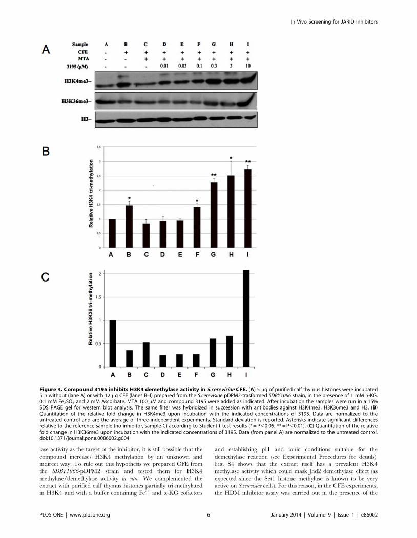
lase activity as the target of the inhibitor, it is still possible that the
compound increases H3K4 methylation by an unknown and
indirect way. To rule out this hypothesis we prepared CFE from
the SDBY1066-pDPM2 strain and tested them for H3K4
methylase/demethylase activity in vitro. We complemented the
extract with purified calf thymus histones partially tri-methylated
in H3K4 and with a buffer containing Fe2+ and a-KG cofactors
and establishing pH and ionic conditions suitable for the
demethylase reaction (see Experimental Procedures for details).
Fig. S4 shows that the extract itself has a prevalent H3K4
methylase activity which could mask Jhd2 demethylase effect (as
expected since the Set1 histone methylase is known to be very
active on S.cerevisiae cells). For this reason, in the CFE experiments,
the HDM inhibitor assay was carried out in the presence of the
Figure 4. Compound 3195 inhibits H3K4 demethylase activity in S.cerevisiae CFE. (A) 5 mg of purified calf thymus histones were incubated5 h without (lane A) or with 12 mg CFE (lanes B–I) prepared from the S.cerevisiae pDPM2-trasformed SDBY1066 strain, in the presence of 1 mM a-KG,0.1 mM Fe2SO4 and 2 mM Ascorbate. MTA 100 mM and compound 3195 were added as indicated. After incubation the samples were run in a 15%SDS PAGE gel for western blot analysis. The same filter was hybridized in succession with antibodies against H3K4me3, H3K36me3 and H3. (B)Quantitation of the relative fold change in H3K4me3 upon incubation with the indicated concentrations of 3195. Data are normalized to theuntreated control and are the average of three independent experiments. Standard deviation is reported. Asterisks indicate significant differencesrelative to the reference sample (no inhibitor, sample C) according to Student t-test results (* = P,0.05; ** = P,0.01). (C) Quantitation of the relativefold change in H3K36me3 upon incubation with the indicated concentrations of 3195. Data (from panel A) are normalized to the untreated control.doi:10.1371/journal.pone.0086002.g004
In Vivo Screening for JARID Inhibitors
PLOS ONE | www.plosone.org 6 January 2014 | Volume 9 | Issue 1 | e86002
Set1 histone methylase inhibitor MTA [40] which depresses the
methylase effect (compare lanes B and C in Fig. 4). We then tested
increasing concentrations of our candidate Jhd2 inhibitor and
observed a concentration-dependent increase of H3K4me3 (Fig. 4,
lanes D–I). The minimal concentration which gave a significant
effect was 0.1 mM, while at 1 mM the increase seems to reach a
plateau. We also tested if this effect was specific for Jhd2. We
provide three evidences that this is the case:
a) The same western blot, shown in Fig. 4A, was hybridized
with antibodies against H3K36me3. CFE addition decreases
H3K36me3 in most of the samples as compared with the
untreated control, showing that a dominant demethylase
activity specific for this modification is present in the extract.
On the other hand, an evident increase in H3K36me3 is
observed only at the highest concentration of 3195 (compare
histograms reported in B and C). Thus although the H3K36
demethylases Jhd1 and Rph1 have catalytic sites similar to
that one of Jhd2 and share the same chemical mechanism,
our inhibitor seems more specific for Jhd2.
b) We tested inhibition of H3K4 demethylation in a CFE
obtained from the untrasformed SDBY1066 strain (Fig. S5).
In this case no significant effect was observed, consistent with
the absence of Jhd2 activity.
c) Since it was still possible that 3195 could induce an increase
of H3K4 tri-methylation by interfering with the MTA
inhibitory action, we tested NEs derived from a strain
carrying a SET1 gene deletion and therefore devoid of H3K4
methylase activity (Fig. S6A). Since the demethylase activity
of KG obtained from this strain was very weak (not shown),
we transformed it with the PDM4 plasmid carrying the JHD2
gene under the control of a strong constitutive promoter. The
NEs obtained by the transformed strain showed a significant
demethylase activity which was inhibited by 3195 at
concentrations §3 mM (Fig. S6B and C). This ruled out
Figure 5. Compound 3195 inhibits H3K4 demethylase activity in HeLa NEs. (A) 5 mg of purified calf thymus histones were incubated 3 hwith 12 mg of NE prepared from HeLa cells, in the presence of 1 mM a-KG, 0.1 mM Fe2SO4 and 2 mM Ascorbate. MTA and compound 3195 wereadded as indicated. After incubation, the samples were run in a 15% SDS PAGE gel for western blot analysis. The filter was hybridized in successionwith antibodies against H3K4me3, H3K9me3, H3K27me3 and H3. (B) Quantitation of the relative fold change in H3K4me3 upon incubation with theindicated concentrations of 3195. Data are normalized to the untreated control and are the average of three independent experiments. Standarddeviation is reported. Asterisks indicate significant differences relative to the reference (no inhibitor, sample C), according to Student t-test results(* = P,0.05; ** = P,0.01).doi:10.1371/journal.pone.0086002.g005
In Vivo Screening for JARID Inhibitors
PLOS ONE | www.plosone.org 7 January 2014 | Volume 9 | Issue 1 | e86002
that the effect of 3195 could be mediated by stimulation of
Set1 methylation activity.
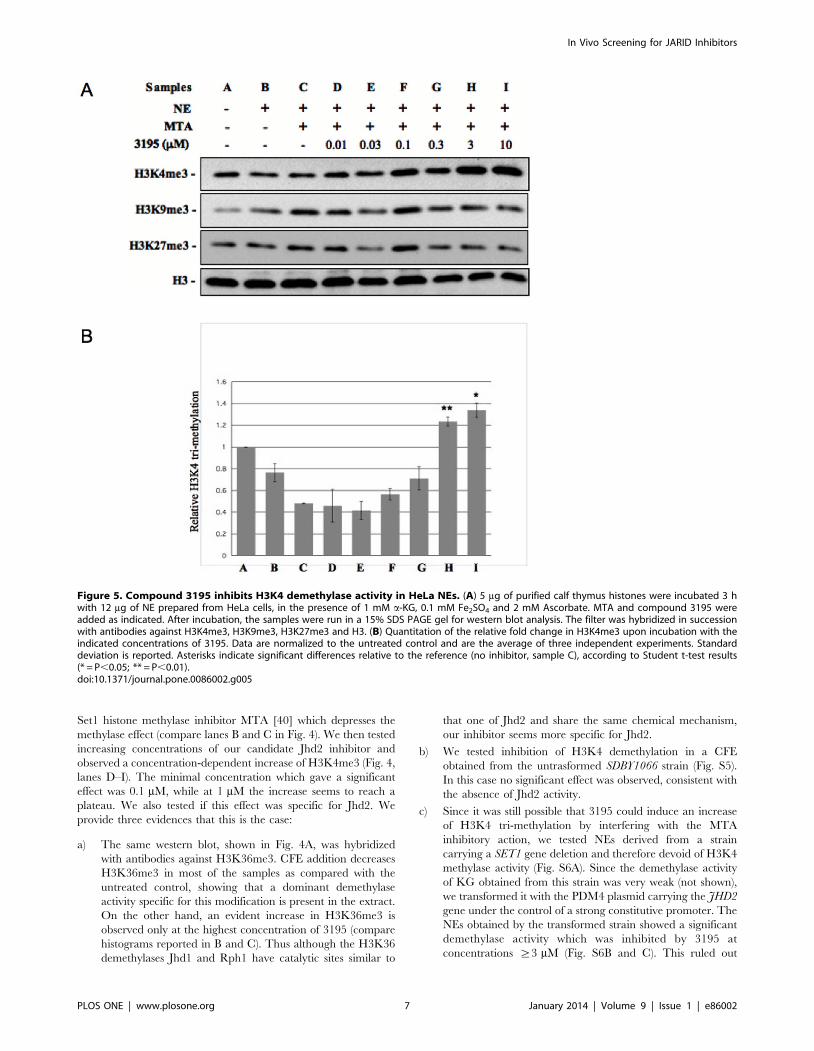
Testing the 3195 compound on mammalian HDMsIn mammalian cells at least four JHDMs exist which specifically
demethylate H3K4: JARID1A, 1B, 1C and 1D [41]. . Since the
catalytic site of all the mammalian JHDMs is very conserved (Fig.
S1) we expected the 3195 compound to be able to inhibit all these
enzymes at least in vitro. Initially we tested an HeLa NE which
should contain all the HDM activities. The experimental
conditions were similar to that of the S.cerevisiae CFE assay
described above. Also in the case of HeLa NE, we added MTA to
repress eventual H3K4 histone methylase activities present in the
extract. From the results which are presented in Fig. 5 it is evident
that:
a) The incubation of histones in the presence of the HeLa NE
determines a slight but appreciable decrease of H3K4 tri-
methylation level suggesting that, contrary to the yeast
situation, in HeLa NE histone methylase activity is not
prevalent on the HDMs activity (Fig. 5B).
b) As expected, MTA enhances the H3K4 demethylation effect
of the HeLa NE (Fig. 5A and B, sample C)
c) 3195 addition at a concentration of 3 or 10 mM leads to a
significant increase in H3K4 tri-methylation (2.7 and 2.9-fold
average increase, as compared with CFE-MTA treated
sample, respectively). We also tested possible effects on
H3K9 and H3K27 tri-methylation. No clear effects were
observed (Fig. 5A). In order to rule out an indirect effect of
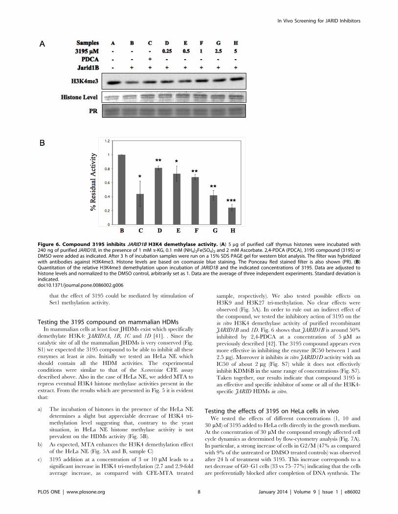
the compound, we tested the inhibitory action of 3195 on the
in vitro H3K4 demethylase activity of purified recombinant
JARID1B and 1D. Fig. 6 shows that JARID1B is around 50%
inhibited by 2,4-PDCA at a concentration of 5 mM as
previously described [42]. The 3195 compound appears even
more effective in inhibiting the enzyme (IC50 between 1 and
2.5 mg). Moreover it inhibits in vitro JARID1D activity with an
IC50 of about 2 mg (Fig. S7) while it does not effectively
inhibit KDM6B in the same range of concentrations (Fig. S7).
Taken together, our results indicate that compound 3195 is
an effective and specific inhibitor of some or all of the H3K4-
specific JARID HDMs in vitro.
Testing the effects of 3195 on HeLa cells in vivoWe tested the effects of different concentrations (1, 10 and
30 mM) of 3195 added to HeLa cells directly in the growth medium.
At the concentration of 30 mM the compound strongly affected cell
cycle dynamics as determined by flow-cytometry analysis (Fig. 7A).
In particular, a strong increase of cells in G2/M (47% as compared
with 9% of the untreated or DMSO treated controls) was observed
after 24 h of treatment with 3195. This increase corresponds to a
net decrease of G0–G1 cells (33 vs 75–77%) indicating that the cells
are preferentially blocked after completion of DNA synthesis. The
Figure 6. Compound 3195 inhibits JARID1B H3K4 demethylase activity. (A) 5 mg of purified calf thymus histones were incubated with240 ng of purified JARID1B, in the presence of 1 mM a-KG, 0.1 mM (NH4)2Fe(SO4)2 and 2 mM Ascorbate. 2,4-PDCA (PDCA), 3195 compound (3195) orDMSO were added as indicated. After 3 h of incubation samples were run on a 15% SDS PAGE gel for western blot analysis. The filter was hybridizedwith antibodies against H3K4me3. Histone levels are based on coomassie blue staining. The Ponceau Red stained filter is also shown (PR). (B)Quantitation of the relative H3K4me3 demethylation upon incubation of JARID1B and the indicated concentrations of 3195. Data are adjusted tohistone levels and normalized to the DMSO control, arbitrarily set as 1. Data are the average of three independent experiments. Standard deviation isindicated.doi:10.1371/journal.pone.0086002.g006
In Vivo Screening for JARID Inhibitors
PLOS ONE | www.plosone.org 8 January 2014 | Volume 9 | Issue 1 | e86002
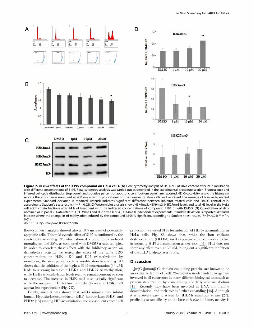
flow-cytometry analysis showed also a 10% increase of potentially
apoptotic cells. This mild cytoxic effect of 3195 is confirmed by the
cytotoxicity assay (Fig. 7B) which showed a presumptive induced
mortality around 25%, as compared with DMSO treated samples.
In order to correlate these effects with the inhibitory action on
demethylase activity, we tested the effect of the same 3195
concentrations on H3K4, K9 and K27 tri-methylation by
monitoring the steady-state levels of modification in vivo. Fig. 7C
shows that the addition of the highest 3195 concentration (30 mM)
leads to a strong increase in H3K4 and H3K27 tri-methylation,
while H3K9 tri-methylation levels seem to remain constant or even
to decrease. The increase in H3K4me3 is statistically significant
while the increase in H3K27me3 and the decrease in H3K9me3
appear less reproducible (Fig. 7D).
Finally, since it was shown that a-KG mimics may inhibit
human Hypoxia-Inducible-Factor (HIF) hydroxylases PHD1 and
PHD2 [43] causing HIF accumulation and consequent cancer cell
protection, we tested 3195 for induction of HIF1a accumulation in
HeLa cells. Fig. S8 shows that, while the iron chelator
desferrioxamine (DFOM), used as positive control, is very effective
in inducing HIF1a accumulation as decribed [44], 3195 does not
show any effect even at 30 mM, ruling out a significant inhibition
of the PHD hydroxylases in vivo.
Discussion
JmjC (Jumonji C) domain-containing proteins are known to be
an extensive family of Fe(II)/2-oxoglutarate-dependent oxygenase
involved in all eukaryotes in many different biological tasks such as
protein stabilization, hypoxia sensing and fatty acid metabolism
[45]. Recently they have been involved in DNA and histone
demethylation, and their role is further expanding [46]. Although
it is relatively easy to screen for JHDMs inhibition in vitro [27],
predicting in vivo efficacy on the base of in vitro inhibitory activity is
Figure 7. In vivo effects of the 3195 compound on HeLa cells. (A) Flow-cytometry analysis of HeLa cell of DNA content after 24 h incubationwith different concentrations of 3195. Flow-cytometry analysis was carried out as described in the experimental procedure section. Fluorescence andinferred cell cycle distribution (top panel) and putative percent of apoptotic cells (bottom panel) are reported. (B) Cytotoxicity assay: the histogramreports the absorbance measured at 450 nm which is proportional to the number of alive cells and represent the average of four independentexperiments. Standard deviation is reported. Asterisk indicates significant difference between inhibitor treated cells and DMSO control cells,according to Student’s t test results (* = P,0.02).(C) Western blot analysis shows H3K4me3, H3K9me3, H3K27me3 levels and total H3 level in the HeLacell acid protein fractions after 24 h of treatment with the indicated concentrations of compound 3195 or with DMSO. (D) Quantitation of dataobtained as in panel C. Data refer to 3 (H3K9me3 and H3K27me3) or 4 (H3K4me3) independent experiments. Standard deviation is reported. Asterisksindicate where the change in tri-methylation induced by the compound 3195 is significant, according to Student t-test results (* = P,0.05; ** = P,0.01).doi:10.1371/journal.pone.0086002.g007
In Vivo Screening for JARID Inhibitors
PLOS ONE | www.plosone.org 9 January 2014 | Volume 9 | Issue 1 | e86002
not straightforward [29]. An interesting in vivo screening system for
small molecules capable to interfere with transcription repressive
epigenetic modifications was recently described [47]. The system
proved to be able to identify a new molecule which inhibits
Jumonji HDMs but with low selectivity between the various class.
We describe here an high-throughput in vivo screening system
which has important features that makes it ideal for the selection of
H3K4-specific inhibitors of JHDMs potentially able to enter
eukaryotic cells and to act on their putative targets without
affecting other HDMs or other important 2-oxoglutarate oxygen-
ases. First, it is based on the yeast S.cerevisiae which has a unique
and not-essential JHDM specific for H3K4 whose catalytic site is
highly homologous to mammalian JHDMs. Second, it makes use
of a double deletion strain (Dnot4/Djhd2) which relies on over-
expression from an episomal copy of Jhd2 (SDBY1066-pDPM2) in
order to efficiently grow in the presence of rapamycin, a feature
which is correlated to an enhanced biological sensitivity to
inhibitors. Third, being the demethylase over-expressed and its
activity less stringently regulated and more comparable with the
strong Set1 histone methylase activity, its inhibition produces an
increase of H3K4me3 much more evident than the 20–30%
increase which is generally observed in jhd2-deleted strains [16,35].
Fourth and most importantly, secondary effects on other Fe(II)/2-
oxoglutarate-dependent oxygenases can be easily identified by
testing the cytostatic effects of candidate compounds in the
absence of rapamycin or on two different kinds of control strains:
wild type strains or strains devoid of Jhd2 activity.
In order to test the potential of the system in identifying selective
HDM inhibitors, we screened a library of 45 putative competitive
inhibitors of a-KG selected by means of a computational
approach. This class of compounds has previously been shown
to be very effective in vitro and also to have the potential to
discriminate among the various groups of Jumonji HDMs and
between them and other important hydroxylases. On the other
end, it is not straightforward to predict the most in vivo effective
and selective compounds based on their structures or on in vitro
results [43]. A considerable effort is currently being made to
identify a-KG mimics which in a certain range of concentration
can inhibit a specific c lysine modification without significantly
affecting others lysines [43]. Moreover, the potential use of this
class of compounds in cancer therapy is seriously limited by their
eventual effect in inhibiting human Hypoxia-Inducible-Factor
(HIF) PHD hydroxylases [43], causing HIF accumulation and
consequent cancer cell protection. For this reasons this class of
inhibitors constituted an ideal challenge for our screening system.
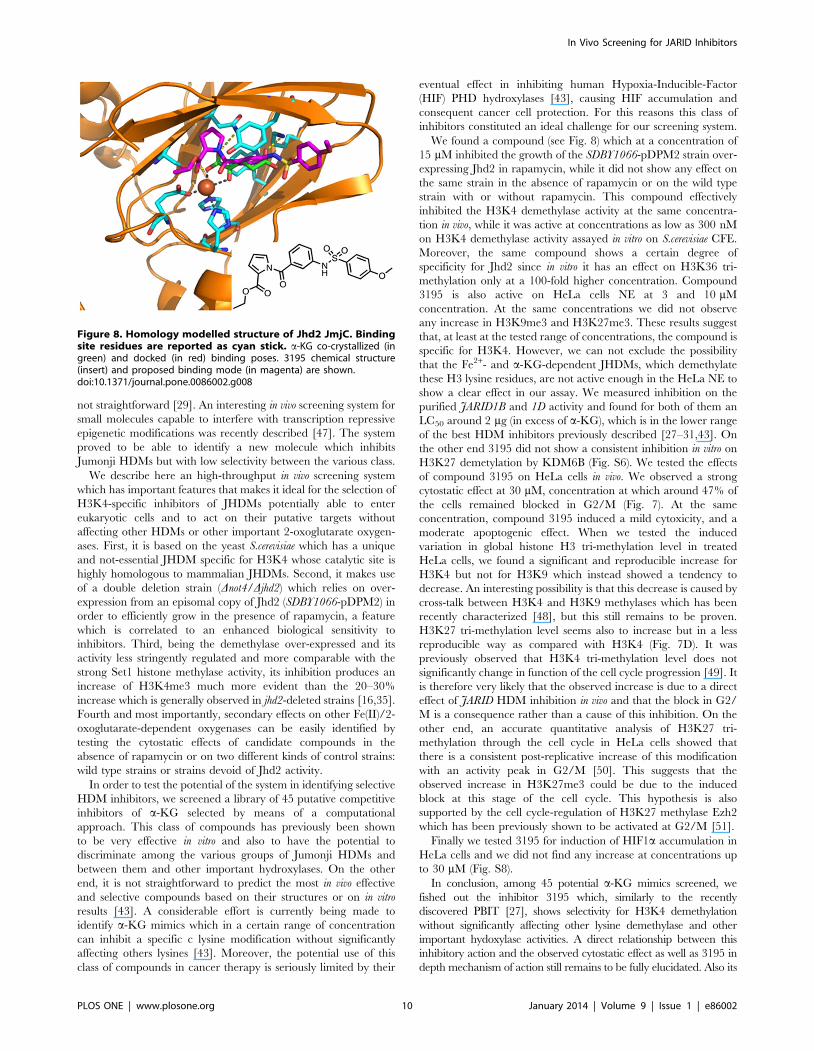
We found a compound (see Fig. 8) which at a concentration of
15 mM inhibited the growth of the SDBY1066-pDPM2 strain over-
expressing Jhd2 in rapamycin, while it did not show any effect on
the same strain in the absence of rapamycin or on the wild type
strain with or without rapamycin. This compound effectively
inhibited the H3K4 demethylase activity at the same concentra-
tion in vivo, while it was active at concentrations as low as 300 nM
on H3K4 demethylase activity assayed in vitro on S.cerevisiae CFE.
Moreover, the same compound shows a certain degree of
specificity for Jhd2 since in vitro it has an effect on H3K36 tri-
methylation only at a 100-fold higher concentration. Compound
3195 is also active on HeLa cells NE at 3 and 10 mM
concentration. At the same concentrations we did not observe
any increase in H3K9me3 and H3K27me3. These results suggest
that, at least at the tested range of concentrations, the compound is
specific for H3K4. However, we can not exclude the possibility
that the Fe2+- and a-KG-dependent JHDMs, which demethylate
these H3 lysine residues, are not active enough in the HeLa NE to
show a clear effect in our assay. We measured inhibition on the
purified JARID1B and 1D activity and found for both of them an
LC50 around 2 mg (in excess of a-KG), which is in the lower range
of the best HDM inhibitors previously described [27–31,43]. On
the other end 3195 did not show a consistent inhibition in vitro on
H3K27 demetylation by KDM6B (Fig. S6). We tested the effects
of compound 3195 on HeLa cells in vivo. We observed a strong
cytostatic effect at 30 mM, concentration at which around 47% of
the cells remained blocked in G2/M (Fig. 7). At the same
concentration, compound 3195 induced a mild cytoxicity, and a
moderate apoptogenic effect. When we tested the induced
variation in global histone H3 tri-methylation level in treated
HeLa cells, we found a significant and reproducible increase for
H3K4 but not for H3K9 which instead showed a tendency to
decrease. An interesting possibility is that this decrease is caused by
cross-talk between H3K4 and H3K9 methylases which has been
recently characterized [48], but this still remains to be proven.
H3K27 tri-methylation level seems also to increase but in a less
reproducible way as compared with H3K4 (Fig. 7D). It was
previously observed that H3K4 tri-methylation level does not
significantly change in function of the cell cycle progression [49]. It
is therefore very likely that the observed increase is due to a direct
effect of JARID HDM inhibition in vivo and that the block in G2/
M is a consequence rather than a cause of this inhibition. On the
other end, an accurate quantitative analysis of H3K27 tri-
methylation through the cell cycle in HeLa cells showed that
there is a consistent post-replicative increase of this modification
with an activity peak in G2/M [50]. This suggests that the
observed increase in H3K27me3 could be due to the induced
block at this stage of the cell cycle. This hypothesis is also
supported by the cell cycle-regulation of H3K27 methylase Ezh2
which has been previously shown to be activated at G2/M [51].
Finally we tested 3195 for induction of HIF1a accumulation in
HeLa cells and we did not find any increase at concentrations up
to 30 mM (Fig. S8).
In conclusion, among 45 potential a-KG mimics screened, we
fished out the inhibitor 3195 which, similarly to the recently
discovered PBIT [27], shows selectivity for H3K4 demethylation
without significantly affecting other lysine demethylase and other
important hydoxylase activities. A direct relationship between this
inhibitory action and the observed cytostatic effect as well as 3195 in
depth mechanism of action still remains to be fully elucidated. Also its
Figure 8. Homology modelled structure of Jhd2 JmjC. Bindingsite residues are reported as cyan stick. a-KG co-crystallized (ingreen) and docked (in red) binding poses. 3195 chemical structure(insert) and proposed binding mode (in magenta) are shown.doi:10.1371/journal.pone.0086002.g008
In Vivo Screening for JARID Inhibitors
PLOS ONE | www.plosone.org 10 January 2014 | Volume 9 | Issue 1 | e86002
potential use in investigating the biological role of H3K4 specific
demethylases in normal and cancer cells remains to be tested. On the
other end our selection system proved to be a very promising tool for
the discovery of H3K4 specific HDM inhibitors active in vivo.
Supporting Information
Figure S1 Sequence alignment of Jmc domains from theJhd2 and the JARID1 family members.
(TIF)
Figure S2 Ectopic over-expression of JHD2 in thepDPM2-trasformed Dnot4/Djhd2 strain. Real time RT-
PCR was performed on cDNA obtained from total RNA extracted
from the indicated strains during exponential growth. Data
represent fold change relative to ACT1 mRNA, used as
endogenous calibrator, and are the average of three independent
experiments. Standard deviation is reported. Asterisk indicate
statistically significant changes as compared with the untreated
wild type strain BY4742. For the untrasformed SDBY1066 strain
no amplification was obtained.
(TIF)
Figure S3 The S.cerevisiae SDBY1066 strain is hyper-sensitive to rapamycin in liquid culture. The indicated
strains were grown at 30uC in YPD until an OD600 of 0.4 was
reached. At this point rapamycin (75 nM) was added and growth
monitored for 7 h.
(TIF)
Figure S4 H3K4 tri-methylation activity is prevalent onS.cerevisiae pDPM2-trasformed SDBY1066 strain CFE.(A) 5 mg of purified calf thymus histones were incubated for 3 h
with the indicated amount of CFE prepared from S.cerevisiae
pDPM2-trasformed SDBY1066 strain, in the presence of 10 mM
a-KG, 1 mM Fe2SO4 and 2 mM Ascorbate. After incubation
samples were run on a 15% SDS gel for western blot analysis. The
filter was hybridized in succession with antibodies against
H3K4me3 and H3. (B) Quantitation of the relative H3K4me3
upon incubation with CFE from S.cerevisiae pDPM2-trasformed
SDBY1066 strain. The intensities of H3K4me3 bands were
normalized to the intensity of the corresponding H3 bands.
Histograms represent the average of three independent experi-
ments. Standard deviation is reported. Asterisks indicate where the
change in H3K4me3 of the CFE treated samples, as compared
with the untreated control, is significant according to Student t-test
results (* = P,0.05; ** = P,0.01).
(TIF)
Figure S5 Compound 3195 has no effect on the un-trasformed S.cerevisiae Dnot4/Djhd2 strain (SDBY1066).5 mg of purified calf thymus histones were incubated for 3 h with
the indicated amount of CFE from S.cerevisiae SDBY1066 strain, in
the presence of 10 mM a-KG, 1 mM Fe2SO4 and 2 mM
Ascorbate. MTA and compound 3195 were added as indicated.
After incubation samples were run on a 15% SDS gel for western
blot analysis. The filter was hybridized with H3K4me3 antibody.
The Ponceau Red staining (PR) is shown as loading control of calf
thymus histones.
(TIF)
Figure S6 Compound 3195 inhibits H3K4 demethylaseactivity in CFEs prepared from a Dset1 strain. Panel A:
20 mg of CFEs prepared from wild type W303 or Dset1 YCVS3
strains, as indicated, were run on a 15% SDS gel for western blot
analysis. The filter was hybridized with anti-H3K4me3 to control
for the absence of H3K4me3 in the Dset1 strain. The Ponceau Red
staining (PR) is shown as loading control. Panel B: 5 mg of purified
calf thymus histones were incubated for 3 h with 12 mg of CFE
prepared from S.cerevisiae Dset1 YCVS3 strain transformed with
pDPM4, in the presence of 10 mM a-KG, 1 mM Fe2SO4 and
2 mM Ascorbate. Compound 3195 was added as indicated. After
incubation samples were run in a 15% SDS gel for western blot
analysis. The filter was hybridized with anti- H3K4me3 and anti-
H3 antibodies. The Ponceau red staining (PR) is shown as loading
control of calf thymus histones. Panel C: Quantitation of western
blot analysis of the relative H3K4me3 demethylation upon histone
incubation with S.cerevisiae Dset1 YCVS3 strain CFEs and different
concentrations of 3195. H3K4me3 data were normalized to the
untreated control (lane A), arbitrarily set as 1, and are the average
of three independent experiments performed with three different
CFEs. Standard deviation is indicated. Asterisks indicate where
changes in H3K4me3 of the CFE treated samples, compared to
the untreated control, are significants according to Student t-test
results (* = P,0.05; ** = P,0.01).
(TIF)
Figure S7 Compound 3195 inhibits JARID1D H3K4demethylase activity but does not affect KDM6B activity.(A) 5 mg of purified calf thymus histones were incubated 1 h with
105 ng of purified JARID1D, in the presence of 1 mM a-KG,
0.1 mM (NH4)2Fe(SO4)2 and 2 mM Ascorbate. Compound 3195
or DMSO were added as indicated. After incubation samples were
run on a 15% SDS page gel for western blot analysis. The fliter
was sequentially hybridized with antibodies against H3K4me3 and
H3. Histone levels are based on coomassie stain. (B) Quantitation
of the relative H3K4me3 demethylation upon incubation with
JARID1D and the indicated concentrations of 3195. Data are
adjusted to histone levels and normalized to the DMSO control,
arbitrarily set as 1. Data are the average of three independent
experiments. Standard deviation is indicated. (C) 5 mg of purified
calf thymus histones were incubated 3 h with 80 ng of purified
KDM6B, in the presence of 1 mM a-KG, 0.1 mM (NH4)2Fe(SO4)2and 2 mM Ascorbate. DMSO, compound 3195 or 2,4-PDCA
(PDCA) were added as indicated. 2,4-PDCA was at 5 mM final
concentration. After incubation samples were run on a 15% SDS
page gel for western blot analysis. The filter was hybridized with
antibodies against H3K27me3. The Ponceau red stained filter
(PR) is shown as loading control of calf thymus histones.
(TIF)
Figure S8 3195 compound does not affect HIF-1 expres-sion in Hela cells. Western blot analysis shows HIF-1 levels in
HeLa cell lysates after 24 h of treatment with the indicated
concentrations of compound 3195, DMSO or DFOM. 50 mg of
total proteins were loaded on SDS-PAGE gels.
(TIF)
Table S1 Effect of compound 3195 on pDPM2-trans-formed SDBY1066 strain cells grown in rapamycin. Cells
were inoculated in 200 ml of YPD plus 50 nM rapamycin at a cell
density corresponding to 0.2 OD600 and incubated at 30uC in the
presence of compound 3195 or DMSO as indicated. OD600 were
read at the indicated times. Data are the average of two wells
inoculated with cells from independent cultures. Variability is
indicated and significant reductions ($75%) are shown in bold.
(DOC)
Acknowledgments
We thank S.D.Briggs for yeast strains and plasmids, Valentina Cerini and
Simone Pippa for technical help, and L. Salvatori for kindly providing
HIF-1 antibody.
In Vivo Screening for JARID Inhibitors
PLOS ONE | www.plosone.org 11 January 2014 | Volume 9 | Issue 1 | e86002
Author Contributions
Conceived and designed the experiments: CM RS RN. Performed the
experiments: CM MP FB EC AA SD TR VF AC GLR. Analyzed the data:
CM MP FB EC AC RS RN. Contributed reagents/materials/analysis
tools: EC TR AC GLR RS RN. Wrote the paper: CM RN.
References
1. Rando OJ, Winston F (2012) Chromatin and transcription in yeast. Genetics190: 351–387.
2. Kouzarides T (2007) Chromatin Modifications and Their Function. Cell 128:
693–705.
3. Martin C, Zhang Y (2005) The diverse functions of histone lysine methylation.Nat Rev Mol Cell Biol 6: 838–849.
4. Pokholok DK, Harbison CT, Levine S, Cole, M . Hannett, et al. (2005)
Genome-wide map of nucleosome acetylation and methylation in yeast. Cell122: 517–527.
5. Pinskaya M, Morillon A (2009) Histone H3 Lysine 4 di-methylation. Epigenetics
4: 302–306.
6. Santos-Rosa H, Schneider R, Bannister AJ, Sherriff J Berstein BE, et al. (2002)Active genes are tri-methylated at K4 of histone H3. Nature 419: 407–411.
7. Dehe PM, Geli V (2006) The multiple faces of Set1. Biochem Cell Biol 84: 536–
548.
8. Margaritis T, Oreal V, Brabers N, Maestroni L, Vitaliano-Prunier A, et al.(2012) Two distinct repressive mechanisms for histone 3 lysine 4 methylation
through promoting 39-end antisense transcription. PLoS Genet 8: e1002952.
9. Zhou BO, Zhou J (2011) Recent transcription-induced histone H3 Lysine 4(H3K4) methylation inhibits gene reactivation. J Biol Chem 286: 34770–34776.
10. Lauberth SM, Nakayama T, Wu X, Ferris AL, Tang Z, et al. (2013) H3K4me3
interactions with TAF3 regulate preinitiation complex assembly and selectivegene activation. Cell 152: 1021–1036.
11. Cloos PA, Christensen J, Agger K, Helin K (2008) Erasing the methyl mark:
Histone demethylase at the center of cellular differentiation and disease. Genes& Dev 22: 1115–1140.
12. Fodor BD, Kubicek S, Yonezawa M, O’Sullivan RJ, Sengupta R, et al. (2006)
Jmj2b antagonizes H3K9 trimethylation at pericentric chromatin in mammaliancells. Genes & Dev 20: 1557–1562.
13. Forneris E, Binda C, Vanoni MA, Mattevi A, Battaglioli E (2005) Histone
demethylation catalysed by LSD1 is a flavin-dependent oxidative process. FEBS
Lett 579: 2203–2207.
14. Shi Y, Lan F, Matson P, Mulligan P, Whetstine JR, et al. (2004) Histone
demethylation mediated by the nuclear amine oxidase homolog LSD1. Cell 119:
941–953.
15. Karytinos A, Forneris F, Profumo A, Clossani G, Battaglioli E, et al. (2009) A
novel mammalian flavin-dependent histone demethylase. J Biol Chem 284:
17775–17782.
16. Liang G, Klose RJ, Gardner KE, Zhang Y (2007) Yeast Jdh2 is a histonetrimethyl demethylase. Nat Struct Mol Biol 14: 243–245.
17. Seward DJ, Cubberley G, Kim S, Schonewald M, Zhang L, et al. (2007)
Demethylation of trimethylated histone H3 Lys4 in vivo by JARID1 JmjCproteins. Nat Struct Mol Biol 14: 240–242.
18. Tu S, Bulloch EM, Yang L, Ren C, Huang W, et al. (2007) Identification of
yeast demethylases in Saccharomyces cerevisiae. J Biol Chem 282: 14262–14271.
19. Spannhoff A, Hauser AT, Heinke R, Sippl W, Jung M (2009) The emerging
therapeutic potential of histone methyltransferase and demethylase inhibitors.Chem Med Chem 4: 1568–1582.
20. Christensen J, Agger K, Cloos PAC, Pasini D, Rose S, et al. (2007) RBP2
belongs to a family of demethylase, specific for triand dimethylated lysine 4 onhistone 3. Cell 128: 1063–1076.
21. Zeng J, Ge Z, Wang L, Li Q, Wang N, et al. (2010) The histone demethylase
RBP2 is overexpressed in gastric cancer and its inhibition triggers senescence ofcancer cells. Gastroenterology 138: 981–992.
22. Lin W, Cao J, Liu J, Beshiri ML, Fujiwara Y, et al. (2011) Loss of the
retinoblastoma binding protein 2 (RBP2) histone demethylase suppressestumorigenesis in mice lacking Rb1 or Men1. Proc Natl Acad Sci USA 108:
13379–13386.
23. Sharma SV, Lee DY, Li B, Quinlan MP, Takahashi F, et al. (2010) Achromatin-mediated reversible drug-tolerant state in cancer cell subpopulations.
Cell 141: 69–80.
24. Hou J, Wu J, Dombkowski A, Zhang K, Holowatyj A, et al. (2012) Genomic
amplification and a role in drug-resistance for the KDM5A histone demethylasein breast cancer. Am J Transl Res 4: 247–256.
25. Mitra D, Das PM, Huynh FC, Jones FE (2012) Jumonji/ARID1 B (JARID1B)
Protein Promotes Breast Tumor Cell Cycle Progression through EpigeneticRepression of MicroRNA let-7e. J Biol Chem 286: 40531–40535.
26. Chicas A, Kapoor A, Wang X, Aksoy O, Evertts AG, et al. (2012) H3K4
demethylation by Jarid1a and Jarid1b contributes to retinoblastoma-mediatedgene silencing during cellular senescence. Proc Natl Acad Sci USA 109: 8971–
8976.
27. Sayegh J, Ran Zou M, Morales A, Blair LP, Norcia M, et al. (2013)Identification of small molecules inhibitors of Jumonji AT-Rich Interactive
Domain 1B (JARID1B) Histone Demethylase by a sensitive High-throughput
Screen. J Biol Chem 288:9408–9417.28. Nielsen AL, Kristensen LH, Stephansen KB, Kristensen JBL, Helgstrand C,
et al. (2012) Identification of catechols as histone-lysine demethylase inhibitors.Febs Letters 586: 1190–1194.
29. Luo X, Liu Y, Kubicek S, Myllyharju J, Tumber A, et al. (2011) A Selective
Inhibitor and Probe of the Cellular Functions of Jumonji C Domain-ContainingHistone Demethylases. J Am Chem Soc 133: 9451–9456.
30. Hamada S, Suzuki T, Mino K, Koseki K, Oehme F, Flamme I (2010) Design,Synthesis, Enzyme-Inhibitory Activity, and Effect on Human Cancer Cells of a
Novel Series of Jumonji Domain-Containing Protein 2 Histone Demethylase
Inhibitors. J Med Chem 53: 5629–5638.31. Rose NR, Woon ECY, Kingham GL, King ONF, Mecinovic J, et al. (2010)
Selective Inhibitors of the JMJD2 Histone Demethylases: Combined Non-denaturing Mass Spectrometric Screening and Crystallographic Approaches.
J Med Chem 53: 1810–1818.32. Korb O, Stutzle T, Exner TE (2006) PLANTS: Application of Ant Colony
Optimization to Structure-Based Drug Design. In: Ant Colony Optimization
and Swarm Intelligence, Proceedings of the 5th International Workshop, Ants33. Goodsell DS, Morris GM, Olson AJ (1996) Automated docking of flexible
ligands: applications of AutoDock. J Mol Recognit 9: 1–5.34. Shechter D, Dormann HL, Allis CD, Hakel SB (2007) Extraction, purification
and analysis of histones. Nat Protoc 2: 1445–1457.
35. Ingvarsdottir K, Edwards C, Lee MG, Lee JS, Schultz DC, et al. (2007) HistoneH3K4 demethylation during activation and attenuation of GAL1 transcription
in Saccharomyces cerevisiae. Mol Cell Biol 27: 7856–7864.36. Mersman DP, Du H, Fingerman IM, South PF, Briggs SD (2009)
Poliubiquitination of the demetylase Jhd2 controls histone methylation and
genes expression. Genes Dev 23: 951–962.37. Azzouz N, Panasenko OO, Deluen C, Hsieh J, Theiler G, et al. (2010) Specific
roles for the Ccr4-Not complex subunits in expression of the genome. RNA 15:277–383.
38. Chen Z, Zang J, Whetstine J, Hong X, Davrazou F, et al. (2006) Structuralinsights into histone demethylation by JMJD2 family members. Cell 125: 691–
702.
39. Klose JR, Kallin EM, Khang Y (2006) JmjC-domain-containing proteins andhistone demethylation. Nat Rev Gen 7: 715–727.
40. Huang J, Kent JR, Placek B, Whelan KA, Hollow CM, et al. (2006)Trimethylation of Histone H3 Lysine 4 by Set1 in the Lytic Infection of
Human Herpes Simplex Virus 1. J Virol 6: 5740–5746.
41. Christensen J, Agger K, Cloos PAC, Pasini D, Rose S, et al. (2007) RBP2belongs to a family of demethylase, specific for tri- and dimethylated lysine 4 on
histone 3. Cell 128: 1063–1076.42. Kristensen LH, Nielsen AL, Helgstrand C, Lees M, Cloos P, et al. (2012) Studies
of H3K4me3 demethylation by KDM5B/Jarid1B/PLU1 reveals strongsubstrate recognition in vitro and identifies 2,4-pyridine-dicarboxylic acid as an
in vitro and in cell inhibitor. FEBS J 279: 1905–1914.
43. Lohse B, Kristensen JL, Kristensen LH, Agger K, Helin K, et al. (2011)Inhibitors of histone demethylases Bioorg Med Chem 19: 3625–3636.
44. Ke Q, Costa M (2006) Hypoxia-inducible factor-1 (HIF-1). Mol Pharmacol 70:1469–1480.
45. Loenarz C, Schofield CJ (2008) Expanding chemical biology of 2-oxoglutarate
oxygenase. Nat Chem Biol 4: 152–156.46. Noma A, Ishitani R, Kato M, Nagao A, Nureki O, et al. (2010) Expanding role
of the Jumonji C Domain as an RNA hydroxylase. J Biol Chem 285: 34503–34507.
47. Wang L, Chang J, Varghese D, Dellinger M, Kumar S, et al. (2013) A smallmolecule modulates Jumonji histone demethylase activity and selectively inhibits
cancer growth Nat Comm 4: 2035.
48. Chaturvedi CP, Somasundaram B, Singh K, Carpenedo R, Stanford WL, et al.(2012) Maintenance of gene silencing by the coordinate action of the H3K9
methyltransferase G9a/KMT1C and the H3K4 demethylase Jarid1a/KDM5A.Proc Natl Acad Sci USA 46: 18845–18850.
49. Mishra BP, Ansari KI, Mandal SS (2009) Dynamic association of MLL1, H3K4
trimethylation with chromatin and Hox gene expression during the cell cycle.FEBS J 276: 1629–1640.
50. Zee BM, Britton LP, Wolle D, Haberman DM, Garcia BA (2012) Origin andformation of histone methylation across the human cell cycle. Mol Cell Biol 32:
2503–2514.51. Kaneco S, Li G, Son J, Xu C-F, Margueron R, et al. (2010) Phosphorylation of
the PRC2 component Ezh2 is cell cycle-regulated and up-regulates its binding to
ncRNA Genes & Dev 24: 2615–2620.52. Corda Y, Schramke V, Longhese M P, Smokvina P, Paciotti V, et al. (1999)
Interaction between Set1p and checkpoint protein Mec3p in DNA repair andtelomere functions. Nat Gen 21: 204–208.
In Vivo Screening for JARID Inhibitors
PLOS ONE | www.plosone.org 12 January 2014 | Volume 9 | Issue 1 | e86002


