Embed Size (px)
Citation preview

Nucleic Acids Research, 2007, 1–17doi:10.1093/nar/gkm066
An improved definition of the RNA-bindingspecificity of SECIS-binding protein 2,an essential component of theselenocysteine incorporation machineryA. Clery1, V. Bourguignon-Igel1, C. Allmang2, A. Krol2 and C. Branlant1,*
1Laboratoire de Maturation des ARN et Enzymologie Moleculaire – UMR 7567 CNRS-UHP, Nancy Universite,Faculte des Sciences et Techniques – BP 239, 54506 Vandoeuvre-les-Nancy Cedex, France and 2Architecture etReactivite de l’arN – CNRS-Universite Louis Pasteur, Institut de Biologie Moleculaire et Cellulaire 15 Rue ReneDescartes, 67084 Strasbourg Cedex, France
Received September 27, 2006; Revised January 20, 2007; Accepted January 22, 2007
ABSTRACT
By binding to SECIS elements located in the 30-UTRof selenoprotein mRNAs, the protein SBP2 playsa key role in the assembly of the selenocysteineincorporation machinery. SBP2 contains anL7Ae/L30 RNA-binding domain similar to that ofprotein 15.5K/Snu13p, which binds K-turn motifswith a 3-nt bulge loop closed by a tandem ofG.A and A.G pairs. Here, by SELEX experiments,we demonstrate the capacity of SBP2 to bind suchK-turn motifs with a protruding U residue. However,we show that conversion of the bulge loop intoan internal loop reinforces SBP2 affinity and toa greater extent RNP stability. Opposite variationswere found for Snu13p. Accordingly, footprintingassays revealed strong contacts of SBP2 withhelices I and II and the 50-strand of the internalloop, as opposed to the loose interaction of Snu13p.Our data also identifies new determinants for SBP2binding which are located in helix II. Among theL7Ae/L30 family members, these determinantsare unique to SBP2. Finally, in accordance withfunctional data on SECIS elements, the identity ofresidues at positions 2 and 3 in the loop influencesSBP2 affinity. Altogether, the data provide a veryprecise definition of the SBP2 RNA specificity.
INTRODUCTION
Based on ribosomal subunit 3D-structure analysis,K-turn motifs were found to be frequent protein-recognition motifs in ribosomal RNAs (1). A total of
8 K-turn motifs were detected in the 23S rRNA fromHaloarcula marismortui and the 16S rRNA from Thermusthermophilus (1–4). K-turn motifs are all characterized bya helix I-loop-helix II structure, and the formation of twonon-Watson–Crick base pairs (most frequently G.A andA.G) within the internal loop extends helix II (1,5). Due tothe stacking onto helix I or helix II of residues in theinternal loop, one of the RNA strand forms a sharp angle(1,5). Only one of the residues in the loop is projected outof the K-turn structure and is located in a pocket ofthe protein in RNA–protein complexes. In addition totheir presence in rRNAs, K-turn motifs are also found inthe U4 and U4atac spliceosomal snRNAs (5,6) andin the numerous C/D box snoRNAs (7), that guide20-O-methylation and cleavages in pre-ribosomal RNA(for review, 8). K-turn motifs were also recently foundin both C/D and H/ACA sRNAs, that guide rRNAmodifications in archaea (9–11). They are thus veryancient RNA-binding motifs. Both in eukarya and inarchaea, small RNAs containing K-turn motifs assembleinto RNP particles and the K-turn motifs play a centralrole in protein assembly (7,9–15). More specifically,the ribosomal L7Ae protein in archaea or its eukaryalhomolog, the Snu13p (yeast)/15.5K (human) protein,first recognizes the K-turn structure and the complexformed then serves as a platform for assembly of the otherproteins (9,10,12–19).The Snu13p/15.5K and L7Ae proteins belong to
the L7Ae/L30 protein family, which is characterized bythe presence of an L7Ae/L30 RNA-binding domain(6,20). The founding member of this protein family, theyeast L30 ribosomal protein recognizes a peculiar K-turnmotif in its own pre-mRNA (21–23). One differencebetween the yeast L30 RNA–protein complex, and theSnu13p/15.5K or L7Ae RNA–protein complexes is the
*To whom the correspondence should be addressed. Tel: 33 383684303; Fax: 33 383684307; Email: [email protected]
� 2007 The Author(s).
This is an Open Access article distributed under the terms of the Creative Commons Attribution Non-Commercial License (http://creativecommons.org/licenses/
by-nc/2.0/uk/) which permits unrestricted non-commercial use, distribution, and reproduction in any medium, provided the original work is properly cited.
Nucleic Acids Research Advance Access published March 1, 2007 by guest on July 14, 2015
http://nar.oxfordjournals.org/D
ownloaded from

identity of the nucleobase located in the protein pocket.Whereas, a strong preference for an U residue is observedfor proteins Snu13p/15.5K and L7Ae (7,24–26), C andA residues are preferentially accommodated in theyeast L30 protein pocket (27). The possibility to binda G residue was however recently observed (28).In vertebrates, SECIS-binding protein 2, another
member of the L7Ae/L30 protein family, binds SECISelements in mRNAs (29,30). The SECIS elements containdeterminants needed for selenocysteine incorporationinto selenoproteins (31,32). Selenocysteine incorporationinvolves reprogramming of a nonsense UGA codon into acodon recognized by the selenocysteine specific tRNASec.Understanding the mechanism of selenocysteine incor-poration into proteins is important as they are key playersin the antioxidant defense system (for review, 33).They are also key participants in a variety of othersystems including thyroid hormone metabolism, musclefunction, transportation and distribution of seleniumto remote tissues and can have roles as structural proteins(for reviews, 34–37). In eukarya, the SECIS elements andSBP2 are two essential components of the selenocysteineincorporation machinery. All SECIS elements consistof a hairpin structure composed of two helices I and II,separated by an internal loop. A highly conserved clusterof four non-Watson–Crick base pairs is located in helix II.It contains a tandem of G.A and A.G pairs, whichis needed for SBP2 binding (29,30). This cluster ofnon-Watson–Crick pairs is an essential determinant forselenocysteine incorporation (31,32). A highly conservedAAR sequence present in a loop of all SECIS elementsis also important for selenoprotein synthesis in vivo,but not for binding of SBP2 in vitro (30,38). As SBP2also binds the specific mSelB/EFSec elongation factor,it is proposed to recruit this dedicated elongation factorin a complex formed with the selenocysteyl-tRNASec tothe ribosomes (39–41). Additionally, according to a recentinvestigation on the selenocysteine incorporation machin-ery (42), the ribosomal protein L30 is able to bind theSECIS motif by displacing protein SBP2. This substitutionwould facilitate the interaction of the Sec-tRNASec
with ribosomes.A prerequisite to fully understand the SBP2 activity is
thus to obtain a more complete picture of the RNAsequence and structural determinants required for SBP2binding. To this end, we combined the SELEX approachand site-directed mutagenesis experiments. As the RNAsrecovered after SELEX experiments could form canonicalK-turn motifs with a protruding U residue, we comparedthe RNA-binding properties of the human SBP2protein with those of a well-characterized member of theL7Ae/L30 protein family, the S. cerevisiae Snu13pprotein. This protein recognizes K-turn motifs in U4snRNA, the C/D box snoRNAs and U3 snoRNA.Altogether, we show here that in contrast to proteinSnu13/15.5K, SBP2 preferentially binds RNA motifswith a large internal loop. In addition, we demonstratethe existence of previously undetected important determi-nants for RNA recognition by SBP2 that are locatedin helix II.
MATERIALS AND METHODS
Strains and growth conditions
The Escherichia coli TG1 strain was used as the hoststrain for plasmid construction. Growth was performedat 378C in Luria Broth medium, complemented with100 mg/ml of ampicillin when necessary. The E. coli strainBL21-CodonPlus (Stratagene) was the host strain forproduction of the recombinant GST/Snu13p, GST/L7Aeand GST/C-SBP2 proteins.
Recombinant DNA
Plasmids pT7SelN (40), pUC18::U3A�2,3,4 (26) andpyU4 (43) were used for the production of matrices forin vitro transcription of the SelN, yU3B/C and yU4RNAs, respectively. The yU3B/C and yU4 matrices wereobtained by PCR amplification, under conditionspreviously described (26). Oligonucleotides yU3B/C-50,yU3B/C-30, yU4-50 and yU4-30, given in Table 1 of theSupplementary Data, were used as primers. PlasmidspGEX-6P-1::SNU13, pGEX-6P-1::L7AE (44) andpGEX-6-P1::C-SBP2 (this work) were used for productionof the recombinant GST/Snu13p, GST/L7Ae andGST/C-SBP2 proteins, respectively. Plasmid pA11 wasused for amplification of the PCR fragment coding forregion 515–854 of human SBP2 protein (45). DNAfragments amplified by RT-PCR from RNAs obtainedafter the fourth cycle of the SELEX experiment werecloned into plasmid pCR2.1 (Invitrogen). Mutagenesisof the RNA Se1 coding sequence was performed by thePCR-based site-directed strategy (primers are listed inTable 1 in the Supplementary Data).
In vitro transcription
The EcoRI linearized pT7::SelN plasmid was used asthe template for SelN RNA transcription. The yU3B/C,yU4, Se1-Se7 and Se1 variant RNA-coding sequenceswere transcribed from PCR amplified DNA fragmentsobtained as described above. Transcriptions were carriedout on 1mg of plasmid DNA linearized with EcoRI or500 ng of PCR product, in a 15 ml reaction as describedin Marmier-Gourrier et al. (26).
RNAs were 50-end labeled using 10 units of T4 poly-nucleotide kinase (MBI-Fermentas), 20 pmol of RNA,5 pmol of [g-32P] ATP, in a 10-ml reaction mixturecontaining 10mM MgCl2; 5mM DTT; 0.1mM spermi-dine; 0.1mM EDTA; 50mM Tris-HCl pH 7.6 at 378C.The 50-end labeled RNAs were purified on a 10%denaturing polyacrylamide gel.
Recombinant protein preparation
The recombinant GST/Snu13p and GST/L7Ae fusionproteins were produced in E. coli as described in Marmier-Gourrier et al. (26). The same procedure was used for theproduction of C-SBP2. For purification of untaggedproteins, they were bound on glutathione-sepharose 4Bas previously described (44) and cleaved on the beadsusing 80 U of PreScission protease (Pharmacia) per mlof glutathione-sepharose bead suspension, under pub-lished conditions (44). The purified proteins were dialyzed
2 Nucleic Acids Research, 2007
by guest on July 14, 2015http://nar.oxfordjournals.org/
Dow
nloaded from

against buffer D (150mM KCl; 1.5mM MgCl2; 0.2mMEDTA; 20mM HEPES, pH 7.9; 10% glycerol) andaliquots were stored at �808C.
SELEX experiment
The starting DNA matrix containing a 18-nt-longdegenerated sequence was produced by PCR amplifica-tion, using two partially complementary oligonucleotides(Table 1 in Supplementary Data): SELEX N18 with a 18-nt-long degenerated sequence and SELEX-50, that gener-ated a T7 RNA polymerase promoter. PCR amplificationwas as previously described (26), except that MgCl2 wasadded at a 4mM concentration in the incubation buffer.About 500 ng of amplified DNA was used for in vitrotranscription with T7 RNA polymerase (26). Transcriptswere purified by electrophoresis on a 6% denaturingpolyacrylamide gel as in Mougin et al. (46). About0.2 nmol of transcripts were used for the first round ofselection. To eliminate RNA molecules having an affinityfor the glutathione-sepharose beads, the RNA mixturewas first incubated with 30 ml of beads in the absenceof the GST/C-SBP2. For RNP complexes, 0.1 nmol oftreated RNAs was incubated with 0.01 nmol of purifiedGST/C-SBP2 for 30min at 48C, in 20 ml of buffer D, in thepresence of 2 mg of a yeast tRNA mixture (Roche). Themixture was then incubated with 15 ml of glutathione-sepharose beads (Amersham) equilibrated in bufferD. After extensive washing with buffer D, the selectedRNAs were released by a 30-min incubation at 378C, with20 mg of proteinase K in buffer D. They were extractedwith a phenol–chloroform mixture, ethanol precipitated,dissolved in sterile water, hybridized with 50 pmol ofSELEX-30 primer, ethanol precipitated, and finallyreverse-transcribed with 25 U of AMV Reverse transcrip-tase (Q.Biogene) for 30min at 428C. Next, 30 cyclesof PCR amplification were performed in the presenceof primers SELEX-50 and SELEX-30 (50 pmol each).The amplified DNA fragments were gel purifiedand used as the matrix for in vitro transcription.At each cycle of the SELEX experiment, a filter-bindingassay was performed after incubation of the uniformlylabeled transcripts produced from the DNA pool withthe GST/C-SBP2 protein. At the fourth cycle of theamplification-selection experiment, DNA fragments werecloned into plasmid pCR2.1 (Invitrogen). Plasmids wereprepared from 30 randomly selected clones and sequencedby the dideoxysequencing method.
Electrophoretic mobility shift assay
About 5 fmol of in vitro transcribed 50-end labeled RNAs,mixed with 2 mg of yeast tRNAs (Roche), were denaturedduring 10min at 658C in 15 ml of buffer D containing1.5mM of MgCl2, followed by a slow cooling toroom temperature for renaturation. To test for the effectof Mgþþ on complex formation, the Mgþþ concentrationwas adjusted to 1.5, 5, 10, 15 or 20mM by additionof MgCl2, without modification of the final volume ofincubation and a control experiment was performed inthe absence of Mgþþ. The Snu13p or C-SBP2 recombi-nant proteins were added at various concentrations
(from 0 to 4 mM) and the mixture was incubated for30min at 48C. RNA–protein complexes were fractionatedby electrophoresis on 6% non-denaturing polyacrylamidegel as in Marmier-Gourrier et al. (26). The amount ofradioactivity in the bands, corresponding to the free andcomplexed RNA, was estimated using a PhosphorImagerand the ImageQuant Software. Using these values,apparent Kds were determined with the SigmaPlotSoftware (SPSS Science Software). For competitionassays with an excess of cold RNA or protein, protein–RNA complexes were preformed as mentioned above,and various amounts of cold competitor RNAs orcompetitor proteins were added, followed by a 30-minincubation at 48C. The remaining complexes weresubjected to gel electrophoresis.
RNA secondary structure analysis
In vitro transcribed 50-end labeled RNAs (25 fmol)were pre-incubated in buffer D for 5min at 658C, in thepresence of 2mg of tRNA followed by a slow coolingfor renaturation. The renatured RNAs were then incu-bated for 30min at 48C in the absence or presence ofC-SBP2 (100, 50 and 30 pmol, respectively), Snu13p(10, 100 and 30 pmol, respectively) or L7Ae (10 pmol),in 10 ml of buffer D. Digestion was for 6min at 208C,in the presence of 0.8 U of T1 RNase (Roche), 2.4 U ofT2 RNase (Gibco) or 0.001 U of V1 RNase (Kemotex).V1 RNase reactions were stopped by addition of 100mMEDTA, followed by phenol extraction. T1 and T2 RNasedigestions were stopped by addition of 20 mg of tRNA,followed by phenol extraction and ethanol precipitation.For production of a ladder, an alkaline hydrolysis ofthe naked RNA was performed for 5min at 968C, using10 fmol of RNA dissolved in 1ml of 100mM sodiumbicarbonate. The cleavage products were fractionated byelectrophoresis on a 10% polyacrylamide–8M urea gel.The free energies of the 2D structures of the selected
RNAs were calculated at 378C and in 1M NaCl with theM-fold software (46).
RESULTS
Protein C-SBP2 does not interact with K-turn motifsrecognized by Snu13p
As ribosomal protein L30 was shown to displace SBP2from SECIS motifs, our first goal was to test whetherSBP2 can bind RNA targets of members of the L7Ae/L30protein family. The large human SBP2 protein (854 aa)has a low solubility. As we wanted to study the RNA-binding property of its L7Ae/L30 domain, we used atruncated version containing this domain. This humanSBP2 fragment encompassing residues 515–854 wasproduced in a soluble form in E. coli. It will be hereafterdesignated as C-SBP2. To test its capacity to bind SECISRNAs, we used the well-characterized SECIS RNA motiffrom the human selenoprotein N mRNA (SelN RNA)(Figure 1A) (30). RNP complexes were formed byincubation of uniformly labeled SelN RNA (5 fmol) withC-SBP2, at concentrations ranging from 50 to 500 nM,in the presence of 2 mg of tRNAs (see the Materials and
Nucleic Acids Research, 2007 3
by guest on July 14, 2015http://nar.oxfordjournals.org/
Dow
nloaded from
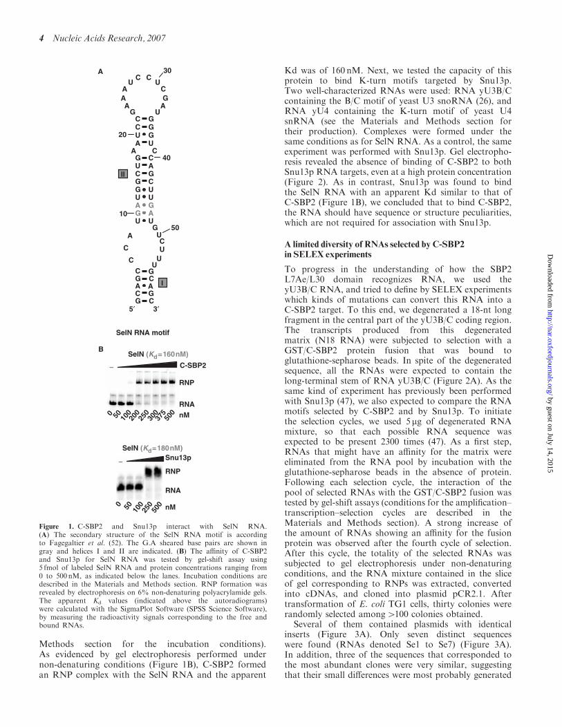
Methods section for the incubation conditions).As evidenced by gel electrophoresis performed undernon-denaturing conditions (Figure 1B), C-SBP2 formedan RNP complex with the SelN RNA and the apparent
Kd was of 160 nM. Next, we tested the capacity of thisprotein to bind K-turn motifs targeted by Snu13p.Two well-characterized RNAs were used: RNA yU3B/Ccontaining the B/C motif of yeast U3 snoRNA (26), andRNA yU4 containing the K-turn motif of yeast U4snRNA (see the Materials and Methods section fortheir production). Complexes were formed under thesame conditions as for SelN RNA. As a control, the sameexperiment was performed with Snu13p. Gel electropho-resis revealed the absence of binding of C-SBP2 to bothSnu13p RNA targets, even at a high protein concentration(Figure 2). As in contrast, Snu13p was found to bindthe SelN RNA with an apparent Kd similar to that ofC-SBP2 (Figure 1B), we concluded that to bind C-SBP2,the RNA should have sequence or structure peculiarities,which are not required for association with Snu13p.
A limited diversity of RNAs selected by C-SBP2in SELEX experiments
To progress in the understanding of how the SBP2L7Ae/L30 domain recognizes RNA, we used theyU3B/C RNA, and tried to define by SELEX experimentswhich kinds of mutations can convert this RNA into aC-SBP2 target. To this end, we degenerated a 18-nt longfragment in the central part of the yU3B/C coding region.The transcripts produced from this degeneratedmatrix (N18 RNA) were subjected to selection with aGST/C-SBP2 protein fusion that was bound toglutathione-sepharose beads. In spite of the degeneratedsequence, all the RNAs were expected to contain thelong-terminal stem of RNA yU3B/C (Figure 2A). As thesame kind of experiment has previously been performedwith Snu13p (47), we also expected to compare the RNAmotifs selected by C-SBP2 and by Snu13p. To initiatethe selection cycles, we used 5mg of degenerated RNAmixture, so that each possible RNA sequence wasexpected to be present 2300 times (47). As a first step,RNAs that might have an affinity for the matrix wereeliminated from the RNA pool by incubation with theglutathione-sepharose beads in the absence of protein.Following each selection cycle, the interaction of thepool of selected RNAs with the GST/C-SBP2 fusion wastested by gel-shift assays (conditions for the amplification–transcription–selection cycles are described in theMaterials and Methods section). A strong increase ofthe amount of RNAs showing an affinity for the fusionprotein was observed after the fourth cycle of selection.After this cycle, the totality of the selected RNAs wassubjected to gel electrophoresis under non-denaturingconditions, and the RNA mixture contained in the sliceof gel corresponding to RNPs was extracted, convertedinto cDNAs, and cloned into plasmid pCR2.1. Aftertransformation of E. coli TG1 cells, thirty colonies wererandomly selected among4100 colonies obtained.
Several of them contained plasmids with identicalinserts (Figure 3A). Only seven distinct sequenceswere found (RNAs denoted Se1 to Se7) (Figure 3A).In addition, three of the sequences that corresponded tothe most abundant clones were very similar, suggestingthat their small differences were most probably generated
RNP
RNA
C-SBP2 −
0 50 100
200
250
300
375
500 nM
SelN (Kd=160nM)
SelN RNA motif
I
II
C
C
5′
C
C
C
C
C
C
CC
C
A
C
G
G
G
G
G
G
G
G
GG
G
G
A
A
A
A
AA
A
AA
U
U
U
U
U
U
U
UU
U
U
U
U
U
U
C
C
C
CG
G
GG
A A
3′
10
20
30
40
50
A
B
RNP
RNA
Snu13p−
0 50 100
250
500 nM
SelN (Kd=180nM)
Figure 1. C-SBP2 and Snu13p interact with SelN RNA.(A) The secondary structure of the SelN RNA motif is accordingto Fagegaltier et al. (52). The G.A sheared base pairs are shown ingray and helices I and II are indicated. (B) The affinity of C-SBP2and Snu13p for SelN RNA was tested by gel-shift assay using5 fmol of labeled SelN RNA and protein concentrations ranging from0 to 500 nM, as indicated below the lanes. Incubation conditions aredescribed in the Materials and Methods section. RNP formation wasrevealed by electrophoresis on 6% non-denaturing polyacrylamide gels.The apparent Kd values (indicated above the autoradiograms)were calculated with the SigmaPlot Software (SPSS Science Software),by measuring the radioactivity signals corresponding to the free andbound RNAs.
4 Nucleic Acids Research, 2007
by guest on July 14, 2015http://nar.oxfordjournals.org/
Dow
nloaded from

by RT-PCR errors in the course of the amplification.Therefore, only four main classes of Se RNAs wereselected in the experiment (Figure 3A). This limiteddiversity of the selected sequences was one major differ-ence, compared to the SELEX experiment performed withSnu13p (31 very different sequences were obtained startingfrom the same initial RNA mixture). This suggested theexistence of strong sequence and/or structural constraintsfor SBP2 recognition. Three of the less frequent Se RNAshad a different length as compared to the initial RNAs:RNA Se2 (Figure 3A) contained an additional residue inthe degenerated sequence, whereas RNA Se1 and Se3lacked two residues compared to the initial RNAs.In addition, all the Se RNAs had a G instead of aU residue at position 38 in the conserved sequence.The same U38G base substitution was also found inseveral of the RNAs selected by the GST/Snu13p protein(47). By using gel-shift experiments, we verified thatthe seven distinct Se RNAs, that were selected, showedan affinity for the untagged C-SBP2 protein (Figure 3C).A wide range of apparent Kd values was observed(from 500 nM to42000 nM) (Figure 3C).
The selected RNAs all form canonical K-turn motifs
In order to understand the structural reasons for thesedifferent affinities, the possible folding of the sevenselected RNAs was investigated. Remarkably, each ofthem could form a canonical K-turn structure withtandem G.A and A.G base pairs and a 3-nt bulgeincluding a U residue at position 3. Most of the proposedstructures were verified by enzymatic probing (Figure 4A).
They are represented in Figure 4B, where they areclassified according to the values of the establishedapparent Kds. The free energies of the proposed 2Dstructures at 378C in 1M NaCl were also calculated byusing the M-Fold software. Based on these structures,nts 17–21 and 38–39 correspond to residues 1–5 and 6-7of the K-turn motif. Hence, residues 1 and 2 in the bulge,the A residue of the first G.A pair in stem II and oneU residue of the third pair in this stem correspondedto invariant residues in the starting RNA mixture.The G residue of the A.G pair corresponded to theabove-mentioned U to G mutation at position 38 inthe constant region. This G residue might have beengenerated by misincorporation in the course of theamplification cycles. Its selection in all the RNAs isin agreement with the high functional importance ofthe A.G pair in K-turn formation. All the selected RNAshad an identical UGAU sequence from position 19 to 22in the randomized segment, which demonstrated a strongpressure for the selection of a perfectly canonical K-turnstructure with two A.G and G.A pairs, and a U residue atposition 3 in the bulge. In all the selected RNAs, exceptRNA Se3 which contains a G.U pair, the constant U37
residue was always facing a U or a C residue in helix II.Interestingly, a U.U pair was almost always selected atthis position of helix II in the SELEX experimentperformed with Snu13p (47). Requirement of a non-Watson–Crick pair on top of the G.A and A.G pairsfor binding of C-SBP2 may explain the absence of bindingof C-SBP2 to yU4 RNA which has a G–C pair at thisposition in helix II (Figure 2B). Finally, in all the selectedSe RNAs, helix II contained at least two Watson–Crick
1000
1250
1500
1750
2000
RNA
C-SBP2−
025
050
075
0 nM25
00
yU3B/C + C-SBP2
RNA
C-SBP2
nM0
250
500
75010
0012
5015
0017
5020
00
−yU4 + C-SBP2A
RNA
Snu13p−
0 50 100
250
500
750
1000
1500
2000 nM
RNP
yU3B/C + Snu13p (Kd=230nM)
RNA
Snu13p−
0 50 100
250
500
750
1000 nM
RNP
yU4 + Snu13p (Kd=230nM)
AAC
G
G
A
AG
UG
A C
U C
G
G
G
U
ACU
U
A
AU
10
40C
C
AU
GG
CC
UG
G
A
A U
30
A
BC
12
543
67
I
II
yU3B/C
3′
U
C CG
AA
U
CA G
U
GAG
20
50
5′
A U
CCG
GG
10
20
U
A
UG
AU
UG
GU
C
C
A
C
I
U
CUA
AG
AG
UG
A
GC
G
A GU
U
ACU
C
12
54
3 6
7
II
G
3′U GUG UG A UUA UA A
50
30
40
5′
B
yU4
G
Figure 2. C-SBP2 does not interact with yU3B/C and yU4 RNAs. The binding of C-SBP2 and Snu13p on yU3B/C or yU4 RNAs was tested bygel-shift assays. The secondary structures of yU3B/C (Panel A) and yU4 (Panel B) are according to Marmier-Gourrier et al. (26) and Mougin et al.(43), respectively. The G.A sheared pairs are in gray and helices I and II are indicated. Nucleotides involved in the K-turn folding are numbered from1 to 7. Complexes were formed between 5 fmol of uniformly labeled yU3B/C (Panel A) or yU4 (Panel B) RNAs, and C-SBP2 or Snu13p atconcentrations ranging from 0 to 2500 nM, as indicated below the lanes. Incubation conditions were as described in the Materials and Methodssection. The autoradiograms obtained after electrophoresis on 6% polyacrylamide gels are shown. For the Snu13p–RNP complexes shown ascontrols, the apparent Kd values were calculated as described in the Materials and Methods section.
Nucleic Acids Research, 2007 5
by guest on July 14, 2015http://nar.oxfordjournals.org/
Dow
nloaded from

base pairs. They are most frequently (RNAs Se3, 4, 5, 6and 7) stacked on the three non-Watson–Crick base pairs.In agreement with the absence of binding of C-SBP2 toyU3B/C RNA (Figure 2A), none of the selected RNAs
had a bulge in the 30 strand and a short helix I. In contrast,no restriction on the size of the bulge, or on the lengthof helices I and II, was found in the SELEX experimentperformed with Snu13p (47). Altogether, the data
Pool 0 RNA
N18 5′ -GGACCUUUGUACCCCAGANNNNNNNNNN.NNNNNNNNUUAUGGGUACAAAUGGCAG-3′
WT 5′ -GGACCUUUGUACCCCAGAGUGAGAAACG.CGAUGAUCUUAUGGGUACAAAUGGCAG-3′
N˚ Selected Sequences:1 5′ -GGACCUUUGUACCCCAGAUGAUGGCUUC...ACUGCUUGAUGGGUACAAAUGGCAG-3′ (1)2 5′ -GGACCUUUGUACCCCAGAUGACGGCUCAUUUCGUGCUUGAUGGGUACAAAUGGCAG-3′ (1)3 5′ -GGACCUUUGUACCCCAGAUGAUGCUUUA..UCAGGCG.GAUGGGUACAAAUGGCAG-3′ (3)4 5′ -GGACCUUUGUACCCCAGAUGAUAGUAAA.GCGCGGCUUGAUGGGUACAAAUGGCAG-3′ (2)5 5′ -GGACCUUUGUACCCCAGAUGAUAGUGAG.GCGCGGCUUGAUGGGUACAAAUGGCAG-3′ (8)6 5′ -GGACCUUUGUACCCCAGAUGAUAGUAAG.GCGCGGCUUGAUGGGUACAAAUGGCAG-3′ (13)7 5′ -GGACCUUUGUACCCCAGAUGAUCCGACG.CGCUUUGGUGAUGGGUACAAAUGGCAG-3′ (2)
10 20 30 40 50
Se1 5′ -GGACCUUUGUACCCCAGAUGAUGGCUUC...ACUGCUUGAUGGGUACAAAUGGCAG-3′ (1)h SelN 5′ -...GCCCAUGAUGGCUG.....CAGCUUGAUGUCUU...-3′ r GPx 5′ -...UUCCAUGACGGUGU.....ACACCUGAUUUCCA...-3′ r 5′ DI 5′ -...GUUUAUGAUGGUCA.....UGACUUGAUUUUUA...-3′ r PHGPx 5′ -...ACUCAUGACGGUCU.....AGUCCCGAGGACCU...-3′ r SelP 5′ -...AUUGAUGAGAACAG.....CUGUUGGAUAGCUC...-3′ m Sel15 5′ -...AUUAAUGAGGAUUA.....AGAUCUGAUAAUUG...-3′ h SelD 5′ -...GUUAAUGACGUCUC.....GAGGCAGAGCAAGC...-3′ d SelD 5′ -...ACUUAUGAGGAUUA.....UAGUCUGAACCUUA...-3′ m SelD 5′ -...GAUAAUGAUGUCUC.....GAGGCUGAACAAAC...-3′ h SelX 5′ -...CUGCAUGAUCCGCU.....AGUGGGGAUGGUCU...-3′ h SelT 5′ -...CAUUAUGAAGGCCU.....AGACCAGAUGCUUU...-3′ h SelZ 5′ -...GAUGAUGACGACCU.....AUGUCCGAGCCCCC...-3′ b TrxR2 5′ -...GAUGAUGAGGACCU.....AUGUCUGAACCCCU...-3′ h TR3 5′ -...GAUGAUGACGACCU.....AUGUCCGAGCCCCC...-3′ m TrxR1 5′ -...GUCCAUGAAGUCAC.....GUGACAGAAGAGCU...-3′ C.e. TrxR 5′ -...CUUUGUGACGACCU.....UGGUCUGAUGCGCC...-3′ z SelW 5′ -...AACAAUGAUGGUGA.....UUGCUUGAUGCUCU...-3′ m Sel15 5′ -...AUUAAUGAGGAUUA.....AGAUCUGAUAAUUG...-3′ h Sel15 5′ -...GUUAAUGAAGACUA.....GGAUCAGAUACAUA...-3′ h SelY 5′ -...GCGGAUGAUAACUA.....UGGUUGGAUGUAGU...-3′ m D12 5′ -...GCGAAUGAUAACUA.....UGGUUGGAUGUAGU...-3′ c D12 5′ -...GUUUAUGAAGAGCA.....UGUUCAGAUGCUCU...-3′ X.l. D13 5′ -...GCAAAUGACGACCG.....GUGUCCGACAUCAA...-3′ c D13 5′ -...CUUUGUGAUGACCG.....GUGUCUGAUGUUGU...-3′ O.n. D13 5′ -...CUCUGUGAAGUUCG.....GACACUGAUGUUUC...-3′ r PHGPx 5′ -...ACUCAUGACGGUCU.....AGUCCCGAGGACCU...-3′ p PHGPx 5′ -...ACCCAUGACAGUCU.....AGACUCGAGAACCU...-3′
Consensus 5′ -........UGAPyGPu........PyCUGA........-3′
d: drosophila, m: mouse, b: bovine, c: chicken, c.e.: c. elegans, z: zebrafish, X.l.: X. laevis, o.n.: O. niloticus, r: rat, p: porcine
A
B
Helix II
123 45 6 5 4321
Figure 3. Sequences of the RNAs recovered from the SELEX experiment and test of their affinities for C-SBP2. (A) Alignment of the WT yU3B/CRNA sequence with the degenerated N18 RNA and the selected Se1-Se7 RNAs sequences. Nucleotides in Se1-Se7 RNA, are numbered according tothe positions of the homolog nucleotides in the WT yU3B/C RNA. The number of sequenced plasmids encoding each selected RNA is indicated inbrackets on the right of the sequences. The nucleotides corresponding to the constant sequence are shown in gray, nucleotides in the degeneratedsequence and nucleotides mutated during the RT-PCR cycles are shown in black. The GA dinucleotides are underlined. (B) The nucleotide sequencesof a series of SECIS motifs from various genes and species (30,52) were aligned with the Se1 RNA sequence taking as references the UGA and GAconserved nucleotides of the K-turn structure (bold characters). A consensus sequence of the SECIS K-turn motifs is deduced from the alignmentand indicated below. The positions of the conserved nucleotides in the two strands of helix II are indicated (C) Estimation of the affinity of C-SBP2for the Se1, Se2, Se3, Se5 and Se7 RNAs by gel-shift assays. RNA–protein complexes formed with 5 fmol of labeled RNA and increasingconcentrations of C-SBP2 (as indicated below the lanes) were fractionated by gel electrophoresis as in Figure 1. The apparent Kd values are indicatedabove the autoradiograms.
6 Nucleic Acids Research, 2007
by guest on July 14, 2015http://nar.oxfordjournals.org/
Dow
nloaded from

suggested that C-SBP2 binding requires a higher stabilityof the helices I and II compared to Snu13p binding.Surprisingly, the three selected RNAs, which showedthe highest stabilities and also the strongest affinities forC-SBP2, were encoded by DNA sequences that wereunderrepresented among the cloned DNA sequences. Thisapparent discrepancy may be explained by the fact thatRNAs Se1, 2 and 3 all have different lengths compared tothe initial RNAs. They might have been generated in a latestep of the selection procedure. The very low affinity foundfor RNA Se7, which has a stability slightly higher thanthose of RNAs Se4, Se5 and Se6, might be explainedby sequence differences in stem II.
Specific requirements in helix II for efficient bindingof protein C-SBP2
Prior to site-directed mutagenesis of Se1 RNA, we testedthe influence of Mgþþ concentration on C-SBP2 bindingto this RNA. Indeed, previous data (42) establishedthe influence of the concentration of this divalent cationon the binding of recombinant SBP2 in vitro. C-SBP2binding was found to be more sensitive to the presenceof Mgþþ ions than Snu13p binding. However, the 1.5mMMgþþ concentration present in the experimental bufferwas found to be sufficient to ensure an efficient binding ofC-SBP2 on Se1 RNA (Figure 1 in Supplementary Data).Thus subsequent experiments were performed under theseconditions. To test the importance of the sequence of helixII for SBP2 binding, we mutated helix II in the winner Se1RNA. The Se1 RNA variants produced are shown inFigure 5A. Their affinities for C-SBP2 and Snu13p werecompared by gel-shift assays. Complexes were formedat different protein concentrations in order to define theapparent Kd values (Figure 5B). Interestingly, Snu13p hada very high affinity for RNA Se1. The estimated Kd
(35 nM) was similar to that found for the winner RNAs inthe Snu13p SELEX experiment (47). A lower affinity wasfound for C-SBP2 (Kd of 500 nM). Most of the base
substitutions in helix II had no marked effect on Snu13paffinity. Only the strong destabilization of helix IIgenerated by substitution of the fifth Watson–Crick basepair (G-C)5 by a G.G pair had a marked deleterious effecton Snu13p affinity (factor of 20). In contrast, several basesubstitutions in helix II, (U.U)3 to (G-C)3, (G-C)5 to(G.G)5 and (C-G)6 to (G.G)6 almost abolished C-SBP2binding. The (G.U)4 to (C-G)4 and, to a lesser extent,the (G.U)4 to (U.U)4 substitutions, also had a markednegative effect. Hence, we concluded that C-SBP2 caninteract with canonical K-turn structures, provided thathelix II contains a triplet of non-Watson–Crick base pairsincluding the G.A and A.G sheared pairs and at leasttwo consecutive Watson–Crick base pairs in helix II.In addition, the base pairs on top of the triplet ofnon-Watson–Crick base pairs should be a Pu.Py pair(G.U, G–C or A–U). This may explain why the Se7 RNA,which has a Py.Pu pair at this position, has a low affinityfor protein C-SBP2.
The presence of a large internal loop instead of thebulge increases C-SBP2 affinity
The apparent Kd of the complex formed by C-SBP2and the winner Se1 RNA was 3-fold lower than that foundfor the natural SelN RNA (Figures 1 and 5B). Inspectionof the 2D structures of these two RNAs suggestedtwo possible explanations for the observed differenceof affinity. The presence of a long stem II in SelN RNA,and/or the presence of a large internal loop insteadof a bulge in this RNA might increase C-SBP2 affinity.We tested whether the insertion of two Watson–Crick basepairs in helix II of RNA Se1 (RNA Se1:Ins) might increasethe affinity of C-SBP2 (Figure 6A). Based on the observedaffinities of RNA Se1:Ins for C-SBP2 and Snu13p(apparent Kds of 300 and 25 nM, respectively), the 2 bpinsertion only had a limited positive effect on C-SBP2affinity and no marked effect on Snu13p affinity. When, inaddition to the extension of stem II, the bulge of RNA Se1
C RNA Se1 + C-SBP2(Kd=500nM)
RNP
RNA
C-SBP2
020
040
060
0 nM800
RNA Se3 + C-SBP2(Kd=750nM)
RNP
RNA
C-SBP2
050
075
010
00 nM
1250
RNA Se7 + C-SBP2(Kd>2000nM)
RNP
RNA
C-SBP2
050
010
0015
00 nM
2000
RNA Se5 + C-SBP2(Kd=1250nM)
RNP
RNA
C-SBP20
500
1000
1500 nM
2000
RNA Se2 + C-SBP2(Kd=700nM)
RNP
RNA
C-SBP2
050
075
010
00 nM
1250
Figure 3. Continued
Nucleic Acids Research, 2007 7
by guest on July 14, 2015http://nar.oxfordjournals.org/
Dow
nloaded from

A
B
RNA Se3 (−14.7 kcal/mol)(Kd=750nM)
AU
G
AU
C
C
U
C
AG
AGU G
A
UG
C
C
GG
GA U
3′5′
UGC
G20
30
40
123
45
67
I
II
C
30
20C
C
CG G
G
G
GA
G
A G
U
U
C
AG
AGU
GA
UU
C
C
GG
GA U
3′5′
40
123
45
67
I
II
C
RNA Se1 (−14.4 kcal/mol)(Kd=500nM)
20
G
C
CU
U
U
C
AG
AGU G
A
UU
C
C
GG
GA U
3′5′
CGG
C
U A
40
123
45
6
7
I
II
30
C
C
CG
G
U
A
AC
G
U
A
C
AG
AGU G
A
UU
C
C
GG
GA U
3′5′
G
GG
20
30
40
123
45
6
7
I
II
C
UUA
C
G CG
G
C
C
AG
AGU G
A
UU
C
C
GG
GA U
3′5′
GUC
G
C
20
30
40
123
45
6
7
I
II
C
N
N
N
NN
NN
U
N
N N N
N
N
C
AG
NNN U
A
N
N
CC
GGG
A U
35′
40
20
30
UGU
ACA
A U
CC
U
GG
AU A
C
U
AG G C G ′A
10
50
II
RNA N18 RNA Se2 (−14.4 kcal/mol)(Kd =700nM)
UU
G
UU
C
U
A
C
AG
AGU G
A
CU
C
C
GG
GA U
CGG
C
G
U
3′5′
C
20
30
40
123
45
67
I
II
C
C
C
CG G
G
G
GA
AA A
U
U
C
AG
AGU G
A
UU
C
C
GG
GA U
3′5′
40
20
30
123
45
6
7
I
II
C
x1 x1 x3
x2 x8 x13 x2
L 1 2 3 4
20
30
RNA Se1
20
30
RNA Se3
20
30
RNA Se5
20
30
RNA Se6
20
30
RNA Se7
L 1 2 3 4 L 1 2 3 4 L 1 2 3 4 L 1 2 3 4
RNA Se4 (−12 kcal/mol)(Kd=1250 nM)
RNA Se5 (−12 kcal/mol)(Kd=1250nM)
RNA Se6 (−12 kcal/mol)(Kd=1250nM)
RNA Se7 (−13.5 kcal/mol)(Kd>2000nM)
Figure 4. All the selected RNAs that recognize C-SBP2 can form a K-turn structure. (A) Secondary structure analysis of RNAs Se1, Se3, Se5, Se6or Se7 by enzymatic probing. The RNAs were 50-end labeled with 32P, renatured and digested with V1 (0.001 U, lane 2), T1 (0.8 U, lane 3) or T2(2.4 U, lane 4) RNases, under conditions described in the Materials and Methods section. As a control, undigested RNA was fractionated in parallel(lane 1). Lane L corresponds to the alkaline hydrolysis of the RNA used for localization of the RNase cleavage sites. Electrophoresis was performedon a 10% 8M urea–polyacrylamide gel. Nucleotide positions are indicated on the left. (B) Secondary structure models proposed for theselected RNAs. Models were proposed based on thermodynamic considerations and the results of the enzymatic digestions are shown in A.Regions corresponding to the degenerated sequences are shown by gray characters. For RNAs Se1, 3, 5, 6 and 7, V1, T1 and T2 RNase cleavages arerepresented by arrows surmounted of squares, dots and triangles, respectively. The color of symbols reflects the intensity of cleavages (gray, darkgray and black for low, medium and strong, respectively). Nucleotide numbering is as in Figure 3A. The apparent Kd values established for eachRNA by gel retardation are indicated. The free energies of the proposed secondary structures, expressed in kcal/mol, were calculated by using theM-Fold software.
8 Nucleic Acids Research, 2007
by guest on July 14, 2015http://nar.oxfordjournals.org/
Dow
nloaded from

was converted into a large internal loop (RNASe1:Insþ loop), the affinity for C-SBP2 was increased bya factor of 4 as compared to RNA Se1. In contrast, theaffinity for protein Snu13p was decreased by a factor of 18(Figure 6B). Hence, the presence of a large internal loop isfavorable for C-SBP2 binding, but not for Snu13pinteraction.
Having selected an RNA (Se1:Insþ loop RNA) with anaffinity for C-SBP2 similar to that of the authentic SBP2RNA target (SelN RNA) (Figure 1B), we then tested
the effect on C-SBP2 affinity of mutations at positions 2and 3 in the internal loop of this RNA (Figure 6C).The results obtained revealed a preference for an A and toa lesser extent a U residue at position 2. The strongestnegative effect on C-SBP2 affinity was observed for anA to C substitution at position 2 and a U to G substitutionat position 3 (Figure 6C). Therefore, the identity ofresidues at positions 2 and 3 in the internal loop has astrong influence on C-SBP2 affinity.
A large internal loop in the RNA confers a higherstability to C-SBP2–RNA complexes
Based on gel-shift experiments, Snu13p and C-SBP2 werefound to have similar affinities for RNA SelN (Kds of 180and 160 nM, respectively) (Figure 1B). However, suchapparent Kds, established by gel-shift assays, mostly reflectthe capacity of the RNA and protein partners to form acomplex which is stable under electrophoresis conditions.Thus, for a better estimation of the stability of the RNPcomplexes, we used competition experiments. Complexeswere formed, as above, with radiolabeled RNA and aprotein concentration about twice that of the apparent Kds(300 nM for C-SBP2 and 1000 nM for Snu13p, for assayson Se1:Insþ loop RNA, and two identical proteinconcentrations, 300 nM, for assays on SelN RNA). ColdRNA was added in excess to destabilize the complex.When complexes were formed with the Se1:Insþ loopRNA (Figure 7), a larger excess of cold Se1:Insþ loopRNA was required to dissociate C-SBP2–RNA complexescompared to Snu13p–RNA complexes and this in spite ofthe higher Snu13p concentration used to form the initialcomplex (Figure 7A). Furthermore, a much strongerdifference was observed when complexes were formedwith the SelN RNA: whereas a 1000-fold molar excess ofSelN RNA was sufficient to destabilize the SelN–Snu13pcomplexes, dissociation of the SelN–C-SBP2 complexesrequired as much as a 40 000-fold excess of cold SelNRNA (Figure 7B). These observations revealed the highstability of complexes formed with C-SBP2.Another approach to verify the high stability of
the SelN RNA–C-SBP2 complexes was to destabilizethe RNA–protein complex by addition of an excess ofa competitor protein (C-SBP2 for complexes formedwith Snu13p and vice versa). As seen in Figure 7C, evenwhen added in large excess (65-fold) to the preformedSelN–C-SBP2 complex, Snu13p could not dissociatethis complex. In contrast, when C-SBP2 was added atthe same concentration as the Snu13p protein used toform the SelN–Snu13p complex, this complex wascompletely converted into a SelN RNA–C-SBP2 complex.This observation argues in favor of a strong specificityof C-SBP2 for the SECIS RNAs.
C-SBP2 protects a larger region of the Se1:Insþ loopand SelN RNAs than Snu13p
One possible explanation for the strong stability ofcomplexes formed by protein C-SBP2 and theSe1:Insþ loop and SelN RNAs was the occurrence ofmore extended RNA–protein contacts in these complexescompared to those formed with Snu13p. To answer this
(G-C)3
(G-C)6, (G.G)6
(C-G)5, (G.G)5
I
IIG
C
CU
U
U
C
AG
AGU G
A
UU
CC
GGG
A U
CGG
C
U A
3′5′
(G-C)4, (C-G)4, (U.U)4, (A-U)4
123
4 5
6
7
RNA Se1
A
B C-SBP2 Snu13p
RNA Se1 500 nM 35 nM
(G-C)3 >4000 nM 45 nM
(G-C)4 600 nM 30 nM
(C-G)4 3000 nM 20 nM
(U.U)4 2000 nM 40 nM
(A-U)4 1050 nM 20 nM
(C-G)5 2000 nM 30 nM
(G.G)5 >4000 nM 740 nM
(G-C)6 1100 nM 40 nM
(G.G)6 >4000 nM 45 nM
Figure 5. Mutations in helix II of RNA Se1 are more deleterious forC-SBP2 than for Snu13p binding. (A) Positions of base substitutions inthe Se1 RNA are represented in gray on the proposed secondarystructure. The nature of the mutations in the variant Se1 RNAs isshown on the right of helix II. (B) The affinities of C-SBP2 and Snu13pfor Se1 RNA and its variants were estimated by gel-shift assaysusing 50-end labeled RNAs and protein concentrations ranging from0 to 4000 nM. The apparent Kd values obtained for each of theRNA–protein complexes are indicated.
Nucleic Acids Research, 2007 9
by guest on July 14, 2015http://nar.oxfordjournals.org/
Dow
nloaded from

question, we probed the RNA accessibilities in thesix RNP complexes formed by the Se1, Se1:Insþ loopand SelN RNAs and each of the C-SBP2 and Snu13pproteins. We used T1 and T2 RNases under conditionssuch that they cleaved single-stranded regions, and V1RNase that cleaves specifically double-stranded andstacked RNA regions. Very similar RNA protectionswere obtained for complexes formed by RNA Se1 andeach of the proteins (Figure 8). Both proteins protectedthe bulge sequence, part of helix II and the 50 strand ofhelix I. In contrast, protections of RNAs Se1:Insþ loopand SelN by Snu13p were very limited compared to thosefound for C-SBP2. Thus, with RNAs containing an
extended internal loop, the architecture of C-SBP2allows tight RNA–protein contacts with both helices andthe 50 strand of the internal loop, which is not the case forSnu13p. Interestingly also, the sensitivity to V1 RNase ofthe 30 strand of helix I was strongly increased by bindingof C-SBP2 or Snu13p to RNA Se1. The same situationwas observed upon binding of C-SBP2 to RNASe1:Insþ loop (Figure 8). This effect was quite lessmarked upon Snu13p binding on this RNA. Altogether,this suggested the occurrence of a profound RNAconformational change when Snu13p or C-SBP2 bindRNA Se1 and when C-SBP2 binds RNA Se1:Insþ loop.This strong RNA conformational change is probably not
BA
RNA Se1:Ins
U
C ACU
U
GCGCCGCG
UUGA
UG
A
A
UG
G
Ins
234
5 67
1
II
AU
3′
5′
I
C
C-SBP2
RNA Se1:Ins+loop
A2 →G
A2 → C
A2 → U
U3 → G
130 nM
1200 nM
>2000 nM
300 nM
>2000 nM
RNA Se1:Ins (Kd=300nM)
RNP
RNA
C-SBP2
020
050
010
00 nM
2000
RNA Se1:Ins+loop (Kd=130nM)
RNP
RNA
C-SBP2
0 50 150
300 nM600
RNA Se1:Ins (Kd=25nM)
RNP
RNA
Snu13p
0 10 20 30 nM40 50 60
RNA Se1:Ins+loop (Kd=500nM)
RNP
RNA
Snu13p
025
050
075
0 nM
1000
1250
RNA Se1:Ins+loop
U
C ACU
U
GCGCCGCG
UUGA
UG
A
A
U
A
3′
5′
GC
UG
CCC
U
UU
U
Ins
Loop
2
34
5 67
1
I
II
RNA Se1:Ins+loop
U
C ACU
U
GCGCCGCG
UUGA
UG
A
A
U
A
3′
5′
GC
UG
CCC
U
UU
U
Ins
Loop
U,C,GG
2
34
5 67
1
I
II
Figure 6. A K-turn motif with an extended internal loop increases C-SBP2 affinity. The variant Se1:Ins (A) and Se1:Insþ loop RNAs (B and C)are shown. The additional residues in these variant RNAs compared to Se1 RNA are shown in gray. The affinities of C-SBP2 and Snu13p for Se1:Ins(A) and Se1:Insþ loop (B) were tested by gel-shift assays. Complex formation was performed as described in Figure 1, using 5 fmol of 50-end labeledRNA and increasing concentrations of C-SBP2 or Snu13p proteins. In Panels A and B, the apparent Kds are indicated above the autoradiograms.(C) The base substitutions generated at positions 2 and 3 in the internal loop of the Se1:Insþ loop RNA are indicated in gray. The table gives theapparent Kd values established by gel-shift assays for complexes formed between C-SBP2 and the variant Se1:Insþ loop RNAs.
10 Nucleic Acids Research, 2007
by guest on July 14, 2015http://nar.oxfordjournals.org/
Dow
nloaded from

induced upon binding of Snu13p to an RNA with a largeinternal loop. Binding of C-SBP2 to SelN RNA alsoinduced a hypersensitivity to V1 RNase, but the RNAsegment concerned was different (extremity of the 50
strand of helix II). No significant hypersensitivity to V1RNase was observed upon Snu13p binding to SelN RNA,which reinforces the idea that only C-SBP2 can establishtight contacts with RNAs containing a large internal loopand as a consequence remodel their conformation. Thearchaeal protein L7Ae is known to interact with bothcanonical K-turn and K-loop structures formed interminal loops (9–11,15,25,48). Thus, by footprintingassays, we tested whether L7Ae can establish a tightinteraction with the SelN RNA, as does C-SBP2 (Figure8). The apparent Kd established by gel-shift assays for theSelN–L7Ae complex revealed a high affinity (Kd of 35 nM,not shown). According to enzymatic footprinting assays(Figure 8), this high affinity may be due to the presence oftwo L7Ae-binding sites in SelN RNA: one of themcorresponds to the quartet of non-Watson–Crick base
pairs, the other one to the terminal loop. Due to thepresence of two G.A dinucleotides in this loop, a K-looprecognized by protein L7Ae can be formed. Interestingly,the protections found in the 50 strand of the internal loop,helix I, and the quartet of non-Watson–Crick base pairs,are very similar in the C-SBP2–SelN and L7Ae–SelNcomplexes. Protein L7Ae protects two additional residuesin the 30 strand of the internal loop as compared to C-SBP2. Hence, concerning the recognition of RNAs withan internal loop, the behavior of protein L7Ae is closer tothat of C-SBP2 than that of Snu13p.
Mutations in helix II of SelN RNA limit C-SBP2 affinity
Since our data suggested a functional importance of helixII for C-SBP2 binding, we tested the effects of mutationsin helix II of the authentic SelN SECIS motif on C-SBP2binding. Substitution of the fifth G.U pair in helix II bya C–G pair as well as substitution of the sixth G–C pair byC–G pair, had less negative effects on C-SBP2 binding(factor of 2) (Figure 9) compared to those found for thecorresponding substitution in RNA Se1 (factor of 4)(Figure 5). However, substitutions of the sixth G–C pairand of the seventh C–G pair by G.G pairs had strongnegative effects on C-SBP2 binding (Kds of 800 and780 nM instead of 160 nM for the WT RNA). Therefore,mutations in an authentic SECIS RNA confirmed ourobservation of the importance of the stability and thesequence of helix II for C-SBP2 binding. Accordingly,Pu–Py pairs are the most frequently observed base pairsat the fifth and sixth positions in helix II of SECISelements (Figure 3B).
DISCUSSION
The present data based on SELEX and site-directedmutagenesis experiments improve our understanding ofthe sequence and structural features required for efficientinteraction of SBP2 with RNA. These findings bringnew insights that will facilitate the understanding of itsmechanism of action in the selenocysteine incorporationmachinery.When used for studying RNA–protein interactions,
the SELEX approach most generally leads to the estab-lishment of an RNA consensus sequence. Here, despite thewide diversity of the initial RNA mixture (184), only sevendifferent sequences were selected, and several of themwere very similar. All of them folded into very similar 2Dstructures that contained a canonical K-turn motif.This limited diversity of the selected sequences indicatednarrow RNA structure requirements for efficient bindingof SBP2. We confirmed this hypothesis by severalexperimental approaches.
DualMgþþ
dependence of SBP2 bindingto different RNA substrates
Earlier work (49) established that SBP2 contained intestis extracts displayed high sensitivity to Mgþþ concen-tration for SECIS binding, the IC50 being around4mM. This sensitivity was however less pronounced(IC50420mM) with a shorter, recombinant version of
A RNA Se1:Ins+loop
1RNP
C-SBP2
0 10 100
10000
1000
0 10
0000
RNA
RNA
RNA Se1:Ins+loop
1
RNP
Snu13p
0 10 100
1 00
00
10 0
0010
0 00
0
RNA
RNA
B
0
RNP
RNA
C-SBP2
RNA0
1000
10 0
0020
000
30 0
0040
000
SelNSelN
RNP
RNA
Snu13p
RNA10010
010
000
C
300
RNP C-SBP2
RNA
Snu13p (nM)
C-SBP2 (nM)
0 030
030
030
030
0
SelN
00
300
300
1000
10 0
0020
000
RNP Snu13p
300
RNA
Snu13p (nM)
C-SBP2 (nM)
0 030
030
030
030
0SelN
00
300 50 100
200
300
RNP C-SBP2 RNP Snu13p
Figure 7. C-SBP2 forms highly stable complexes with RNAs containingan extended internal loop. The stabilities of the complexes formedbetween C-SBP2 and Snu13p and the Se1:Insþ loop (A) and SelN(B) RNAs were tested by competition experiments. RNA–proteincomplexes were formed by using 5 fmol of 50-end labeled Se1:Insþ loopor SelN RNAs and C-SBP2 (300 nM) or Snu13p (1000 or 300 nM).The RNA–protein complexes were challenged with increasing con-centrations of cold Se1:Insþ loop or SelN RNAs (10–100 000- and10–40 000-fold molar excess, respectively, as indicated below the lanes).The remaining complexes were fractionated by gel electrophoresis.(C) Comparison of the relative stabilities of the Snu13p–SelN andC-SBP2–SelN complexes. RNP complexes formed with C-SBP2 at300 nM were challenged by addition of an excess of Snu13p protein andvice versa. Complexes formed with Snu13p at 300 nM were challengedby addition of an excess of C-SBP2. The remaining complexes werefractionated by gel electrophoresis. The identities and concentrations ofthe protein competitors used in the assays are indicated below the lanes.
Nucleic Acids Research, 2007 11
by guest on July 14, 2015http://nar.oxfordjournals.org/
Dow
nloaded from

A
20
10
30
40
V1 T1 T2 V1 T1 T2
C-SBP21000 nM
Snu13p100 nM
L
RNA Se1
20
10
30
40
50
V1 T1 T2 V1 T1 T2
C-SBP2500 nM
Snu13p1000 nM
L
RNA Se1: Ins+loop
20
10
30
40
50
V1 T1 T2
C-SBP2300 nM
Snu13p300 nM
L V1 T1 T2 V1 T1 T2
L7Ae100 nM
SelN RNA
U
C ACU
UGCAUUACG
UUGA
UG
A
A
U
A
CG
CG
CG
C
AGG
A U
UA
GC
UA
UA
UA
UC
G
CG
CAG
3′
5′
G C
UG
CCC
U
UUU
10
20
30
40
50
60
2
34
5 67
1
I
II
B
RNA Se1: Ins+loop+ C-SBP2
RNA Se1: Ins+loop+ Snu13p
U
C ACU
UGCAUUACG
UUGA
UG
A
A
U
A
CG
CG
CG
C
AG
G
A U
UA
GC
UA
UA
UA
UC
G
CG
CAG
3′
5′
G C
UG
CCC
U
UUU
10
20
30
40
50
60
2
34
5 67
1
I
II
RNA Se1 + C-SBP2/Snu13p
I
II
U
C ACU
UGCCG
UUG
A
UG
A
AU
A
CG
CG
CG
C
AG
G
AU
UA
GC
UA
UA
UA
U
CG
CG
CAG
3′
5′
G
UG
10
20
30
40
50
12
34
56
7
SelN RNA+ Snu13p
C
C
5′
C
C
C
C
C
C
CC
C
A
C
G
G
G
G
G
G
G
G
GG
G
G
A
A
A
A
AA
A
AA
U
U
U
U
U
U
U
UU
U
U
U
U
U
U
C
C
C
CG
G
GG
A A
3′
10
20
30
40
50
I
II
SelN RNA+C-SBP2
C
C
5′
C
C
C
C
C
C
CC
C
A
C
G
G
G
G
G
G
G
G
GG
G
G
A
A
A
A
AA
A
AA
U
U
U
U
U
U
U
UU
U
U
U
U
U
U
C
C
C
CG
G
GG
A A
3′
10
20
30
40
50
II
I
SelN RNA+ L7Ae
C
C
5′
C
C
C
C
C
C
CC
C
A
C
G
G
G
G
G
G
G
G
GG
G
G
A
A
A
A
AA
A
AA
U
U
U
U
U
U
U
UU
U
U
U
U
U
U
C
C
C
CG
G
GG
A A
3′
10
20
30
40
50
II
I
Figure 8. C-SBP2 protects a larger region of the Se1:Insþ loop and SelN RNAs than Snu13p. (A) The in vitro transcribed 50-end labeled Se1,Se1:Insþ loop and SelN RNAs (25 fmol) were incubated in the absence (�) or presence (þ) of C-SBP2, Snu13p or L7Ae. The protein concentrationsused in the assays are indicated above each panel, 2mg of tRNA were added in each assay and the digestion was carried out for 6min at 208C, inbuffer D, in the presence of 0.8 U RNase T1, 2.4 U RNase T2 or 0.001 U RNase V1, as described in the Materials and Methods section. Thecleavage products were fractionated on a 10% polyacrylamide–8M urea gel. L: alkaline hydrolysis. Nucleotide positions are indicated on the left. (B)Schematic representation of the results shown in panel A on the secondary structures proposed for the three studied RNAs. Helices I and II areindicated. V1, T1 and T2 RNase cleavages are represented by arrows surmounted of squares, dots and triangles, respectively. The color of symbolsreflects the intensity of cleavages (green, orange and red for low, medium and strong, respectively). Nucleotides with decreased sensitivity to RNasein the presence of the proteins are circled in blue (pale and dark for low and strong protection, respectively). Nucleotides with increased sensitivityto RNase in the presence of the proteins are indicated by a red star. The number of stars reflects the increased sensitivity to cleavage (one, two andthree representing low, medium and strong, respectively).
12 Nucleic Acids Research, 2007
by guest on July 14, 2015http://nar.oxfordjournals.org/
Dow
nloaded from

SBP2, and PHGPx SECIS RNA as the RNA partner (42).Interestingly, here we found that binding of C-SBP2 to theSe1 RNA, which forms a canonical K-turn structure,requires a 1.5mM Mgþþ concentration, higher concentra-tions being innocuous. At first glance, the two series ofresults may appear contradictory. Nevertheless, thesedifferential behaviors toward Mgþþ are likely explainedby the use of different RNA partners. Se1 RNA is agenuine K-turn RNA, and it is known that divalentcations favor the closed conformation of canonical K-turnmotifs (50). SECIS RNAs possess a large internal loopand thus contain a K-turn like motif (32). A high Mgþþ
concentration may induce a conformational change intoSECIS RNAs, which is not favorable for SBP2 binding.For instance, based on our data, we can imagine that a
high Mgþþ concentration promotes closing of the internalloop, and we show that a large internal loop is neededfor maximum binding efficiency of SBP2. The Se1 RNA isa typical Snu13p partner. As expected, no marked Mgþþ
requirement is observed for Snu13p binding to this RNA.In contrast, as Se1 RNA does not contain an internalloop, a prior stabilization of the kink structure may beneeded to reinforce SBP2 binding. Altogether, the previousand present data strongly suggest that each memberof the L7A/L30 family is perfectly suited for binding toits authentic partner at the physiological concentrationof divalent cations. When RNA partners are exchangedin in vitro experiments, the Mgþþ ion concentration hasto be adapted in order to form the heterologousinteraction. Accordingly, a high Mgþþ ion concentration
A B
I
II
C
C
5′
C
C
C
C
C
C
CC
C
A
C
G
G
G
G
G
G
G
G
GG
G
G
A
A
A
A
AA
A
AA
U
U
U
U
U
U
U
UU
U
U
U
U
U
U
C
C
C
CG
G
GG
A A
3′
10
20
30
40
50
RNA
C-SBP2−
0 50 100
200
400
650
900
1250
1500
nM
RNP
SelN (G.U)5→ U.U (Kd=280nM)
2000
RNA
C-SBP2−
0 50 100
200
400
650
900
1250
1500 nM
RNP
SelN WT (Kd=160nM)
2000
C, UG
G SelN (G.U)5→ C-G (Kd=300nM)
RNA
C-SBP2−
010
020
040
080
013
0018
00
nM
RNP
SelN (G-C)6→ C-G (Kd=270nM)
RNA
C-SBP2−
010
020
040
080
013
0018
00
nM
RNP
GC
RNA
C-SBP2−
0 50 100
200
400
650
900
1250
1500 nM
RNP
SelN (G-C)6→ G.G (Kd=800nM)
2000 0 50 100
200
400
650
900
1250
1500
2000
RNA
C-SBP2 −
nM
RNP
SelN (C-G)7→ G.G (Kd=780nM)
SelN RNA motif
Figure 9. The sequence and stability of helix II are important for C-SBP2 binding onto SelN RNA. (A) The base-pair substitutions generated atpositions 5, 6 and 7 in helix II of the SelN RNA are shown. (B) Complexes were formed with 5 fmol of radiolabeled WT or mutated SelN RNA andincreasing concentrations of the C-SBP2 protein (from 50 to 2000 nM). The RNP complexes were fractionated on 6% polyacrylamide 8–M urea geland apparent dissociation constants were determined by measuring the radioactivity in the bands of gel corresponding to free RNA and the RNP.The determined Kds are indicated above each autoradiogram.
Nucleic Acids Research, 2007 13
by guest on July 14, 2015http://nar.oxfordjournals.org/
Dow
nloaded from

was found to be required for efficient in vitro bindingof protein L30 to a SECIS element, in the presence ofSBP2 (42).
Specific sequence requirements in helix II
Site-directed mutagenesis, performed on the winner Se1RNA obtained by SELEX experiments, demonstratedthat binding of C-SBP2 requires the presence of a stablehelix II containing at least two Watson–Crick base pairs.In agreement with this observation, all the SECISelements identified so far contain a series of Watson–Crick base pairs on top of the non-Watson–Crick base-pair quartet (30,51–53; A.K., unpublished data).Accordingly, we showed that their individual disruptionin SelN RNA decreases C-SBP2 affinity. Not only helix IIstability but also its sequence has an influence on C-SBP2affinity. The presence of a Pu–Py pair at the fourthposition in helix II was found to be of high importancefor C-SBP2 binding to Se1 RNA, a Pu–Py pair at thisposition being also more favorable for C-SBP2 binding toSelN RNA. This is in contrast with the absence ofsequence requirement in helix II, except for the G.A andA.G base pairs and the adjacent U.U pair found forproteins Snu13p/15.5K and L7Ae (6,7,16,24,47,54–56).Up to now, little attention was given to the importanceof the identity of base pairs in the upper part of helix II ofSECIS elements. However, at position 4 of helix II, a Puresidue (most frequently a G residue) is almost alwaysfound in the 50 strand and a Py residue (most frequently aC residue) is observed in the 30 strand (30,51–53; A.K.,unpublished data). Although less strictly conserved, thefifth base pair in helix II is predominantly a Pu–Py pair(Figure 3B). Together with our experimental data, thesephylogenetic observations strongly suggest a functionalimportance of these conserved Pu.Py base pairs atpositions 4 and 5 in helix II. In accordance with thishypothesis, the G–C pair at the fourth position in RNASe1 was protected against V1 RNase digestion in thecomplex formed with C-SBP2, but not in the complexformed with Snu13p (Figure 8). Accordingly, the verylimited V1 RNase cleavage, which is located betweenresidues G13 and G14 in free SelN RNA, disappeared inthe presence of C-SBP2, but not with Snu13p.Remarkably, this V1 RNase cleavage was also abolishedin the presence of protein L7Ae.Comparison of the Se1 to Se7 RNAs and site-directed
mutagenesis of the Se1 RNA also show the importancefor a non-Watson–Crick base pair on top of the A.Gand G.A pair tandem (Figures 4 and 5). Accordingly,U.U pairs are frequently encountered pairs at this positionin SECIS elements (30,51–53; A.K., unpublished data)and a U.U pair was also preferentially selected at thisposition of helix II, in the SELEX experiment performedwith Snu13p. The presence of a U.U base pair atthis position, with a C10–C10 distance of the ribose ringclose to that in G.A pairs, is very likely required tofavor the smooth transition from the non-Watson–Crickto the Watson–Crick section of helix II. Noticeably also,in K-turn structures found in ribosomal RNAs, thenucleobase of one of this unpaired couple of nucleotides
was proposed to interact with one nucleobase in helix I,and thus to reinforce the inter-helical angle between helix Iand helix II (57).
Importance of a large SECIS internal loop
Increasing the size of both helix II and the internal loopof the winner Se1 RNA obtained by SELEX, yieldedan RNA with an affinity similar to that of SelN RNA(Figure 6B). Such an RNA could not be obtained in theSELEX experiment, because of limitation in size of thedegenerated sequence that can be used (18 nt) in theseexperiments. Indeed, due to the necessity to cover allthe possible sequences during the screening, one cannotuse largely extended degenerated sequences (58,59).In agreement with the importance of the size of helix II,all the identified SECIS elements contain a long helix II.Based on our footprinting data, the high affinity ofC-SBP2 for RNAs with an internal loop, as well as thestability of the complexes formed, are due to its capacityto contact helices I and II and the 50 strand of the internalloop in these RNAs (Figure 8). Interestingly, Martin et al.(38) showed that closing of the internal loop of the ratD1 SECIS element almost completely abolished seleno-cysteine incorporation in vivo. In agreement with theobserved requirement of at least one base pair closingthe 3-nt bulge loop of K-turn motifs for efficient bindingof Snu13p (44), Snu13p establishes very loose contactswith RNAs containing an internal loop. The presence ofa closing base pair is not required for L7Ae and thisprotein is able to bind open K-loop structures (9,44,48).The presence of an arginine at position 95 in the 15.5K/Snu13p protein, that forms a salt bridge with the50 phosphate of the residue at position 1 in the bulge,and its replacement by a valine in L7Ae, were proposed toexplain this difference between proteins 15.5K/Snu13pand L7Ae (60). Interestingly, like L7Ae, SBP2 containsa valine at the corresponding position in the L7A/L30domain (29). This may explain our observation of similarbinding properties of proteins SBP2 and L7Ae on RNAscontaining a large internal loop.
In free RNAs containing a canonical K-turn structurewith a bulge loop, helices I and II form a 768 angle. UponSnu13p/15.5K binding, the RNA undergoes furtherfolding, so that the helix I–helix II angle is reduced to488 (56,61). This folding likely explains the tight contact ofSnu13p with both helices of RNA Se1 that we detected byfootprinting assay. As very similar footprinting resultswere obtained with C-SBP2, it probably also induces afolding of this RNA. However, C-SBP2 but not Snu13pmay induce a similar folding in both the Se1:Insþ loopand SelN RNAs.
Sequence requirement in the internal loop
Our site-directed mutagenesis experiments on theSe1:Insþ loop RNA revealed the importance of theidentity of residues at positions 2 and 3 in the internalloop for C-SBP2 binding. The most deleterious basesubstitution at position 2 was the A to C replacement.Interestingly, a 66% decrease of selenocysteine incorpora-tion was observed when the same A to C substitution
14 Nucleic Acids Research, 2007
by guest on July 14, 2015http://nar.oxfordjournals.org/
Dow
nloaded from

was generated in the SECIS element of the rat GPxmRNA while A to G and A to U changes only led to a lossof 30 and 22% of the incorporation, respectively (52). Inaccordance with the decrease of the C-SBP2 affinity uponU to G substitution at position 3 in RNA Se1:Insþ loop,selenocysteine incorporation was decreased by 88%when this base substitution was generated in the SECISelement of the rat GPx mRNA (38). In addition, a U toC substitution at this position abolished the binding ofSBP2 to SelN RNA and is responsible for a human geneticdisease, the rigid spine muscular dystrophy (62). As theresidue at position 3 in canonical K-turns is located inthe protein pocket, its mutation also has a deleteriouseffect on 15.5K/Snu13p and L7Ae binding (6,24–26).Residues E61 and K86 in 15.5K, and D54 and K79 inArchaeoglobus fulgidus L7Ae, are involved in the interac-tion with the nucleobase at position 3. Their counterpartsin SBP2 (E699 and R730) probably play a similar role,since they are crucial for binding to SECIS RNAs(5,29,56). The specificity of L7Ae/L30 protein memberstowards the residue at position 2 in the K-turn motifis variable. Whereas an A or G residue at position 2increases 15.5K/Snu13p affinity, substitutions at position2 have no marked effect on L7Ae affinity (25). Concerningposition 2, SBP2 exhibits a behavior closer to that of15.5K/Snu13p than to L7Ae.
The ribosomal protein L30 was recently shownto compete with SBP2 for binding to SECIS RNA (42).L30 was found to recognize a K-turn structure of itspre-mRNA that contains a protruding A residue in a smallinternal loop (21–23). SELEX experiments performedwith L30 revealed its preference for K-turn motifs withprotruding C or A residues (27). Later, it was shown thatL30 also has the ability to accommodate K-turn structureswith a protruding G (28). However, its interaction withK-turn motifs containing a protruding U residue hasnot been demonstrated yet. Consequently, the bindingof L30 to SECIS elements, that all contain a U residue atposition 3, raises the question of how it can achieve this.
CONCLUSION
Assembly of the selenocysteine incorporation machineryis proposed to be initiated by SBP2 association toSECIS elements in the nucleus, and more likely in thenucleolus (63). Protein 15.5K/Snu13p is abundant inthe nucleolus and SBP2 shares several common featureswith Snu13/15.5K. However, our data reveal importantdifferences in RNA specificities that may ensure thespecific association of SBP2 to SECIS elements inthe nuclear compartment.
ACKNOWLEDGEMENTS
V. Senty-Segault and S. Massenet are acknowledgedfor helpful discussions. S. Sonkaria is thanked for carefulreading of the manuscript. A. Clery was a fellow fromthe French ‘Ministere de la Recherche et des NouvellesTechnologies’. The work was supported by the CentreNational de la Recherche Scientifique, the French
‘Ministere de la Recherche et des NouvellesTechnologies’, the ACI ‘Biologie Cellulaire, Moleculaireet Structurale’ n8BCMS226, the PRST ‘Bioingenierie’of the ‘Conseil Regional Lorrain’ and the ToxNuc-EProgramme ‘Toxicologie Nucleaire Environnementale’.Funding to pay the Open Access publication charge wasprovided by CNRS-Sciences de la vie.
Conflict of interest statement. None declared.
REFERENCES
1. Klein,D.J., Schmeing,T.M., Moore,P.B. and Steitz,T.A. (2001)The kink-turn: a new RNA secondary structure motif. EMBO J.,20, 4214–4221.
2. Wimberly,B.T., Brodersen,D.E., Clemons,W.M.Jr, Morgan-Warren,R.J., Carter,A.P., Vonrhein,C., Hartsch,T. andRamakrishnan,V. (2000) Structure of the 30S ribosomal subunit.Nature, 407, 327–339.
3. Schluenzen,F., Tocilj,A., Zarivach,R., Harms,J., Gluehmann,M.,Janell,D., Bashan,A., Bartels,H., Agmon,I. et al. (2000) Structureof functionally activated small ribosomal subunit at 3.3 angstromsresolution. Cell, 102, 615–623.
4. Ban,N., Nissen,P., Hansen,J., Moore,P.B. and Steitz,T.A. (2000)The complete atomic structure of the large ribosomal subunit at2.4 A resolution. Science, 289, 905–920.
5. Vidovic,I., Nottrott,S., Hartmuth,K., Luhrmann,R. and Ficner,R.(2000) Crystal structure of the spliceosomal 15.5kD protein boundto a U4 snRNA fragment. Mol. Cell, 6, 1331–1342.
6. Nottrott,S., Hartmuth,K., Fabrizio,P., Urlaub,H., Vidovic,I.,Ficner,R. and Luhrmann,R. (1999) Functional interaction of anovel 15.5kD [U4/U6.U5] tri-snRNP protein with the 50 stem-loopof U4 snRNA. EMBO J., 18, 6119–6133.
7. Watkins,N.J., Segault,V., Charpentier,B., Nottrott,S., Fabrizio,P.,Bachi,A., Wilm,M., Rosbash,M., Branlant,C. et al. (2000) Acommon core RNP structure shared between the small nucleoar boxC/D RNPs and the spliceosomal U4 snRNP. Cell, 103, 457–466.
8. Terns,M.P. and Terns,R.M. (2002) Small nucleolar RNAs: versatiletrans-acting molecules of ancient evolutionary origin. Gene Expr.,10, 17–39.
9. Charpentier,B., Muller,S. and Branlant,C. (2005) Reconstitutionof archaeal H/ACA small ribonucleoprotein complexes active inpseudouridylation. Nucleic Acids Res., 33, 3133–3144.
10. Omer,A.D., Ziesche,S., Ebhardt,H. and Dennis,P.P. (2002) In vitroreconstitution and activity of a C/D box methylation guideribonucleoprotein complex. Proc. Natl. Acad. Sci. U.S.A., 99,5289–5294.
11. Rozhdestvensky,T.S., Tang,T.H., Tchirkova,I.V., Brosius,J.,Bachellerie,J.P. and Huttenhofer,A. (2003) Binding of L7Ae proteinto the K-turn of archaeal snoRNAs: a shared RNA binding motiffor C/D and H/ACA box snoRNAs in Archaea. Nucleic Acids Res.,31, 869–877.
12. Granneman,S., Pruijn,G.J., Horstman,W., van Venrooij,W.J.,Luhrmann,R. and Watkins,N.J. (2002) The hU3-55K proteinrequires 15.5K binding to the box B/C motif as well as flankingRNA elements for its association with the U3 small nucleolar RNAin Vitro. J. Biol. Chem., 277, 48490–48500.
13. Nottrott,S., Urlaub,H. and Luhrmann,R. (2002) Hierarchical,clustered protein interactions with U4/U6 snRNA: a biochemicalrole for U4/U6 proteins. EMBO J., 21, 5527–5538.
14. Watkins,N.J., Dickmanns,A. and Luhrmann,R. (2002) Conservedstem II of the box C/D motif is essential for nucleolar localizationand is required, along with the 15.5K protein, for the hierarchicalassembly of the box C/D snoRNP. Mol. Cell. Biol., 22, 8342–8352.
15. Baker,D.L., Youssef,O.A., Chastkofsky,M.I., Dy,D.A., Terns,R.M.and Terns,M.P. (2005) RNA-guided RNA modification: functionalorganization of the archaeal H/ACA RNP. Genes Dev., 19,1238–1248.
16. Tran,E.J., Zhang,X. and Maxwell,E.S. (2003) Efficient RNA20-O-methylation requires juxtaposed and symmetrically assembledarchaeal box C/D and C0/D0 RNPs. EMBO J., 22, 3930–3940.
Nucleic Acids Research, 2007 15
by guest on July 14, 2015http://nar.oxfordjournals.org/
Dow
nloaded from

17. Rashid,R., Aittaleb,M., Chen,Q., Spiegel,K., Demeler,B. and Li,H.(2003) Functional requirement for symmetric assembly of archaealbox C/D small ribonucleoprotein particles. J. Mol. Biol., 333,295–306.
18. Bortolin,M.L., Bachellerie,J.P. and Clouet-d0Orval,B. (2003)In vitro RNP assembly and methylation guide activity of anunusual box C/D RNA, cis-acting archaeal pre-tRNA(Trp).Nucleic Acids Res., 31, 6524–6535.
19. Schultz,A., Nottrott,S., Hartmuth,K. and Luhrmann,R. (2006)RNA structural requirements for the association of the spliceosomalhPrp31 protein with the U4 and U4atac small nuclear ribonucleo-proteins. J. Biol. Chem., 281, 28278–28286.
20. Koonin,E.V., Bork,P. and Sander,C. (1994) A novel RNA-bindingmotif in omnipotent suppressors of translation termination,ribosomal proteins and a ribosome modification enzyme?Nucleic Acids Res., 22, 2166–2167.
21. Vilardell,J. and Warner,J.R. (1994) Regulation of splicing at anintermediate step in the formation of the spliceosome. Genes Dev.,8, 211–220.
22. Chao,J.A. and Williamson,J.R. (2004) Joint X-ray and NMRrefinement of the yeast L30e-mRNA complex. Structure, 12,1165–1176.
23. Mao,H., White,S.A. and Williamson,J.R. (1999) A novel loop-looprecognition motif in the yeast ribosomal protein L30 autoregulatoryRNA complex. Nat. Struct. Biol., 6, 1139–1147.
24. Kuhn,J.F., Tran,E.J. and Maxwell,E.S. (2002) Archaeal ribosomalprotein L7 is a functional homolog of the eukaryotic 15.5kD/Snu13p snoRNP core protein. Nucleic Acids Res., 30, 931–941.
25. Charron,C., Manival,X., Clery,A., Senty-Segault,V., Charpentier,B.,Marmier-Gourrier,N., Branlant,C. and Aubry,A. (2004) Thearchaeal sRNA binding protein L7Ae has a 3D structure verysimilar to that of its eukaryal counterpart while having a broaderRNA-binding specificity. J. Mol. Biol., 342, 757–773.
26. Marmier-Gourrier,N., Clery,A., Senty-Segault,V., Charpentier,B.,Schlotter,F., Leclerc,F., Fournier,R. and Branlant,C. (2003) Astructural, phylogenetic, and functional study of 15.5-kD/Snu13protein binding on U3 small nucleolar RNA. RNA, 9, 821–838.
27. Li,H. and White,S.A. (1997) RNA apatamers for yeast ribosomalprotein L32 have a conserved purine-rich internal loop. RNA, 3,245–254.
28. White,S.A., Hoeger,M., Schweppe,J.J., Shillingford,A., Shipilov,V.and Zarutskie,J. (2004) Internal loop mutations in the ribosomalprotein L30 binding site of the yeast L30 RNA transcript. RNA, 10,369–377.
29. Allmang,C., Carbon,P. and Krol,A. (2002) The SBP2 and 15.5kD/Snu13p proteins share the same RNA binding domain:identification of SBP2 amino acids important to SECIS RNAbinding. RNA, 8, 1308–1318.
30. Fletcher,J.E., Copeland,P.R., Driscoll,D.M. and Krol,A. (2001)The selenocysteine incorporation machinery: interactions betweenthe SECIS RNA and the SECIS-binding protein SBP2. RNA, 7,1442–1453.
31. Walczak,R., Carbon,P. and Krol,A. (1998) An essential non-Watson–Crick base pair motif in 30UTR to mediate selenoproteintranslation. RNA, 4, 74–84.
32. Allmang,C. and Krol,A. (2006) Selenoprotein synthesis: UGA doesnot end the story. Biochimie, 88, 1561–1571.
33. Krol,A. (2002) Evolutionarily different RNA motifs andRNA-protein complexes to achieve selenoprotein synthesis.Biochimie, 84, 765–774.
34. Gladyshev,V.N. (2001). Selenium in biology and human health:controversies and perspectives. In Hatfield,D.L. (ed), Selenium:Its Molecular Biology and Role in Human Health. Kluwer, Boston,pp. 313–317.
35. Flohe,L., Andreesen,J.R., Brigelius-Flohe,R., Maiorino,M. andUrsini,F. (2000) Selenium, the element of the moon, in life on earth.IUBMB Life, 49, 411–420.
36. Rayman,M.P. (2000) The importance of selenium to human health.Lancet, 356, 233–241.
37. Rederstorff,M., Krol,A. and Lescure,A. (2006) Understandingthe importance of selenium and selenoproteins in muscle function.Cell Mol. Life Sci., 63, 52–59.
38. Martin,G.W.III, Harney,J.W. and Berry,M.J. (1998) Functionalityof mutations at conserved nucleotides in eukaryotic SECIS elements
is determined by the identity of a single nonconserved nucleotide.RNA, 4, 65–73.
39. Tujebajeva,R.M., Copeland,P.R., Xu,X.M., Carlson,B.A.,Harney,J.W., Driscoll,D.M., Hatfield,D.L. and Berry,M.J. (2000)Decoding apparatus for eukaryotic selenocysteine insertion.EMBO Rep., 1, 158–163.
40. Fagegaltier,D., Hubert,N., Yamada,K., Mizutani,T., Carbon,P. andKrol,A. (2000) Characterization of mSelB, a novel mammalianelongation factor for selenoprotein translation. EMBO J., 19,4796–4805.
41. Zavacki,A.M., Mansell,J.B., Chung,M., Klimovitsky,B.,Harney,J.W. and Berry,M.J. (2003) Coupled tRNA(Sec)-dependentassembly of the selenocysteine decoding apparatus. Mol. Cell, 11,773–781.
42. Chavatte,L., Brown,B.A. and Driscoll,D.M. (2005) Ribosomalprotein L30 is a component of the UGA-selenocysteinerecoding machinery in eukaryotes. Nat. Struct. Mol. Biol., 12,408–416.
43. Mougin,A., Gottschalk,A., Fabrizio,P., Luhrmann,R. andBranlant,C. (2002) Direct probing of RNA structure andRNA-protein interactions in purified HeLa cells and yeastspliceosomal U4/U6.U5 tri-snRNP particles. J. Mol. Biol., 317,631–649.
44. Charron,C., Manival,X., Charpentier,B., Branlant,C. and Aubry,A.(2004) Purification, crystallization and preliminary X-ray diffractiondata of L7Ae sRNP core protein from Pyrococcus abyssii. ActaCrystallogr. D Biol. Crystallogr., 60, 122–124.
45. Lescure,A., Allmang,C., Yamada,K., Carbon,P. and Krol,A. (2002)cDNA cloning, expression pattern and RNA binding analysis ofhuman selenocysteine insertion sequence (SECIS) binding protein 2.Gene, 291, 279–285.
46. Mathews,D.H., Sabina,J., Zuker,M. and Turner,D.H. (1999)Expanded sequence dependence of thermodynamic parametersimproves prediction of RNA secondary structure. J. Mol. Biol., 288,911–940.
47. Clery,A., Senty-Segault,V., Leclerc,F., Raue,H.A. and Branlant,C.(2007) Analysis of sequence and structural features that identify theB/C motif of U3 small nucleolar RNA as the recognition site forthe Snu13p-Rrp9p protein pair. Mol. Cell. Biol., 27, 1191–1206.
48. Nolivos,S., Carpousis,A.J. and Clouet-d’Orval,B. (2005)The K-loop, a general feature of the Pyrococcus C/D guide RNAs,is an RNA structural motif related to the K-turn. Nucleic AcidsRes., 33, 6507–6514.
49. Copeland,P.R. and Driscoll,D.M. (1999) Purification, redoxsensitivity, and RNA binding properties of SECIS-binding protein2, a protein involved in selenoprotein biosynthesis. J. Biol. Chem.,274, 25447–25454.
50. Matsumura,S., Ikawa,Y. and Inoue,T. (2003) Biochemical char-acterization of the kink-turn RNA motif. Nucleic Acids Res., 31,5544–5551.
51. Kryukov,G.V., Castellano,S., Novoselov,S.V., Lobanov,A.V.,Zehtab,O., Guigo,R. and Gladyshev,V.N. (2003) Characterizationof mammalian selenoproteomes. Science, 300, 1439–1443.
52. Fagegaltier,D., Lescure,A., Walczak,R., Carbon,P. and Krol,A.(2000) Structural analysis of new local features in SECIS RNAhairpins. Nucleic Acids Res., 28, 2679–2689.
53. Lescure,A., Gautheret,D., Carbon,P. and Krol,A. (1999) Novelselenoproteins identified in silico and in vivo by using a conservedRNA structural motif. J. Biol. Chem., 274, 38147–38154.
54. Szewczak,L.B., DeGregorio,S.J., Strobel,S.A. and Steitz,J.A. (2002)Exclusive interaction of the 15.5 kD protein with the terminal boxC/D motif of a methylation guide snoRNP. Chem. Biol., 9,1095–1107.
55. Szewczak,L.B., Gabrielsen,J.S., Degregorio,S.J., Strobel,S.A. andSteitz,J.A. (2005) Molecular basis for RNA kink-turn recognitionby the h15.5K small RNP protein. RNA, 11, 1407–1419.
56. Moore,T., Zhang,Y., Fenley,M.O. and Li,H. (2004) Molecularbasis of box C/D RNA-protein interactions; cocrystalstructure of archaeal L7Ae and a box C/D RNA. Structure, 12,807–818.
57. Razga,F., Spackova,N., Reblova,K., Koca,J., Leontis,N.B. andSponer,J. (2004) Ribosomal RNA kink-turn motif – a flexiblemolecular hinge. J. Biomol. Struct. Dyn., 22, 183–194.
16 Nucleic Acids Research, 2007
by guest on July 14, 2015http://nar.oxfordjournals.org/
Dow
nloaded from

58. Tuerk,C. and Gold,L. (1990) Systematic evolution of ligands byexponential enrichment: RNA ligands to bacteriophage T4 DNApolymerase. Science, 249, 505–510.
59. Klug,S.J. and Famulok,M. (1994) All you wanted to know aboutSELEX. Mol. Biol. Rep., 20, 97–107.
60. Hamma,T. and Ferre-D’Amare,A.R. (2004) Structure of proteinL7Ae bound to a K-turn derived from an archaeal box H/ACAsRNA at 1.8A resolution. Structure, 12, 893–903.
61. Wozniak,A.K., Nottrott,S., Kuhn-Holsken,E., Schroder,G.F.,Grubmuller,H., Luhrmann,R., Seidel,C.A. and Oesterhelt,F. (2005)Detecting protein-induced folding of the U4 snRNA kink-turn by
single-molecule multiparameter FRET measurements. RNA, 11,1545–1554.
62. Allamand,V., Richard,P., Lescure,A., Ledeuil,C., Desjardin,D., Petit,N.,Gartioux,C., Ferreiro,A., Krol,A. et al. (2006) A single homozygouspoint mutation in a 30untranslated region motif of selenoprotein NmRNA causes SEPN1-related myopathy. EMBO Rep., 7, 450–454.
63. de Jesus,L.A., Hoffmann,P.R., Michaud,T., Forry,E.P.,Small-Howard,A., Stillwell,R.J., Morozova,N., Harney,J.W. andBerry,M.J. (2006) Nuclear assembly of UGA decodingcomplexes on selenoprotein mRNAs: a mechanism for eludingnonsense-mediated decay? Mol. Cell. Biol., 26, 1795–1805.
Nucleic Acids Research, 2007 17
by guest on July 14, 2015http://nar.oxfordjournals.org/
Dow
nloaded from