Embed Size (px)
Citation preview

ELSEVIER Hearing Research 94 (1996) 14-23 REr .,dKH
An RT-PCR analysis of mRNA for growth factor receptors in damaged and control sensory epithelia of rat utricles
Linda D. Saffer a, Rende Gu a, Jeffrey T. Corwin a.b., Department g['Otolao,ngology-HNS. Unicersity q/Virginia School qf Medicine. Charlottesl ille, VA 22908, USA
b Department q/'Neuroscience, Unit'ersi O' o1 Virginia School o[Medicine. Charlottescille. VA 22908, USA
Received 12 June 1995; revised 29 November 1995" accepted 3 December 1995
Abstract
Sensory epithelia from normal rat utricles and those cultured with and without neomycin treatment were assayed for the presence of growth factor receptor mRNAs by RT-PCR (reverse transcriptase-polymerase chain reaction). Both undamaged and damaged utricles showed mRNA for Insulin receptor, IGF-I receptor, FGF receptor I, EGF receptor, and PDGF c~ receptor. Neomycin-damaged sensory epithelia showed less PDGF c~ receptor mRNA than undamaged epithelia, suggesting that this message may be expressed at higher copy levels in hair cells than in supporting cells. Consistent with that hypothesis, immunohistochemistry revealed much stronger PDGF c~ receptor staining in the hair cells than in the supporting cells. Preliminary evidence suggests that IGF-I receptor message also may be lowered in neomycin-damaged epithelia.
Keywords: Growth factor receptor; Reverse transcriptase-polymerase chain reaction; Hair cell: Regeneration: Balance: Vestibular
1. Introduct ion
Hair cells are the mechanoreceptive transducers for sound and balance in the inner ear. Hair cell loss caused by loud sound, drug treatments, infections, or aging results in permanent hearing and balance deficits that affect millions of people. In fish and amphibians these cells are produced throughout life and damaged cells can be replaced (Corwin, 1981, Corwin, 1983, Corwin, 1985; Lombarte et al., 1993; Baird and Torres, 1993). In birds and mammals, all audi- tory hair cells normally are produced before birth (Ruben, 1967; Katayama and Corwin, 1989). Birds can, however, regenerate their hair cells. Loss of hair cells is fol lowed by self-repair s temming from trauma-evoked mitosis in sup- porting cells and the differentiation of replacement hair cells at sites of lesions (Cotanche, 1987; Corwin and Cotanche, 1988; Ryals and Rubel, 1988). In fact, the supporting cells of a wide range of auditory, vestibular, and lateral line epithelia of non-mammalian vertebrates can divide to give rise to new cells which can either continue to proliferate, or differentiate as supporting cells or hair cells (Corwin, 1986; Cotanche, 1987; Corwin and
* Corresponding author. Tel.: (804)924-1568: Fax: (804)982-3966.
0378-5955/96/$15.00 © 1996 Elsevier Science B.V. All rights reserved SSD10378-5955(95)00228-6
Cotanche, 1988; J~argenson and Mathiesen, 1988; Girod et al., 1989; Balak et al., 1990; Jones and Corwin, 1993; Raphael, 1992, Raphael, 1993; Weisleder and Rubel, 1993).
Supporting cells often survive trauma that kills neigh- boring hair cells. This is true in all vertebrates, including mammals, so the surviving supporting cells may be a source of new epithelial cells that could be manipulated to enhance regenerative responses. The vestibular organs in mammals share structural characteristics with the sensory epithelia of the ears of non-mammalian species that pro- duce hair cells during postembryonic life, suggesting that the mammalian vestibular epithelia might have a capacity for regeneration of hair cells (Corwin, 1986). Recently, morphological studies demonstrated the reappearance of immature-appearing hair bundles and hair cell bodies at sites of lesions in the utricles of guinea pigs that were fixed 4 weeks alter the end of a 10-day treatment with gentamicin (Forge et al., 1993). Proliferation of supporting cells was demonstrated in a parallel study after hair cell loss caused by gentamicin and neomycin treatments of utricles from juvenile and adult guinea pigs and adult humans in vitro (Warchol et al., 1993). Labeled putative replacement hair cells were observed in utricles that had been cultured for 4 weeks after the aminoglycoside treat- ment.

L.D. Saffer et al. / Hearing Research 94 (1996) 14-23 15
In order for non-cancerous cells to proliferate, they require the binding of mitogenic growth factors to specific receptors on their cell surfaces. Binding and the subse- quent dimerization that occurs for many types of tyrosine kinase receptors activates signaling cascades within the cell to take the cell from a resting state, G 0, back to G~ and into S-phase, where DNA is replicated (Pardee, 1989). Growth factors can also participate in the regulation of cell differentiation and are required as positive signals for cell survival (McKay and Leigh, 1993; Raft et al., 1993). The nature of a tissue's responsiveness to growth factors is regulated by the availability of growth factor ligands and the expression of specific growth factor receptor types on the surfaces of the cells. The sequences for many messen- ger RNAs have been determined for rats, and a number of sequences for growth receptors have been published. Since the mammalian utricle has at least some regenerative capabilities, we chose to assay the sensory epithelium of rat utricles for growth factor receptor messages so as to identify signals that may be required to control regenera- tion in those epithelia.
RT-PCR (reverse transcriptase-polymerase chain reac- tion) was chosen as a rapid, sensitive method to assay small numbers of cells from the utricular sensory epithe- lium for the expression of 5 receptor messages. Utricles were harvested from young rats and processed in three groups. For the in vivo control group, the epithelia were removed immediately after decapitation. For the in vitro damaged group and the in vitro undamaged group, the utricles were cultured for 24 h in the presence or absence of neomycin and then cultured an additional 24 h without neomycin. All undamaged sensory epithelia were found to contain mRNA for each of the growth factor receptor messages examined: insulin receptor (Insulin R), insulin- like growth factor I receptor (IGF-IR), fibroblast growth factor receptor 1 (FGFR-1), epidermal growth factor recep- tor (EGFR), and platelet-derived growth factor a receptor (PDGF-c~R). Neomycin-damaged sensory epithelium con- tained mRNA for the same receptors, but consistently appeared to have less mRNA for PDGF-aR and in many cases appeared to have less mRNA for IGF-IR.
2. Materials and methods
2.1. Dissection
Utricles were removed from 36 female Sprague-Dawley rats (100-149 g) and dissected in ice-cold Hanks' bal- anced saline solution containing 10 mM HEPES at pH 7.4 (HHBSS). The utricles were divided into 3 groups: (1) in vivo undamaged controls, (2) in vitro neomycin-damaged, and (3) in vitro undamaged controls. The sensory epithelia for the in vivo undamaged group were harvested immedi- ately after decapitation of control rats. Utricles were dis- sected from the inner ears, and the membranous roof of the
utricle, the otoconia, and the otolithic membrane were removed. A thermolysin digestion method was used to separate sensory epithelia from non-sensory tissue. This was adapted from a procedure used to harvest human keratinocytes. (Germain et al., 1993, Corwin et al., 1995). For this each utricle was incubated for 1.5 h at 37°C in Medium 199 (M-199) containing Earle's salts and ther- molysin (Sigma protease type X) at 500 /~g/ml in a 5% CO 2 atmosphere. Then the enzyme solution was diluted with excess ice-cold HHBSS and kept cold as the epithelia were harvested. After the thermolysin treatment, the sen- sory epithelium was gently lifted from the underlying connective tissue, and the surrounding regions of non- sensory epithelia were cut away using double-edge razor blades. Isolated sheets of cells, composed of only the central part of a sensory epithelium, were harvested in this way, individually transferred to an Eppendorf tube contain- ing 1 ml of fresh, ice-cold HHBSS, and centrifuged at 5200 × g for 5 rain. Then all but a few microliters of solution was removed, the tube was immersed in liquid nitrogen, and transferred to storage at -80°C.
2.2. Cultures of utricles
Other utricles were cultured in M-199 with Earle's salts, supplemented with 5% fetal bovine serum (GIBCO), 10 U / m l penicillin (GIBCO), 0.025 mg/ml Fungizone (GIBCO), 1.2 pM cholera toxin (Sigma), either with or without 10 mM neomycin sulfate (Sigma) for 24 h at 37°C in a 5% CO 2 atmosphere, depending upon whether they were in the in vitro damaged or the in vitro undamaged treatment groups. The media were removed and twice the volume of M-199 (at 37°C) was added as a rinse and removed. Then M-199, supplemented as above but without neomycin, was added to each dish and the utricles were cultured for an additional 24 h. The sensory epithelia were treated next with thermolysin, removed from each of the cultured utricles, and handled as described above.
2.3. Extraction of RNA and RT-PCR
Total RNA was extracted from each sensory epithelium individually using Trizol (GIBCO), a phenol guanidine isothiocyanate solution (Chomczynski and Sacchi, 1987). Murine leukemia reverse transcriptase and random primers (Perkin-Elmer-Roche, Branchburg, NJ) were used for re- verse transcription at 42°C for 15 rain. The resulting cDNA was amplified using specific primers under opti- mized conditions of annealing temperatures, MgC1 z con- centrations, and pHs. Those conditions were determined for each PCR reaction using rat brain tissue and an Opti- Prime PCR optimization kit (Stratagene, La Jolla, CA). The pH was 8.3 or 8.8 and the ionic concentrations were 10 mM Tris-HCl, 1.5 mM or 3.5 mM MgC12, and 25 mM KC1. Samples were overlaid with mineral oil. The cDNA was denatured for 5 min at 95°C before adding 2.5 U of

16 L.D. Sql~'r et al./Hearing Research 94 (1996) 14 23
Taq polymerase (Perkin-Elmer-Roche). Each of the 35 cycles of amplification included denaturation at 94°C for 120 s, reannealing of primer to target sequences for 105 s at 55-65°C, depending on the sequence, and extension for 120 s at 72°C. The final extension was for 5 rain.
All PCR products were electrophoresed in 3% Metaphor agarose gels (FMC, Rockland, ME), visualized on a UV illuminator with Sybr Green l (Molecular Probes, Eugene, OR), and photographed.
2.4. Oligonucleotide primers
Insulin R, IGF-IR, EGFR, PDGF-o~R, and /3-actin primers were identical, or nearly identical, to previously published oligonucleotide sequences that had been used to successfully amplify fragments of these growth factor re- ceptor messages (Watson et al., 1992; Pletsch et al., 1990). In the case of Insulin R and /3-actin a single base was changed to bring about agreement with the rat sequences. FGFR-I primers were designed on the basis of published sequences for rat FGFR cDNA (Yazaki et al., 1993). The primers 5'-GAC CGT TCT GGA AGC CCT TGG AA-3' and 5 '-AGA CCG ATG GCT TCG GCC AAC AC-3' were used to amplify a piece of cDNA for a region of the protein extending from the proximal part of the extracellular do- main through one-third of the intracellular domain. The amplified cDNA fragments for/3-actin, EGFR, and FGFR- 1 contained splice sites, so any artifactual amplification of genomic DNA would have produced larger fragments than expected for the cDNA. The predicted sizes of the ampli- fied cDNA fragments were 243 base pairs (bp) for/~-actin, 300 bp for Insulin R, 359 bp for IGF-IR, 434 bp for FGFR-1, 640 bp for EGFR, and 234 bp for PDGF-olR.
2.5. RNase precautions and controls
RNase-free plastic ware and diethyl pyrocarbonate (DEPC)-treated glassware and water were used. Three controls were included in each experimental run. Rat brain was used as a positive control and for PCR optimization, because it is known to contain mRNA for all the growth factor receptors which were assayed here. DEPC-treated water was used as a negative control to confirm that the solutions were not contaminated with cDNA. Each sample also was run in duplicate, with and without reverse tran- scriptase in the reaction mixture, to confirm that the ampli- fied bands came from RNA and not from the amplification of genomic DNA or contaminating cDNA.
2.6. Semi-quantitatit, e RT-PCR
To compare the levels of messages quantitatively it is necessary for the PCR amplification to be in the exponen- tial phase. We removed aliquots of the PCR products from the same tube after 10, 15, 20, 25, 30, 35, 40, and 45 cycles and examined the bands on agarose gels after
staining with Sybr Green 1 to determine the curve of amplification. The intensity of each band was measured using an Eagle Eye I video system (Stratagene, La Jolla, CA), that was calibrated using DNA standards (GIBCO) run in the same gels and analyzed with NIH Image soft- w are.
2.7. hnmunohistochemical localization of PDGF-e~R
Utricles were fixed in 4% paraformaldehyde for 30 min at room temperature. After rinsing in phosphate-buffered saline (PBS), they were incubated in 5% sucrose in PBS overnight at 4°C. They were then incubated in 15% su- crose in PBS for 1 h at 4°C followed by a similar incubation in 20% sucrose in PBS and embedding in 7.5% gelatin with 20% sucrose. After snap freezing in 2-methyl butane, the blocks were stored at - 80°C . Frozen sections, 8 /zm thick, were cut in a cryostat, placed on gelatin-sub- bed slides and stored at - 8 0 ° C .
For immunostaining, slides were allowed to stand at room temperature for 30 rain and rinsed 3 times with warm PBS to remove gelatin. Non-specific binding sites were blocked by a 30 rain treatment with 1% bovine serum albumin (BSA), containing 0.2% Tween-20 (Sigma) in PBS. Tissue was incubated with a polyclonal anti-PDGF- c~ R, C-20 (Santa Cruz Biotechnology, Santa Cruz, CA) at 0.5 p ,g /ml in blocking solution overnight at 4°C. After washing 3 times in PBS for 5 rain, the sections were reacted with an anti-rabbit immunoglobulin antibody con- jugated to Cy3 fluorochrome (Jackson ImmunoResearch Laboratories, West Grove, PA) for 1 h at room tempera- ture. Nuclei were counterstained with YOPRO-I iodide (Molecular Probes, Eugene, OR).
2.8. Methac;3:late sections
Four rat utricles were cultured as above, two in control conditions and two in the neomycin treatment. After rins- ing twice with PBS, they were fixed in 3% glutaraldehyde in phosphate buffer for 1 h, postfixed in 1% OsO 4 for 1 h, dehydrated in graded ethanols and embedded in methacry- late (Historesin, Leica). Sections, 3 /zm thick, were mounted on slides and stained with toluidine blue for morphological analysis of tissue changes.
The care and use of the animals reported in this study was approved by the Animal Care Advisory Committee of the University of Virginia.
3. Results
3.1. Undamaged sensor), epithelia from utricles express mRNA fi)r Insulin R, IGF-IR, FGFR- 1, EGFR, and PDGF- c~R
We assayed 4 - 8 individual sensory epithelia for each growth factor receptor message. Where the samples showed

L.D. Saffer et a l . /Hearing Research 94 (1996) 14-23 17
detectable levels of mRNA for/3-actin, they also showed a fragment of the expected size for the targeted growth factor receptor cDNA in every case except one. In 1 case, a sensory epithelium sample yielded a band for /3-actin, but bands for Insulin R and FGFR-1 were not detected. However, samples from 4 other sensory epithelia prepared according to the same protocol yielded the amplified band for Insulin R and 3 other sensory epithelia showed the amplified band for FGFR-1. The results were consistently like those illustrated in Figs. 1-4. All amplified cDNA fragments of growth factor receptor messages were of the expected sizes. The identity of the PCR band for PDGF-ceR was verified by digesting the cDNA product with the endonuclease Rsa 1 (Fig. 5). The digestion gave 2 frag- ments of 163 bp and 71 bp, the expected sizes based on the sequence specificity of the endonuclease. PCR products amplified for IGF-IR gave a pair of bands of similar size. Both bands appear to be fragments of IGF-IR cDNA because both were digested with endonuclease Msp I to
369 bp --
InsulinR
246 bp - -
A - SE~ SE 2 +
Actin
give bands of expected sizes (data not shown). The smaller of the 2 bands was a doublet. A fragment of IGF-IR has been reported to amplify as a doublet, because a single base addition occurs inconsistently during the Taq poly- merase reaction (Pedrini et al., 1994).
3.2. Hair cell loss was produced by culturing sensory epithelia with neomycin
At least 75% of the hair cells were lost from the utricles that were cultured in medium containing 10 mM neomycin for 24 h, followed by 24 h in medium without neomycin. Most remaining hair cells appeared abnormal with many containing pycnotic nuclei. The supporting cells appeared unchanged. In contrast, utricles cultured over the same period, but without neomycin, had normal appearing hair cells (Fig. 6).
3.3. Neomycin-damaged sensory epithelia showed euidence of mRNA for Insulin R, FGFR-1, and EGFR
Fig. 1 shows products from the RT-PCR assay for Insulin R message in control and neomycin-damaged sen- sory epithelia as well as the products from the RT-PCR assay for /3-actin message from the same samples of sensory epithelia. Three of 3 cases of control sensory epithelia showed the correct size PCR product for Insulin R message as did 4 of 4 cases for the neomycin-damaged sensory epithelia. In the case of FGFR-1, 4 of 4 control epithelia and 5 of 6 neomycin-damaged epithelia yielded bands of the appropriate size (Fig. 3). Assays for EGFR message yielded a single band of the appropriate size in 4 of 4 control and in 5 of 6 neomycin-damaged sensory epithelia (Fig. 4).
369 bp - -
- - I nsu l i n R
B - SEa SF_. SE5 +
Fig. 1. Amplified fragments for Insulin R mRNA and /3-actin mRNA from control (A) and neomycin-damaged (B) sensory epithelia of rat utricles. The mRNA was reverse transcribed to cDNA before amplifying. Fragments of predicted size, 300 bp for Insulin R and 243 bp for actin, were visualized in a 3% metaphor gel with Sybr Green I dye. Numbers refer to base pairs of DNA standards; SE, sensory epithelium; - , negative control, amplification of sequences in DEPC water; + , positive control, amplification of sequences in rat brain.
3.4. Levels of the PDGF-ceR and IGF-IR message de- creased after neomycin damage
Assays for the presence of PDGF-o~R mRNA yielded bands in 8 of 8 cases for sensory epithelia cultured in the control medium, whereas the RT-PCR reactions for neomycin-damaged sensory epithelia showed no bands in 13 cases and weak bands in 2 of the 15 epithelia assayed with 35 cycles of PCR amplification (data not shown).
The exponential phase for the amplification of PDGF- olR was determined. The products amplified with primers for the PDGF-aR fragment did not yield detectable bands after 10, 15, 20, and 25 cycles of PCR, but a band of the expected size was detected after 30, 35, 40, and 45 cycles in samples from sensory epithelia cultured in control medium. The reactions using samples from sensory epithe- lia that had been damaged by neomycin yielded no de- tectable bands after 10, 15, 20, 25, and 30 cycles, but did yield bands after 35, 40, and 45 cycles (Fig. 7). Fluoromet- ric measurements of the amount of cDNA in each of the

18 L.D. Sqffer et al. / Hearing Research 94 (1996) 14 23
Con Neo
o - " d ~ - SE~ SE2 SE 3 SE4 +
Fig. 3. Amplified fragments for FGFR-1 mRNA in control and neomycin-damaged sensory epithelia. Fragments of predicted size, 434 bp for FGFR-I, were visualized in a 3% metaphor gel with Sybr Green I dye. Numbers refer to base pairs of DNA standards; Con, control; Neo, neomycin-damaged: SE, sensory epithelium; - , negative control. amplification of sequences in DEPC water: + , positive control, amplifi- cation of sequences in rat brain.
738 bp __
6 1 5 bp - -
Con Neo
EGFR
- SE1 SE2 SE3
Fig. 4. Amplified eDNA fragments for EGFR in control and neomycin- damaged sensory epithelia. Fragments of predicted size, 640 bp for EGFR, were visualized in a 3% metaphor gel with Sybr Green I dye. Numbers refer to base pairs of DNA standards: Con. control; Neo, neomycin-damaged: SE, sensory epithelium; , negative control, amplification in DEPC water.
bands provided quantitative evidence that neomycin- damaged sensory epithelia contained decreased levels of the PDGF-o~R mRNA (Fig. 8). At 35 cycles the control sensory epithelia samples yielded on average 7.48 ng of amplified eDNA, whereas the neomycin-damaged sensory epithelia yielded 2.68 ng. At 40 cycles the control sensory epithelia yielded 21.69 ng of amplified cDNA, while the neomycin-damaged sensory epithelia yielded 8.59 ng. The exponential phase of PCR amplification for the PDGF-oeR fragment amplified using the conditions and the primers we chose extends from at least 30-40 cycles, so these semi-quantitative results and the results from the earlier reactions that used 35 cycles of PCR both occurred in the appropriate range to reflect real differences in the levels of this mRNA.
As a control for potential variations in the amount of message extracted and as a control for variation in mes- sage stability, the PCR products for ,8-actin mRNA were assayed after 10, 15, 20, and 25 cycles in aliquots of the same samples used for the PDGF-c~R mRNA. Neither
COnIToI
3 6 9 bp - - IGF- I R
SE~ SE 2 SE 3 SE4 +
Neomycin-Damaged
3 6 9 bp - - IGF- I R
- SE1 SE 2 SE 3 SE4 4-
Fig. 2. Amplified fragments for IGF-IR mRNA in control and neomycin- damaged sensory epithelia from rat utricles. Fragments of predicted size, 359 bp for IGF-IR, were visualized in a 3% metaphor gel with Sybr Green I dye. Both bands of the doublet appear to be amplified fragments of IGF-IR. The upper band is likely to be the result of extra base additions which have been reported to occur inconsistently during the Taq polymerase reaction (Pedrini et al., 1994). Numbers refer to base pairs of DNA standards; SE, sensory epithelium: , negative control, amplifica- tion of sequences in DEPC water: + . positive control, amplification of sequences in rat brain.
control nor neomycin-damaged sensory epithelia yielded detectable bands for the /3-actin fragment after 10 or 15 cycles of PCR. Both control and damaged samples yielded weak bands after 20 cycles and stronger bands after 25, 30, and 35 cycles (data not shown). Fluorometric measure- ments showed that the exponential phase for the /3-actin amplification extended from at least 25 to 30 cycles.
Repetition of the semi-quantitative PCR experiment using two control and two neomycin-damaged sensory epithelia, and assaying for EGFR, as well as for PDGF-aR, after 30, 35, 40, and 45 cycles of PCR, and for /3-actin after 20, 25, 30, and 35 cycles added further evidence of a specific neomycin-induced decrease in PDGF-c~ R mRNA. The mRNA for EGFR was chosen for parallel control reactions in this experiment, because the EGFR eDNA fragment had yielded a weak band in the earlier assays that used 35 cycles of PCR. The weak appearance of the EGFR band in each of those experiments suggested that EGFR message might be present in a low number of copies in the samples of sensory epithelia, so that any potential variation in the efficiency of RNA extraction or in the stability of RNA, that could have confounded the PDGF-oeR results, might be expected to appear in the semi-quantitative PCR of the EGFR fragment. As in the other semi-quantitative assays, the exponential phases of PCR amplification for PDGF-o~R and EGFR fragments were found to extend from at least 30 to 40 cycles. The exponential range for the
236 b p -
123 bp
1 2 Fig. 5. The identity of the PDGF-aR fragment was verified by digestion with RsaI, an endonuclease that cuts DNA at the sequence 5'-GTAC-3'. The targeted PDGF-oeR sequence contains a single RsaI cleavage site. Digestion of the amplified fragment at the Rsal site yielded two frag- ments of the sizes predicted from the sequence, one of 163 bp and another of 71 bp. Lane 1, undigested fragment (predicted size, 234 bp)" Lane 2, digested fragment (predicted size, 163 and 71 bp).

L.D. Saffer et al . / Hearing Research 94 (1996) 14-23 19
Con#ol
J m SE~ SE~
Neomycin-Damaged
400 bp 200 bp P D G F R
SE3 SE 4 3O 35 40 45 30 35 40 45
Cycles of PeR Amplification
Fig. 7. Semi-quantitative PCR showed less mRNA for PDGF-c~R in neomycin-damaged sensory epithelia than in control sensory epithelia. For comparison of the levels of messages in two different samples, it is necessary that the PeR reaction be in the exponential phase. At low numbers of cycles PeR amplification is exponential, but at high cycle numbers the efficiency decreases because reactants are depleted or the accumulated product interferes. Aliquots of the same sample were re- moved after different numbers of cycles of PeR and were visualized in a 3% metaphor gel with Sybr Green I dye. The exponential phase for the PeR reaction with the primers and conditions used here extends from at least 30 to 40 cycles. While the levels of two other messages measured in the same samples in their exponential phase appeared similar for neomycin-damaged and control epithelia, the levels of message for PDGF-aR showed a specific and reproducible decrease in neomycin- damaged sensory epithelia as compared with control epithelia.
Fig. 6. A: A cross-section of an undamaged rat utricle which was cultured as a control. B: A cross-section of a neomycin-damaged rat utricle. Arrows point to hair cell nuclei. Note that many of the remaining nuclei in the upper (hair cell) stratum of the neomycin-damaged sensory epithe- lium of the utricle are pycnotic.
/3-actin fragment extended from at least 20 to 30 cycles. The results again showed clearly detectable decreases in the mRNA for PDGF-c~R in the neomycin-damaged sen- sory epithelia, whereas the message for EGFR, another growth factor receptor, and the message for /3-actin, a cytoskeletal protein, did not show detectable decreases after the neomycin damage.
Five of 5 control sensory epithelia showed the presence of mRNA for IGF-IR. The PeR products amplified for IGF-IR from cultured epithelia gave 2 bands, as had the in vivo epithelia. The sample from one of the control sensory epithelia yielded faint bands for IGF-1R that did not repro- duce in the photograph (Fig. 2). Only 7 of 13 neomycin- damaged sensory epithelia gave evidence of IGF-IR mes- sage after 35 cycles of PeR. Therefore, this message was investigated by semi-quantitative PeR. The exponential range of PeR amplification extended from 30 to 40 cycles for the message. Using 2 in vitro control sensory epithelia and 2 neomycin-damaged sensory epithelia, 35 cycles of PCR yielded on average 13.9 ng of amplified cDNA for the controls, whereas the neomycin-damaged epithelia yielded no detectable eDNA. At 40 cycles, the controls
yielded 113.5 ng of amplified eDNA, while the neomycin-damaged sensory epithelia yielded 27.4 ng.
3.5. Localization o f the PDGF-o~R to the hair cells
Fig. 9A shows a section of a control utricle that was incubated with an antibody to PDGF-c~R. A fluorescently tagged secondary antibody was used to visualize the loca- tion of the primary antibody bound to PDGF-o~R. Staining
40 35 cycles 40 cycles 45 cycles
Amount of eDNA 20
27.03
21.69 m
7.48
2.68
8.59
Control Neomycin- Control Neomycin- Control Neomycin- Damaged Damaged Damaged
Fig. 8. Measurements of the amounts of eDNA in the individual bands amplified from the sequence of PDGF-ozR as illustrated in Fig. 7 showed that neomycin-damaged sensory epithelia yielded less eDNA than control sensory epithelia at 35 and 40 cycles of amplification. The intensity of each band was measured and compared to DNA standards which were run in the same gels. Numbers are nanograms of eDNA. Each band represents the average of 2 samples.
20 L.D. Sq~Or et al. / Hearing Research 94 (1996) 14-23
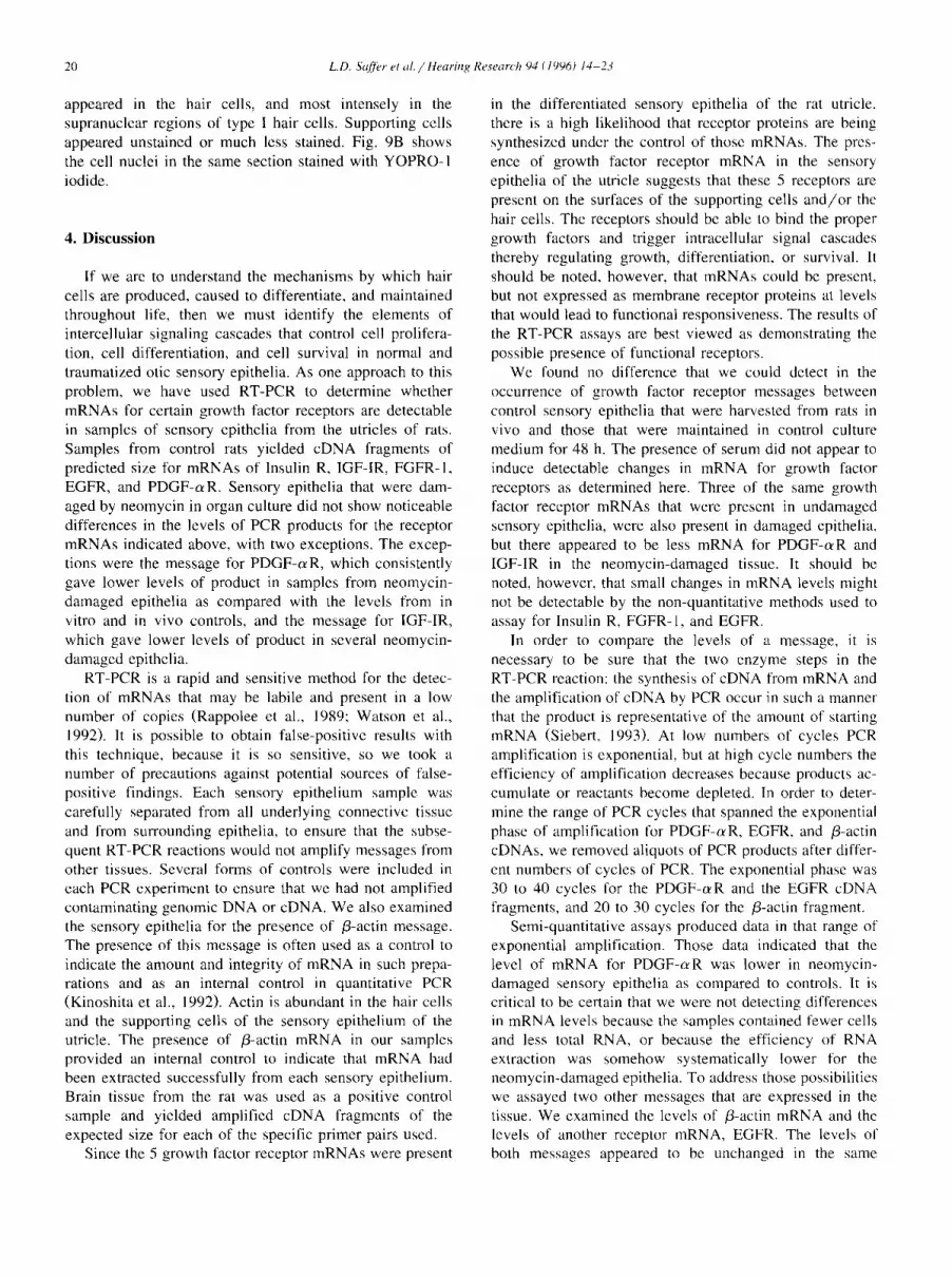
appeared in the hair cells, and most intensely in the supranuclear regions of type I hair cells. Supporting cells appeared unstained or much less stained. Fig. 9B shows the cell nuclei in the same section stained with YOPRO-1 iodide.
4. Discussion
If we are to understand the mechanisms by which hair cells are produced, caused to differentiate, and maintained throughout life, then we must identify the elements of intercellular signaling cascades that control cell prolifera- tion, cell differentiation, and cell survival in normal and traumatized otic sensory epithelia. As one approach to this problem, we have used RT-PCR to determine whether mRNAs for certain growth factor receptors are detectable in samples of sensory epithelia from the utricles of rats. Samples from control rats yielded cDNA fragments of predicted size for mRNAs of Insulin R, IGF-IR, FGFR-1, EGFR, and PDGF-c~R. Sensory epithelia that were dam- aged by neomycin in organ culture did not show noticeable differences in the levels of PCR products for the receptor mRNAs indicated above, with two exceptions. The excep- tions were the message for PDGF-c~R, which consistently gave lower levels of product in samples from neomycin- damaged epithelia as compared with the levels from in vitro and in vivo controls, and the message for IGF-IR, which gave lower levels of product in several neomycin- damaged epithelia.
RT-PCR is a rapid and sensitive method for the detec- tion of mRNAs that may be labile and present in a low number of copies (Rappolee et al., 1989; Watson et al., 1992). It is possible to obtain false-positive results with this technique, because it is so sensitive, so we took a number of precautions against potential sources of false- positive findings. Each sensory epithelium sample was carefully separated from all underlying connective tissue and from surrounding epithelia, to ensure that the subse- quent RT-PCR reactions would not amplify messages from other tissues. Several forms of controls were included in each PCR experiment to ensure that we had not amplified contaminating genomic DNA or cDNA. We also examined the sensory epithelia for the presence of /3-actin message. The presence of this message is often used as a control to indicate the amount and integrity of mRNA in such prepa- rations and as an internal control in quantitative PCR (Kinoshita et al., 1992). Actin is abundant in the hair cells and the supporting cells of the sensory epithelium of the utricle. The presence of /3-actin mRNA in our samples provided an internal control to indicate that mRNA had been extracted successfully from each sensory epithelium. Brain tissue from the rat was used as a positive control sample and yielded amplified cDNA fragments of the expected size for each of the specific primer pairs used.
Since the 5 growth factor receptor mRNAs were present
in the differentiated sensory epithelia of the rat utricle, there is a high likelihood that receptor proteins are being synthesized under the control of those mRNAs. The pres- ence of growth factor receptor mRNA in the sensory epithelia of the utricle suggests that these 5 receptors are present on the surfaces of the supporting cells and/or the hair cells. The receptors should be able to bind the proper growth factors and trigger intracellular signal cascades thereby regulating growth, differentiation, or survival. It should be noted, however, that mRNAs could be present, but not expressed as membrane receptor proteins at levels that would lead to functional responsiveness. The results of the RT-PCR assays are best viewed as demonstrating the possible presence of functional receptors.
We found no difference that we could detect in the occurrence of growth factor receptor messages between control sensory epithelia that were harvested from rats in vivo and those that were maintained in control culture medium for 48 h. The presence of serum did not appear to induce detectable changes in mRNA for growth factor receptors as determined here. Three of the same growth factor receptor mRNAs that were present in undamaged sensory epithelia, were also present in damaged epithelia, but there appeared to be less mRNA for PDGF-c~R and IGF-IR in the neomycin-damaged tissue. It should be noted, however, that small changes in mRNA levels might not be detectable by the non-quantitative methods used to assay for Insulin R, FGFR-1, and EGFR.
In order to compare the levels of a message, it is necessary to be sure that the two enzyme steps in the RT-PCR reaction: the synthesis of cDNA from mRNA and the amplification of cDNA by PCR occur in such a manner that the product is representative of the amount of starting mRNA (Siebert, 1993). At low numbers of cycles PCR amplification is exponential, but at high cycle numbers the efficiency of amplification decreases because products ac- cumulate or reactants become depleted. In order to deter- mine the range of PCR cycles that spanned the exponential phase of amplification for PDGF-c~R, EGFR, and /3-actin cDNAs, we removed aliquots of PCR products after differ- ent numbers of cycles of PCR. The exponential phase was 30 to 40 cycles for the PDGF-oeR and the EGFR cDNA fragments, and 20 to 30 cycles for the /3-actin fragment.
Semi-quantitative assays produced data in that range of exponential amplification. Those data indicated that the level of mRNA for PDGF-aR was lower in neomycin- damaged sensory epithelia as compared to controls. It is critical to be certain that we were not detecting differences in mRNA levels because the samples contained fewer cells and less total RNA, or because the efficiency of RNA extraction was somehow systematically lower for the neomycin-damaged epithelia. To address those possibilities we assayed two other messages that are expressed in the tissue. We examined the levels of /3-actin mRNA and the levels of another receptor mRNA, EGFR. The levels of both messages appeared to be unchanged in the same

L.D. Sqff~r et al. / Hearing Research 94 (1996) 14-23 21
A 13 Fig. 9. A: Localization of PDGF-o~R to the hair cells of an undamaged rat utricle by fluorescent antibody staining. Note the intense fluorescence of the supranuclear regions and the necks of type I hair cells (arrows) and the relative absence of labeling in the supporting cells. B: A view of the same section photographed with different optics to show the cell nuclei counterstained with YOPRO- 1 iodide. The hair cell nuclei form an incomplete row in the apical half of the epithelium (the upFer half in this micrograph). The nuclei of supporting cells form a nearly complete row at the base of the epithelium just above the connective tissue.
samples where the levels of P D G F - a R mRNA showed clear decreases (4 samples). These results suggest that the decrease in PDGF-c~R mRNA that resulted from neomycin treatment was specific to that message and was not the result of decreased total mRNA extraction.
What might this finding mean? From in vitro studies and histology, we know that many hair cells were kil led by the neomycin treatment, whereas the supporting cells ap- peared unharmed. As these experiments proceeded the hypothesis developed that PDGF-o~R is expressed at higher levels in hair cells than in supporting cells. Consequently, the loss of hair cells caused by the neomycin treatment might have resulted in the measured drop in the level of mRNA for PDGF-o~ R. Subsequent application of immuno- histochemistry with an antibody against P D G F - a R sup- ported this idea, revealing much stronger staining in hair cells than in supporting cells (Fig. 9).
PDGF is a dimeric protein composed of two similar chains linked by disulfide bonds. It exists in three forms, the homodimers AA, and BB, and the heterodimer AB. There are two receptors for the isoforms of PDGF, c~ and /3 receptors, which are membrane proteins with an extra- cellular domain that binds the growth factor and an intra- cellular tyrosine kinase domain. Ligand binding causes PDGF receptors to dimerize in the membrane resulting in kinase activation. PDGF-c~R can bind both A and B chains of PDGF, where as PDGF-/3R binds only the B chain. PDGF is a mitogen for connective tissue, glia, and smooth muscle cells. In some fibroblasts, PDGF can stimulate synthesis of collagens and other matrix proteins. PDGF also can have neurotrophic effects (Raines et al., 1991; Smits et al., 1991). Mature hair cells have not been reported to undergo mitosis, so it seems likely that PDGF binding to hair cells, if active, would function in another role.
A convincing argument has been made that all mam- malian cells, with the exception of blastomeres of the early embryo, require extracellular signals from other cells in order to survive (Raf t et al., 1993). Such survival factors bind to cell surface receptors. Cells that do not receive the required amount of survival factors, cells that have lost the receptors for available survival factors, and cells in which the intracellular survival signaling cascades have been blocked all die by programmed cell death (apoptosis). Hair cells in vestibular and auditory sensory epithelia which have been damaged by aminoglycoside antibiotics or by acoustic overstimulation can also die by apoptosis (JCrgen- son, 1991; Li and Forge, 1995; Kil et al., 1995; Mason et al., 1995).
An elegant series of experiments on the growth and survival requirements of glial cells in the optic nerve have demonstrated that PDGF can promote the survival of immature ol igodendrocytes by preventing activation of the apoptotic suicide program which would kill cells in the absence of such a factor (Barres and Raft, 1994). By analogy it seems reasonable to hypothesize that the indica- tions of mRNA and protein for the PDGF-o~R observed in hair cells may relate to a survival control function for extracellular PDGF in this cell type. In the future it will be important to test that hypothesis experimentally.
Evidence from PCR at 35 cycles and preliminary evi- dence from semi-quantitative PCR have suggested that mRNA for IGF-IR also decreases in utricular sensory epithelia along with decreases in the number of hair cells that survive after neomycin treatment. Like PDGF, IGF-I is a known survival factor for immature oligodendrocytes, but it does not induce proliferation of those cells or their precursors. IGF-I also has survival promoting effects on cells from the kidney and motomeurons (Raf t et al., 1993). In view of the survival promoting effects of IGF-I in those

22 L.D. Sa~]~'r el al. / Hearing,, Re,~earch 94 (1996) 14 .23
cell types and evidence for decreased levels of IGF-IR mRNA occurring along with neomycin-induced loss of hair cells, it seems reasonable to suggest that IGF-I may have a role in the normal survival of hair cells in the mammalian ear. Hair cell loss in response to a variety of insults can occur via an apoptotic mechanism which de- pends on the activation of a suicide program within the cell. As the results of this investigation have shown, hair cells express receptors specific for extracellular signaling factors that are known to prevent the execution of the apoptotic suicide program in other cell types. The results suggest the potential for the development of pharmacologi- cal methods to protect ears from the loss of hair cells caused by traumatic insults. Age-related loss of hair cell may also be preventable by the development of such survival factor therapeutics if they can contribute to the replacement of decreased levels of normal survival signals or provide augmentation of the resistance of hair cells to insults that might otherwise trigger the activation of an apoptotic suicide program.
Acknowledgements
This work was supported by a grant from the National Organization for Hearing Research and Grant RO I- DC00200 from the National Institute on Deafness and other Communication Disorders to J.T.C. We are indebted to Federico Gonzalez-Fernandez for guidance, comments, and use of equipment. We also wish to acknowledge Jim Finley's fine immunohistochemistry. We thank Mark War- chol and Mark Marchioni for helpful discussions and Ellen Van Niel, James Finley, Bet Xia, and Met Deng for many and various fol~ms of help. We thank Pam Neff/'or instruc- tion and assistance in image analysis for quantification of cDNA in gels, and we thank Joel Linden for use of the Eagle Eye image capture system.
References
Barres, B.A. and Raft, M.C. (1994) Control of oligodcndrocytc number in the developing rat optic nerve. Neuron 12, 935 942.
Baird, R.A. and Torres, M.A. (1993) Hair cell regeneration in the vestibular otolith organs following arninoglycoside toxicity. Hear. Res. 65, 164 174.
Balak, K.J., Corwin, J.T. and Jones, J.E. (1990) Regenerated hair cells can originate from supporting cell progeny: evidence from photoxicity and laser ablation experiments in the lateral line system. J. Neurosci. 10. 2502-2512.
Chomczynski, P. and Sacchi. N. (1987) A rapid method of RNA isolation by acid guanidinium thiocyanate-phenol-chloroform extraction. Anal. Biochem. 162, 156-159.
Corwin. J.T. (1981) Postembryonic production and aging of inner car hair cells in sharks. J. Comp. Neurol. 201, 541-553.
Corwin. J.T. (1983) Postembryonic growth of the macula neglecta audi- tory detector in the ray, Raja clalata: continual increase m hair cell number, neural convergence, and physiological sensitivity. J. Comp. Neurol. 217. 345-356.
Corwin. J.T. (1985) Perpetual production of hair cells and maturational changes in hair cell ultrastructure accompany postembryonic growth in an amphibian ear. Prec. Natl. Acad. Sci. USA 82. 3911-3915.
Corwin, J.T. (1986) Regeneration and self-repair in hair cell epithelia experimental evaluation of capacities and limitations. In: R.J. Ruben, T.R. Van DeWater and E.W Rubel (Eds.). Biology of Change in Otolaryngology, Elsevier, New York. pp. 291-304.
Corwin, J.T. and Cotanche, D.A. (1988) Regeneration of sensory hair cells after acoustic trauma. Science 240, 1772-1774.
Corwin, J.T., Finley. J.E., SalTer, L.. Gu, R.. Cunningham, L., Xia. B. and Warchol. M. (1995) Isolation of pure living hair cell epithelia b,~ use of thermolysin. Assoc. Res. Otolaryngol. Abstr. 18, 87.
Cotanche, D.A. (1987} Regeneration of the hair cell stereociliary bundles in the chick cochlea following severe acoustic trauma. Hear. Res. 30. 181-196.
Forge, A., Lin, L., Corwin, J.T. and Nevill. G. (1993) Ultrastrucmral evidence for hair cell regeneration in the mammalian inner ear. Science 259, 1616-1619.
Germain. L.. Rouabhia, M. Guignard, R,. Carrier. L,, Bou~ard, V.. and Auger, F.A, (1993). hnprovement of human keratinocyte isolation and culture using thermolysin. Burns 19, 99-104.
Girod. D.A.. Duckerl, L.G. and Rubel. E.W (1989) Identification of potential regenerating hair cell precursors in the avian cochlea follow- ing acoustic trauma. Assoc. Res. Otolaryngol. Abstr. 12, 84-85.
Jones, J.E. and Cor~,~in. J.T. (1993) Replacelnent o[" lateral line sensory organs during tail regeneration in sahunanders: identificalion of pro- genitor cells and analysis of leukocyte activity. J. Neurosci. 13, 1022 [034.
Jorgcnson..I.M. (I 991) Regeneration of lateral line and inner ear vestibu- lar cells. In: Regeneration of Vertebrate Sensor? Receptor Cells. Wiley. Chichester (Ciba Found. Syrup. 160) pp. 151-170.
Jorgcnson. J.M. and Mathiesen. C. (1988) The avian inner car: continu- ous production of hair cells in vestibular sensory organs, but not in the auditory papilla. Naturwissenschafien 75. 319-320.
Katayama, A. and Corwin, J,T. (1989) Cell production in the chicken cochlea. J. Comp. Neurol. 28 l. 129-135.
Kil. J.. Warchol. M.E. and Corwin, J.T. (1995) On-going and aminogly~ coside induced apoptotic cell death in the vestibular cpilhelia of chicks. Assoc. Rcs. ()tolaryng. Abstr. 18. 82.
Kinoshila. T., lmanmra. J,, NaTal. H. and Shimotohno. K. (1992)Quan tification of gone expression o'~er a wide range by' thc polymerase chain reaction. Anal. Biochcm. 206, 231-235.
Li. L.. Nevill. G. and Forge, A. (1995) Two modes of hair cell loss from the vestibular sensory epithelia of Ihe guinea pig inner car. J. Getup. Neurol. 355. 4{)5 417.
Lombarte. A.. Yan. H,Y., Popper, A.N.. Chang, J.S. and Platt, C. (1993) Damage and regeneration of hair cell ciliary bundles in a fish car following treatmcm with geulamiciu. Hear. Res. 64. 166-174.
Mason, J.C.. Kil, J. Warchol. M.E. and Corwin. J.T. (1995) Acoustic traunm resulls in apoptotic death of cochlear hair cells. Soc. Neurosci. Abstr. 20. 396.
McKay, 1. and Leigh, I. (1993)Growth Factors: a Practical Approach. Oxford University Press, New York. NY.
Pardee, A.B. (1989)GI events and regulation of cell proliferation. Science 246. 603-608.
Pedrini, M.T., Giorginow. F. and Sinith. R.J. (1994) cDNA cloning of the rat IGF1 receptor: structural analysis of rat and htmlan IGF-1 and insulin rcceplors reveal differences in ahernative splicing and receptor specific domain conservation. Bio. Biophys. Res. Com. 2{)2. 1038 [ (140.
Pletsch. L.A., Harris. J., Raymond. V.W., I~lasband. A., l.ec, D.C. and Earp, H.S. (1990) A truncated secreted form of the epidermal growth factor receptor is encoded by an alternatively spliced transcript in normal rat tissue, Mol. Cell. Biol, 10, 2973 2982.
Raff. M,C.. Barres, B.A.. Burne. J.F., Coles. H.S., lshizaki, Y. and Jacobsen, M.D. 11993) Programmed cell dcalh and the control of cell sur\ival: lessons from the nervous system. Science 262, (~95-7()().

L.D. Sr(~'er el a l . / Hearing Research 94 (1996) 14-23 23
Raines, E.W., Bowen-Pope, D,F. and Ross, R. (1991) Platelet-derived growth factor. In: M.B. Sporn and A.B. Roberts (Eds.), Peptide Growth Factors and Their Receptors I, Springer, New York, NY.
Raphael, Y. (1992) Evidence for supporting cell mitosis in response to acoustic trauma in the avian inner ear. J. Neurocytol. 21, 663-671.
Raphael, Y. (1993) Reorganization of the chick basilar papilla after acoustic trauma. J. Comp. Neurol. 330, 521-532.
Rappolee, D.A., Wang, A., Mark, D. and Werb, Z. (1989) Novel method for studying mRNA phenotypes in single or small numbers of cells. J. CelI Biochem. 39. I 11.
Ruben, R.J. (1967) Development of the inner ear of the mouse: a radioautographic study of terminal mitoses. Acta Otolarnygol. Suppl. 220, 1-44.
Ryals, B,M. and Rubel, E.W (1988) Hair cell regeneration after acoustic trauma in adult Coturnix quail. Science 240. 1774-1776.
Siebert, P.D. (1993) In: Y. Munch, K. Mayo and A. Miller (Eds.), Quantitative RT-PCR: Methods and Applications, Clontech, Palo Alto, CA.
Smits, A,, Kato, M., Westermark, B., Nister, M., Heldin, C. and Funa, K. ( 1991 ) Neurotrophic activity of platelet-derived growth factor (PDGF): rat neural cells possess functional PDGF /3-type receptors and re- sponse to PDGF. Proc. Natl. Acad. Sci. USA 88, 8159-8163.
Warchol. M.E., Lambert, P.A., Goldstein, B.J., Forge, A. and Corwin~ J.T. (1993) Regenerative proliferation in inner ear sensory epithelia from adult guinea pigs and humans. Science 259, 1619-1622.
Watson, A.J., Hogan, A., Hahnel, A., Wiemer, K.E. and Schultz, G.A. (1992) Expression of growth factor ligand and receptor genes in the preimplantation bovine embryo. Mol. Reprod. Dev. 31, 87-95.
Weisleder, P. and Rubel, E.W (1993) Hair cell regeneration after strepto- mycin toxicity in the avian vestibular epithelium. J. Comp. Neurol. 331, 97-110.
Yazaki, N., Fujita, H., Ohta, M., Kawasaki, T. and Itoh, N. (1993) The structure and expression of the FGF receptor- 1 mRNA isoforms in rat tissues. Biochim. Biol. Acta 1172, 37-42.


