Embed Size (px)
Citation preview
Analysis of the Substrate Specificity of the Dim-5 Histone LysineMethyltransferase Using Peptide Arrays
Philipp Rathert1, Xing Zhang2, Christian Freund3, Xiaodong Cheng2, and Albert Jeltsch1,*1Biochemistry Laboratory, School of Engineering and Science, Jacobs University Bremen, CampusRing 1, 28759 Bremen, Germany2Department of Biochemistry, Emory University School of Medicine, 1510 Clifton Road, Atlanta, GA30322, USA3Leibniz-Institut für Molekulare Pharmakologie, Robert-Rössle-Str. 10, 13125 Berlin, Germany
SUMMARYHistone methylation is an epigenetic mark essential for gene regulation and development. Weintroduce peptide SPOT synthesis to study sequence specificity of the Dim-5 histone-3 lysine-9methyltransferase. Dim-5 recognizes R8-G12 of the H3 tail with T11 and G12 being the mostimportant specificity determinants. Exchange of H3 tail residue S10 and T11 by E strongly reducedmethylation by Dim-5, suggesting that phosphorylation of S10 or T11 may regulate the activity ofDim-5. In the Dim-5/peptide structure, E227 interacts with H3R8 and D209 with H3-S10. Mutationsof E227 or D209 caused predictable changes in the substrate preference, illustrating that peptiderecognition of histone methyltransferases can be altered by protein design. Comparative analyses ofpeptide arrays with wild-type and mutant enzymes, therefore, are well suited to investigate the targetspecificity of protein methyltransferases and study epigenetic crosstalk.
INTRODUCTIONEpigenetic regulation by covalent modification of histone proteins and methylation of DNAcontrols gene activity during development and disease processes (Egger et al., 2004; Feinbergand Tycko, 2004; Jones and Baylin, 2002; Li, 2002). Histones are posttranslationally modifiedby various enzymatic reactions including methylation, primarily at their flexible N termini(Berger, 2007; Margueron et al., 2005). Methylation of lysine residues occurs in histone H3 atresidues K4, K9, K27, and K36; in histone H4 at K20; and in histone H1b at K26. All thesemodifications have different biological functions (Berger, 2007; Martin and Zhang, 2005); forexample, H3K9 methylation leads to condensation of the chromatin and inhibition of geneexpression, H3K27 methylation is correlated to gene silencing, while H3K4 methylation marksactive chromatin (Li et al., 2007).
Most histone lysine methyltransferases (HKMTs) contain a SET (Su(var)3−9, Enhancer-of-zeste, Trithorax) domain which comprises approximately 130 amino acids and harbors theactive center of the enzymes (Cheng et al., 2005). Different HKMTs not only vary in their
© 2008 Elsevier Ltd All rights reserved*Correspondence: [email protected] DataSupplemental Data include one figure that shows an example of Bromophenol blue stained peptide membrane and SupplementalExperimental Procedures that give a detailed description of data analysis and are available with this article online athttp://www.chembiol.com/cgi/content/full/15/1/5/DC1/.
NIH Public AccessAuthor ManuscriptChem Biol. Author manuscript; available in PMC 2009 August 10.
Published in final edited form as:Chem Biol. 2008 January ; 15(1): 5–11. doi:10.1016/j.chembiol.2007.11.013.
NIH
-PA Author Manuscript
NIH
-PA Author Manuscript
NIH
-PA Author Manuscript
substrate specificity but also in product pattern because lysine can be mono-, di-, ortrimethylated (Zhang et al., 2003). Here, we investigate the target specificity of the H3K9Dim-5 HKMT from Neurospora crassa. The enzyme generates trimethylated H3K9(H3K9me3) in a processive reaction (Zhang et al., 2003) that controls DNA methylation in N.crassa (Selker et al., 2002; Tamaru and Selker, 2001; Tamaru et al., 2003). The three-dimensional structure of the protein alone and in complex with a peptide has been solved andallowed to identify the directly interacting moieties in atomic detail (Zhang et al., 2002,2003).
While the sequence specificity of substrate and ligand interaction of proteases (Overall,2002), kinases (Kreegipuu et al., 1998), and antibodies (Mariuzza et al., 1987) has beenanalyzed comprehensively, such in-depth studies have not yet been carried out for HKMTsbecause a large number of different peptides have to be analyzed to derive a completespecificity profile for one enzyme. Peptide array SPOT synthesis on cellulose membranes hadbeen introduced to prepare large libraries of different peptides at amounts sufficient forbiochemical assays and at moderate costs (Frank, 2002; Hilpert et al., 2007; Reineke et al.,2001; Wenschuh et al., 2000). The method was used to analyze the specificity of kinases andproteases (Hohne and Hilpert, 2005; Tegge and Frank, 1998) and to study the bindingspecificity of protein/protein interaction (Bialek et al., 2003; Frank, 2002; Hilpert et al.,2000, 2007). Here, we demonstrate that peptide arrays are an ideal approach to analyze thesubstrate specificity of HKMTs.
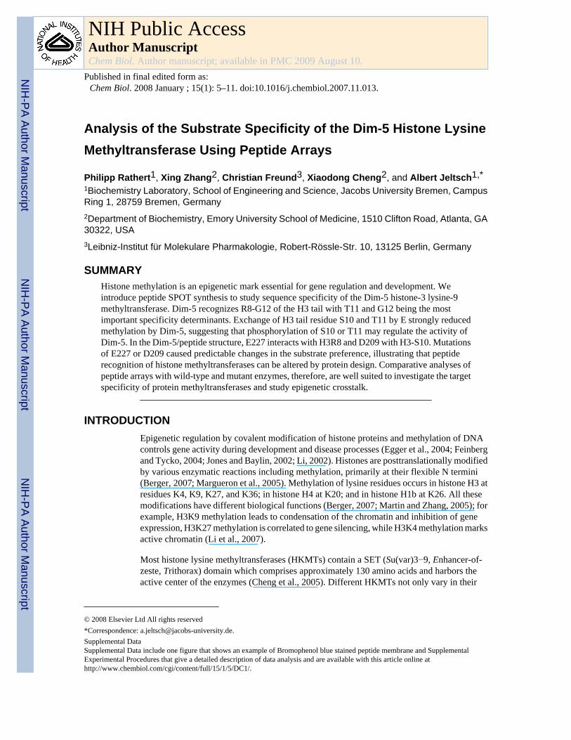
RESULTS AND DISCUSSIONWe synthesized 21-residue peptides on a functionalized cellulose membrane by the SPOTsynthesis method (Frank, 2002; Reineke et al., 2001; Wenschuh et al., 2000). The methylationof the respective substrates was analyzed by following the enzymatic transfer of radioactivelylabeled methyl groups from the coenzyme S-adenosyl-L-methionine (AdoMet) to theimmobilized peptides. As a pilot experiment, several spots with the sequence of the first 21amino acids of the N-terminal tail of histone H3 and spots of the K9A peptide variant in whichthe target lysine had been replaced by alanine were prepared. After incubation with Dim-5, aclear methylation signal was detected at the H3 peptides, while no methylation occurred onH3 K9A peptides (Figure 1A). The methylation signal at H3 peptides was linearly increasingwith incubation time and enzyme concentration (Figures 1B and 1C). To analyze if the activityof Dim-5 on immobilized substrates is comparable with data obtained in solution, wedetermined the Km value for AdoMet by using H3 tail peptides as substrates either bound tothe cellulose membrane or in solution. For both experiments, similar Km values between 1.2and 1.3 μM were obtained (data not shown).
Substrate Specificity of Dim-5To study the influence of each residue on peptide recognition by Dim-5, an alanine scanningexperiment was performed by synthesizing a small array of 21 peptides each carrying anexchange of a single residue against alanine (Figure 1D). The reduced methylation of peptidescarrying substitutions at positions 8−12 demonstrated an important role of R8, K9 (the targetof methylation), S10, T11, and G12 in the peptide recognition by Dim-5. The residualmethylation activities were measured on four independent membranes, and standard deviationswere determined (Figure 1E). The average standard error in all four experiments was ± 7%;standard errors of individual substrates were generally smaller than ± 15%, indicating that theassay is reliable and accurate. We conclude that the peptide methylation assay on solid supportallows rapid methylation analysis of several target peptides. One inherent advantage of themethod is that all peptide spots are methylated in competition, which ensures that equalamounts of active enzyme and cofactor are available for all substrates. Under the experimentalconditions used here, the relative rates of methylation correspond to ratios of kcat/Kd values
Rathert et al. Page 2
Chem Biol. Author manuscript; available in PMC 2009 August 10.
NIH
-PA Author Manuscript
NIH
-PA Author Manuscript
NIH
-PA Author Manuscript
for the respective peptide, the latter representing an established parameter for quantificationof enzyme specificity (Fersht, 1998) (Supplemental Experimental Procedures, see theSupplemental Data available with this article online).
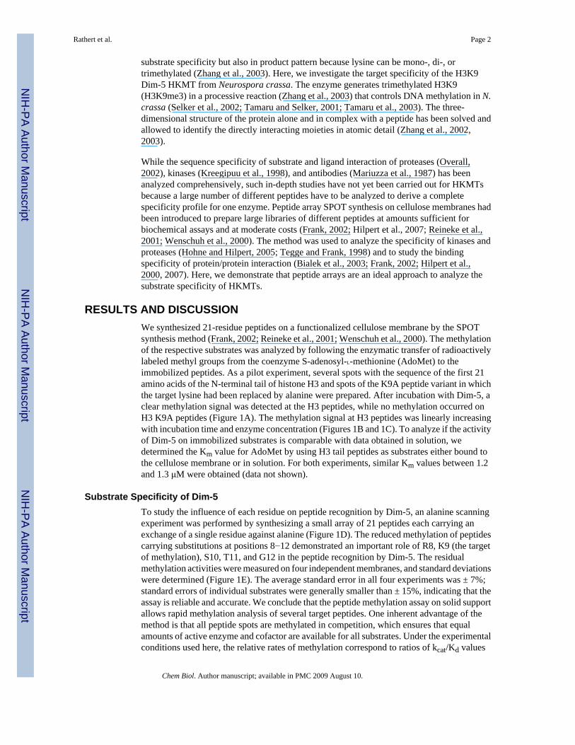
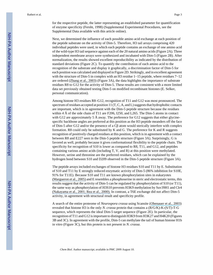
Next, we determined the influence of each possible amino acid exchange at each position ofthe peptide substrate on the activity of Dim-5. Therefore, H3 tail arrays comprising 420individual peptides were used, in which each peptide contains an exchange of one amino acidof the wild-type H3 tail sequence against each of the 20 natural amino acids (Figure 2A). Threeindependent membrane arrays were synthesized and incubated with Dim-5 (Figure 2B). Afternormalization, the results showed excellent reproducibility as indicated by the distribution ofstandard deviations (Figure 2C). To quantify the contribution of each amino acid to therecognition of the substrate and display it graphically, a discrimination factor of Dim-5 foreach position was calculated and displayed in Figure 2D. Strikingly, and in excellent agreementwith the structure of Dim-5 in complex with an H3 residue 1−15 peptide, where residues 7−12are ordered (Zhang et al., 2003) (Figure 3A), the data highlights the importance of substrateresidues R8 to G12 for the activity of Dim-5. These results are consistent with a more limiteddata set previously obtained testing Dim-5 on modified recombinant histones (E. Selker,personal communication).
Among histone H3 residues R8–G12, recognition of T11 and G12 was most pronounced. Thespectrum of residues accepted at position 11 (T, C, A, and L) suggests that hydrophobic contactsare important, which is in agreement with the Dim-5 peptide structure because the residueswithin 4 Å of the side chain of T11 are F206, I250, and L265. The Dim-5 atoms in contactwith G12 are approximately 5 Å away. The preference for G12 suggests that either glycine-specific backbone angles are preferred at this position as the H3 peptide meanders off the faceof Dim-5 after G12 and/or the presence of a Cβ atom would sterically interfere with complexformation. R8 could only be substituted by K and G. The preference for K and R suggestsrecognition of positively charged residues at this position, which is in agreement with a contactbetween R8 and E227 seen in the Dim-5 peptide structure (Figure 3A). Surprisingly, G isfavored as well, probably because it gives conformational flexibility to the peptide chain. Thespecificity for recognition of S10 is lower as compared to R8, T11, and G12, and peptidescontaining various amino acids (including T, V, and R) at this position were methylated.However, serine and threonine are the preferred residues, which can be explained by thehydrogen bond between S10 and D209 observed in the Dim-5 peptide structure (Figure 3A).
The peptide arrays included exchanges of histone H3 residues S10 and T11 by E. Substitutionof S10 and T11 by E strongly reduced enzymatic activity of Dim-5 (86% inhibition for S10E,91% for T11E). Because S10 and T11 are known phosphorylation sites in eukaryotes(Margueron et al., 2005) and E resembles a phosphoserine in steric and electrostatic terms, thisresults suggest that the activity of Dim-5 can be regulated by phosphorylation of S10 (or T11),the same way as phosphorylation of H3S10 prevents H3K9 methylation by Suv39H1 and Clr4(Nakayama et al., 2001; Rea et al., 2000). In contrast, a T6E exchange did not affect Dim-5activity, in agreement with structural result and specificity profile.
A search of the entire proteome of Neurospora crassa using Scansite (Obenauer et al., 2003)revealed that histone H3 is the only N. crassa protein that contains a (R/G/K)-K-(S/T)-T-Gsequence, which represents the ideal Dim-5 target sequence (Figure 2E). In particular, therecognition of T11 and G12 is important to distinguish H3K9 from H3K27 and H4K20 (Figures3B and 3C). In agreement with the profile, Dim-5 can methylate the tail of human histone H1bin vitro (Figure 3C), but this protein is not present in N. crassa.
Rathert et al. Page 3
Chem Biol. Author manuscript; available in PMC 2009 August 10.
NIH
-PA Author Manuscript
NIH
-PA Author Manuscript
NIH
-PA Author Manuscript
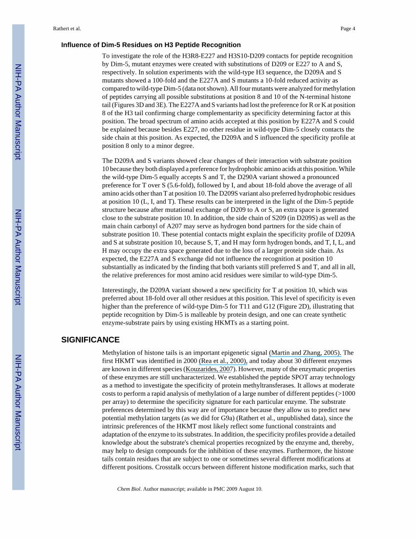
Influence of Dim-5 Residues on H3 Peptide RecognitionTo investigate the role of the H3R8-E227 and H3S10-D209 contacts for peptide recognitionby Dim-5, mutant enzymes were created with substitutions of D209 or E227 to A and S,respectively. In solution experiments with the wild-type H3 sequence, the D209A and Smutants showed a 100-fold and the E227A and S mutants a 10-fold reduced activity ascompared to wild-type Dim-5 (data not shown). All four mutants were analyzed for methylationof peptides carrying all possible substitutions at position 8 and 10 of the N-terminal histonetail (Figures 3D and 3E). The E227A and S variants had lost the preference for R or K at position8 of the H3 tail confirming charge complementarity as specificity determining factor at thisposition. The broad spectrum of amino acids accepted at this position by E227A and S couldbe explained because besides E227, no other residue in wild-type Dim-5 closely contacts theside chain at this position. As expected, the D209A and S influenced the specificity profile atposition 8 only to a minor degree.
The D209A and S variants showed clear changes of their interaction with substrate position10 because they both displayed a preference for hydrophobic amino acids at this position. Whilethe wild-type Dim-5 equally accepts S and T, the D290A variant showed a pronouncedpreference for T over S (5.6-fold), followed by I, and about 18-fold above the average of allamino acids other than T at position 10. The D209S variant also preferred hydrophobic residuesat position 10 (L, I, and T). These results can be interpreted in the light of the Dim-5 peptidestructure because after mutational exchange of D209 to A or S, an extra space is generatedclose to the substrate position 10. In addition, the side chain of S209 (in D209S) as well as themain chain carbonyl of A207 may serve as hydrogen bond partners for the side chain ofsubstrate position 10. These potential contacts might explain the specificity profile of D209Aand S at substrate position 10, because S, T, and H may form hydrogen bonds, and T, I, L, andH may occupy the extra space generated due to the loss of a larger protein side chain. Asexpected, the E227A and S exchange did not influence the recognition at position 10substantially as indicated by the finding that both variants still preferred S and T, and all in all,the relative preferences for most amino acid residues were similar to wild-type Dim-5.
Interestingly, the D209A variant showed a new specificity for T at position 10, which waspreferred about 18-fold over all other residues at this position. This level of specificity is evenhigher than the preference of wild-type Dim-5 for T11 and G12 (Figure 2D), illustrating thatpeptide recognition by Dim-5 is malleable by protein design, and one can create syntheticenzyme-substrate pairs by using existing HKMTs as a starting point.
SIGNIFICANCEMethylation of histone tails is an important epigenetic signal (Martin and Zhang, 2005). Thefirst HKMT was identified in 2000 (Rea et al., 2000), and today about 30 different enzymesare known in different species (Kouzarides, 2007). However, many of the enzymatic propertiesof these enzymes are still uncharacterized. We established the peptide SPOT array technologyas a method to investigate the specificity of protein methyltransferases. It allows at moderatecosts to perform a rapid analysis of methylation of a large number of different peptides (>1000per array) to determine the specificity signature for each particular enzyme. The substratepreferences determined by this way are of importance because they allow us to predict newpotential methylation targets (as we did for G9a) (Rathert et al., unpublished data), since theintrinsic preferences of the HKMT most likely reflect some functional constraints andadaptation of the enzyme to its substrates. In addition, the specificity profiles provide a detailedknowledge about the substrate's chemical properties recognized by the enzyme and, thereby,may help to design compounds for the inhibition of these enzymes. Furthermore, the histonetails contain residues that are subject to one or sometimes several different modifications atdifferent positions. Crosstalk occurs between different histone modification marks, such that
Rathert et al. Page 4
Chem Biol. Author manuscript; available in PMC 2009 August 10.
NIH
-PA Author Manuscript
NIH
-PA Author Manuscript
NIH
-PA Author Manuscript
methylation of H3K9 precludes H3K4 methylation or phosphorylation of H3S10 (Margueronet al., 2005) and methylation of H3R2 prevents H3K4 methylation (Guccione et al., 2007;Kirmizis et al., 2007). Such epigenetic crosstalk can occur at the level of the modificationreading domains or the modifying enzymes. For example, H3R8, which is an importantspecificity determinant of Dim-5, can be methylated by Prmt5 (Pal et al., 2004)in vivo. Thepeptide spot array method is well suited to investigate the sensitivity of HKMTs towardmutations on the histone tails—that can be easily expanded to include pre-existingposttranslational modifications. This allows to decode the effect of the complexposttranslational modification pattern of histone tails on HKMT activity and is, therefore, ofgreat value for an understanding of the cellular role of these enzymes.
EXPERIMENTAL PROCEDURESEnzyme Purification—Dim-5 wild-type and variants were expressed and purified asdescribed previously (Zhang et al., 2002). The E. coli strain XL1 blue was used as cloning hostfor Dim-5 variants. The plasmid pXC379 (Zhang et al., 2002) encoding Dim-5 was mutatedat different positions to yield Dim-5 D209A/S and E227A/S variants by site-directedmutagenesis methods (Jeltsch and Lanio, 2002). All mutants were sequenced to verify thepresence of the intended mutation and the absence of additional mutations.
Peptide Methylation in Solution—A synthetic peptide corresponding to the first 19 aminoacids of histone H3 tail plus a methionine (Bt-MARTKQTARKSTGGKAPRKQ), whichcontains a biotin at its N terminus, was purchased from IRIS Biotech (Marktredwitz, Germany)in HPLC-purified form and was dissolved in water. Purity of the peptide was greater than 95%,as confirmed by HPLC and MALDI-TOF mass-spectrometric analysis. Methylation reactionson micro plates were carried out as described (Gowher et al., 2005).
Synthesis of Peptide Spot Arrays—Peptide arrays were synthesized as described by theSPOT synthesis method (Frank, 2002; Wenschuh et al., 2000). Successful synthesis of eachpeptide was confirmed by bromphenol blue staining of the membranes (Figure S1). The peptidespots used in our assay had diameters of 2 mm and contained approximately 9 nmol of peptide(Autospot Reference Handbook, Intavis AG).
Methylation of Peptide Spot Arrays—For methylation, the membranes containing 420peptide spots were washed for 20 min in methylation buffer containing 50 mM glycine (pH9.8), 2 mM dithiothreitol (DTT), 25 μg/ml BSA, 10% glycerol, and subsequently incubatedwith 20 nM enzyme in methylation buffer at ambient temperature in the presence of 0.35 μMlabeled [methyl-3H]-AdoMet (2.93 × 10¹⁵ Bq/mol) (NEN Life Sciences). After 45 min, themembranes were washed four times with 50 mM NH4HCO3, dried between whatman papers(Whatman GmbH, Dassel, Germany), and washed once with Amplify NAMP100V solution(GE Healthcare, Munich, Germany). The membranes were incubated on Hyperfilm highperformance autoradiography films (GE Healthcare, Munich, Germany) in the dark. After 3−7 days, the films were developed by using AGFA Curix 60 developing machine (AgfaDeutschland Vertriebsgesellschaft mbH & Co. KG, Cologne, Germany).
Data Analysis—All experiments were carried out at least in triplicate. Data are reported asmean values and standard errors. To compare the accuracy of recognition of each residue inthe substrate quantitatively, the relative contribution of each amino acid i at position x forpeptide recognition was calculated by a discrimination factor D:
Rathert et al. Page 5
Chem Biol. Author manuscript; available in PMC 2009 August 10.
NIH
-PA Author Manuscript
NIH
-PA Author Manuscript
NIH
-PA Author Manuscript
where vi is the rate of modification of peptide carrying amino acid i and is the average rateof methylation of all 19 peptides carrying a different amino acid j ≠ i at position x (includingthe wild-type sequence). For example in Figure 2D, the discrimination factor of 10 for a Thrat position 11 indicates that the peptide with Thr at that position is methylated ten times fasterthan the average of all peptides carrying any of the other amino acids at this site. Since thedetection limit of the experiments was at about 3% of the full activity, the discrimination factorfor K9, which could not be replaced by any other residue, was 30.
Supplementary MaterialRefer to Web version on PubMed Central for supplementary material.
ACKNOWLEDGMENTSThis work has been supported by National Institutes of Health GM068680 and the Bundesministerium fuer Bildungund Forschung Biofuture program. We thank C. Behn (Intavis AG) for technical advice. Technical assistance by M.Schwerdtfeger is gratefully acknowledged.
REFERENCESBerger SL. The complex language of chromatin regulation during transcription. Nature 2007;447:407–
412. [PubMed: 17522673]Bialek K, Swistowski A, Frank R. Epitope-targeted proteome analysis: towards a large-scale automated
protein-protein-interaction mapping utilizing synthetic peptide arrays. Anal. Bioanal. Chem2003;376:1006–1013. [PubMed: 12677339]
Cheng X, Collins RE, Zhang X. Structural and sequence motifs of protein (histone) methylation enzymes.Annu. Rev. Biophys. Biomol. Struct 2005;34:267–294. [PubMed: 15869391]
Egger G, Liang G, Aparicio A, Jones PA. Epigenetics in human disease and prospects for epigenetictherapy. Nature 2004;429:457–463. [PubMed: 15164071]
Feinberg AP, Tycko B. The history of cancer epigenetics. Nat. Rev. Cancer 2004;4:143–153. [PubMed:14732866]
Fersht, A. Structure and Mechanism in Protein Science. W.H. Freeman and Company; New York: 1998.Frank R. The SPOT-synthesis technique. Synthetic peptide arrays on membrane supports—principles
and applications. J. Immunol. Methods 2002;267:13–26. [PubMed: 12135797]Gowher H, Zhang X, Cheng X, Jeltsch A. Avidin plate assay system for enzymatic characterization of a
histone lysine methyltransferase. Anal. Biochem 2005;342:287–291. [PubMed: 15935324]Guccione E, Bassi C, Casadio F, Martinato F, Cesaroni M, Schuchlautz H, Luscher B, Amati B.
Methylation of histone H3R2 by PRMT6 and H3K4 by an MLL complex are mutually exclusive.Nature 2007;449:933–937. [PubMed: 17898714]
Hilpert K, Hansen G, Wessner H, Schneider-Mergener J, Hohne W. Characterizing and optimizingprotease/peptide inhibitor interactions, a new application for spot synthesis. J. Biochem2000;128:1051–1057. [PubMed: 11098149]
Hilpert K, Winkler DF, Hancock RE. Peptide arrays on cellulose support: SPOT synthesis, a time andcost efficient method for synthesis of large numbers of peptides in a parallel and addressable fashion.Nat. Protoc 2007;2:1333–1349. [PubMed: 17545971]
Hohne W, Hilpert K. Unraveling sub-site specificities of peptidic serine protease inhibitors bysubstitutional and structural analysis. Protein Pept. Lett 2005;12:449–456. [PubMed: 16029157]
Jeltsch A, Lanio T. Site-directed mutagenesis by polymerase chain reaction. Methods Mol. Biol2002;182:85–94. [PubMed: 11768980]
Jones PA, Baylin SB. The fundamental role of epigenetic events in cancer. Nat. Rev. Genet 2002;3:415–428. [PubMed: 12042769]
Kirmizis A, Santos-Rosa H, Penkett CJ, Singer MA, Vermeulen M, Mann M, Bahler J, Green RD,Kouzarides T. Arginine methylation at histone H3R2 controls deposition of H3K4 trimethylation.Nature 2007;449:928–932. [PubMed: 17898715]
Rathert et al. Page 6
Chem Biol. Author manuscript; available in PMC 2009 August 10.
NIH
-PA Author Manuscript
NIH
-PA Author Manuscript
NIH
-PA Author Manuscript
Kouzarides T. Chromatin modifications and their function. Cell 2007;128:693–705. [PubMed:17320507]
Kreegipuu A, Blom N, Brunak S, Jarv J. Statistical analysis of protein kinase specificity determinants.FEBS Lett 1998;430:45–50. [PubMed: 9678592]
Li B, Carey M, Workman JL. The role of chromatin during transcription. Cell 2007;128:707–719.[PubMed: 17320508]
Li E. Chromatin modification and epigenetic reprogramming in mammalian development. Nat. Rev.Genet 2002;3:662–673. [PubMed: 12209141]
Margueron R, Trojer P, Reinberg D. The key to development: interpreting the histone code? Curr. Opin.Genet. Dev 2005;15:163–176. [PubMed: 15797199]
Mariuzza RA, Phillips SE, Poljak RJ. The structural basis of antigen-antibody recognition. Annu. Rev.Biophys. Biophys. Chem 1987;16:139–159. [PubMed: 2439094]
Martin C, Zhang Y. The diverse functions of histone lysine methylation. Nat. Rev. Mol. Cell Biol2005;6:838–849. [PubMed: 16261189]
Nakayama J, Rice JC, Strahl BD, Allis CD, Grewal SI. Role of histone H3 lysine 9 methylation inepigenetic control of heterochromatin assembly. Science 2001;292:110–113. [PubMed: 11283354]
Obenauer JC, Cantley LC, Yaffe MB. Scansite 2.0: proteome-wide prediction of cell signalinginteractions using short sequence motifs. Nucleic Acids Res 2003;31:3635–3641. [PubMed:12824383]
Overall CM. Molecular determinants of metalloproteinase substrate specificity: matrix metalloproteinasesubstrate binding domains, modules, and exosites. Mol. Biotechnol 2002;22:51–86. [PubMed:12353914]
Pal S, Vishwanath SN, Erdjument-Bromage H, Tempst P, Sif S. Human SWI/SNF-associated PRMT5methylates histone H3 arginine 8 and negatively regulates expression of ST7 and NM23 tumorsuppressor genes. Mol. Cell. Biol 2004;24:9630–9645. [PubMed: 15485929]
Rea S, Eisenhaber F, O'Carroll D, Strahl BD, Sun ZW, Schmid M, Opravil S, Mechtler K, Ponting CP,Allis CD, et al. Regulation of chromatin structure by site-specific histone H3 methyltransferases.Nature 2000;406:593–599. [PubMed: 10949293]
Reineke U, Volkmer-Engert R, Schneider-Mergener J. Applications of peptide arrays prepared by theSPOT-technology. Curr. Opin. Biotechnol 2001;12:59–64. [PubMed: 11167074]
Selker EU, Freitag M, Kothe GO, Margolin BS, Rountree MR, Allis CD, Tamaru H. Induction andmaintenance of nonsymmetrical DNA methylation in Neurospora. Proc. Natl. Acad. Sci. USA2002;99(Suppl 4):16485–16490. [PubMed: 12189210]
Tamaru H, Selker EU. A histone H3 methyltransferase controls DNA methylation in Neurosporacrassa. Nature 2001;414:277–283. [PubMed: 11713521]
Tamaru H, Zhang X, McMillen D, Singh PB, Nakayama J, Grewal SI, Allis CD, Cheng X, Selker EU.Trimethylated lysine 9 of histone H3 is a mark for DNA methylation in Neurospora crassa. Nat.Genet 2003;34:75–79. [PubMed: 12679815]
Tegge WJ, Frank R. Analysis of protein kinase substrate specificity by the use of peptide libraries oncellulose paper (SPOT-method). Methods Mol. Biol 1998;87:99–106. [PubMed: 9523264]
Wenschuh H, Volkmer-Engert R, Schmidt M, Schulz M, Schneider-Mergener J, Reineke U. Coherentmembrane supports for parallel microsynthesis and screening of bioactive peptides. Biopolymers2000;55:188–206. [PubMed: 11074414]
Zhang X, Tamaru H, Khan SI, Horton JR, Keefe LJ, Selker EU, Cheng X. Structure of the NeurosporaSET domain protein DIM-5, a histone H3 lysine methyltransferase. Cell 2002;111:117–127.[PubMed: 12372305]
Zhang X, Yang Z, Khan SI, Horton JR, Tamaru H, Selker EU, Cheng X. Structural basis for the productspecificity of histone lysine methyltransferases. Mol. Cell 2003;12:177–185. [PubMed: 12887903]
Rathert et al. Page 7
Chem Biol. Author manuscript; available in PMC 2009 August 10.
NIH
-PA Author Manuscript
NIH
-PA Author Manuscript
NIH
-PA Author Manuscript
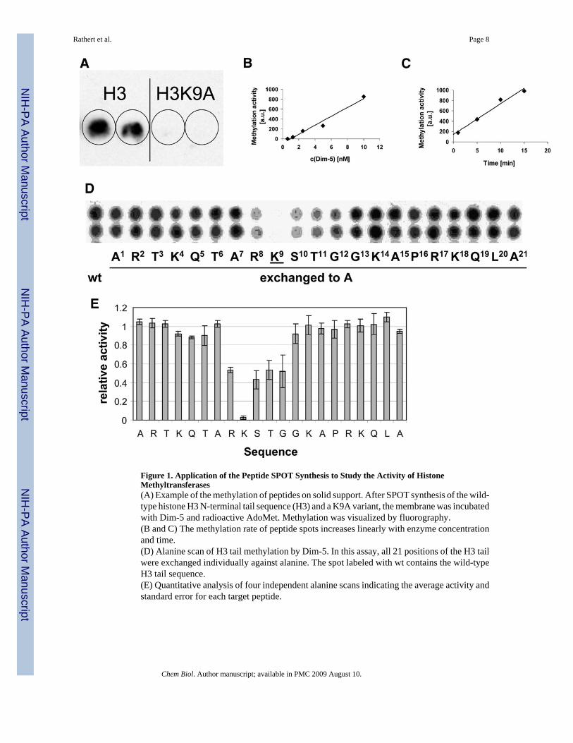
Figure 1. Application of the Peptide SPOT Synthesis to Study the Activity of HistoneMethyltransferases(A) Example of the methylation of peptides on solid support. After SPOT synthesis of the wild-type histone H3 N-terminal tail sequence (H3) and a K9A variant, the membrane was incubatedwith Dim-5 and radioactive AdoMet. Methylation was visualized by fluorography.(B and C) The methylation rate of peptide spots increases linearly with enzyme concentrationand time.(D) Alanine scan of H3 tail methylation by Dim-5. In this assay, all 21 positions of the H3 tailwere exchanged individually against alanine. The spot labeled with wt contains the wild-typeH3 tail sequence.(E) Quantitative analysis of four independent alanine scans indicating the average activity andstandard error for each target peptide.
Rathert et al. Page 8
Chem Biol. Author manuscript; available in PMC 2009 August 10.
NIH
-PA Author Manuscript
NIH
-PA Author Manuscript
NIH
-PA Author Manuscript
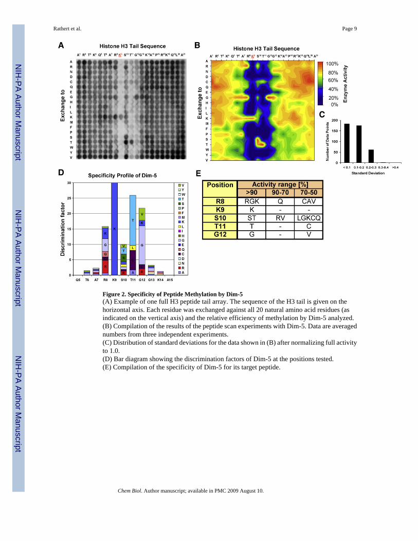
Figure 2. Specificity of Peptide Methylation by Dim-5(A) Example of one full H3 peptide tail array. The sequence of the H3 tail is given on thehorizontal axis. Each residue was exchanged against all 20 natural amino acid residues (asindicated on the vertical axis) and the relative efficiency of methylation by Dim-5 analyzed.(B) Compilation of the results of the peptide scan experiments with Dim-5. Data are averagednumbers from three independent experiments.(C) Distribution of standard deviations for the data shown in (B) after normalizing full activityto 1.0.(D) Bar diagram showing the discrimination factors of Dim-5 at the positions tested.(E) Compilation of the specificity of Dim-5 for its target peptide.
Rathert et al. Page 9
Chem Biol. Author manuscript; available in PMC 2009 August 10.
NIH
-PA Author Manuscript
NIH
-PA Author Manuscript
NIH
-PA Author Manuscript
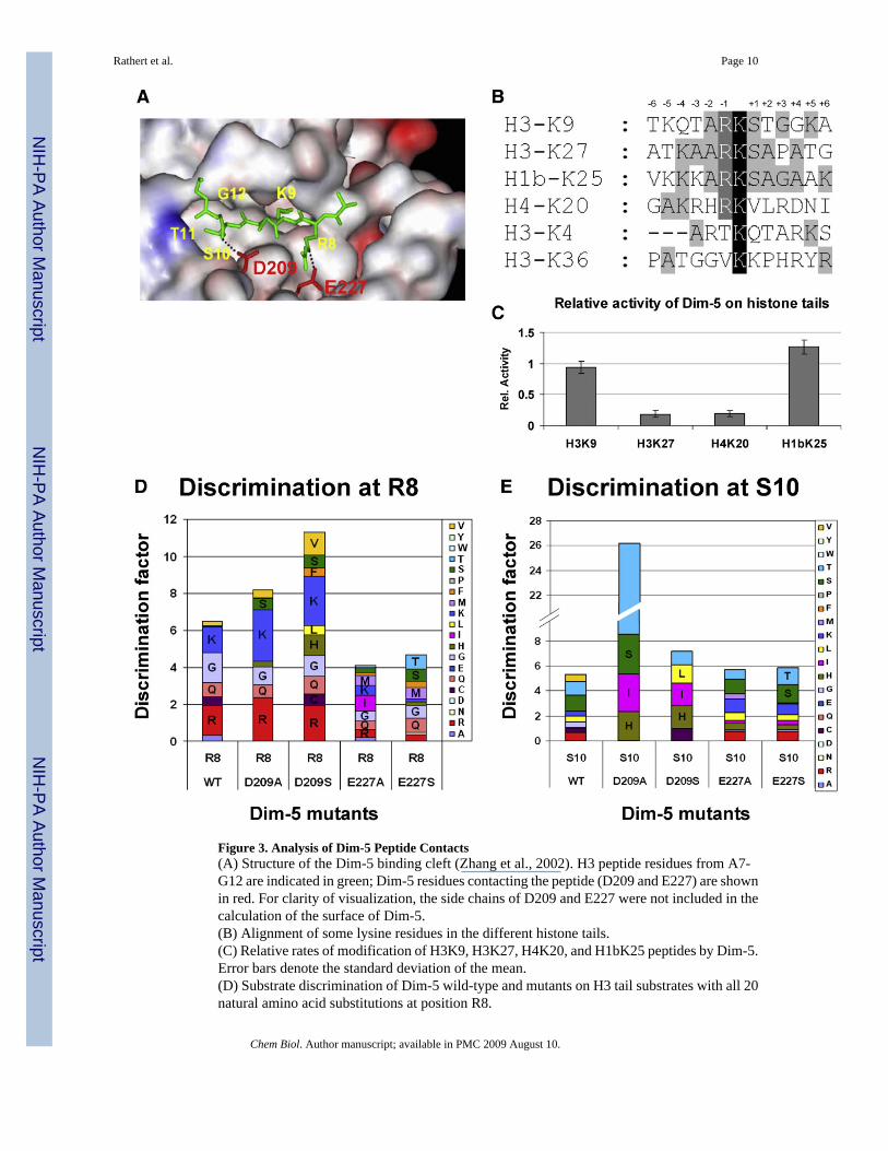
Figure 3. Analysis of Dim-5 Peptide Contacts(A) Structure of the Dim-5 binding cleft (Zhang et al., 2002). H3 peptide residues from A7-G12 are indicated in green; Dim-5 residues contacting the peptide (D209 and E227) are shownin red. For clarity of visualization, the side chains of D209 and E227 were not included in thecalculation of the surface of Dim-5.(B) Alignment of some lysine residues in the different histone tails.(C) Relative rates of modification of H3K9, H3K27, H4K20, and H1bK25 peptides by Dim-5.Error bars denote the standard deviation of the mean.(D) Substrate discrimination of Dim-5 wild-type and mutants on H3 tail substrates with all 20natural amino acid substitutions at position R8.
Rathert et al. Page 10
Chem Biol. Author manuscript; available in PMC 2009 August 10.
NIH
-PA Author Manuscript
NIH
-PA Author Manuscript
NIH
-PA Author Manuscript

(E) Substrate discrimination of Dim-5 wild-type and mutants on H3 tail substrates with all 20natural amino acid substitutions at position S10.
Rathert et al. Page 11
Chem Biol. Author manuscript; available in PMC 2009 August 10.
NIH
-PA Author Manuscript
NIH
-PA Author Manuscript
NIH
-PA Author Manuscript


