Embed Size (px)
Citation preview

Eur. J. Biochem. 267, 2617±2623 (2000) q FEBS 2000
Analysis of the thyrotropin-releasing hormone-degrading ectoenzyme bysite-directed mutagenesis of cysteine residuesCys68 is involved in disulfide-linked dimerization
Theofilos Papadopoulos, Heike Heuer and Karl Bauer
Max-Planck-Institut fuÈr Experimentelle Endokrinologie, Hannover, Germany
Thyrotropin-releasing hormone-degrading ectoenzyme is a member of the M1 family of Zn-dependent
aminopeptidases and catalyzes the degradation of thyrotropin-releasing hormone (TRH; Glp-His-Pro-NH2).
Cloning of the cDNA of this enzyme and biochemical studies revealed that the large extracellular domain of the
enzyme with the catalytically active site contains nine cysteine residues that are highly conserved among species.
To investigate the functional role of these cysteines in TRH-DE we used a site-directed mutagenesis approach and
replaced individually each cysteine by a serine residue. The results revealed that the proteolytically truncated and
enzymatically fully active enzyme consists of two identical subunits that are associated noncovalently by
protein±protein interactions but not via interchain S-S bridges. The eight cysteines contained within this region
are all important for the structure of the individual subunit and the enzymatic activity, which is dramatically
reduced in all mutant enzymes. This is even true for the four cysteines that are clustered within the C-terminal
domain remote from the Zn-binding consensus sequence HEICH. In contrast, Cys68, which resides within the
stalk region seven residues from the end of the hydrophobic membrane-spanning domain, can be replaced by
serine without a significant change in the enzymatic activity. Interestingly, this residue is involved in the
formation of an interchain disulfide bridge. Covalent dimerization of the subunits, however, does not seem to be
essential for efficient biosynthesis, enzymatic activity and trafficking to the cell surface.
Keywords: metallopeptidase; Zn-dependent aminopeptidase; cysteine residues; covalent dimerization; site-
directed mutagenesis.
Thyrotropin-releasing hormone (TRH) acts both as a hypo-thalamic hypophysiotropic hormone (reviewed in [1,2]) and aneuromodulator or neurotransmitter that elicits a wide range ofpharmacological and behavioural effects in extrahypothalamicbrain areas (reviewed in [3±5]). These functions are mediatedby high-affinity TRH-receptors [6±8] and imply the existenceof an efficient inactivation system.
Inactivation was found to be initiated by cleavage of thel-pyroglutamyl/histidinyl bond by a particular ectoenzyme(reviewed in [9,10]) that, similar to the TRH-degrading serumenzyme, exhibits a high degree of substrate specificity and doesnot cleave any of the other known pyroGlu-containingneuropeptides [11±14]. Thus, this enzyme has been termed
thyroliberinase, TRH-specific aminopeptidase, and TRH-degrading ectoenzyme (TRH-DE; EC 3.4.19.6). It is alsoknown as pyroglutamyl-peptidase II or pyroglutamyl-amino-peptidase II (PAP II) to distinguish it from the unspecific,cytosolic pyroglutamyl-aminopeptidase I (PAP I). It is found insynaptosomal [12] and adenohypophyseal [15] membranefractions. In cell cultures from murine brain it is found on thesurface of neuronal cells but not on glial cells [16,17], and isthus adequately located to terminate neurotropic TRH signals.This notion is strengthened by the analysis of the region-specific expression of TRH-DE in the rat nervous system [18].
Following purification of the enzyme [19] and cloning ofthe cDNA of the rat TRH-DE [20], the cDNA of thehuman TRH-DE has also been cloned recently [21]. Ratand human TRH-DE reveal an unusually high degree ofconservation. Overall, there are 96% identical residues aroundthe Zn-binding consensus sequence. In fact, there is a stretchof 224 amino acids without any substitution. Based onbiochemical studies and cDNA cloning data, the rat andhuman TRH-DE were be identified as a glycosylated, type IImetalloproteinase [19,20,22]. The extracellular domaincontains the HEXXH 1 E motif that is characteristic ofZn-dependent aminopeptidases (reviewed in [23]). A compari-son of the complete amino-acid sequence of rat TRH-DE andthose in the translated GenBank revealed significant homologywith rat aminopeptidase N [20,24] (34%) and mouse amino-peptidase A (32%) [25]. While the intracellular parts and thetransmembrane-spanning regions of these three enzymes arecompletely different, some sequences in the extracellulardomain display a high degree of homology. Notably, an
Correspondence to K. Bauer, Max-Planck-Institut fuÈr Experimentelle
Endokrinologie, PO Box 610309, D-30603 Hannover, Germany,
Fax: 1 49 5115359 203, Tel.: 1 49 5115359 200,
E-mail: [email protected]
Abbreviations: TRH, thyrotropin-releasing hormone; TRH-DE,
TRH-degrading ectoenzyme; PAP, pyroglutamyl-aminopeptidase; APN,
aminopeptidase N; APA, aminopeptidase A; GFP, green fluorescent protein;
BHK, baby hamster kidney; DMEM, Dulbecco's modified Eagle's medium;
LHRH, luteinizing-hormone-releasing hormone; pyroGlu, pyroglutamic
acid; BNA, b-napthylamide; EGFP, enhanced green fluorescence protein.
Enzymes: pyroglutamyl-aminopeptidase II (EC 3.4.19.6); pyroglutamyl-
aminopeptidase I (EC 3.4.19.3); aminopeptidase A (glutamyl
aminopeptidase; EC 3.4.11.7); aminopeptidase N (EC 3.4.11.2) meprin A
(EC 3.4.24.18); membrane dipeptidase (EC 3.4.13.19); endothelin-
converting enzyme-1 (EC 3.4.24.71); dipeptidyl peptidase IV (EC 3.4.14.5).
(Received 31 January 2000, revised 3 March 2000, accepted 6 March 2000)
2618 T. Papadopoulos et al. (Eur. J. Biochem. 267) q FEBS 2000
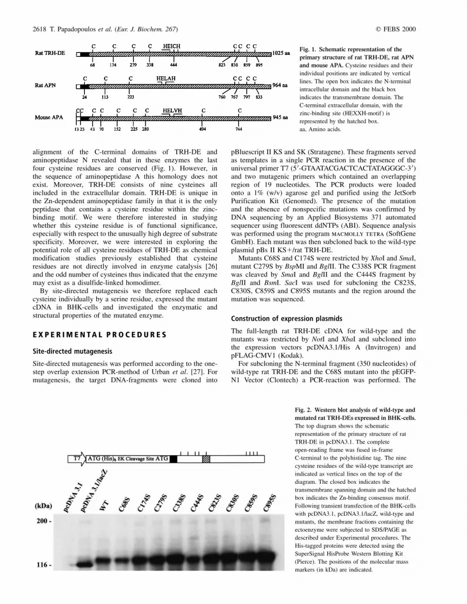
alignment of the C-terminal domains of TRH-DE andaminopeptidase N revealed that in these enzymes the lastfour cysteine residues are conserved (Fig. 1). However, inthe sequence of aminopeptidase A this homology does notexist. Moreover, TRH-DE consists of nine cysteines allincluded in the extracellular domain. TRH-DE is unique inthe Zn-dependent aminopeptidase family in that it is the onlypeptidase that contains a cysteine residue within the zinc-binding motif. We were therefore interested in studyingwhether this cysteine residue is of functional significance,especially with respect to the unusually high degree of substratespecificity. Moreover, we were interested in exploring thepotential role of all cysteine residues of TRH-DE as chemicalmodification studies previously established that cysteineresidues are not directly involved in enzyme catalysis [26]and the odd number of cysteines thus indicated that the enzymemay exist as a disulfide-linked homodimer.
By site-directed mutagenesis we therefore replaced eachcysteine individually by a serine residue, expressed the mutantcDNA in BHK-cells and investigated the enzymatic andstructural properties of the mutated enzyme.
E X P E R I M E N T A L P R O C E D U R E S
Site-directed mutagenesis
Site-directed mutagenesis was performed according to the one-step overlap extension PCR-method of Urban et al. [27]. Formutagenesis, the target DNA-fragments were cloned into
pBluescript II KS and SK (Stratagene). These fragments servedas templates in a single PCR reaction in the presence of theuniversal primer T7 (5 0-GTAATACGACTCACTATAGGGC-3 0)and two mutagenic primers which contained an overlappingregion of 19 nucleotides. The PCR products were loadedonto a 1% (w/v) agarose gel and purified using the JetSorbPurification Kit (Genomed). The presence of the mutationand the absence of nonspecific mutations was confirmed byDNA sequencing by an Applied Biosystems 371 automatedsequencer using fluorescent ddNTPs (ABI). Sequence analysiswas performed using the program macmolly tetra (SoftGeneGmbH). Each mutant was then subcloned back to the wild-typeplasmid pBs II KS1/rat TRH-DE.
Mutants C68S and C174S were restricted by XhoI and SmaI,mutant C279S by BspMI and BglII. The C338S PCR fragmentwas cleaved by SmaI and BglII and the C444S fragment byBglII and BsmI. SacI was used for subcloning the C823S,C830S, C859S and C895S mutants and the region around themutation was sequenced.
Construction of expression plasmids
The full-length rat TRH-DE cDNA for wild-type and themutants was restricted by NotI and XbaI and subcloned intothe expression vectors pcDNA3.1/His A (Invitrogen) andpFLAG-CMV1 (Kodak).
For subcloning the N-terminal fragment (350 nucleotides) ofwild-type rat TRH-DE and the C68S mutant into the pEGFP-N1 Vector (Clontech) a PCR-reaction was performed. The
Fig. 1. Schematic representation of the
primary structure of rat TRH-DE, rat APN
and mouse APA. Cysteine residues and their
individual positions are indicated by vertical
lines. The open box indicates the N-terminal
intracellular domain and the black box
indicates the transmembrane domain. The
C-terminal extracellular domain, with the
zinc-binding site (HEXXH-motif ) is
represented by the hatched box.
aa, Amino acids.
Fig. 2. Western blot analysis of wild-type and
mutated rat TRH-DEs expressed in BHK-cells.
The top diagram shows the schematic
representation of the primary structure of rat
TRH-DE in pcDNA3.1. The complete
open-reading frame was fused in-frame
C-terminal to the polyhistidine tag. The nine
cysteine residues of the wild-type transcript are
indicated as vertical lines on the top of the
diagram. The closed box indicates the
transmembrane spanning domain and the hatched
box indicates the Zn-binding consensus motif.
Following transient transfection of the BHK-cells
with pcDNA3.1, pcDNA3.1/lacZ, wild-type and
mutants, the membrane fractions containing the
ectoenzyme were subjected to SDS/PAGE as
described under Experimental procedures. The
His-tagged proteins were detected using the
SuperSignal HisProbe Western Blotting Kit
(Pierce). The positions of the molecular mass
markers (in kDa) are indicated.

q FEBS 2000 Cysteine mutants of rat TRH-DE (Eur. J. Biochem. 267) 2619
forward primer 5 0-ATATCCAGCGAATTCGCGGCCGCCAT-GG-3 0 used included the Start ATG, the NotI restriction site anda new EcoRI site. As reverse primer 5 0-GTTGCGCGGATCC-GATCCTGGGTAGCTG-3 0 was selected to introduce a BamHIsite for the continuous in frame cloning with GFP. Theamplified-fragment was digested by EcoRI and BamHI andsubcloned into the corresponding site of the pEGFP-N1-vector.Conditions for the PCR reaction were the following: initial2 min at 95 8C, 30 s at 50 8C, 1 min at 72 8C followed by 2cycles of 1 min at 95 8C, 30 s at 55 8C, 1 min at 72 8Cfollowed by 30 cycles of 20 s at 95 8C, 20 s at 60 8C, 1 min at72 8C, with an additional extension at 72 8C for 3 min in thelast cycle.
Tissue culture and tranfection of BHK-cells
BHK-cells were grown in Dulbecco's modified Eagle's medium(DMEM; Life Technologies) supplemented with 10% fetal
bovine serum in a 37 8C incubator with 10% CO2. For transientexpression, BHK-cells were plated onto six-well plates at2 � 105 cells per well or on 150-cm2 Petri dishes at 2 � 106
cells. After 18 h of growth, the cells were washed twice withDMEM and transfected (2 mg of DNA per well or 10 mg ofDNA per dish) by using LipofectAmine-PLUS (LifeTechnologies) as cationic lipid (1 mg DNA per 6 mg lipid).The cells were incubated for 4 h, and DMEM containing 10%fetal bovine serum was added. At 48 h after transfection, thecells were collected, washed with NaCl/Pi and homogenized inice-cold 20 mm potassium phosphate, pH 7,4. After centri-fugation (20 000 g for 30 min at 4 8C) the cell pellet waswashed in the same buffer and then resuspended in ice-cold20 mm potassium phosphate, 500 mm NaCl, pH 7,4. After30 min on ice the membranes were collected by centrifugation(20 000 g for 30 min at 4 8C). This step was repeated once. Thefinal membrane pellet was homogenized in 20 mm potassiumphosphate, pH 7,4 and stored at 280 8C until used for Westernblot analysis and enzyme assays. The protein was determinedby a modification of the Lowry method as described byPeterson [28].
Confocal fluorescence microscopy
BHK-cells expressing recombinant enhanced green fluores-cence protein (EGFP)-tagged rat TRH-DE were harvested 24 hafter transfection and cultured on glass coverslips for 48 h,washed in NaCl/Pi, fixed with 3% formaldehyde in NaCl/Pi for20 min at room temperature and washed in NaCl/Pi again. Cellswere viewed under a Leica confocal fluorescence microscopeequipped with the appropriate filter set and photographed.
SDS/PAGE and Western blot analysis
Polyacrylamide gel electrophoresis was carried out accordingto Laemmli [29] using a 4% stacking and a 7.5% separating gel.After blotting [30] onto nitrocellulose membranes (BAS83,Schleicher and Schuell) by use of a tank blot (Biorad Trans BlotCell) the proteins were detected either by the chemicalluminescence technique (SuperSignal HisProbe WesternBlotting Kit, Pierce) or by an immunochemical method usingeither anti-FLAG monoclonal antibody M2 (Kodak) as primary
Fig. 3. Enzymatic activity of rat TRH-DE cysteine-mutants. Rat TRH-
DE activity in the membrane fractions of transfected BHK cells is expressed
as percentage of wild-type rat TRH-DE activity. Measurements are the
mean ^SD of three independent transfections. The mean value for wild-
type rat TRH-DE corresponds to 250 nmol of substrate hydrolyzed per min
per mg enzyme.
Fig. 4. Immunoblot analysis of recombinant
TRH-DEs and truncated ectoenzyme.
(A) SDS/PAGE and immunoblotting of the
membrane proteins from BHK cells transfected
with the FLAG-tagged wild-type TRH-DE cDNA
(lanes 2 and 5), the corresponding C68S mutant
(lanes 3 and 6) and the pFLAG-CMV1/BAP
cDNA (lanes 1 and 4). The samples were
incubated on ice for 30 min in SDS buffer in the
presence (1) or absence (±) of dithiothreitol
(DTT). After SDS/PAGE, the anti-FLAG
monoclonal antibody M2 was used for
immunoblotting. (B) SDS/PAGE and
immunoblotting of the purified ectoenzyme
from rat brain (trypsin-fragment) under reducing
(lane 7) and nonreducing (lane 8) conditions.
After SDS/PAGE separation the samples were
analyzed by immunoblotting using an
affinity-purified polyclonal rabbit antibody
against rat TRH-DE.

2620 T. Papadopoulos et al. (Eur. J. Biochem. 267) q FEBS 2000
antibody and alkaline-phosphatase conjugated goat anti-(mouseIgG) Ig (Biorad) as secondary antibody or the living colorspeptide antibody-AP conjugate (Clontech). The proteinexpression levels were estimated by densitometric scanning(Duoscan T1200, Agfa) and calculated using the programnihimage (NIMH).
Enzyme assay
The activity of the TRH-DE was measured by the radio-chemical assay in the presence of the inhibitors of the solubleTRH-degrading enzymes as described in [16] using [pyro-Glu-3H]TRH as substrate. The kinetic parameters (Km andVmax) were determined from Lineweaver±Burk plots.
R E S U L T S
Characterization of cysteine mutants of rat TRH-DEexpressed in BHK-cells
Each of the nine cysteine residues in rat TRH-DE was mutatedindividually to serine. The mutant cDNAs were expressed inBHK-cells. Western blot analysis of the membrane fractionsprepared from these cells showed that each mutant enzymehad the same apparent molecular mass (130 kDa) as therecombinant wild-type enzyme, and thus appeared to besimilarly glycosylated (Fig. 2).
The activity of each mutant enzyme was determined in themembrane fractions by radiochemical assay and expressedrelative to the amount of the wild-type transcript and thepurified brain ectoenzyme (trypsin-fragment), as quantified byWestern blot analysis (densitometric scanning of bands,Duoscan T1200, Agfa/NIH-Image, NIMH). Using the sameamount of recombinant enzyme we detected specific enzymaticactivities similar to that of the wild-type enzyme only for theC68S mutant (Fig. 3). The Km and Vmax values were also notsignificantly different (Table 1). The C-terminal mutants
C823S, C830S and C895S were completely inactive, indicatingthat even the elements remote from the active site are importantfor the overall structure of the enzyme. The specific activities ofall the other mutants were markedly decreased. The specificactivities of the C279S and the C338S mutants were 10-foldand fivefold lower and correspondingly a decrease in the Vmax
values could be demonstrated, while the Km values were notsignificantly different.
Fig. 5. Heat inactivation of wild-type and C68S mutant. Membrane
preparations of recombinant wild-type (B) or the C68S mutant (A) enzyme
were preincubated for 40 min and then assayed at the temperatures
indicated. [pyroGlu-3H]TRH was used as substrate.
Fig. 6. Subcellular distribution of the truncated GFP-fusion proteins
with either cysteine or serine at position 68, as analyzed by confocal
laser fluorescence microscopy. BHK cells were transfected either with the
empty pEGFP-N1 vector (A), the EGFP-tagged, truncated wild-type cDNA
of TRH-DE (B) or the corresponding C68S mutant (C) and processed as
described in Experimental procedures.

q FEBS 2000 Cysteine mutants of rat TRH-DE (Eur. J. Biochem. 267) 2621
In the Zn-binding HEICH domain, substitution of thecysteine by serine resulted in a 60-fold decrease in the specificenzymatic activity of the mutant enzyme, suggesting that thiscysteine is an important structural element that is not limited torestraining only the substrate specificity of the enzyme.
Cys68 is involved in disulfide-linked dimerization
For the proteolytically truncated enzyme a molecular mass of230 kDa has been estimated by gel-filtration chromatography,whereas a single band with an apparent molecular mass of116 kDa could be detected after SDS/PAGE under bothreducing and nonreducing conditions [19]. From the alignmentof the amino-acid sequences we speculated that Cys68, the onlyCys residue of the N-terminal peptide fragment that could beleft in the membrane after trypsin cleavage, might be involvedin disulfide-linked dimerization. Therefore, the wild-type andthe C68S mutant were fused in-frame C-terminal of theFLAG-epitope of pFLAG-CMV1 (see Experimental pro-cedures) and expressed in BHK-cells. Membrane fractionsfrom BHK-cells expressing the wild-type or the mutantenzyme were subjected to SDS/PAGE under reducing andnonreducing conditions and analyzed by immunoblotting
with the anti-FLAG M2 monoclonal antibody (Kodak). Underreducing conditions (1.5% dithiothreitol), a band of immuno-reactive protein with a molecular mass of 130 kDa could bedetected for both proteins, the wild-type and the C68S mutant(Fig. 4A, lanes 2 and 3). Under nonreducing conditions, thesame band was also visualized for the C68S mutant (Fig. 4A,lane 6) but not for the wild-type protein (Fig. 4A, lane 5),which migrated as a high molecular mass protein. These dataare compatible with the interpretation that the mutant enzymemay exist as a monomeric fusion protein, while the wild-typeprotein represents a disulfide-linked homodimer. This hypoth-esis is further supported by the analysis of the proteolyticallytruncated, catalytically fully active ectoenzyme from rat brain.Under both reducing and nonreducing conditions, this proteinmigrates on SDS/PAGE as a single band with an apparentmolecular mass of 116 kDa (Fig. 4B) indicating that othercysteine residues are not involved in dimer formation. Toanalyze whether the homodimer formation via the Cys68 S-Sbridges might contribute to the stability of TRH-DE, we studiedthe temperature-dependant inactivation of the wild-type and theC68S mutant but found no difference (Fig. 5).
To study whether the interchain bridge at Cys68 might beimportant for the proper intracellular trafficking and insertion
Table 1. Kinetic parameters for the hydrolysis of [pyroGlu-3H]-TRH by wild-type and mutant rat TRH-DE. Specific activities were determined at
2 mm substrate concentration. Reactions were carried out as described under Experimental procedures, values are the mean ^SD from at least three
independent transfections. ND, not detected.
Enzyme
Specific activity
(nmol´min21´mg21)
Km
(mm)
Vmax
(nmol´min21´mg21)
Vmax/Km
(mL´min21´mg21)
Wild-type 250 �̂ 44 32.5 �^ 1 3727 �^ 104 114�.67
C68S 210 �̂ 36 29.3 �^ 0.4 2900 �^ 489 98�.97
C174S 6.25 �^ 0.2 �± �± ±
C279S 24.56 �^ 4.15 27 �^ 4 238 �^ 51 10�.48
C338S 46.38 �̂ 5.11 33 �^ 3 716 �^ 28.27 21�.69
C444S 4.37 �^ 2 �± �± ±
C823S ND �± �± ±
C830S ND �± �± ±
C859S 9.37 �^ 1.37 �± �± ±
C895S ND �± �± ±
Fig. 7. Expression of wild-type and C68S
mutant as truncated GFP-fusion proteins in
BHK-cells. The large extracellular domain of
TRH-DE with the eight cysteine residues was
replaced by EGFP. BHK-cells were transfected
either with the empty pEGFP-N1 vector (lanes 1
and 4), the EGFP-tagged, truncated wild-type
cDNA of TRH-DE (lanes 2 and 5) or the
corresponding C68S mutant (lanes 3 and 6).
The membrane fractions were subjected to
SDS/PAGE (12% gel) in the presence (1) or
absence (±) of dithiothreitol and analyzed by
immunoblotting using the living colors peptide
antibody-AP conjugate (Clontech). The
positions of the molecular mass markers (in kDa)
are indicated.
2622 T. Papadopoulos et al. (Eur. J. Biochem. 267) q FEBS 2000
into the plasma membrane, fusion proteins were constructed inwhich the large extracellular domain of TRH-DE with the eightcysteine residues were deleted and replaced by EGFP. Thetruncated proteins thus contained at position 68 either the onlyCys in the sequence or alternatively a Ser residue. As expected,cells transfected with the vector alone exhibited brightfluorescence distributed throughout the whole cell (Fig. 6A),whereas the truncated fusion protein with either cysteine orserine at position 68 was found to be localized on the plasmamembrane (Fig. 6B,C).
On SDS/PAGE, the truncated mutant protein exhibited thesame mobility irrespective of whether dithiothreitol waspresent. The same pattern was observed when the truncatedwild-type protein was subjected to SDS/PAGE under reducingconditions, whereas in the absence of dithiothreitol a band ofabout twice the molecular mass could be visualized (Fig. 7).
D I S C U S S I O N
Site-directed mutagenesis is an important and well establishedtechnique in protein chemistry to study the functional role ofdefined amino-acid residues. We used this method toinvestigate the structural and functional importance of thenine cysteine residues in TRH-DE by replacing individuallyeach cysteine by a serine residue, and thus identified thisenzyme as a new member of mammalian cell-surface pep-tidases that exist as disulfide-linked dimers. Previously, amino-peptidase A [31,32], meprin [33,34], membrane dipeptidase[35] and endothelin-converting enzyme-1 [36] have been theonly established examples.
In agreement with previous biochemical data [19], the resultsof the present study clearly demonstrate that the eight cysteineresidues within the large extracellular domain all contribute tothe intrachain S-S-bridging of the subunits, the conformation ofthe catalytic domain and the enzymatic activity. The specificactivities of all these mutant enzymes were either considerablyreduced or completely abolished.
Within the M1 family of Zn-dependent aminopeptidases,TRH-DE and aminopeptidase N share some sequence similarityin the C-terminal region, especially with regard to the cluster offour cysteine residues [20,24] (see Fig. 1). Although remotefrom the active site, these cysteine residues are clearly ofmajor importance to the structural integrity of the coredomain, whereas in aminopeptidase A the differentlystructured C-terminal end is separated from the catalyticcore domain by a protease susceptible region [32]. Although thefunctional significance of the C-terminal domain of TRH-DEand sequence-related enzymes awaits determination by othermethods (e.g. crystallographic analysis), it is interesting to notethat the porcine coronavirus transmissible gastroenteritis virusbinds to the C-terminal domain of aminopeptidase N betweenresidues 717 and 813 [37].
Among the large family of metalloproteases, TRH-DE isunique in that it contains a cysteine residue within the HEXXHzinc-binding domain. Mutation of this cysteine (Cys444) toserine resulted in a 60-fold decrease of the enzymatic activity.As part of an intrachain S-S bridge, this cysteine apparentlycontributes to establishing the tertiary structure of the activepocket. On the basis of the high substrate specificity ofTRH-DE, we assumed that replacement of Cys444 by serinemight `open' the active pocket and thus might result in anenzyme that could hydrolyze different substrates other thanTRH. However, with pyroglutamyl-b-naphthylamide, a lowaffinity substrate that is slowly hydrolyzed by TRH-DE, releaseof b-naphthylamine by the mutant enzyme could not be
detected (data not shown). Furthermore, LH-RH, a deca-peptideamide with the sequence pyroGlu-His-Trp-Ser-Tyr-Gly-Leu-Arg-Pro-Gly is also not hydrolyzed by the mutant enzyme(data not shown). LH-RH was tested because this peptide is nothydrolyzed by TRH-DE, yet it is apparently recognized by theenzyme as the degradation of TRH by TRH-DE is effectivelyinhibited by this peptide [13].
In contrast to the cysteines constituting the catalytic domain,Cys68, which resides very close to the membrane surface (justfour residues away from the hydrophobic membrane-spanningregion), could be substituted by serine without affectingsignificantly the specific activity of the mutant enzyme. Thiscysteine is positioned within the presumed stalk region, astretch of about 40 amino-acid residues that is a general featureof ectoenzymes and is thought to be important for surfaceexposition [38]. The hypothesis that this stretch forms aseparate domain is supported by the fact that in TRH-DE [21],as in other enzymes such as in aminopeptidase N (reviewed in[39]), this region is considerably less conserved than thecatalytic domain. As in aminopeptidase A [31,32], this cysteineis obviously responsible for intersubunit S-S bridging and thusfor the formation of the homodimeric wild-type TRH-DE.Surprisingly, however, covalent dimerization seems not to beessential for the formation and expression of the catalyticallyactive enzyme. Moreover, as also shown with the truncatedmutant protein in which the catalytic domain was replaced byGFP, the intermolecular disulfide link via Cys68 appeared notto be essential for trafficking and transport of the protein to thecell surface. In agreement with these results are the data ofprevious biochemical studies on the dimeric assembly ofenterocyte brush border enzymes (e.g. aminopeptidase N andA, dipeptidyl peptidase IV) which demonstrated that homo-dimerization is not an absolute requirement for transport ofthese enzymes to, and through, the Golgi complex [40]although dimeric assembly may increase the rate of intra-cellular transport. For the mouse meprin peptidase, site-directedmutagenesis studies also demonstrated that the covalentdimerization of subunits is not essential for efficient bio-synthesis, trafficking or post-translational processing of thesecreted protease [33]. Moreover, for membrane dipeptidase,another enzyme with a single disulfide bond close to themembrane surface, mutation of the critical cysteine residue toglycine neither influenced the expression of the mutant enzymeto the cell surface nor the enzymatic properties of thepeptidase [35].
Taking these data together, it might be reasonable to assumethat the noncovalent hydrophobic domain±domain interactionsof the catalytic subunits may represent the major elements thatare critical for the structure and function of TRH-DE andrelated dimeric proteins.
A C K N O W L E D G E M E N T S
We thank Andre Wolfstein and Beate Sodeik (Institut fuÈr physiologische
Chemie, Medizinische Hochschule, Hannover) for experienced support with
confocal fluorescence microscopy, Lutz Schomburg for advice and helpful
discussions, Uwe Grunenberg for excellent technical assistance, Valerie
Ashe for linguistic help and the Deutsche Forschungsgemeinschaft for
financial support.
R E F E R E N C E S
1. Guillemin, R. (1978) Peptides in the brain: the new endocrinology of
the neuron. Science 202, 390±302.

q FEBS 2000 Cysteine mutants of rat TRH-DE (Eur. J. Biochem. 267) 2623
2. Schally, A.V. (1978) Aspects of hypothalamic regulation of the
pituirary gland. Science 202, 18±28.
3. Morley, J.E. (1981) Neuroendocrine control of thyrotropin secretion.
Endocr. Rev. 2, 396±436.
4. Prasad, C. (1984) Thyrotropin-releasing hormone. In Handbook of
Neurochemistry (Lajtha, A., ed.), Vol. 8, pp. 175±200. Plenum, New
York.
5. Kelly, J.A. (1995) Thyrotropin releasing hormone: basis and potential
for its therapeutic use. Essays Biochem. 30, 133±149.
6. Gershengorn, M.C. & Osman, R. (1996) Molecular and cellular biology
of thyrotropin-releasing hormone receptors. Physiol. Rev. 76,
175±191.
7. Itadani, H., Nakamura, T., Itoh, J., Iwaasa, H., Kanatani, A.,
Borkowski, J., Ihara, M. & Ohta, M. (1998) Cloning and
characterization of a new subtype of thyrotropin-releasing hormone
receptors. Biochem. Biophys. Res. Commun. 250, 68±71.
8. Cao, J.O., Donnell, D., Vu, H., Payza, K., Pou, C., Godbout, C., Jakob,
A., Pelletier, M., Lembo, P., Ahmad, S. & Walker, P. (1998) Cloning
and characterization of a cDNA encoding a novel subtype of rat
thyrotropin-releasing hormone receptor. J. Biol. Chem. 273,
32281±32287.
9. O'Cuinn, G.O., Connor, B. & Elmore, M. (1990) Degradation of
thyrotropin-releasing hormone and luteinising hormone-releasing
hormone by enzymes of brain tissue. J. Neurochem. 54, 1±13.
10. Bauer, K., Schomburg, L., Heuer, H. & SchaÈfer, M.K.-H. (1999)
Thyrotropin releasing hormone (TRH), the TRH-receptor and the
TRH-degrading ectoenzyme; three elements of a peptidergic
signalling system. In Regulatory Peptides and Cognate Receptors
(Richter, D., ed.), pp. 13±42. Springer-Verlag, Berlin, Heidelberg.
11. Bauer, K., Nowak, P. & Kleinkauf, H. (1981) Specificity of a serum
peptidase hydrolyzing thyroliberin at the pyroglutamyl-histidine
bond. Eur. J. Biochem. 118, 173±176.
12. O'Connor, B. & O'Cuinn, G. (1984) Localization of a narrow-
specificity thyroliberin hydrolyzing pyroglutamate aminopeptidase in
synaptosomal membranes of guinea-pig brain. Eur. J. Biochem. 144,
271±278.
13. O'Connor, B. & O'Cuinn, G. (1985) Purification of and kinetic studies
on a narrow specificity synaptosomal membrane pyroglutamate
aminopeptidase from guinea-pig brain. Eur. J. Biochem. 150, 47±52.
14. Wilk, S. & Wilk, E.K. (1989) Pyroglutamyl peptidase II, a thyrotropin-
releasing hormone degrading enzyme: purification and specificity
studies of the rabbit brain enzyme. Neurochem. Int. 15, 81±89.
15. Horsthemke, B., Leblanc, P., Kordon, C., Wattiaux-De Coninck, S.,
Wattiaux, R. & Bauer, K. (1984) Subcellular distribution of particle
bound neutral peptidases capable of hydrolyzing gonadoliberin,
thyroliberin, enkephalin and substance P. Eur. J. Biochem. 139,
315±320.
16. Bauer, K., Carmeliet, P., Schulz, M., Baes, M. & Denef, C. (1990)
Regulation and cellular localization of the membrane-bound
thyrotropin-releasing hormone-degrading enzyme in primary cultures
of neuronal, glial and adenohypophyseal cells. Endocrinology 127,
1224±1233.
17. Cruz, C., Charli, J.-L., Vargas, M.A. & Joseph-Bravo, P. (1991)
Neuronal localization of pyroglutamate aminopeptidase II in primary
cultures of fetal mouse brain. J. Neurochem. 56, 1594±1601.
18. Heuer, H., Ehrchen, J., Bauer, K. & SchaÈfer, K.H. (1998) Region-
specific expression of thyrotropin-releasing hormone-degrading
ectoenzyme in the rat central nervous system and pituitary gland.
Eur. J. Neurosci. 10, 1465±1478.
19. Bauer, K. (1994) Purification and characterization of the thyrotropin-
releasing hormone-degrading ectoenzyme. Eur. J. Biochem. 224,
387±396.
20. Schauder, B., Schomburg, L., KoÈhrle, J. & Bauer, K. (1994) Cloning of
a cDNA encoding an ectoenzyme that degrades thyrotropin-releasing
hormone. Proc. Natl Acad. Sci. USA 91, 9534±9538.
21. Schomburg, L., Turwitt, S., Prescher, G., Lohmann, D., Horsthemke, B.
& Bauer, K. (1999) Human TRH-degrading ectoenzyme ± cDNA
cloning, functional expression, genomic structure and chromosomal
assignment. Eur. J. Biochem. 265, 415±422.
22. Czekay, G. & Bauer, K. (1993) Identification of the thyrotropin-
releasing hormone-degrading ectoenzyme as a metallopeptidase.
Biochem. J. 290, 921±926.
23. Hooper, N.M. (1994) Families of zinc metalloproteases. FEBS Lett.
354, 1±6.
24. Watt, V.M. & Yip, C.C. (1989) Amino acid sequence deduced from a
rat kidney cDNA suggests it encodes the Zn-peptidase amino-
peptidase N. J. Biol. Chem. 264, 5480±5487.
25. Wu, Q., Lahti, J.M., Air, G.M., Burrows, P.D. & Cooper, M.D. (1990)
Molecular cloning of the murine BP-1/6C3 antigen: a member of the
zinc-dependent metallopeptidase family. Proc. Natl Acad. Sci. USA
87, 993±997.
26. Czekay, G. (1993) Zur Charakterisierung und Klonierung des
TRH-abbauenden Ektoenzyms aus Rattenhirn. PhD Dissertation.
UniversitaÈt Hannover, Germany.
27. Urban, A., Neukirchen, S. & Jaeger, K.E. (1997) A rapid and efficient
method for site-directed mutagenesis using one-step overlap exten-
sion PCR. Nucleic Acids Research 25, 2227±2228.
28. Peterson, G.L. (1977) A simplification of the protein assay method of
Lowry et al. which is more generally applicable. Anal. Biochem. 83,
346±356.
29. Laemmli, U.K. (1970) Cleavage of structural proteins during the
assembly of the head of bacteriophage T4. Nature 227, 680±685.
30. Towbin, H., Staehelin, T. & Gordon, J. (1979) Electrophoretic transfer
of proteins from polyacrylamide gels to nitrocellulose sheets:
procedure and some applications. Proc. Natl Acad. Sci. USA 76,
4350±4354.
31. Wu, Q., Li, L., Cooper, M.D., Pierres, M. & Gorvel, J.P. (1991)
Aminopeptidase A activity of the murine B-lymphocyte differen-
tiation antigen BP-1/6C3. Proc. Natl Acad. Sci. USA 88, 676±680.
32. Hesp, J.R. & Hooper, N.M. (1997) Proteolytic fragmentation reveals
the oligomeric and domain structure of porcine aminopeptidase A.
Biochemistry 36, 3000±3007.
33. Marchand, P., Volkmann, M. & Bond, J.S. (1996) Cysteine mutations in
the MAM domain result in monomeric meprin and alter stability and
activity of the proteinase. J. Biol. Chem. 271, 24236±24241.
34. Chevallier, S., Ahn, J., Boileau, G. & Crine, P. (1996) Identification of
the cysteine residues implicated in the formation of alpha 2 and
alpha/beta dimers of rat meprin. Biochem. J. 317, 731±738.
35. Keynan, S., Habgood, N.T., Hooper, N.M. & Turner, A.J. (1996) Site-
directed mutagenesis of conserved cysteine residues in porcine
membrane dipeptidase. Cys 361 alone is involved in disulfide-linked
dimerization. Biochemistry 35, 12511±12517.
36. Shimada, K., Takahashi, M., Turner, A.J. & Tanzawa, K. (1996) Rat
endothelin-converting enzyme-1 forms a dimer through Cys412 with a
similar catalytic mechanism and a distinct substrate binding
mechanism compared with neutral endopeptidase-24.11. Biochem.
J. 315, 863±867.
37. Delmas, B., Gelfi, J., Sjostrom, H., Noren, O. & Laude, H. (1994)
Determinants essential for the transmissible gastroenteritis virus±
receptor interaction reside within a domain of aminopeptidase-N that
is distinct from the enzymatic site. J. Virol. 68, 5216±5224.
38. Kenny, A.J. & Turner, A.J. (1987) What are ectoenzymes? In Mam-
malian Ectoenzymes (Kenny, A.J. & Turner, A.J., eds), pp. 1±13.
Elsevier Science Publishers B.V., Biomedical Division, City.
39. NoreÂn, O., SjoÈstroÈm, H. & Olsen, J. (1997) Aminopeptidase N. In Cell-
Surface Peptidases in Health and Disease (Kenny, A.J. & Boustead,
C.M., eds), pp. 175±191. BIOS Scientific Publishers Ltd, Oxford,
UK.
40. Danielsen, E.M. (1994) Dimeric assembly of enterocyte brush border
enzymes. Biochemistry 33, 1599±1605.


