Embed Size (px)
Citation preview

www.elsevier.com/locate/cogbrainres
Cognitive Brain Research
Research Report
Anarchic hand syndrome: Bimanual coordination and sensitivity to
irrelevant information in unimanual reaches
Ada Kritikosa,*, Nora Breenb, Jason B. Mattingleyc
aSchool of Psychology, Victoria University of Technology, St Albans Campus, Melbourne, Victoria, 8001, AustraliabDepartment of Psychology, Royal Prince Alfred Hospital, Sydney, Australia
cCognitive Neuroscience Laboratory, Department of Psychology, School of Behavioural Science, University of Melbourne, Melbourne, Australia
Accepted 16 March 2005
Available online 25 May 2005
Abstract
Anarchic hand syndrome is characterised by unintended but purposeful and autonomous movements of the upper limb and intermanual
conflict. Based on predictions of internal models of movement generation, we examined the role of visual cues in unimanual and bimanual
movements in a patient with anarchic hand syndrome and in a matched control. In Experiment 1, participants made unimanual movements in
a sequential button-pressing task. The cue for the next target in a sequence appeared either prior to (exogenous) or after (endogenous) the
initiation of movement. For the patient, performance of the anarchic left hand was selectively impaired in the endogenous condition. In
Experiment 2, participants made unimanual movements on a digitising tablet to a target, which appeared either alone or with a distractor.
While the presence of a distractor was associated with increased Initiation time in general, the patient’s anarchic left hand was particularly
vulnerable to disruption by the distractor. The findings of Experiments 1 and 2 indicate excessive reliance on salient environmental stimuli
for movement production in anarchic hand syndrome. We conclude that in AHS goal-directed actions of the affected limb are particularly
vulnerable to disruption by non-relevant information. Finally, in Experiment 3, participants performed unimanual and mirror-image
bimanual movements on a digitising tablet to targets in the left or right hemispace. Coupling of the parameters of the two hands was evident
such that, compared with a unimanual baseline, Initiation time of the intact right hand deteriorated while it improved for the anarchic left
hand.
D 2005 Elsevier B.V. All rights reserved.
Theme: Neural basis of behaviour
Topic: Cognition
Keywords: Anarchic hand syndrome; Unimanual action; Bimanual coordination
Alien hand syndrome is a striking phenomenon charac-
terised by unintended but purposeful and autonomous
movements of the upper limb following brain lesions. Brion
and Jedynak [3] originally defined alien hand as the inability
to differentiate while blindfolded the affected hand from that
of a stranger’s placed in the unaffected hand. Feinberg et al.
suggested that in fact two distinct syndromes exist, depend-
ing on the site of the lesion [8]. One syndrome is seen after
anterior cortical lesions involving damage to the SMA,
anterior cingulate and medial prefrontal cortex of the left
0926-6410/$ - see front matter D 2005 Elsevier B.V. All rights reserved.
doi:10.1016/j.cogbrainres.2005.03.015
* Corresponding author.
E-mail address: [email protected] (A. Kritikos).
hemisphere, together with lesions to the anterior corpus
callosum. Symptoms of this syndrome consist of reflexive
grasping, groping and compulsive tool manipulation [8,51].
Recent formulations have termed this Fanarchic hand
syndrome_ (AHS) [5,6,8]. Crucially, patients with this
impairment acknowledge the hand as theirs, although they
are frustrated by its unintended actions. A second syndrome,
the alien hand proper, seen after callosal damage and
hemispheric disconnection, is characterised by intermanual
conflict and right-limb exploratory reflexes, with release
from an asymmetrically distributed, mostly left hemisphere
inhibition [5–7,53]. In contrast to individuals with anarchic
hand syndrome, patients with the alien hand syndrome tend
24 (2005) 634 – 647

A. Kritikos et al. / Cognitive Brain Research 24 (2005) 634–647 635
to dissociate themselves from the hand and its actions,
frequently remarking on the hand’s behaviour as if it does
not belong to them [8]. In this paper, we report the results
from a series of experiments on a patient with AHS. The
aim of these experiments was to characterise the underlying
impairments in unimanual and bimanual actions.
AHS has been reported in patients with lesions of the right
parietal lobe [27], anterior cingulate, anterior prefrontal
cortex and supplementary motor area (SMA) and medial
primary motor cortex [10–12,15], and in some cases include
the corpus callosum [1,3,6,11,13,50]. Each of these struc-
tures has a role in the control of movement and our ability to
interact successfully with the environment. The parietal lobe,
SMA and corpus callosum are crucial in selection, program-
ming and execution of goal-directed actions in general, as
indicated by findings in studies of motor control in humans
and monkeys (e.g. [23–25,38,39,47,48]). In particular, the
parietal lobe represents objects in terms of their physical
features such as size and shape and in terms of their direction
and distance [36–38,40]; it is also crucial in directing and
maintaining attention to goal-relevant objects [18,19,32].
The parietal lobe also maintains a representation of body
parts in space [55,56]. Work in primates indicates that the
parietal lobe is involved in the formation of goal-appropriate
grasp postures [36,37,46]. Thus, damage to the parietal lobe
may be expected to manifest in symptoms usually associated
with AHS, such as heightened susceptibility to non-relevant
objects, impaired proprioception in the affected limb and
impaired programming and execution of goal-directed
actions. The SMA, conversely, receives inputs from both
cortical and subcortical areas [12] and is activated in tasks
requiring selection between various possible actions and
sequences of actions [4,43]. Damage to the SMA, therefore,
may lead to selection and release of inappropriate actions of
the contralateral hand and thus to manual actions that are
inconsistent with current task demands, as observed in AHS.
In this study, we examined two main deficits in motor
control associated with AHS, namely, impaired generation
of goal-appropriate actions and impaired bimanual coordi-
nation. We examined these deficits in a patient with right
hemisphere damage involving the parietal lobe and the
SMA, in whom the corpus callosum remained intact.
Clinically, the patient exhibited frequent, unwanted grasp-
ing and groping actions with his left hand. Although he
complained that the unwanted hand seemed to act with a
‘‘will of its own’’, he never denied that it belonged to him.
Although such behavioural manifestations of AHS have
been well described in the literature, until recently, few
accounts have been proposed in terms of the perceptual
processing of environmental information and subsequent
programming, generation and coordination of actions
within the environment. The current experiments were
designed to examine different aspects of unimanual and
bimanual actions and to explain the impairments in these
abilities in terms of contemporary theories of perception
and action.
1. Case report
At the time of testing, MA was a 61-year-old, right-
handed man with a history of hypertension, hypercholestero-
laemia and ischaemic heart disease. He was admitted to a
hospital in September 2000 after experiencing sudden onset
of left leg weakness while he was a spectator at a sporting
event.
He was found to have a monoplegia with some mild left-
hand clumsiness. A cerebral CT scan performed on
admission revealed several small focal hypodensities in
the left and right thalamus, and the left corona radiata,
consistent with old lacunar infarcts. There was also some
evidence of periventricular hypodensity around the posterior
horns of the lateral ventricles consistent with chronic
ischaemia. The basal cisterns, ventricles and cerebral sulci
were within normal limits. The following day, MA under-
went a series of MRI scans (see Fig. 1). Sagittal T1 and axial
FLAIR T2 and diffusion-weighted images were obtained.
The diffusion-weighted images revealed an area of acute
infarction in the right frontal lobe, involving the apex of the
precentral gyrus. There were also other small areas of acute
infarction posteriorly in the paracentral parietal lobe and
extending into the right cuneus. The radiological report
indicated that the areas of infarction were most likely due to
an embolus in the right anterior cerebral artery that had
fragmented and resulted in small areas of distal branch and
watershed infarction.
MA was examined by a neuropsychologist (N.B.) 2
weeks after his stroke. At this time, he showed left-sided
neglect and a left anarchic hand. He failed to copy the left
side of geometric shapes, omitted numbers from the left side
of a clock face and missed targets on the left side of a visual
cancellation task. He also failed to insert his left arm into its
sleeve while dressing, suggesting some degree of personal
neglect. His copies of geometric objects were generally
good, though somewhat distorted due to his visual neglect.
He scored 16 for his copy of the Rey Figure (age-scaled
mean = 30.79, SD = 4.21; see [42]) and 9.5 after a delay of
30 min (age-scaled mean = 14.21, SD = 7.50; see [42]).
With eyes closed, he was able to identify objects with both
his left and right hands and to identify letters traced onto the
palm of either hand. There was no evidence of problems
with language production or comprehension, reading or
writing. His verbal learning and recall, as measured by the
Rey Auditory Verbal Learning Test, were in the low–
normal range. His performance on the Boston Naming Test
and the Controlled Oral Word Association Test were also
within the normal range.
MA’s most significant problem involved the loss of
voluntary control of his left hand, which did not act
according to his intentions and was sometimes in conflict
with the actions of his right hand. An intriguing aspect of
this problem was the apparent dissociation between MA’s
inability to perform left-hand actions according to intentions
and his preserved ability to make the same movements

Fig. 1. Depiction of lesion sites for patient MA on standardised templates.
A. Kritikos et al. / Cognitive Brain Research 24 (2005) 634–647636
under external command. For example, when asked to grasp
the examiner’s fingers tightly with his left hand, he was
unable to let go despite strenuous efforts to do so. When the
examiner commanded him to let go, however, he imme-
diately did so. When handed a shirt, he grasped it firmly
with his left hand but could not release his grip, even with
the assistance of his right hand. Once again, however, he
relinquished the garment following the verbal command of
the examiner. MA recounted several incidents of a similar
nature that had occurred during his stay in hospital. On
several occasions, he had opened a door and started to walk
through only to find himself unable to let go of the
doorknob. He would remain Fattached_ to the door in this
way until a nurse passed and told him to Flet go_. In the
bathroom, he had noticed that his left hand would continue
unfurling toilet paper from its roll until it lay sitting in a pile
at his feet, unless he was told to desist by one of the nursing
staff. In most cases, his attempts to bring the left hand back
under control using his right hand were unsuccessful.
MA gave his voluntary consent and participated in the
experimental tasks approximately 8 weeks after his stroke.
At this time, he no longer showed any signs of neglect on
standard clinical tests such as line bisection or cancellation,
though he did have a residual ipsilesional (rightward)
attentional bias on the greyscales task [31]. He reported
ongoing problems with voluntary control of his left hand,
but these had clearly diminished in severity since his stroke.
We compared MA’s performance to that of TM, a
neurologically intact 65-year-old right-handed male with
corrected-to-normal vision. Both MA and TM gave their
informed consent for participation.
2. Experiment 1
Successful generation of goal-appropriate actions involves
selection of relevant, salient information amongst multiple
environmental stimuli, as well as inhibition of inappropriate
actions [49]. Internal models of movement generation
assume that actions and the environment are mimicked or
internally represented (e.g. [54,56]). According to these
models, there are three types of representation: the current
state of the system, the desired state and the predicted state
(that is, the sensory consequences of a motor command are
estimated). A close and bi-directional relationship between
actions and their outcomes is assumed.
A crucial element of the recent model of movement
generation proposed by Frith et al. [9] is the emphasis on

A. Kritikos et al. / Cognitive Brain Research 24 (2005) 634–647 637
environmental cues in the selection and generation of
appropriate, goal-specific movements. The authors propose
that these affordances are particularly instrumental in the
manifestations of AHS. Specifically, they postulate that the
crucial deficit in AHS is activation of representations, and
therefore actions, by irrelevant objects in the environment.
These superfluous actions are not suppressed by intended
actions. The rest of the motor control system is intact,
however, and representations of the intended and actual
positions of the hand are available. Thus, patients know that
the behaviour of the anarchic hand does not conform to their
intentions. Hence, consistent with clinical descriptions of
AHS, patients can recognise discrepancies between desired
and actual actions of the hand [9].
Clinical and experimental work in other neurological
disorders reinforces the importance of affordances in motor
control. For example, Frith et al. [9] point out that, in optic
ataxia, after lesions to the inferior parietal sulcus, patients
can see and name objects in their environment. Due to
impaired coding of object properties, however, reaching for
and grasping these objects is unsuccessful [20]. Another
indication of the importance of affordances is the descrip-
tion of the visual form agnosic, D.F., who sustained diffuse
damage including the inferior temporal cortex bilaterally
[14]. Clinical and experimental work with this patient
clearly demonstrates the role of affordances: while she is
unable to see or name objects in her environment, she is
nonetheless able to reach for and grasp them successfully
[14]. This dissociation has been attributed to intact pathways
from the primary visual areas to the parietal lobe (the Fhow_stream; see [28]), such that coding of object properties
remains functional.
In Experiment 1, we tested whether goal-directed move-
ments are impaired when they need to be generated
internally, i.e., when salient affordances are not provided.
Mushiake et al. showed that, in macaques, the SMA showed
greater activation than the premotor cortex during the
execution of well-learned and internally guided sequences
[33]. Moreover, it has been suggested that in AHS the
behavioural impairment is greater for internally generated
(endogenous) movements than for externally cued (exoge-
nous) movements [29]. Here, we focus on the postulated
over-reliance on external cues during movement program-
ming and generation in AHS. We used the sequential
movement task described in Mattingley et al. [29,30] to
compare movement production under conditions of exter-
nally and internally cued movement programming and
generation in patient MA and in the age- and sex-matched
healthy control participant TM. Participants made visually
guided movements in a sequential button-pressing task,
using their right or left hand. Sequences of cued movements
were randomised within each trial. The visual cue for the
next target in a sequence could appear prior to the initiation
of movement (exogenous condition) or after initiation of the
movement (endogenous condition). Thus, whereas in the
exogenous condition a visual cue was available to trigger
the movement initiation (i.e., provide the location of the
next button in the sequence), in the more complex
endogenous condition, this visual cue did not appear until
after initiation (i.e., after the finger had been lifted off the
previous button), requiring the participant to programme
and generate movement internally. Based on the findings of
Mattingley et al. [29,30], we expected that, in healthy
control, endogenously generated movements would be
slower than exogenous movements. Moreover, we expected
that movements executed by the non-dominant hand would
be slower than those executed by the dominant hand. If,
according to Frith et al., AHS involves an over-reliance on
external cues for action generation, these differences should
be magnified in AHS. MA should be significantly slower to
perform visually guided movements in the endogenous
compared to the exogenous condition. Moreover, this
difference should be more pronounced for the affected than
for the unaffected hand. Importantly, however, the decre-
ment in performance between externally and internally
generated movements should be a significantly greater in
MA than TM.
2.1. Materials and methods
2.1.1. Apparatus
The response board, described in Mattingley et al. [29],
consisted of a laminated wooden surface (480 mm � 100
mm). Twenty-three spring-loaded circular buttons were set
into the top of the board (see Fig. 2). Each button was made
from highly visible white plastic and was placed within a
translucent circular ring. Red LEDs were embedded within
the rings to provide the visual cues. Twenty buttons were
arranged in two parallel rows of 10, with three additional
buttons, two of which were always pressed at the start (S1,
S2) and one at the finish (F) of the sequence. Each of the ten
pairs of buttons was separated by 30 mm, and the rows were
30 mm apart; each button was 13 mm in diameter.
A Pentium II lap top computer controlled all aspects of
visual cue production and sequence structure via the parallel
port. Cue duration was set at 1000 ms. Movement time was
recorded on-line for each segment of the movement
sequence. This was the time from release of one button in
the sequence to the release of the next.
2.1.2. Procedure
Participants alternately used their right and left hands to
execute the visually guided movement (button pressing)
sequences. The response board was positioned directly in
front of them in the radial plane, such that the S1 and S2
buttons were away from the body and the F button was
towards the trunk (see Fig. 2).
Participants were required to press a sequence of buttons
with their index finger as quickly as possible in response to
the visual cues (LEDs). In all trials, visual cues initially
appeared at buttons S1 and S2. The computer then randomly
generated one of eight sequences of cues. In all eight

Fig. 2. Diagram of the tapping board used in Experiment 1. Participants
pressed buttons S1 and S2 in sequence then performed visually guided
movements towards their body along the pairs of buttons, ending with the F
button.
A. Kritikos et al. / Cognitive Brain Research 24 (2005) 634–647638
sequences, one of each of the 10 pairs of buttons was
illuminated. Thus, each sequence consisted of two predict-
able movements (button presses) at buttons S1 and S, and
then ten unpredictable movements proceeding in the same
direction to the finish button (F). The number of linear and
diagonal movements was held constant in each sequence,
such that the total distance moved was equal in all eight se-
quences. MA and TM received eight practice trials with each
hand. If a cue was missed, or if the wrong button was pressed,
the trial was aborted and repeated from the beginning.
There were two visual cueing conditions, and hence two
conditions of movement initiation, exogenous and endoge-
nous. In the exogenous condition, each cue in the sequence
was activated upon completion of the previous movement,
i.e., pressing of the previous button in the sequence. Thus,
Fig. 3. Total Time for endogenously and exogenously guided movements
each new action (lifting of the finger off the button) could be
initiated on the basis of an external visual cue. By contrast,
in the endogenous condition, each cue in the sequence was
activated after initiation of the movement, i.e., on release of
each button in the sequence. Thus, each new action had to
be initiated internally. Because sequences varied randomly
from trial to trial, participants were unable to learn the
correct movements in a sequence prior to receiving each
cue.
2.1.3. Data analysis and design
There were four blocks of trials and eight sequences per
block. Participants executed 2 blocks with the right hand
and 2 blocks with the left hand in a counterbalanced ABBA
design. Data from movements to buttons S1 and S2 were not
analysed. Means were calculated for Total Time required to
complete each movement of a sequence. Planned compar-
isons were conducted between left and right hands for the
endogenous and exogenous cue conditions separately for
MA and TM.
2.2. Results and discussion
Consistent with expectations, patient MA was signifi-
cantly slower in the endogenous than exogenous cue
condition with both the left and the right hands (t15 =
9.617, P < 0.0001 and t15 = 5.774, P < 0.0001 respectively;
see Fig. 2). The pattern was the same for the matched
control (t15 = 4.508, P < 0.0001 for the left hand and t15 =
6.614, P < 0.0001 for the right hand; see Fig. 3).
We calculated an Efficiency Cost index by subtracting
exogenous from endogenous response times. This was taken
as a measure of the relative impact of endogenous as
opposed to exogenous cues on performance. The Efficiency
Cost difference for patient MAwas significant, such that the
affected left hand was slowed to a markedly greater extent
than the right for the endogenous compared with the
exogenous condition (t15 = 3.648, P < 0.0001; see Fig. 3).
By contrast, the Efficiency Cost difference was not
significant for the matched control (t15 = 2.420, P < 0.05;
see Fig. 3).
(left and right hand) for participants MA and TM in Experiment 1.
A. Kritikos et al. / Cognitive Brain Research 24 (2005) 634–647 639
In general, movements were impaired for the affected
hand in the endogenous condition compared with the
exogenous condition. For the endogenous condition, the
difference in Movement time between the two hands for
MA was considerably greater than the difference for the
exogenous condition. In other words, patient MA was
significantly slower in generating and executing internally
cued movements with the anarchic left than with the intact
right hand.
The critical finding of this experiment is that, when
there are no salient environmental cues giving specific
information (location, distance) about the movement to be
programmed and executed, performance for the affected
hand deteriorates. This pattern of results is consistent
with the predictions for AHS in the model outlined by
Frith et al. [9] and suggests an over-reliance on external
cues for successful and efficient movement generation
and execution.
The impact of visual cues on movement planning and
execution raises the question of what happens in the
presence of environmental cues which give equally clear
and salient information about upcoming movements, but
which are accompanied by non-relevant information or
affordances. In Experiment 2, we show that the presence of
non-relevant visual information disrupts programming of
movement by the anarchic hand.
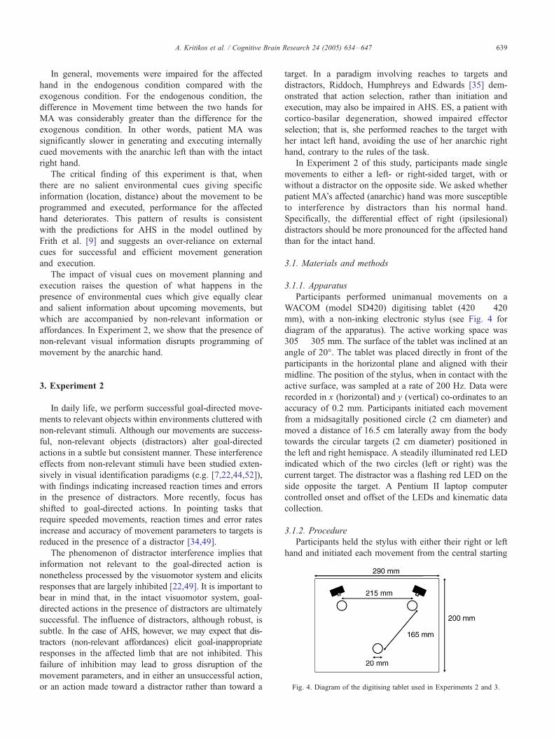
Fig. 4. Diagram of the digitising tablet used in Experiments 2 and 3.
3. Experiment 2
In daily life, we perform successful goal-directed move-
ments to relevant objects within environments cluttered with
non-relevant stimuli. Although our movements are success-
ful, non-relevant objects (distractors) alter goal-directed
actions in a subtle but consistent manner. These interference
effects from non-relevant stimuli have been studied exten-
sively in visual identification paradigms (e.g. [7,22,44,52]),
with findings indicating increased reaction times and errors
in the presence of distractors. More recently, focus has
shifted to goal-directed actions. In pointing tasks that
require speeded movements, reaction times and error rates
increase and accuracy of movement parameters to targets is
reduced in the presence of a distractor [34,49].
The phenomenon of distractor interference implies that
information not relevant to the goal-directed action is
nonetheless processed by the visuomotor system and elicits
responses that are largely inhibited [22,49]. It is important to
bear in mind that, in the intact visuomotor system, goal-
directed actions in the presence of distractors are ultimately
successful. The influence of distractors, although robust, is
subtle. In the case of AHS, however, we may expect that dis-
tractors (non-relevant affordances) elicit goal-inappropriate
responses in the affected limb that are not inhibited. This
failure of inhibition may lead to gross disruption of the
movement parameters, and in either an unsuccessful action,
or an action made toward a distractor rather than toward a
target. In a paradigm involving reaches to targets and
distractors, Riddoch, Humphreys and Edwards [35] dem-
onstrated that action selection, rather than initiation and
execution, may also be impaired in AHS. ES, a patient with
cortico-basilar degeneration, showed impaired effector
selection; that is, she performed reaches to the target with
her intact left hand, avoiding the use of her anarchic right
hand, contrary to the rules of the task.
In Experiment 2 of this study, participants made single
movements to either a left- or right-sided target, with or
without a distractor on the opposite side. We asked whether
patient MA’s affected (anarchic) hand was more susceptible
to interference by distractors than his normal hand.
Specifically, the differential effect of right (ipsilesional)
distractors should be more pronounced for the affected hand
than for the intact hand.
3.1. Materials and methods
3.1.1. Apparatus
Participants performed unimanual movements on a
WACOM (model SD420) digitising tablet (420 � 420
mm), with a non-inking electronic stylus (see Fig. 4 for
diagram of the apparatus). The active working space was
305 � 305 mm. The surface of the tablet was inclined at an
angle of 20-. The tablet was placed directly in front of the
participants in the horizontal plane and aligned with their
midline. The position of the stylus, when in contact with the
active surface, was sampled at a rate of 200 Hz. Data were
recorded in x (horizontal) and y (vertical) co-ordinates to an
accuracy of 0.2 mm. Participants initiated each movement
from a midsagitally positioned circle (2 cm diameter) and
moved a distance of 16.5 cm laterally away from the body
towards the circular targets (2 cm diameter) positioned in
the left and right hemispace. A steadily illuminated red LED
indicated which of the two circles (left or right) was the
current target. The distractor was a flashing red LED on the
side opposite the target. A Pentium II laptop computer
controlled onset and offset of the LEDs and kinematic data
collection.
3.1.2. Procedure
Participants held the stylus with either their right or left
hand and initiated each movement from the central starting

A. Kritikos et al. / Cognitive Brain Research 24 (2005) 634–647640
position. They were instructed to move quickly to the left or
right target position, corresponding to the steadily illumi-
nated LED, and to disregard the flashing LED that could
appear on the opposite side. There were four blocks of 40
trials, two blocks with the left hand and two with the right,
in an ABBA design. In each block, there were ten trials with
a left and ten with a right target alone and a further 10 trials
to each target in the presence of a simultaneous distractor on
the opposite side. Variables calculated were Initiation time
(time to initiate the movement), Movement time and the
Number of Submovements (cycles of acceleration and
deceleration) as a measure of Movement efficiency [29].
Planned comparisons were conducted separately on data
from MA and TM, with trials as observations. Preliminary
analysis showed that serial autocorrelation between succes-
sive trials was low (r < 0.2), thus permitting the use of
parametric statistical analyses.
3.2. Results and discussion
Visual inspection of the mean scores indicates that, in
general, consistent with predictions, movements of the left
hand were initiated and executed more slowly and were
less efficient than movements of the right hand for both
patient MA and the healthy control (Figs. 5A–C). Also
consistent with predictions, visual inspection of the mean
scores indicates that both participants in general initiated
and executed movements to targets more slowly when a
distractor was present, compared with movements to
targets alone (see Figs. 5A–C). These distractor effects
were only evident in the timing of movements; neither
patient MA nor the control moved to the distractor in any
of the trials. Visual inspection of patient MA’s performance
indicates that, averaged over hand and distractor presence,
he tended to initiate movements to the left hemispace more
slowly and less efficiently than movements to the right
hemispace.
We now consider specifically the effects of distractors on
movements made by the patient’s affected left hand and the
healthy control’s left hand.
3.2.1. Initiation time
Both patient MA and the healthy control initiated left-
hand movements to a left-sided target significantly more
slowly in the presence of a distractor on the right side,
comparedwith no distractor (t18 = 8.379,P < 0.0001 and t18 =
5.000, P < 0.0001 respectively; see Fig. 5A). Crucially, a
between-subjects comparison indicated that the distractor
Cost for left hand movements to left-sided targets was
significantly greater for MA than for the healthy control
(mean Cost = 462.63 SD = 297.02 and 98.42 SD = 92.09
respectively; t18 = 5.960, P < 0.0001), indicating that the
left (anarchic) hand of the patient was more susceptible to
non-relevant information than the left hand of the control.
Equally importantly, as a between-subjects comparison, the
distractor interference cost for the right hand moving to a
left-sided target was not significantly different for the patient
and healthy control (mean Cost = 337.37 SD = 255.74 and
129.47 SD = 79.63 respectively; t18 = 0.922, P > 0.05).
3.2.2. Movement time
For Movement time, a distractor interference effect was
not evident for either MA (t15 = 0.546, P > 0.05) or the
healthy control (t19 = 1.098, P > 0.05; see Fig. 5B).
Therefore, the distractor interference cost was also non-
significant for both patient MA and the healthy control
(t15 = 0.278, P > 0.05 and t17 = 0.031, P > 0.05
respectively). Moreover, as a between-subjects comparison,
the distractor interference cost for the right hand moving to
a left-sided target was not significantly different for MA
and the healthy control (t19 = 0.686, P > 0.05).
3.2.3. Number of Submovements
A distractor interference effect again was not evident for
the Number of Submovements for either the patient MA
(t16 = 1.191, P > 0.05) or the healthy control (t19 = 0.567,
P > 0.05; see Fig. 5C). Therefore, the distractor interfe-
rence cost was also non-significant for both MA and the
healthy control (t15 = 0.333, P > 0.05 and t17 = 0.438, P >
0.05 respectively). As a between-subjects comparison, the
distractor interference cost for the right hand moving to a
left-sided target was not significantly different for MA and
the healthy control (t19 = 1.000, P > 0.05).
In summary, the results for MA clearly indicate that the
presence of distractors disrupted initiation of movements by
the left hand to a left-sided target. Based on the between-
subjects comparison of the distractor interference cost
measure showed that this disruption was particularly marked
for MA’s left (anarchic) hand compared with his intact right
hand; that is, there was a significantly greater distractor
interference cost, as indicated by IT, for the left compared
with the right hand. This distractor interference cost
difference was not evident for the healthy control.
The findings of Experiment 2 are consistent with the
predictions of Frith et al. [9]: the anarchic left hand was
more sensitive than the intact right hand to the presence of
non-relevant information in the right hemispace. This would
indicate that patient MA had difficulty in selecting relevant
cues and suppressing non-relevant ones when planning and
programming actions with his anarchic hand, manifesting in
increased Initiation time.
The significant findings for Initiation time are also
consistent with the reaction time literature in distractor
interference, showing increased response times to targets in
the presence of distractors (e.g. [7,22,49]). Interestingly, in
the present experiment, these effects were significant only
for Initiation time, not for Movement time or the Number of
Submovements. This is likely to reflect an impairment
restricted to the planning and programming of movement
rather than to its execution. It should be noted, however, that
this is in contrast to findings in healthy controls in distractor
interference tasks, which show alterations in on-line

Fig. 5. Initiation time (A), Movement time (B) and Number of Submovements (panel C) for the left and right hand unimanual movements of participants MA
and TM in Experiment 2. RH = right hand, LH = left hand, RT = right target, LT = left target.
A. Kritikos et al. / Cognitive Brain Research 24 (2005) 634–647 641
execution parameters of movement [17,21,34]. This point
will be explored further in Conclusions.
4. Experiment 3
The second category of motor deficits in AHS is poor
programming and execution of actions requiring bimanual
coordination. Such actions involve transfer and integration
of information about object properties between the hemi-
spheres of the brain via the corpus callosum. The program-
ming and execution of bimanual movements, therefore, are
of particular interest here because the corpus callosum is
intact in patient MA.
Bimanual actions may be congruent (identical, mirror-
image movements to targets of the same level of difficulty)
or incongruent (either mirror-image movements of different
levels of difficulty or non-mirror-image movements). In the
intact visuomotor system, there is a cost relative to
unimanual actions associated with both congruent and
incongruent bimanual actions. Congruent movements tend
to be slower to initiate and to complete than the analogous
unimanual movements and have lower velocities [26].
Similar to congruent movements, incongruent movements
are also characterised by slower initiation and duration
[16,26]. Importantly, however, there is coupling between
the parameters of the two hands in incongruent movements
such that the parameters of the hand performing the

A. Kritikos et al. / Cognitive Brain Research 24 (2005) 634–647642
movement of lesser difficulty (for example, shorter
distance, lower weight) begin to approach the parameters
of the hand performing the movement of greater difficulty
(for example, longer distance, greater weight) and vice
versa [16,26]. Thus, in both congruent and incongruent
bimanual movements, parameters such as response time,
movement duration and amplitude tend to become
synchronised.
It has been suggested that bimanual synchronisation and
the tendency towards symmetry in incongruent bimanual
movements arise from neural cross-talk [41,45]. This concept
assumes that there is an interaction between pathways of
motor control at all levels of the CNS and that, for successful
bimanual coordination, information is exchanged at the
cortical level through the corpus callosum. Importantly,
cross-talk is postulated to occur at both the planning and
execution stages of bimanual control [16,26,45]. The
information exchanged is both temporal and spatial in nature.
Spijkers and Heuer [41] also argued that cross-talk has an
impact on temporal parameters. They had healthy partic-
ipants make bimanual symmetric movements to far and near
targets in the parallel plane, with a manipulandum grasped in
each hand. The authors demonstrated amplitude coupling,
that is, the amplitude of one hand depended on the amplitude
of the other hand. Moreover, the increasing frequency of
movement of one hand influenced the constant frequency of
the other hand.
In Experiment 3, we quantified bimanual control in
patient MA, using a modified version of the cued
movement task introduced in Experiment 2. In AHS after
corpus callosum damage, unwanted actions of the anarchic
hand disrupt the actions of the other hand during tasks
requiring bimanual movements. This particular impairment
is distinct from the unintended actions toward non-relevant
objects that may follow parietal lobe and SMA damage
[9]. Because the corpus callosum is intact in patient MA,
transfer of information between the sensorimotor areas of the
two hemispheres is possible; thus the two hands should be
able to act in concert. Importantly, however, the actions
performed by the anarchic left hand are impaired relative to
the intact right hand and relative to the actions of the healthy
control participant. This means that the level of difficulty of
the actions programmed and performed by the anarchic hand
is higher even for congruent, mirror-image actions. What we
would expect to see then is a synchronisation or coupling of
the parameters of the two hands, such that the movement
parameters of the intact right hand deteriorate and those of
the anarchic left hand would improve. In other words, an
averaging of the parameters of the two hands should be
evident, compared with a unimanual baseline.
4.1. Materials and methods
4.1.1. Apparatus
All movements were recorded on a WACOM SD420
digitising tablet, with targets being indicated by a steadily
illuminated LED. The layout of the start, and left and
right target positions, was the same as in Experiment 2
(see Fig. 4).
4.1.2. Procedure
Participants executed unimanual and bimanual move-
ments, from the midsagittal starting position to one or both
laterally placed targets. They held the electronic stylus with
the right or the left hand and a second plastic Fdummy_ penof similar colour, size and weight with the other hand.
Position information was not recorded from the Fdummy_pen. Participants were instructed to move quickly with the
left, right or both hands towards to the target position(s), as
cued by a steadily illuminated LED. The left hand always
moved to the left-sided target and the right hand always
moved to the right-sided target. On bimanual trials,
participants were instructed to move both hands simulta-
neously rather than consecutively. Four blocks of 20 trials
each were administered in an ABBA design; these included
two blocks with the active stylus in the left hand and two
blocks with the active stylus in the right hand. In each
block, ten trials included unilateral targets (five left, five
right) and ten included bilateral targets presented in
pseudorandom order. As in Experiment 2, dependent
variables were Initiation time, Movement time and Move-
ment efficiency. Planned comparisons (with Bonferroni
adjustment) were conducted separately on data from patient
MA and the healthy control, with trials as observations.
Preliminary analysis showed that serial autocorrelation
between successive trials was low (r < 0.2), thus
permitting the use of parametric statistical analyses.
4.2. Results and discussion
4.2.1. Initiation time
As expected, Initiation time for the healthy control was
significantly slower for bimanual movements than for
unimanual movements for both the left and right hands
(t18 = 3.197, P < 0.001 and t19 = 5.892, P < 0.0001 for left
and right hand respectively; see Fig. 6A).
For patient MA, however, Initiation time with the left
hand was not significantly different for unimanual versus
bimanual conditions (t19 = 2.132, P < 0.125; see Fig. 6A).
Mean Initiation time increased for the affected left hand
as well as for the intact right hand in the bimanual
compared with the unimanual condition. Importantly,
however, the relative direction of the deterioration in
performance in the bimanual condition was the reverse of
the unimanual condition.
Thus, it may be argued that, because no significant
deterioration was evident under bimanual conditions, the
performance of the affected left hand improved to unima-
nual levels when a simultaneous, congruent movement was
made with the intact right hand. Moreover, there was no
significant difference between the left and right hands under
bimanual conditions (t19 = 1.519, P > 0.0125), whereas

A. Kritikos et al. / Cognitive Brain Research 24 (2005) 634–647 643
right hand performance deteriorated in the bimanual versus
unimanual condition (t19 = 5.959, P < 0.0001; see Fig. 6A).
The performance of the left hand in bimanual actions was
consistent with this pattern: for the healthy control, there was
no significant difference in Initiation time for the left hand
versus the right hand (t18 = 1.788, P > 0.0125; see Fig. 6A).
Importantly, for patient MA, there was no difference in
Initiation time between right and left hands for the bimanual
condition, indicating that performance of the right hand was
scaled down to that of the left in the bimanual condition.
4.2.2. Movement time
For the healthy control, there was no significant
difference in Movement times between right and left hands
Fig. 6. Initiation time (A), Movement time (B) and number of Submovements (p
participants MA and TM in Experiment 3.
for bimanual movements (t18 = 1.716, P > 0.103), and
each hand had longer Movement times in bimanual
compared with unimanual conditions (t19 = 7.330, P <
0.01 and t19 = 3.197, P < 0.001, respectively, for right and
left hands; see Fig. 6B).
For patient MA, however, there was no re-scaling evident
in Movement times though the pattern for this parameter
indicated a greater general impairment of the left hand. As
expected, under bimanual conditions, the left hand was
significantly slower than the right (t19 = 3.289, P <
0.001; see Fig. 6B). The left hand was also slower during
bimanual than unimanual movements (t19 = 6.722, P <
0.0001; see Fig. 6B). Finally, there was no difference in
Movement time for the left hand under bimanual compared
anel C) for the left and right hand unimanual and bimanual movements of

A. Kritikos et al. / Cognitive Brain Research 24 (2005) 634–647644
with unimanual conditions (t19 = 7.407, P < 0.0001; see
Fig. 6B).
4.2.3. Number of Submovements
For the healthy control, the left and right hands made a
comparable Number of Submovements under bimanual
conditions (t18 = 2.535, P > 0.0125), and there was no
difference in the Number of Submovements in the
bimanual and unimanual conditions for the right hand
(t19 = 1.710, P > 0.0125). Finally, there was no difference
in the Number of Submovements in the bimanual
compared with the unimanual condition for the left hand
(t19 = 0.438, P > 0.0125).
In contrast, patient MA’s affected left hand was
generally more impaired than the right, and there was no
re-scaling evident for this parameter. The left hand made
more submovements than the right (t19 = 5.393, P <
0.0001; see Fig. 6C). Furthermore, under bimanual
conditions, the left hand made more submovements than
the right (t19 = 5.329, P < 0.0001). Finally, under
bimanual conditions, the right hand made more submove-
ments than under unimanual conditions (t19 = 4.982, P <
0.0001; see Fig. 6C).
We had predicted that, due to a putative cross-talk
mechanism, the pattern in bimanual actions would be
similar to that of the control, and parameters of the left and
right hands should be equalised. This was borne out for
Initiation time only, such that, in the congruent bimanual
actions performed, Initiation time for the right hand was
slowed or scaled down to match that of the affected left
hand. This pattern conforms to the predictions of Heuer et
al. [16] and Marteniuk et al. [26]: whereas parameterisation
of the left anarchic hand improves to match more closely
that of the right hand, the parameterisation of the right
hand deteriorates or is scaled down to that of the left.
These findings indicate that the actions of both the
anarchic and intact hands are affected during bimanual
coordination in AHS.
The fact that only Initiation time showed this scaling
down for the right hand has implications for the level at
which neural cross-talk impacts on action in AHS. It has
been suggested that cross-talk occurs both during the
programming and execution of action [16,26,45]. In our
study, however, only Initiation time was affected in the
predicted manner. If Initiation time is assumed to reflect
processes in the planning of action (e.g. [16]), then it may be
postulated that, in AHS, cross-talk impacts on actions at the
level of programming but not beyond.
The pattern of findings in this experiment may also
suggest a trade-off between Initiation time and Movement
time and Efficiency. Whereas left hand performance
improves in terms of Initiation time in the bimanual
condition relative to the unimanual condition, thereafter,
the movement is slower and less efficient. The reverse may
be true for the right hand: Initiation time deteriorates, but
Movement time and Efficiency are unaffected.
5. Conclusions
The purpose of this series of experiments was to
investigate (a) the role of relevant and irrelevant environ-
mental information in movement generation; and (b)
bimanual coordination in a patient with anarchic hand
syndrome, within the context of recent influential models
of visuomotor control. The findings of Experiments 1 and
2 support our postulation of the crucial role of stimuli in
the environment in movement generation. In Experiment
1, we compared performance for exogenously and
endogenously triggered movements in a visually cued,
sequential button-pressing task. For patient MA, perform-
ance of the left hand was selectively impaired in the
endogenous condition, in which movements had to be
initiated internally prior to the appearance of an external
visual cue.
For both patient MA and the healthy control, salient
cues giving information about the location of forthcoming
targets facilitated performance when they were presented
prior to movement initiation. When such cues were
presented after movement initiation, however, performance
deteriorated. This pattern is consistent with previous
reports [29,30]. In this study, moreover, the Efficiency
Cost of internal movement generation was exaggerated in
patient MA, and particularly in his anarchic left hand,
indicating that movement generation relies excessively on
externally provided information.
In Experiment 2, we compared the effects of visual
distractors for unimanual actions directed towards targets
in the left and right hemispace. The presence of a distractor
slowed movement initiation times for actions performed
with the right and left hands for both patient MA and the
healthy control. For patient MA, actions to the left
hemispace were adversely affected, consistent with residual
left-sided inattention. The important finding from this
experiment, however, was that MA’s anarchic left hand
was particularly vulnerable to disruption by non-relevant
cues. Again, this interference from distractors is a robust
phenomenon in healthy controls, as seen in Experiment 2
of this paper and in other studies (e.g. [7,17,34]). The
distractor interference cost in Initiation time was exagger-
ated, however, for MA’s anarchic hand. Moreover, the
effect was in excess of any impairment attributable to left-
sided inattention.
Thus, the main deficit in motor control that MA
demonstrated in Experiments 1 and 2 was heightened
susceptibility environment stimuli leading to increased
activation of representations and therefore actions. This
was indicated by slow movement initiation, regardless of
whether the stimuli were relevant (Experiment 1) or
irrelevant (Experiment 2). The superfluous actions are not
suppressed by intended actions.
According to the model proposed by Frith et al. [9],
knowledge of the actual state is based on the current motor
commands and sensory feedback, while the knowledge of

A. Kritikos et al. / Cognitive Brain Research 24 (2005) 634–647 645
the predicted state is an estimate based on future
commands. The end goal or desired state is also held
concurrently in the system. In patient MA, correct
information regarding the desired state, the actual state
and the predicted state was available at all times to the
visuomotor system. Moreover, the feedback mechanisms
between these three states were also intact; thus a
comparison of any discrepancy and the ability to make
corrections was possible. Therefore, although he com-
mented on the actions of his anarchic hand, MA did not
Fdisown_ its actions. That is, he recognised a discrepancy
between what his hand was doing and what he wished it to
do but did not attribute this discrepancy to an external
cause.
We argue that the essential deficit in patients with
anarchic hand syndrome, as shown by distractor interfer-
ence paradigms (this series of experiment s and [35]), is
increased susceptibility to non-relevant cues in the
environment and thus impaired selection of appropriate
motor programmes. That is, inappropriate actions are
elicited, initiated and executed by patients with AHS.
Once the programmes have been selected, however, they
are executed successfully, that is, they are ‘‘purposeful’’
and made towards a specific object in the environment.
These executed programmes, particularly in the acute
stages of the syndrome, may not be wanted or appropriate,
and hence the hand performing them is described as
Fanarchic_.The pattern of findings are consistent with this anarchic
hand syndrome as a disorder of intrafrontal disconnection
(see Boccardi et al. [2]). While the lateral premotor cortex
is involved in externally guided movements (that is,
actions guided by environmental stimuli), the medial
system or SMA is involved with internally guided actions
and the inhibition of unwanted, inappropriate responses to
environmental stimuli. Thus, if the SMA is damaged, the
contralateral premotor cortex is susceptible to external
stimuli and the actions they generate.
In Experiment 3, we examined bimanual coordination
for actions directed to simultaneous left- and right-sided
targets. For bimanual movements, initiation times
increased for both the anarchic left hand for the intact
right hand in patient MA, but the relative direction of the
increase was the opposite to that of unimanual movements.
This pattern is consistent with coupling between the two
hands for this parameter. No such coupling was evident for
the healthy control. The findings of Experiment 3 show
that coupling and therefore synchronisation of the para-
meters of the two hands occurred in AHS, as would be
expected when the corpus callosum is intact and informa-
tion can be transferred across the two hemispheres. Thus,
we argue that poor bimanual coordination of congruent
movements in AHS is due to the impaired paramete-
risation of the anarchic hand impacting on the parameters
of the intact hand. This is an important finding not
previously reported in the AHS literature and has
implications for the actual mechanisms of bimanual
incoordination in AHS.
As described above, synchronisation of movement in
incongruent bimanual movements has been attributed to
neural cross-talk [41,45]. Cross-talk is postulated to occur at
both the planning and execution stages of bimanual control
[26,45]. The information exchanged is both temporal and
spatial in nature. Recent work, however, indicates that cross-
talk may occur only at the planning stage of movement
production and is reflected by alterations in Initiation time
[16].
In general, findings emphasise the crucial role of
environmental stimuli in goal-directed actions in AHS.
Goal-directed actions made towards objects within our
environment are modified by the presence or absence of
relevant and irrelevant information. In particular, damage
to the visuomotor system leads to increased disruption by
non-relevant information in the affected limb. Finally, in
the case of bimanual synchronous goal-directed actions to
target objects, the movement of the affected hand
improves.
Acknowledgments
We are grateful to MA and TM, for giving us their time
and for their patience throughout the testing sessions. We
also thank Professor John Bradshaw for the use of the
technical equipment, Ms. Nadja Berberovic for her help
with the literature search, Drs. Chris Chambers and Mark
Williams for their insightful comments during the prepa-
ration of the manuscript and the two anonymous reviewers
for their suggestions in the revision process.
References
[1] K. Baynes, M.J. Tramo, A.G. Reeves, M.S. Gazzaniga, Isolation of a
right hemisphere cognitive system in a patient with anarchic (alien)
hand sign, Neuropsychologia 8 (1997) 1159–1173.
[2] E. Boccardi, S. Della Sala, C. Motto, H. Spinnler, Utilisation
behaviour consequent to bilateral SMA softening, Cortex 38 (2002)
289–308.
[3] S. Brion, C.P. Jedynak, Le signe de la main etrangere, Rev. Neurol.
126 (1972) 257–266.
[4] M.-P. Deiber, R.E. Passingham, J.G. Colebatch, K.J. Friston, P.D.
Nixon, R.S.J. Frackowiak, Cortical areas and the selection of move-
ment, Exp. Brain Res. 84 (1991) 393–402.
[5] S. Della Sala, C. Marchetti, H. Spinnler, Right-sided anarchic
(alien) hand: a longitudinal study, Neuropsychologia 29 (1991)
1113–1127.
[6] S. Della Sala, C. Marchetti, H. Spinnler, The anarchic hand: a fronto-
mesial sign, in: F. Boller, J. Grafman (Eds.), Handbook of Neuro-
psychology, vol. IX, Amsterdam, Elsevier, 1994.
[7] B.A. Eriksen, C.W. Eriksen, Effects of noise letters upon the
identification of a target letter in a non-search task, Percept.
Psychophys. (1974) 143–149.
[8] T.E. Feinberg, R.J. Schindler, S. Gilson, N. Flanagan, L. Haber, Two
alien hand syndromes, Neurology 4 (1992) 19–24.
[9] C.D. Frith, S.-J. Blakemore, D.M. Wolpert, Abnormalities in the

A. Kritikos et al. / Cognitive Brain Research 24 (2005) 634–647646
awareness and control of action, Philos. Trans. R. Soc. Lond., Ser. B
Biol. Sci. 355 (2000) 1771–1788.
[10] C. Gerloff, B. Corwell, R. Chen, M. Hallett, G. Cohen, Stimulation
over the human supplementary motor area interferes with the
organization of future elements in complex motor sequences, Brain
120 (1997) 1587–1602.
[11] D.H. Geschwind, M. Iacobini, M.S. Mega, D.W. Zaidel, T. Cloughesy,
E. Zaidel, Alien hand syndrome: interhemispheric motor disconnec-
tion due to a lesion in the midbody of the corpus callosum, Neurology
43 (1995) 802–808.
[12] G. Goldberg, Supplementary motor area structure and function: review
and hypotheses, Behav. Brain Sci. 8 (1985) 567–616.
[13] G. Goldberg, K. Bloom, The alien hand sign. Localization,
lateralisation and recovery, Am. J. Phys. Med. Rehabil. 69 (1990)
228–238.
[14] M.A. Goodale, A.D. Milner, L.S. Jakobson, D.P. Carey, A neuro-
logical dissociation between perceiving objects and grasping them,
Nature 349 (1991) 154–156.
[15] U. Halsband, N. Ito, J. Tanji, H.-J. Freund, The role of premotor cortex
and the supplementary motor area in the temporal control of move-
ment in man, Brain 116 (1993) 243–266.
[16] H. Heuer, W. Spijkers, T. Kleinsorge, H. Van Der Loo, C. Steglich,
The time course of cross-talk during the simultaneous specification
of bimanual movement amplitudes, Exp. Brain Res. 118 (1998)
381–392.
[17] L.A. Howard, S.P. Tipper, Hand deviations away from visual
cues: indirect evidence for inhibition, Exp. Brain Res. 113 (1997)
144–152.
[18] M. Husain, J.B. Mattingley, C. Rorden, C. Kennard, J. Driver,
Reaching in parietal neglect: response to D.P. Carey: Action,
perception, cognition, and the inferior parietal cortex, Trends Cogn.
Sci. 2 (1998) 164–166.
[19] M. Husain, J.B. Mattingley, C. Rorden, C. Kennard, J. Driver,
Distinguishing sensory and motor biases in parietal and frontal
neglect, Brain 123 (2000) 1643–1659.
[20] M. Jeannerod, D. Decety, F. Michel, Impairment of grasping move-
ment following a bilateral posterior lesion, Neuropsychologia 32
(1994) 369–380.
[21] A. Kritikos, K.M.B. Bennett, J. Dunai, U. Castiello, Interference from
distractors in reach-to-grasp movements, Q. J. Exp. Psychol. 53 (2000)
131–151.
[22] N. Lavie, Y. Tsal, Perceptual load as a major determinant of the
locus of selection in visual attention, Percept. Psychophys. 56
(1994) 183–197.
[23] F. Macar, F. Vida, L. Casini, The supplementary motor area in motor
timing: evidence from slow brain potential changes, Exp. Brain Res.
125 (1999) 271–280.
[24] C. Marchetti, S. Della Sala, Disentangling the alien and anarchic hand,
Cogn. Neuropsychiatry 3 (1998) 191–207.
[25] C.D. Marsden, The mysterious motor function of the basal ganglia: the
Robert Wartenberg lecture, Neurology 323 (1982) 514–539.
[26] R.G. Marteniuk, C.L. Mackenzie, D.M. Baba, Bimanual movement
control: information processing and interaction effects, Q. J. Exp.
Psychol. 36a (1984) 335–365.
[27] J. Martı-Fabregas, J. Kulisevsky, E. Baro, G. Mendoza, C. Valencia,
J.-L. Martı-Vilalta, Alien hand sign after a right parietal infarction,
Cerebrovasc. Dis. 10 (2000) 70–72.
[28] J.B. Mattingley, J. Driver, Distinguishing sensory and motor deficits
after parietal damage: an evaluation of response selection biases in
unilateral neglect, in: P. Thier, H.-O. Karnath (Eds.), Parietal Lobe
Contributions to Orientation in 3D Space, Springer-Verlag, Heidel-
berg, 1997, pp. 309–338.
[29] J.B. Mattingley, J.L. Bradshaw, J.G. Phillips, Impairments of
movement initiation and execution in unilateral neglect:
directional hypokinesia and bradykinesia, Brain 115 (1992)
1849–1874.
[30] J.B. Mattingley, L.A. Corben, J.L. Bradshaw, J.A. Bradshaw, J.G.
Phillips, M.K. Horne, The effects of competition and motor
reprogramming an visuomotor selection in unilateral neglect, Exp.
Brain Res. 120 (1998) 243–256.
[31] J.B. Mattingley, N. Berberovic, L. Corben, M.J. Slavin, M.E.R.
Nicholls, J.L. Bradshaw, The greyscales task: a perceptual measure of
attentional bias following right hemisphere damage, Neuropsycholo-
gia 42 (2004) 387–394.
[32] A.D. Milner, M.A. Goodale, The Visual Brain in Action, Oxford Univ.
Press, Oxford, 1995.
[33] H. Mushiake, I. Masahiko, J. Tanji, Neuronal activity in the primate
premotor, supplementary, and precentral motor cortex during visually
guided and internally determined sequential movements, J. Neuro-
physiol. 66 (1991) 705–718.
[34] J. Pratt, A. Abrams, Action-centred inhibition: effects of distractors
on movement planning and execution, Hum. Mov. Sci. 13 (1994)
245–254.
[35] M.J. Riddoch, G.W. Humphreys, M.G. Edwards, Neuropsychological
evidence distinguishing object selection from action (effector) selec-
tion, Cogn. Neuropsychol. 17 (2000) 547–562.
[36] G. Rizzolatti, G. Gentilucci, M. Fogassi, G. Luppino, M. Matelli, S.
Ponzoni-Maggi, Neurons related to goal-directed motor acts in
inferior area 6 of the macaque monkey, Exp. Brain Res. 67 (1987)
220–224.
[37] G. Rizzolatti, R. Camarda, M. Fogassi, G. Gentilucci, G. Luppino, M.
Matelli, Functional organisation of inferior area 6 in the macaque
monkey ii: area f5 and the control of distal movements, Exp. Brain
Res. 71 (1988) 491–507.
[38] H. Sakata, M. Taira, M. Kusunoki, A. Murata, Y. Tanaka, The parietal
association cortex in depth perception and visual control of hand
action, Trends Neurosci. 20 (1997) 350–357.
[39] D.J. Serrien, A.C. Nirkko, A. Lovblad, M. Wiesendanger, Damage to
the parietal lobe impairs bimanual coordination, NeuroReport 12
(2001) 2721–2724.
[40] E. Shikata, Y. Tanaka, H. Nakamura, M. Taira, H. Sakata, Selectivity
of the parietal visual neurones in 3d orientation of surface of
stereoscopic stimuli, NeuroReport 7 (1996) 2389–2394.
[41] W. Spijkers, H. Heuer, Structural constraints on the performance of
symmetrical bimanual movements with different amplitudes, Q. J.
Exp. Psychol. 48a (1995) 716–740.
[42] O. Spreen, E. Strauss, A Compendium of Neuropsychological Tests—
Administration, Norms, and Commentary, Oxford Univ. Press, New
York, 1991.
[43] K.M. Stephan, F. Binofski, U. Halsband, C. Dohle, G. Wunderlich, A.
Scnitzler, P. Tass, G. Posse, H. Herzog, V. Sturm, K.S. Zilles, R.J.
Seitz, H.-J. Freund, The role of ventral medial wall motor areas in
bimanual coordination: a combined lesion and activation study, Brain
122 (1999) 351–368.
[44] J.R. Stroop, Studies of interference in serial verbal reactions, J. Exp.
Psychol. 18 (1935) 643–662.
[45] S.P. Swinnen, Intermanual coordination: from behavioural principles
to neural-network interpretations, Nat. Rev. 3 (2002) 350–359.
[46] M. Taira, A.P. Georgopoulos, A. Murata, H. Sakata, Parietal cortex
neurons of the monkey related to the visual guidance of movement,
Exp. Brain Res. 83 (1990) 29–36.
[47] J. Tanji, K. Okano, K.C. Sato, Relation of neurons in the
nonprimary motor cortex to bilateral hand movement, Nature 327
(1987) 618–620.
[48] J. Tanji, K. Okano, K.C. Sato, Neuronal activity in cortical motor areas
related to the ipsilateral, contralateral, and bilateral digit movements of
the monkey, J. Neurophysiol. 60 (1988) 325–343.
[49] S.P. Tipper, L.A. Howard, S.R. Jackson, Selective reaching to
grasp: evidence for distractor interference effects, Vis. Cogn. 4
(1997) 1–38.
[50] C. Trevarthen, Integrative functions of the cerebral commissures, in:
R.D. Nebes, S. Corkin (Eds.), Handbook of Neuropsychology,
Elsevier, New York, 1991, pp. 49–83.
[51] L. Trojano, C. Crisci, B. Lanzillo, R. Elefante, G. Caruso, How many

A. Kritikos et al. / Cognitive Brain Research 24 (2005) 634–647 647
alien hand syndromes? Follow-up of a case, Neurology 43 (1993)
2710–2712.
[52] G. Underwood, Semantic interference from unattended printed words,
Br. J. Psychol. 67 (1976) 327–338.
[53] M.G. Ventura, S. Goldman, J. Hildebrand, Alien hand syndrome
without a corpus callosum lesion, J. Neurol., Neurosurg. Psychiatry 58
(1995) 735–737.
[54] D.M. Wolpert, M. Kawato, Multiple paired forward and inverse
models for motor control, Neural Netw. 11 (1998) 1317–1329.
[55] D.M. Wolpert, Z. Gharamani, M.I. Jordan, An internal model for
sensorimotor integration, Science 269 (1995) 1880–1882.
[56] D.M. Wolpert, S.J. Goodbody, M. Husain, Maintaining internal
representations: the role of the human superior parietal lobe, Nat.
Neurosci. 1 (1998) 529–533.


