Embed Size (px)
Citation preview
META-IODOBENZYLGUANIDINE:
LNVESTIGATIONS INTO A M E C W I S M OF ACTION
AND APPLICATION TO RESTENOSIS
LORRAINE YAU
A Thesis
Submitted to the Faculty of Graduate Studies
in Partial Fulfillment of the Requirements
for the Degree of
DOCTOR OF PHILOSOPHY
lnstitute of Cardiovascular Sciences
Department of Physiology, Faculty of Medicine
University of Manitoba
Winnipeg, Manitoba
O January, 200 1

National Library Bibliothèque nationale du Canada
Acquisitions and Acquisitions et Bibliogmphic Services services bibliographiques 395 Wellington Street 395. rue Wellington OttawaON K1AON4 Ottawa ON K I A ON4 Canada Canada
Y w r file Votre rëkirenœ
Our fïk Notre rBrence
The author has granted a non- exclusive licence aliowing the National Library of Canada to reproduce, loan, distriiute or sell copies of this thesis in microform, paper or electronic formats.
The author retains ownership of the copyright in this thesis. Neither the thesis nor substantid extracts fkom it may be printed or othervvise reproduced without the author's permission.
L'auteur a accorde une licence non exclusive permettant à la Bibliothèque nationale du Canada de reproduire, prêter, distribuer ou vendre des copies de cette thèse sous la fome de microfiche/nlm, de reproduction sur papier ou sur format électronique.
L'auteur conserve la propriété du droit d'auteur qui protège cette thèse. Ni la thése ni des extraits substantiels de celle-ci ne doivent être imprimés ou autrement reproduits sans son autorisation.
THE UNIVERSITY OF MANITOBA
FACULTY OF GRADUATE STUDIES *f ***
COPYRIGHT PERMISSION PAGE
Me&-Iodobenzylguanidine: Investigations into a Mechanism of Action
and Application to Restenosis
Lorraine Yau
A ThesislPracticum submitted to the Facuity of Graduate Studies of The University
of Manitoba in partial fulnllment of the requirements of the degree
of
Doctor of Philosophy
LORRAINE YAU 02001
Permission has been granted to the Library of The University of Manitoba to lend or seil copies of this thesidpracticum, to the National Library of Canada to micronlm this thesis and to Iend or s a copies of the film, and to Dissertations Abstracts International to publish an abstract of this thesis/practicum.
The author reserves other publication rights, and neither this thesis/practicum nor extensive extracts fkom it may be printed or otherwise reproduced without the author's written permission.

This body of work is dedicated to:
My parents who have always believed in me and been my biggest fans;
My ioving husband who has always been my pillar of strength;
My daughter/child(ren) who will always be my greatest joy;
My fnends who never let me give up hope;
My Lord and Saviour, Jesus Christ, Who gave me a new life and purpose to go on.
The wise man accomplishes what must be done without dwelling on it.
- Lao T m
In the pursuit of learning, everyday sornething is acquired.. .
- Lao Tzu
There is a limit to our life, but there is no limit to knowledge.
- Chuang Tzu
TABLE OF CONTENTS
ACKNOWLEDGEMENTS ....................................................................... x ............................................................... ............. LIST OF FIGURES ,.., xi
LIST OF TABLES ................................................................................ xvi
LIST OF ABBREVIATIONS .................................................................. xvii
...................................................................................... ABSTRACT xxi
...................................................................................................... 1 . 0 INTRODUCTION 1
..................................................................................... 2-0 REVIEW OF LITERATURE 6
2.1 Heart disease in North Arnerica .................................................................................. 6
2.2 Coronxy artery disease ............................................................................................... 6
2.2.1 Definition of coronary artery disease .................................................................. 6
2.2.2 Classification of coronary artery disease ............................................................ 7
2.2.3 Interventions for the treatment of coronary artery disease ...................... ..... 8
2.2.4 Surgical treatrnent of coronary artery disease ..................................................... 9
2.2.5 Non.surgica1, non-medical treatrnents for coronary artery disease .................. 10
2.2.5.1 Percutaneous transluminal coronary angioplasty .......... .,.,. .......................... 11
2.2.5.2 Coronary stenting .......................................................................................... 11
2.2.5.3 Atherectomy .................................................................................................. 12
2.3 Revasculanzation-induced restenosis ....................................................................... 12
2.3.1 Pathogenesis of restenosis ................................................................................ 14
2.3.2 Wound healing as the paradigm for restenosis ................................................. 16
2.3 -3 Animal models of restenosis ................................................................................ 17
2.3.3.1 Rat carotid injury mode1 of restenosis .......................................................... 17
2.3.3.2 Larger animal models of restenosis ........................................................... 19
2.3.4 Benefits of animals models of restenosis ......................................................... 20 . .
2.4 Vascular injury and remodeling ................................................................................ 20
2.4.1 Remodeling and restenosis ............................................................................... 21
2.5 Pharmacological approaches for the prevention of restenosis .................................. 23
2.5.1 Perspectives on pharmacological treatment of restenosis .............................. 25
2.6 Vascular smooth muscle cells ................... .-. ........ .-... ........................................... 26
2-6-1 Multifbnctional nature of smooth muscle cells ................................................. 26
2.6.2 Smooth muscle ce11 phenotypic modulation ..................................................... 27
2.6.3 Smooth muscle ce11 culture systems ................................................................. 29
............................................. 2.6.4 Regulation of smooth muscle ce11 differentiation 29
........... 2.6.5 Factors controlling the phenotypic modulation of smooth muscle cells 30
2.6.6 Induction o f smooth muscle ce11 proliferation (and migration) .................... .... 30
2.6.7 Factors affecting smooth muscle proliferation (and migration) ....................... 32
1.6.7.1 Hormones ...................................................................................................... 33
2.6.7.2 Eicosanoids .................................................................................................. 34
.............. 2.6.8 Ce11 signaling systems involved in injury and the response of SMCs 34
2.6.8.1 Phosphatidylinositol 3-kinase ....................................................................... 35
2.6.8.2 p21-Ras ......................................................................................................... 36
2.6.8.3 MAP kinase ................................................................................................... 37
2.6.9 New frontiers in ceil signaling: Regulation by non-traditional signal
molecules.. ........................................................................................................ 38
.................................................................................................. 2.7 ADP-ribosylation 4 0
..................................................................... 2.7.1 Poly(ADP-nbosy1)ation reactions 40
2.7.1 . 1 Poly(ADP-ribose) polymerase (PARP) ....................................................... 41
2.7.1.2 Enzymology of PARP ......................... .. .................................................... 42
................................... 2.7.1.3 PARP substrates .........,.............................................. 42
2.7.1.4 Function of PARP within cells .................................................................. 43
2.7.1 -5 PARP in the cardiovascular system ............................................................. 45
2.7.1.6 Lessons learned from PARP knockout mice .............................................. 45
.................................................................. 2.7.2 Mono(ADP-ribosy1)ation reactions 46
2.7.2.1 Mono(ADP-ribosy1)transferase (rnART) ...................................................... 50
.................................................................................. 2.7.2.2 Enzymology of mART 51
........................................................................................... 2.7.2.3 mART substrates 52
2.7.2.4 Function of rnART within cells ................................................................... 53
2.7.2.5 Mono(ADP-ribosy1)ation in the cardiovascular system ............................... 56
2-8 Inhibitors of mART and PARP ................................................................................ 57
2.8.1 MIBG ................... ..-. ......................... ... .... 58
2.8.1.1 Biodistribution and phannacokinetics of MIBG ...................................... 59
2.8.1.2 Cellular uptake and retention of MBG ........ .......... ...................................... 60
2.8.1.3 Metabolism of MIBG .................................................................................. 62
2.8.1.4 ExcretiodElimination of MIBG ................................................................... 62
2.8.1.5 Toxicity of MLBG ......................................................................................... 63
2.8.1.6 Clinical application of MIBG ............................... .....-. ........... 64
2.8.1.7 Clinical application of MIBG in the cardiovascular system ......................... 66
2.8.1.8 Mode of action of M B G .............................................................................. 67
2.8.1.9 MIBG as an inhibitor of arg-mART ........................................................... 69
................................................................................................. 2-9 General Perspectives 70
........... 3.0 STATEMENT OF THE PROBLEM: HYPOTHESIS AND OBJECTIVES 72
.............................................................................. 4.0 MATERIALS AND METHODS 74
4.1 Materials ................................................................................................................... 74
4.2 Experimental Systerns ............................................................................................... 77
4.2.1 H4LIE ce11 culture .............................................................................................. 77
4.2.2 L6 myoblast/tube ce11 culture ............................................................................ 78
4.2.3 Primary smooth muscle ceIl cufture .................................................................. 78
4.2.4 In vivo pig mode1 - fernoral angioplasty ....................................................... 79
4.2.4.1 Femoral angiography .............................................. ,., . . . 80
4.3 Ce11 growth and viability assays ............................................................................... 81
...... 4.3. I Radiotracer incorporation for measurements of DNA and RNA synthesis 81
4.3.2 Radiotracer incorporation for measurement of cell cycle re-entry ................... 82
................................................................................................... 4.3.4 CelI number II 83
4.3.5 Brornodeoxyuridine (BrdU) ce11 labeling ....... .. ................................... .. .......... 83
4.3.6 FACS analysis ................................................................................................... 84
4.3 -7 LDH assay ........................................... .. ........................................................ 85
4.3.8 [ '~ ]~ lucose uptake assay .................................................................................. 85
.................................................................................................... 4.4 Ce11 differentiation 86
4.5 Cell migration (Boyden chamber) ........................................................................... 86
* * 4.6 Fibnn glue preparation ..... .. ....................................................................................... 87
4-7 Histology ........ .. ......................................................................................................... 88
4.7.1 Paraformaldehyde preparation ......... ., ............................................................... 88
4.7.2 Histology of tissue sections ............................................................................. 88
...................................................................................... 4.7.3 Immunocytochemistry 88
4.7.4 Photography ..................................................................................................... 89
....................................................................................... 4.8 Protein analysis techniques 89
4.8.1 BCA protein assay ......................................................................................... 89 . . . ......................................................................................... 4.8.2 Immunoprecipita~on 90
4.8.3 Immunoblotting (Western blotting) ............................................................ 90
4.8.4 SubcelluIar fractionation ................................................................................... 91
........................................................................................ 4.8.5 Ligand binding assay 92
............................................................................... 4.8.6 Nuclear extract preparation 92
4.8.7 UV-crosslinking ................................................................................................ 93
4.9 Enzyme Assays ......................................................................................................... 93
Activity gel MAP kinase assay .................... .. ............................................... 93
In vitro MAP kinase assay ........................................................................... 94
................................ Phosphatidylinositol 3-kinase (PD-kinase) assay, in vivo 94
PI3-kinase assay, Ni vitro .................................................................................. 95
...................................................................................... p2 1 -Ras activity assay 96
Poly(ADP-ribose) polymerase (PARP) assay ................................................. 97
Mono(ADP-ribosy1)transferase (mART) assay ................................................ 97
Metabolic labeling of mono(ADP-ribosy1)ated proteins .................................. 98
In situ gel assay for mART ............................................................................... 99
In situ l abe lhg of intact cells for detection of an extracel1uIar mART ........... 99
.................................................................................... 4.1 O Nucleic acid manipulations 100
4.1 O . 1 OIigodeoxynucleotide preparation .................................................................. 100
4.10.2 RNA preparation ............................................................................................. 100
4.10.3 RT-PCR ............................................. ... ................. 101
4 1 1 Data measurement and statistical analysis ............................................................. 101
5.0 H4LE RAT HEPATOMA CELLS AS A MODEL OF CELL
PROLIFERATION ................................................................................................ 103
5.1 Introduction .............................................................................................................. 103
...... 5-2 Mitogenic actions of insulin and IGF-1 ........................................................... 106
5.2.1 Background and rationale ............................................................................. 1 O6
.............. 5.2.2 Specific aims .. .......................................... ...................................... 108
. ........... 5.2.3 Experimental design .................................................................... 109
5.2-4 Results ............................................................................................................ 109
5.2.4.1 Growth charactenstics of WIIE cells in response to insulin ................-..... 109
5.2.4.2 Growth and metabolic response of H4IIE cells to insulin and IGF-1 ...,..... 115
... 5.2.4.3 Signaling components of the H4IIE response to insulin and IGF- I .....,.. 1 18
5-2.4.4 Effect of an inhibitor of MEK (PD98059) on insulin-mediated H4II.E
......................................................................................................... growth 128
5.2.5 Discussion ............................................................... . . . . . . . . . . . . . . . 133
5.3 Contribution of ADP-ribosylation to H4IIE cell proliferation .........................-..... 140
5.3.1 Background and rationale ........................................................... ............ 140
5.3 -2 Specific aims ............................................................................ ...............-... 142
............ ............. 5.3.3 Experimental design ... ................................. 142
5.3.4 Results ............................................................................................... . . . . 143
5.3.4.1 Insulin stimulates ADP-ribosylation ........................................................... 143
5.3.5 Discussion ... ................................................................................................... 159
5-4 Modulation of ceIl proliferation by inhibitors of mART ........................................ 162
5.4.1 Background and rationale ..................................................................... . 162
5.4.2 Specific Aims . ............................................................................................. . 163
5 .4.3 Experimental design ........................................................................................ 164
5.4.4 Results ......................................................................................................... . 164
5.4.4.1 Effects of ADP-ribosylation inhibitors on ce11 growth:
involvement of mART in H41IE ce11 growth and proliferation ............-..... 164
5.4.4.2 Toxicity of MIBG ................................. .... ...................................... 178
5.4.4.3 Mechanism of action of MLBG .. .............................................................. 189
5.4.5 Discussion ................................................................ ...................................... 194
5.5 Summary ................................................................................................................. 201
.......... 6-0 L6 SKELETAL MYOBLASTS AS A MODEL OF DIFFERENTIATION 204
6.1 Introduction ....... ... ................................................................................................ 204
........... 6.2 Contribution of mono(ADP-ribosy1)transferase to myogenesis .. ................ 208
...................................................................................... 6.2.1 Background/rationale 208
6.2.2 Specific Aims ................. ... ....................................................................... 209
6.2.3 Experimental Design ....................................................................................... 209
6.2.4 Results ................... .-. ...................... .... .................................................... ,,.. 209
6.2.4.1 Differentiation ofL6 skeletal myoblasts into rnyotubes ............ .. ............ 209
6.2.4.2 ADP-nbosylation inhibitors and their effect on L6 skeletal
myoblast differentiation ...... .. ...................................................................... 210
............... ................ 6.2.4.3 Expression of myogenic proteins affected by MIBG ,. 230
......................................................... 6.2.4.4 ADP-ribosylation and differentiation 238
....................................................................................................... 6.2.5 Discussion 243
6.2.6 Summary ....................................................................................................... 250
7.0 SMC AS A MODEL OF DE-DIFFERENTIATION, PROLIFERATION
AND MIGRATION ................................................................................................ 251
7.1 Introduction ............................................................................................................. 251
.............................. 7.2 Mitogenic response of SMC's to growth factors: role of PGE2 254
...................................................................................... 7.2.1 Background/rationale 254
7.2.2 Specific Aims .................................................................................................. 256
........ ............................................................................. 7.2.3 Experimental Design .. 256
.............................. ........................................................................... 7.2.4 Results ... 257
7.2.4.1 Growth characteristics of SMCs in response to serurn and
other growth factors .................................................................................... 257
............................................................ 7.2.4.2 Growth response of SMCs to PGEz 263
7.2.4.3 Activation of MAP kinase and p2 1-Ras by PGEl ........................... ... ........ 282
7.2.4.4 Involvement of PI3-kinase in PGEz-dependent SMC growth .................... 294
7.2.5 Discussion ...................................................................................................... 297
7.3 ADP-ribosylation and SMC proIiferation ........ .. ..................................................... 316
...................................................................................... 7.3.1 Backgroundrationale 316
7-3 -2 Specific Aims .................................................................................................. 317
7.3 -3 Expenrnental Design ....................................................................................... 317
............................................................................................................. 7.3.4 R e s t s 317
7.3.4.1 Activation of an arg-mART in porcine SMCs ........,. .......... ... ..................... 317
........................................... 7.3.4.2 Effect of LWBG on SMC proliferation/growth 318
7.3.4.3 Cytotoxic effects of MIBG on SMCs ............... ,. ........................................ 330
7.3.5 Discussion ....................................................................................... ,,.. ........... 330
7.4 Modulation of intracellular signaling pathways by MIBG ..................................... 338
7.4.1 Background/rationale ...................................................................................... 338
............................................................................................... 7.4.2 Specific Aims ... 339
. 7.4.3 Experimental design ...................................................................................... 339
7.4.4 Results ............................................................................................................. 340
7.4.4.1 Effect of MIBG on MAP kinase activation ................................................ 340
7.4.4.2 Effect of MLBG on p2 1-Ras activation .................................................. 340
................................................... 7.4.4.3 Effect of MIBG on P13-kinase activation 340
................................................... 7.4.4.4 Effect of MIBG on c-fus gene expression 345
7-45 Discussion ................................................................................................... 345
........................................................................ 7.5 Effect of MIBG on SMC migration 351
7.5.1 B a c k o r a t i o n a e ...................................................................................... 351
................................................................................................... 7.5.2 Specific aims 353
7.5.3 Experimental design ..........................~............................................................. 353
............................................................................................................. 7-54 Results 354
............................................ 7.5.4.1 Effect of PGEz and AngII on SMC migration 354
.............. ............ 7.5.4.2 Effect of MIBG on AnglI-mediated SMC migration ..... .. 354
....................................................................................................... 7-55 Discussion 354
...................................................... ..................................................... 7.6 Surnmary ., 359
8.0 MODELS OF NEOINTIMAL LESION FORMATION ........................................ 361
8.1 Introduction ............................................................................................................. 361
. 8.2 In vivo porcine femoral angioplasty mode1 of restenosis ......... ,. ..................... 369
8.2.1 Background/rationale ...................................................................................... 369
8.2.2 Specific Aims .................................................................................................. 369
....................................................................................... 8.2.3 Expenmental Design 370
............................................................................................................. 8.2.4 Results 370
8.2.4.1 Effect of MLBG on femoral artery balloon injury and restenosis ............... 370
....................................................................................................... 8.2.5 Discussion 378
................................................................................................................. 8.3 Summary 382
9 . 0 CONCLUSIONS AND SIGNIFICANCE .............................................................. 384
............................................................ 10.0 REFERENCES ...................,................... 393

ACKNOWEDGEMENTS
First, 1 would like to thank n y supervisor, Dr. Peter Zahradka, for taking me on as a
graduate student after that fm t and fateful summer at the University of Gueiph. I would
also like to thank him for his patience, support and guidance, and for still sticking by me
after al1 these years- It's been quite the joumey!
Second, 1 would like to thank rny advisory cornmittee members, Dr- Peter Cattini, Dr.
Janice Dodd, Dr. David Litchfield and Dr- Barb Triggs-Raine for al1 their hard work and
guidance that has helped me through this process.
Third, 1 would like to acknowledge d l those whom 1 have had the opportwiity and
pnvilege to work with through the years, including Laura Saward (fiend and comrade-
in-anns), David Wilson, Mirei Nguyen, Michael Moon, Jeff Werner, Julieta Werner,
Brenda Litchie, Jason Voldeng, Shawn Thomas, Shelly Buhay, Cameron Wong, Helena
Lukes (in memoriurn) and many others. Thank you for al1 the discussion periods, superb
technical assistance, advice and support, and rnost of al1 for
making it fun along the way-
Fourth, I would like to thank rny family and friends who have fieely given of their love
and support, especiaiIy my husband Nobby who has had to put up with everything, and
my daughter Kendra who has grown up wonderfülly despite it all.
Also, 1 would like to give praise and thanks to my God in Heaven and my Lord and
Saviour, Jesus Christ, Who takes care of my life and Who was there
every step of the way.
Finally, many thanks to the Medical Research Council of Canada and the St. Boniface
Research Foundation for their funding support,
LIST OF FIGURES
............................................. FIGURE 1 : Arginine-dependent Mono(ADP-ribosy1)ation 45
................... FIGURE 2: Response of I34IIE Cells to Insulin Withdrawal and Addition 112
FIGURE 3: Effect of Lnsulin Exposure on CeIl Growth Response ................................ 114
FIGURE 4: Comparison of Mitogenic Effects of Insulin and IGF- 1 on Quiescent H4IIE
........................................................................................................ Hepatoma Cells 117
FIGURE 5: Comparative Metabolic EMècts of Insulin and IGF-1 on Quiescent H4IIE
Hepatoma Cells ........................................................................................................ 120
FIGURE 6: Tyrosine Phosphorylation of H41IE Proteins after Treatrnent with Insulin and
IGF-1. ....................................................................................................................... 123
...................................... FIGURE 7: Stimulation of MAP Kinase by Insulin and IGF-1 127
FIGURE 8: Nuclear Translocation of MAP Kinase in Response to Insulin and IGF-1 . 130
FIGURE 9: Insurin- and IGF-1-dependent PhosphoryIatior, of MEK ............................ 132
FIGURE 10: Inhibition of MAP Kinase Phosphorylation and Ce11 Proliferation by the
............................................................ ...................... MEK Inhibitor PD98059 .-.. 135
.......... FIGURE 1 I: Protein Poly(ADP-nbosy1)ation in H4IIE Hepatoma CelIs in vivo 148
FIGURE 12: Stimulation of Mono(ADP-ribosy1)ation in H4IIE Cells by Insulin. ........ 151
.................. FIGURE 13 : Effect of Insulin on Mono(ADP-ribosy1)ation in H4IIE Cells 154
FIGURE 14: In virro Mono(ADP-ribosyl)transferase Activity in Insulin-treated H4IIE
Celis .......................................................................................................................... 156
...................... FIGURE 1 5: Sensitivity of Mono(ADP-ribosy1)transferase to Inhibition 158
FIGURE 16: Sensitivity of Insulin-mediated Ce11 Growth in H4IIE Cells to 3-
Aminobenzamide ..................................................................................................... 167
FIGURE 17: Sensitivity of Insulin-mediated RNA Synthesis to Inhibitors of Mono(ADP-
ribosy1)ation Reactions ............................................................................................. 169
FIGURE 18: Sensitivity of Insulin-mediated Ce11 Growth in H4IIE Cells to MIBG and its
Analog MIBA ........................................................................................................... 172
......... FIGURE 19: Tirne Course of MIBG Addition to Insulin-stimulated H4IIE Cells 175
FIGURE 20: Time Course of M B G Washout After Addition to Insuiin-treated H411E
C e k Effect on Insulin-mediated Ce11 Growth ....................................................... 177
FIGURE 2 1 : Effect of MIBG and MIBA on Growing and Quiescent H4IIE Cells . MTT
Assay as a Measure of Ce11 Cytotoxicity ................................................................. 180
FIGURE 22: Effect of MIBG and MIBA on Growing H411E Cells . LDH Release as a
. ................................................................................... Measure of Cell Cytotoxicity 183
FIGURE 23: Effect of MIBG on Ce11 Cycle .................................................................. 186
FIGURE 24: Effect of MIBG and MIBA on CeIl Viability . Coulter Counting as a Method
........................................................................... for Determining CeIl Cytotoxicity 188
.................... FIGURE 25: Effect of MIBG on Glucose Uptake in WIIE Cells ... ..... 191
FIGURE 26: Effect of Inhibitors of ADP-ribosylation, 3-Aminobenzamide and MIBG, on
MAP Kinase Activation by Insulin in H4IIE Cells ................................................. 193
FIGURE 27: Effect of Inhibitors of ADP-ribosylation, 3-Aminobenzamide and MIBG, on
p2 1 -Ras Activation by Insulin in H4IIE Cells ....................................................... 196
FIGURE 28: Nornarsky micrography of L6 Di fferentiation: Transition from Myoblast to
Myotube. .................................................................................................................. 212
F I G U E 29: L6 Differentiation: Effect of MIBG ......................... ....... ................... 214
FIGURE 30: Effect of ADP-ribosylation Inhibitors on L6 Differentiation .................... 216
FIGURE 3 1: Effect of MIBG Concentration on Inhibition of L6 Differentiation ......... 219
FIGURE 32: Effect of MIBG Concentration on Inhibition of L6 Differentiation .
Quantification of L6 Myotube Formation .......................................................... 221
xii
FIGURE 33: Effect of 3-aminobenzamide and MIBG on L6 Differentiation . Quantitation
...................................... of L6 Myotube Formation: Fusion Index ......................... ,, 224
.................... FIGURE 34: Effect of Delayed Addition oTMIBG on L6 Differentiation 226
..................... FIGURE 35: Effect of Transient MIBG Treatment on L6 Differentiation 229
FIGURE 36: Effect of MIBG on L6 Ce11 Survival: Test of MLBG Cytotoxicity ........... 232
FIGURE 37: Association of El2147 with MIyoD in L6 Skeletal Myotubes ................ ... 235
.............. FIGURE 38: Effect of MIBG on Protein Markers of L6 Ce11 Differentiation- 237
..................... FIGURE 39: Effect of MIBG on Protein Bknding to the MEF2 Element- 240
FIGURE 40: ADP-ribosylation and L6 Differentiation . In situ ADP-ribosylation of
... Extracellular Proteins ........................-.................................................................. 242
FIGURE 41: Response of SMCs to Serum Withdrawal and Addition ........................... 260
FIGURE 42: Immunocytochemistry of Smooth Muscle Cells ....................................... 262
.................. FIGURE 43: Response of SMCs to Serum and Growth Factor Stimulation 265
FIGURE 44: Response of SMCs to Semm and Angiotensin II ...................................... 267
FIGURE 45: Response of SMCs to a Spectrum of Growth Factors and Growth
. Stimulating Agents ..............................-...................... .......................................... 269
FIGURE 46: Response of SMCs to Prostaglandi r. E2- ................................................ 272
FIGURE 47: Prostaglandin E2 Specificity f o r its own Receptor During the Growth
.................................... ..... Response ........................................................................ 274
FIGURE 48: Involvement of a Specific Proastaglandin E2 Receptor Subtype in
. ................................................................... Prostaglandin-mediated SMC Growth - 277
FIGURE 49: Response of SMCs to Serum and Prostaglandins: Hypertrophie vs .
. Hyperplastic Growth ...........................-................................................................. 279
FIGURE 50: Response of SMCs to Senim and Prostaglandins: Other Growth
Parameters .................................... ..................................... ...................................... 281
FIGURE 5 1: RT-PCR Analysis of PGEz-mediated c-fos Gene Expression ................... 284
FIGURE 52: Tyrosine Phosphorylation Stimulated by Prostaglandin Ez ...................... 286
FIGURE 53: Activation of MAP Kinase by Prostaglandins and Serum- . ,..... ............ .... 288
FIGURE 54: Inhibition of CeII Proliferation by the MEK Inhibitor PD98059 .............. 292
FIGURE 55: InvoIvement of PI3-Kinase in PGEî-mediated SMC Growth. .................. 296
FIGURE 56: Activation of PU-Kinase by Prostaglandins: In vivo Assay ..................... 299
FIGURE 57: Activation of P13-Kinase by Prostaglandins: In vitro Assay .................... 301
FIGURE 58: Activation of P13-Kinase by Prostaglandins: Concentration Effect of
Prostaglandin Ez- ...................................................................................................... 303
FIGURE 59: Effect of P13-Kinase Inhibitors on PGE2-mediated Activation of MAP
Kinase ....................................................................................................................... 305
FIGURE 60: Effect of PI3-Kinase Inhibitors on PGEz-mediated Expression of c-fos ... 307
FIGURE 61: Activation of -Mono(ADP-ribosy1)tr~tnsferase by Angiotensin 11 and
Prostaglandin E2 ....................................................................................................... 320
FIGURE 62: Sensitivity of Mitogen-stirnulated SMC Growth to MIBG ...................... 322
FIGURE 63: Sensitivity of ProstagIandin El-mediated Ce11 Growth in SMCs to
MIBG ....................................................................................................................... 325
FIGURE 64: Sensitivity of Serurn-mediated Ce11 Growth in SMCs to MIBA ............... 327
FIGURE 65: Effect of MIBG and MIBA on Serum-stimulated SMC Proliferation ...... 329
FIGURE 66: Effect of MIBG on Quiescent SMCs . MTT Assay as a Measure of Ce11
Cytotoxicity. ........................................................................................................ 332
FIGURE 67: Effect of MIBG a.id MIBA on the Morphology of Quisecent SMCs ....... 334
FIGURE 68: Effect of MIBG on Prostaglandin Ez-stimulated MAP Kinase
. . Activation ................................................................................................................. 342
xiv

FIGURE 69: Effect of MIBG on P13-Kinase Activation in Prostaglandin Ez-treated
SMCs .,........c.-...-.......~-~-~.~~~-..~.-..-.--.--.-.....,.. 344
FIGURE 70: Effect of MIBG on Prostaglandin Ez-stirnulated c-fos Gene Expression.. 347
FIGURE 7 1 : Activation of Migration in SMCs by Angiotensin II and Prostaglandin E2.
Inhibition by MIBG ...........,.. - -...-.+......-...+.........-...---.--. . . . . . . . . . . . . . . . . . 356
FIGURE 72: Organ Culture Model of Balloon Angioplasty. ..,--.. ..... .......... ...----............ 3 65
FIGURE 73: Effect of MIBG on Neointimal Formation after Balloon Angioplasty in an
Organ Culture Model. ..-..... .. .... .. ................................................................... --......, 367
FIGURE 74: Angiogram of Porcine Femoral Artery Region, .-....-..-....--...-. -..- .. -.-.--.-.... 372
FIGURE 75: Porcine Femoral Artery Balloon-Injury Model. Effect of MIBG on
Neointimal Formation. .....,...,...............~~.-~~-...~..~............................... 374
FIGURE 76: Quantitative Analysis of Vesse1 Sections fiorn the Porcine Femoral Artery
Injury Model ................................................... ......................................................... 377
FIGURE 77: Mode1 of Signal Intermediates Associated with c-fos Gene Induction by a
LIST OF TABLES
TABLE 1: Responsiveness of ADP-Ribosylation to Insulin Stimulation in H4IIE
Hepatomas ................................................................................. 144
TABLE 2: Sensitivity of Poly(ADP-ribose) Polymerase and Mono(ADP-
ribosyl)transferase to Inhibition by 3.aminobenzarnide. PD 128763 and
MIBG ....................................................................................... 146
.................. TABLE 3: Modulation of p2l-Ras Activity in SMCs by Prostaglandins 293
xvi
LIST OF ABBREVIATIONS
3AB
a-MEM
ACE
AngII
arg
arg-rnART
ART
BCA
bFGF
BrdU
BSA
CABG
CAD
CPK
CRE
CREB
Cy3
D-MEM
DTT
EC
ECM
EDTA
EGTA
ERK
3-aminobenzamide
a-modified minimal essential media
angiotensin converting enzyme
angiotensin II
arginine
arginine-dependent mono(ADP-ribosy1)transferase
ADP-ribosyltrans ferase
bicinchoninic acid
basic fibroblast growth factor
bromodeoxyurïdine
bovine serum albumin
coronary artery bypass graft
coronary artery disease
creatine phosp ho-hase
CAMP responsive element
CAMP responsive element binding protein
indocarbocyanine
Dulbecco's modified Eagle media
dithiothreitol
endothelial ce11
extracellular matrix
ethylenediaminetetraacetic acid
ethyieneglycol-bis(beta-aminoethyl ether)-N,N ' -tetraacetic acid
extracellular signal-regulated kinase
xvii
FACS
FBS
FITC
GPI
H B S
HBSS
HMBA
HRP
IGF- 1
IL- 1
IL-6
rRS- 1
LAD
LAME
LDH
LNAME
LY
mART
MAP kinase
MBP
MEF
MEK
MIBA
MIBG
MIHA
Fluorescence-Activated Cell Sorting
fetal bovine serum
fluorescein isothiocyanate
glycosylphosphatidylinositol
Hepes buffered saline
Hank's balanced salt solution
hexamethy lenebisacetamide
horse radish peroxidase
insulin-like growth factor- l
interleukin- 1
interleukin-6
insulin receptor substrate- l
left anterior descending (coronary artery)
L-arginine methy 1 ester
lactate dehydrogenase
L-nitro-arginine rnethyl ester
LY294002
mono(ADP-ribosy1)transferase
mitogen activated protein kinase
myelin basic protein
rnyocyte-specific enfiancer-binding factor
MAP/ERK kinase
meta-iodobenzylamine
meta-iodobenzylguanidine
meta-idodohippuric acid
xviii
MRF
MTT
NE
NO
NOS
ODN
PABA
PAGE
PAK
P m
PBS
PCD
PCNA
PCR
PDGF
PEPCK
PG
PGE2
PGF2a
PGIî
PI
PI3-kinase
PIP
PIP2
P I P ~
myogenic regulatory factor
3-(4,5-dimethyl-thiazol-2-y1)-2,5-dip h e n o i bromide
norepinephnne
nitric oxide
nitric oxide sythase
oligodeoxynucleotide
para-aminobenzoic acid
polyacylamide gel electrophoresis
p2 1 activated serîne/threonine kinase
poly(ADP-ribose) polymerase
phosphate buffered saline
programmed ce11 death
proliferating ce11 nuclear antigen
polymerase chain reaction
platelet denved growth factor
phosphoenolpyruvate carboxykinase
prostaglandin
prostaglandin E2
prostaglandin F2,
prostaglandin Iz/prostacyclin
phosphatidylinositol
phosphatidylinositol3-kinase
phosphatidy linositol3-phosphate
phosphatidyIinositoI4,5-bisphosp hate
phosphatidylinositol 3,4,5-triphosphate
xix
PMSF
PTCA
PVDF
RT-PCR
ROK
SDS
SMC
SRF
TBS
TBS-T
TCA
TGF-P
TLC
TxA2
VEGF
WM
p henylmethylsulfonyL£'iuoride
percutaneous transluminal coronary mgioplasty
poly(viny1idene difl uoride)
reverse transcription PCR
Rho-b -inding se~e/threonine kinase
sodium dodecyl sulphate
smootih muscle ce11
serum response factor
Tris buffered saline
Tris baiffered saline + Tween-20
trichl<rroacetic acid
transforming growth factor-p
thin layer chromatograp hy
thromboxane A2
vascul ar endothelium derived growth factor
wortnnannin
ABSTRACT
Failure of revasculanzation procedures for the treatment of coronary artery
disease, such as balloon angioplasty, typically occurs as a result of restenosis, a
renarrowing of the lumen at the site of intervention. Restenosis is characterized by the
formation of a neointimal lesion, a process that involves migration of smooth muscle
cells fiom the medial to intimai region of a blood vesse1 followed by ce11 proliferation
and extraceIlular ma& deposition. No intervention to date has proven to be clinically
significant with regards to efficacy. As a result, a unique compound, MIBG, that inhibits
post-translational modification of proteins associated with signal transduction pathways
that modulate ce11 migration, proliferation and differentiation was investigated- H4IIE
hepatoma cells were used to establish that MJBG inhibited ce11 growth and proliferation.
MIBG was also shown to prevent differentiation of L6 skeletal myoblasts into myotubes,
an effect that was reversible on removal of the compound. The effect of MIBG on
differentiation was found to be a consequence of its action on the expression of key
elements in the myogenic program, Smooth muscle cells are the primary ce11 type
involved in the events leading to restenotic Iesion formation, and studies with MIBG
demonstrated that this compound is capable of inhibiting both smooth muscle ce11
migration and proliferation in response to growth factors and other stimulating agents.
Furthemore, there was an indication that the compound may affect several signaling
pathways in smooth muscle cells, as demonstrated by MBG-dependent inhibition of
l'GEz-mediated c-fos gene expression. An organ culture model of balloon angioplasty
was used to show that MIBG is capable of inhibiting restenosis under controlled
experimental conditions. Finally, femoral angioplasty in vivo was used to confirrn the
data generated with the organ culture model in a physiologically relevant system. MIBG
xxi
significantly reduced neointirnal formation in the injured regions of a porcine femoral
artery when compared to arteries without MLBG treatment. These studies indicate that the
inhibition of cell migration, proliferation and differentiation by MIBG may be beneficial
in the treatment of restenosis post-angiopiasty and therefore applicable to therapeutic
intervention in a clinical setting.
xxii
1.0 Introduction
Coronary artery disease (CAD) is one of the leading causes of death in North
America (49% of al1 cardiovascular disease deaths in the United States in 1997). With
our aging population, the burden to the health care system associated with CAD will only
get worse. Many rnedical, surgical and mechanical procedures are availabfe to treat the
symptoms of CAD, including percutaneous transluminal coronary angioplasty (PTCA), a
cornmon revascularization technique for the treatment for coronary artery disease.
Unfortunately, Iate-stage procedural failure of thÏs technique is prevalent (occurs in 30-
50% of patients) due to the development of intima1 hyperplîsia at the site of angioplasty
(Liu et al. 1989; McBride et al. 1988). The clinical consequences of restenosis, which
include recurrence of angina, myocardial infarction and sudden death, typically
necessitate the need for re-operation (Califf et al. 199 1 ; McKema et al. 1994).
Restenosis, defined as the loss of lumenal gain following revascularization of a
lesion-filled vessel, represents the vascular response to arterial injury. The paradigm for
restenosis post-angioplasty has three distinct but integrated phases: i) formation of a
mural thrombus (involving both coagulation and inflammation), ii) migration and
proliferation of cells, and iii) secretion of extracelluIar matrix (ECM) with subsequent
remodeling (Glagov et al. 1987; Jackson & Schwartz 1992; Ross & Fuster 1996;
Schwartz et al. 1992a; Schwartz et al. 1995a; Schwartz & Reidy 1996; Thyberg 1998). It
is now clearly established that the (neo)intimal lesion forms as a result of smooth muscle
ce11 (SMC) migration from the media1 to intimai layer of the vessel, followed by
proliferation of the cells and deposition of ECM (Schwartz et al. 1994; Schwartz et al.
1992a; Schwartz et al- 1995a). From this description, it would be assumed that an
intervention that inhibits any of these SMC functions should have a significant impact on
both the patient and the disease process. Howcver, neither pharmacological nor
mechanical interventions targeted to these functions have been chical ly effective to date
(de Smet et al. 1997; Landzberg et al, 1997; Lefkovits & Topo1 1997; Post et al. 1997).
This would suggest that not al1 contributors to the compfex process of restenosis have
been defined, and that there may exist other targets that wodd be amenable to
intervention.
Smooth muscle cells (SMCs) inhabit a unique position in the spectrum of cells
that undergo differentiation. In cornpanson to skeletal and cardiac muscle cells that
undergo terminal differentiation, SMCs do not appear to be teminaily differentiated as
demonstrated by their capacity to modulate behveen differentiated and de-differentiated
States (Katoh & Periasamy 1996). As a result, the SMC is "phenotypically plastic", and
retains the ability to proliferate and replicate in response to environmental cues and
injury. However, rnuch like skeletal and cardiac muscle cells, expression of phenotypic-
specific genes in SMCs is regulated by distinct families of transcription factors (Owens
1995; Sartore et al. 1999; Sobue er ul. l998). The expression of these genes appears to
provide the impetus for SMC activation and subsequent phenotype alteration in response
to signals present in the immediate cellular environment.
The role and contribution of SMCs to restenotic lesion formation is significant
(Bauters & Isner 1997; GIagov 1994; Schwartz et al. l992a; Schwartz et al. 1995a). In
response to vascular injury, activated SMCs undergo a change in phenotype frorn a
quiescent, contractile state to a proliferative, synthetic state (Le. de-differentiation) which
is capable of repairing the damage (Sobue er al. 1999; Thyberg 1998). This change in
function is characterized by an ability to migrate from the media to the intima (Le.
directional response), proliferatc and secrete components of ECM (Majesky et al. 1992),
and is accon-ipanicd by cxtensivc structural rc-organization (Thybcrg 1998). The
nicchanism bchind tl-ic shift in phcnotypc is still poorly undcrstood, howcvcr, it is a
reversible process since the SMCs return to a contractile state after formation of the
intima1 plaque and completion of repair (Hedin et ai. 1999b; Thyberg 1998).
Lnterestingly, during nomal wound healing, it has been suggested that remodeling
continues indefinitely (Koopmann 1995; Kudravi & Reed 2000). Therefore, it is possible
that SMCs do not truly return to their initial quiescent and contractile state, as seen in
vivo in the normal media, but remain in an interrnediary state that is capable of
responding to certain stimuli (Gibbons & Dzau 1994; Owens et a[. 1996; Thyberg 1996).
In this scenario, it is then pIausible that under certain circurnstances, SMCs behave more
like a transformed tumour ce11 capable of indefinitely responding to specific cues in its
environment. Within the context of the vesse1 wall, this would constitute an exaggerated
repair with resultant encroachment of the neointirnal into the lumen, the hallmark of
restenosis. Thus, the cellular events associated with de-differentiation, as defined by
migration, proliferation and secretion of ECM proteins, ultimately precede restenotic
lesion formation post-injury.
A vanety of ce11 signaling molecules are coordinately regulated in the activated
SMC, and these intracellular messengers have discrete functions with respect to the
regulation of ce11 proliferation, migration, differentiation and secretion of ECM. The best
characterized of the intracellular signal pathways involve either tyrosine phosphorylation
cascades or the activation of GTP-binding proteins (including G protein-coupled
receptors and small GTP-binding proteins). Exarnples of signai moiecules involved with
each type of signaling cascade include mitogen activated protein ( M M ) kinase,
phosphatidylinositol 3-kinase (PI3-kinase), and p21-Ras. These molecules have been
associated with cellular processes such as ce11 proliferation, migration, differentiation,
contraction and apoptosis (Adam et al. 1995; Cowley et al. 1994; Di Salvo et al. 1997;
Force & Bonventre 1998; Inagami & Eguchi 2000; Khalil & Morgan 1993; Klemke et al.
1997; Mansour et al. 1994; Nelson et al. 1998; Pfitzer & Amer 1998; Soltoff et al. 1993;
Varticovski et al. 1994). Many of these intracellular signaling cascades affect processes
within the nucleus of the cell, including modulation of gene expression and regulation of
ce11 cycle proteins (Cook et al. 2000; Marshall 1999; Treisman 1996; Zhang er al.
1993b). Also worth considenng are novel intracellular regulatory modifications such as
ADP-ribosylation which has been shown to occur endogenously in eucaryotic cells and
has been demonstrated to influence a vanety of ce11 finctions (D'Amours et al. 1999;
Okazaki & Moss 1996a; Okazaki & Moss 1996b; Zolkiewska et al. 1994). While this
particular regulatory system is not as well characterized as traditional signal pathways,
ADP-ribosylation of proteins by exogenous mono(ADP-nbosy1)transferases ( W T s ) of
bacterial origin has been shown to influence certain ce11 signaling cascades (Aktones
1994; Aktories 1997; Gierschik 1992; Moss 1987; Okazaki & Moss 1996b). Moreover,
rnART activity has been shown to increase following mitogen stimulation, and inhibition
of this enzyme can prevent ce11 proliferation (Yau et al. 1998). Interestingly, the rnolecule
at the centre of my studies, meta-iodobenzylguanidine (MIBG), is a known inhibitor of
arginine-dependent mono(ADP-ribosy1)transferase (arg-rnART) (Loesberg et al. 1990b;
Smets et al. 1988a; Smets et al. 1988b; Smets et al. 1990b) and it rnay be this enzyme
target that is central to the anti-proliferative effects of MIBG.
MIBG, an analog of norepinephrine (NE), is used clinically in the radio-iodinated
form for the detection and therapy of neuroendocrine tumours. More recently, unlabelled
MIBG has been used in the treatment of small bowel tumours (Akle et al. 1997) and has
been tested for the therapy of neuroblastoma in experimental models and carcinoid
syndrome clinically (Cornelissen et al. 1995a; Kuin et ai. 1999; Ramage et al. 1995;
Smets et al. l988b; Taal el al. 1999; Taal el al- 1996; Zuetenhorst et al. 1999). Presently,
its mechanism of action as a chernotherapeutic anti-tumour agent are not known,
although it appears to have both anai-proliferative and anti-metastatic properties
(Cornelissen et al- 1997b; Cornelissen et al. 1995a; Prvulovich et al. 1998; Srnets et al.
1988b). Furthemore, in experïmental culture systems, MIBG has demonstrated an ability
to inhibit both cellular proliferation and differentiation (Graves et al. 1997; Huang et al.
1996; Kharadia et al. 1992; Thybergg et al. 1995b). Interestingly, differentiated
neuroblastoma cells exhibit an increased affinity for and uptake of MIBG (Iavarone et al.
1993; Montaldo et al. 1996). This information is highly suggestive that MIBG could be
applied in the treatment of restenosis, simce it affects not only cell proliferation but also
ce11 migration and ce11 differentiation. This senes of studies was designed to determine
the efficacy of MIBG as an inhibitor of .ceIl proliferation, migration and differentiation
using a spectrum of cell types that encompass the various ceIlular properties
representative of the target SMC populartion that contributes to restenosis. In addition,
experiments were conducted to elucidate a mechanism of action for MIBG with respect
to ceit proliferation. Finally, models of vascular injury and (neo)intimal lesion formation
were employed to examine the feasibility .of using MIBG for the prevention of restenosis.
2.0 Review of Literature
2.1 Heart disease in North Arnerica
Cardiovascular disease is one of the leading causes of morbidity and mortality in
North Arnerica. In 1997, it was estirnated that close to 1,000,000 people died from a
cardiovascular disorder in the United States- In Canada, although the number of deaths is
not equivalent, a similar percentage of the population is affected (581/100,000 in US vs.
430/100,000 in Canada). Furthemore, it is estirnated that the total cost of cardiovascuIar
disease (and stroke) to the health care system was $286.5 billion (in 1999, and projected
to be $326-6 billion in 2000) in the United States (AHA 1999) and $7.3 billion (in 1995)
in Canada (Canada 1995). Moreover, because cardiovascular disease is prïmarily a
disease of aging, the incidence of cardiovascular disease and the burden to the health care
system is increasing, mainly due to an increase in the percentage of individuals over the
age of 55. Although a varïety of different conditions fall under the umbrella of
cardiovascular disease, including hypertension, myocardial infarction, stroke, congestive
heart failure and coronary artery disease (CAD) (atherosclerosis or arteriosclerosis), the
majority of deaths are attributable to coronary artery disease (greater than 50%) and its
sequelae (Le. myocardial infarction, angina pectoris and congestive heart failure).
2.2 Coronary artery disease
2.2.1 Definition of coronary artery disease
CAD is defined as obstruction of the arteries that feed the heart. The obstruction
is the result of arteriosclerosis, a group of diseases characterized by thickening,
hardening, loss of elasticity and restntcturing of arteriat walIs (Dorland 1988; Woolf
1982). There are three distinct foms of arteriosclerosis: 1) Monckeberg's arterioscIerosis,
which involves calcification, hyalinization and necrosis in the media of the artery, 2)
arterioIosclerosis, which involves smaller arteries and 3 ) atherosclerosis, an extrernely
common form of arteriosclerosis in which deposits of yellowish plaques (atheromas)
containing cholesterol, Iipid material and foam ceIls (macrophage- and/or smooth muscle
cell-denved) forrn within the intima and inner media of large (elastic arteries) and
medium-sized (muscular) artenes (DorIand 1988). Although atherosclerosis develops
over decades, it is nevertheless considered to be pnmarily a phenomena associated with
the aging process. However, this may be a consequence of the rnechanism and
progression of atherosclerosis and the ability of the body to compensate for early stages
of disease, since signs and symptoms of atherosclerosis arise during the more severe
stages of disease when progressive detenoration is no longer tolerated by the body.
2.2.2 Classification of coronary artery disease
In the past, atherosclerosis was classified in purely descriptive terms. The
classical nomenclature distinguished three stages of atherosclerotic plaque progression:
1) fatty streak, 2) fibrous plaque, and 3) cornplicated lesion (Grundy 1990; Yutani et al.
1999). More recently (past 10 years), these classifications have been revised according to
information concerning the morphological and biochemical details of the processes
involved in the progression of coronary atherosclerosis. Stary's classification, which
separates lesion progression into £ive phases defined according to rnorphological
characteristics established by the AHA cornmittee on Vascular Lesions, is now
universally accepted (Stary et al. 1992; Stary er al. 1995; Stary et al. 1994; and as
reviewed in Yutani et al. i999). Phase 1 represents the small lesion. These lesions
progress slowty over a nurnber of years, are commonly found in individuals under the age
of 30 and are further subdivided into type I - III lesions. Type I lesions contain only
macrophage-denved foam cells, while type II lesions contain both macrophages and
srnootli muscle cells, and type III lesions are made up largely of smooth muscle cells. At
this stage, al1 three lesion types contain lipid deposits. Phase 2 represents the plaque with
a high lipid content. These plaques do not significantly reduce blood flow at this stage but
are prone to rupture. Phase 2 plaques are classified as type IV or V. Type IV lesions are
highly cellular and contain a large arnount of lipid, while type V lesions are more fibrotic.
Phase 3 Iesions are considered to be cornplicated type VI lesions with the potential of
initiating mural tiirombus formation. These lesions may then progress to a fibrotic, non-
occlusive phase 5 lesion or gradually evolve into an occlusive lesion. On the other hand,
phase 4 Iesions are complicated type VI Iesions sternrning from pIaque rupture, tlirornbus
formation and sudden occlusion, The occlusive thrombus rnay be fibrotic in phase 5.
(Stary et al. 1992; Stary et ut- 1995; Stary et al, 1994). In al1 cases, advanced
atherosclerotic lesions (phases 2 to 5; types IV to VI) result in loss of local blood flow,
tissue ischemia and possible episodes of angina pectoris (Gotlieb & Havenith 1991;
Woolf 1982). Furthemore, the resulting occlusive lesion of advanced atherosclerosis
(phase 3 to 5; types IV to VI) may lead to myocardial infarction, stroke or ischernic
damage to other end organs ( eg kidney), with subsequent complications resulting in
death (Gotlieb & Havenith 199 1 ; Woolf 1982). Treatment options have included medical
interventions, surgical interventions and, more recently, percutaneous revascularization
techniques (Favaloro 1994; Frishrnan et al. 1998; Gnintzig et a[- 1979).
2.2.3 Interventions for the treatment of coronary artery disease
The cost of atherosclerosis to the individual is I-iigh since its sequelac often
include niyocardial infarction, stroke, heart failure and other end organ failure, tliercby
limiting the quality of lifc and the rangc of activities for that individua1. Furthemore, the
moriality rate from this disease is estimated to be 50%, accounting For an estimatcd
520,189 dcaths in North America in 1996 (44,065 Canada, 476,124 United Statcs). As a
rcsult, both mcdical and surgical intcrvcntions havc bccii dcvclopcd to improvc
symptoms and outcomes (Le. morbidity and mortality), and as preventative measures for
the onset of associated disease States. Medical interventions employ drug therapy to
alleviate symptoms of angina pectoris, a consequence of ischemic CAD. These therapies
include a host of vasodilating agents such as nitroglycerin, calcium channel blockers,
angiotensin converting enzyme (ACE) inhibitors and, in some cases, beta-blocker therapy
(Fr ishan et al. 1998; O'Neill 2000). Although these medical interventions are effective
in improving the symptoms, thereby allowing patients to resume their normal daily
activities, this treatment modality neither ameliorates the underlying atherosclerotic
lesion, nor improves blood flow into the region that has been affected by the plaque,
except on a sornewhat cyclical, per treatrnent basis. Newer therapies based on prevention,
including lipid-lowering agents (Siegel 1997) have had success, but only with subgroups
of patients. Therefore, revascularization procedures have been developed as a rneans of
restoring normal blood flow to the affected region of the heart. These procedures
effectively reduce the mortality rate fiom myocardial infarction and heart failure resulting
from coronary artery obstuction.
2.2.4 Surgical treatment of coronary artery disease
The primary surgical intervention for coronary artery obstruction Is coronary
artery bypass-grafting (CABG). In this procedure, a segment of saphenous vein is grafied
from the aorta to the coronary artery, thereby circumventing the region of obstruction.
While the autogenous vein graft still remains the standard conduit for al1 forms of
revascularization because of its verasatility and ease of implantation, the use of arterial
conduits, such as internal mammary and radial arteries, is gaining favour (Favaloro 1994;
Green 1989). Arterial conduits, especially the internal mammary artery, are an advantage
since they demonstrate excellent sustained patency (failure rate of 5 to 10% at 10 years),
tending to fail much less frequently from the development of intima1 hyperplasia
compared to vein grafts (failure rate of 50% at 10 years) (Galbut et al. 1990; Lvert et al.
1988; Loop et al. 1986; Lytle et al- 1985; Lytle et al. 1987; Zeff et al. 1988). This allows
for longer-term survival of patients without the need for repeat revascuiarization
procedures.
2.2.5 Non-surgical, non-medical treatments for coronary artery disease
While surgical intervention is considered to be the best treatment option for those
with either multivessel disease or extensive atherosclerotic Iesions, percutaneous
transhminal coronary angio plasty (PTCA) is often preferred as the method of treatment
in patient populations with atherosclerosis, since it is noninvasive and "non-surgical". For
these reasons, recovery times are much shorter and the cost to the health care system is
considerably less. Since the initial paper on PTCA by Gruntzig et al. (1979), the creation
of newer methodologies based on similar principles of plaque "ablation" demonstrated by
PTCA has flourished (Brown et ai- 1996). Among the more popular options are
directional atherectomy, coronary stenting and excimer laser angioplasty. While no
rnethod has proven better than any other, it does deserve mention that al1 of these
techniques, including PTCA, have been improved upon over the last 20 years. There have
not only been significant technical advances but there has also been an increase in
operator experience and training, more refined patient selection criteria and improvement
in angiographic selection criteria (Dangas & Fuster 1996; Fischman et al. 1994; Mudra et
al. 1997)- In addition, increased understanding of the prïmary post-operative
complications has resulted in the development of adjunct drug therapy designed to
decrease thrombus and clot formation. As an addendum, al1 thc techniques discussed in
previous sections and in the Collowing scctions for treatmcnt of CAD can also bc uscd to
control peripheral vascular lesions and its sequelae.
2.251 Percutaneous trânsluminal coronary angioplasty
Since the introduction of PTCA in 1979 by Gruntzig et al. ( 1979), the procedure
has gained widespread use and acceptance. In the United States alone, it is estimated that
447,000 PTCA procedures were perfomed in 1997, and this number is growing every
year (greater than 5% annually). In Canada, an estimated 16,933 PTCA procedures were
camed out in 1995. When Europe, Australia and Japan (and parts of SE Asia) are
factored into the equation, close to 1,000,000 PTCA procedures are performed in any
given year worldwide. PTCA involves positioning a catheter fitted with an inflatable
balloon on its tip at the site of the obstruction, using the body's system of arteries as a
conduit to the heart, and then inflating the balloon. Balloon inflation opens the arterial
lumen which leads to restoration of blood flow (Brown et al. 1996; Lyon et a/- 1987;
Waller 1985). Although the precise mechanism by which PTCA increases Lumen
diameter is not defined, it is now accepted that plaque fracture, separation of the plaque
€rom the underIying artery wail and stretching of the artery wall contribute to
enlargement of the artenal Lumen (Alexander et al. 1998; Lyon et al. 1987; WalIer 1985;
Yutani et al. 1999).
2.2.5.2 Coronary stenting
Coronary stenting is a method of revascularization that has been employed in the
treatment of CAD for about the same period of time as balloon angioplasty. Stenting is
rea1ly an adjunct of PTCA since it involves permanent pIacernent of a metal coi1 into a
vesse1 at the site of an atherosderotic lesion while piggy-backed on a balIoon catheter.
PIacement of the stent restores the luminal size and patency of the vesse1 by cornpressing
the plaque in a radial fashion, which redistributes the plaque mass circurnferentially
without tissue removal (Laskey et al. 1993). The impressive and immediate angiographic
results with stent placement has made this the preferred method of treatment for CAD in
the United States (F i schan et al, 1994; Sermys et al. 1994).
2.2.5.3 Atherectomy
Directional atherectomy involves insertion of 2 catheter fitted with small blades to
the site of an atherosclerotic lesion and allowing the blades to either core out the Iesion
and remove the atheromatous tissue (Kimball et al. 1992; Lau & S i w a r t 1995; Tenaglia
et al. 1992) or pulverize the atheromatous plaques into small microparticles (Brogan et
al. 1993; Lau & Sigwart 1992; Lau & Sigwart 1995). Laser (excimer laser) angioplasty
works on the same principle, with the exception that the blades are replaced with a laser
that burns away the atheromatous tissue (Litvack et al. 1994; Margolis & Mehta 1992;
Reis et al. 1991; Spears et al. 1990)- Both atherectomy and laser angioplasty debulk the
obstructive lesion in an effort to restore the lumen diameter. These experimental
revascularization procedures were popular for a penod of time but dernonstrated no
advantage over PTCA (Adelman et al. 1993; Topo1 et al. 1993; Umans et al. 1993) with
respect to restenosis.
2.3 Revascularization-induced restenosis
The long-term success for revascularization procedures of a11 types (CABG,
bailoon angioplasy, intracoronary stents, atherectomy, laser angioplasty, etc.) is limited
by the development of restenosis within 6 to 10 months (Ellis er al- 1992; Kastrati et al.
1993; Lau & Sigwart 1995; Litvack et al. L994; Liu el al. 1989; Lytle et al. 1987;
McBride et al. 1988; Reis et al. 1991; Spears et al. 1990; Spray & Roberts 1977). It is
well documented that 30-50% of patients undergoing revascularization procedures
demonstrate clinical restenosis (defined by angiography) within 6 months, usually
necessitating another revascularization procedure. Restenosis can be defined as the re-
appearance of luminal narrowing (Le. renewed narrowing) in the region where a
successfuI revasculanzation procedure had entarged the lumen diameter. The initial
stenosis (constriction or narrowing) is the result of an atheromatous lesion. The
subsequent stenosis, or restenosis, is the end result of vascular repair processes (Le.
wound repair process or response to vascular injury) that are typified by not only a
vascular proliferative response involving srnooth muscle cells (and fibroblasts), but also a
remodeling of the vessel (Andersen et al. 1996; Ellis & Muller 1992; Glagov et al. 1987;
Lafont et aC. 1995; Lau & Sigwart 1995; Liu et al. 1989; Mintz et ai. 1996; Post er aC.
1994; Schwartz el ai. 1992a; Schwax-tz et al. I995a). The proliferative response results in
the development of a thickened neointima that encompasses and eclipses the p r i r n q
atheromatous lesion (Ip et al. 1990; Schwartz et al. 1995a; Ueda et al. 199 1). Deposition
of extracellular matrix (ECM) is also a part of the sequelae of restenosis, as are the early
events of thrombus formation and vascular recoil (Harker 1987; La11 & Sigwart 1995;
Schwartz et aL 1992a; Schwartz et al. 1995a; Schwartz & Reidy 1996; Steele et al, 1985;
Wilentz et al- 1987). Moreover, the vessel undergoes a remodeling, either adaptive or
constrictive, over the time course of restenosis (Andersen et al. 1996; GIagov et ai. 1987;
Lafont et al. 1995; Mintz et al. 1996; Post et al. 1994). The resuiting restenotic lesion, in
comparison to the atherornatous lesion, is usually composed of smooth muscle cells
(SMCs) in a matrix of connective tissue (as opposed to deposits of fat and foam cells)
(Casscells 1992; Jackson & Schwartz 2992; Schwartz et al. 1995a). The cellularity or
acellularity of this lesion is deterrnined by not only biological variability, but possibly
ako by the elapsed timc ovcr which the lesion has progressed.
Restcnosis is used as a broad terrn to describe the re-narrowing of a lumen after a
procedure to create a patent, open lumen, However, in the clinical setting, multiple
definitions for restenosis exist and, as a result, it is important to understand and consider
the definition that is being used within a specific context, especially when covering the
literature. In general, clinical restenosis is the more important end point. Nonetheless,
angiographic restenosis is the standard used to define the degree of restenosis (especially
when the efficacy of a pharmacological intervention is being evaluated). Clinical
restenosis is defined as recurrent ischemic syrnptoms within 6 months after a
revascularization procedure (PTCA) (Le. patient symptomology, exercise stress testing,
need for repeat revascularization, myocardial infarction and death (Landzberg er al.
1997)). In contrast, angiographic restenosis is defined as: i) an increase in occlusive
diameter from the irnrnediate post-procedure stenosis on follow-up angiography of 30%,
ii) a loss of 50% of the gain from the procedure (PTCA), or iii) a greater than 50%
stenosis on foilow-up angiography (Holmes et al. 1984; Landzberg et al. 1997).
Therefore, a patient with angiographic restenosis could be living symptom free whiIe
having silent ischemia, whereas a patient with clinical restenosis may not have
angiographically significant restenosis. Nevertheless, some degree of Luminal narrowing
occurs in al1 lesions after any catheter-based treatment or revascularization procedure
(Kuntz & Baim 1993). Therefore, it may be desirable to view restenosis as not "simply an
undesirable side effect that occurs in a minorïty of cases, but rather as a process that
occurs to some extent in virtually al1 patients, following a gaussian distribution"
(Landzberg et al, 1997; Rensing et al. 1992). Furthemore, since controlled injury to the
artenal wall occurs in al1 coronary interventions, the vesse1 wiII repair itseIf and, thus,
restenosis may be seen as an exaggerated healing response.
2.3.1 Pathogenesis of restenosis
The exact etiology of restenosis remains elusive, although it wouId not be argued
that there are roles for vascular injury, platelet aggregation, inflammation, soluble factors,
elastic recoil, hemodynarnic factors, proliferation, migration and vascular remodeling.
There are many theones as to the pathophysiology of restenosis, including: 1)
contribution of human cytornegalovirus infection (Landzberg et al. 1997; Speir et al.
L994), 2) restenosis as a remodehg process in which the critical factors are wall shear
stress and wall t ende strength (Glagov 1994; Landzberg et al. 1997), 3) contribution of
elastic recoil (Fischell et al. 1988; Landzberg et al. L997), 4) restenosis as an
autoimmune process (Eber el al. 1992; Landzberg et al. 1997), and 5) the wound healing
theory of restenosis (Forrester et al. 1991; Landzberg et al. 1997). However, the
possibility does exist that the complex nature of restenosis may encompass several of the
above-rnentioned processes during its progression. The wound healing theory of
restenosis is most widely accepted in the literature and is reviewed by Landzberg et al.
(1997) and will be dealt with here only briefly. Vascular and plaque injury after balloon
angioplasty results in platelet aggregation at the surface of the injured vessel.
Thromboxane Az (TxAz) is then released from degranulated platelets, resulting in
vascoconstriction and further platelet aggregation. PlateIets, coronary endothelial cells,
vascular smooth muscle cells and inflammatory cells (macrophages, leukocytes) then
release a variety of soluble factors such as thrornbin, platelet-derived growth factor
(PDGF), basic fibroblast growth factor (bFGF), vascular endothelial growth factor
(VEGF), angiotensin I I (AngII), interleukin- 1 (IL-1) and transforming growth factor-beta
(TGF-P) into the local area. Since the endothelium is not present to secrete inhibitory
factors such as nitric oxide (NO) and prostacyclin (PG12), stimulation of srnooth muscle
migration into the neointima, followed by proliferation and deposition of ECM occurs.
The end result is the formation of a thickened neointima that decreases the lumen
diameter of the vessel (Le. occludes the vessel) (Landzberg el al. 1997; Schwartz el al.
1995a). Furthemore, in al1 animal models and in patients, it is suspected that the degree
of injury influences the size of the restenotic lesion (Asada et al- 1996; Kimura et al.
1998; Schulz er al. 2000; Schwartz et al. 1992b). The processes of adaptive andor
constrictive (geometric) remodeling also contribute to progression of restenotic lcsion
formation. These events have been compared to an exaggerated wound healing response
as is seen in an hypertrophied scar or keloid formation (Sherris et al. 1995; Tuan &
Nichter 1998; Unoste et al. 1999)-
2.3.2 Wound healing as the paradigm for restenosis
The vascular injury response follows the wound healing paradigm. Although both
wound healing and restenosis are modeled and studied independently, the basic princip les
are the same. Wound healing is a dynamic, integrated, interactive process of three
overlapping phases involving blood cells, ECM, soluble mediators and
fibroblast/parenchymal cells: 1) an inflammatory phase in which there is coagulation,
increased permcability of capillaries, and aggregation of platelets and leukocytes at the
site of injury, 2) a proliferative phase which involves the movernent of macrophages,
fibroblasts and new blood vessels into the injured area, followed by proliferation of
fibroblasts and deposition of ECM, and 3) a maturation phase which involves
myofibroblasts, compaction of connective tissue and contraction of the wound followed
by collagen remodeling and apoptosis (Hofstadter 1995; Koopmann 1995; Kudravi &
Reed 2000; Mutsaers et al. 1997; Singer 62 Clark 1999). The corollary in restenosis also
has three distinct but integrated phases: 1) formation of a mural thrombus (involving both
coagulation and inflammation), 2) migration and proliferation of SMCs, and 3) secretion
of ECM with subsequent remodeling (Schwartz 1994; Schwartz et al. I995a; Schwartz &
Reidy 1996; Thyberg 1998). Whiie repair processes are clearIy activated by
revascularization procedurcs, their contribution to the development of the rcstcnotic
lesion remains to be clarified. Ncvcrthelcss, much has been lcarncd about both rcpair
processes, and especially restenosis, fiom the study of animal models. particularly the rat
(Libby & Tanaka 1997; Schwartz et al. 1995a).
23.3 Animal models of restenosis
Our understanding of the fundamental mechanisms of restenosis cornes from
studies camed out in animal models. Although much insight into the process of restenosis
and the biology of the vascular response to injury has been derived fiom these models,
either the mode1 andor the injury differs sufficiently from the human disease that
translation of laboratory findings (with respect to mechanism and progression of
restenosis and the application of pharmacological interventions for inhibiting the
development of restenosis) to the level of the patient is rarely observed. However, for the
sake of completeness and historicai perspective, a review of animal models and restenosis
is warranted-
2.3.3.1 Rat carotid injury mode1 of restenosis
The rat carotid artery model, as reviewed by Schwartz et al. (1995a) and Libby &
Tanaka (1 997), is probably the most widely used and accepted expenmental preparation
for the study of restenosis. The rat carotid model gave rise to the multiwave model of
restenosis. In this model, four phases or waves are observed after arterial injury The first
wave consists of the replication of SMCs within the tunica media. A role for basic
fibroblast growth factor (bFGF) in the stimulation of medial smooth muscle proliferation
has been demonstrated (Libby & Tanaka 1997; Lindner et al. 199 1; Lindner et al. 1990).
The second wave involves migration of SMCs From the tunica media across the interna1
elastic lamina to the intima. Platelet-derived growth factor (PDGF) is required for this
phenornena (Clowes et al. 1989; Libby & Tanaka 1997). Replication of smooth muscle
cells in the intima constitutes the third wave, and, finally, the fourth wave is the increased
responsiveness of the neointima to mitogens and other soluble factors (Schwartz et al.
1995a). Mural thrombus formation caused by the injury rnay additionally contribute
cytokines, chernoattractants and growth factors tliat not only initiate but help to promote
neointirnal thickening. The final result is a space-occupying iesion that impinges on the
arterial lumen. The problem with the rat carotid injury model is that it differs from
angioplasty of diseased human vessels in many ways. First and forernost, an
atheromatous lesion that may or may not already contain SlMCs is not present in the rat
carotid rnodel, Secondly, the first and second waves of the arterïal response to injury as
mapped out in the rat carotid model (Le. proliferation followed by migration) may not
apply to the response of human vessels, at Ieast not in the precise order observed with the
rat model. Moreover, the actual rate of replication of SMCs in human coronary arteries
(and rabbit or pig arteries (Carter et al. 1994; Clozel et al. 1991; Schwartz et al. 1992b))
is very low in cornparison to experimental rat models (O'Brien et al. 2000; O'Brien et al.
1993; Schwartz et al. 1996). Thirdly, the vascular structure and responses of the rat are
distinct from those of human vessels. A major difference is the lack of a subintimal layer
in the rat vasculature prior to injury (Sims 1989). As well, rat arteries contain less medial
elastin and do not have a vas0 vasorum (Badimon er al. 1998; Sims 1989). Finally, the
time course to experirnental restenosis in animal rnodels does not tit with the time course
of restenosis seen in human patient populatioiis, even when aging rates are factored in. As
a result of these differences, successes in trials of phannacoIogical agents in experimental
rat models have not translated to the clinical setting (Johnson et al. 1999)- For cxampk,
the use of ACE inhibitor therapy in a rat carotid injury model proved to prevent
restenosis (Fernandez-Alfonso et al. 1997; PowelI et ul. 199 1). Based on thesc succcssful
results, large human clinical trials of ACE inhibitors were initiated. Unfortunately, it was
found that long-terrn ACE inhibition did not prevent intima1 lesion formation (Dcsmct et
ul. 1994; MERCATOR 1992).
2.3.3.2 Larger animal models of restenosis
Other models of restenosis, employing larger animals such as rabbits, dogs and
pigs, are now used more frequently since they fare slightly better in their ability to mimic
the development of restenosis in humans. Rabbits are utilized for the5r similarity to
hurnans in their response to cholesterol-loading (Barry et al. 1 997; Fuhxyama et ai. 1996;
Lawrence el al- 1995). Pigs are preferred for their ability to more closely mode1 the
human vasculature and the responses of the human vasculature to injury CBadimon et al.
1998; Ferrell et al, 1992; Fuster el al, 1991; Johnson et al. 1999; Karas et al, 1992;
Muller et al. 1 9Wa; Schwartz et al- 1992a; Schwartz et al. i992b). P r ï m ~ t e models have
also been used, but to a lesser degree. Target vessels have included the nabbit iliac and
rabbit carotid arteries, and pig coronary and pig femoral arteries. The rabbit arterial
response to injury is similar to that of the rat, so a discussion of this medel will not be
necessary (Finking & Hanke 1997; Johnson et al. 1999). Ongoing cytokine expression
and inflarnmatory responses may further contribute to lesion developrnent in rabbits
(Libby & Tanaka 1997; Tanaka et al. 1993)- The porcine arterial injury rnodel (coronary
or femoral) has also been well studied, and use of this mode1 is now more widespread due
to the considerabie simiiarities between pig and human responscs (Badimon et al. 1998;
Ferreli et al. 1992; Karas et al. 1992; Muller et al. 1992a). Three distin-ct phases have
been identified in the porcine coronary artery after injury (Schwartz et a l , 1992a). The
immediate response to arterial injury involves formation of a thrombus (made up of
aggregated platelets, fibrin and trapped erythrocytes). Cellular recruitment into the
thrombus is then observed (macrophages and lymphocytes). Finally, SMCs migrate into
the thrombus where they then proliferate and elaborate ECM. The ongoing involvement
of inflammation and cytokine expression is also well documented in this mode1 (Fuster et
al- L99 1). Neverthcless, although the pig modcl would seem better-suited Eor thc study of
restenosis, the manageable size of the rabbit, the size of its vessels, and the potential for a
more cornplex and intermediary response, rnakes the rabbit model favoured over the pig
model. However, many limitations still exist with these mode1s and data reported in the
literature should be interpreted with great care prior to considering application to humans.
2.3.4 Benefits of animals models of restenosis
Regardless of the short-comings of animal models of restenosis, they have
provided an insight into the biology, ce11 biology and molecular bioIogy underlying the
arterial response to injury (Johnson et a l 1999; Schwartz et a(. 1 %Da; Schwartz el al-
1995a). Used appropriately, with an appreciation of the distinct features of natural or
simulated disease in each species and the importance of proper design and execution of
experiments, animals models c m prove to be very valuable (Narayanaswarny et al.
2000). Animal rnodels are relatively inexpensive (in comparison to human clinical trials)
and allow for direct measurernents of absolute intima1 thickness and lumen/medial-
intima1 area ratios, which is not possible in humans unless at post-mortem (but results are
less accurate). It would be ideal to study the development of restenosis in humans, but
human samples are difficult to obtain and ethical considerations are a priority. However,
there are now in vitro human models that are showing promise. For exarnple, a recently
developed human arterial organ culture model of post-angioplasty restenosis may offer a
satisfactory intermediary model and adjunct for future studies of restenosis (Voisard et al.
1999)-
2.4 Vascular inj ury and remodeling
A relatively new concept developed for restenosis is the idea of remodehg.
Vascular remodeling, as viewed by Faxon et al- (1997), is a very important part of the
restenosis process. Although once ignored, it is now known, with the advent of
intravascular ultrasound (IVUS), that vascular remodeling (or more accurately, geometnc
remodeling) contributes to the Iate lumen loss seen during restenosis (Mintz et al. 1996)-
In fact, its contribution may be more significant to the [oss of lumen diameter than the
increase in intima1 mass, and the contribution of SMC migration and proliferation
associated therein (Kakuta et al. 1994; Mintz ef al. 1996; Post et al- 1994). However, it
shouId be noted that bi-directional remodeling is observed after balloon angiopfasty (Le.
early adaptive enlargement followed by Iate constrictive remodeling of the vessel)
(Kimura et al- 1997; Min& et al. 1996). Adaptive remodeling results in the preservation
of lumen size and therefore blood flow in the face of a thickened neointima (i-e. the
diameter of the vessel itself is enlarged). On the other hand, constrictive remodeling (Le.
decrease in the area of the vessel media) exacerbates the effect of a thickened neointima
and the resulting decrease in lumen diameter greatly decreases blood flow. The resultant
lumen diameter therefore represents a balance between the reparative response to injury
that promotes neointimal formation and the contribution of shear-stress to expanding the
lumen (Gibbons & Dzau 1994)- More importantly, it is critical to understand that even
relatively small changes in vessel diameter or area can lead to very major alterations in
lumen diameter and blood flow (Faxon et al, 1997).
2.4-1 Remodeling and restenosis
The term remodeling refers to a change in size or structure and, in the
cardiovascular field, this term Ilas been applied to a varkty of conditions (eg. ventricular
remodeling and atriaI remodeling). In the blood vessel, remodeling can refer to a chronic
change in diameter or to an alteration in the components of the vesse1 wall with or
without a change in size andor shape (Faxon et al. 1997; Gibbons & Dzau 1994).
Remodeling can also be good or bad (Le. adaptive or maladaptive, compensatory or not).
For example, during atherosclerosis, the vessel can enlarge and compensate for the loss
of lumen as a result of the encroaching atheromatous lesion (Giagov et ai. 1987).
However, the opposite is aIso seen during atheroscierosis, whereby the vessel decreases
in size, exacerbating the loss of lumen and precipitating cardiovascular complications and
events (Herity et al. 1999; Mintz et ai- 1997; Pasterkamp er al. 1997)- During restenosis,
alterations in vessel size have the greatest impact, thus the term geometric remodeling has
been coined to refer to changes in total vessel area (Faxon et al. 1997). Geometric
rernodeling can be defined as "lasting structural changes in the vessel wall in response to
Iiemodynamic stimuli and is distinct from those acute variations in arterial tone that
follow changes in blood pressure, flow and circulating vasoreactive substances" (Herity
el al. 1999). This definition can be narrowed down to describing changes in cross-
sectional area within the external elastic lamina (Le. changes in media1 area), thereby
distinguishing it from the thickening of the vesse1 wall that is often associated with
hypertension (Herity et al. 1999). Since vascular remodeling is ongoing, changes in at
Ieast four cellular processes are seen. These include: i) ce1 1 proli feratiodgrowth; ii) ce11
migration; iii) apoptosiskell death; and iv) synthesiddegradation of extracellular matix
components. Dynamic interactions between a variety of factors (local generation of
growth factors and soluble mediators, vasoreactive substances, hemodynamic stimuli)
influence the relative contribution of these four processcs to the eventual outcome of thc
rernodeling process and its potential contribution to the restenotic lesion (Faxon et al.
1997; Gibbons & Dzau 1994; Herity et cri. 1999). The concept of vascular remodeling, its
mechanisms and its contribution to vascular restenosis arc revicwed by Gibbons & Dzau
(1 994), Hcrity et al. (1999) and Faxon er ai. (1997), and I refcr you to thesc articlcs for
the intricatc details. Intcrestingly, the expansion of the paradigm of restcnosis to includc
rcmodcling and its nicchanisms has important in~plications to tlic thcrapcutic approaclics
uscd for trcatmcnt of rcstcnosis.
2.5 Pharrnacologicai approaches for the prevention of restenosis
A variety o f pharmacologicaI agents have been tested, botii clinically and in
animal models, for their ability to prevent restenosis. Unfortunately, to date, very few
have been deemed successful. 1 refer you to reviews by Landzberg et al. (1997) and
Lefkovits & Topol (1997) for a comprehensive discussion of the pharmacological agents
already evaluated- Other approaches to control the development of restenosis include the
use of stents, P-radiation and genetic therapies that include the Iocal delivery of antisense
oligodeoxynucleotides, dominant-negative mutants, suppressors of cell cycle and
recombinant chimeric toxins (Landzberg et aL 1997; Lefkovits & Topol 1 997). However,
these approaches will not be discussed here and 1 refer you to recent reviews for further
details (Chougule 2000; Kibbe et al. 2000; Kuntz & Baim 2000; Landzberg et al. 1997;
Lau & Sigwart 1993; O'Brien & Simari 2000; Oesterle et al. 1998; Smith & Walsh 2000;
Tio et al. 1998).
The process of restenosis can potentially be divided into three phases, thrombosus
(thrombus formation and organization), granulation (proliferation and migration of SMCs
followed by ECM deposition) and remodeling (organization of ECM), each amenable to
phamacological intervention. Pharmacological treatments have employed anti-platelet
agents, anti-thrombotidanti-coagulant agents, anti-inflammatory drugs, growth factor
antagonists, vasodilators, anti-proliferative and anti-neoplastic agents, selective and non-
selective AngIl receptor antagonists, ACE inhibitors, lipid-lowering agents, and anti-
oxidants (Landzberg et al. 1997; Lefkovits & Topol 1997). The bulk ofthese agents have
shown little, if any, benefit. Howevcr, those agents that have rcacked clinical trial and
thus far show potcntial include platelet glycoprotein IIb/IIla receptor inhibitors (chimeric
7E3 monoclonal antibody or abciximab, Tirofiban, integrilin), prostacyclin analogs such
as Ciprostcnc (natural anti-platelct agent), non-stcroidal anti-inflammatory açcnts such as
Ebselen and tranilast, specific growth factor antagonists such as trapidil (PDGF/TxA2
receptor antagonist) and angiopeptin (somatostatin analog), vasodilators such as calciun1
channel antagonists (eg- verapamil), and nitric oxide donors (Landzberg et al. 1997;
Lefkovits & Topo1 1997). Other agents and approaches which have met with success in
experimental animal models of restenosis include Taxol (an anti-microtubule agent),
photochemotherapy with 8-methosypsora~en (8MOP), antagonism of endothdial and
leukocyte adhesion molecules, tissue plasminogen activator, anti-statin and a 1 adrenergic
blockers (Landzberg et al. 1997). Careful clinical testing will deterrnine the suitability of
these approaches in the arsenal against restenosis. Nevertheless, the intense study of the
pharmacological prophylaxis of' restenosis has led to improvements in patient mortality
and morbidity. For example, the implementation of aspirin and heparin therapy has
clearly reduced the acute compIications of percutaneous revascularization procedures,
although it has had little effect on restenosis itself(Landzberg et al. 1997).
Therapeutic approaches that focus on the inhibition of specific cellular processes,
such as proliferation andor migration, have the potential to fail because these therapies
may not influence the contribution of vascular remodeling in its entirety. In the past,
treatment strategies for controlling restenosis have focused primariiy on ce11 proliferation
and/or migration. AIthough cellular proliFeration may be the initial event that activates
the artenaI remodeling process (in the rat), treatment strategies should encompass the
contributions of ce11 proliferation, migration and arterial remodeling. Pharmacologie
approaches that have attenuated arterial remodeling (when examined by IVUS) include
probucol and PDGF antagonism (Cote et cd. 1999; Maresta et al. 1994; Yokoi et al-
1997). Intravascular irradiation has also been dcnionstratcd to have similar effccts (King
et al. 1998; Mccrkin el ul. 1998; Sabatc et ul. 1999). Stcnts have becn deploycd as a
mcchanical approach for thc prcvcntion of inward rcmodcling and rcstcnosis (Lau &
Sigwart 1993; Lau & Sigwart 1995; Oesterle er al- 1998). However, the progression of
restenosis with stent placement is characterized by increased neointimal thickening,
which eventually obstructs the lumen diameter (Gershlick & Baron 1998; Mintz et al.
1998; Virmani & Farb t 999) and ironically reverts the problem of restenosis back to one
of cellular proliferation andior migration. As a result, it appears that combination therapy
designed to inhibit al1 of the aforementioned cellular processes involved in restenosis, in
a temporal manner, will have the most success with respect to treatment.
2.5.1 Perspectives on pharmacological treatment of restenosis
Although we are now at the stage where there are a variety of pharmacological
agents that may prove beneficial for the treatrnent of restenosis, it is Iikely that no one
therapeutic agent will be the 'magic bulle;'. Likely, combination therapy and/or a
combination of approaches will be required to control restenosis, since the progression of
restenosis is rnultifactorial in nature and in part governed by independent nsk factors
such as diabetes and smoking. Furthemore, biological variation in the population rnay
result in different pathogenetic mechanisms leading to restenosis in different individuals
(Waller et ai. 1991). Therefore, successful treatrnent of restenosis may lie with tailoring
specific pharmacological approaches to specific patient populations. Moreover,
elucidation of specific risk factors may identify and categorize patients into those who
require and those who do not require prophylactic therapy, and those who should not
undergo percutaneous revascularization procedures (Landzberg et al. 1997).
Why has the clinical success rate of pharrnacoIogica1 agents not been reflective of
either the effort that has been put into thc research and testing of compounds or the
apparent success seen in expenmental anima1 models? Firstly, the relative lack of success
of pharmacologica1 agents for the prevention of restenosis may lie with an incornpiete
understanding of its pathogenesis (Glagov 1994; Landzberg et al. 1997). For example, as
elaborated in section 2.4, the importance of vascular remodehg to restenosis complicates
the scenario. n-iaking attempts to block a single event in the process untenable. Secondly,
inadequate drug dosing in the clinical setting may arise due to toxicity limitations or an
inability to deliver sufficient dmg (Landzberg et a[. 1997; Lincoff et al. 1994). As well,
pretreatment periods necessary to reach therapeutic levels are often inadequate in clinica!
trials. Thirdly, the duration of treatment rnay not be Iong enough. Fourthly, results are
otten confounded due to concomitant administration of medications. Fifthly, follow-up
periods and/or follow-up end-points are often of insufficient duration in the human
population, and end-points are not consistent between animal and human studies, making
evaluations difficult. Finally, interspecies differences in both vessel physiology, response
to injury and b a s e h e of the mode1 make it difficult to translate findings from an animal
mode1 to the clinical setting. Although these deficiencies appear to be encompassing,
advances in Our understanding of the pathogenesis of restenosis, the more critical
evaIuation of experimental details, the generation of newer technologies and the use of
more adequate animal models, will no doubt make translation of pharmacological agents
and methodologies for the treatment of restenosis more dependable.
2.6 Vascular smooth muscle ceIIs
2.6.1 Multifunctional nature of smooth muscle cells
The vessel wall is typically cornposed of three layers scparated by an elastin
matrix-containing lamina: an outer advcntitial laycr composed n-iainly of fibroblasts, an
inner luminal Iayer composed of cndothelial cclls and, in betwcen, a mcdial iayer of
smooth muscle cclls (SMCs). SMCs arc norn-ially the only ce11 type Found in the media of
inammalian artcrics. In this sctting, undcr norn~al circumstanccs, thc spccializcd function
of t17~ SMC is contraction for thc purposc of imintaininç blood prcssurc and flow
(Somlyo & SomIyo 1994a; Somlyo & Somlyo 1994b). However, the SMC is a
remarkably plastic cell, and during the pathogenesis of certain vascular diseases and
following injury the SMC can modulate its phenotype and perform a variety of functions
as dictated by the environrnental conditions set out by the pathology. Under these
abnormal circumstances, as a result of environrnental ancilor autocrine/paracrine cues, the
function of sorne media1 SMCs will change fiom contraction to migration, proliferation
and degradatiodsecretion of extracellular matrix (ECM)- This modulation occurs in an
effort to either adapt to local stresses or to repair portions of the vessel wall that have
been damaged (Owens 1995; Schwartz et al- 1986; Schwartz & Ross 1984). This
scenario is also applicable to normal developmental vascuIogenesis, whereby
proliferation, migration and ECM deposition are also required events (Owens 1995;
Schwartz & Ross 1984). Since the processes involved in repairing vessels, specifically
SMC proliferation, migration, differentiation and ECM synthesis, are sirnilar to those
active during new vesse1 formation, it has been suggested that SMCs involved in the
vascular repair might re-express a fetalheonatal phenotype (Kim et al. 1994; Majeslq et
al. 1992).
2.6.2 Smooth muscle ce11 phenotypic modulation
SMCs exist in two distinct differentiated States that were originally believed to be
mutually exclusive, as postulated by Chamley-Campbell et al. (1979). The 'contractile'
phenotype is observed in the normal mature media of the vessel wall, while the
'synthetic' phenotype is characteristic of the developing vessel wall and observed upon
injury of the vesse1 wall (Chamley-Campbell et al. 1979; Schwartz et al. 1995a). The
'contractile' phenotype is characterized by a ce11 cytoplasm occupied by myofilaments. In
contrast, the 'synthetic' phenotype is distinguished by a marked loss of myofilaments and
an increase in organelles such as the endoplasmic reticulum and Golgi complex (Thyberg
1998; Thyberg et al. 1997)- This classification is now known to be an oversimplification
and that sorne degree of differentiation is compatible with proliferation in SMCs. For
exarnple, SMCs during vascular development express many proteins characteristic of a
differentiated SMC even though these cells are rapidly proliferating and laying d o m
ECM (Duband et ai. 1993). Similarly, SMCs within intima1 (atheroscleortic) lesions in
human and experimental animal models continue to show an altered differentiated
phenotype even wlien the proliferation rates have returned to normal levels (Clowes et al-
1988; Gordon er al. 1990; O'Brien et al, 1993)- Since there has been Iittle success in
equating the appearance and disappearance of particdar rnarker genes or proteins with a
definitive phenotypic state, either 'contractile' or 'synthetic' SMCs may not typically
exist (Owens 1998; Owens el al- 1996; Owens & Wise 1997). Rather, it has been
proposed that media1 SMCs consist of two or more subpopulations, each with distinct but
overlapping properties (Bochaton-PialIat et al. 1994; Frid et al. 1994; Holycross et al.
1992; McNamara et al. 1993; Vernon et al. 1993). Support for this concept is provided by
the detection of SMCs expressing these intermediary properties in vessels (Campbell &
Campbell 1997; Shanahan & Weissberg 1998)- Interestingly, it appears that SMC growth
and differentiation can be dissociated in culture more cornpletely. For example,
individual growth Factors for SMCs (eg. PDGF, thrombin and bFGF vs. serum) display
very distinct effects on the expression of SMC differentiation rnarker proteins such as
smooth muscle a-actin, smooth muscle myosin heavy chain and smooth muscle
tropomyosin (Holycross et al. 1992; Kallmeier et al. 1995; McNamara et al. 1993;
Vernon et al. 1993). Nevertheless, neither phenotype is a terminal event, as SMCs can
revert back to a 'contractile' state after undergoing modulation from the 'contractile' to
the 'synthetic' phcnotype (Kocher et al- 199 1).
2.6.3 Srnooth muscle ceil culture systems
Much of the information that has been gathered concerning SMC physiology,
phenotypic modulation and SMC proliferation has corne from studying isolated SMCs in
culture (Chamley-Campbell et al. 1979; Owens 1995). SMCs have been isolated via
explantation techniques (Jarmolych et al. 1968; Ross 197 1) and enzymatic isolation
techniques (Chamley et al. 1977), and several ce11 lines of supposed vascular SMC origin
have been described in the literature (Kimes & Brandt 1976; Schubert et al. 1974). Each
SMC culture model has its own unique advantages and disadvantages, and consideration
of the age of the donor animals, the species of the donor animal and the inherent
heterogeneity within the original ceIl populations is essential (Bochaton-PialIat et ai.
1993; Lemire et al- 1994). Additionally, culture conditions will influence behaviour
(Burke & Ross 1979; Carey 199 1; Davies 1990; Davies & O'Connor 1990; Spahr & Piper
1990; Thyberg et al. 1990a; Thyberg et ai. 199Ob). In general, it has been noted that
SMCs derived by enzymatic digestion retain their differentiated properties longer than
explant-derived cells (Owens 1995). On the other hand, explant-derived cells may
provide a model system that more suitably mimics the pathological state of SMCs within
intima1 lesions (Owens 1995). Taken together, these points woula suggest that it is more
important to keep in mind the experimental question that is being addressed when
choosing a SMC culture method than it is to decide which experimental system is the
most advantageous. Furthern-iore, it is critical to keep in mind that not al1 experimental
systems will generate the same findings, since SMCs respond very differently depending
on a multitude of environmcntd- and cell-related conditions.
2.6.4 Regulation of smooth muscle ceIl diffcrcntiation
The factors that influence SMC differentiation have Iong bcen topics of study.
However, differentiation control gcnes cquivalcnt to those present in other ce!l types (eg.
MyoD in skeletal muscle (Molkentin & Olson 1996a; Yun & Wold 1996), and Soxg in
chondrocytes (de Crombrugghe et al- 2000)) have not been identified in SMCs.
Nevertheless, a number of genes expressed in SMCs contain regions that are highly
homologous to homeobox genes coding for proteins that bind to specific promoter
elements that either activate or inhibit the corresponding genes (Laughon 1991).
Expression of three homeodomain-containing genes, Mhox (BIank et al, 1995; Cse jesi et
al. 1 992), Hox 1-1 1 (Patel et al- 1992) and Gax (Gorski et al- 1993) has been
demonstrated in SMCs, Of these, Gax appears to be the favoured candidate for a vascular
SMC differentiation control gene, since its expression is largely confined to the
cardiovascular system of the adult (Gorski et al- 1993). Furthemore, Gax is rapidly
downregdated du&g the GdGi transition in cukured SMCs, and it is suspected that this
gene pIays an important roIe in the control of SMC differentiation,
2.6.5 Factors controlling the phenotypic modulation of smooth muscle cells
The changes in SMC phenotype observed both in primary SMC culture and in the
injured vesse1 wall are not only associated with &nctional and structural reorganization
of the cells (Le. differentiated SMCs), but also with alterations that can be brought about
by ECM components and other physiologie factors (Owens 1995; Thyberg 1996;
Thyberg 1998). These concepts are reviewed by Thyberg (1996 and 1998) and Owens
(1995) and will not be covered here. However, the contribution of growth factors,
cytokines, hormones and prostanoids to SMC phenotypic modulation, differentiation,
proliferation and migration will be covered briefly.
2.6.6 Induction of smooth muscle ce11 proliferation (and migration)
Proliferation of SMCs is a phenotype- and rnitogen-dependent process that is
important in fetal life during the development of blood vessels and also in adult life
during repair of injured blood vessels (Ross 1993). The regulation of SMC proliferation
is thus an important area of study and has been reviewed by many (Bobik & Campbell
1993; Nikol & Hofling 1995; Sanz-Gonzalez et al. 2000; Thyberg 1996). However,
because of the many animal models used, the differences in disease status of the models,
the differences in age of the anirnals, and the inherent heterogeneity of SMCs, the results
are complicated and often difficult to interpret and apply to the general process of SMC
proliferation (Berk & Alexander 1989; Bochaton-Piallat et al- 1993; Dartsch et al. 1990a;
Gordon et al- 1986; Grunwald et al. 1987; Hadrava et al. 1989; Haudenschild &
Grunwald 1985; Lemire ei al. 1994; Orlandi et al. 1994a; Rosen et al. 1985; Sakis &
Bobik 1992; Yoshida et al. 1988). Interestingly, the modulation of SMCs fkom a
contractile to a synthetic phenotype precedes and is necessary but is not sufficient to
initiate the proliferation of SMCs (Chamley et al. 1977; Chamley-Campbell et al. 198 1 ;
Fritz et al- 1970; Thyberg et al. 1983). Thus, SMCs appear to require serum or another
source of mitogens in order for replication to begin (Dartsch et al. 1990b; Hoshi et al.
1988; Hwang et al. 1992; Ross el al. 1974; Ross et al. 1978; Thyberg 1996; Weinstein et
al. 1981; Wren et al. 1986; Yamamoto et al. 1993b).
Migration of smooth muscle cells is also phenowe-dependent, but does not
necessady require a stimulus in the form of a chemoattractant factor since other matnx
and physical cues can cause SMCs to become motile (Banai et ai. 1990; CassceIls 1992;
Ingber & Folkman 1989). SMC migration is an important component of wound healing
and the formation of hyperplastic lesions which contribute to restenosis post-angioplasty
(Raines & Ross 1993; Ross et al. 1986). It is also known to occur dunng embryogenesis,
tumour metastasis and angiogenesis (Casscelk 1992). Ce11 migration invol-/es regulated
attachrnent/detachment to the ECM, contraction of non-muscle myosin and actin,
cytoskeletal plasticity (Le. re-organization of the cytoskeleton) and a requirement for
oxygen and protein synthesis (Caterina & Devreotes 199 1 ; Kelley et al. 199 1 ; Madn el
al. 199 1). How these events are coordinated and what intracellular signaling pathways are
involved is at present not fully understood (Saxty et al- 1998b; Stossel 1993).
Extracellular signals that regulate migration include physical forces (Ingber & Folkman
1989), soluble regulators (Le. vasoactive hormones) (Bell & Madri 1989; Bell & Madn
1990), peptide growth factors (eg. PDGF) (Abedi et al. 1995; Abedi & Zachary 1995)
and ca2+, M~", pH and oxygen tension (Banai et ai. 1990; Casscells L992).
It is reasonable to assume that SMCs must undergo a change in the cytoskeleton
in order to migrate, so phenotypic modulation of SMCs with loss of contractiIe structures
likely functions to activate cells for both proliferation and migration (Schwartz 1997;
Schwartz et al. 1995a). A similar loss of contractile structure is observed in other muscle
cells migrating into a wound, including skeletal muscle (Snow 1977a; Snow 1977b), and
it is possible that migration of any differentiated ce11 into a wound requires de-
differentiation. In SMCs, this de-differentiation may also be Iinked to an ability to enter
the ceIl cycle (Schwartz et ai. 1995a). On the other hand, migrating cells are not
necessarily synthesizing DNA (Casscells 1992; Clowes & Reidy 199 1). Another feature
that distinguishes migration from proliferation is that migration is not affecte6 by doses
of radiation or drugs that inhibit ce11 division (Thorgeirsson et al. 1979). TypicaIIy,
migration also precedes proliferation and the concentration of stimulus required (eg.
PDGF) is much lower than that required for proliferation (Grotendorst et al. 1982)-
2.6.7 Factors affecting smooth muscle proliferation (and migration)
Growth factors and cytokines have been detected in the irnmediate area of a
vesse1 wall injury (Corson & Berk 1993; Libby et al. 1986; Loppnow & Libby 1992;
Warner et al. 1987; Wilcox 1993). PDGF, bFGF and TGF-P 1, for example, known
stimulators of SMC migration and proliferation, originate from platelets, endothelial cells
and SMCs imrnediately after injury (Bernstein et al. 1982; Blank & Owens 1990; Bowen-

Pope & Ross 1982; Bowen-Pope et al- 1985; C o j a y er al. 1989; Corson & Berk 1993;
Grotendorst et al. 1982; Grotendorst er al. 198 1; Janat & Liau 1992; Kocher & Madri
1989; Nilsson et al. 1983; Orlandi et al. 1994b; Raines et al. 1990; Ross 1993; Saltis &
Bobik 1992; Saltis et al. 1995; van Neck et ml, 1995; Weinstein et al, 198 1; Wilcox
1993). Peptide mitogens that also have a positive effect on SMC replication include EGF,
IGF-1, and thrombin (Banskota et al. 1989; B ar-Shavit et al. 1990; CIernrnons 1984;
Clernmons & Van Wyk 1985; Delafontaine e E al- 199 1 ; Gospodarowicz et al. 198 1;
Grainger et al. 1994; Hwang et al. 1992; McNamara el al- 1993; Pfeifle et al. 1987a;
Pfeifle el al. l987b; Saltis et al. 1995; Thyberg L 996; Weinstein et al. 198 1). And finally,
the effect of cytokines, such as IL-1 and TNF-a, on SMC migration and proIiferation has
been demonstrated (Bonin et a i 1989; Bourcier el al- 1995; Gay & Winkles 199 1; Hajjar
gL Pornerantz 1992; Libby & Hansson 199 1 ; Libby et al. 1988; Nilsson 1993; Raines et
al. 1989; Sawada el al. 1990; Wamer & Libby 1989).
2.6.7-1 Hormones
Hormones such as angiotensin II (AngIE) and endothelin-l have been shown to
play an important role in the behaviour of SMCs, although their predominant effects are
associated with the control of vascular tonc (Levin 1995; RegoIi et al. 1993).
hrevertheless, thesc hormones and others (Le. vasoactive peptides) have been
demonstrated to promote migration and proli feration of SMCs (Battistini et al. 1993;
Bobik et al. 1990; Dalsgaard et al. 1989; Hahn EJ al. 199 1 ; Hahn et al. 1990; Hirata et al.
1989; itoh et al. 1993; Janakidevi et al, 1992; Komuro et al. 1988; Naftilan et al. 1989;
Ncwby & George 1993; Owens 1989; Stouffer & Owens 1992; Weber et al. I994a;
Wcber el cri. 1994b). On thc othcr hand, their ability to moddate SMC phcnotype appear
to bc distinct from thosc elicitcd by PDGF (Andrawis et al. 1993; Van Putten et al.
1994). For example, PDGF decreases a-actin whereas AngII increases a-actin, and yet
both positively influence proliferation and migration.
2.6.7.2 Eicosanoids
Prostanoids (eg. PGEin, PGIz and Tx&) are arachidonic acid metabolites that
modulate vascular tone. As well, they can rnodulate the SMC phenotype and
consequently proliferation (Cornwell et al. 1979; Hara et al. 1995; Koh el al. 1993;
Loesberg er al. 1985; Monnelli et al. 1994; Monsaki et al. 1988; Nilsson Sr Olsson 1984;
Owen 1986; Pietila el al- 1980; Pomerantz & Hajjar 1989; Sachinidis et al. 2995;
Thyberg et al. 1990a; Uehara et al. 1988). They are produced locally by endothelial cells,
SMCs and macrophages in the vesse1 wall after injury. Release of T A z has been shown
to increase SMC growth (Morinelli et al. 1994) while prostacyclin has been found to
have inhibitory effects (CIara et al. 1995; Isogaya a al. 1995; Koh et al. 1993; Schror &
Weber 1997; Wu 1997). hterestingly, PGEi was found to increase the conversion of
SMCs to the synthetic phenotype and stimulate DNA replication. However, as soon as
the SMCs had entered a synthetic state, PGEl was demonstrated to have an inhibitory
effect on SMC growth (Sjolund et al. 1984). Others have shown similar effects including
Owen (1986), and recently Schror & Weber (1997) postulated a dual role for PGs during
vascular injury and repair.
2.6.8 Ce11 signaling systems involved in injury and the response of SMCs
Arterial injury typically results in darnage to SMCs and the exposure of medial
layer SMCs and possibly adventitial layer fibroblasts to multiple blood borne and
inflammatory factors, with ensuing phenotypic moduiation and activation of the SMCs.
These factors, including growth factors such as PDGF, bFGF and TGF-Pl, cytokines
such as TNFa, IL-1 and IL-6, hormones such as AnglI and endothelin-1, and prostanoids
such as TxA2 and PGEln, bind to specific ce11 surface receptors on SMCs and thereby
activate multiple effector proteins. The ce11 surface receptors on binding of the ligand,
undergo a conformational change that allows for subsequent activation of signaling
cascades. Although the initial events are different for each receptor, it would appear that a
common set of signal molecules must be activated for proliferation (and/or migration) to
occur and these prominent signal moieties include PI3-kinase, p2 1-Ras and MAP kinase.
AI1 three have the potential to influence nucfear activation of immediate-early growth
response genes such as c-~ôs, c-jzrn and c-mye (Bravo 1990; Marshall 1999). It is
presumed that activation of these genes, since they encode transcription factors, regulates
the subsequent activation and entry of the ce11 into the ce11 cycle. Interestingly, the
magnitude of the vascular response to al1 of these growth factors and hormones must be
coordinated by the cell, since it is unlikely that any of these factors acts individually-
2.6.8.1 Phosphatidylinositol3-kinase T -
PI3-kinase is a lipid kinase that is activated by a number of growth factors and is
an integral cornponent in the cascade of signal transduction events involved in ce11
growth and transformation (Soltoff et al. 1993). It is a heterodimeric protein composed of
p85 and pl10 subunits that catalyzes the synthesis of 3-phosphorylated
phosphoinositides. It is considered to be 2 key component in receptor-mediated
mitogenesis (Varticovski et ai- 1994). PI3-kinase iinks the autophosphorylated and
activated receptor to adapter proteins such as IRS-1 and Shc for recruitment to
Grb/mSOS complexes, with subsequent activation of Ras/Raf and the MAP kinase
cascade (Kapeller & Cantley 1994; Sun et al. 1993). The 3-phosphorylated modified
lipids generated by PI3-kinase can also serve as intermediates for specific downstream
signal events that will eventually detemine the celtutar response to a particular growth
stimulus (Li et al. 1995; Rameh er al. 1995; Saward & Zahradka 1997a; Shepherd et al-
1996). Inhibition of P 13-kinase by speci fic inhibitors (i-e. LY294002) has demonstrated
the importance of this enzyme for the activation of DNA synthesis, MAP kinase, p70
ribosornal S6 kinase, protein synthesis and glucose uptake (Cheatham et al. 1994; Ferby
et al. 1994; Petntsch et (11. 1995; Sanchez-Margalet et al. 1994), thereby being an
important aspect of the intraceMar signaling cascades that are generated by growth
factor receptors with intrinsic tyrosine kinase activity. PU-kinase is also demonstrated to
be an important component of G protein-coupled receptor activation of MAP kinase and
growth related pathways, since it can be activated by Gp, subunit of G proteins and
behaves as the tyrosine kinase pathway intermediate that links G proteins to kinase
signaling pathways (Della Rocca et al. 1997; Force & Bonventre 1998; Lopez-Ilasaca er
al. 1997).
2.6.8.2 p21-Ras
p21-Ras is a guanine nucleotide binding protein that sits at the threshold of the
MAP kinase cascade and is considered to be important for cell growth and proliferation,
since constitutive activation of Ras leads to a cctransformed" phenotype (Bos 1995;
Mansour et al. 1994; Maruta & Burgess 1994; Seuwen et al. 1988) and dominant-
negative mutants of Ras inhibit MAP kinase activition and ce11 proliferation (Bos 1995;
de Vries-Smits et al. 1992; Mansour et al. 2 994; Manita & Burgess 1994; Robbins et al.
1992; Seuwen et ai. 1988; Thomas et al. 1992; Wood et al. 1992). The Ras proteins
function as signal transducers in the GTP-bound state and are inactive in the GDP-bound
state. Although activation of M M kinase is not necessarily dependent on the activation
of this protein, a direct link between RasRaf and MAP kinase activation has been
established (Bos 1995; de Vries-Smits ef al. 1992; Force & Bonventre 1998; Mansour et
al- 1994; Maruta & Burgess 1994; Robbins et al. 1992; Seuwen et a(. 1988; Thomas et al.
1992; Wood et al. 1992). The Ras pathway has aiso been associated with activation of
signals othcr than MAP kinase (OIson & Marais 2000). These effectors include p21-
activated serine/threonine kinase (PAK) and the Rho-binding senne/threonine kinase
(ROK) (Lim et al. 1996; Marshall 1996). Activation of p21-Ras is also involved in Rho
directed cytoskeletal rearrangement which potentially influences ce11 migration (Hall
1993; Lim et al- 1996; Marshall 1996).
2.6.8.3 MAP kinase
Of the three MAP kinase cascades that have been characterized in mamrnalian
ceils, the best understood is the ERK (extracellular signal-regulated protein kinase)
cascade. This particular pathway is activated by treatment of SMCs with growth factors
that induce ce11 proliferation, and is also a critical component of cellular differentiation,
contractility and ce11 migration processes (Adam el al. 1995; Force & Bonventre 1998;
Khalil & Morgan 1993; Klemke el al. 1997; Nelson et al. 1998). The importance of the
ERK pathway in cell growth and proliferation is dernonstrated by the ability of
constitutively active foms of MEK- 1 to activate mitogenesis in fibroblasts (Cowley et al.
1994; Mansour et al. 1994) and the ability of dominant-negative mutants of Ras, Raf or
MEK- I to block growth factor-induced mitogenesis. Moreover, sustained ERK activation
seems to be required for cells to pass through the Gi/S restriction point (Marshall 1995;
Pages el al. 1993). The importance of the ERK pathway in SMC migration vs.
proliferation was reported by Nelson et al. (1998) who demonstrated that distinct phases
of MAP kinase activation were required to stimulate these two discrete cellular events
(Le. MAP kinase activity within the first 15 minutes was required for SMC fi~igration,
whereas MAP kinase activity behveen 1 and 4 hours was required for SMC proliferation).
The other two MAP kinase cascades, the SAPWINWp54 MAP kinase pathway and the
p38 MAP kinase pathway, are induced by cellular stress. Activation of al1 three MAP
kinase cascades results in the stimulation of transcription factors, and modulation of these
transcription factors regulates the induction of genes that will determine the biological
response of the ce11 (Cook et al- 2000).
The MAP kinases, ERKl @42) and ERK2 (PM), are activated by stimulation of
SMCs with PDGF (Graves et al- 1993; Rakhit et al. 2000), h g 1 1 (Mii et al, 1994;
Sadoshima et al. 1995), TxA2 (Monnelli et al. 1994), endothelin (Malarkey et al. 1995;
Wang et al. L 992) and thrornbin (Papkoff et al. 1994). Subsequently, M A P kinase
translocates to the nucleus and activates transcription factors c-nzyc and EIk-1 (Seger &
Krebs 1995; Treisman 1995; Treisman 1996; Zhang et al. 1993b). Elk-1 is a ternary
complex factor which, along with other temary complex factors (Le. SAP-1, SAP-S),
binds to the promoter of genes Iike c-fos that contain serum response elements (Gille et
al. 1992). ERKs also activate other serinehhreonine kinases such as p90'1k, PLA2 and the
EGF receptor (Lin et al- 1993; Nemenoff et al. 1993; Sturgill et al. 1988; Takishima et al.
1991; Zhao et al. 1995). c-fos is an immediate early gene, and its activation by signaling
cascades such as MAP kinase, has been linked to the stimulation of both SMC
proliferation and migration (Law et al- 1996; Miano et al- 1993; Suggs et al. 1999).
Interestingly, both c-fos and c-jun are elevated after vascular injury and Fos and Jun
proteins have been localized in the nuclei of SMCs at the luminal border (Miano ei al.
1993). Moreover, anfisense oligonucleotides to c-fus and c-jun have been shown to
inhibit intima1 lesion formation after vascular injury (Suggs et al. 1 999).
2.6.9 New frontiers in ce11 signaling: Regulation by non-traditional signal
molecules
The traditional signaling systems linking ce11 surface receptors to ceIl migration
and proliferation involve the three pathways discussed above. In general, activation of a
receptor leads to PI3-kinase activation which results in p2LRas activation with
subsequent activation of MAP kinase leading to stimulation of cellular events involved in
cell migratory and proliferative processes, including activation of c-fos. However, other
essential pathways may also participate, but these have not been mapped to the same
degree. Exarnples would include: 1) integrhs and ECM, both of which alter cytoskeletal
components and regulate c-fos (Brizzi et al. L999; Dike & Lngber 1996; Lee et al. 2000;
Li & Xu 2000); 2) Rho, which is independently required for activation of c-fos via S M
(Le. other ternary complex factors are required to activate c-fos) (Montaner et al. 1999;
Poser et al. 2000; Sun et al. 1999; Wang et al. 1998); and 3 ) mono(ADP-
rÏbosy1)transferase (Le. ADP-ribosylation reactionsj, which has been demonstrated to be
an important endogenous component of many ce11 activities, including differentiation and
ce11 communication (Brune et al. 1994; Hilz 1997; Kharadia et al. 1992; Zolkiewska et
ai. 1992). Al1 have been shown to be active in SMCs (Li & Xu 2000; Li er al. 1999;
Thyberg et al. 1995b; Yamakawa et al. 2000). The latter pathway is of particular interest
because of its involvement in SMC differentiation and the regulation of G protein
receptors. In SMCs, the relative importance of this signal pathway has already been
documented by Thyberg et al. who showed the potential importance of a mART in the
differentiation status of rat aortic pnrnary SMCs (Thyberg et ul. 1995b). And more
recently, Li er al. (1999) demonstrated that an endogenous mono(ADP-
ribosy1)transferase was present in bovine coronary artery SMCs, and that it may mediate
intracellular signal transduction and play a role in activating Kt channeIs in response to
I 1, L 2-epoxyeicosatrienoic acid ( 1 1,12-EET) . In other muscle ce11 types, the importance
of this signal molecule has also been documented, although a precise biological function,
mode of action and associated signaling has not yet been identified (Kharadia er al. 1992;
ZoIkicwska & Moss 1993).
ADP-ribosylation is a post-translational modification of proteins that involves the
transfer of an ADP-ribose moiety fiom NAD+ to an acceptor amino acid. ADP-
ribosylation reactions can be divided into 2 distinct classes: mono(ADP-ribosy1)ation and
poly(ADP-ribosyl)ation. Both are considered important for a variety of cellular functions
including cell signaling, cell differentiation, ce11 growth, ce11 proliferation, ceII death and
DNA repair. However, rnono(ADP-ribosy1)ation controls enzymatic activity directly - - while poly(ADP-ribosy1)ation modulates the binding of proteins to nucleic acids,
2.7.1 Poly(ADP-ribosy1)ation reactions
Poly(ADP-ribosy1)ation is catalyzed by the enzyme poly(ADP-ribose) polymerase
(PARP, EC 2.4.2.30), which transfers ADP-ribose to nuclear acceptor proteins. This
reaction is thought to be ubiquitous in eukaryotic cells, with possibly the exception being
yeast. Interestingly, a correlation exists between PARP activity in rnononuclear
leukocytes of mamrnals and species-specific life span (Burkle et al. 1994; Grube &
Burkle 1992; Lagueux et al. 1994). Poly(A-DP-ribosy1)ation is associated with a variety
of DNA-related processes including chromatin condensation, replication, recombination,
DNA repair, gene expression, cellular differentiation, growth and transformation
(D'Amours et al. 1999; de Murcia et al. 1 994; Lagueux et al. 1994; Shall 1994) and,
more recently, the phenornena of apoptosis (Negri et al. 1997; Pieper er al. 1999;
Rosenthal et al. 1997; Scovassi & Poirier 1999). ADP-ribosylation is a mechanism for
the alteration of chromatin structure and th is property may be the foundation of its effects
on several cellular functions (Hayaishi & Ueda 1977; Kleczkowska & AIthaus 1996;
Purne11 et al. 1980). A shuttle mechanism that links the synthesis and degradation (Le.
ADP-ribosylation cycle) of poly(ADP-ribose) polymers wi th changes in PARP activity
and DNA binding (de Murcia et al. 1983; Ferro & Olivera 1984; Zahradka & Ebisuzaki
1982) also accounts for the contribution of PARP to vanous nuclear processes (Lindahl er
al. 1995). Furthemore, the ADP-ribosylation cycle has been described in a variety of ceIl
types including lymphocytes (Schraufstatter et al. 1986), endothelial cells (Kirkland
199 1; Thies & Autor 1991), fibroblasts (Yamamoto et al. 1993a), SMCs (Szabo et al.
1997) and hepatocytes (Bowes et al. 1998; Stubberfield & Cohen 1988). Degradation of
poly(ADP-ribose) polymers is carried out by the enzyme poly(ADP-ribose)
glycohydrolase (Lagueux er al. 1994). The apparently constitutive activation of this
enzyme accounts for the short half-life of poly(ADP-ribose) polymers within the cell
nucleus. Poly(ADP-ribose) glycohydrolase has been reviewed in the past (Desnoyers er
al. L995; Lagueux et al. 1994) and will not be discussed within the body of this work.
2.7.1.1 Poly(ADP-ribose) polymerase (PARP)
PARP is a nuclear enzyme of MW 1 16,000 that consists of 10 14 amino acids. The
gene encoding PARP is located on chromosome lq, the same as that for the proto-
oncogene trk and for the cytokine TGF-P (Smulson 1994). Frorn biochemical and genetic
studies, three functional dornains have been identified: i) an N-terminal 46-kDa DNA
binding domain that contains two zn2+ finger structures, a nuclear localization signal and
the newly descnbed 24-kDa apoptotic domain (which is generated after cleavage of
PARP by caspase-3 (Smulson et al. 1998)); ii) a 22-kDa automodification domain, and
iii) a 54-kDa NAD' binding domain at the C-terminus (Alvarez-Gonzalez et al. 1994).
Within this protein, two functional modules have been identified: the N-terminus, which
acts as the sensor for DNA nicks, and the C-terminus which synthesizes the poly(ADP-
ribose) polymers. Within each of these domains, thcre are strongly conserved rcgions (eg.
znZ' fingers, NAD- binding domain, DNA binding domain, P-sheet structures, Rossman
fold structure), and homology of this enzyme is greater than 90% arnong mammals (with
61% similarity and 43% identity to Drosophila) (Alvarez-Gonzalez el al. 1994). It is
interesting that PARP is so highly conserved in eukaryotes despite the high energy cost
associated with the ADP-ribosylation cycle, which points to an important role for this
enzyme in cellular hnction-
2.7.1.2 Enzymology of PARP
PARP is a mu'ttifunctional enzyme having the capacity to catalyze 3 separate
reactions: i) initiation (1 reaction per acceptor site), ii) elongation (greater than 200
reactions per site), and iii) branching (5-7 reactions per site) (Alvarez-Gonzalez &
Jacobson 1987; Alvarez-Gonzalez et al, 1994; Kiehlbauch et al. 1993). Polyrners of
greater than 100 units in Iength have been detected, Using NAD- as a substrate, PARP
catalyzes a reaction that involves the transfer and subsequent synthesis of ADP-nbose
polyrners, either linear or branched, on nuclear proteins. Furthermore, P A W is a catalytic
dirner with an intermolecular automodification reaction (Bauer et al. 1990; Mendoza-
Alvarez & Alvarez-Gonzalez 1993). Experiments utilizing purine-base substituted NAD'
analogs have conclusively demonstrated that the presence of an amino group within the
purine base at a specific position is important for efficient cotalysis (Oei el al. 1996).
Furtherrnore, PARP is dependent on the presence of DNA strand breaks for its full
enzymatic activity and PARP is an important cornponent of efficient DNA excision repair
(Satoh & Lindahl 1992; Shall 1994).
2.7.1 -3 PARP substrates
Over the years, more than 30 nuclear proteins have been proposed as targets for
poly(ADP-ribosyl)ation, including histones (H 1, H2B) (Boulikas 1988; Boulikas 1990;
HuIetsky et al. 1985), DNA polymerase-a (Simbulan et al. 1993; Yoshida & Simbulan
1994), endonucleases (Tanaka et al. 1984), topoisomerase 1 (Ferro & Olivera 1984;
Kasid et ai. 1989), DNA Iigase II (Creissen & Shall 1982), HMG proteins (Tanurna &
Johnson 1983; Tanuma et ai. 1985), RNA polymerases (Furneaux & Pearson 1980;
Taniguchi et al. 1983), Fos (Amstad et ai. 1992), p53 (Maianga et ai. 1998; Pleschke et
ai. 2000; Simbulan-Rosenthal et al. 1999) and PARP itself (Le. automodification)
(Adamietz 1987: Kawaichi et al. 1981; Kreimeyer et al. 1984: Yoshida & Simbulan
1994; Yoshihara er al. 1977). However, wliether these proteins are m e endogenous
substrates of PARP should be considered carefully since many of the experimental
procedures employed for the identification of these acceptor proteins invoived the use of
permeabilized cells and nuclear extracts from cells. These procedures in and of
themselves would have resulted in cellular, nuclear and DNA damage, and thus PARP
activation, with the consequence of having artifactually induced ADP-ribosylation of
protein.
2.7.1.4 Function of PARP within ceiIs
PARP is a cntical regulatory component of the cellular response to stress,
including DNA damage. PARP is expressed constitutively and usually found bound to
chromatin and requires the presence of DNA nicks or strand breaks (single or double
stranded) for activation (Shall 1994). It participates in DNA repair by associating with
strand breaks, and the subsequent elaboration of poly(ADP-ribose) polymers then signais
the cet 1 to switch off DNA synthcsis temporarily (Satoh & Lindahl 1992). This serves as
a mechanism to ensure that lesions are not replicated bcfore DNA repair can be
completed and may indicate that PARP is a survival factor that plays an essential and
positive rolc during DNA damagc recovery (de Murcia er (11. 1997). Intercstingly, PARP
has also bccn sl-iown to play a rolc in apoptosis or programmcd cc11 death (PCD)
(Scovassi & Poirier 1999). The mcchanism bchind this action is postulatcd to involve a
dccrcasc in a ccll's cncrçy pools (ive. NAD') upon massive activation of PARP (by DNA
damagc/strand brcaks) (Bcrgcr 1985; Carson el al. 1986; Tanizawa el ai. 1989;
Wintcrsbcrgcr & Wiiiicrsbcrgcr 1 985). S incc tlic dcgrcc of PARP activation is dcpcndcni
on the amount of DNA damage, this may be the signal that determines whether the ce11
either repairs the damage or, if the damage is too great, induces PCD. Activation of the
apop totic program involves cleavage of PARP by proteases suc h as protease resembling
ICE (interleukin-f3 converting enzyme) (prlCE) or caspase-3 (Lazebnik et al. 1994;
Scovassi et al. 1998; Srnulson et al. 1998) and, ultimately, PARP cleavage may represent
a critical juncture at which point a ce11 must decide whether it is able to recover or
undergo PCD or necrotic ce11 death. The role of PARP in apoptosis was confimied by
observations that 3-aminobenzamide (3AE9, an inhibitor of PARP, could rescue cells
from undergoing PCD (Kuo et ai- 1996; Malomi er al. 1995; Scovassi & Poirier 1999;
Yamamoto et al. 1993a). Furthemore, PARP has been reported to modie p53, a tumour
suppressor protein rhat is associated with activation of apoptosis (Beneke et al. 2000;
Kapasi & Singhal 1999; Simbulan-Rosenthal et al. 1999).
Increased PAEW activity has also been found in protiferating cells (Simbulan-
Rosenthal et al. 1998; Yoshida & Simbulan 1994) and the transcription of PARP has
been detected before the onset of DNA synthesis early in the cell cycle (Lagueux et al.
1994). Moreover, activation of PARP rnay contribute to regulation of gene activity as has
been observed with phosphoenolpymvate carboxykinase (PEPCK) and procollagen gene
expression (Ghani et al. 1992; Leverence e< al. 1988; Zahradka & Yau 1994).
Furthermore, PARP has been reported to exist associated with the nuclear matrix in rat
testis (Quesada et al. 1994). Intcrestingly, activation of PARP in this system involves
both automodification and heteromodification reactions at the nuclear matrix level with
both histones and nuclear matrix acting as acceptors (Quesada et al. 2000)- Finally, NF-
KB expression may be regulated by PARP (Hassa & Hottiger 1999; Karneoka et al. 2000;
Oliver et al. 1999), having repercussions in the sequelae of borh apoptosis and
inflammatory proccsses (Pero et ul. 1999).
2.7.1.5 PARP in the cardiovascular systern
In the cardiovascular system, inhibition of PARP by 3AB has been shown to
attenuate ce11 death associated with oxidant stress in isolated cardiac myoblast
preparations and in experimental models of cardiac ischemia (Bowes et a l 1999;
Thiemerrnann et al- 1997). As a result, inhibition of PARP may be therapeutic for
myocardial infarction due to a prevention in the decline of high energy phosphates during
ischemia~reperfusion (Bowes et aL 1999; Bowes et al. 1998). Conversely, activation of
PARP rnay contribute to the pathophysiology of reperfusion injury via the PARP suicide
hypothesis (Thierneimann et al. 1997). It is suspected that ischemia/reperfusion results in
DNA damage and therefore activates PARP which then depletes energy pools (both
NAD+ and ATP) and results in activation of the cell death program (Le. PARP suicide
hypothesis) (Berger 1985; Carson et al. 1986; Tanizawa et al. 1989; Thiemermann et ai.
1997; Wintersberger & Wintersberger 1985). In the vasculature, PAEU? may play a role in
the pathogenesis of endothelid dysfunction, having a role in peroxynitrite-induced
cytotoxicity (Szabo et al. 1997). Development of endothelia1 dysfunction in this mode1
has been demonstrated to improve with 3AB treatment (Szabo et al. 1997).
2.7.1.6 Lessons learned from PARP knockout mice
The preparation of PARP" knockout mice (KO-1: (Wang el al. 1995), KO-2:
(Tmcco et al. 1998), KO-3: (Masutani et al. 1999)) has pemitted the detection of
additional PARP isofotms within the celi, since PARP" is not a developrnentally lethal
phenotype (Wang el al. 1995), and poly(ADP-ribose) polymers are present in PARP"
mice. Furthemore, cells and cell lines derived from neuronal cells and fibroblast cells of
PARP" mice are able to undergo DNA repair after exposure to reactive oxygen species
(Shieh et al. 1998). Nevertheless, cells derived from the KO- 1 mouse had a marginally
sIower rate of proliferation and skin hyperplasia was observed in about 1/3 of older mice
(Wang et al. 1995). Interestingly, PARP-'- mice are resistant to dmg-induced
inflammation (Wang et al. 1995) and this may be related to a requirement for PAEW in
the activation of NF-KB-dependent target genes (Hassa & Hottiger 1999). Recently, both
PARP-2 and PAW-3 have been identified and ctoned (Arne et al. 1999; Johansson
1999), and PARP-2, a 62-kDa protein that is also DNA damage-dependent, rnay account
for the residual poly(ADP-ribose) synthesis observed in PARP-1 deficient celk exposed
to DNA damaging agents (ShaIl & de Murcia 2000). Taken together, it rnay be concluded
that PARP is dispensible during ernbryogenesis and that its DNA repair function is
sufficiently important for genome protection to warrant a redundant enzyme activity.
Altematively, PARP-2 may play a more significant role in DNA repair, since,
interestingly, the generation of knockout rnice has indicated that PARP (or PARP-1 as it
should now be referred to or identified) may play a critical role in ce11 death and in
various inflammatory processes (Shall & de Murcia 2000). The presence of a PARP gene
family may suggest that specific PARP enzymes have specific cellular functions and
provide the ce11 with an additional regulatory rnechanism for distinct ce11 processes.
2.7.2 Mono(ADP-ribosy1)ation reactions
Mono(ADP-ribosy1)ation reactions are catalyzed by a family of mono(ADP-
ribosyl)transferases (rnART) found in viruses, bacteria and eucaryotic cells (Moss et al.
1997; Okazaki & Moss 1996a; Zolkiewska et ai, 1994). This is a reversibie post-
translational modification of proteins in eucaryotic and prokaryotic cells that involves the
transfer of an ADP-ribose rnoiety from NAD' to an acceptor amino acid (Figure 1). The
reaction catalyzed by mART's is distinct from that catalyzed by PARP in severaI ways.
Most notably, the chain length of ADP-ribose units is shorter (one compared to 100-200),
but the chemical nature of the glycosidic Iinkage, the character of the enzymes involved
and thc site of reaction are also distinct (Soman & Graves 1988; Zolkicwska et al. 1994).
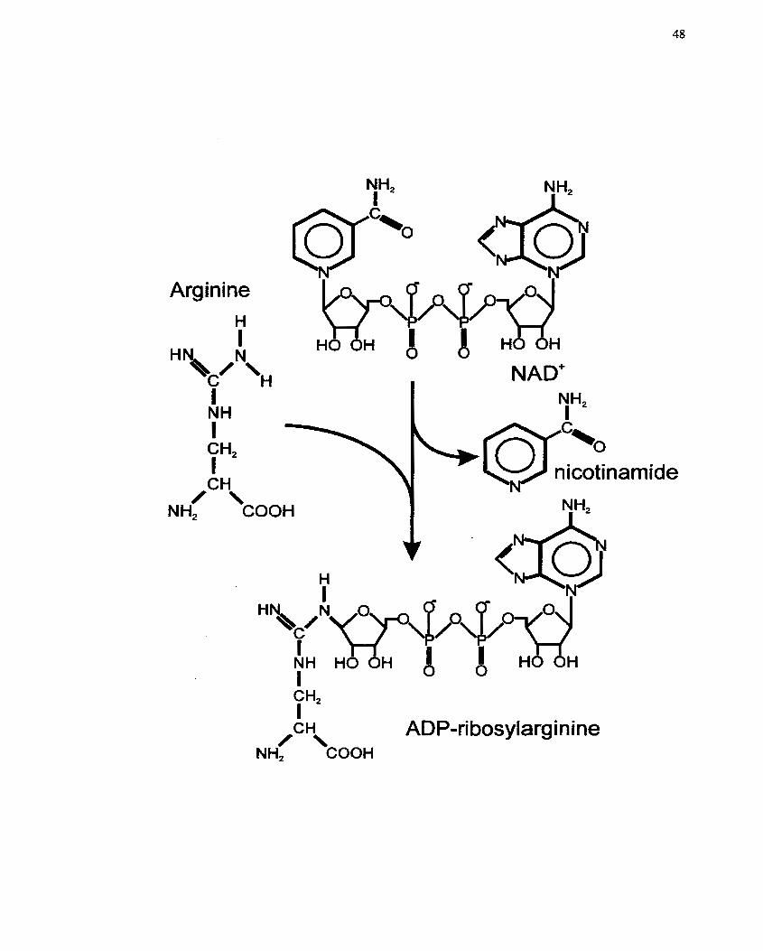
Arginine H I
H I NH I CH* I
I NAD'
,CH\ ADP-ri bosylarginine
NH2 COOH
The role of mono(ADP-ribosy1)ation in ce11 function is related to cellular metabolism and
has been associated with cellular differentiation (Kharadia et al. 1992), cellular
proiiferation (Thyberg et al. 1995b), ce11 migration (Norgauer et al. 1988; Saxty et al.
1998a; Saxty et al. 1998b; Stasia et al. 1991) and both intra- and extra-ceIlular
cornniunication, (Brune et al- t994; Hilz 1997; ZoIkiewska & Moss 1993; Zolkiewska et
al. 1992).
Catabolisxn of mono(ADP-ribose) is catalyzed by ADP-ribosyl(amino
acid)transferase hydrolases (EC 3.2.2.19). The end result is the regeneration of free,
unmodified amino acid (arginine, cysteine, diphthamide or asparagine). Hydrolases have
been identified in bacterial, avian and mammalian systems, appearing to be ubiquitous in
eukaryotic tissues, and thus supporting the prernise that ADP-ribosylation reactions are
important mediators of cellular regulation. The best characterized of the hydrolases is the
ADP-ribosylarginine hydrolase in eucaryotic cells (Moss et a[. 1997; Takada et al. 1994;
Williamson & Moss 1990). This hydrolase activity was detected in the soluble fraction of
turkey erythrocytes (Moss et al. 1988), cultured mouse cells (Smith el al. 1985), rat
skeletal muscle (Chang et al. 1986) and rat brain (Moss et al. 1992). Thus a eucaryotic
ADP-ribosylation cycle is hypothesized based on the reversible modification of proteins
by transferases and hydrolases (Moss et al. 1997; Williamson & Moss 1990), much like
the reversible phosphorylation modification of proteins by kinases and phosphatases
(Graves & Krebs 1999). Additionally, ADP-ribose can be processed by
phosphodiesterases and phosphatases as seen with the GPL-anchored skeletal muscle
mART, generating a phosphonbosyl-integrin and S'AMP, although the physiological
consequences of this structure have yet to be examined (Okazaki & Moss 1999;
Zolkiewska & Moss 1997).
2.7.2.1 Mono(ADP-ribosy1)transferase (mART)
The discovery of eucaryotic protein ADP-ribosylation (Gill & Meren 1978;
Honjo et al. 1968; Moss & Vaughan 1977) resulted in the identification of mARTs.
Bacterial toxins such as choiera toxin, pertussis toxin, diphtheria toxin and botulinum C3
endotoxin ADP-ribosylate a variety of eucaryotic and prokaryotic acceptor proteins, and
modulate their associated cellular functions (Aktones 1994; Aktories 1997; Gierschik
1992; Lem et aL 2000; Moss 1987; Okazaki & Moss 1996b; Popoff 1998). A variety of
animal tissue and organs are also now known to contain mART and rnART activity
(Soman & Graves 1988). Vertebrate mART activity was first detected in turkey
erythrocytes (Moss & Stanley 198 1b; Moss et al- 1980), rat Iiver homogenates (Moss &
Stanley 1981a) and Xenopus tissues (Godeau et al. 1984; Okazaki & Moss 1999;
Tsuchiya & Shirnoyama 1994). Presently, 6 avian mARTs (4 turkey erythrocyte ARTs:
A, B, C and A', and chicken ARTs from hen liver and heterophil grandes) and more than
13 mammalian rnARTs (eg. ART 1-5, ART 1-IV from rat brain, and 4 mARTs from rat
adrenal medulla) have now been purified andor cloned and characterized (Fujita et al-
1995; Matsuyama & Tsuyama 1991; Okazaki & Moss 1999). This subject has been
reviewed recently by Okazaki & Moss (1999), and 1 refer you to this article for more
details.
Of the rnarnmalian mARTs, expression was initially identified in cardiac and
skeletal muscle, leukocytes/lymphocytes, brain and testis (Duman et al, 199 1; Hara et al.
1987; Kharadia et al. 1992; Obara et al. 1989; Obara et al. 199 1; Peterson er al. 1990;
Piron & ~McMahon 1990; Soman & Graves 1988; Soman et al. 199 1; Soman et al. 1984a;
Soman et al. 1983; Yamashita et al- 1991). Subsequently, mART expression was
demonstrated in hcpatoma cclls (Ledford & Lcno 1993; Lcno & Ledford 1989), SMCs
(Li et al. 1999) and endothelial cells where ihese enzymes participated in signal
transduction and prostacyclin production (Halldorsson et al. 1992).
Vertebrate mARTs (eg. NAD:arginine(ADP-ribosy1)transferases (EC 2.4.2.3 1))
are a family of enzymes ranging in molecular weight frorn approxirnately 25- to 36-kDa
and 66- and 69-kDa (4 distinct ART'S from rat brain - 1 to IV and rat adrenal medulla)
(Okazaki & Moss 1999). Despite a lack of overall similarïty in the amino acid sequences
(i.e. Iack of conservation among sequences), there appear to be 3 regions (regions 1, II,
III) of sequence hornology that make up the catalytic site. A precise configuration of
these regions is required for the ADP-ribose transfer reaction (Domenighini & Rappuoli
1996; Okazaki & Moss 1999; Takada et al. 1995). Conserved regions house an NAD-
binding cleft composed of an a-helix bent over a B-strand, along with an arginine or
histidine residue and an active-site glutamate which are critical for enzymatic activity
(Domenighini & Rappuoli 1996; Okazaki & Moss 1999). Furthemore, 2 separate groups
of enzymes can be defined by the presence of either arginine (cholera and pertussis toxin-
like) or histidine (diphtheria toxin-like) in region I. Region II contains a specific arnino
acid motif (aromatic-hydrophobic-Ser-X-Ser-hydrophob) and region III contains the
active-site glutamate. For more details, please refer to the review by Okazaki & Moss
(1999). Interestingly, several of the mammalian mARTs have regions of hornology to
glycosylphosphatidylinositol (GP1)-anchored proteins, and thus may be membrane bound
with an extracellular orientation (Ferguson & Williams 1988; Low 1989; Zolkiewska et
al. 1994). Secretory and intracellular counterparts also exist and there may therefore be 3
discreet farnilies of transferases (Okazaki & Moss 1998).
2.7.2.2 Enzymology of mART
A common mechanism of substrate binding and catalysis apparently exists
between and among both bacterial and eucaryotic mARTs. Mammalian and bacterial
mARTs are beIieved to act via an SN2-Iike mechanism in which nucleophilic attack on
NAD' is followed by displacement of the nicotinamide and inversion of configuration
(Takada et al. 1995). In this enzymatic reaction, a single ADP-ribose moiety is
transferred ta the acceptor substrate. Amino acid-specific acceptors for mARTs include
arginine, cysteine, diphthamide. histidine and asparagine of which arginine-mARTs are
the best characterized thus far (Zolkiewska et al- 1994). Mechanisms for regulating
mARTs have not been described since Little research has been done in this area, however,
Weng et al, (1999) did recently report on the auto(ADP-rîbosy1)ation of ARTS as a
mechanism for replation of mART activity,
2.7.2.3 mART substrates
Vertebrate mARTs are typically species- and tissue-specific with respect to
acceptor proteins (Taniguchi et al. 1993). Although a large variety of proteins are ADP-
ribosylated, the majority of mARTs are arginine-specific (and can also modiQ other
simple guanidino derivatives) and a variety of potential substrates have been described
(Huang et ai. 1993; Okazaki & Moss 1999; Wang et ai. 1996; Zolkiewska & Moss 1993).
Acceptors of ADP-ribose in these mammalian (in vitro) systems include casein,
ovalbumin and simple guanidino compounds such as arginine and agmatine (Zolkiewska
et ai- 1994). Additional target proteins include Ha-ras-p2 1, transducin, protein kinases
(with subsequent reduction in activity), and skeletal C ~ " - A T P ~ S ~ (with subsequent
decrease in activity) (Okazaki & Moss 1996a; Tsuchiya & Shimoyama 1994). Finally,
endogenous arginine-dependent ADP-ribosylation of G proteins (Le. G,, Gi and Go) is
well documented in mammalian cells and tissues (Donnelly et ai. 1992; Duman et al.
199 1 ; Jacquemin et ai. 1986; Molina y Vedia et ai. 1989; Obara et al. 199 1).
Endogenous vertebrate ADP-ribosylation of cysteine residues is less comrnon, but
has been observcd in human and bovinc crythrocytcs via the actions of a cysteine-speci fic
mART put-ified from erythrocyte and platelet membranes. This cys-mART is also
reported to modify Gia (Okazaki & Moss 1999; Saxty & van Heyningen 1995; Tanuma
et al. 1988). Interestingly, ADP-ribosyl-cysteine linkages can occur as the result of a
nonenzymatic reaction involving ADP-ribose and NAD' jlycohydrolases. In this case, an
ADP-ribosyl-thiazolidine linkage is generated in place of the thioglycoside AD P-rïbosyl-
cysteine Iinkage, the type normally observed with pertussis toxin catalyzed reactions
(McDonald et al. 1992; Okazaki & Moss 1999)-
2-7.2.4 Function of mART within cells
Bacteriai mARTs have a variety of biological consequences and functions within
eucaryotic cells and organisms. For exarnp le, cholera toxin ADP-ribos y (ates an arginine
residue of G,a, the stimulatory a-subunit of the heterotrimeric GTP-binding (G) proteins,
and irreversibly inhibits its GTPase activity. The result is activation of adenylyl cyclase
and a subsequent increase in cellular cAMP levels (Cassel & Pfeuffer 1978; Gill &
Meren 1978; Moss & Vaughan 1977). On the other hand, pertussis toxin ADP-ribosylates
a cysteine residue in the a-subunit of Gi and Go which blocks the inhibitory effect of the
a;-subunit on adenylyl cyclase and results in activation of the cAMP cascade and/or
uncoupling of the G protein frorn its receptor (Takada et al. 1994; Ui & Katada 1990).
Nevertheless, both modifications result in abnornlalities in ccllular clectrolyte flux and
fluid secretion in the small intestines (Field 1976; Moss & Vaughan 1988; Sharp 1973).
Diphtheria toxin (and Psez~rloimmzs aeniginosa exotoxin A) ADP-ribosylates a
diphthamide (i.e. a modified histidine) residuc in elongation factor-2 (EF-2) resulting in
the inactivation of EF-2. This disrupts the rcgulatory mechanism for translocation of
elongating protcins across thc endoplasmic rcticulum membrane and resuIts in inhibition
of protcin synthesis (Collier 1967; Collicr 1975; lglewski 1994; Wick et al. 1990).
Intcrestingly, a fusion protcin of IL-2 and diphthcria toxin has bcen succcssfully applied
for the treatment of neoplastic diseases. This fusion protein targets cells with IL-2
receptors and the resultant ADP-ribosylation of EF-2 inhibits ce11 proliferation
(vanderspek et al. 1994). Finally, exotoxin C2 and C3 both rnodify components of the
cytoskeletal network. Botulinum C2 exotoxin ADP-ribosyIation of an arginine causes
inhibition of (non-muscle) actin polynierization and subsequent degradation of the
microfilament/cytoskeIetal network in cells (Aktories 19 94). Botulinum C3 exotoxin
ADP-ribosylation of an asparagine residue on the small GTP-binding protein Rho results
in the inactivation and attenuation of Rho-associated signal ing pathways (Kumagai et al.
1993). The resultant decrease in tyrosine phosphorylation and subsequent
phosphatidyIinositol3-kinase (PU-kinase) activation has t h e potential for modulating ce11
proliferation and differentiation as a result of aItered GTP-binding protein interactions
with the cytoskeletal system and a disturbance in t h e organization of the actin
cytoskeleton (Aktorks 1994; Kishi et al. 1993; Maehama e r al. 1994; Nishiki et al. 1990;
Paterson et al. 1990; Ridley & Hall 1992; RidIey et al. 1992).
As rnentioned previously, eucaryotic mARTs are involved in a variety of ce11
functions, including ceII differentiation, proliferation, migration and cellular
communication (Brune et al. 1994; Hilz 1997; Kharadia et al. 1992; Norgauer el al,
1988; Saxty et al. 1998a; Saxty et al. 1998b; Stasia et aC. 1991; Thyberg et al. 1995b;
Zolkiewska & Moss 1993; Zolkiewska et al. 1992). Specific modification of certain
cytoskeletal/intermediate filament proteins wouid result irx the alteration of the above-
mentioned ce11 functions. For example, desmin is ADP-rEbosylated by an endogenous
arg-mART in skeletal muscle (Graves et al. 1997; Huang e t al. 1996; Yuan et al. 1999).
This modification results in desmin disassembly (Yuan et al. 1999), with consequences
related to ce11 differentiation, migration, proliferation an& intracellular communication
(Huang et al. 1993). ADP-ribosylation of desmin is an important rcgulatory mechanism
in differentiating muscle cells, with mART activity increasing dramaticatly after
myoblast fusion (Kharadia et al. 1992). In addition, desmin from SMCs has been shown
to react with an antibody specific for ADP-ribosylated-arginine (Graves & Krebs 1999).
A second exarnple would be the modification of actin filaments. ADP-ribosylation of
actin in human neutrophils is associated with inhibition of neutrophii chemotaxis (Stasia
et al. 1991). AdditionaHy, non-muscle Ply-actin, skeletal muscle a-actin and smooth
muscle y-actin are ADP-ribosylated in vitro with resuItant inhibition of actin
pdymerization (Terashima et al. 1992). Furthemore, endogenous ADP-nbosylation of
heterophil non-muscle actin was proposed as a mechanism for reguiating heterophil
phagocytosis, secretion and chemotaxis (functions dependent on actin de/polymerization)
(Terashima et al. 1993). The final example is the ADP-ribosyiation of integrin a7, the
receptor for the extracellular protein Iaminin, on skeletal muscle cells by mART. This
modification may modulate cell-ce11 or cell-matnx interactions, with effects on cellular
differentiation and/or proliferation (Zolkiewska & Moss 1993; Zolkiewska & Moss 1997;
Zolkiewska el al. 1998).
Other eucaryotic ce11 functions that may be altered by mART include neuronal
transmission (in the brain, ADP-ribosylation of G proteins may modie neuronal signal
transmission) (Durnan et al. 1991; Matsuyama & Tsuyama 1991), and regdation of T-
ce11 proliferation and cytolytic activity (ADP-ribosylation of a p56'ck-associated protein)
(Wang et al. 1996). More recently, Lupi et al. (2000) have reported the modification of
the P-subunit of Go, by arg-mART as a novel mechanism of regulating G protein-
mediated signal transduction. This modification prevented the inhibition of calmodulin-
stimulated type 1 adenylyl cyclase. Finally, modification of MARCKS and tubuIin by
rnART has been demonstrated to lead to changes in cytoskeletal and microtubular
assembly/organization (Aktories & Wegner 1992; Chao et al. 1 994; Scaife et al. 1 %Q),
as well as alterations in the abiIity of MARCKS to bind calrnodulin and interact with the
plasma membrane (Chao et al- 1994).
From a more clinicai point of view, in experimental diabetic neuropathy, mART
activity has been associated with a reduction of substance P axonal transport and
subsequent onset of diabetic perïpheral neuropathy (Donadoni et aL 1995). Et was
observed that exogenous ADP-ribosylation reactions were reduced in extracts fiorn
diabetic retinas, suggesting increased cytosolic endogenous ADP-ribosylation activity.
Treatment with an inhibitor of ADP-ribosylation (silybin) increased the extent of
exogenous ADP-ribosylation (Donadoni et al. 1995)-
2.7.2.5 Mono(ADP-ribosy1)ation in the cardiovascular system
In the cardiovascular system, arg-mART has been found in relatively high Ievels
in neonatal and adult rat hearts (Piron & McMahon 1990). In cardiac muscle, ADP-
ribosylation reactions have been associated with developmental regulation (Piron &
McMahon 1990). Furthemore, Jones and Baird (Jones & Baird 1997) have shown that
bFGF, a growth factor intimately associated with the cardiovascular system, angiogenesis
and the muscu1oskeletal system, is ADP-ribosylated on the surface of adult bovine arch
endotheIia1 cells (and in human hepatoma cells). This modification may represent an
additional regulatory mechanism, in conjunction with phosphorylation, for this potent
mitogen. In the past, bFGF has been found to be both phosphoryIated and ADP-
ribosylated in vitro (Boulle et al- 1995; Feige & Baird 1989; Vilgrain & Baird 199 1;
Vilgrain et al. 1993).
Nitric oxide (NO) is a second messenger produced by NO synthase (NOS) in
response to a variety of signaIs that is able to stimulate endogenous ADP-ribosylation in
vitro. Moreover, NO, acting as a free radical molecule, can activate ADP-ribosylation of
several proteins in vivo (Brune & Lapetina 1989; Schuman et al. 1994; Zhang & Snyder
1992) and result in cellular damage (Parrado et al. 1999). In the vasculature, NO-
dependent ADP-ribosylation of G proteins by an endogenous ART in srnooth muscle
increases the activity of adenylyl cyclase and results in vasodilatation via a decrease in
the activation of phospholipase C (Kanagy et al. 1995). As a result, it is postulated that
hypertension-associated decreases in eNOS expression and activity would decrease the
arnount of NO produced, thereby leading to decreased ADP-ribosylation of G proteins
and, consequently, increased vesse1 constriction since activation of G proteins by agonists
is unopposed (Kanagy et al. 1995). Moreover, ADP-ribosylation was discovered in SMCs
derived from bovine coronary artery (Li et al- 1999). Here, this modification was found
to modulate G,a and thereby play a role in activating K' channels as induced by 1 1,12-
EET. Taken together, these studies suggest that ADP-ribosylation rnay constitute a novel
participant in the cardiovascular systern and vasculature, playing a role in regulation of
cellular processes such as developrnent, differentiation, proliferation and SMC
contractility (Le. vasoactivity).
2.8 Inhibitors of mART and PARP
Over the years, numerous inhibitors of mART and PARP have been discovered
and used successfully (and prirnarîly) to study the biological function of these enzymes.
A number of compounds have been shown to inhibit mART, however, specificity is
typically lacking because of the sirnilarity of the rnART and PARP catalyzed reactions
(reviewed in Banasik et al. (1992), Banasik & Ueda (1994) and Rankin er al. (1989). This
ambiguity bas impacted on our understanding of ADP-nbosylation, since data
interpretation can be biased if it is based on an inhibitor that is not seiective for either
mART or PARP. For example, many of the cellular functions of PARP were identified by
extensive use of 3-aminobenzamide (3AB) (Sims et al. 1982). However, the fact that this
compound also inhibits rnART suggests a re-examination of the conclusions should be
made (Banasik et al- 1992; Milam & CIeaver 1984; Rankin et al. 1989; Zahradka & Yau
1994). Other examples of PARP inhibitors include benzarnide and nicotinamide. A more
complete Iisting of inhibitors is provided in articles that have reviewed this topic in detail
(Banasik et ai- 1992; Banasik & Ueda 1994; Rankin et al. 1989). Examples of arg-rnART
inhibitors include benzamide, napthaIimide, novobiocin, and vitamin K. Although more
PARP and mART inhibitors have since been identified, few are selective for arginine-
dependent mART. The best examples are silyrnarinkilybin and meia-
iodobenzylguanidine (MIBG) (Donadoni et al. 1995; Loesberg et al. 1990b; Smets et al.
1990b), and 1 will be concentrating on the latter compound, MIBG, since it is the focus
of this thesis.
2.8.1 MIBG
MIBG was synthesized as a norepinephnne analogue, and is structurally similar to
guanidines such as the antihypertensive dmg guanethidine (Montaldo et al. 1996; Short
& Darby 1967). Its discovery was brought on by the search for agents that could be used
for the visualization of the adrenal medulla and possibly other related neoplasms
(Wafelman el ai. 1994b). MIBG is a ring-substituted alkylguanethidine and this family of
agents and its potency was first reported by Short & Darby (1967) as a series of
aralkylguanidines with high sympathetic nerve blocking capacity (Wafelman et al.
1994b). The evaluation of radioiodinated forrns of the aralkylguanidines as adrenal
medulla visualizing agents then followed and, in 1980, Wieland et al. (1980) investigated
and reported on the effects of radiolabelled O, nz and p position iodine-substituted-
benzylguanidine. The investigators found that the nz forrn was more resistant to in vivo
deiodination and, for this reason, radiolabelled MIBG was selected for further
investigations which lcd to its widespread use today (Wieland et al. 1980). Presently,
MIBG in its radiolabelled form (?- or 131~-labelled) is used for both diagnostic and
therapeutic purposes. Due to its selective uptake, radiolabelled MIBG enables the
detection and localization of neuroendocrine turnours and adrenergic tumours (Le.
adrenal medullary hyperplasia) . Typically, the radiolabeIIed MIBG is used for diagnostic
scintigraphy of neural crest-derived tumours (Hoefnagel 1994; Hoefnagel et al. 1991;
Srnets et al. 1988b) and also for the scintlgraphic assessrnent of cardiac sympathetic
neuronal integrity (Le. detection of cardiac sympathetic dysinnervation) in a variety of
disease States that include ischemia and diabetes (Nakata et al. 1996; SchneIl et al. 1996;
Valdes Otmos et ai. 1993). Although targeted radiotherapy of neuronal (neuroendocrine)
tumours by radiolabelled MIBG is practical and benefcial, non-radioactive forms of
MIBG have been tested for the treatment of carcinoid tumours and their rnetastases
(Cornelissen et al. 1995a; Kuin et al. 1999; Smets et al. 1988b; Taal et al. 1999; Taal et
al, 1996; Zuetenhorst et al. 1999). Furthemore, MLBG can be used for the detection,
diagnosis and therapy of smooth muscle tumours of the small bowel (Akle et al. 1997).
Finally, MIBG is a known inhibitor of arg-mART reactions (Loesberg et al. 1990b;
Smets et al. 1990b) and has been used as a tool for the study of ADP-ribosylation
reactions and their biological functions (Richter 1990). Its utility in this capacity is based
on its structure, which contains a guanidino group that mimics the acceptor portion of an
arginine moiety, and perrnits it to act as a pseudosubstrate (high affinity substrate) for
arg-mART and thereby inhibit its activity.
2.8.1.1 Biodistribution and p harmacokinetics of MIBG
After intravenous administration, radiolabeiled MIBG is distributed from the
vascufar compartment within I hour and then is slowly redistributed from the perpiheral
compartment to the central cornpartment (Wafelman et al. 1994a). Interestingly, the
initial uptake of MIBG involves both neuronal and nonneuronaI organs, and early
clearance (less than 4 hours) occurs €rom the nonneuronal sites (Wafelman et al. 1994a).
Within 24 hours, MLBG is observed mainly in the liver and to a lesser extent in the lungs,
heart, spleen salivary glands and urinary bladder (Wafelman et al. 1994a). Uptake in the
normal adrenal glands is very low, but hyperplastic adrenals, and tumours such as
pheochrornocytoma and neuroblastoma, dong with other tumours that have
neurosecretory granules, have increased rates of uptake (Wafelman et al. 1994a).
Significant clearance of radiolabelled MIBG fiom the liver and spleen occurs within 72
hours (Wafelman et al. 1994a). The biodistribution of MIBG can be altered by interaction
with other dmgs, especially agents that act in, and interfere with, the sympathetic nervous
sytem (eg. labetalol, reserpine and cocaïne) (Wafelman et al. 1994a).
2.8.1.2 Cellular uptake and retention of MIBG
MLBG shares the same mechanism of transport and storage as norepinephrine
(NE) (Jaques et ai. 1984; Tobes et al. 1985; Wieland et al. 1980). In general, there are
two mechanisms of uptake by which MIBG enters the ce11 (eg. adrenomedullary cells).
The first route is an active uptake system at the ce11 membrane specific to catecholarnines
(Uptake-1) and the second is a non-specific uptake system, which is presumed to be
passive diffusion (Jaques et al. 1984; Tobes et al. 1985). MIBG has specific affinity for
uptake- 1 mechanism of catecholamines and serotonin transporter (Jaques et al. 1987;
Rutgers et al. 1993; Smets et al. 199 1). The NE transporter/Uptake-1 is a neuron-specific,
ATPase-dependent catecholamine-~a symport that is driven by a ~ a ' gradient generated
by N ~ + / K + - A T P ~ s ~ (Gasnier et al. 1986; Jaques et al. 1984; Tobes et al. 1985). Uptake- 1
is also temperature-dependent, high-affinity, Iow-capacity, saturable, oubain- (inhibitor of
N~';/K'-ATP~s~) sensitive, imipramine- or cocaine-sensitive and energy-dependent (Le.
decreased uptake when glucose is low in the medium) (Jaques et al. 1984; Tobes et ai.
1985). At relativcly high concentrations of MIBG (i-e. 10" M), non-specific uptake (Le.
passive diEusiion) dominates over active uptake as the transporter becomes saturated
(Jaques el ai- 1987; Lashford et ai. 1991). The non-specific uptake system is aIso
temperature-despendent, but is not energy-dependent, oubain-sensitive or saturable
(Jaques et al. 1484; Tobes et al. 1985)- Although passive diffusion is assumed to
characterize this non-specific uptake system, some in vivo studies have now indicated
that nonspecifilc membrane binding of MIBG provides the explanation for non-specific
uptake as opposed to simply passive diffiision through the plasma ce11 membrane
(Glowniak et al. 1993)- Although MIBG is highly protonated (pK of about 13 in vivo
(Wieland et a(. 1984)), it lacks polar hydroxyl groups that decrease the Iipophilicity of the
phenyl ring. Tbus, MIBG may be able to associate with the cell membrane. It has also
been suggestee that this high positive charge results in accumulation of MD3G in cells
and mitochondoia via an electro-chernical gradient (Smets et al. 1990b). In cells that have
active uptake ( e g . neuroblastoma), the typical time frame for maximal intracelluIar MIBG
concentration i:s 1-2 hours incubation (Iavarone et al- 1993; Smets et al. 1989). In cells
that do not contain active uptake mechanisms (eg. L1210, a murine lymphoblastic
leukemia ce11 Inne), saturation of the ce11 with MIBG wiH take about 3 hours (Smets et al.
1990b).
Within cells, MIBG is stored in intracellular storage vesicles (Montaldo et ai.
1991) and as freely diffusable cytoplasmic molecules, and is not associated with
rnacromoleculax compIexes (Smets et al. 1990a). However, the process of MIBG
retention is alsm considered to involve dynamic uptake, release and re-uptake processes.
Mechanisms o~ release are three-fold: exocytosis, carrier-mediated efflux and passive
diffusion, and ~ h e contribution of each will Vary depending on environmental conditions
(Servidei et ai. 1995). This uptakehelease mechanism allows for the rapid homogeneous
distribution of MIBG within a ce11 or tissue (Smets et ai. 1989). For example, in
neuroblastoma ceIls, MIBG is stored mainly in the cytoplasm and only to a smaI1 extent
in the chromophilic granules. Coupled with the dynamic process of MIBG retention
mentioned above, this rnechanism of storage facilitates the homogeneous distribution of
MIBG within a tumour (Servidei et al. 1995; Weber et al, 1996) and allows for efficient
visualization and treatment. Interestingly, if MIBG is ADP-rÏbosylated by arg-mART, the
rnodified rnolecule, which will contain an ADP-ribose moiety, will likely not move out of
the ce11 by the aforementioned mechanisms, and should increase the cellular retention of
MIBG. The rnodified MIBG should stay within the ce11 until the reverse enzyme reaction
(Le. via arg-mART hydrolase) occurs. However, since the modification with ADP-ribose
is typically reversible, this possibility should not influence the cellular retention and
rehptake of MIBG.
2.8.1.3 Metabolism of MIBG
Within the body, MiBG is not metabolized by monoamine oxidase (MAO) nor by
catchol-O-methyItransferase, but is rapidly excreted in urine (Iversen et al. 197 1 ;
Mangner et al. 1986; Schnell et al. 1996; Sisson et al. 1987; Tobes et al. 1985). MIBG is
primarily excceted by the kidneys and, within 24 hours, 60% of the dose is eliminated
(Kuin et al. 1998). As a result, M B G has been docurnented to cause stress-related
syrnptoms associated with the release of bioamines, thereby affecting renal perfusion
(Kuin et ai. 1994).
2.8.1.4 Excretion/Elimination of MIBG
The rate of MIBG excretion is similar for either a diagnostic or a therapeutic dose
(Mangner et ai. 1986). MIBG is eliminated from the body via the rend pathway, mostly
in the unaltered form, and glomerular filtration rate has a major influence on MIBG
kinetics (Blake et al. 1989). Within 24 hours, 40-50% is eliminated, and by 72-96 hours,
70-90% is excreted, prirnarily as unchanged drug (Mangner el al. 1986). The importance
of renal excretion is confimed by fecal recovery of the dmg that is usually less than 2%
within 0-24 hours and less than 2% at 0-4 days (Kline et al. 198 1; Mangner et al. 1986;
Shulkin et al. 1986). The primary metabolites of MIBG are meta-iodohippux-ic acid
(MIHA) (2-16%), iodine (2-6%) (typically in the radiolabelled form and thus likely to
affect the thyroid gland), meta-iodobenzoic acid (less than 0.5%) and para-hydroxy-
meta-iodobenzylguanidine (less than 0.5%) (Mangner et al. 1986). Since most of the
metabolic studies with MIBG have used the radiolabelled forms, rnetabolism has been
easy to follow. As weli, sensitive HPLC techniques have been developed for detection in
plasma and urine (Mangner et al. 1986).
2.8.1.5 Toxicity of MIBG
MIBG has been shown to have cytotoxic effects in the unlabelled form (Slosrnan
et al. 1988; Smets et al. l988b; Wieland et al. 198 1). Within the cell, cytotoxicity may be
felated to its ability to inhibit mitochondrial activity (Loesberg et al. 1990b; Smets et al.
1990b). MIBG is selectively accumulated by cells, and it is predominantly the
mitochondria that actively accumulate MLBG (Gaze et al. 1991). The uptake of MIBG
into mitochondria is passive, since mitochondria do not possess uptake I (Cornelissen et
al. 1995a). Specifically, MIBG can inhibit both cornplex I and cornplex III of the
rnitochondrial respiratory chain (Cornelissen et al. 1995b; Loesberg et al. 1990a;
Loesberg et al. 1991; Slosman et al. 1993). Increased glycolytic flux is observed 2.5
hours after exposure to 31 FM MIBG and this has been interpreted as a compensatory
mechanism to the inhibition of mitochondrial respiration. This increased glycolytic flux is
accompanied by an increase in lactate production and leads to intracellular acidification
(Loesberg et al. 1990a). An interesting benefit to this cytotoxic effect is that MIBG can
potentiate the anti-neopiastic actions of certain cancer drugs that require a lower
intracellular pH for maximai function (Kuin et al. 1999; Kuin et al. 1994). The possibility
also exists that the cytotoxic effects of MIBG may be retated to the inhibition of arg-
mART, since 3 classes of ADP-ribose acceptors have been identified in mitochondria
(Althaus & Richter 1987). However, the contribution of rnARTs to mitochondrial
function has not yet been documented or studied in detail (Kuin et al. 1998).
At the tissue level, the MIBG toxicity primarily involves the renal system (Kuin et
al. 1998). MIBG at a dose of 40 mg/kg results in decreased renal blood perfusion and the
induction of reversible kidney darnage (Le. decrease in EDTA clearance and histological
darnage in the distal tubules). Although the onset of renal toxicity would be assumed to
be associated with inhibition of mitochondrial respiration, interestingly, benzylguanidine
(de-iodinated form of MIBG) does not affect renal clearance even though
benzylguanidine also decreases mitochondrial respiration. As a result, it is liKely that
decreased mitochondrial respiration is not the main mechanism of MIBG-induced renal
damage (Kuin et al. 1998). Recent work has shed sorne light on this matter. MIBG
inhibits al1 three forms of human NOS (Kuin et al. 1998) and it is suspected that MIBG at
100 pM causes the inhibition of NOS (nitric oxide synthase). It is this inhibitory act that
may thus contribute to the decrease in renal clearance that is observed (Kuin et al. 1998).
2.8.1.6 Clinical application of MIBG
Because of its high affinity for the norepinephrine (NE) transporter, MIBG is used
mostly as an adrenal irnaging agent via scintigraphic visuaIization of tumours and
metastases (Montaldo el al. 1996). Clinically, MIBG labelled with radioactive iodine ('9
or "'1) is used for scintigraphy of neuroendocrine tumours such as pheochromocytorna,
neuroblastoma, medullary thyroidoma and tumours of the APUD (amine precursor
uptake and decarboxylation) series (Smets er al, 1990a). Since MIBG is taken up by
neuroendocrinc tumours, radioiodinated MIBG has becn used for treatrncnt of these very
samc tumours (Smets et al, 1990a). Aftcr uptakc of radiolabellcd MIBG into thc tumour,
the cancerous celIs are slowly ablated by the effects of Ply-radiation release.
Unfortunately, one of the consequences of radiolabelled MIBG treatment for cancer is
thrombocytopenia resulting frorn the uptake of MIBG via the serotonin transporter into
platelets and rnegakaryocytes with subsequent death of the ptatelets by radiation (Lode et
al, 1995; Tytgat et ai- 1995).
UnIabelled MIBG is also cytostatic, and even cytotoxic in various ce11 Iines
(Srnets et al. 1988b). For example, MIBG has been shown to almost completely inhibit
the proliferation of a neuroblastoma ce11 Iine (SK-N-BE(2c)), and also decreases the
ATP/ADP ratio in this ce11 line (Cornelissen et ai. 1995a). As a result, MIBG has now
been tested in its unlabelled f o m for the palbation of cardinoid syndromes and has
shown some success (Taal et al. 1999; Taal et al. 1996; Zuetenhorst et al- 1999). In its
unlabeIled form, 60 mg/kg is the tolerated dose by oral administration with a
bioavailability of 59%. The onIy evident toxicity is a decrease in renal function that
correlates with histologically detectable changes in the distal tubule (occurs at 40 mgkg)
(Kuin et al. 1998). At these levels, however, the darnage is entirely reversible (Kuin et al.
1999). Moreover, there appear to be no histological abnorrnalities in the kidney,
intestines, Iiver, heart, lungs, thymus, salivary glands and testes. As a result, repeated oral
administration of MIBG is a promising alternative for the long-term palliation of the
carcinoid syndrome without the need for hospitalization (Kuin et al. 1999) and without
the requirement for radiation therapy.
It is interesting to note that MIBG uptake increases on differentiation of human
neuroblastoma cells and the intracelluIar half-life is correspondingiy increased (MontaIdo
et al. 1996). This is mainly due to greater uptake capabilities of the differentiated cells
and does not reflect an increase in the storage capacity (Montaldo et al. 1996). A
correlation also exists between ce11 maturity and the ability of MIBG to accumulate
within those cells (Montaldo et al. 1996). Within certain neuroblastorna ce11 popuIations,
MIBG resistance is observed. This is likeIy due an inability to take up the MIBG and
corresponds to the presence of a very immature cell subpopulation (Montaldo et ai.
1996)-
2.8.1.7 Clinical application of MIBG in the cardiovascular system
In the cardiovascular system, MIBG scintigraphy has been used for the
assessment of cardiac dysinnervation (Le. cardiac sympathetic dysfunction) (Schnell et
al. 1996). In this application, the affinity of MlBG for post-ganglionic sympathetic nerve
endings pennits a direct assessment of sympathetic nervous activity in cardiac tissue.
This method has become a relatively reliable method for detection of cardiac
dysinnervation and is effective at identiQing changes that occur even before the onset of
ECG-based abnorrnalities (Schnell et al* 1996). For example, MIBG scintigraphy can be
used to assess the extent of myocardial damage after idarction since sympathethic nerve
fibres are damaged throughout the infarct zone. It has also been used to evaluate the
cardiac autonornic newous system defects at the onset of IDDM where a decrease in
global uptake is observed (Turpeinen et al- 1996).
The diagnostic efficiency for MIBG tornography for the detection of coronary
artery disease (CAD) is limited due to nonspecific reductions in MIBG uptake in the
inferior and postlateral regions of the heart (Nakata et al. 1996). Interestingly, the cardiac
sympathetic nerves travel with coronary arteries, and congenital defects in coronary
anatomy can also result in regional sympathetic denervation (Wichter et al. L994)I.
Nevertheless, the severity of coronary stenosis can be correlated with MIBG uptake
defect (Hartikainen et al. 1997). In C m , the extent of denervation is related to the local
severity of ischemia and not related to the size of myocardium with impaired perfusion
defect (Hartikainen et al. 1997; Nakata et al. 1996). It has also bcen noted that cardiac
adrenergic tissue is very sensitive to ischemia and regional cardiac sympathetic
denervation can occur in patients with stable CAD without previous MI. Interestingly, 3-
4 months after a coronary angioplasty, there is recovery of MIBG uptake if the residual
stenosis after angioplasty is less than 40% of the Lumen diameter (Guertner et al. 1993).
The sympathetic nervous system also pIays an important role in the development
and progression of heart failure, since it has been documented that myocardial NE
content falls with the progression of heart failure (Takeishi et ai. L997). Impairment of
both neuronal uptake and vesicular storage function is apparent in the failing heart (Seto
et al. 1996) and there is a correlation between cardiac MIBG uptake and myocardiar NE
content (Schofer et ai. 1988). Moreover, myocardial MIBG uptake can reflect the effect
of treatments and therapies for cardiomyopathy (eg. ACE inhibitors, verapamil improve
uptake), in addition to the progression of cardiomyopathy (Somsen et al. 1996;
Wakabayaslii et al. 1997). For example, ACE inhibitors act not onIy on systemic
vasodilation but also directiy to inhibit the release of NE from sympathetic nerve
terminais and as a result improve rnyocardial fibrosis (Le. ACE inhibitor suppression of
neurohormonal system may contribute to sorne of the beneficial effects) (Somsen et al.
1996; Wakabayashi et al. 1997). Furthemore, AngII prevents the neuronal uptake of NE
which then in tum leads to a decrease in P-adrenoceptors during heart faiIure (Somsen et
al. 1996).
2.8.1.8 Mode of action of MIBG
In many ce11 Iines, the anti-proliferative effect of MIBG is suggested to result
from inhibition of compIexes 1 and III of the mitochondrial respiratory chain, as stated
previously. However, a complete proliferation arrest is observed at concentrations of
MIBG rnuch higher than those required for the complete inhibition of mitochondrial ATP
synthcsis (Cornelissen et ai- 1995a; CorneIissen el al. 1995b). The maximum inhibition
for ATP synthesis was seen a t 10 pM while the optimum concentration for inhibition of
ce11 proliferation was observed at concentrations greater than 25 FM. Therefore,
inhibition of mitochondrial respiratory chain activity is not entirely responsible for the
ce11 proliferative inhibition properties of MIBG, and MIBG must influence cellular
processes apart from rnitochomdrïal ATP synthesis (Cornelissen et al. 1997b; Cornelissen
et al. 1995a).
Initially it was suggested that the differential inhibition of ce11 proliferation and
mitochondrial activity may b e due to generation of superoxide and lipid peroxidation of
the mitochondrial inner membrane (via inhibition of complex 1), since increasing MIBG
concentrations increase the levels of malondialdehyde (MDA) which defines the extent of
lipid peroxidation (Cornelissem et al. 1997a). When MlBG was used to inhibit complex I,
superoxide formation occurred maximally at concentrations of MLBG seven times higher
than those required for optimal inhibition of mitochondrial ATP synthesis (Cornelissen et
al. 1997b). Interestingly, the authors could use either vitamin E or GSH to scavenge free
radical fornation and, as a result, there was little or no MDA fonned with increasing
concentrations of MIBG (O - 25 PM; 25 p.M produced the maximal concentration of
MDA) incubated with cells ovemight- However, despite the protective effect of the
scavengers on MIBG-induced increase in MDA levels, no increase in the ce11
proliferation rate was observed when cells were cultured for 5 days in the presence of
MIBG with vitamin E (Comelissen et al. 1997b). This would suggest that still other
cellular mechanisms are being influenced by MIBG in its anti-proliferative effect. One
possibility is that MIBG i s inhibiting an arg-mART enzyme activity. Thus, the
mitochondrial proteins that have been shown to be mono(ADP-ribosy1)ated could
mediate the dismption of rnitechondrial function by MIBG (Althaus & Richter 1987).
2.8.1.9 MIBG as an inhibitor of arg-mART
Smets and colleagues demonstrated that MLBG was an inhibitor of arg-mART
(Loesberg et al. 1990b; Smets et al. 1988a; Srnets et al- 1988b; Smets et al. 1990b). Their
initial studies revealed that non-radiolabelled MIBG had several cellular effects,
including cytotoxicity, which they ascribed to interference with cellular rnARTs (Smets
et al. 1988a; Smets et al. 1988b). The reasons for this assumption were two-fold: i)
MIBG could potentiate the leukernolytic actions of glucocorticoid hormones similar to
inhibitors of ADP-ribosylation such as nicotinamide and 3AB (Smets et al. L988a), and
ii) MLE3G was structurally similar to arginine and guanyltycamine, two known substrates
for N-linked mART activity of choiera toxin (Althaus & Richter 1987). Upon hirther
investigation, they found that MIBG could serve as an acceptor for the mART activity of
not only cholera toxin, but also of an endogenous mART activity fiom elythrocyte
membranes (Loesberg et al. 1990b), and MlBG could compete with intracellular
acceptors of arg-rnART enzymes (Smets et al. 1990b). Moreover, this activity was
dependent on the presence of the guanidino-group, since MIBA (nieta-iodobenzylamine),
the related monoamine precursor, had no effect (Loesberg et al. 1990b). This would
suggest that MIBG competes effectively with endogenous acceptors via its guanidino-
group, making it a valuable tool for the study of the physiological functions of cellular
mARTs.
Arrned with the knowledge that MIBG is an anti-tumour agent and that it also
inhibits arg-mART enzyme activity (Comelissen et al. 1995a; Loesberg et al. 1990b;
Taal et al. 1996), it is conceivable that the anti-turnour activity of MIBG may be linked
with its ability to inhibit mono(ADP-ribosy1)ation reactions. Studies in this laboratory
have dernonstrated that arg-mART activity increases following mitogen stimulation, and
that inhibition of this enzyme activity prevents ce11 proliferation (Yau et al. 1998). These
studies certâinly support the contention that the anti-proliferative actions are mediated
through inhibition of arg-mART and that ceIl proliferative processes rnay be associated
with activation of this enzyme. Furthemore, the ability of MIBG to inhibit an arg-mART
which is linked to ce11 proliferation would emphasize the findings of Cornelissen et a(.
(1997b and 1995a) that a differential inhibition of mitochondrial respiration and ce11
proliferation was observed, with the latter requiring much higher concentrations of
MIBG- Taken together, these data would suggest that MIBG must influence cellular
processes apart €rom mitochondrial ATP synthesis with respect to its ability to inhibit ce11
proli feration, and a li kely candidate is N-linked mono(ADP-nbosy1)at ion of endogenous
acceptor proteins.
2.9 General Perspectives
The preceding literature review dernonstrates that considerable knowledge exists
concerning restenosis, its pathogenesis and the involvement of vascular remodeling and
SMCs, as well as the cellular processes activated within the SMC population of the
restenotic lesion. However, even with this understanding, and the extensive amount of
research that is still ongoing, the phannacological therapies and mechanical strategies
that have been implemented to date have not brouglit about a significant reduction in the
incidence of restenosis after revascularization procedures. Thus, it is still of paramount
importance to continue defining the temporal sequence of events that take place during
and after vascular injury. In particular, the cellular systems associated with the repair
processes that bring about the fibroproliferative and remodeling events characteristic of
restenosis must bc dcfined, since there is a lack of continuity of information rcgarding the
progression of thc cntirc disease proccss. Novcl playcrs may yct still be discovcrcd and
charactcrizcd. Intcrcstingly, many of ilic ccIlular proccsscs activated in vascular rcpair
and restenosis are sirnilar to those seen during tumour progression and metastases.
AIthough many chemotherapeutic agents, especially those that inhibit ce11 proIiferative
processes, have been tested without rnuch success, the spectrum of agents has not been
exhausted. Nevertheless, more details conceming the progression of restenosis will
improve the likelihood that a successful therapeutic modality will be found. Armed with
the knowledge that restenosis is in part a disorder of activated vascuIar (srnooth muscle)
cells and in part a consequence of vascular remodeling, it is evident that combination
therapy will be required- AIternatively, a single compound that will interfere with
multiple processes within the restenotic program must be identified. Thus, the
investigation of novel compounds for the treatrnent of restenosis is stil1 a burgeoning
field. It is the potential for MIBG to inhibit ceil proliferation, ce11 migration and ce11
differentiation, and the ability of MIBG to influence a novel ce11 regdatory rnechanism
potentially related to cell proliferation, migration and differentiation that brought me to
investigate this molecule in a variety of cellular systems, and finally apply MIBG to the
treatment of restenosis post-angioplasty.
3.0 Statement of the Problem: Hypothesis and Objectives
The information summanzed in the introduction (section 1.0) allowed the
formation of a general hypothesis under which studies reported in this thesis were carried
out. To review briefly, the incidence of restenosis after revasculanzation procrdures is
close to 50%. The clinical consequences of restenosis include angina, myocardial
infarction and sudden death. A variety of pharmacological agents have been tested as
therapeutic agents for the inhibition of restenosis, none of which has had measurable
clinical success. MIBG, synthesized as a norepinephrine analog, is used clinically for the
detection of neuroendocrine tumours and cardiac sympathetic dysinnervation. When
tagged with radioactive iodine, MIBG has been successfully used as therapy for
neuroendocrine tumours (Le. neuroblastoma). More recently, favourable results have
been found for non-radiolabelled MIBG as a chemotherapeutic agent for neuroblastoma
and as therapy for small bowel tumours, since it effectively abrogates turnour growth and
metastases. This information would infer that MIBG has the potential to prevent
neointimaI formation, and thus be employed clinically to suppress restenosis post-
angioplasty.
GENERAL HYPOTHESIS - Meta-iodobenzylguanidine (MIBG), an inhibitor of
rnono(ADP-ribosy1)transferase (mART), will effectively reduce restenosis
(neointimal lesion formation) post-baIloon angioplasty by interfering with thc
cellular processes that mediate prolifcration, migration and differentiation. As a
corollary to the hypothesis, MIBG is an inhibitor of mART, and thcrcfore, mART
activity may be essential for ce11 prolifcration, ce11 migration and cell differentiation.
73
Within the context of the stated General Hypothesis. huo objectives were
advanced:
GENERAL OBJECTIVES -
1) To characterize the respons
action.
e of cell .s to MIBG and to identify its mechanism of
2) To evaluate MIBG as an inhibitor of restenosis.
It is anticipated that the information provided by these studies will lead to a noveI
therapeutic application for MIBG îhat will benefit patients who require revascularization
procedures each year. Furthemore, it is expected that these studies will generate renewed
interest in the basic cellular mechanisms that contribute to the restenotic process.
4.0 Experimental Procedures/Materials and Methods
4.1 Materials
Al1 media, antibiotics, sera and other reagents used for ce11 culture experïments
were obtained from Gibco/BRL - Life Technologies (Burlington, ON), unless otherwise
noted. Reagents for the SMC serum-free defined suppiement (pyruvate, ascorbate, hoIo-
transfemn, selenium and insulin) were obtained from Sigma Chemicals (St. Louis, MO).
Nunc plastic tissue culture plates were from Gibco/BRL - Life Technologies (Burlington,
ON), while Linbro plastic multiwell tissue culture dishes were acquired h m ICNIFlow
Technologies (Costa Mesa, CA).
individual growth promoting agents were acquired from a variety of commercial
sources. Insulin used for the H4IlE experirnents was purchased either from Sigma
Chemicals (St. Louis, MO) or from Eli Lilly Company (Scarborough, ON). IGF-1 and
PDGF were obtained from Peprotech lncorporated (Rockyhill, NJ). Angiotensin II
(AngLI) was purchased frorn Sigma Chemicals (St. Louis, MO), Peninsula Laboratories
(Belrnont, CA) or Calbiochem (Cedarlane Laboratories Ltd.; Hornby, ON). PGE2, PGI2,
PGF;?, and cTxA2 were obtained from Cayman Chemicals (Ann Arbor, MI). Thrombin
was acquired from Sigma Chernicals (St. Louis, MO). Inhibitors used throughout the
studies were also procured from a variety of commercial sources. MIBG, MIBA, 3AB
and PABA were purchased from Sigma Chemicals (St. Louis, MO). PD128763 was a
generous gift of Parke-Davis (Ann Arbor, MI). PD98059, SC-5 1322 and LY294002 were
€rom BioMol Research Laboratories Inc. (Plymouth Meeting, PA), while wortrnannin
was obtained from Sigma Chernicals (St. Louis, MO).
Radioiabelled compounds ([methyl-3~]thyrnidine, [5,6- luri ri di ne, [ y - 3 Z ~ ] ~ ~ ~ ,
~ - [ 6 - ~ ~ ( N ) ] ~ l u c o s e , [3Z~]orthophosphate, [ 3 ~ ] ~ ~ ~ 2 , I~ 'P]~CTP, (2.8-3~]adenosine,
[ 3 ' ~ ] ~ ~ ~ t , [ 3 ~ ] ~ ~ ~ 3 and Aquasol were purchased from Dupont-NENIMandcl-NEN
(Guelph, ON), while Cytoscint ES was acquired from ICN/Flow Technologies (Costa
Mesa, CA). Trichloroacetic acid (TCA) was supplied by Fisher Scientific (Nepean, ON).
Cell Lifiers manufactured by CoStar were obtained from Fisher Scientific (Nepean, ON).
Whatman GF/A gIass fibre filters, GF/C glass fibre filters, DE-81 filters,
polyethylenirnine-cellulose (PEI-cellulose) chromatography plates, and silica gel TLC
plates were acquired througl-i Fisher Scientific (Nepean, ON). Ionex-25 SA-Na plates
were purchased Eom Machery-Nagel (Easton, PA). ADP-ribose, NAD', L-arginine
methyl ester and L-nitro-arginine methyl ester were obtained frorn Sigma Chemicals (St.
Louis, MO) or Boehringer-Mannheirn/Roche Diagnostics (Laval, QC).
Immobilon-P poly(viny1idene difluoride) (PVDF) membranes used for Western
blot analysis were from Millipore as purchased through Fisher Scientific (Nepean, ON).
The ECL chemiluminescent detection system was provided by Amersham Lik Sciences
(Oakville, ON) both in the regular format and in the super sensitive fomlat. Reflection
film and Kodak X-OMAT high sensitivity film were from Dupont-NEN/MandeI-NEN
(Guelph, ON) and VWR Scientific (Edmonton, AB), respectively. Kodak film and
developing reagents were purchased from Don's Camera (Winnipeg, MB). Myelin basic
protein (MBP) was supplied by Sigrna Chemicals (St. Louis, MO) while bovine serum
albumin (BSA) (fraction 'V) was purchased from Boehringer-MannheimRoche
Diagnostics (Laval, QC). Protein G Sepharose and imrnunoprecipitin was from
Amersham Phannacia Biotech (Baie dYUrfe, QC). The bicinchoninic acid (BCA) protein
assay kit was supplied by Pierce through BioLynx Inc, (Brockville, ON). Protease
inhibitors (phenylrnethylsulfonylfluoride (PMSF), sodium orthovanadate, sodium
fluoride, aprotinin, leupeptin, etc-) were from Sigma Chemicals (St. Louis, MO). The
LDH cytotoxici ty assay kit was purchased from Boehringer-MannheimiRoche
Diagnostics (Laval, QC). The T4 DNA polymerase labeling kit was obtained from
GibcoBRL - Life Technologies (Burlington, ON).
Balloon angioplasty catheters (different sizes) were acquired from Boston
Scientific/SciMed (Maple Grove, MN) or Cook Inc. (Stouffirille, ON). Suture was from
Ethicon (Johnson & Johnson, Somerville, NJ). TisseelTM was supplied by Baxter, Canada
(Mississauga, ON). OCT/Tissue Tek was obtained from Ted Pella Inc (Redding, CA).
Superf?ost Plus glass slides and paraformaldehyde used for irnrnunological staining were
purchased from Fisher Scientific (Nepean, ON) and TAAB Laboratories Equipment Ltd.
(Reading, Berks, England), respectively. Streck tissue futative was obtained from Streck
Laboraties (Omaha, NE). CrystalMount anti-fade mounting media was supplied by
Biomeda (Foster City, CA). JB-4 embedding resin was purchased fiom Polysciences Inc.
(Warrington, PA).
TRizoI reagent for RNA extraction was obtained fi-om Gibco/BRL - Life
Technologies (Burlington, ON). Reverse Transcriptase-Polymerase Chain Reaction (RT-
PCR) kits were purchased from Perkin-Elmer/Cetus Corporation (Ville St. Laurent, QC),
while extra nucleotides were supplied by Boehringer-Mannheim/Roche Diagnostics
(Laval, QC). SYBR Green I was purchased from Molecular Probes (Eugene, OR),
Prirnary antibodies were obtained from Sigma Chernicals (St. Louis, MO) [anti-
(smooth muscle a-actin), anti-(smooth muscle myosin)], Transduction Laboratories (San
Diego, CA) [anti-(phosphotyrosine PY20) IgG coupled to horseradish peroxidase (HW),
anti-(p2 1 -Ras)], New England Biolabs (Mississauga, ON) [anti-(de/phosphoMEK2/2),
anti-(de/phosphoMAP kinase)], Promega through Fisher Scientific (Nepean, ON) [anti-
(phosphoMAP kinase)], Santa Cruz Biotechnologies (Santa Cruz, CA) [anti-(MyoD),
anti-(myogenin), anti-(p21c'P'), anti-(MEFZ)], Boehnnger MannheidRoche Diagnostics
(Laval, QC) [anti-(bromodeoxyuridine)], DAKO (Mississauga, O N ) [anti-(proliferating
ce11 nuclear antigen - PCNA)], Trevigen (Gaithersburg, MD) [anti-(poly-(ADP-ribose))],
and Upstate Biotechnologies (Lake Placid, NY) [anti-(p85)]. Secondary antibodies were
obtained from Bio-Rad (Mississauga, ON) [HRP-coupled anti-(rabb it IgG), HRP-coupled
anti-(mouse IgG)], Jackson ImmunoResearch Laboratones (BioCan Scientific,
Mississauga, ON) [Cy-3 conjugated anti-(rabbit IgG), Cy-3 conjugated anti-(rnouse IgG),
Cy-3 conjugated anti-(goat IgG), Cy-3 conjugated anti-(rat IgG)]. Hoescht No. 33342
nuclear stain was acquired from Sigma Chemicals (St. Louis, MO). Fluorescein (F1TC)-
conjugated mouse, monoclonal anti-BrdU antibody was purchased fiorn Becton
Dickinson (San Jose, CA), Propidium iodide and DNAse 1 globuIar actin stain was
purchased from Molecular Probes (Eugene, OR). Bromodeoxyuridine (BrdU) was
acquired from Boehringer-Mannheim/Roche Diagnostics (Laval, QC).
General laboratory chernicals were purchased from either Sigma Chemicals (St.
Louis, MO) or Fisher Scientific (Nepean, ON) unless othenvise noted. Ultrapure
chernicals (Tris, glycine, SDs, acrylamide, glycerol, Tween 20, Triton X-100 etc.) were
supplied by Gibco-BEU - Life Technologies (Burhgton, ON), Bio-Rad (Mississauga,
ON) or Boehringer-MannheidRoche Diagnostics (Laval, QC)- 3-(4,5-dimethyl-thiazol-
2-YI)-2,s-dipheoLtetrazolium bromide (MTT) was purchased from Sigma Chemicals (St.
Louis, MO).
4.2 Experimental Systems
4.2.1 H4IIE ce11 culture
Rat H411E hepatoma cells (American Type Culture Collection, CRL 1548) were
grown in 150-mm culture dishes in a-modified minimal essential media (a-MEM)
containing 10% fetal bovine serum (FBS), 2 mM glutamine, 50 pg/mL streptomycin and
50 pg/mL penicillin. The celIs were passaged every second day at a 1% dilution. Cells
were placed into serum-free a-MEM for 72 h prior to the start of experimentation to
ensure entry into a quiescent state (Yau et a[. 1998).
4.2.2 L6 myoblast/tube celi culture
Rat L6 skeletal myoblast cells (obtained from Dr. Ted Lo, University of Western
Ontario) were grown in 150-mm culture dishes in a-MEM containing 10% FBS, 2 rnM
glutamine, 50 pg/mL streptomycin and 50 pJmL penicilIin. The cells were passaged
every second day at a 1:3 dilution. Confluency was maintained at Iess than 70% to ensure
that the cells did not begin to differentiate spontaneously. To induce differentiation, ce1Is
were placed into a-MEM containing 2.5% horse serurn for 96 h at which point rnyotube
formation was essentially cornplete (Zahradka et ai. 1989).
4.2.3 Primary smooth muscle ce11 culture
Primary cultures of porcine coronary artery smooth muscle cells (SMCs) were
generated from the Ieft anterior descending coronary artery (LAD) by an explant organ
culture method (Saward & Zahradka 1997b). The LAD was dissected free of the heart,
cut into segments of 2-5 mm, placed into Dulbecco's modified Eagle media (D-MEM)
containing 20% FBS, 2 rnM glutamine, 50 pg/rnL streptomycin, 50 pg/rnL penicilIin and
10x antibiotic/antimycotic (TMFungizone, Gibco-BRL), and incubated at 37°C in 5%
COz. After approximately 2-3 weeks in culture, SMCs migrating from the free-floating
explants populated the tissue culture dish and were ready for passaging. SMCs were
propagated in D-MEM containing 20% FBS, 2 mM glutamine, 50 pg/rnL streptomycin,
50 pg/mL penicillin and l x antibiotic/antimycotic. Cells were used only after the second
passage to maintain consistency between cultures. To obtain a quiescent ce11 population,
SMCs were grown to 70% confluence, rïnsed with phosphate-buffered saline (PBS) and
incubated in serum-free D-MEM supplemented with 11 pg/mL pyruvate, 5 pg/rnL
transferrin, 1 oe9 M sclenium, 2x 10" M ascorbate and 1 O-' M insulin for 5 days.
4.2.4 In vivo pig mode1 of femoral angioplasty
Male castrated pigs, 30-35 kg, were obtained locally and kept in quarantine for a
week. Prior to the procedure, the pigs were sedated (zolazepam). The pigs were then
anaesthetized (tiletamine, xylazine), intubated and maintainea on isofluorane gas
anaesthesia according to University of Manitoba Use Protocol 97-060 until the surgery
was cornpleted. The animal protocol was approved by the University of Manitoba Animal
Care cornmittee, and the procedure c a ~ e d out in accordance to the guidelines set out by
the protocol and the Canadian Council on Animal Care. With the pig in the supine
position, an incision was made to expose the left femoral artery extending from the
bifurcation of the femoral and superficial femoral arteries to the point of femoral artery
insertion into the groin area. Once the tissue planes were cleared and the area was
exposed, a 1 : 1 mixture of nitrog1ycerine:papaverine ( 1 .O rnL of 5 mg/mL nitroglycerine
and 1.0 mL 8.125 m g h L papavenne) was applied to the femoral artery. The right
femoral artery was then exposed and treated in the same manner. Exposed tissue was kept
moist with a saline-soaked sponge. Once the femoral artery was vasodilated, atraumatic
vascular clamps were placed at the proximal site (groin area) and at the distal site, just
past the bifurcation of the femoral. A #11 scalpel was then used to create an arteriotomy
at the site of bifurcation. An approprïately sized balloon (typically 6.0 x 20 mm - in a
few cases 5.0 x 20 mm - based on intravascular ultrasound measurements of the femoral
artery diameter of test pigs and a bal1oon:artery diameter ratio of 1.2 to 1.4) was inserted
through the arteriotomy in a retrograde manner and the proximal vascular clamp
removed. Once the balloon was in place, 20-mm from the point of insertion, the balloon
was inflated to 6 atm for I min. The balloon was then deflated and renlovcd, and the
proximal vascular clamp rcapplied. The arteriotomy was then closed with 6.0 Prolene
suture. If rcquircd, Tissecl (1.0 mL) was applied along thc lcngth of the femoraI artery
from the arteriotomy to the proximal region of the vessel, encompassing the site of
balloon injury. The incision, including the deep fascia, was then closed with 3.0 Vicryl or
3-0 Dexon suture, and the skin stapled to close the wound. Pigs were given antibiotics
(amoxicillin) for 5 days folIowing the procedure in accordance to Pifanitoba Animal Care
cornmittee guidelines. The pigs were euthanized 14 days after the angioplasty procedure,
and the femoral arteries harvested. The femorals were divided into 4 regions
(arteriotomy, distal, balloon and proximal) and each region cut into 2 sections, Al1
sections were placed into OCT/Tissue Tek, blocked, flash fiozen in a dry ice/ethanol bath
and stored at -80°C.
4.2.4.1 Femorai angiograp hy
Femoral artery angiograms were conducted prior to euthanization of the pigs. The
pigs were sedated, intubated endotracheally, anaesthetized and maintained on isofluorane
anaesthesia for the duration of the procedure. With the pigs in supine position, exposure
of the left main carotid artery was conducted using a combination of sharp and brunt
dissection, Upon exposure, silk Loops were placed to secure the carotid artery. Hemostatic
clamps were placed proximally and distally to gain vascular control and allow an
arteriotorny in the vessel- A 6 French (6F) cannula was pIaced into the carotid artery and
secured in place with silk ligature. A 6F angiocatheter was advanced under fluoroscopic
guidance into the aorta and then the left common iliac artery- The pigs were identified
using radio-opaque nurnbers, and the letter "L" to denote thc left side. Fluoroscopic
imaging and the use of radio-opaque dye confirmed the field of view. Once satisfactory,
60 mL of radio-opaque dye was injected while the image was being recorded ont0 a VCR
tape. The same procedure was repeated for the right common iliac artery/fernoral. The
right side was identificd on VCR tapc by the lettcr "O" to rcpresent the right side.
Following the completion of the angiogram, the pigs were euthanized to harvest the
femoral arteries.
In those instances where the carotid approach was unsuccessful, angiography was
initiated at the most proximal portion of the femoral artery. Briefly, with the pig in supine
position, the fernoral artery was carefully exposed, as proximally as possible. Silk loops
were placed to secure the femoral artery. Hemostatic clamps were placed proximally and
distally to gain vascular control to allow an arteriotomy. A 6F cannula was placed into
the femoral artery and secured with silk ligature. Fluoroscopic imaging and the use of
radio-opaque dye confmed the appropnate field of view. A pair of hemostat clamps
identified the entry point of the catheter and a second pair was placed distally to identie
the positioning of the leg joint. When the field of view was satisfactory, 60 mL of radio-
opaque dye was injected while the image was being recorded ont0 a VCR tape. Upon
completion of the angiogram, the fernoral arteries were harvested for histologie
processing.
4.3 Ce11 growth and viability assays
4.3.1 Radiotracer incorporation for measurements of DNA and RNA synthesis
Cells (H4IIE, SMC, L6) were plated in 24-well muitiwell dishes and allowed to
grow over 2 days. Quiescent cells were prepared with the serum-free conditions
described in 4.2.1, 4.2.2 and 4.2.3, and stimulated by direct addition of the indicated
compounds without replacing the media. When inhibitors were used, they were added 10-
15 min prior to addition of stirnulating agents. To measure RNA synthesis, cells were
incubated with 2 pCi l lur ri di ne for 6 h after addition of stimulating agents. Similarly,
DNA synthesis was measured by incubating the H4IIE cells with 2 pCi [ j ~ l t h ~ m i d i n e for
24 h. For SMC cultures, the [ '~l th~rnidine was added 24 h after the initial stimulation,
and the incubation continued for 48 h. The cells were subsequently lysed with 1.0 mL
lysis buffer containing 10 mM Tris-HCI (pH 7.4), 100 mM NaCI, 1 mM EDTA and 0.5%
SDS, and the nucleic acids precipitated with an equal volunle of ice-cold 20%
trichloroacetic acid (TCA)- The precipitate was collected on Whatnlan GF/A glass fiber
filters, which were washed 4 times with 5% TCA, once with ethanol and then ailowed to
air-dry. Radioactivity was determined by liquid scintillation counting using a Beckman
LS6500 Multipurpose Scintillation Counter,
4.3.2 Radiotracer incorporation for measurement of ce11 cycle re-entry
Cells (H411E and SMC) were plated in 24-well rnuItiweI1 dishes and allowed to
grow over 2 days- Quiescent cells were prepared with the serum-free conditions
described in 4.2- 1 and 4.2.3. To evaluate the time required to re-enter the ce11 cycIe, DNA
synthetic activity was measured by pulse-labeling cultures with 2 pCi [ '~ l th~rnid ine for
30 min at varying times after mitogen stimulation. Cells were then lysed and analyzed as
described in section 4.3.1.
4.3.3 Ce11 Number I
Cells (H4IIE and SMC) prepared in 96-well multiwell culture dishes (Nunc, 200
pL volume) were either maintained for 96 h in serum-freekerum-free supplen~ented
media containing varying concentrations of inhibitor, or were stimulated with a variety of
agents + inhibitors over 96 hours without replacing the media. If inhibitors were used,
they wcre added 15 min prior to addition of stimulating agents. CeIl nurnber (Le.
mitochondriat density) was measured after a 4 h incubation with MTT according to Shi et
al. (1993). CoIour dcvelopment was quantified using a Molecular Devices ThemoMAX
plate reader with a 550-nnl filter. A dcclinc in ce11 numbcrs may bc obscrved under
conditions that pronlotc cc11 dcath. For th is rcason, thc MTT assay can also bc cmploycd
to measurc cytotoxicity (Dcnizot & Lang 1986; Mossi~ian 1983).
4.3.4 Ce11 number II
Cultures of quiescent cells (H4IIE and SMC) in either 12- or 24-well culture
dishes were stimulated by direct addition of the indicated compounds without replacing
the media. When inhibitors were used, they were added 15 min pnor to addition of
stimulating agents. At the selected times (24, 48, 72 or 96 h), cells were trypsinized in
200 PL and neutralized with 200 pL of media containing FBS. The celis were then added
to vials containing 9.0 mL of filtered PBS. Each well was then washed twice with 300 pL
of PBS and each wash added to the vials. Ce11 number was determined with a Coulter
Counter set to count 500 pL volumes. Each condition was conducted in triplicate and
each via1 was counted three times.
4.3.5 Bromodeoxyuridine (BrdU) ce11 labeling
Cells (H41IE and SMC) were grown on Superfrost Plus glass slides and made
quiescent by incubating in serum-free media (with or without supplement, depending on
ce11 type). Quiescent cells were then stimulated by direct addition of agents without
replacing the media. BrdU (50 PM) was added for 2-24 h before harvest of the slides at
the selected times (24,48, 72 or 96 h). Control slides were incubated with BrdU over the
same time period in the absence of stimulating agent. Additional controls were generated
in the absence of BrdU. Slides were washed in PBS and fixed by placing in ice-cold
methanol for 5 min. The slides were allowed to air-dry and were subsequently rehydrated
in PBS (3 changes, 5 min each). The slides were incubated for 1 h at 37OC in 2 N HC1
and neutralized with 3 changes of O. 1 M borate ( 5 min each). After 3 washes with PBS,
the slides were incubated for 1 h at room temperature with monoclonal anti-BrdU
antibody (diluted I:200) in 1% bovine serum albumin (BSA) in TBS-T containing 10
mM Tris-HC1 (pH 7.5), IO0 mM NaCl and 0. 1°!4 Twcen 20. Thc slides were then washed
in PBS and anti-mouse secondary antibody conjugatcd to Cy-3 (dilutcd 1:4OO) in 1%
BSA/TBS-T was applied for I h at room temperature. The slides were washed in PBS
and allowed to dry slightly before rnounting with an antifade agent (CrystalMount or
VectaShield). The slides were then viewed with an epifluorescence microscope and the
number of nuclei showing BrdU staining recorded.
4.3.6 FACS analysis
1 x 1 o6 - 2.5 x 10⁶ cells (H4IIE) were prepared in 6-well multiwell culture dishes.
The cells were then treated with agonist or inhibitor for 48 h, and 50 pM BrdU/SO pM 5-
fluoro-2'-deoxyuridine (BrdU Labeling Solution, Amersham Life Sciences, Oakville,
ON; Catalogue number EWN 20 1) was added 2 h prior to ce11 harvest. The cells were then
trypsinized and transferred to 10 rnL conical tubes and centrifuged for 5 min at 2400xg.
The ce11 pellet was washed in PBS/l% BSA/O.S% Tweed20 mM EDTA, transferred to
microfuge tubes and centrifuged for 5 min at 2400xg. The supernatant was removed and
the ce11 pellet resuspended in 1 rnL ice-cold 0.9% saline. The cells were then fixed by
adding the resuspended cells dropwise to 5 mL of 70% ethanol while vorîexing. The tube
was rinsed in 100 pL saline and the cells incubated for 30 min on ice or stored at -20°C
for a maximum of 5 days. The cells were centrifuged for 5 min at 3000xg. The
supernatant was removed, the ce11 pellet vortexed vigorously and the cells resuspended in
0.5 mL PBS/2O mM EDTA/O.S% Tween. The çells were passed through a 26 gauge
needle to break up clumps and transferred to a glass tube. 1 mL of 4 M HCY1% Triton
was added while vortexing. The cells were then incubated at room temperature for 30
min and centrifuged at 3000xg for 5 min. The acid was neutralized with 1.5 mL of O. 1 M
borate (pH 8.5) and the cells vortexed vigorously. The cells were collected by
centrifugation for 5 min at 3000xg and resuspended in 100 PL of PBS/l% BSA/O.S%
Tweed20 mM EDTA. After adding 20 pL of FITC-mouse anti-BrdU, the sample was
mixed for 2 h in thc dark at room temperature before washing in PBS/20 mM
EDTA/OS% Tween. The cells were pelleted and 500 pL of propidium iodide sbin
solution (15 p g h L ) was added and the ce11 suspension mixed for an additional 2 h in the
dark, before FACS analysis,
Irnmunofluorescence was quantified with an EPICS Mode1 753 Fluorescence
Activated CeIl Sorter (Coulter Electronics, Wialeah, FL) equipped with a 488-nm (500
mW) line from an argon laser. To enable the detection of FITC fluorescence (emission
peak of 520-MI), a 540-nm band pass filter was used. Propidium iodide fluorescence
(emission peak of 620-nm) was determined with a 610-nm long pass filter. Data
collection and analysis was conducted by Dr. Ed Rector (Department of Immunology).
4.3.7 LDH assay
The LDH cytotoxicity assay was camed out according to the method provided
with the kit purchased from Boehringer-MannheimRoche Diagnostics (Laval, QC).
Brïefly, cells (H411E) were plated in 96-well multiwell tissue culture plates. Growing or
serum-deprived H4IIE cells were then treated with varying concentrations of compound
and, 24 h later, a 20 pL aliquot of media was mixed with 80 pL o f PBS and transferred to
a new 96-well plate. Reaction mixture (100 pL of a mixture of catalyst,
diaphorase/NAD-, dye solution, iodotetrazolium chloride and sodium lactate) was then
added to each well and the plates incubated at room temperature for 30 min. Conversion
of the dye by released LDH was measured colorimetrically by absorbance at 490-nrn with
a ThermoMAX plate reader.
4.3.8 [ 3 ~ ] glucose up take assay
Quiescent cells (H41IE) were prepared in 24-well culture dishes. The media was
replaced with glucose-free buffer (137 mM NaCl, 4.7 mM KCI, 10 mM sodium
phosphate (pH 7.4), 0.5 mM MgQ, 1 mM CaCIZ, 0.2% (wlv) BSA), and the cells were
subsequently stimulated with insulin or LGF-1. After 10 min, 10 mM [3~]glucose (0.5
pCi) was added to each well and the incubation continued for a further 10 min (Kato et
al. 1993). The cells were then washed three times with ice-cold PBS and solubilized with
1 M NaOH. The lysate was neutralized with acetic acid, mixed with 5 mL Aquasol, and
the radioactivity monitored by liquid-scintillation counting on a Beckrnan LS6500
Multipurpose Scintillation Counter.
4.4 Ce11 differen tiation
L6 skeletaI myoblasts were grown in 6-well culture dishes and switched into a
2.5% horse serum media as described in 4.2.2. The media was refieshed after 48 h. Ce11
fusion was routinely monitored by phasekontrast microscopy. To quantify
differentiation, cells were fixed in methanol, stained with Wright's-Giemsa stain, rinsed
in water and 70% ethanoi and the number of nuciei in fused myotubes quantified by
counting. If agents or inhibitors were added to assess their effect on L6 myoblast
differentiation, they were added directly to the wells afier the growth media had been
replaced with differentiation media.
4.5 Ce11 migration (Boyden chamber)
SMCs were prepared in a standard 48-well Boyden chamber (suppIied by
Neuroprobe). In the lower chamber, 30 pL of serum-free D-MEM t chernotactic factors
was pIaced into each of the 48 wells. A membrane with pore size of 5 p m was then
placed over the top of the wells and the upper plate fitted over the membrane. SMCs (1 -5
x 10) cells) were then loaded into the wells of the top chamber in 35 pL of senim-free D-
MEM. If inhibitors were added, they were added to the top chamber. The Boyden
chamber was then incubated at 37OC in 5% COz for 48 h- The membrane was
subscquently rcmovcd from betwccn the two plates and the cells fixed by immersion in
methanol for 5 min. Cells that adhered to the upper surface of the membrane were
scraped off. The underside of the membrane was then nnsed in dd&O and the cells
stained with Gienlsa stain for 60 min- The membrane was then cut into sections and the
membrane mounted on glass slides. The nurnber of cells present in cach well area was
then counted under light microscopy (n = 6 per condition).
4.6 Fibrin glue preparation
Fibrin glue (Tisseel) was prepared according to the manufacturer's instructions.
Al1 components were premeasured and used completely unless othenvise specified.
Briefly, the vials containing lyophilized fibnn and thrombin were warmed to 37°C in the
apparatus provided- Aprotinin was then added to the fibt-in vial and the contents allowed
to mix for 10 min and kept at 37OC until use. A CaC12 (40 mM) solution was then added
to the thrombin 500 vial (quick setting), mixed and kept at 37OC until used. For
application of Tisseel, equal amounts of fibrin and thrombin were drawn up in the
syringes provided and the syringes placed into the specially designed Duploject clip for
simultaneous ejection and mixing of the components. The Tisseel was then applied as
descrïbed in 4.2.4. I f application of M[BG was being tested in the TisseeI, the inhibitor
was dissolved in CaC12 solution and filter-sterilized before addition into the vial of
thrombin 500. The final concentration of inhibitor upon application was 25 mM. In
control pigs, rigl-it and left femora1 artcries were randomly treatcd with either Tisseel or
no Tisseel. In cxpcrimcntal pigs, fcmoral arterics were treated with either Tisseel alone or
Tissecl plus inhibitor in a random doubIe-blind manner. Blinding of the experiment was
not revealed unlil al1 tissue samples had been processed and ncointimal index quantified.
4.7 Histology
4.7.1 Paraformaldehyde preparation
A solution consisting of 40 mL water, 10 rnL 9.0% NaCl and 3-4 drops NaOH
was heated to 55"C, after which 4 g of paraformaldehyde was added. Once dissolved, the
solution was removed from the heat and 20 mL of 0.5 M NaPOj (pH 7.4) added before
allowing to cool to room temperature. The pH of the solution was adjusted to 7.4 and the
volume increased to 100 rnL with dd&O before filtration. The 4% paraformaldehyde
solution was kept at 4 O C until used (maximum 24 h).
4.7.2 Histology of tissue sections
Tissue frozen in OCT was cryosectioned (6-7 pm thickness), placed onto
Superfrost Plus glass slides and stored at -80°C. SIides were allowed to warm to room
temperature for processing, The sections were then treated with Streck tissue fixative for
10 min at room temperature, rinsed 3 x with TBS (20 rnM Tns-HCI (pH 7 3 , 150 mM
NaCl) and stained for 1 min with Lee's methylene blue (0.026% methylene blue, 0.026%
basic fuchsin, 25% ethanol in PBS) for identification of interna1 elastic lamina, nuclei and
muscular tissue. The slides were then rinsed sequentially with distilled water, 70%
ethanol and distilled water, and allowed to air-dry before viewing by light microscopy.
The images were captured with a DAGE-MT1 CCD camera and associated software.
4.7.3 Immunocytochemistry
Ce!ls (H41IE, SMC) grown on Superfrost Plus g l a s slides and washed 3x with
TBS were fixed pnor to antibody treatrnent. Fixatives included 4% paraformaldehyde,
Streck tissue fixative, methanol, rnethano1:acetone (L : 1) or acetone for 5- 15 min at either
4OC or room temperature depending on the antibody being used. If the slides were fixed
with an organic solvent (methanol, acetone), the slides were air-dried before rehydration
in TBS or PBS and treatment of the stides with blocking buffer (3% bovine serum
albumin (BSA) in TBS-T containing 20 mM Tns-HCl (pH 7.9, 150 mM NaCl, and 0.1%
Tween 20) for 60 min at room temperature. If the slides were fixed with a cross-linking
agent, the slides were washed 3x in TBS or PBS before incubation with O-[% Triton X-
100 (in TBS or PBS) for 15 min to permeabilize the cells. After 3 washes in TBS or PBS,
the slides were treated with blocking buffer for 60 min at room temperature. Afier
treatment \vit11 blocking buffer, the slides were rinsed with TBS and incubated with
primary antibody (diluted with 1% BSAîTBS-T) for 60 min at room temperature. After
washing in TBS, the pnmary antibody was detected with a Cy3-, Cy2- or Oregon Green-
coupled secondary antibody (diluted with 1% BSNTBS-T). The slides were then washed
in TBS and Hoescht No. 33342 (0.5 mg/mL diluted 1:4000 in TBS), a nuclear stain, was
applied for 1 min before washing extensively with TBS. Photographs were taken with an
Olympus BH-2 RFCA microscope fitted with a 35-mm camera using Fuji Provia400 slide
film or Tri-X Pan black and white 400 ASA film. Altematively, images were captured
with a DAGE-MT1 CCD camera and associated software,
4.7.4 Photography
Cells (SMC, L6) prepared in 6-, 12-, or 24-well culture dishes were photographed
with an Olyrnpus CK-2 inverted phase-contrast microscope fitted with a 35-mm camera
containing Tri-X Pan black and white 400 ASA film.
4.8 Protcin analysis techniques
4.8.1 BCA protcin assay
Protcin assays wcre based on tlic instructions providcd by Pierce, supplier of the
BCA protcin assay kit, for ttic standard protocol. The niethod was inodified to a 96-well
format in conjunction with a Molecular Dcvices ThemioMAX platc rcadcr fitted with a
550-nm filtcr. Bricfly, 1 O p L of cc11 lysatc (or an appropriatc dilution) was addcd to each
welf (each condition was nieasured in triplicate) followed by addition of 200 pL BCA
solution (150 mixture). The plate was then incubated at 37OC for 30 min- After colour
developrnent, the plate was aIlowed to cool and the absorbance read at 550-nm. BSA
standards were prepared in trîplicate and the standard curve produced by the standards
was used to quantitate the amount of protein in the ce11 lysates.
4.8.2 Immunoprecipitation
Ce11 lysates were prepared from 6-weII or 100-mm culture dishes by addition of
0.5 mL or 1.0 mL lysis buffer, respectively, containing 1% NP-40, 20 mM Tris-HCI (pH
7.3, 10% glycerol, 137 m-M NaCl, 1 mM MgC12, 1 mM PMSF, 0-4 rnM orthovanadate
and 1 mM NaF. The plates were scraped and the lysates cleared by centrifugation (10 min
at 12,000xg at 4OC). Protein concentrations were measured and aliquots of 100 pg protein
were mixed for 2 h at 4°C with Protein G Sepharose. Protein G Sepharose was
subsequently removed by centrifugation at 12,000xg for 5 min at 4OC. Each aliquot was
then mixed over 2.5 - 4 h at 4OC with 2-5 pg of antibody. Protein G Sepharose was added
for an additional 0.5 - 2 h and the Protein G Sepharose beads collected by centrifugation
(12,000xg for 5 min at 4OC)- The Protein G Sepharose was washed 4 times with 1.0 mL
lysis buffer. The beads were then either resuspended directly in 50 pL of 2x SDS/gel
ioading buffer (lx buffer contains 62.5 mM Tris-HC1 (pH 6.8), 1% SDS, 10% glycerol,
0.005% bromophenol blue and 5% P-mercaptoethanol) for Western blot analysis or
further rnanipulated before measurement of PI3-kinase activity (see section 4.9.4).
4.8.3 Immunoblotting (Western blotting)
In~inunoprccipitates or aliquots of ce11 lysatcs prepared by detcrgent lysis (celis
trcatcd with agents and then lysed in 2 x SDS/gel loading buffer minus bromophcnol blue
and P-mcrcaptocthanol) containing cqual protein concentration wcrc mixcd with an cqual
volumc of 2 x SDS/gcl loadiiig buffcr and Iicatcd for 5 min al 9S°C. SainpIcs wcrc thcn
loaded ont0 7.5% or 10% polyacrylamide gels. FoIIowing eIectrophoresis (BioRad Mini-
Protean II) at 20 mA constant current for I h, protein was transferred to PVDF membrane
at 90 V (0.5 A) over 60 min in 15-20% methanol, 25 mM Tris and 130 mM glycine.
Membranes were treated for 60 min at room temperature with blocking buffer (3%
BSA/TBS-T). Primary antibody (dihted with 1% BSA/T%S-T) was added and incubated
for 60 min at room temperature, The membranes were then washed 3x with TBS-T over
15 min and horse-radish-peroxidase-(HRP)-conjugated-secodary antibody ( 1 : 10,000
diluted) applied in 1% BSA/TBS-T for 60 min at room temperature. After 3 washes in
TBS-T, the HRP was detected using the ECL cherniluminescent system- intensity of
bands appearing on the autoradiographs was quantified by scanning densitometry using a
BioRad Model-670 Imaging Densitometer and Molecular Analyst software.
4.8.4 Subcellular fractionation
Cells (H4IIE, SMC and L6), prepared in 150-mm diameter tissue culture dishes
(Nunc), were washed twice in PBS following treatment, harvested by scraping in 3.0 mL
of PBS and collected by centifugation (Varïfuge, 5 min at 3,000xg at 4OC). The cells
were disrupted in 2.5 ce11 pellet volumes of homogenization buffer (0.25 M sucrose, 5
mM Tris-HCI (pH 8-O), 3 rnM CaCl?, I mM EDTA, 0.5 mM EGTA, 0.2 mM PMSF, 25
k U h L aprotinin, 25% glycerol) using a Pro200 homogenizer (Pro Scientific Inc.) fitted
with a 5-mm generator. Nudei were rernoved by centrifugation (10 min at 8,000xg at
4°C) and the supernatant further centifuged to separate the membranc and cytoplasmic
fractions (70,000xg for 60 min at 4OC). The microsomal.pel1et was resuspended in 200
pL homogenization buffer. Additionally, the nuclear pellet was extracted with 3 volumes
of 175 mM K2HP04, 0.1 mM EDTA and thc debris removed by centrifugation (3000xg
for 10 min).
4.8.5 Ligand binding assay
Quiescent SMC cultures prepared in 12-well muItiweI1 tissue culture dishes were
washed twice in PBS and 0.5 mL binding buffer was added to each well. The binding
buffer was essentially a Hepes buffered saIt solution with the following composition: I O
nlM Hepes (pH 7.4), 2 mM glutamine, 1.25 mM CaCL, 0.5 mM MsC12, 0.45 mM
MgSol and 150 mM NaCl. 10 p M of competing "coId" PGE? was added just prior to the
addition of radiolabelled L ~ H ] P G E ~ (0, 20, 50, 100, 200, 300 nM) to create a cornpetition
dose response curve. The plates were then incubated at room temperature for 30 min with
rotation, followed by three washes with ice-cold binding buffer. The cells were lysed in
0-25 mL of 1 N NaOH for 30 min with rotation and neutralized with 0.25 mL 1/10
diluted glacial acetic acid for 30 min with rotation. The material was then transferred to
scintillation vials and 4 mL of Aquasol added to each vial. The vials were vortexed
vigorousIy, allowed to sit overnight and vortexed again before liquid scintillation
counting witl'i a Beckman LS6500 Multipurpose Scintillation Counter.
4-8-6 Nuclear extract preparation
L6 cells prepared in 150-mm culture dishes were washed three times in TBS, and
2.5 mL of lysis buffer (20 mM Hepes (pH 7.6), 20% glyceroi, 10 mM NaCI, 1.5 mM
MgCl?, 0.2 mM EDTA, 0.1% Triton X-LOO, 1 mM DTT, 1 mM PMSF, 10 pg/mL
pepstatin, IO0 p g h L aprotinin) was added to each plate. Cells were scraped and
collected by centrifugation at 4°C (5 min at 2000 rpm). Nuclei were suspcnded in 2.5
volumes of nuclear extraction buffer (lysis buffer with 500 mM NaCI), gently rocked for
1 h at 4°C and centrifuged for 10 min at 10,000 rpm. The supernatant was collected,
aliquoted and stored at -80°C until use.
4.8.7 UV-crosslinking
Oligodeoxynuc~eotides (ODNs) containing the rnyogenin binding site of MEF2
(see 4.10.1) were Iabeled with [ 3 ' ~ ] d ~ ~ ~ (800 Cilmmol) using a T4 DNA polymerase
labeling kit. 0.5 - 5 pL of nuclear extracts (see section 4.8.6) and labelled ODN (5000
cprn) were incubated for 15 min at 37°C in 15 PL of solution containing 1 10 mM KCl,
12.5 mM Hepes (pH 7.9), 5 mM MgC12 and 10% glycerol, in addition to 0.1 nig/mL
poly[d(I-C)]. Samples were then subjected to UV-irradiation (1200 x 100 p.Jlcrn2) over
LO min (samples were maintained on ice), loaded onto 10% SDS/polyacrylamide gels and
protein-ODN complexes were visualized by autoradiography at -80°C with one
intensifying screen.
4.9 Enzyme Assays
4.9.1 Activity gel MAP kinase assay
Cells (H4IIE, SMC) prepared in 6-well multiwell or 100-mm culture dishes were
treated, rinsed twice with ice-cold PBS and samples prepared by detergent lysis (50 mM
P-glycerphosphate (pH 7.4), 0.5% (v/v) Triton X-100, 25% (vlv) glycerof, 2 mM EGTA,
1 mM orthovanadate, 1 mM dithiothreitol, 0.5 mM PMSF, 0.1 mM bacitracin and 20
p g h L aprotinin) followed by homogenization or sonication ( 5 sec at 60 Hz.). After
centrifugation (10 min at 12,000xg at 4OC), aliquots of the lysate were mixed with 3x
SDS/gel loading buffcr and loaded (witliout heating) ont0 a 10% polyacrylamide gel
containirlg 0.5 rng/mL niyclin basic protein, Following electropliorcsis (20 n-iA for I h),
SDS was rcrnoved from the gcls by washing twice in 20% isopropanoIl50 mM Tris-HCI
(pH 8.0) and oncc in 50 mM Tris-HCl (pH 8.0)/5 niM P-mercaptoethanol for 1 h each at
room tcmpcraturc. Protcins wcrc thcn denaturcd by treating the gel with 6 M
guanidincIHC1 in 50 inM Tris-HCI (pH 8.0)/5 niM (3-rncrcaptocthanol for 1 h at room
temperature, and renatured in 50 mM Tris-HC1 (pH 8.0) containing 0.04% Tween 40 and
5 rnM P-rnercaptoethanol with 5 changes over 16 hours at 4OC. Phosphorylation of
myelin basic protein was assayed by incubating the gel with 25 pCi [ y - 3 ' ~ ] ~ ~ ~ in 50
rnM Tris-HC1 (pH 8.0), 2 mM DTT, O. 1 mM EGTA, 5 mM MgCl2 and 100 pM ATP for
1 h at room temperature. Unincorporated label was removed by washing 5x in 1%
sodium pyrophosphate/5% trichloroacetic acid. The gel was then dried and exposed to
Reflection film (Dupont) at -80°C with one intemiQing screen. Intensity of bands
appearing on the autoradiographs was quantified by scaming densitometry with a
BioRad Model-670 Imaging Densitometer and Molecular Analyst software.
4.9.2 Iir vitro MAP kinase assay
Cleared ce11 tysates (4 PL), prepared as described in 4.9.1, were incubated at 30°C
for 5 min in a final volume of 24 pL with 0.25 mg/rnL myelin basic protein and 2 pCi [y-
3 2 ~ ] ~ ~ ~ in kinase reaction buffer (50 rnM P-glycerphosphate (pH 7 4 , 1 mM DTT, 1.5
mM EGTA, 10 mM MgOAc and 100 pM ATP). The reaction was terminated by addition
of 8 pL 4x SDS/gel Ioading buffer. Samples were heated for 5 min at 95"C, loaded ont0 a
12% polyacrylamide gel and electrophoresed at 20 mA for 1 h. The gel was dried ont0
Whatman 3MM paper using a BioRad gel dryer (80°C stepped cycle for 1.5 h) and
subjected to autoradiography at -80°C. Intensity of bands appearing on the
autoradiographs was quantified by scanning densitometry with a BioRad Model-670
Imaging Densitometer and Molecular Analyst software.
49.3 Phosphatidylinositol3-kinase (PI3-kinase) assay, in vivo
To rnetabolicaIly label phosphatidylinositoi pools in vivo, quiescent cells (SMCs
prepared in 4-well culture dishes containing 0.8 rnL media) were incubated with 200
pCi/rnL [32~]orthophosphate for 4 h in phosphate-free media after preincubation for 1 h
in phosphate-free serum-free D-MEM. Cells were then stimulated with agonist for 15
min. Inhibiton were added 10 min prior to addition of agonists. After incubation, the
media was removed and replaced with ice-cold 5% perchlonc acid and kept on ice for 20
min. The material on the plate was removed by scraping, transferred to 15 mL conical
tubes, and the precipitate collected by centrifugation (10 min at 3,000xg at 4OC).
Phosphatidylinositides were extracted from the precipitate (pellet) with
methanol:chloroform (1:l) and analyzed by TLC. Silica G plates (pre-run with 1.2%
potassium oxalate in methanokwater (2:3) and heated at 110°C for 20 min) were
developed in chloroform:acetone:methanol:acetic acid:water (80:30:26:24: 14) and
product separation was visualized by autoradiography at -80°C. Intensity of spots
appearing on the autoradiographs was quantified by scanning densitometry with a
BioRad Model-670 Imaging Densitometer and Molecular Analyst software.
4.9.4 PI3-kinase assay, in vitro
PI3-kinase was assayed itz vitro after irnmunoprecipitation of the p85 subunit
from ce11 lysates (refer to section 4.8.2). Protein G Sepharose beads were washed 3x with
assay buffer (20 mM Tris-HCI (pK 7.6), I O mM MgC12, and LOO mM NaCI) and
resuspended in 90 pL assay buffer containing 20 p M [ 3 Z ~ ] ~ ~ ~ . The reaction was
initiated by addinz 10 PL of a phosphoinositide mixture (equal volumes of
phosphatidylserine, phosphatidylinositol, and PI(4,5)P2 prepared in assay buffer, final
concentration of 200 pg/rnL). After a 20 min incubation at 37"C, the reaction was
terminated by the addition of 200 pL 1 M HC1:methanol ( 1 : 1) and the samples extracted
twice with 200 pL chloroform. The lipids, recovered from the combined organic phases
by cvaporation, wcre resuspendcd in 10 pL chloroform and analyzed by TLC as
describcd in section 4.9.3.
4.9.5 p2 1-Ras activity assay
Quiescent cells (WIIE and SMC prepared in 4-well culture dishes) were treated
with agonist for 15 min after incubation with 200 mCi/L [3Z~]orthophosphate as
described in section 4.9.3. Inhibitors were added 10 -15 min pior to addition of agonists.
The cells were then washed once in ice-cold PBS and lysed in 0.5 mL lysis buffer (50
mM Trk/HCI, pH 7.5, 150 rnM NaCl, 5 mM MgCl*, 1 mM NaV04, 1 mM PMSF, 1 mM
DTT, 10 &mL aprotinin, 10 pJmL leupeptin, 1% Triton X- 100) for 20 min - 1 h on ice.
The weIIs were then scraped and the lysate collected and centrifuged for 2 min at
12,000xg. The supernatant was then removed and aliquoted before storage at -80°C if not
used immediately. A 0.5 mL aliquot of supernatant was cleared by rotating with Protein
G Sepharose (25 pL) and/or 10% activated charcoal(100 PL) for 1 h at 4°C. The samples
were then centrifuged for 5 min at 5000 rpm and the supematant transferred to fresh
tubes. Radioactivity in a 2 PL aliquot was then measured by liquid scintillation counting
to ensure al1 cornparisons were based on an equal lever of radioactivity in each sample.
The Ras immunoprecipitation protoco! is identical to the procedure described in 4-8.2.
Bnefly, 15 pL of anti-Ras antibody was added to tubes containing 0.5 - 5 x 10⁷ cpm and
mixed for 2.5 h at 4OC. Protein G Sepharose (25 pL) was added for an additional 30 min
while mixing and the beads collected by centrifugation (13,000 rpm for 1 min) in a fixed
angle rotor. The Protein G Sepharose beads were then washed three times with 0.5 mL
lysis buffer and twice with 0.5 mL PBS. The beads were then resuspended in 30 pL of
0.75 M KH2P04 (pH 3.4), and boiled for 5 min. The beads were collected by
centrifugation (13,000 rpm for 1 min) and, with care not to disturb the beads, the
supematant was spotted ont0 PEI-cellulose plates, 10 PL at a time (20 pL total
supematant spottcd onto plates). The PEI-cellulose plates had been prc-treated with
methanol and prc-spottcd with 10 mM GDP and 10 mM GTP. The plates werc thcn
developed in 0.75 M -PO4 (pH 3.4). When the running h n t was 2 cm fiom the top of
the plate, the plate was removed fkom the developing chamber, allowed to air dry and
autoradiography camed out at -80°C with one intensifying screen. Prior to this, the
position of the GDP and GTP were marked ont0 the plates with pencil. A hand-held UV
light source was used to illuminate the nucleotide phosphates. Once the autoradiographs
were developed, the regions on the PEI-cellulose plate corresponding to the spots and the
initial rnarked regions, were cut out and the radioactivity quantified by Iiquid scintillation
counting.
4.9.6 Poly(ADP-ribose) polyrnerase ( P m ) assay
Poly(ADP-ribose) polyrnerase activity was measured according to Zahradka &
Ebisuzaki (1982) without inclusion of histone in the reaction. To each tube was added
1 10 PL of the reaction mix (0.1 M TrisMc1 (pH &O), 10 mM MgC12, 1.4 rnM DTT, O. 1
rnM NAD+, 0.25 pCi [ 3 H l ~ ~ ~ ' and 10% glycerol), 20 pL calf thymus DNA and 20 pL
of the prepared nuclear extract (section 4.8.4). The mixture was then vortexed and placed
into a 30°C water bath for 5 min. The enzyme reaction was stopped by placing the tube
on ice and adding I mL of 20% TCA to the reaction mixture. The sarnple was then
filtered ont0 GF/C glass fibre filters, washed extensively with 5% TCA and the
radioactivity remaining on the filter quantified by liquid scintillation counting using a
Beckman LS6500 Multipurpose ScintiIlation Counter.
4.9.7 Mono(ADP-ribosy1)transferase (mART) assay
The cytosolic and microsomal fractions obtained in section 4.8.4 were assayed for
mono(ADP-ribosy1)transferase activity. Inhibitors of mAE2T were first added to the
designated tubes. To each tube was then added 70 pL of the reaction mix (70 mM
Tris/HCl (pH 7.5), 0.1 mM NAD+, 0.25 pCi L~~P]NAD+, 0.1 mM PMSF), 10 pL acceptor
protein (2 mg/mL polyarginine or 1 mg/rnL histone Hl) and 20 pL of cytosolic or
rnicrosomai extract- The mixture was then vortexed, and incubated for 30 min at 30°C,
The enzyme reaction was stopped by placing the tube on ice and adding 1 mL of 20%
TCA. The sample was then filtered onto GF/C glass tibre filters or DE-8 1 filter paper,
washed extensively with 5% TCA and the remaining radioactivity on the filter quanti fied
by liquid scintillation counting using a Beckrnan LS6500 Multipurpose Scintillation
Counter.
Alternatively, arginine-dependent mART activity was assayed in a total volume
of 50 PL (50 rnM Tris-HCI (pH 7 3 , 100 pM [ 3 ~ ] ~ ~ ~ T ) using 75 mM arginine
methylester as the acceptor substrate (Tanigawa et al. 1984). A 30 pL aliquot was
analyzed by TLC on lonex-25 SA-Na plates (Machery-Nagel) with water as the
developing solution. Since the position of ADP-ribosylated arginine-methylester could
not be routinely visualized, the entire lane was cut into 1 cm sections and the
radioactivity of each square quantified by liquid scintillation counting using a Beckman
LS6500 Multipurpose Scintillation Counter.
4.9.8 Metabolic labeling of mono(ADP-ribosy1)ated proteins
Quiescent cells (H41IE) were incubated in 12-we11 tissue cu1ture dishes with 20
pCi [2,8-3~]adenosine for 16 hours (Aboul-Ela et al. 1988). Following treatrnent with
insulin, the cells were harvested by two possible methods. In method A, the cclis werc
washed once with ice-cold PBS, and solubilized directly with 1%) ,LL 2x SDS/gel loading
bufir . The sample was then clarified by centrifugation (l2,000xg, I O min) and an aliquot
of25 pL was subsequently loaded ont0 a 10% polyacrylamide gel. In method B, the ceI1s
wcre scraped from the wells into 0.5 n1L PBS using Ce11 Lifters (CoStar), collected by
centrifugation (3,000~9, 5 min) and cxtracted with 25 pL lysis buffèr (1% Nonidct P-40,
20 mM TrisIHCI, pH 7.5, 10% glyccrol, 137 mM NaCl, 1 mM MgCII). Aliquots
containirig cquivalcnt atnounts of protcin (25 pg), as dctcrmincd by BCA protcin assay,
were then mixed with an equal volume of Z x SDS/gel loading buffer and subjected to
SDS/PAGE using a 10% polyacrylamide gel. The radioactivity present in the gels was
detected by salicylate-enhanced fluorography (Zahradka & Ebisuzaki 1982), with
Reflection film and one intensifying screen.
4.9.9 111 situ gel assay for mART
This method was based on the MAP kinase in gel activity assay method of 4.9.1.
Microsomal fractions prepared according to 4.8.4 were mixed with 3x sarnple buffer
without P-mercaptoethanol (20 pL sarnple t 10 pL sarnp1e buffer), allowed to sit for 5
min at room temperature, and Ioaded onto a 10% polyacryramide gel containing 0.2
m g h L polyarginine. Following eIectrophoresis at 4°C for 1 h at 200 V, the gels were
washed twice for 30 min in 2.5% Triton X-IO0 and once for 30 min in ice-cold ddH20.
The gels were then incubated for 1 h at 37OC in 10 mL of incubatior? buffer (50 rnM
TrisMc1 (pH &O), 5 mM DTT, LO pM ADP-ribose, 10 p M NAD* and 25 pCi
[ 3 ' ~ ] ~ ~ ~ ' ) followed by extensive wasliing in 5% TCNI% sodium pyrophosphate (5
washes)- The geIs were dried onto 3MM Whatman paper and autoradiography camed out
at -80°C with one intensifying screen.
4.9.10 l i r situ labelling of intact cells for detection of an extracellular mART
Cells (L6) were prepared in 6-well tissue culture dishes as described previousty
(section 4-2.2). The cells were rinsed in PBS and then incubated for 1 h at 37°C in 1 mL
incubation buffer (PBS with 1 n1M ADP-ribose, 5 p M NAD' and 25 pCi PINAD AD^ with or without prior addition of ADP-ribosylation inhibitors (Le. 3AB, PABA, MIBG,
MIBA or PD 128763). The celk were then washed twice in PBS and disrupted in 0.5 mL
lysis buffer (3% SDS, 0.1 M NaOAc (pH 6-8) and 5 m M EDTA), followed by boiling for
10 min. An equal volume of 20% TCA was then added to thc samples which were then
placcd on icc for 10 min- Thc sarnpIes wcre ccntrifugcd (12,000 rpm for 10 min) and the
pellet washed twice with acetone and once with 70% ethanol. The pellet was dried and
resuspended in 40 pL of 3x sample buffer containing 5% f3-rnercaptoethanol. The
samples were run on a 10% poIyacrylamide gel which was dried ont0 Whatman 3 MM
paper and subjected to autoradiography at -80°C with two intensifying screens.
4.10 Nucleic acid manipulations
4.10.1 Oligodeoxynucleotide preparation
OIigodeoxynucleo tides (ODNs) were prepared using an O Iigo 1 000 DNA
Synthesizer (Beckman) according to the manufacturer's recommended procedure. ODNs
were synthesized for RT-PCR or UV-crosslinking.
ODNs generated:
1) GAPDH (s) 5'-CGGTGTGAACGGATTTGGCCGTAT-3 '
GAPDH (as) 5'-AGCCTTCTCCATGGTGGTGAAGAC-3 ',
2) C- OS (s) 5'-GAATAAGATGGCTGCAGCCAAGTGC-3 '
c-fos (as) 5'-AAGGAAGACGTGTAAGCAGTGCAGC-3 '
3) MEF2 (s) 5'-CGTTGGCTATATTTATCTCT-3'
MEF2 (as) 5'-AGAGATAAATATAGCCAACG-3 '
4.10.2 RNA preparation
Total RNA was isolated from cells (SMC) prepared in 6-wel1 culture dishes using
TRIzol. After treatment, celk were rinsed twice in PBS and 1.0 mL TRIzo! was added to
each well. Lysates were transferred to microfuge tubes, extracted with 500 PL
chloroform and centrifuged for 10 min at I1,OOOxg. The aqueous phase was transferred
to fresh microfuge tubes and precipitated with an equal volume of isopropanol.
Prccipitatcd RNA was collcctcd (10 min at 11,000xg) and the pcllcts waslicd and storcd
in 70% ctIianol at -20°C.
4.10.3 RT-PCR
RNA stored in 70% ethanol was collected by centrifugation (10 min at 11,000xg)
and allowed to air dry. The RNA was resuspended in 25 PL RNase-free water containing
25 IU/L of RNase inhibitor, and concentration determined by spectrophotornetic
absorbance at 260-nm. Reverse transcription of l pg of RNA was conducted according to
the protocol recommended for the GeneArnp kit from Perkin Elmer-Cetus using oligo-dT
as the primer and 1 IU of reverse transcriptase enzyme (MuLV Reverse Transcriptase).
For PCR, 25 ng sense and antisense pnrners and 1.25 IU Arnplitaq Taq DNA polyrnerase
(GeneArnp kit) were added to each RT-reaction (50 PL final volume). Amplification was
conducted over 35 cycles using a three-step program (1 min at 95"C, 1 min at SOC, 1
min at 72OC; annealing and elongation temperatures were varied slightly depending on
the pnmers being used) that was concluded with 7 min at 72OC. Sarnples were analyzed
by electrophoresis in 1.7% agarose gels and visualized with SYBR Green 1 (Molecular
Probes). Photographs were then taken using a Polaroid DS34 Direct Screen Instant
Camera fitted with an EP-H6 0.8~ hood and green filter and Polaroid 667 black and white
instant film, Band intensity on the photographs was quantified by scanning densitometry
with a BioRad Model-670 Imaging Densitometer and Molecular Analyst software.
Control reactions (minus RNA, minus RT and minus primers) were used to demonstrate
the specificity of the PCR reaction.
4.11 Data measurement and statistical analysis
Radiotracer incorporation, ce11 number and morphornetric data were quantified
and presented as means t SEM of individual experiments conducted in triplicate.
Student's t-test (paired and unpaired) was used to compare treatment means versus
controls. Statistical significance was set at p c 0.05,
Quantification of data obtained on film or autoradiograp hs was accomplished with
a BioRad Model-670 Imaging Densitometer under non-saturating conditions.
Background subtraction was achieved by reading the absorbance of an equal sized region
directly adjacent (above, below or beside) to the band. Although multiple exposures were
acquired to ensure the absence of film saturation, the experimental figures typically show
Ionger exposures selected specifically for visual presentation and not used for data
analysis.
5.0 H4IIE rat hepatoma cells as a mode1 of ce11 proliferation
5.1 Introduction
Histoncally, interest in the marnmalian hepatic cell began when the unique
function that the ce11 serves in governing the relationship of the extemal environment to
the interna1 "milieu" was recognized (Pitot et al. 1964). The metaboIic flexibility of
hepatic cells was identified by early workers who studied the effect of dietary intake and
composition on the enzymatic content of the liver (Knox et al. 1956). It was found that
specific small molecules controiled the alteration in liver enzymes (Civen & Knox 1960;
Feigelson & Greengard 196 1; Knox 195 1; Lee 1956). Moreover, hepatic cells respond to
environmental cues that influence enzyme levels and modulate the metabolic plasticity of
these cells (Pitot et a[. 1964). Of note, biologic alterations such as regeneration and
cirrhosis do not alter the response to environmental stimuli (Pitot 1959; Thomson &
Moss 1955).
The neoplastic hepatic cell, or hepatoma, has mainly been studied in relation to
enzyrnatic differences with normal hepatic cells. It was hoped that perhaps a "simple"
alteration in the biochemistry of the ce11 wouId account for its malignant phenotype. On
the contrary, it was found that different types of hepatic carcinomas contained vastly
different modifications with respect to cellular metabolism and function. Moreover, some
hepatic carcinomas exhibited Little similarity to the hepatocyte while others very cIosely
resembled liver, and each type of tumour differed in terms of its morphology and
behaviour (Novikoff 196 1 ; Pitot 1962; Pitot er al. 1964). As a result, it became of interest
to study these cellular systems in isolation, to define the absolute characteristics and
enzymatic responses in the absence of other environmental cues (Murray & Kopech
1953).
The H41IE hepatoma ce11 line is an expenmental (solid) tumour ce11 line
originating fkom the Reuber H-35 hepatoma, a well-differentiated hepatocellular
carcinoma induced by a chemical carcinogen (Morse et ai. 1961; Pitot et al. 1964; Reuber
1961). These cells have been in culture since 196 1, and are used primarily for
biochemical and cytogenetic investigations (DeLuca et al. 1972; Haggem et al- 1975;
Kovacs et al. 1977; Miller et ai. 1972; Potter et aL 1967; ReeI & Kenney 1968). The
generation of this particular line was an attempt to achieve a virhxally pure culture of a
serially propagated ce11 line that could be used for determining the levels of metabolic
enzymes in cornparison to Iiver and other hepatomas (Pitot et al. 1964). It was hoped that
the biochemical properties of normal and malignant liver growing in vitro could be
rationally compared, even though at the time a "normal liver" equivalent of the hepatoma
was not available. Furthemore, the H41IE tumour ce11 system provides an opportunity to
investigate quantitatively several general areas of both solid tumour therapy and of
radiobiological principles in neoplastic cells, along with the ability to study tumour
growth and ce11 proliferation and to rnonitor the clonogenic potential of tumour cells
following therapy (Evans & Kovacs 1977).
The H4IIE ce11 Iine can be maintained and studied either in ce11 culture or as a
transplantable solid tumour in AC1 male rats (Evans & Kovacs 1977). In addition, it
allows for the in vitro assay of ce11 survivai following treatment of animal tumours in situ
(Le. clonogenicity of pnmary tumour to treatment efficacy) (Evans & Kovacs 1977).
H41IE hepatoma cells are considered to be a poorly differentiated hepatoma, with loss of
connective tissue and an increased growth rate (Evans & Kovacs 1977). H4IIE cells form
rather typical epitheIial colonies, and the morphology in vivo is almost identical to its
original parent line, the H-35 (Pitot et al. 1964). The ce11 line retains the ability to cause
neoplasms that are almost indistinguishabk biochemically and morphoIogically from the
original explanted hepatocellular carcinoma tissue (Pitot et a[. 1964). Interestingly,
several enzymes present in die iiver and in H-35 cells are absent from the WLIE,
including tryptophan pyrrolase, glucose-6-phosphate dehydrogenase and proline oxidase.
However, the hepatic marker enzymes histidase, omithine transaminase, tyrosine
transaminase, thymine reductase and glucokinase are present at measurable levels (Pitot
et al- 1964)-
In this laboratory, interest in H4IIE cells came frorn earlier work aimed at
defining the regulation of the phosphoenolpyruvate carboxykinase gene (PEPCK) by
insulin (Yau and Zahradka, unpublished observations; Yau et al. 1998). The function and
regulation of this enzyme was of interest due to its strategic positioning in the
glycolytic/gluconeogenic pathway, which is of primary importance in liver cells, and the
expression of a i s gene is acuteIy regulated by several different hormones including
insulin and glucagon (Beale et al. 1984; Christ et al. 1988). H4IIE cells provided a
convenient ce11 culture system that, although phenotypically different frorn pnmary
hepatocytes in many ways, was similar enough with respect to the metabolic pathways of
glucose storage and release that the system was feasible to work with,
As the work evolved, it became clear that H4IIE hepatoma cells would provide a
good mode1 of ce11 proliferation with the ability of this ce11 line to proliferate in response
to a single growth factor, insulin. Moreover, with insulin being a key factor in both
proliferation and metabolic regulation, interactions between the two events could be
monitored (especially with respect to the possibility of neoplasia and malignancy).
Furtherrnore, because this hepatoma ce11 line was considered to be less differentiated than
other hepatorna ce11 lines, especially in cornparison to hepatocytes, the potential to
examine the spectrum between differentiation and proliferation was available. As a result,
when studies dealing with MIBG were first proposed, it was with respect to ADP-
ribosylation and its control of metabolic events in H4IIE cells (Le. PEPCK gene
expression via insulin). As the growth inhibitory properties of MIBG came to light, the
utility of the H4IE mode1 as a mode1 of ce11 proliferation became apparent. H4IIE cells
were abundant, easy to culture and responded to insulin by initiating growth events. Thus,
the initial studies with MIBG were carried out in this ce11 line-
Based on this information, a specific hypothesis was proposed: Insulin-mediated
p w t h and proliferation of H4IIE hepatorna cells wiZZ be ïnhibited by M B G flirough
inhibition of a spec$c cellular signaling parhway that will involve an ADP-ribosylation
reaction.
This goal was achieved by i) defining the cellular system, ii) testing the efficacy
of the inhibitor in the system, and iii) examining the signaling components that might be
involved.
5.2 Mitogenic actions of insulin and IGF-1
5.2.1 Background and rationaIe
The liver plays a central role in glucose metabolism and is one of the major
targets for insulin. GIycolysis, glycogenolysis and gluconeogenesis are among the
processes controlled by insulin in response to nutritional status. For exarnple, the
elevation of blood glucose levels leads to insulin release from the pancreas, which results
in increased glucose uptake by the liver, stimulation of glycogen synthesis and inhibition
of both giuconeogenesis and glycogenolysis. Although regulation of liver metabolism is
the major function for insulin, insulin also promotes liver regeneration following
hepatectomy (Bucher & Swaffield 1975). Since liver regeneration involves hepatocyte
growth and proliferation, insulin rnust function as a growth factor for certain ce11 types,
much like IGF-1 (Gressner et al. 1995). However, it is unclear whether the hepatic
growth response to insulin is mediated solely by the insulin receptor and its affiliated
signaling systems or if participation of IGF- l is also required-
While insulin, IGF-1 and their respective receptors share a high degree of
homology, the cellular response to each peptide is weIl defined. In mammaIs, insulin is
the primary regulator of glucose metabolism infiuencing both glucose uptake and
utilization. These efkcts occur as a result of changes in both protein activity, as mediated
by post-translational modification, and de novo protein synthesis (Czech 1977). In
contrast, the effect of IGF-1 on the metabolic pathways associated with glucose
utiIization is limited (Cohick & Clemmons 1993), although it has been shown to
modulate certain aspects of hepatic glucose metabolism (Freemark et al. 1985; Heaton et
al. 1980; Verspohl et al. 1984). Nevertheless, IGF-1 cannot fiilly substitute for insulin in
mice Iacking the insulin receptor (Di Cola et al. 1997). For these reasons, IGF-1 is
primarily considered a growth factor that influences ce11 proliferation by directly
stimulating transit through the ce11 cycle. Support for this function has been provided by
the identification of IGF-1 as a critical factor for G1 progression (Baserga et al. 1994),
and by its mitogenic actions on skeletal muscle, NIH-3T3 cells and fibroblasts (Coolican
et ai, 1997; Knebel et al. 1997; Scnmgeour et al. 1997; Takata er al. 1996)- Although the
functions of insulin and IGF-1 differ, the structural similarity of both the peptides and
their receptors results in heterologous binding. Sufficient specificiy exists, however, that
these heterologous interactions occur with an approximately 100-fold reduced affinity
(Massague & Czech 1982).
Insulin and IGF-I receptors are a& heterotetramers. The a subunit present on
the external surface of the ceIl contains the ligand-binding dornain. Signals from the a
subunit are conducted into the ce11 by the P subunit, a transrnembrane polypeptide with
an intrinsic tyrosine kinase activity (De Meyts et al. 1994; White & Kahn 1994).
Activation of the kinase results in the stimulation of an intracellular phosphorylation
cascade which influences vanous processes associated with ceIl growth and metabolism
AIthough the cellular response to insulin and IGF-1 is usually distinct, a similar set of
intraceliular mediators participate in both signal-transduction systems. For exarnple,
insulin receptor substrate-1 (IRS-l), phosphatidylinositol 3-kinase (PU-kinase) and Syp
interact directly with insulin and IGF-1 receptors (Seely et al. 1995). Ligand-binding to
the receptor also triggers p21-Ras and leads to activation and translocation of MAP
kinase to the nucleus where it mediates the phosphorylation of specific transcription
factors (Kann 1996). MAP kinase is therefore important for the propagation of signals
from ce11 surface receptors into the nucleus.
The stimulation of H4IIE hepatoma ce11 growth and glucose metabolic pathways
by insulin makes this ce11 line an ideal model for characterizing the role of both insulin-
dependent and insulin-independent signaling systems (Lauris et al. 1986; Mohn et al.
1991; Taub et al. 1987). This laboratory also recently found that H4IIE cells also express
IGF-I receptors (Zahradka et al. 1998). The present study was thus initiated to compare
the effect of insulin and IGF-1 on ce11 proliferation and rnetabolisrn, with an emphasis on
the signaling pathways that mediate these cellular processes.
5.2.2 Specific aims
1. To establish the growth and metabolic response of H4IIE celIs to insulin and
IGF- 1.
2. To identify the signaling pathways stimulated in H4IIE cells in response to
insulin and IGF- I administration.
3. To determine the relative importance of MAP kinase activation for the
activation of H4IIE cellular growth by insulin.
5.2.3 Experimental design
H4IIE celis were cultured according to standard conditions as suggested by
ATCC (section 4.2.1). Cells were passaged every second day and maintained in 10%
FBS/u-MEM, For most experiments, H41IE celts were employed when quiescent, a state
obtained by incubating ce1Is in serum free a-MEM for 72 h. Growth of cells in culture
was measured by [ '~ l th~midine and un uridine incorporation, and ceIl nurnber was
measured with the MTT (mitochondnal reduction of tetrazoliurn compound) assay after
administration of both insulin and IGF-1. Western blot analysis was used to monitor
changes in protein phosphorylation and activation of specific protein kinases and adapter
proteins participating in the receptor-mediated signal transduction pathways that
contribute to insulin and IGF-1 stimuIated growth of H4IIE cells. An inhibitor of the
MAP kinase pathway, PD98059, which is specific for MEK (MAP kinase kinase), was
included to determine the relative importance of this signaling pathway to insulin-
mediated H4IIE ce11 growth.
5.2.4 Results
5.2.4.1 Growth characteristics of H411E cells in response to insulin
The response of H411E cells to serum withdrawal and insuiin addition was
monitored over set periods of time. Subconfluent H4IIE cells were placed into serum-free
a-MEM and the rate of DNA synthesis at specific time points measured by pulse-IabeIing
with [ ' ~ l th~mid ine over a 30 min period. Over 140 h, the rate of DNA synthesis declined
considerably in cornparison to growing cells (Figure 2A). By approximately 72 h in
serum frec a-MEM, a Iow, steady-state level of DNA synthesis indicative of quiescence
was achicvcd (Le. the celIs have stopped progressing through and possibly have exited
from the cc11 cycle). Altliough cc11 viability assays werc not conducted, there was no
evidence of cell death by microscopie examination (data not shown), and the ability of
quiescent H4IIE cells to re-enter the ce11 cycle was assumed to indicate that the cells
remained healthy and viable (Figure 2B). As a result of this expenment, H4IIE cells were
subjected to a 72 h incubation in serurn free a-MEM before initiation of most
experïrnents,
AIthough the ability of insulin to function as a mitogen in H4IIE cells has already
been described previously by our laboratory (Yau et al. 1998; Zahradka & Yau 1994),
synchronous re-entry of quiescent H4IIE cells into the ce11 cycle was established by
measuring the rates of both DNA and RNA synthesis over a period of 24 h afier
stimulating the cells with insulin (10-~ M). Insulin was able to stimulate the H4IIE cells to
re-enter the celi cycle as indicated by the onset of DNA synthesis at approximately 6-8 h
after treahnent. This experïment also showed that S phase peaked at approximately 14 h
(Figure 2B). In addition, insulin was able to stimulate the incorporation of lundine ne
which was Iinear for a perïod of more than 10 h (Figure 2C) indicating an increase in
cellular activity, This RNA synthesis expenment provided the tirne fiame (6 h) for al1
subsequent studies that employed [3~]undine incorporation as a measure of cellular
activityandgrowth. .
To detennine whether insulin was required for the entire period of a growth assay,
an insulin washout study was carried out, monitoring both the rates of R I A and DNA
synthesis. I t was observed that a 5 min exposure to insulin was sufficient to maximally
stimulate both RNA and DNA synthesis (Figure 3). These data indicate that insulin works
rapidly and "early" as a growth factor in this particular ce11 type, and that the intracellular
signals generated by the binding of insulin to its receptor are suficient to initiatc the
cascade of events required for enhanced growth-
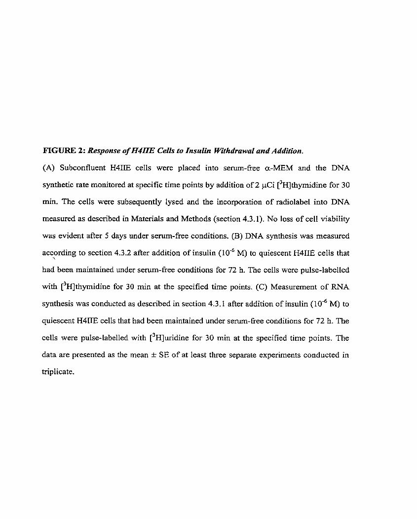
FIGURE 2: Response of H4ITE Celis to Insuiin Withdrawd and Addition.
(A) Subconfluent H4IIE cells were placed into senun-free a-MEM and the DNA
synthetic rate monitored at specific time points by addition of 2 pCi [3~]thymidine for 30
min, The cells were subsequently lysed and the incorporation of radiolabel into DNA
measured as described in Materials and Methods (section 4.3.1). No loss of ce11 viability
was evident after 5 days under serum-fiee conditions. (B) DNA synthesis was measured
according to section 4.3.2 after addition of insulin (10-~ M) to quiescent H4IIE cells that i
had been maintained under serum-free conditions for 72 h. The cells were pulse-labelled
with [-'HJthyrnidine for 30 min at the specified time points. (C) Measurement of RNA
synthesis was conducted as described in section 4.3.1 after addition of insulin (104 M) to
quiescent H4IIE cells that had been maintained under senun-fkee conditions for 72 h. The
cells were pulse-labelled with [3~]uridine for 30 min at the specified time points. The
data are presented as the mean t SE of at least three separate experiments conducted in
triplicate.
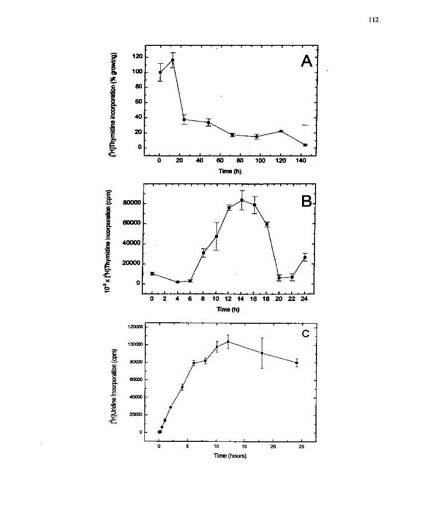
120000-
1OOOOO
g c O 80000 - H B - - 8 C - ?!
40000-
=
I 1 1 1 1 I - C .
-
- p\- /- I l + - : -
5 ,, Y
1 GL i
= O -
1 1 1 I 1 I
O 5 10 15 20 25
Tirne (hwrs)
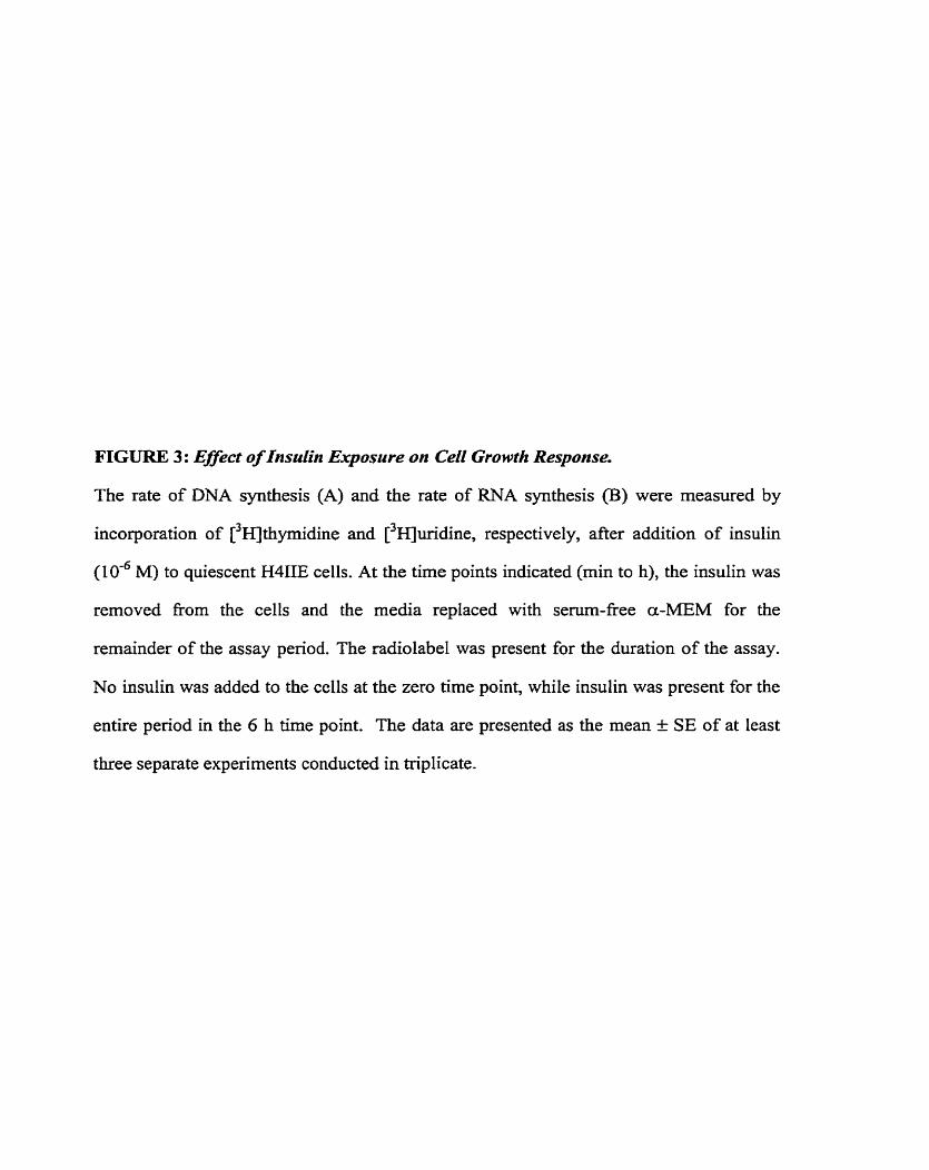
FIGUlU3 3: Effect of Insuiin Exposure on Ceii Growrh Response.
The rate of DNA synthesis (A) and the rate of RNA synthesis (B) were measured by
incorporation of [3~thymidine and [3~urïdine, respectively, after addition of insulin
(10" M) to quiescent H4IIE cells. At the time points indicated (min to h), the insulin was
removed fiorn the cells and the media replaced with senun-free a-MEM for the
remainder of the assay period. The radiolabel was present for the duration of the assay.
No insulin was added to the cells at the zero time point, while insulin was present for the
entire period in the 6 h time point. The data are presented as the mean f SE of at least
three separate experiments conducted in triplicate.
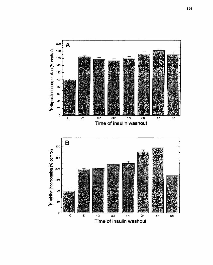
10' 30' 1h 2h
Time of insulin washout
Time of insulin washout
5.2.4.2 Growth and metabolic response of H4IIE cells to insulin and IGF-1
In many ce11 systems, IGF-1 is considered to be a potent stimulator of growth and
is classified as a progression factor with respect to ce11 cycle kinetics (Baserga et al.
1994). Based on the studies in section 5.2.4.1, it could not be ruled out that IGF-1 andior
its receptor participate in the process of insulin-mediated H411E growth. As a result it was
of interest to determine if H41IE cells, which are reported to be responsive to insulin
aIone (Figure 2; Yau et al. 1998), are also influenced by IGF-1. 30th insulin and IGF-1
were able to significantly stimulate RNA and DNA synthesis (Figure 4A,B) in a
concentration-dependent manner (IO-" to 1 0 - ~ M), reaching a maximum effect at 10-' M.
However, insulin was able to stimulate a greater response than IGF-1 at a concentration
two orders of magnitude lower (IO-' M compared to 1 0 - ~ MM, respectively) (Figure 4A,B).
Furthemore, although increased DNA synthesis is a good indicator that ce11 prohferative
processes have been triggered, it does not guarantee that ce11 division will occur.
Therefore, the effect of insulin and IGF-I on ce11 number was monitored 72 h after
addition of the peptides to quiescent H4IIE cells (Figure 4C). As observed with the
growth assays, insulin increased ce11 number at IO-' M while 1 O-' M IGF- 1 was iequired
before an equivalent increase in ce11 number was obtained. Taken together, these results
indicate that insulin is a significantly stronger mitogen than IGF-1 in this ceil type.
Moreover, the difference in effective concentrations of insulin and IGF-1 eliminates the
possibility that the growth response occurs as a result of insulin cross-reacting with IGF-1
receptors. Furthemore, since the relative affinity of IGF- 1 for the insulin receptor is 100-
fold less than that for insulin (Massague & Czech 1982) the converse is also unlikely.
The results obtained with this experiment thus indicate that mitogencsis is mediated by
the insulin receptor of H4IIE cells.
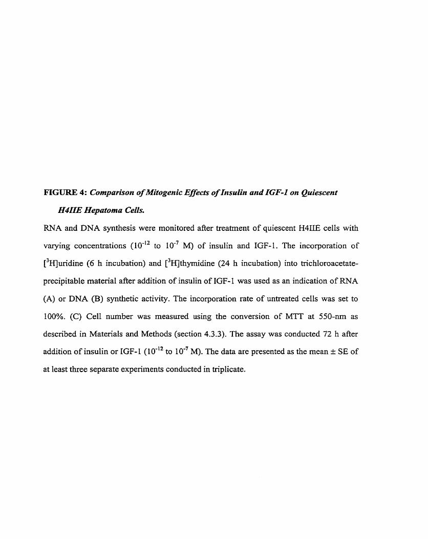
FIGURE 4: Cornparison of Mitogenic Effects of Insulin and IGF-I on Quiescent
H4IIE Hepatoma Cells.
RNA and DNA synthesis were monitored after treatment of quiescent H4IIE celIs with
varying concentrations (10-'' to IO-' M) of insulin and IGF-1. The incorporation of
~ ~ ~ ~ ] ~ u i d i n e (6 h incubation) and ['HJthyrnidine (24 h incubation) into trichloroacetate-
precipitabie material after addition of insulin of IGF-1 was used as an indication of RNA
(A) or DNA (B) synthetic activity. The incorporation rate of untreated cells was set to
100%. (C) Ce11 number was measured using the conversion of MTT at 550-nrn as
described in Materials and Methods (section 4.3.3). The assay was conducted 72 h afier
addition of insulin or IGF-1 (10''~ to 10" M). The data are presented as the mean t SE of
at least three separate experiments conducted in triplkate.
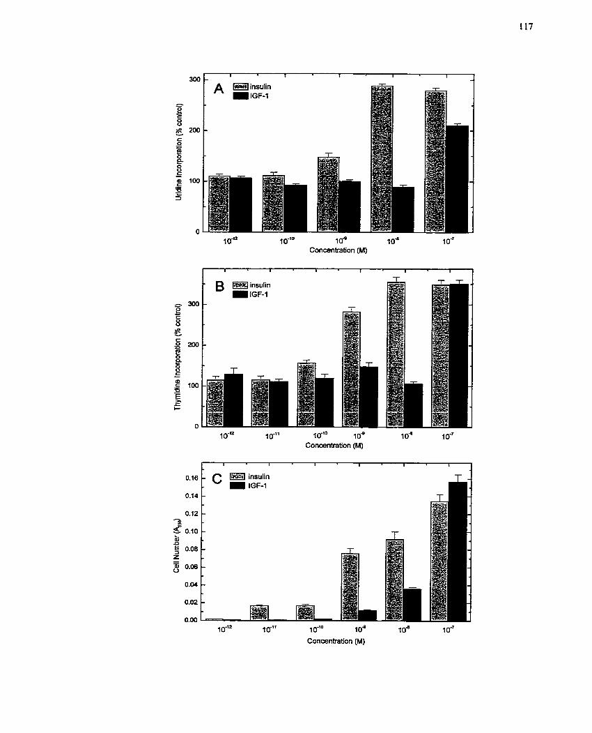
A insulin IGF-1
1 0*1° 10" 3 0" 1 O" Concentration (M)
insulin IGF-1
1 0-l2 1 O-" 1 O-'" 1 O= lo" Concentration (M)
1 0-l2 1 O-" 1 0 - ' O 1 0" 1 0"
Concentration (M)

fnsulin in most ce11 systems (eg. skeletal muscle and liver) is a metabolic activator
and regulator of events such as glucose uptake and usage (Czech 1977). For example,
insulin controls glucose utilization by the Iiver, influencing glucose uptake, glycogen
synthesis, gIycoIysis and gluconeogenesis. Under the experimental conditions
established, H4IIE cells responded to insulin (10" M) by taking glucose up into the ce11
as would be expected in physiologically relevant systems such as the liver (Figure 5). In
contrast, IGF-1 was unable to enhance glucose uptake over a small concentration range
(IO-' to IO-' M). Serurn (2% v/v FBS), which contains insulin and many other growth
factors, was used as the positive control for comparing the increased glucose uptake
generated by either insulin or IGF-1 (Figure 5). These results clearly demonstrate that the
IGF-1 reccptor does not modulate glucose metabolism in H4IIE cells.
5.2.4.3 Signaling components of the H4IIE response to insulin and IGF-1
Binding of insulin to its receptor initiates an intracellular s i gnahg cascade that
involves protein tyrosine phosphorylation (Frattali et al. 1992; Wilden et al. 1992).
Although TGF-1 activates a similar cascade in most systems responsive to this peptide
(Kato et al, 1993; LeRoith et al. 1995), it remains unclear how the IGF-1 s i g n a h g
system produces a response that is different from insuIin. To address this issue, a study of
the relationship between protein tyrosine pliosphorylation events and insulin or IGF-I
ligand binding was conducted. Treatment of H411E cells with insulin ( L O - ~ M) resulted in
a rapid phosphorylation of proteins with molecular mass 60-, 9 5 , 125- and 180-kDa, as
detected with an antibody speci fic for phosphotyrosine (Figure 6A), although the c hangc
in the 125-kDa protein (assumed to be p125FAK) was much less obvious. Thesc proteins
remained phosphorylated over the 120 min time course of the experiment- In contrast,
phospl.iorylation of thcse same proteins was considerably less whcn tlic cells were trcatcd
with 1 o - ~ M IGF- 1. A concentration curvc of insulin and IGF-1 indicatcd that IGF- i was
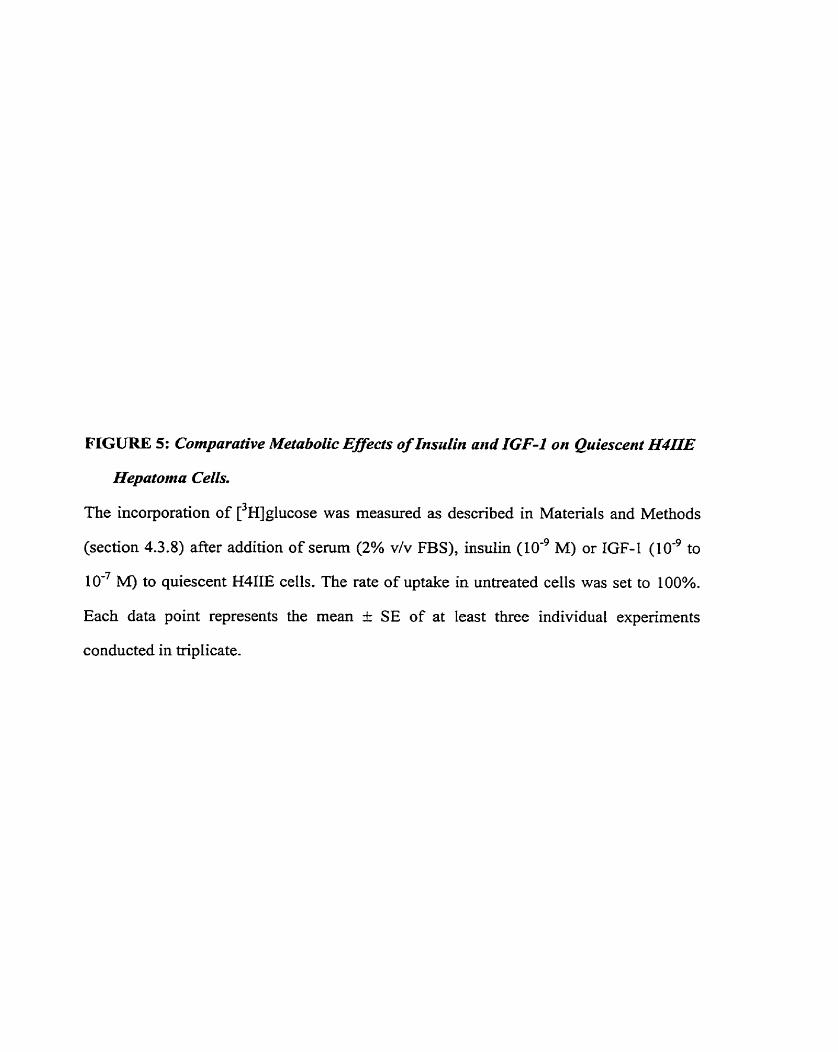
F I G U R E 5: Comparative Metabolic Effects of Xnszdin and XGF-1 on Quiescent H4nE
Hepatonza Cells.
The incorporation of [3~]glucose was measured as descnbed in Materials and Methods
(section 4.3.8) after addition of serum (2% v/v FBS), insulin (10-~ M) or IGF-1 (10~' to
10" M) to quiescent H4IIE cells. The rate of uptake in untreated cells was set to 100%.
Each data point represents the mean t SE of at least three individual experiments
conducted in triplicate.
Serum: Insulin: IGF-1:
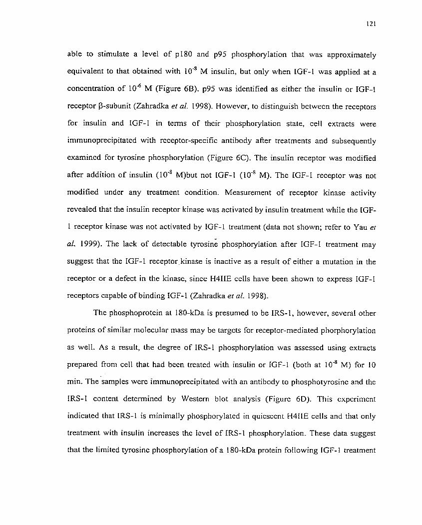
able to stimulate a level of pl80 and p95 phosphorylation that was approximately
equivalent to that obtained with 10" M insulin, but only when IGF-1 was applied at a
concentration of 10" M (Figure 6B). p95 was identified as either the insulin or IGF-1
receptor B-subunit (Zahradka et al. 1998). However, to distinguish between the receptors
for insulin and IGF-1 in terms of their phosphorylation state, ce11 extracts were
immunoprecipitated with receptor-specific antibody afier treatments and subsequently
examined for tyrosine phosphorylation (Figure 6C). The insulin receptor was modified
after addition of insulin (10" M)but not IGF-1 (10.' M). The IGF-1 receptor was not
rnodified under any treatment condition. Measurement of receptor kinase activity
revealed that the insulin receptor kinase was activated by insulin treatment while the IGF-
1 receptor kinase was not activated by IGF-1 treatment (data not shown; refer to Yau sr
al. 1999). The lack of detectable tyrosiné phosphorylation after IGF-1 treatrnent may
suggest that the IGF-1 receptor-kinase is inactive as a result o f either a mutation in the
receptor or a defect in the kinase, since H4IlE cells have been shown to express IGF-1
receptors capable of binding IGF-1 (Zahradka et al. 1998).
The phosphoprotein at 180-kDa is presumed to be IRS-1, however, several other
proteins of similar molecular mass may be targets for receptor-mediated phorphorylation
as well. As a result, the degree of IRS-1 phosphorylation was assessed using extracts
prepared from ce11 that had been treated with insulin or IGF-1 (both at 1 0 - ~ M) for 10
min. The samples were immunoprecipitated with an antibody to phospliotyrosine and the
IRS-1 content determined by Western blot analysis (Figure 6D). This experiment
indicated that IRS-1 is rninimally phosphorylated in quiescent H4IIE cells and that only
treatment with insulin increases the Ievel of IRS-1 phosphorylation- These data suggest
that the limited tyrosine phosphoryIation of a 180-kDa protein following IGF- I treatment
FIGURE 6: Tyrosine Phosphoryiation of H4LE Proteins after Treatmenf with Insuiin
and IGF-1.
(A) Quiescent H4IIE cells, prepared in 12-well culture dishes containing I rnL media,
were treated with 10'~ M insulin or 10" M IGF-1 and harvested by direct addition of
SDS/gel loading buffer (as described in Materials and Methods, section 4.8.3) at the
indicated thne points. Sarnples were subjected to SDSPAGE (7S% gel), transferred to
PVDF membrane and imrnunostained with PY2O anti-phosphotyrosine monoclonzl
antibody (diluted 15000). The antibody was visualized with HRP-coupled anti-mouse
IgG and the ECL chemiluminescent detection system. Pre-stained molecular mass
markers were nin in parallel with the samples and were used to identiQ the bands.
Identical results were obtained in two independent experirnents of which one
representative blot is shown. (B) Cells were harvested for Western blot analysis 5 min
after addition of insulin or IGF-1 (10-" to lo4 M). After SDSPAGE and transfer to
PVDF membrane, PY20 antibody was used to monitor tyrosine phosphorylation. Pre-
stained molecular mass markers were run in parallel with the sarnples and were used to
identie the bands. Identical results were obtained in three independent experiments of
which one representative blot is shown. (C) Cells were lysed for immunoprecipitation
(IP) as descnbed in Materials and Methods (section 4.8.2) 5 min after addition of insulin
or IGF-I (IO-* MJ. The protein content of each sample was measured by BCA protein
assay, and equal 100 pg protein aliquots were incubated with antibody (3 pg) to either the
b u l i n receptor P-subunit (insulin-R) or the IGF-1 receptor B-subunit (IGF1-R). The
antibody-antigen complexes were collected with protein G Sepharose, solubilized by
heating in SDS/gel loading buffer and subjected to SDSPAGE. After transfer of the
proteins to PVDF membrane, tyrosine phosphorylation was detected with PY20 antibody
(diluted 15000). Pre-stained molecular mass markers were run in parallel with the
sarnples and were used to i d e n t a the bands. Comparable results were obtained in two
independent expenments. (D) Proteins were resolved by SDSPAGE after
immunoprecipitation with ami-phosphotyrosine antibody (3 pg). The membrane was
subsequently probed for IRS-I using an IRS- 1 specific antibody (diluted 1: 1000). The
cells were untreated (lane C) or treated with 10-~ M insulin (lane Ins) or 1om8 M IGF-1
(lane IGF) for 5 min before ce11 lysis. Comparable results were obtained in three
independent experiments.
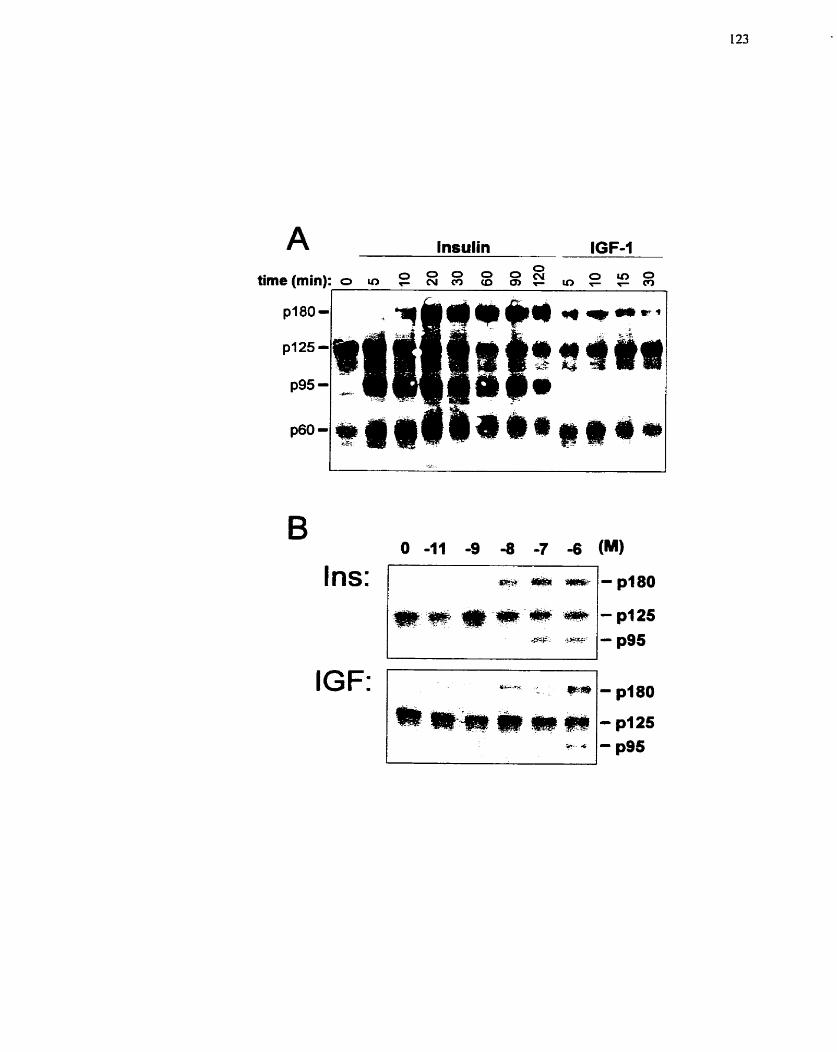
IGF: yp4,.= - ** -pl80
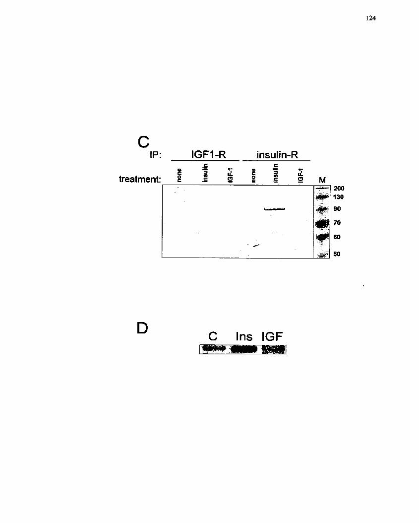
IP: IGFI -R insulin-R r - - E
4) 4) -
T - r 3 LL E 3 7
b d L L treatment: E - C 0 E - E 52
C Ins IGF
(Figure 6A) does not represent modification of IRS-1, but that of another protein with
sirnilar molecular rnass, possibly IRS-2.
Mitogen activated protein (MAP) kinase is considered to be an essential signaling
cornponent for the growth of a variety of ce11 types (Adam el al. 1995; Force &
Bonventre 1998; KhaIil & Morgan 1993; Klemke et al. 1997; Nelson et al. 1998)- The
activation of p42 (ERK1) and p 4 4 (ERK2) MAP kinase is an early event in the signaling
cascades activated by both insulin and IGF-1 (Kim & Kahn 1997)- As a result, MAP
kinase is considered to be a required intemediate in the cellular response to these
peptides. The stimulation and activation of MAP kinase involves the phosphorylation of
both tyrosine and threonine residues by MAP kinase kinase (MEK) (Payne et al. 1991).
In this study, the activation of MAP kinase was monitored by Western blot analysis,
using an antibody specific for the phosphorylated f o m of this protein (Xu et ai. 1996;
Yau & Zahradka 1997). Insulin and IGF-1 induced a similar transient increase in p42/p44
MAP kinase phosphorylation at 1 oA8 M (Figure 7A) with a peak at 10 min and a return to
basal levels after 20 min. To verify that MAP kinase pliosphorylation correlated with an
increase in activity, an in situ polyacrylamide gel assay was used to measure MAP kinase
activity. This method was chosen for its ability to distinguish the myelin basic protein
phosphorylated by p42 vs, p 4 4 MAP kinase (Yau & Zahradka 1997). Increased
modification of myelin basic protein was observed with increasing concentrations of
insulin and IGF-1 (IO-" to 10-' M) with maximal phosphorylation observed at 1 0 - ~ M for
both peptides (Figure 7B). The results indicate that both peptide ligands are equaliy
effective at activating MAP kinase, which is in contrast to the tyrosine phosphoryIation
data presented above (Figure 6). Although it was observed that several other proteins are
capable of phosphorylating rnyelin basic protein (most notabIy proteins of moIecular
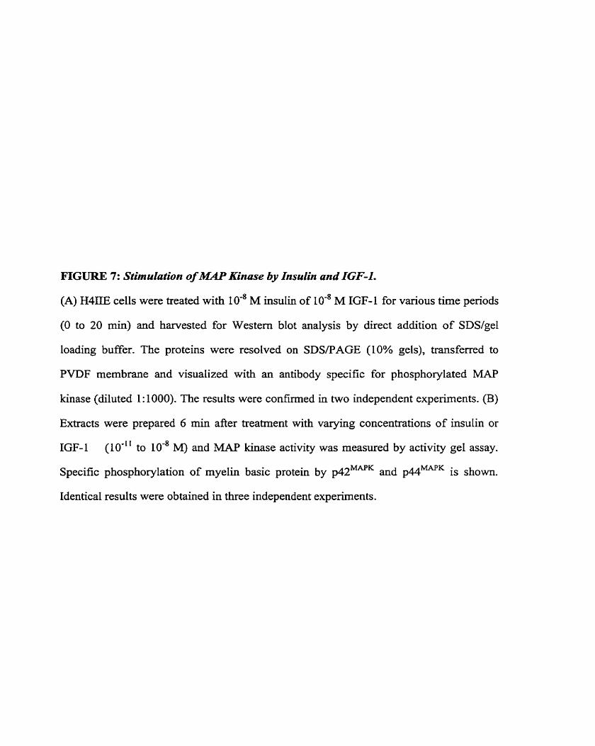
FIGURE 7: Stimulation of MAP Kinase by Insulin and IGF-1.
(A) H4IlE cells were treated with IO-* M insulin of M IGF-1 for various time periods
(O to 20 min) and harvested for Western blot analysis by direct addition of SDS/gel
loading buffer. The proteins were resolved on SDSPAGE (IO% gels), transferred to
PVDF membrane and visualized with an antibody specific for phosphorylated MAP
kinase (diluted 1: 1000). The results were c o n f i e d in two independent experiments. (B)
Extracts were prepared 6 min afier treatment with varying concentrations of insulin or
IGF- 1 (10-Il to 10-* M) and MAP kinase activity was measured by activity gel assay.
Specific phosphorylation of myelin basic protein by ~ 4 2 ~ ~ ' ~ and p44MAPK is shown.
IdenticaI results were obtained in three independent experirnents.
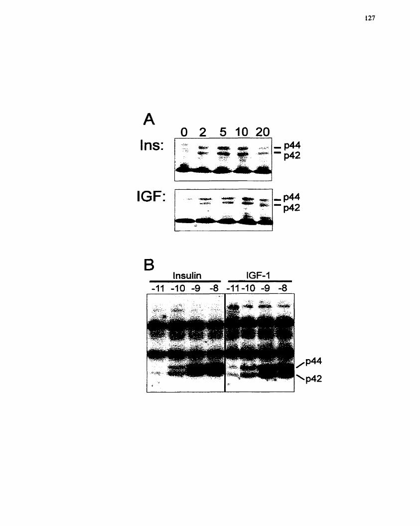
I ns:
IGF:
B lnsulin IGF-1
mass 58- and 112-kDa), these proteins were constitutively active and did not respond to
mitogen stimulation (Figure 7B).
To veri& that MAP kinase activation correlated with nuclear translocation, the
subcellular distribution of phosphorylated MAP kinase was monitored by
immunocytocliemical staining, Phosphorylated MAP kinase was at the lower limits of
detection in quiescent H4II.E cells, and no nuclear staining was evident (Figure 8A). After
treatment with both insulin and fGF-1 (10" M), nuclear translocation of phosphorylated
MAP kinase was evident (Figure 8B,C). Since equimolar concentrations of insulin and
IGF- 1 activated MAP kinase, these data suggest that IGF- 1 -dependent induction of MAP
kinase signaling is mediated through the IGF-1 receptor.
To confirm that the signaling system responsible for MAP kinase activation is
triggered by both insulin and IGF-1, the phosphorylation status of MEK, the kinase that
phosphorylates MAP kinase (Payne et al. 1991), was monitored. Western blot analysis,
with antibodies specific to de/phosphorylated M E K M kinase, showed that MEK
phosphorylation was increased by both insulin and IGF-1 in a concentration-dependent
marner (10-" to 1 0 - ~ M), with maximal stimulation observed at 1 0 - ~ M (Figure 9A). The
ratio of phosphorylated MEK and total MEK band intensities (Xu et al. 1996) was similar
over the entire concentration range (Figure 9B), thus confinning that IGF- 1-mediated
MEK phosphorylation results from stimulation of the IGF- 1 receptor.
5.2.4.4 Effect of an inhibitor of MEK (PD980591 on insulin-mediated H4ILE growth
As mentioned previousiy, MAP kinase is an essential signaling molecule in the
activation and growth of a variety of ce11 types. Therefore, the importance of the MAP
kinase cascade in mediating insulin-stimulated growth events in H4IIE c e k was assessed
using PD98059, a specific inhibitor of MEK (Alessi et ai. 1995; Dudley et al. 1995). By
inhibiting MEK1/2, PD98059 prcvents thc phosphorylation and subsequent activation of
FrCURE 8: Nuclear Translocation of MAP Kinase in Response to Insulin and IGF-1.
Epifluorescence microscopy was used to monitor the subcellular localization of
phosphorylated MAP kinase. Quiescent H4IIE cells (A), cells treated with IO-' M insulin
(B) and cells treated with IO-' M IGF-1 (C) were used in these experiments.
Magnification: 120~.
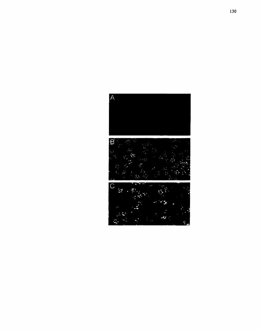
FIGURE 9: I~sulin- nnd lGF-1-dependent Phosphorylation of MEK.
(A) MEK phosphorylation in response to various concentrations of insulin or IGF- 1 ( 10"O
to IO-' M) was assessed by Western blot analysis using antibodies (diluted 1: 1000)
specific to either the phosphorylated (pMEK) or al1 forms of MEK. One of three
independent experiments is presented, all of which exhibited the sarne response. (B) The
ratio of pMEUMEK was calculated by combining densitometric values obtained from
three separate blots for each condition. This ratio provides a control for variation in
protein loading.
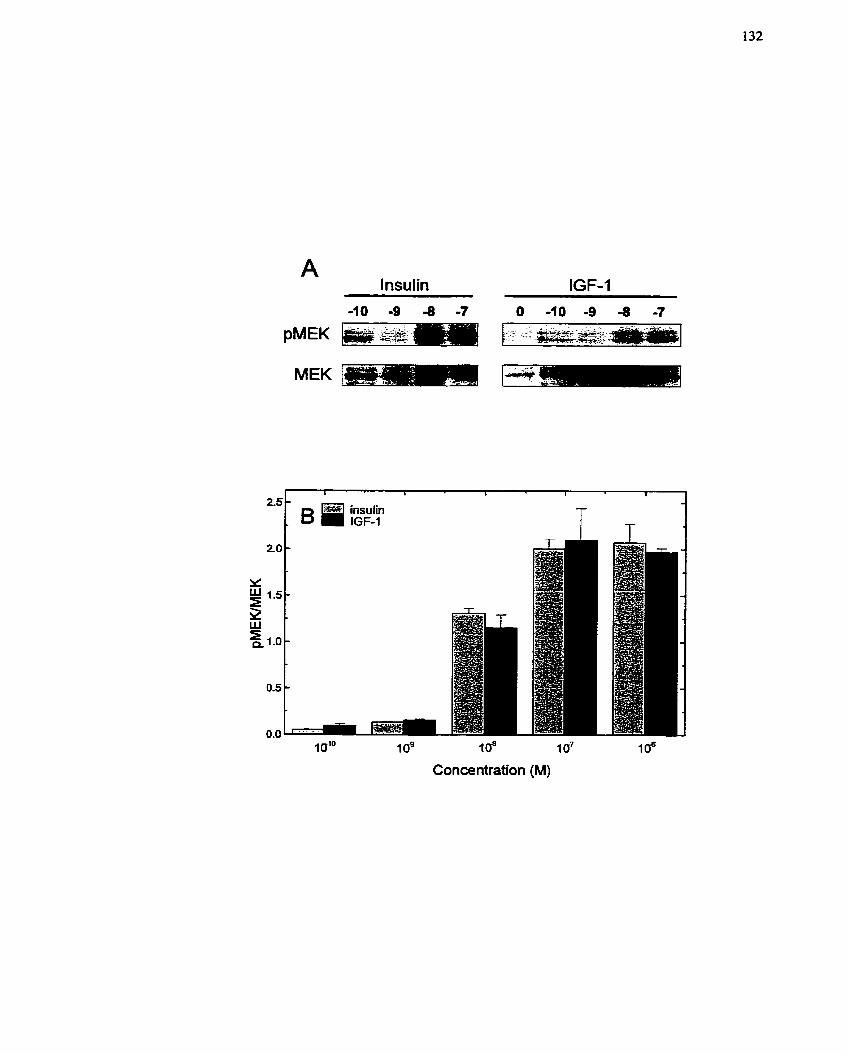
A lnsulin
1 al0 1 o9 ioa 1 o7 10"
Concentration (M)
MAP kinase, This event was verified in H4IIE cells by pretreating H41IE cells with
PD98059 for 10 min before stimulation with insulin (IO-' M). PD98059 significantly
decreased the phosphorylation of MAP kinase in a concentration-dependent rnanner (10"
to 1 0 ~ M) (Figure 10A). Quantification of these data indicated that PD98059 at 104 M
inhibited MAP kinase phosphorylation (both p42 and p44 MAP kinase) by 90% (Figure
108). PD98059 also had a marked effect on the growth response of H4IIE cells. This
inhibitor reduced insulin (10" M)-mediated RNA and DNA synthesis, with lo4 M
PD98059 decreasing RNA and DNA synthesis by 52% and 88%, respectively (Figure
10C,D). These data clearly establish that MAP kinase activation is necessary for the
growth and proliferation of H4HE cells.
5.2.5 Discussion
Insulin elicits a variety of biological responses in the liver reIating predominantly
to glucose metabolism. As well, this hormone is noted for its influence on ceIl growth
during liver regeneration. The response of H4IIE hepatoma cells to insulin also includes
modification of their glucose metaboIic pathways and initiation of ce11 growth (Mohn et
al. 1991; Taub et al. 1987). We have confirmed that both DNA and RNA synthesis are
increased upon insulin stimulation (Yau et al. 1998; Zahradka & Yau 1994) and have
extended this to demonstrate that H411E celIs traverse the entire ce11 cycle and complete
ce11 division (Figure 4). These findings establisli that insulin is a potent mitogen for
H4IIE cells, especially considering the ineffectiveness of so-called strong mitogens such
as platelet-denved growth factor (PDGF) and epidennal growth factor (EGF) in this
systern (P. Zahradka, unpublished observations). In contrast, IGF- 1, an agent more noted
for its growth stimulatory effects, does not trigger ceII growth unless high concentrations
are used (Figure 4). The 100-fold or greater concentration difference indicates the
FIGURE 10: inhibition of MXP Kinase Phosphorylniion and Celi Proliferution by the
M2CK In hibifor PD98059.
(A) Quiescent H4IIE cells were treated with 10 -~ M insulin in the presence of various
concentrations (10-~ to 104 M) of PD98OS9. Samples were nui on SDSRAGE (10% gel)
and phosphorylation of MAP kinase was monitored by Western blot analysis using an
antibody specific for phosphorylated MAP kinase (diluted 1:1000). The response
produced by PD98059 alone (lo4 M) is shown in the Iast lane. The relative positions of
the molecular mass markers (in kDa) is shown. This experiment was conducted in
duplicate for treatrnent with both 1 0 - ~ M and IO-' M insulin; one representative gel
depicting the effect with 10'~ M insulin is shown. Similar results were obtained for both
experiments. (B) Band densities were determined by Censitometry and the data plotted
for both the 42- and 44-kDa bands. (C, D) Insulin (10" M)-stimulated RNA (C) and
DNA @) synthesis was measured in the presence of varying concentrations of the MEK
inhibitor PD98059 (IO-' to 104 M). Each data point represents the mean k SE of at least
three individual experiments conducted in triplicate. Values are presented relative to a
non-insulin-treated control which was set to 100%.
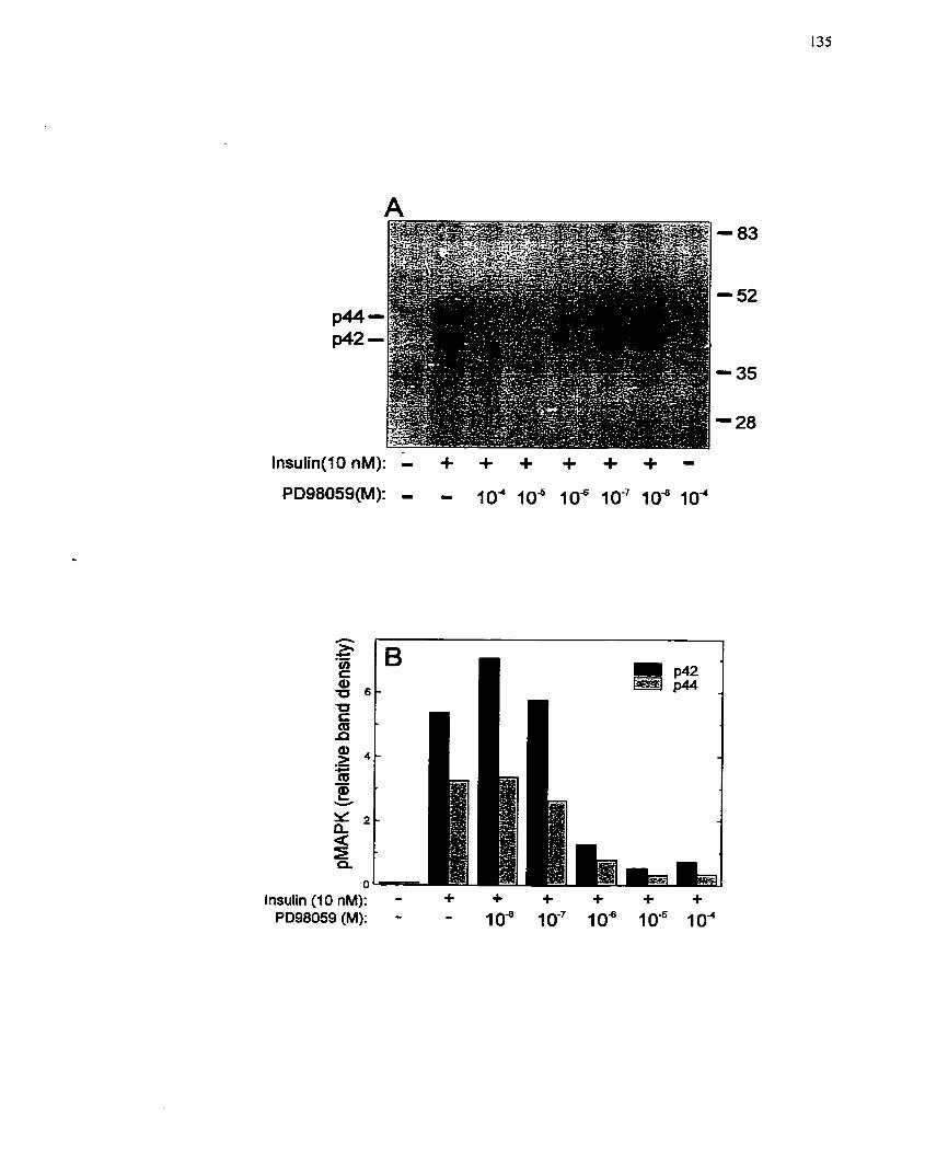
Insulin ( I O nM): - + + + + + + PD98059 (M): - - 10" IO-' los 105 104
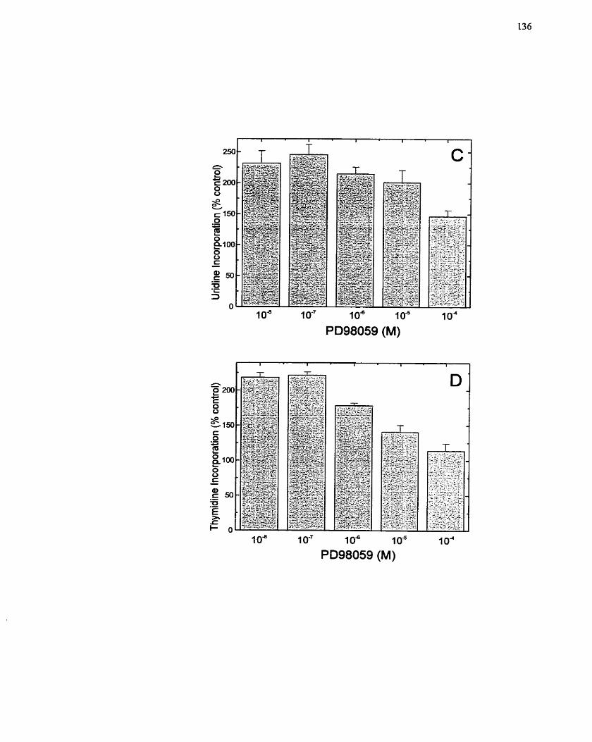
1 o4 1 o - ~ 1 o6 1 o6 i o4 PD98059 (M)
1 O" 1 oa I o5 1 o4 PD98059 (M)
stin~ulation of WIIE ce11 growth by IGF-1 is likely mediated by the insulin receptor
rather than the IGF-1 receptor (Koontz 1984; Massague & Czech 1982). An examination
of the effects of insulin and IGF-1 on glucose metabolic systems revealed a similar
pattern, since IGF-1 did not increase glucose uptake at a concentration below 10" M
(Figure 5). While the inability to detect glucose uptake with IGF-1 at IO-' M cannot be
explained, although it was obsenred at 1 o ' ~ M (data not shown), it is plausible that at this
concentration IGF-L does not stimulate glucose uptake beyond 10 min, and uptake has
therefore ceased before addition of the radiolabelled substrate. These data, nevertheless,
confirm that LGF-1 does not influence the regulatory systems responsible for mediating
the metabolic changes associated with insulin in this ce11 type. Nevertheless, H4IIE cells
cxpress receptors capable of binding IGF-L (Zahradka et al. 1998) and it is generally
accepted that common elements exist in the respective signaling pathways of IGF-1 and
insulin (Kadowaki et al. 1996; Prager & Melmed 1993; Sasaoka et ai. 1996).
Experiments utilizing kinase-defective insulin receptor mutants have
demonstrated that induction of the P subunit tyrosine kinase activity is essential for both
mitogenesis and changes in glucose metabolism (McClain et al. 1990). The insulin
receptor kinase stimulates various cytoplasrnic signaling cascades through IRS-1
(Kadowaki et al. 1996; Myers et al- 1994a; Myers et al. 1994b), an intermediary protein
that recmits proteins vital for further signal transmission. In general, the phosphorylation
of IRS- 1 results in the binding of proteins with SH2 domains, such as p85, Syp and Grb2,
that have been linked to the production of phosphatidylinositol 3-phosphates, tyrosine
dephosphorylation and p2 1 -Ras/MAP kinase activation, respectively (Myers et ai. 1994b;
White & Kahn 1994). IRS-1 has an equivalent role in the signaling system for IGF-1 (De
Meyts et al. 1994; Frattali el ai. 1992; Myers et ai. 1994a) and an inability to initiate IRS-
1 phosphorylation (Figure 6D) could thcrefore account for the lack of effcct by IGF- 1 on
H411E cc11 growth and metabolism. Since these cclls obviously have Eûnctional IGF- 1
rcceptors. as demonstrated by the stimulation of both MEK pliosphorylation and MAP
kinase activity with M IGF-1 (Figures 7, 9), the defect could potentially exist in the
receptor kinase. This assumption was confirmed by directly measuring receptor kinase
activity where it was observed that insulin, but not IGF-1, could elicit tyrosine
phosphorylation of the respective P subunit (data not shown; refer to Yau et al. 1999).
This detail explains the lack of IGF-1 receptor autophosphorylation (Figure 6C) and the
absence of IRS-1 tyrosine phosphorylation (Figure 6D) when H41IE cells are treated with
IGF- 1.
The lack of correlation between IRS-1 phosphorylation and M M kinase
activation has been previously observed in ceIIs expressing mutated insulin receptors
deficient in tyrosine kinase activity (Krook et al. 1996; Takata el al. 1996). Interestingly,
these reports noted tliat IRS-1 tyrosine phosphorylation still occurred following insulin
treatment, yet MAP kinase activation was suppressed. In conjunction with the data
presented in this report, support for the prernise that IRS-1 is not a component of the
MAP kinase cascade becomes apparent. Although it has been well established that IRS- 1
directs certain elements of the signaling cascade leading from both insulin and IGF-1
receptors (Waters et al. 1993), it has also becorne evident that alternative pathways for
regulating specific intracellular processes exist.
These data confim that MAP kinase (p42/p44) is an essential intracelluIar
mediator of the proliferative response to insulin (Figure IOC, D), and it is most likely that
MAP kinase promotes ce11 prolireration by stimulating gene expression and protcin
synthcsis. On the othcr Iiand, i t has bcen shown tliat activation of MAP kinase alone is
insufficicnt for thesc cvcnts (Scrimgcour et al. 1997). Mcchanistically, MAP kinase is
prcsunicd to opcratc by stiriiulating gcnc cxprcssion, pai-tiçularIy of kcy proto-oncogcncs
such as c-fos and c-jun (Karin 1996)- It has become evident, however, that modulation of
transcription factors can occur independent of MAP kinase (Kim & Kahn 1997). Similar
findings relate to specific metabolic processes that are controlled by insulin. For instance,
MAP kinase does not participate in the stimulation of gluconeogenesis (Sutherland et al.
1998). As well, there is consensus in the literature that the physiological role of MAP
kinase does not encompass the metabolic effects of insulin (Combettes-Souverain &
Issad 1998; Lazar et al. 1995). Unfortunately, a relevant mode1 of intracellular s ignahg
that defines the role of MAP kinase in ce11 proliferation and ce11 metabolism has not been
elucidated, presumably because the inter-relationships between various signal
transduction pathways, including the MAP kinase cascade, have yet to be clarified.
Nevertheless, it has been recently suggested that MAP kinase activation by IGF-1
requires the participation of G proteins through their By subunits (Luttrell et al. 1995; van
Biesen et al. 1995). It has generally been assumed, however, that tyrosine
phosphorylation events must precede G protein activation. These results, on the other
hand, support a paradigm wherein activation of MAP kinase by IGF-1 occurs in the
absence of receptor-mediated tyrosine phosphorylation events. These characteristics are
emphasized by three critical points: 1) IGF-1 does not trigger tyrosine phosphorylation of
either IRS-1 or the IGF-1 receptor (Figure 6), 2) the H4IIE IGF-1 receptor apparently
Iacks tyrosine kinase activity (data not shown; Yau et al. 1999), and 3) MAP kinase
activation occurs at IGF-1 concentrations that are below the threshold necessary for
insuIin receptor stimulation (Figures 7, 9). It is therefore credible that the lack of tyrosine
kinase activity displayed by the IGF-1 receptor of H411E cells (data not shown, Yau et ai.
1999) may be irrelevant with respect to the MAP kinase cascade. Evidence that G protein
activation is an essential component for intracellular signalling by insulin (Rizzo &
Romero 1998; Uehara er al. 1999) thereby provides a rnechanisrn by which receptor
tyrosine kinase-independent events may by implemented- Further exploration of these
alternative pathways is clearly warranted, and H4IIE hepatomas represent an excellent
system for defining the divergent signaling pathways associated with LGF -1 receptor
function.
5.3 Contribution of ADP-ribosylation to H4IIE ce11 proliferation
53.1 Background and rationale
ADP-ribosyiation reactions can be divided into two groups. Those involving
PARJ? occur in the ce11 nucleus and those involving M T s are not simiIarly restricted to
a specific cellular cornpartment (D'Amours et al. 1999; Moss et a[. 1997; Okazaki &
Moss 1996a; ShaIl 1994; Zolkiewska et al. 1994). Both of these reactions have been
implicated in ce11 proliferation. One of the known functions of PARP is participation in
DNA repair and replication (Cesarone et ai. 1990), while m . T s have been
demonstrated to be direct or indirect regdatory factors in the growth processes of a
varies of ce11 types (Kharadia et al. 1992; Loesberg et al. 1990b; Smets et al- 1990b;
Thyberg et al. 1995a).
ADP-nbosylation is characterized by the transfer of ADP-ribose from NAD* to an
acceptor protein (Moss & Vaughan 1988; Ueda & Hayaishi 1985). O-linked polymers of
ADP-ribose are produced by the enzyme poly(ADP-ribose) polyrnerase. Each molecule of
poly(ADP-ribose) is anchored via the terminal ADP-ribose moiety to the acceptor protein by
an ester linkage to a glutamate residue. Polymerization is achieved through the formation of
glycosidic bonds which coupIe adjacent ADP-ribose units. Due to the specific cellular
location of poly(ADP-ribose) polyrnerase, poly(ADP-ribosy1)ation occurs only in the
nucleus. Since the principal acceptors for poly(ADP-ribose) are DNA-binding proteins, and
attachment of poly(ADP-ribose) reduces their affmity for DNA (de Murcia et al. 1983;
Zahradka & Ebisuzaki 1982), changes in gene expression may be coupled to the
modification of specific transcription factors that regulate either gene activation or
repression. Alternatively, gene transcription could be controlled indirectly by poly(ADP-
ribosy1)ation of histones, a process that modulates chromatin condensation (de Murcia et al.
1988).
N-linked mono(ADP-ribosy1)ation occurs in the nuclear, cytosoiic and membrane
fractions of a cell (Moss & Vaughan 1988; Ueda & Hayaishi 1985). Arginine-dependent
mono(ADP-ribosyl)transferases (mg-mART) couple a single ADP-ribose to the guanidino
moiety of arginine. Transferases that catalyze the ADP-ribosylation of cysteine, serine,
lysine, asparagine and histidine residues have also been detected (Zolkiewska et al. 1994). It
has become apparent that mono(ADP-ribosy1)ation can modulate numerous enzyrnatic
processes, especially those associated with GTP-hydrolysis. For example, G protein function
is stimulated by ADP-ribosylation of the Gs a-subunit at the same arginine that is modified
by choiera toxin (Jacquemin et al. 1986). At this point, however, there is no indication that
mono(ADP-rïbosy1)ation participates directly in the regdation of gene expression.
in the liver, the ribosylation of proteins has been associated with both
regenerating liver (Cesarone er al. 1990), the onset of hepatocellular carcinoma
(McKillop et al, 1998) and inhibition of hepatocyte growth via nitrïc oxide (Dalrnau et al.
1996). Moreover the actions of insulin have been linked to the increased expression of G;
proteins (Livingstone et al. 1994; Sanchez-Margalet et al. 1999) and expression is altered
by exogenous pertussis toxin ribosyIation events (Sanchez-Margalet et al. 1999).
Furthermore, in diabetes, Gi cxpression is decreased (Gawler et al. 1987). As a result, the
question of whether ADP-ribosylation events are linked to and with insulin-mediated
H411E ce11 growth becomes an extremely reIevant question and this study is the first to
directly examine this cellular phenornena and its participation in specific cellular
processes-
This section describes the results of experiments designed to dari@ the relevance
of ADP-ribosylation to insulin-mediated ce11 growth. Furthermore, it describes attempts
to differentiate between the cIass of ribosylation reaction contributing to ce11 growth
through the use of different inhibitors of the ribosylation process. A potential role for
protein modification with mono(ADP-ribose) rather than poly(ADP-ribose) in the
regdation of -gowth by insulin in WIIE cells was identified.
5.3.2 Specific aims
1. To determine the contribution of ADP-ribosylation reactions to H4IIE ce11
growth and proliferation.
2. To identi@ the class of ADP-ribosylation reaction involved in H41IE ce11
growth and proliferation through the use of specific ADP-ribosylation
inhibitors.
5.3.3 Experimental design
H4IIE cells prepared as described in section 4.2.1, will be stimulated with insulin
and the extent of ADP-ribosylation activity correlated with the activation state as it
relates to ce11 proliferation. The experiments in this section are based on two previous
publications. The first report found that insulin-dependent inhibition of the gluconeogenic
pathway was prevented by inhibitors of ADP-ribosylation, specifically 3-
am inobenzamide (3AB) and PD 128763. Furthermore, these inhibitors of ADP-
ribosylation could themselves induce PEPCK gene expression (Yau er ai. 1998). it was
postulated in this paper that control of PEPCK gene expression, a key component of the
oluconeogenic pathway, may involve a mART enzyme rather PARP. The second 3
publication established that 3AB was an effective inhibitor of H4IIE ce11 growth
(Zahradka & Yau 1994). The class of ADP-ribosyIation reaction associated with insulin
stimulation of H4IIE cells will be identified through the use of specific inhibitors of
either arg-mART or PARP. At this point, it should be noted that 3AB has been shown to
inhibit both PARP and arg-mART activities depending on the concentrations that are
being used, with lower concentrations more specific to P A U and higher concentrations
influencing arg-mART activity as well (Banasik et al. 1992; Milam & Cleaver 1984;
Rankin et al. 1989). PD 128763, on the other hand, is reported to be a specific inhibitor of
PARP activity (Arundel-Suto et al. 1991; Burkart et aL 1999; Yau et al. 1998).
5.3.4 ResuIts
5.3.4.1 Insulin stimulates ADP-ribosylation
Based on a previous publication that demonstrated 3AB and PD128763 both had
unique effects on the expression of PEPCK mRNA (Yau et al. 1998), two scenarios were
generated: i) ADP-ribosylation participates in the insulin-mediated control of PEPCK
gene activity, or ii) ADP-ribosylation is responsive to insulin trcatmcnt. To define these
events, the activities of both PARP and arg-mART were measured in extracts from
quiescent and insulin-stimdated H4IIE celk. Significant stimulation of PARP \vas not
detected in the nuclear fraction of H411E cells prepared without and with a 30 min insulin
treatrnent ( 1 O-%) (Table 1). On the other hand, the microsomal Fraction dcmonstrated a
profound incrcase in arg-iiiART activity after stimulation with insulin I IO-^ M) compared
to without insulin treatrncnt (Table 1). A panei of ADP-ribosylation inhibitors was then
tcstcd in conjunction witli both PARP and arg-mART assays il? idro. This not only
hclpcd to cstabl isli the spccificity of thc ADP-ribosylatioii cvcnts occuring in thc
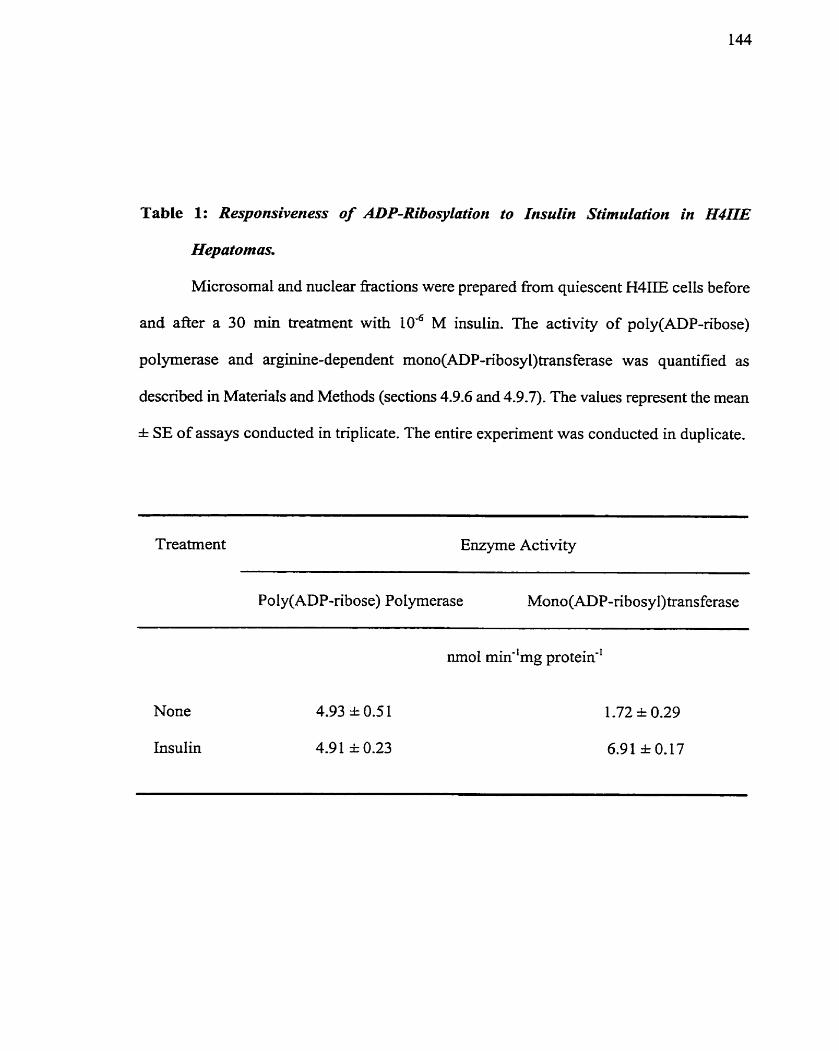
Table 1: Responsiveness of ADP-Ribosyhtion to Insu& Stimulation tn H411E
Hepa tom as.
Microsomal and nuclear fiactions were prepared fiom quiescent H4IIE cells before
and afier a 30 min treatment with 106 M insulin. The activiiy of poly(ADP-ribose)
polymerase and arginine-dependent mono(ADP-ribosy1)transferase was quantified as
descnbed in Matenais and Methods (sections 4.9.6 and 4.9.7). The values represent the mean
* SE of assays conducted in triplkate. The entire experiment was conducted in duplicate.
Treatrnent Enzyme Activiw
Poly(ADP-ribose) Polyrnerase Mono(ADP-ribosyl) transferase
Noue
Insulin
nuclear and microsomal fractions of growing H4IIE cells, but ako validated the
selectivity of the inhibitors for use in future assays. ADP-ribosylation inliibitors assessed
induded: i) PD 138763, a specific PARP inliibitor (Arundel-Suto et al- 199 1 ), i i ) 3AB, a
dual inhibitor of PARP and mART (Banasik et ul. 1993; Milani & Clcal-er 1984; Rankin
er al. I989), and i i i ) MIBG, a specific argmART inhibitor (Loesbcrg et al. 1 WOb; Smets
et cd. 1990b). I t was observed that growing H41iE ceIls have a relativcly high basal levet
of ADP-ribosylation activities (Table 2)- 3AB (2 n1M) \vas a b k to inliibit both PARP
activity of nuclear fractions and arg-mART activity of niicrosomal fractions, confirming
its dual inhibitory role (Table 2). PD138763 (10 pM), on the other hand, \vas only
capable of inhibiting the PARP activity of nuclear fractions. thereby confirming its
specificity for PARP (Table 2). Finally, MlBG (50 FM) inhibited arg-niART activity
without significantly reducing PARP activity, thereby validating its specificity for arg-
mART. Taken together, these data support the specificity of the ADP-ribosylation
reactions tliat are being observed in H41IE cells iiz vitro (Table 1). Similar magnitudes of
inhibition werc observed using estracts Froni insulin-stiniulated H4IIE cells (data not
shown).
To confimi the rcsults of PARP cnzyme activity measurements iii vitro, which are
not always reprcscntative of changes in activity fiz vivo (Lautier cr cd. 1993), an
alternative approacli was utilized. Western bIot analysis with an antibody spccific to
poly(ADP-ribose) was uscd to idcntify proteins modilicd with poly(ADP-ribose).
Nun~crous protcins wcrc detcctcd, including protcins of niolccular m i s s 1 IO-, 74-, 62-,
SI- , 46-, 3 1-, 29-, 27-, and 25-kDa (Figurc 1 1)- The promincnt bands bctwccn 15- and
30-kDa prcstiiiiabiy rcprcscnt various histonc proteins that arc known acccptors of
poty(ADP-ribosc) (Figurc 1 1 ) . Thc lack of dctcctablc variation in tlic 116-kDa PARP
batid, wliich irncicrgocs autoniodification in rcsponsc to cc11 daniagc (Kawaichi et ui.
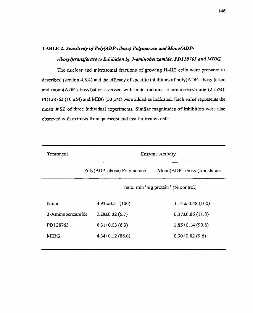
TABLE 2: Sensitivity of Poly(ADP-ribose) Poly~nerase and Mono(ADP-
ri&osyl)transferase to Inhibition by 3-arnino benzumide, PD128 763 and MIBG.
The nuclear and microsomal fiactions of growing H4IIE cells were prepared as
described (section 4.8.4) and the efficacy of specific inhibitors of poly(ADP-ribosy1)ation
and mono(ADP-nbosyl)ation assessed with both fhctions. 3-arninobenzarnide (2 mM),
PD128763 (10 PM) and MIBG (50 FM) were added as indicated. Each value represents the
mean SE of three individual experirnents. Similar magnitudes of inhibition were also
observed with extracts fiom quiescent wid insulin-treated cells.
Treatrnent Enzyme Activity
Poly(ADP-ribose) Polyrnerase Mono(ADP-ribosy l)trans ferase
nmo1 min-'mg protein-' (% control)
FIGURE 1 1 : Protein Poly(ADP-ribosyl)arion NI H4DE Hepatotna Cells in vivo.
H4IE ceIls maintained for 30 h in sem-£ree a-MEM media were harvested at various
time points after addition of insulin (10'~ M) and the protein modification monitored by
Western blot analysis using polyclonal a-pADP anti-poly (ADP-ribose) serum (diluted
1: 1000). Positions of molecular mass markers are indicated. Antibody binding was shown
to be specific using an auto-rnodified poly(ADP-ribose) poiyrnerase control. Similar
results were obtained with the monoclonal 10H antibody.
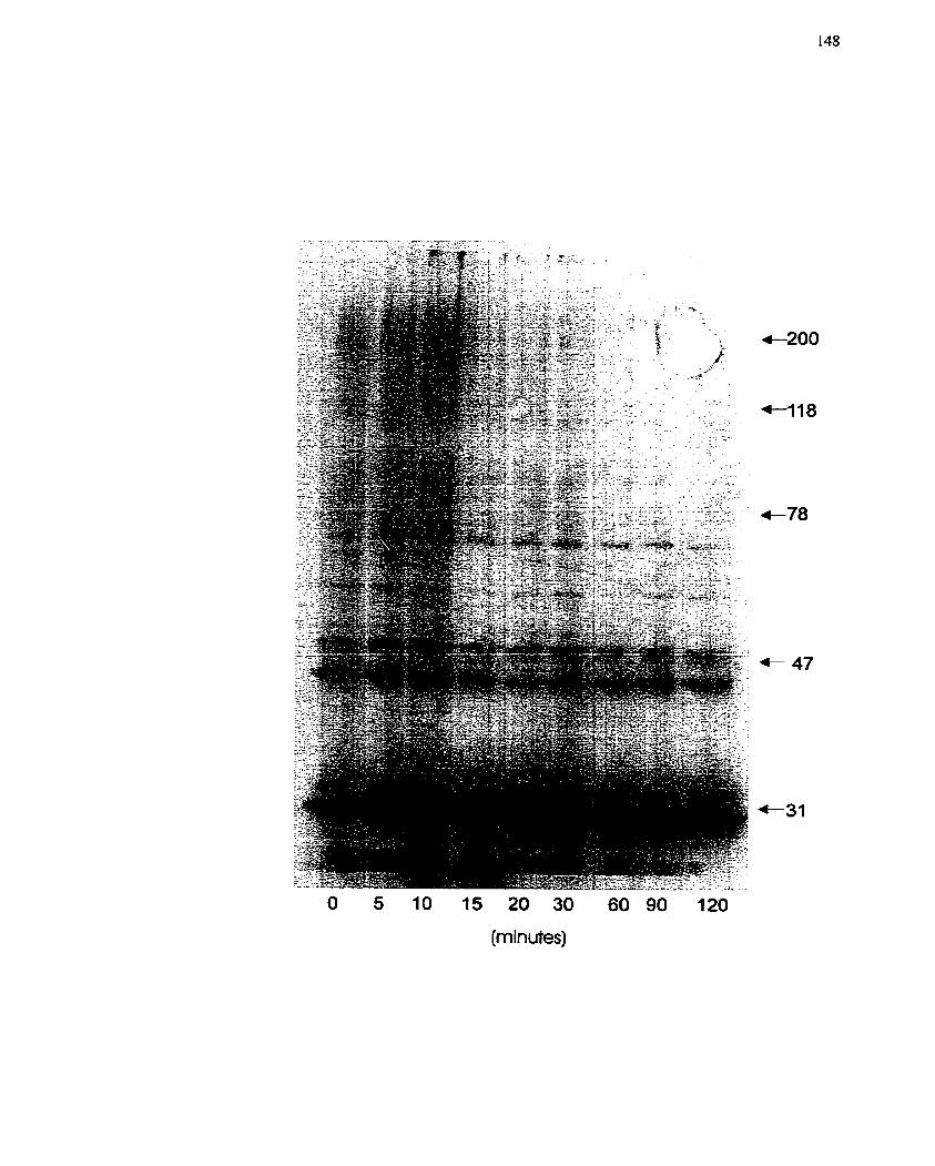
O 5 10 15 20 30 60 90 120
(min utes)
198 1; Schraufstatter et al. 1986), indicates that cellular repair systems n-ere not activated
(Figure I l ) . Over a time course of insulin treatment (W6 M), significant changes in the
pattern of poly(ADP-ribosy1)ation were not obscrved. thus supponing the enzyme
activity data shown in Table 1.
A more cellular and less invasive approach was also uscd to monitor mono(ADP-
ribosy1)ation in H41IE cclls in order to confim~ the results in Table 1. HLFIIE cells were
labeled with [3~]adenosine for 16 h pnor to insulin addition and the modified proteins
were then visualized by fluorography after separation of the proteins by SDS/PAGE. This
analysis showed that modification of a 53-kDa band was increased foliowing insuiin
treatrnent (Figure 12A). Furthemore, modification of this protein was slightly decreased
in the presence of' 3AB, but not PD128763 (Figure 12B). To verify that radiolabehg
under these experimental conditions represented ADP-ribosylation, the samples were
treated with 1 M NaOH or 1 M hydroxylamine, agents which disrupt the argininyl-ADP-
ribose bond (Figure 12B) (Jacobson et ai. 1994). The loss of protein-associated radiolabel
with these two treatments confim~s that the 53-kDa protein is rnono(ADP-ribosy1)ated on
an arginine residue (Figure 12B). The correlation between insulin-dependent mono(ADP-
ribosy1)ation (Figure 12A) and insulin-stimulated ars-mART activity (Tablc 1) suggests
that this process, and not poly(ADP-ribosy1)ation is activated upon insulin stimulation of
H4IIE cells and may be involved in insulin-mediatcd H411E ceIl growth and proliferation.
Cellular arg-mART activity has becn dctccted in scveral cc11 fractions. For
cxample, Zol kicwska and Moss (Zolkiewska & Moss 1993; Zolkicwska et al. 1992) have
demonstratcd a n3A RT associatcd with tlic cxtcrnal plasma rncni brane of chick skclctal
muscle cclls, and Gravcs and colIcagucs (Huang ci ul. 1996; Kharadia et cd. 1992) have
sliown a microsonial arg-iiiART activity i n chick niusclc cclls. For this rcason, arg-
mART assays wcrc ciiip loycd to dctcriiiinc wliich cc1 l fkxiions containcd t Iic arg-niART
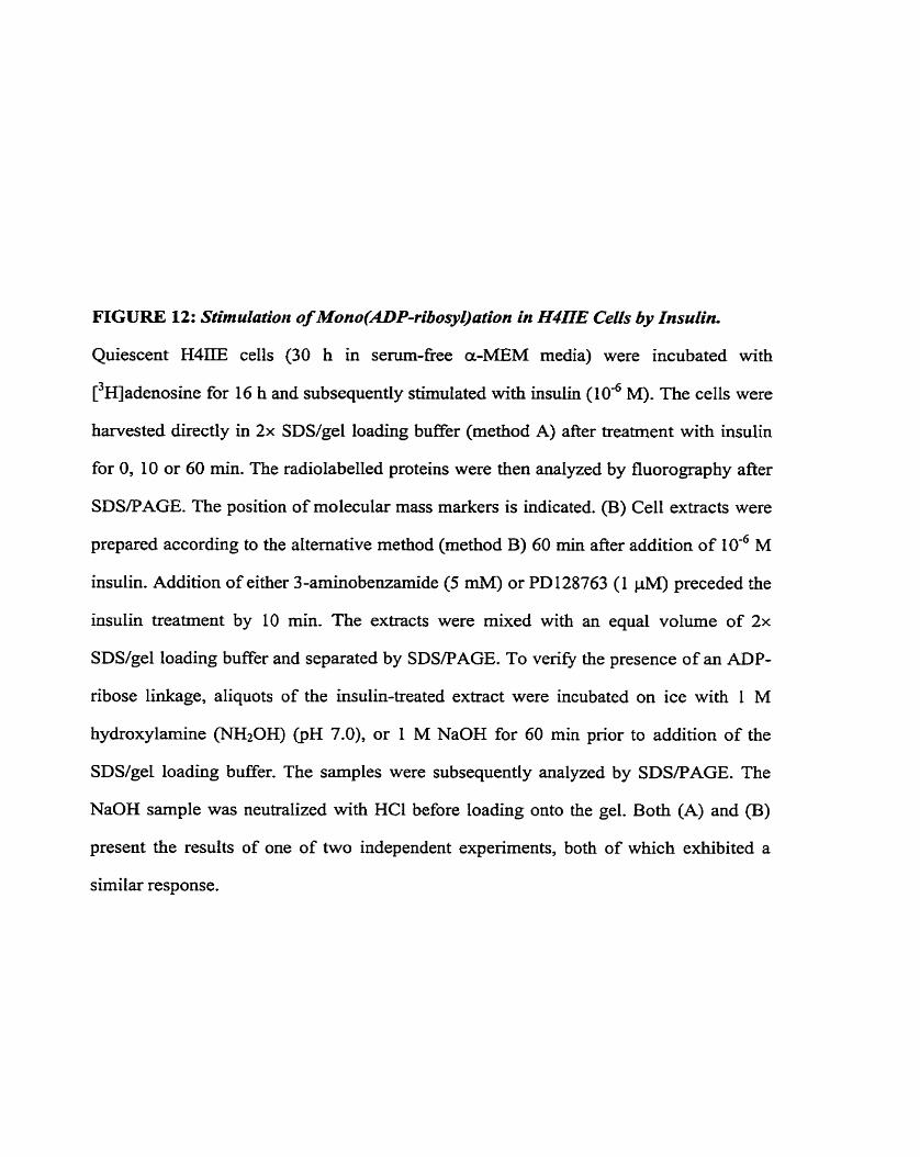
FIGURE 12: Stimulatio~t of Mono(ADP-ribosyl)afiun in H417E Cells by Insulin.
Quiescent H4IIE cells (30 h in sem-fkee a-MEM media) were incubated with
[3~adenosine for 16 h and subsequently stimulated with insulin ( 1 0 ~ M& The cells were
harvested directly in 2x SDS/geI loading buffer (method A) after treatment with insulin
for 0, 10 or 60 min. The radiolabelled proteins were then analyzed by fluorography &er
SDSPAGE. The position of molecular mass markers is indicated. (B) Ce11 extracts were
prepared according to the alternative method (rnethod B) 60 min after addition of 10-6 M
insulin. Addition of either 3-arninobenzamide (5 mM) or PD 128763 (1 pMJ preceded the
insulin treatment by 10 min. The extracts were mixed with an equal volume of 2x
SDS/gel loading buffer and separated by SDSFAGE. To ven@ the presence of an ADP-
ribose linkage, aliquots of the insulin-treated extract were incubated on ice with 1 M
hydroxylamine (NfiOH) (pH 7.0), or 1 M NaOH for 60 min prior to addition of the
SDS/gel loading buffer. The samples were subsequently analyzed by SDSPAGE. The
NaOH sarnple was neutralized with HC1 before loading onto the gel. Both (A) and (B)
present the results of one of two independent experiments, both of which exhibited a
sirnilar response.
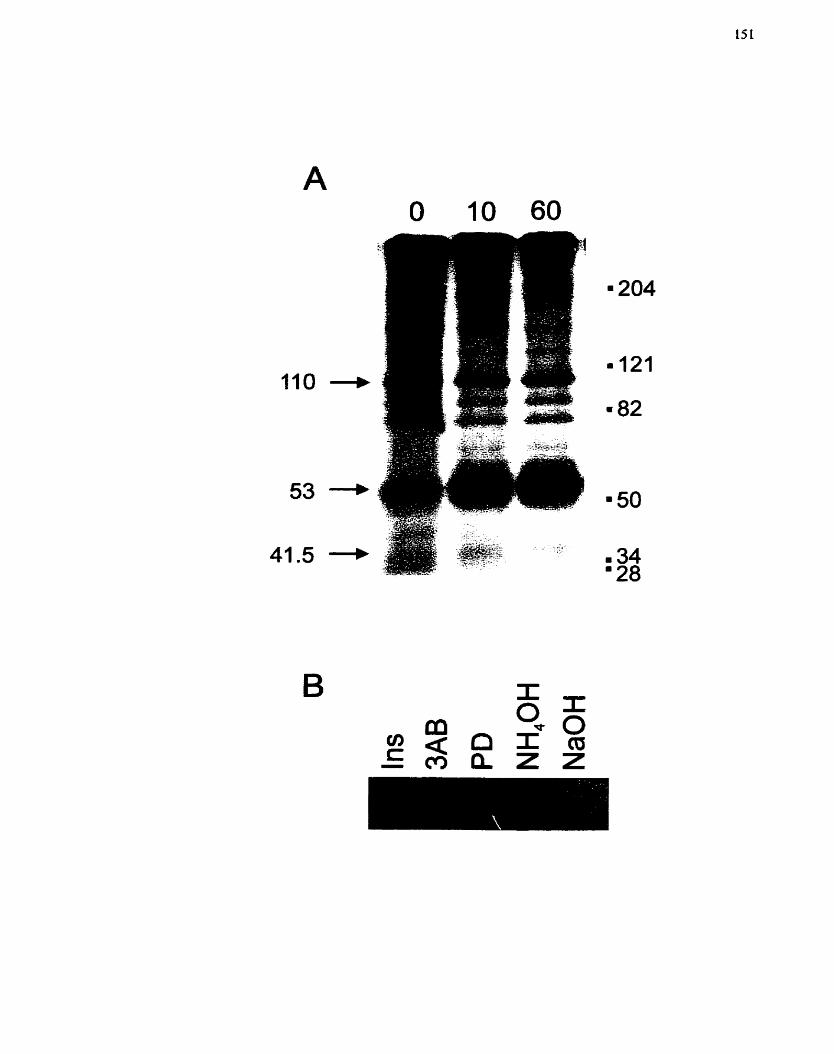
activity that increased after insulin stimulation, An iiz sirrr ADP-ribosyltransferase
activity assay indicated arg-rriART activity was increased in the microsomal fraction over
a 3 h period afier insulin stimulation (lad M) (Figure 13), while the cytosolic fraction
had no detectable activity by this method (data not s h o w ) - In vitro arg-mART activity
assays were then conducted with both cytosolic and microsomal fractions of H4IIE ce11
extracts that had been treated over a 2 h tirne course with insulin (10-~ M). The
microsomal fraction showed significantly more arg-mART activity in comparison to the
cytosolic fraction even in the basal state (Figure 11A), and the microsomal fraction
demonstrated an increase in arg-mART activity in response to insulin stimulation. Since
peak activity was observed at I h, a separate in vitro assay was camed out with
subcellular fractions prepared from H41IE cells 1 h after insulin treatment. This assay
showed that insulin increased microsoma1 arg-rnART activity by 1.5-fold over basal
levels (Figure 14B)- The change in arg-mART activity of the cytosolic fraction was not
more than 1.15-fold (Figure I4B). These data confirm the observation that insulin is able
to stimulate arg-mART activity in H4IIE cells (Table 1, Figure 13), and that the
activation of this enzyme may be linked to insulin-mediated ce11 growth and proliferation.
To venfy the specificity of the microsoma1 arg-mART activity stirnulated by
insulin (Table 1, Figure 14), an iiz vitt-o arg-mART activity assay was carrïed out in
which selective ADP-ribosylation inhibitors were included during the incubation time of
the assay. 3A8 (5 mM) or MIBG (50 FM) was added exogenously to the microsomal
fraction of 1 h insulin-stimulated H41IE cells and compared to microsomal extracts from
quiescent H4IIE cells. As seen previously (Figure 24), insulin treatment increased the
arg-mART activity from basal levels in quiescent cells (Figure 15). Both 3AB and MIBG
were able to decrease the arg-mART activitics to control, untreated levels (Figure I S ) ,
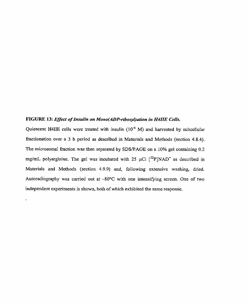
FIGURE 13: Effect of Insulin on Mono(ADPribosyl)ation in H411E Cells.
Quiescent H4IIE cells were treated with insulin ( 1 0 ~ ~ M) and harvested by subcellular
fiactionation over a 3 h period as described in Matenals and Methods (section 4.8.4).
The rnicrosomal fraction was then separated by SDS/PAGE on a 10% gel containing 0.2
mg/mL polyarginine. The gel was incubated with 25 pCi [ 3 2 ~ ] ~ A D f as descnbed in
MatenaIs and Methods (section 4.9.9) and, following extensive washing, dried.
Autoradiography was carried out at -80°C with one intensi*g screen. One of two
independent expenments is shown, both of which exhibited the same response.
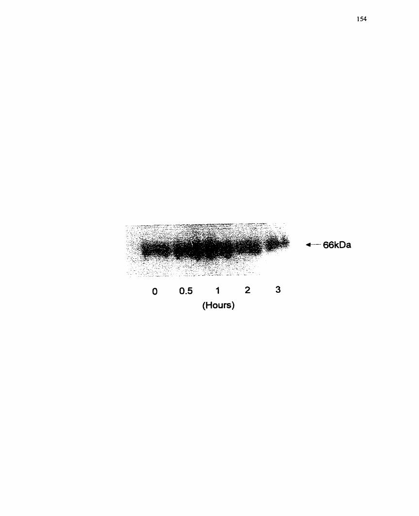
O 0.5 1 2 (Hours)
FIGURE 14: In vitro Mono(ADP-ribosyl)transferase Activity in Insuiin-treated H411E
Ceils.
Quiescent H4IIE cells were treated with insulin (lod M) and subcellular hct ions were
fiarvested at various times (O - 120 min) as described in Materials and Methods (section
4.8.4). (A) An in vitro rnono(ADP-ribosy1)ation assay was used to measure transfer of
ADP-ribose in both the microsomal (membrane) and cyiosolic (cytosol) £?actions over a
120 min t h e course as described in Materials and Methods (section 4.9.7). Polyarginine
(2 mg/rnL) was used as the acceptor protein for the assay. Each data point represents the
pooIed data from two independent experiments. (B) An in vitro mono(ADP-ribosy1)ation
assay was carried out using both the microsomal (membrane) and cytosolic (cytosol)
Eactions, and the changes in mono(ADP-ribosy1)transferase activity compared in insulin-
treated (1 h stimulation) and untreated cells. Polyarguiine (2 mg/mL) was used as the
acceptor protein for the assay. The activity present in untreated cells was set to 1.0 for
cornparison. These data represent one of two independent experiments, both of which
exhibited the sarne response.
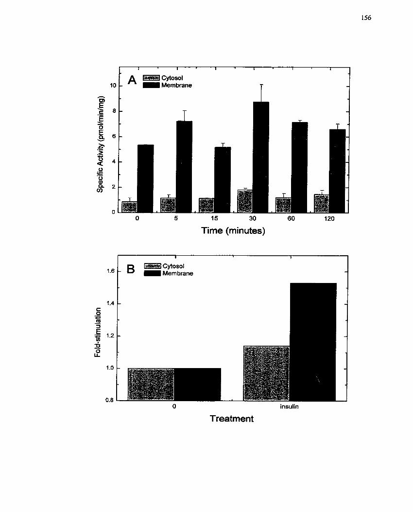
O 5 15 30 60 120
Time (minutes)
O insulin
Treatment
FIGURE 15: Serrsitivity of Mono(ADP-ribosyC)fransferase to inhibition.
Quiescent WIIE cells were treated with insulin (104 M) and harvested after 1 h by
subcellular hctionation as described (section 4.8.4). rnART activity in the microsomal
fraction was assayed in the presence of 3-aminobenzamide (5 m . ) or MIBG (50 CIM)
Polyarginine (2 mg/mL) was used as the acceptor protein for the assay. Data represent
one of two independent experiments, both of which exhibited the same response.
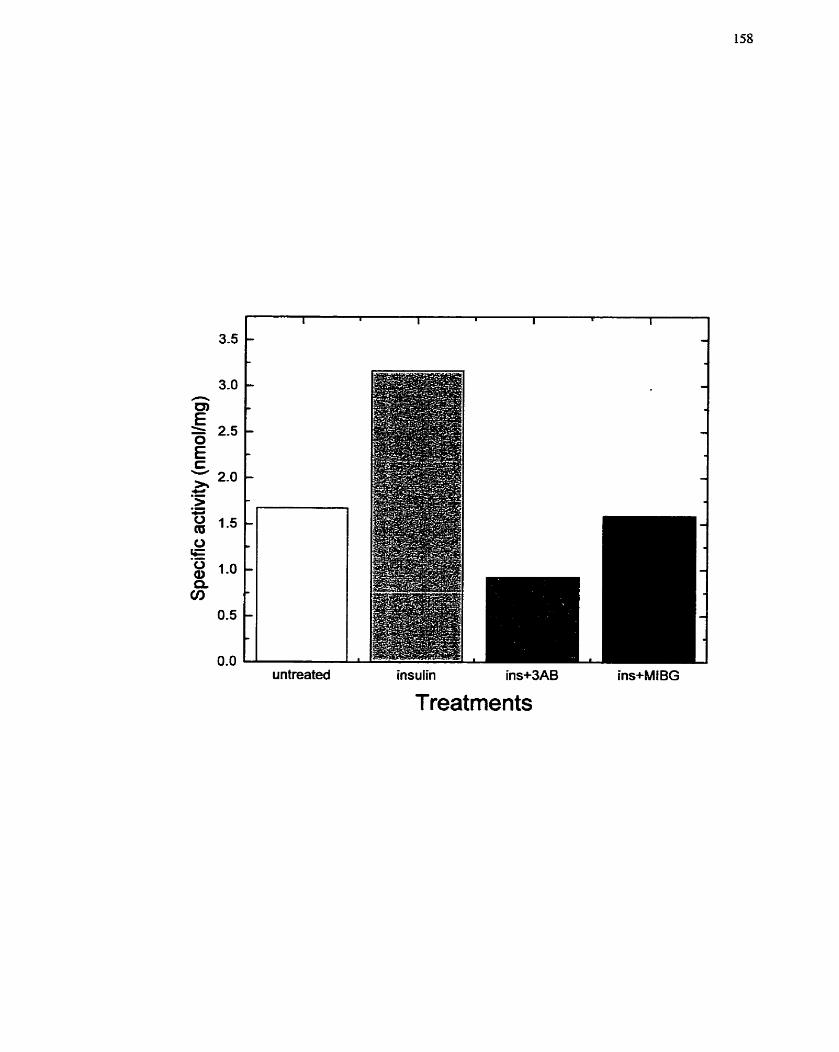
untreated insulin ins+3AB ins+MIBG
Treatments
demonstrating specificity of the cellular ADP-ribosylation response after insulin
treatrnent,
5.3.5 Discussion
Several cytosolic and rnicrosomal monotransferases have been identified
(Tsuchiya & Shimoyama 1994; Zolkiewska et al. 1994), however, none have been linked
to a specific regulatory process. The purpose of this study was to clan@ the roIe of ADP-
ribosylation and ADP-ribosyItransferases in the signal transduction processes associated
with the regulation of H4IIE ce11 growth by insulin. A previously published paper (Yau et
al. 1998) had examined the participation of ADP-ribosylation in the regulation of gene
expression by insulin- In that paper, expression of the PEPCK gene as controlled by
insulin was examined in conjunction with the ADP-nbosylation inhibitor 3-
aminobenzamide (3AB). It was noted here that the activity of PARP was not significantly
changed between insulin-stimulated and control cells. In contrast, both mART activity
and protein mono(ADP-ribosy1)ation were elevated in cells treated with insulin, the first
indication that mono(ADP-ribosy1)ation events are responsive to growth factors such as
insulin- Moreover, this activity could be inhibited by 3AB. Although these findings did
not confirrn a role for rnART activity in insulin-mediated changes in gene expression, the
data did emphasize that mART may have a role in mediating insulin-dependent s ignahg
systems.
In the previous section, it was demonstrated that insulin alone was able to act as a
growth factor for H4IIE ceIls and that certain signaling pathways such as MAP kinase
piayed a critical role in the insulin-mediated ce11 growth and proliferation. The data
generated in this section suggested a mono(ADP-ribosy1)transferase participates in the
ce11 growth effects mediated by insulin in H411E cells. The data showed that nuclear
PARP activity was not increased on ce11 stimulation with insulin and that there was no
change in the pattern of poly(ADP-rïbosy1)ation following insulin stimulation (Table 1).
In contrast, an increase in arg-mART activity was observed and this was consistent with
the increase in ADP-ribosylation of the 53-kDa protein (Figure 12). Although the identity
of this protein was not clarified, a band at 53-kDa has been observed in other ce11 systems
(Graves et al. 1997). PARP is typicaIly associated with ce11 growth and replicative
events- Its activity is increased in Ziver regeneration (Cesarone et al. 1990), and
upregulation occurs in many tissues and ce11 types dunng proliferation. On the other
hand, PARP can also be activated during ce11 senescence, apoptosis and by negative
growth regulatory factors such as nitric oxide (Dalrnau et al. 1996; Pieper et al. 1999;
Scovassi & Poirier 1999)- Nevertheless, in our ce11 system, PARP does not appear to play
a significant role in the ce11 growth processes mediated by insulin.
Arg-mART reactions on the other hand participate directly in cellular events in
the cytoplasm. In this scenario, ADP-ribosylation behaves as a post-translational
modification much like phosphorylation that signals cells to undergo a particular event.
In several different ce11 systems, incIuding smooth muscle and skeletal muscle, arg-
mART has been shown to participate in the cellular events leading to ce11 proliferation
and differentiation (Kharadia et al. 1992; Thyberg et al- 1995a). Moreover, the success of
MIBG therapy for the treatment of neuroblastoma, carcinoid tumours and metastases
suggests that perhaps an arg-mART event may be involved in tumour progression, which
involves ce11 proliferation (Cornelissen et al. 1995a; Hoefnagel et al. 199 1; Srnets et al.
1988b; Smets et al. 1989). In H41IE cells, it was demonstrated that particular cel1uIar
fractions exhibited elevated arg-mART activity after insulin stimulation (Figures 13, 14).
The microsomal fraction was shown by both in situ ADP-ribosyltransferase labeling
(Figure 13) and with an in vitra assay (Figure 14), that insulin was able to increase the
arg-mART activity in that particular ce11 fraction. The stimulation was approximately
1.5-fold (Figure 14B). On the other hand, the cytosolic fraction did not appear to contain
arg-mART detectable with the in sirll ADP-nbosyltransferase labeling assay (data not
show) , and insulin was only able to marginally increase the activity measurable with the
Nt vitro biochemical assay (Figure 14). These results suggest that the arg-mART activity
is associated with the ce11 membranes and that its compartmentalization within the ce11 is
via membranous components. This finding is in support of the findings of Zolkiewska
and colleagues (Zolkiewska & Moss 1993; Zolkiewska er al. 1992) and Kharadia et al.
(1992) who have also found that the arg-mART activity is more prominent in the
microsomal fraction. With respect to the ability to transmit signals, having the arg-mART
within the membrane fraction indicates that the activity is likely localized to one place
and that the enzyme does not shuttle behveen cornpartments. Considering that G proteins
and small GTP-binding proteins are such good targets for ADP-ribosylation reactions,
and that G proteins and GTP-binding proteins are mostly associated with the membrane
(Milligan & Grassie 1997; Rodbell 1997; Willard & Crouch 2000), the membrane-bound
nature of mART likely places it at an advantage for modulating G protein reactions.
With respect to the specificity of the reaction, the initial findings that mART
activity, but not PARP, was increased upon insulin stimulation (Table 1) led to the use of
specific inhibitors of both enzymes in order to confirm this finding. PD128763, a
reportedly specific inhibitor of PARP, only inhibited PARP activity, while MIBG only
inhibited arg-mART activity. 3AB on the other hand, inhibited both PARP and mART
activities (Table 2, Figure 15), confirming the dual inliibitory capabilities of this
compound. Furthemore, at the concentrations used in these studies (2 2 mM), 3AB will
inhibit mART activity (Banasik el al. 1992; Milam & Cleaver 1984; Rankin et al. 1989).
Thus, these data furthcr validate the contention that insulin is stimulating an arg-mART
and that activation of this mART is a key mediator of insulin-dependent WIIE cell
growth and proliferation. Although the studies in this section do not necessarily establish
that mART is a component of insulin-mediated H4IIE growth, the fact that insulin
stimulates this enzyme and that insulin correspondingly stimulates H4IIE ceil growth
suggests that a link exists between these events. The studies descnbed in the next section
were designed to establish that link and confirm the requirement of mART activation for
H41E hepatoma ce11 proliferation in response to insulin.
5.4 Modulation of ce11 proliferation by inhibitors of mART
5.4.1 Background and rationale
Mono(AJ3P-ribosy1)transferase is an enzyme present in the nuclear, cytosolic or
rnicrosomal fractions of cells and is involved in the post-translational modification of
proteins (Moss & Vaughan 1988; Ueda & Hayaishi 1985; Zolkiewska et al. 1994). This
enzyme transfers a single ADP-ribose moiety from NAD' to acceptor proteins that
usually contain an arginine, cysteine or diphthamide moiety (Zolkiewska et al. 1994).
This modification, much like phosphorylation, is reversible and is a component of
modulating signaling processes and cellular events (coordinating the processes within a
cell) (Williamson & Moss 1990; Zolkiewska et al. 1994). The most studied and
commonly modified proteins are those associated with G proteins and proteins that
undergo GTP binding and hydrolysis, such as G,,, Ci and srnaIl GTP-binding proteins
such as Rho and Ras (Ali & Agrawal 1994; Di Girolamo et al. 1997; Gierschik 1992;
Kanagy et al. 1995; Lem et al. 2000; Schmidt & Aktories 1998; Tsuchiya & Shimoyama
1994)-
Inhibitors of mART corne in many foms. The most popuIar of those ernployed
for biochemical studies of this protein include novobiocin, benzarnidc, naphthalimide,
hydroxyisoquinoline, silybin (silymarin) and meta-iodobenzylguanidine (MIBG). Many
of these compounds inhibit the action o f the enzyme directly. However, others such as
MIBG (and other compounds containing guanidino groups or guanylhydrazones) act as a
decoy substrate, mimicking the arginine present on the acceptor protein of the reguiar
substrates. Although MIBG does not directly inhibit the action of arg-mART, by
preventing the modification of the actual acceptor protein the potency of the mART
enzyme is lost.
MIBG was first recognized as a diagnostic molecule for the identification and
radiotherapy of adrenal turnours and other neuroendocrine tumours in its radioiodinated
forrn (Hoefnagel 1994; Hoefnagel et al- 1987; Lode et al. 1995; Smets et al. 1990a;
Wafelman et al. 1994b; Wieland et al. 1980). Subsequently, the non-radiolabelled
cornpound was shown to decrease turnour progression (Kuin et al. 1999; Smets et al.
1988b; Taal et al. 1999; Taal et al. 1996; Zuetenhorst et al. 1999). Although the
mechanism of action for the anti-tumour activity of MLBG without the radiolabel is not
presently known, it has been postulated to involve inhibition of mitochondrial respiration
(Cornelissen et al. 1995b; Loesberg et al. 1990a; Loesberg et ai. 1991; Smets et al.
1 99Ob). However, the arg-mART inhibitory activity of MIBG may also explain its anti-
neoplastic properties (Loesberg et al. 1990b; Smets et al. 1990b). Since these issues have
not been adequately addressed, an understanding of how ADP-ribosylation contributes to
ce11 proliferation will help to clarify these points.
5.4.2 Specific Aims
1. To examine the effect of inhibitors of ADP-ribosylation on insulin-mediated
H4IIE ce11 growth and proiiferation,
2. To confirm the link between ADP-ribosylation and insulin-mediated H41IE
ceil growth and proliferation.
3. To clanfi the adverse effects of MLBG on H4IIE cells.
4- To identify a potential mechanism of action for MIBG by determining the
effects of MIBG on two signaling intermediates stimuiated by insulin: MAP
kinase and p2 1 -Ras.
5.4.3 Experimental design
H 4 E celk, prepared as descnbed in section 4.2.1, will be pretreated for 10 min
with inhibitors of ADP-ribosylation (eg. 3AB, MIBG, precursor analog MIBA, etc.),
stimulated with insulin, and monitored for growth according to parameters such as RNA
and DNA synthesis- The exposure time required for MIBG to exert an inhibitory effect
will also be defined, along with an assessrnent of inhibitor reversibility. The relative
toxicity of MIBG will also be examined using an MTT cytotoxicity assay, an LDH
formation assay and FACS analysis of MIBG-treated H41IE celis. Finaily, potential
mechanisms of MIBG action on insulin-stimulated H4IIE cells will be defined using
assays to detect activation of two prominent signaling enzymes: MAP kinase and p21-
Ras -
5.4.4 Results
5.4.4.1 Effects of ADP-ribosylation inhibitors on ceH growth: involvement of mART
in H4IIE ce11 growth and proliferation
In the previuus section (section 5.3), it was established that insulin activates an
arg-rnART. Based on the findings presented in section 5.2 regarding insulin-mediated
H41IE cell growth and proliferation, it was speculated that stimulation of arg-mART
might be a component of the signaling systems that influence H4IIE ce11 growth post-
insulin treatrnent. As a result, to fiirther define the system and to c o n h the participation
of this protein modification in H4IIE ce11 growth and proliferation mediated by insulin,
inhibitors of ADP-ribosylation were tested for their ability to affect incorporation of
lundine ne and [ '~l th~midine. H41IE cells were pretreated for 10 min with the inhibitors
before the addition of insulin, The dual PARP/rnART inhibitor, 3AB, showed a modest
inhibition of insulin-stirnulated RNA synthesis (Figure 16A1, v~hile it did not affect
insulin-stimulated DNA synthesis (Figure 16B). Interestingly, at the high concentrations
(2 to10 mM) of 3AB required for modest inhibition of RNA synthesis, 3AB inhibits both
mART and PARP activity (Table 2, FigurelS). The analog of 3AB that lacks inhibitory
action, para-aminobenzoic acid (PABA), also did not affect DNA synthesis (data not
shown). Furthermore, neither agent was toxic to H4IIE cells as confirmed by lactate
dehydrogenase (LDH) release assay afier 72 h incubation with varying concentrations
(0.1 to 10 mM) of 3AB or PABA (data not shown). These data suggest that 3AB is
working via mART to inhibit insulin-mediated RNA synthesis, and that perhaps mART
rnay not participate in the events leading to DNA synthesis.
The evidence that 3AB influenced the cellular response to insulin by inhibiting
mART activity led to experiments with selective inhibitors of arg-mART. Several
different ADP-ribose acceptors (i-e. pseudosubstrates) for arg-mART catalyzed reactions
were tested including MIBG, L-arginine methylester (LAME) and L-nitro-arginine
rnethylester (LNAME). The cornmon feature of these compounds is the presence of an
argininyl rnoiety, the group to which the ADP-ribose is transferred. In addition,
norepinephrine (NE) was tcsted in these experirnents since NE is a physiological analog
of MIBG. MIBG was capable of significantly decreasing insulin- and FBS-rnediated
H4IIE cc11 growth (Figure 17). The other agcnts were unable to inhibit RNA synthesis in
FIGURE 16: Sensitivity of Insuiin-mediuted Ceil Growth in H411E Ceils to 3-
Aminobenzarnid~
Quiescent H 4 E cells were pretreated with 3-aminobenzamide (0.5 to 10 rnM) for 10
min pnor to addition of insulin (106 M). The incorporation of [ 3 ~ ] ~ d i n e (6 h
incubation) and [3~]thymidine (24 h incubation) into trichioroacetate-precipitable
material after addition of insulin was used as an indication of RNA (A) or DNA (B)
synthetic activity in the presence and absence of the inhibitor. For DNA synthetic activity
(B), a single dose of 3-aminobenzamide (5 mM) was tested. The incorporation rate of
untreated cells was set to 100%. The data are presented as the mean + SE of at least three
individual experirnents conducted in triplicate. Cornparisons were made between insulin
13-aminobenzamide treated tells using Student's t-test (*, p c 0.05).
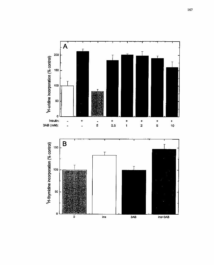
Insulin: - 3AB (rnM): -
ins
FIGURE 17: Sensitivity of Insuiin-ntediated RNA Synthesii to Inhibitors of
Mona(ADP-ribosyC)ation &actions.
Quiescent H 4 E cells were pretreated with a variety of inhibitors for 10 min prior to
addition of insulin (104 M) or FBS (10% viv). Inhibitors tested included: MIBG (50
PM), 3-aminobenzamide (3AB, 10 mM), L-arginine methylester (LAME, 1 pM), L-nitro-
arginine methylester (LNAME, 1 pMJ and norepinephrine (NE, 1 FM). The
incorporation of uridine (6 h incubation) into tnchloroacetate-precipitable material
after addition of insulin or FBS was used as an indication of RNA synthetic activity in the
presence and absence of the inhibitors. The incorporation rate of untreated cells was set
to 100%. The data are presented as the mean +_ SE of at least three separate expenments
conducted in triplicate. Cornparisons were made between growth factor treated and
growth factor + each inhibitor treatrnent with the Student's t-test (*, p < 0.05).
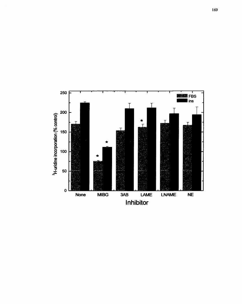
- 1 . 1 1 I 1 1 1 1 1 1
FBS ins
None MIBG 3AB LAME LNAME NE
lnhibitor
either insulin- or FBS-treated H411E cells (Figure 17), although LAME had a slight and
statistically significant effect on the FBS-treated cells- These data validate the premise
that insulin-stimulated H4IIE ce11 growth may be mediated by an arg-mART-dependent
pathway. Moreover, these data are suggestive of a requirement for not only structural
similarity (Le. arginine-iike guanidino group), but also for physicochemical
cornpatability.
The effect of MIBG on insulin-mediated H41E ceII growth and proliferation was
then exarnined in greater detail. MIBA was included as a control since the body of the
molecule is identical to MLBG, but it lacks the argininyl (guanidino) rnoiety, MIBG
inhibited insulin-stimulated RNA synthesis in a concentration-dependent manner (2.5 to
50 pM) (Figure 18A)- in contrast, MIBA did not appreciably inhibit insulin-stimulated
RNA synthesis at any concentration (Figure 18A)- Sirnilarly, MIBG inhibited insulin-
stimulated DNA synthesis in a concentration-dependent manner to below basal levels,
while MIBA had little effect (Figure 18B). Since MIBG is a specific inhibitor of arg-
mART, these data further support the prernise that an arg-mART may be part of the ce11
growth pathway stimulated by insulin. The lack of effect of MIBA to inhibit insulin-
mediated ce11 growth suggests that the argininyl (guanidino) moiety is the critical portion
of the molecule for the inhibitory effect and confirms that MIBG rnay be inhibiting arg-
mART activity by acting as a pseudosubstrate.
To determine when MIBG exerts its inhibitory effects as celIs progress from the
initial stimulus to beginning DNA synthesis, MLBG (50 FM) was added at specific time
points after insulin addition to H4ILE cells, and both [3~]uridine and [3~]thyrnidine
incorporation monitored. Over the 6 h period of an RNA synthesis assay, it was observed
MIBG reduced insulin-mediated uridine incorporation at every point of additior, (Figure
19A)- Most significant is the fact that MIBG does not reduce incorporation to basal
F I G U R E 18: Sensitivity of ilnsulin-mediated Cell Gro wth in H4HE Celk to kîZüG and
its Analog MIBA.
Quiescent H4IIE hepatoma cells were pretreated with MlBG or MIBA (2.5 to 50 pM) for
10 min ptior to addition of insulin (10'~ M). The incorporation of uridine (6 h
incubation) and [.'Hlthyrnidine (24 h incubation) into trichloroacetate-precipitable
material after addition of insulin was used as an indication of RNA (A) or DNA (B)
synthetic activity in the presence and absence of the inhibitor. For RNA synthesis (A), the
control analog MIBA was only tested at the highest concentration (50 PM) with insulin
(10" M). The incorporation rate of untreated cells was set to 100%. The data are
presented as the mean k SE of at least three separate experiments conducted in triplicate.
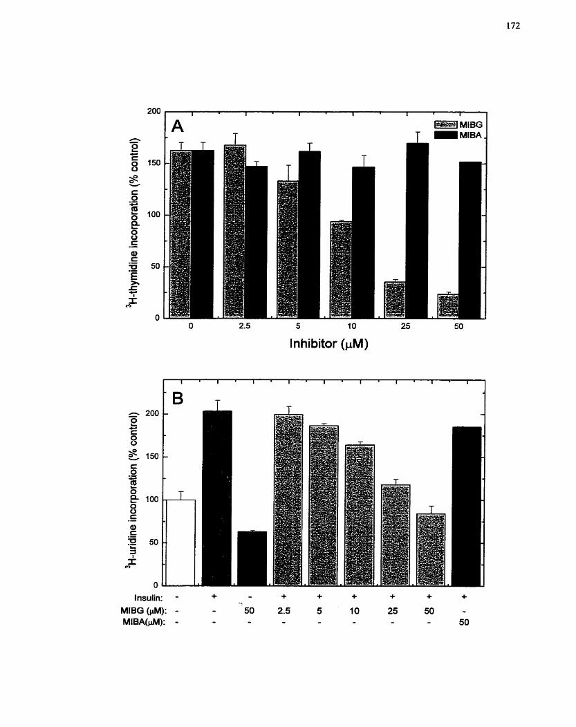
O 2.5 5 1 O 25
lnhibitor (FM)
- 1- MIBG
MlBA
Insulin: - + - + + + -k f + MIBG (FM): - - 50 2.5 5 10 25 50 - MlBA(pM): - - - - - - - - 50
levels, but produces an inhibition that linearly correlates with the time remaining in the
assay. These data suggest that MIBG must be present throughout the assay to completely
suppress RNA synthesis. Moreover, this relationship indicates that there is no single point
a t which the inhibitor is effective and impiies that mART activity is required
continuously for this process. On the other hand, it was showm that addition of MIBG up
to 3 h after insulin administration effectively reduced insulin-stimuiated DNA synthesis
to baseIine levels, with addition of MIBG at the earlier time points being more effective
(Figure WB). Addition of MIBG at 5 h or later did not produce an appreciable decrease
in DNA synthesis, which suggests that the inhibition by MIBG as it pertains to DNA
synthesis contributes to an early event and is no longer required after 3-5 h- For this
reason, MIBG has no effect on insulin-mediated ce11 growth beyond this time penod.
To confirm the above findings, MIBG washout assays were conducted in which
H41IE celIs were treated with MIBG, stimulated with insulin and then the media
containing MIBG and insulin rernoved and replaced with media containing insulin only.
These expenments were devised to determine the Iength of time that the cells must be
exposed to MIBG in order for an effect to be seen. For the RNA synthesis assay, it was
demonstrated that the length of MIBG exposure correlated with the degree of inhibition
(Figure 20A), with the levels of RNA synthesis returning to baseline after 4 h treatrnent
with MIBG and declining even more by the end of the 6 h assay. These findings are
consistent with the previous data (Figure 19A) and suggest that MIBG may either inhibit
multiple steps in the RNA synthesis program or that the target is required throughout. In
contrast, inhibition of DNA synthesis was only observed if MIBG was present for the
first 6 h of the assay or longer (Figure 20B) (i.e, 24-48 h). Rernoval of MIBG prior to 4 h
prevented inhibition. While these data apparently do not agree with the result of the
MIBG addition expenment (Figure 19), it is worth noting that insulin need only be
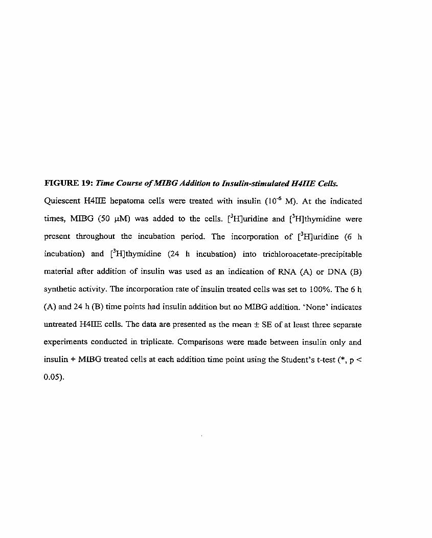
FIGURE 19: Time Course of M B G Addition to Insulin-sfimulated H 4 I E Ceiïs.
Quiescent H4IIE hepatoma cells were treated with insulin (10-~ M). At the indicated
tirnes, MIBG (50 pkf) was added to the cells. [3auridine and [3~]thyrnidine were
present throughout the incubation period. The incorporation of ['H)~ridine (6 h
incubation) and [ - ' ~ ~ t h ~ r n i d i n e (24 h incubation) into trichloroacetate-precipitable
rnaterial after addition of insulin was used as an indication of RNA (A) or DNA (B)
synthetic activity. The incorporation rate of insulin treated cells was set to 100%. The 6 h
(A) and 24 h (B) time points had insulin addition but no MIBG addition. 'None' indicates
untreated H4IIE cells. The data are presented as the mean c SE of at least three separate
experiments conducted in triplicate. Cornparisons were made between insulin only and
insulin + MIBG treated cells at each addition time point using the Student's t-test (*, p <
0.05).
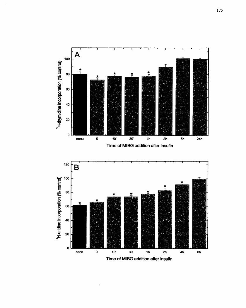
I O ' 30' I h 3h
Time of MlBG addition after insulin
m O 10' 30' 1h 2h 4h 6h
Time of Ml& addition aiter insulin
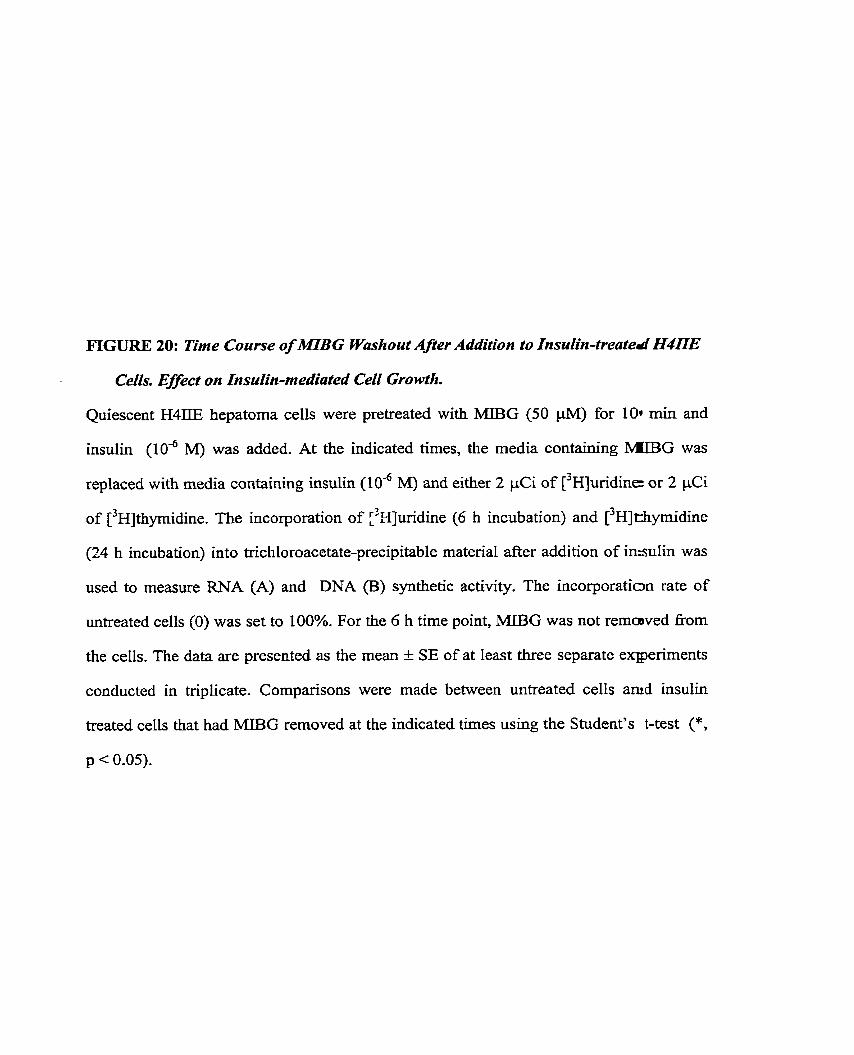
FIGURE 20: Tirne Course of MIBG Washout Aper Addition to Insulin-treated H4UE
Cells. Effect on Insulin-medialed Cell Growth.
Quiescent H4IIE hepatorna cells were pretreated with MIBG (50 pM) for 104 min and
insulin (1W6 M) was added. At the indicated times, the media containing lMIBG was
replaced with media containing insulin (10" M) and either 2 pCi of [ 3 ~ u r i d i n e or 2 pCi
of thpidine. The incorporation of :'9]uridine (6 h incubation) and [ 3 ~ ] thymidine
(24 h incubation) into tnchloroacetate-precipitable material after addition of insulin was
used to measure RNA (A) and DNA (B) synthetic activity. The incorporation rate of
untreated cells (0) was set to 100%. For the 6 h time point, MIBG was not remcwed from
the cells. The data are presented as the mean f SE of at least three separate exlperiments
conducted in triplicate. Cornparisons were made between untreated cells amd insulin
treated cells that had MIBG removed at the indicated times using the Student's t-test (*,
p < 0.05).
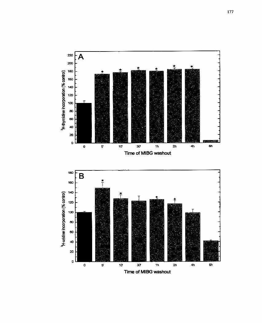
O S 10' 3CY l h 2h 4h 6h
Tirne of MlBG washout
O S 10 30 l h 2h 4h 6h
Tirne of MlBG washout
present for 5 min to induce ce11 proliferation (Figure 3)- Therefore, addition of MIBG at
10 min may have missed the cntical MIBG-sensitive event activated by insulin.
Nevertheless, these results, as well as those of the addition study (Figure 19), indicate
that MIBG also inhibits a specific event occumng 3 to 6 h foIlowing insulin stimulation.
5~4.4~2 Toxicity of MIBG
As a result of the findings in section 5.4.4.1, where MIBG completely inhibited
DNA synthesis, it was of interest to ascertain if the inhibition of H4ZIE ce11 growth by
MIBG was the result of a direct effect on ce11 cycle re-entry or the result of cellular
toxicity. MTT cytotoxicity assays (Le. mitochondrial reduction of a tetrazolium dye)
employed a concentration range of both MIBA and MIBG (0-200 PM), and data were
collected (Le. cells lysed) at both 48 and 96 h after addition of the compounds. Growing,
quiescent H4IIE cells and insulin-stimulated H4IIE cells were used to cover the entire
growth spectrum for this assay. In growing and quiescent H4IIE cells, MIBA had Little
effect on MTT conversion after either 48 or 96 h of incubation, although quiescent cells
appeared to be more sensitive to the MIBA (Figure 2 lA,C)- The significance of the extra-
sensitivity of the qüiescent state may reflect the altered metabolic rate in quiescent tells,
Taken together, these data suggest that MIBA is not cytotoxic to the cells and is
minimally affecting the mitocliondria of the cells. On the other hand, MIBG treatment
decreased MTT conversion by both growing and quiescent H4IIE cells, with increasing
concentrations of MIBG having a significantly greater effect (Figure 2 lB,D). This effect
was time-dependent as well, with 96 h of incubation having a greater effect than 48 h
(Figure 21B,D). Most importantly, these experirnents identified the dose, 20 FM, at
which MlBG (mitochondrial) toxicity may affect the cells. Moreover, H4IIE cells treated
with insulin (data not shown) responded similarly to both treatments as growing and
quiescent H4IIE celIs. Thesc data would indicate that MIBG is cytotoxic to the ceIls at
FIGURE 21: Effect of MlBG and MlBA on Growing and Quiescent H4DE C e k
MTT Assay as a Measure of Cell Cyfotoxicity.
Growing (A, B) and quiescent (C, D) H4IIE hepatoma cells were treated with varying
concentrations (O to 200 PM) of MIBA (A, C) or MIBG (B, D) for 48 and 96 h. MTT
was then added to the cells for an additional 4 h, and colour development in the 96-welI
microtitre plates monitored at 0D550. The data are presented as the mean + SE of at least
three separate experirnents conducted in quadniplicate.
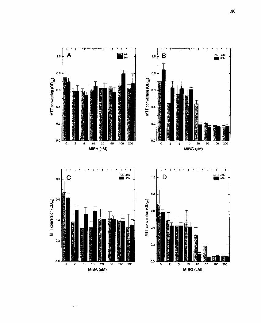
O 2 5 10 20 50 100 200
MIBA (PM) O 2 5 10 20 50 100 200
MIBG (FM)
- .-
O 2 5 10 20 50 100 200
MIBG (PM)
the higher concentrations andor is having a detrimental effect on mitochondrial
function, as previously reported (Loesberg et al. 1990a; Smets el al. 1990b). It should be
noted however, that tlie MTT assay is limited in its ability to detect ce11 death. Since
conversion of MTT is mitochondrial dependent, non-lethal effects on mitochondrial
activity would aIso be observed with this assay.
To distinguisli between cytotoxicity and mitochondrial activity inhibition by
MIBG, a second assay was employed. LDH release from cells has been used as a
measure of cytotoxicity and cellular membrane integrity, H411E ceils were exposed to
both MIBA and MIBG (0-200 PM) for 24 and 72 h, and LDH activity in the media was
measured. After 24 h, MIBA did not increase LDH levels (Figure 22A). On the other
hand, a 24 h treatment with MIBG increased LDH levels, but only at concentrations
greater than 50 p M (Figure 22B). At 72 h, only the highest MIBA concentration
increased LDH levels (Figure 22A), while LDH release was obtained with 20 pM or
more MIBG (Figure 22B). These data support the MTT findings and imply that MIBG is
having a cytotoxic effect. However, this interpretation must also take into account the
effects of MIBG on mitochondrial respiration. MIBG (and analogs such as
benzylguanidine) are provcn inhibitors of complex [/ILI of the mitochondrial respiratory
chain (CorneIissen et al. 1995a; Cornelissen et ai. 1995b; Loesberg ct al- 1990a;
Loesberg el a[. 199 1; Smets et ai. 1990b). If mitochondrial respiration is inliibited, a ceIl
must obtain energy from the anaerobic glycolytic pathway which increases the lactate
content of a cell. This incrcase in lactate content would be expectcd to induce LDH, and
it may be this increase in LDH levcls that is being detected with this assay. Although the
LDH released may not Iiavc bcen tlie result of cytotoxicity, nevcrthclcss, membrane
integrity must have becn compromised since LDH activity was detcctable in the media.
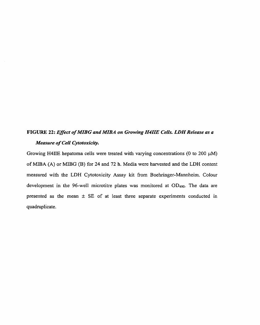
FIGURE 22: Effecf of M'BG and MIBA on Growing H4ILE CeiCs. LDH Reiease as a
Measure of Cell Cy fo foxicity.
Growing H4IIE hepatoma cells were treated with varying concentrations (O to 200 jA4)
of MIBA (A) or MIBG (B) for 24 and 72 h. Media were harvested and the LDH content
measured with the LDH Cytotoxicity Assay kit from Boehringer-Mannheim. Colour
development in the 96-well microtitre plates was rnonitored at 0D490. The data are
presented as the mean & SE of at least three separate experiments conducted in
quadruplicate.
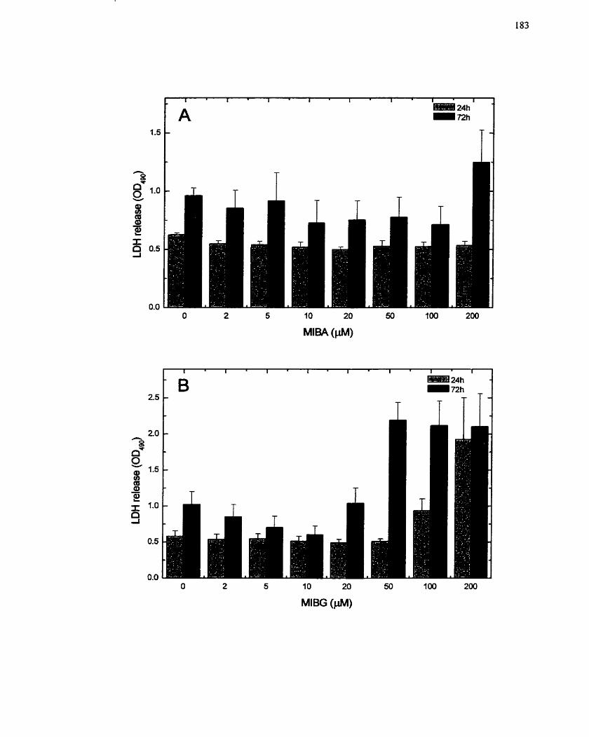
The uncertainty in detecting MIBG cytotoxicity with the MTT and LDH assays
led to the inclusion of a third assay in this study. FACS analysis was used to deterrnine
the extent of cellular damage produced by MIBG, to determine whether ce11 death was
occumng and to determine how MIBG affects progression of WIIE cells through the ce11
cyc!e. Untreated, growing H41IE cells demonstrated the typical distribution of cells in
various stages of the ce11 cycle, with a broad band of cells in the S phase of the cell cycle
(Figure 23). M e r 48 h in the presence of MIBG (50 PM), H411E cells were prirnanly
distributed in the Gr phase of the ce11 cycle, with a noticeable absence of S phase ceIIs
(Figure 23)- Interestingly, the cells were distributed in a pattern suggestive of cells that
were viable, but not undergoing replication. Moreover, distribution of cells by size on the
axes indicated that the cells had not fragmented or undergone lysis, since the size of
MIBG treated cells was comparable to that of untreated cells (Figure 23). These
particular data wouId suggest that MIBG is not exerting cytotoxic effects on H4IIE cells,
which contrasts with the previous assays (Figures 21,22), and rather that MIBG is having
a cytostatic effect.
To further clan@ whether MIBG has adverse effects on H 4 I E cells, H4IIE ce11
number was measured after 48 and 96 h of MIBG and MIBA treatrnent (0-1000 PM)-
MIBG significantly decreased ce11 number in both growing and quiescent H4IIE cells,
with the higher concentrations demonstrating an almost complete loss of cells (Figure
24C,D). However, ce11 losses were not detected at concentrations below 20-50 PM. In
contrast, MIBA did not significantly affect cell number unless very high concentrations
(greater than 200 PM) were used (Figure 24A,B). These data would suggest and support
the earlier results (Figures 21, 22) that indicate MIBG is cytotoxic. Further studies, not P
contained within the body of this work, are attempting to clearly establish whether MIBG
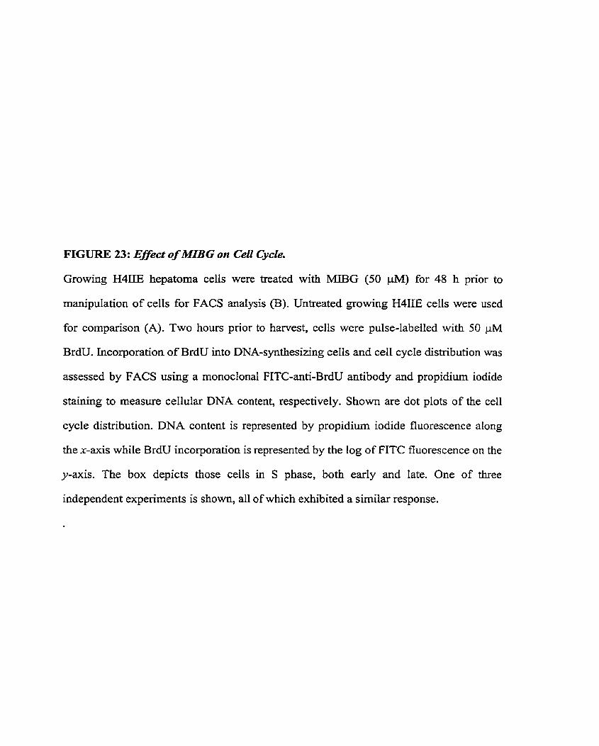
FIGURE 23: Effect of 2MIBG on Ceil Cycle.
Growing H4IIE hepatoma ceils were treated with MIBG (50 pM) for 48 h prior to
manipulation of cells for FACS analysis (B). Untreated growing H4IIE cells were used
for cornparison (A). Two hours pnor to harvest, cells were pulse-labelled with 50 p M
BrdU. Incorporation of BrdU into DNA-synthesizing cells and cell cycle distribution was
assessed by FACS using a monoclonal FITC-anti-BrdU antibody and propidiurn iodide
staining to measure cellular DNA content, respectively. Shown are dot plots of the ceil
cycle distribution. DNA content is represented by propidium iodide fluorescence along
the x-axis while BrdU incorporation is represented by the log of FITC fluorescence on the
y-axis. The box depicts those cells in S phase, both early and iate. One of three
independent experiments is shown, al1 of which exhibited a sirnilar response.
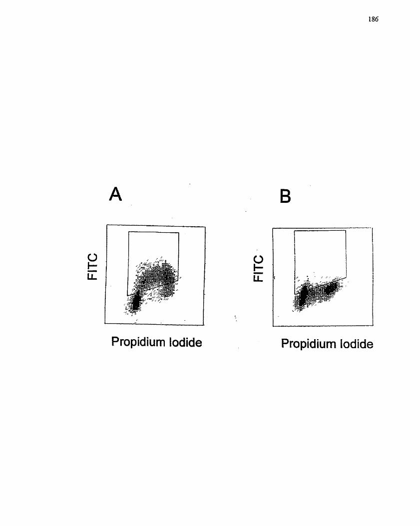
1
Propidium lodide Propidium lodide
FIGURE 24: Effect of MIBG and MIBA on Cell Viability. Coulter Counring as a
Method for Determinittg Cell Cytotoxicity.
Growing (A, C) and quiescent (B, D) H4IIE hepatoma cells were treated with MIBG (C,
D) or MD3A (A, B) (0-1000 pM) for 96 h. The wells were then rinsed, the cells released
by trypsinization and ce11 number assessed with a Coulter Counter. One of two
experirnents is shown, both of which exhibited a similar response. Each data point was
counted in triplicate fkom samples prepared in duplicate.
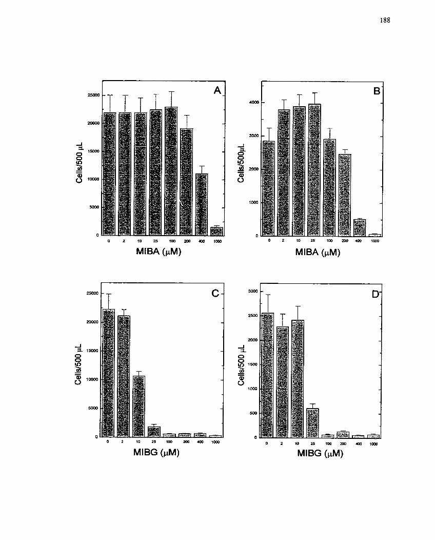
O 2 10 25 100 200400 lm
MlBG (PM)
O 2 IO 25 100 m.«)[) lm
MlBA (PM)
O 2 10 2a i m mo 4oa 1000
MlBG (PM)
is cytotoxic or cytostatic and to identify the mechanism by which ce11 cycle and S phase
progression are inhibited,
Finally, to determine if MIBG has an effect on the metabolism of H4IIE cells,
glucose uptake was measured after stimulation with insulin in the presence and absence
of MIBG (10-100 FM). It was observed that MIBG significantly reduced the amount of
insulin-mediated [3~]glucose uptake by H4IIE cells (Figure 25) in a concentration-
dependent marner. These data support the contention that MIBG may be having a
metabolic effect and may also suggest a second mechanism by which it affects ce11
growth and proliferation.
5.4.4.3 Mechanism of action of MIBG
MlBG has a potent inhibitory effect on ce11 growth induced by insulin
stimulation. As a result, it was of interest to identie growth-associated signaling
pathways activated by insulin that are inhibited by the actions of MIBG. Two signaling
systems were identified as possible candidates: MAP kinase, which is generally activated
in a variety of ceil types that undergo growth, and p2 1-Ras which is upstream of M M
kinase, but also participates in signaling events independent of MAP kinase (Hall 1993;
Lim et al. 1996; Marshall 1996; Olson & Marais 2000).
Insulin stimulated the activation of MAP kinase as measured by an in gel activity
assay. Activity was transiently increased over a time course of 20 min, with peak
activation occurring at 2 to 5 min (Figure 26 and Figure 7 h m section 5.2.4.3).
Pretreatment of H4LIE cells with 3AB (5 mM) for IO min before insulin addition resulted
in a diminution of the MAP kinase response (Figure 26). In contrast, pretreatment with
MIBG (50 PM) for 10 min before insuiin addition resulted in an apparent enhancement of
MAP kinase activity (Figure 26). The importance of MAP kinase in thc insuiin-mediated
growth response is alrcady documented (Figure 10). Thereforc, these tindings suggest
FIGURE 25: Effecf of MIBG on Glucose Upiake in H4IIE Cells.
The incorporation of [3~g lucose was measured as described in Matenals and Methods
(section 4.3.8) after addition of insulin (IO-' M) to quiescent H 4 E cells subsequent to
pretreatment with varying concentrations (10 to 100 PM) of MIBG. The rate of uptake in
untreated cells was set to 100%. FBS (2% v/v) was used as a negative control. Each data
point represents the mean + SE of two experirnents conducted in triplicate.

FBS: - + - - - - - Insulin: - - + + + + +
MlBG (FM): - - - 100 50 20 10
FIGURE 26: Effect of Inhibitors of ADP-ribosyiation, 3-Arnirt obenzamide and MIBG,
on MXP Rinase Activation by Insulin in H4I.E Cells.
MAJ? kinase activity was measured over 20 min by activity geI assay. Cells were
stirnulated with insulin (106 M) and pretreatment with either 3-arninobenzamide (5 mM)
or MIBG (50 pMJ occurred 10 min prior to insulin addition. Cells were harvested at the
indicated time points. Densitornetry of the p42M*K/p44MAPK bands is plotted from one
experiment. Three independent experiments were conducted, al1 of which exhibited a
similar response.
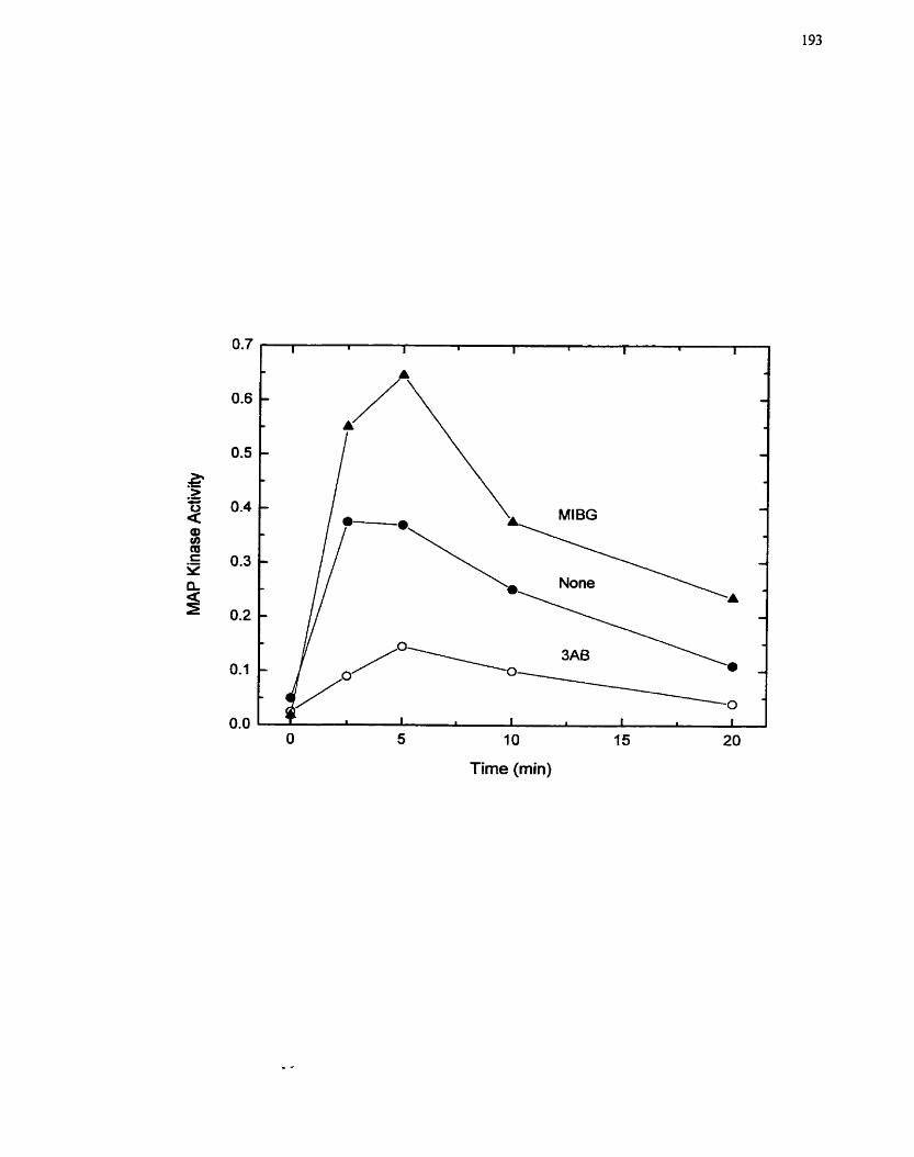
O 5 10 15 20
Time (min)
that the inhibition of cell growth and proliferation by MIBG is not the result of inhibition
of M M kinase activity.
p21-Ras was investigated because it is frequently activated in growing cells (de
Vries-Smits et al. 1992; Mansour et al. 1994) and, because as a small GTP-binding
protein, the possibility that it might be ADP-rïbosylated is quite high (Lem et al. 2000;
Schmidt & Aktories 1998; Tsuchiya & Shimoyama 1994). Ras activation was detected by
measuring the ratio of GTP and GDP bound Ras. This assay involved
[32~]orthophosphate labeling of cellular phosphate pools followed by
immunoprecipitation with a specific anti-Ras antibody and subsequent separation of
GTP/GDP pools by TLC (section 4.9.5). Addition of insulin ( 1 0 ~ ~ M) to quiescent H4IIE
cells for 10 min resulted in a significant increase in GTP:GDP ratio in the cells (Figure
27). This activation could not be blocked by either pretreatment with 3AB (5 mM) or
MIBG (50 PM), which would suggest that p21-Ras is not the target for MIBG action.
5-4.5 Discussion
The initial studies in this section, dealing with a variety of different inhibitors of
ADP-ribosylation reactions, indicqted that only the specific inhibitor of arg-mART,
MIBG, was capable of inhibiting insulin-stimulated ce11 growth and proliferation in
H41IE cells (Figures 16, 17, 18). The dual ADP-ribosyltransferase inhibitor 3AB, had
only a modest effect on RNA synthesis and no effect on DNA synthesis (Figures 16, 17).
Structural analogues of MIBG, that should behave as pseudosubstrates for arg-mART,
did not have apparent inhibitory effects on H4IIE ce11 growth or proliferation (Figure 17).
Specifically, based on structure, it was assumed that LAME and LNAME would behave
in a similar fashion to MIBG if arg-mART was indeed the target of inhibition. However,
physicochemical constraints were notcd that could have prevented these compounds from
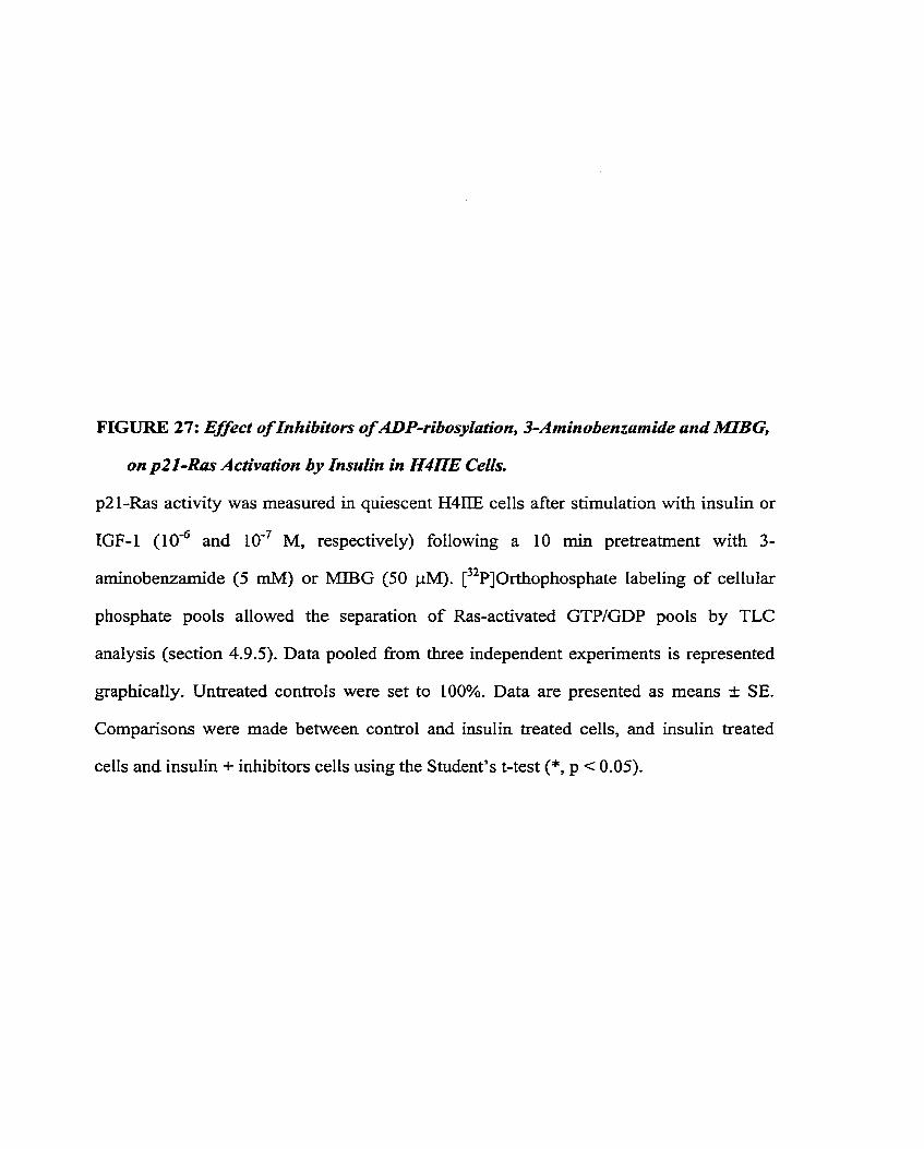
FIGURE 27: Effect of In hibitors of ADP-ribosylation, 3-Aminobenzamide and MBG,
on p21-Ras Activation by Insulin in H4LTE Cells.
p21-Ras activity was measured in quiescent H4IIE cells after stimulation with insulin or
IGF-1 (106 and IO-' M, respectively) following a 10 min pretreatrnent with 3-
arninobenzamide (5 mM) or MIBG (50 pM). [32~]0rthophosphate labeling of cellular
phosphate pools allowed the separation of Ras-activated GTPIGDP pools by TLC
analysis (section 4.9.5). Data pooled from three independent expenments is represented
graphically. Untreated controls were set to 100%. Data are presented as rneans -t SE.
Cornparisons were made between control and insulin treated cells, and insulin treated
cells and insulin + inhibitors cells using the Student's t-test (*, p c 0.05).
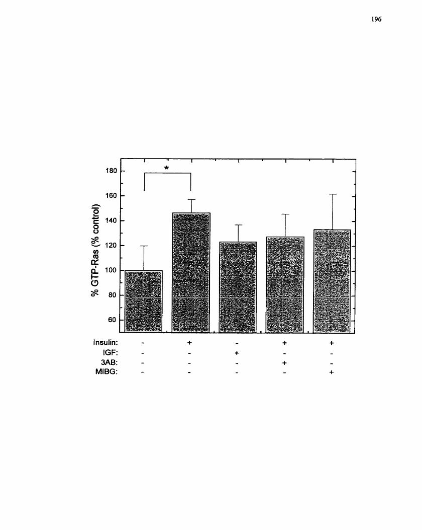
Insulin: - + - + + IGF: - - + - -
3AB: - - - + - MIBG: - - - - +
behaving as pseudosubstrates for arg-rnART. In particular, LNAME rnay not behave as a
pseudosubstrate due to steric hindrance from the presence of a nitro group on the
guanidino rnoiety. On the other hand, LAME is highly positively charged, and the
presence of the charges would reduce ce11 entry via passive diffusion (although the
charges rnay a1Iow the molecule to be more attractive to the ce11 membrane). As a result,
although LAME is a substrate for arg-mART Ni vitro, it rnay not be able to reach its point
of action Nz vivo. Thus, lack of inhibitory effect of these compounds (LAME and
LNAME) does not necessarily irnply that MIBG is not effectively inhibiting an arg-
mART. In fact, these studies support the concept that an arg-rnART is involved in the cet1
proliferative processes activated by insulin and that MIBG is inhibiting this enzyme
activity. Since MIBG was able to inhibit the activation of an arg-mART in vitro (section
5.3.4.1), it rnay be assumed that inhibition of ce11 growth by MIBG involves the
inhibition of an arg-mART that regdates a process that is critical for the cell proliferative
program. Moreover, the steps that rnay be affected are related to progression of the cells
from quiescence into the ce11 cycle. MIBG rnay be behaving as a cytostatic agent through
the blockade of events required for the entry of cells into the ce11 cycle, or transition from
the Gr to S phase. Although studies were not carried out to delineate in more detail the
mechanism of action of MIBG and how it relates to inhibition of the ceIl cycle, these
studies are planned. It is aIso possible that MIBG could just be inhibiting the mechanism
for the uptake of the radiolabelled agents and thus the appearance of inhibition of cell
proliferation would be observed. However, this scenario does not appear to be borne out
by the FACS analysis, where it was shown that MIBG could specifically inliibit S phase
entry (Figure 23). it is possible that the absence of cells in S phase could indicate ceIl
death, but the presence of cells in other phases of the ce11 cycle would not support this
concIusion. Furthemore, the cells did not appear to be dead or dying, or undergoing
necrosis, since ceil fragmentation was not apparent, and a downshift of the curve was not
observed. Moreover, if uptake of BrdU was being prevented, the height of the curve
wouId have shifted to reflect this.
It is generaILy accepted that mitogenic stimulation initiates a cascade of events
that are temporally ordered to ensure regulated progression through the ce11 cycle. As part
of this process, the cell is required to provide the necessary macromolzcular components
for each step of the mitogenic program, as well as supply energy in the form of ATP.
While MIBG could inhibit ce11 growth by either mechanism, there is strong evidence in
the literature to support interference with mitochondrial function as the pnmary route by
which MIBG operates (Loesberg et al. 1990a; Smets et al. 1991; Van den Berg et al.
1997). Additional support for this concept is provided by the MTT and LDH release
expenments (Figures 21, 22), since the FACS analysis (Figure 23) indicates the cells
exposed to MIBG are not actually dying. The results of both MTT and LDH studies can
be explained by the known inhibitory effects of MlBG on mitochondnal respiration
(Cornelissen et al. 1995a; Cornelissen et al. 1995b; Loesberg et al. 1990a; Smets et al.
1991). Inhibition of mitochondrial respiration would prevent the conversion of the
tetrazolium dye by the mitochondria, since this process requires electron transfer.
Similarly, inhibition of mitochondrial respiration leads to an elevated rate of glycolysis in
order to rnaintain ATP levels. During anaerobic respiration, the amount of Iactate
generated by the ce11 would increase. Concomitantly, LDH levels would rise. Thus,
inhibition of mitocliondrial function does not necessarily lead to cell death, but could
interfere with ce11 hnctions essential for growth and proliferation. On the other hand, an
analog of MIBG, benzylguanidine, has been shown to also inhibit mitochondrial
respiration (Van den Berg et al. 1997). However, this compound was not able to inhibit
insulin-mediated H4IlE ce11 proliferation (Bernardin, Litchic & Zahradka, unpublished

observations), which wouid definitely suggest that rnitochondrial inhibition is not linked
to the anti-proliferative effects of MIBG. This latter point would support the participation
of a mART in the ce11 proliferative processes mediated by insulin in H4IIE cells, and a
role for MIBG in preventing ce11 proliferation via an arg-mART. Moreover, Cornelissen
et al. (1995a; 1995b) have observed a differential concentration of MIBG required for
complete inhibition of mitochondrial ATP synthesis in cornpanson to inhibition of cell
proliferation (10 ph4 vs. 25 pM, respectively). Thus, the inhibition of mitochondnal
respiration is not entirely responsible for the ce11 proliferative inhibitory properties of
MLBG, and MLBG rnust influence cellular processes apart from mitochondrial ATP
synthesis (Cornelissen et al. 1997b; Cornelissen et nL 1995a), lending further support for
the participation of a mART,
The possibility also exists that MIBG may De inliibiting ce11 growth via a non-
ADP-ribosyltransferase pathway. Since MIBG resembles arginine, which is a donor for
nitric oxide (NO) synthesis, it is possible that MIBG also behaves as a donor for NO
synthesis, and NO is a known inhibitor of ce11 proliferation (Cornwell et al. 1994;
Kolpakov et al. 1995; Nakaki et al. 1990). However, Kuin et al. (Kuin et al. 1998)
demonstrated that MIBG (100 FM) inhibits al1 three forms of nitric oxide synthase,
which would prevent NO synthesis and the inhibitory effects typically associated with
NO. If the converse were true, that inhibition of NO synthesis is required for inhibition of
cell proliferation, then it would have been expected that LNAME, an inhibitor of nitric
oxide synthase, would have been able to inhibit ce11 growth and proliferation in this ce11
system (Figure 16). Thus, the participation of NO in these events is unlikely, and lends
further credence to the participation of an arg-mART in H4IIE ce11 growth and
proliferation that is inhibitable by MIBG,
Ln addition to inliibiting ce11 growth processes, MiBG decreased glucose uptake in
response to insulin (Figure 25). Glucose is transported into cells via the GLUT-2/4
transporter and MIBG could potentially be having a direct effect on the transporter
(Czech 1995; FIetcher & Tavare 1999; Kahn 1996; MueckIer 1994; Olson & Pessin
1996; Thorens 1992). Alternatively, MIBG might reduce the cellular energy pools (via its
effects on the mitochondria) and indirectly halt this energy-dependent transporter
process. Ironically, a ce11 with inhibited mitochondrial respiration relies more heavily on
glucose and the glycolytic pathway for the generation of ATP. Thus, while it would be
more advantageous for the ce11 to increase glucose uptake for the glycolytic pathway, if
the ce11 does not, disruption of a wide variety of energy-dependent process and,
subsequently, cell death may instead ensue. This rnay be part of the mechanism of
inhibition of ce11 growth for MIBG.
H41IE cells may be more sensitive to the effects of MIBG because hepatocytes
and hepatomas have a high metabolic rate and would therefore rely heavily on their
mitochondria for metabohsm, including ce11 growth and proliferation. On the other hand,
exclusion of a direct effect on the mitogenic program by MIBG is not possible on the
basis of the delayed addition and washout studies. The effect of MIBG on DNA
synthesis, in particular, would suggest that the target is a specific process required for
progression to S phase that occurs within the first 3 to 6 h after treatment with insulin
(Figures 19,20). Thus, the first 5 to 6 h are the most critical for the inhibitory action to bc
evident, although MIBG shouId be present over the cntire time period to produce the
most profound effect. In contrast, MIBG was required to be present for the entire lençth
of thc assay in ordcr to bc effective at reducing RNA synthesis (Figures 19, 20). Thcsc
rcsults may suggcst that MIBG is affccting this proccss on a morc global lcvel or that
constitutive activation of a single target is required for the insulin-dependent increase in
RNA synthesis.
Since the mechanism by which MIBG inhibits ce11 growth could not be
consistently and conclusively linked to an inhibition of mitochondrial respiration, the
effect of MIBG on certain signaling pathways was examined. MAP kinase, which was
established as being critical for insulin-stimulated H4IIE ce11 proIiferation (Figure IO),
was not inhibited by MIBG (Figure 26). This rnay indicate that inhibition of ceIl
proliferation by MIBG occurs downstrearn of MAP kinase or invoives a separate but
parallel pathway. Similarly, MIBG did not decrease the GTP:GDP ratio of p21-Ras
stimulated by insulin addition (Figure 27). The latter molecule was chosen because of the
importance of p21-Ras in growth regulatory signaling pathways, and also because it is a
small GTP-binding protein that may be modified by ADP-ribosylation (Tsuchiya &
Shimoyama 1994). These results would indicate that the action of MIBG lies after both of
these s ignahg steps have occurred, possibly in the nucleus.
5.5 Sumrnary
H4IiE hepatoma ceIls, used primarily for the study of the biochemical and
metabolic changes in the liver dunng malignancy, have also been used for the study
insulin-dependent ce11 proliferation. To define the proliferative response to insulin in Our
ce11 system, and to identify the signaling molecules associated with the ce11 proliferative
responses, the responses of H4IIE cells to insulin and IGF-1 were studied. In this series
of studies it was observed that insulin, but not IGF-1 is capable of stimulating both
mitogenesis and glucose utilization in H41IE hepatoma cells. The distinct response to
insulin and IGF-1 exhibited by H4IiE cells made it possible to examine the early
s ignahg events triggered by these ligands. The data suggest that phosphorylation of IRS-
1 via the receptor tyrosine kinase is required for both ce11 growth and metabolism, but it
does not necessarily participate in the pathways leading to MAP kinase activation.
Furthemore, MAP kinase activation appears to play an important role in the ce11 growth
and proliferation events activated by insulin in H4IIE cells. However, it is recognized that
distinct and independent pathways other than MAP kinase also contribute to ceIl growth
and proliferation.
The activation of cell growth in H411E cells appears to be Iinked to the activation
of an arg-rnART, since insulin increases the activity present in the rnicrosomal fraction of
H4IIE cells. Enhanced nuclear PARP activity was not observed and inhibition of this
enzyme did not prevent ce11 growtk In contrast, inhibitors of arg-mAEU were able to
abrogate the insulin-stimulated RNA and DNA synthesis in H41IE cells. Although the
precise mechanism of action of MIBG to prevent ce11 growth is still not clear, despite the
numerous attempts made to clarify this issue, it can be concluded that the insulin-
mediated H4IIE ce11 growth is sensitive to MIBG and not to its analog MIBA. This
concIusion speaks of the importance of the argininyl (guanidino) moiety for the inhibltory
activity of these compounds. Two distinct mechanisms of action were considered: i)
MIBG is acting at specific points within the ceil to inhibit ce11 growth and proliferation,
and ii) MIBG is affecting ce11 growth processes indirectly through its ability to inhibit
mitochondrial activation. Experiments to define the Iatter mechanism were inconcIusive
while experiments targeted to the former mechanism revealed that MIBG must be
affecting signal molecules downstream to MAP kinase, or an unrelated but parallel signa1
pathway that is also critical for cell growth and proliferation. At this point, regardless of
its mechanism of action, it is sufficient to mention that MIBG is an effective inhibitor of
ce11 growth. Future studies will need to clanfy these issues and to further define the

cytotoxic effects of MIBG. Direction for these additional studies will be based on the
results presented in section 7-4.
6.0 L6 skeietal myoblasts as a mode1 of differentiation
6.1 Introduction
The development of skeletal muscle is controlled by a highly synchronized series
of events. In this process, called myogenesis, cells that have no specialized characteristics
are converted into muscle fibres which permit body rnovement. Vanous signaIs from
outside and inside the cell regulate the switch from unspecialized to muscle cell.
The conversion of multipotential mesodermal stem cells present in the ventral
sornites into muscle fibres occurs through a multistep process directed by the sequential
expression and activation of specific transcription factors (Sassoon 1993; Venuti &
Cserjesi 1996). Two distinct stages are involved in the establishment of the skeletal
muscle phenotype (Ludolph & Konieczny 1995). The first, termed cornmitment or
determination, requires the synthesis of a single myogenic factor (either MyoD or myf5)
and establishes the ce11 ' s ability to eventually express the s keletal muscle p henotype. The
ensuing second stage, which governs the actual differentiation process, requires the
sequential expression of myogenin and p21C1P', and is characterized by expression of
muscle-specific contractile proteins (eg. rnyosin heavy chain), fusion of the individual
myoblasts into multinucleated myotubes and cessation of ce11 division (Dias et ai- 1994;
Walsh 1997). Expression of p21C'P' is required for withdrawal of cells from the ce11
cycle. Bo th stages of the myogenic program are ternporally distinct aiid independentl y
regulated (Lassar & Munsterberg 1994).
The rnyogenic lineage is established once mesodermal stem cells, which have the
potential to differentiate into either chondroblasts, adipoblasts or rnyoblasts, express
either MyoD or myf5. MyoD was first identified when Davis et al. (1987), showed that
expression of a single gene was sufficient to convert a fibroblast into a myoblast. Similar
experimental approaches wcre used to isolate myf5, myogenin and MRF4, and thcir
importance for skeletal muscle formation was confinned by their ability to direct
myogenesis when ectopically expressed in non-myogenic cells (Weintraub et al. 1991).
Expression studies of these myogenic regdatory factors (MRFs) during embryonic
development revealed that a11 four proteins are synthesized within the somites in a
temporally ordered pattern (Smith et al. 1993; Yun & Wold 1996). It was further
dernonstrated that both MyoD and myf5 are expressed in proliferating myogenic cells
(myoblasts) pnor to their differentiation into myotubes (Rudnicki et al. 1993). The
correlation between these factors and cornmitment to a myogenic lineage was also
evident in myogenic ce11 lines where MyoD or myf5 are expressed in the myoblast.
Moreover, recently generated knockout rnice for MyoD and myf5 suggest that absence of
one of these MRFs does not abrogate the myogenic program while the absence of both
results in mice with no skeletal muscle (Rudnicki et al. 1993; Valdez et al. 2000).
MyoD, myf5, myogenin and MRF4 belong to the bHLH family of proteins. These
proteins contain two functional domains, a helix-loop-helix (HLH) motif that mediates
protein dimerization and a basic region that facilitates DNA binding. The DNA binding
activity of these bHLH proteins is triggered once they form heterodimers with a distinct
class of bHLH proteins, the E proteins E12/E47, constitutively present in most cells
(Lassar et al. 199 1). The resultant MRWE protein cornplex binds to a cis-element termrd
the E-box, present in the promoter regions of most skeletal muscle-specific genes.
Although the bHLH MRFs have an important roIe in myogcnesis, the E-box is absent
from several key skeletal muscle-specific genes such as myosin heavy chain and a-actin
(Takeda et al- 1995). A novel DNA-binding protein was found to bind to a distinct
sequence present in the promoter of these and other genes. This myocyte-specific
enhancer-binding factor (MEF2), while not uniquely present in cells of skeletal muscle
lineage, is expressed at an early point in somite development (Olson er al. 1995) and
mutation of the MEF2 site severely reduces gene expression (Kausha1 et al. 1994). Thus,
a ternary complex between MRF, E-protein and MEF2 has been proposed as the
hnctional unit required for expression of skeletal muscle-specific genes during
differentiation (Molkentin & Olson 1996a; Molkentin & Olson 1996b; Yun & WoId
1996). This ability to interact constitutes one of the principal mechanisms by which the
differentiation of skeletal muscle precursors can be controlled-
Skeletal muscle differentiation is driven by the ordered expression of specific
transcription factors that control the synthesis of muscle-specific contractile proteins and
concomitant fusion of mononucleated myoblasts into multinucleated myotubes. The
function of these transcription factors is moduiated through their interaction with either
positive (eg. E12/E47) or negative (eg. Id and Twist) regulatory proteins that determine
their ability to bind to specific prornoter elements (eg. E-box). Two mechanisms govern
the balance between positive and negative components, protein abundance and post-
trandational modification, and these are regulated in turn by intracellular signaling
systerns (eg. PKC) that respond to extracelIular cues (eg. bFGF, IGF and TGF-P) (Hardy
et al. 1993; Lim et al. 1995; Miles & Wagner 2000; Zappelli et al. 1996).
Skeletal muscle comprises one of the best systems to study terminal
differentiation. Myogenic differentiation involves the withdrawal of proliferating
myoblasts from the ce11 cycle and the coordinated activation and expression of muscle
specific genes, giving rise to the myogenic phenotype which is characterized by the
fusion of mononucleic myoblasts into multinucleated myotubes. Although molecular
studies to define the temporal recruitment and coordination of muscle specific genes are
ongoing in other laboratories, very little is known about the cellular signals that cause
myoblasts to exit from the ce11 cycle and initiate the terminal differentiation program. To
this cnd, the signals that activate the recruitment and coordinated exprcssion of muscle
specific genes dunng myogenesis are best studied using an isolated system, and L6
myoblasts, a well established myogenic system (Yaffe 1968), provide a good rnodel.
L6 skeletal myoblast cells, an established ce11 line originally derived from rat
skeletal hindlimb muscle, are a committed skeletal muscle precursor which can be
induced to differentiate into multinucleated, terminally differentiated myotubes. The L6
ce11 line has theoreticaily also been transformed by the carcinogen methylcholanthrene in
an effort to rnaintain the cultures by serial passage (Yaffe 1968). This cell line c m also be
kept in a frozen state (at -80°C) for extended periods of time without loss of viability and
without loss of differentiation properties. The conditions for differentiation involved the
removal of growth media (10% FBS/a-MEM) and replacement with mitogen-poor
differentiation media (2.5% horse seruda-MEM). Upon differentiation, the immature
skeletal myotubes enter a quiescent state and are unable to re-enter the ce11 cycIe.
Terminal differentiation is accompanied by a change in cell morphology (fusion of
myoblasts into myotubes), the synthesis of contractile muscle proteins and the
organization of muscle bundle filaments (Zahradka et al. 1989). Interestingly, L6 cells
constitutively express MyoD, suggesting that these cells are firmly committed to the
myogenic lineage (Lognonne & Wahrmann 1986). However, the spectrum of MRF
activation and association thereafter thereby controls the final outcome. Since relatively
little is known about the mechanisms that determine how myogenesis is modulated by
extemal factors and intracellular signaling systems, the role of mART in this cellular
program will be examined. Several published reports have already indicated that
inhibitors of rnART, including MIBG, are capable of preventing the differentiation of
skeletal myoblasts into myotubes,
Based on this information, a specific hypothesis was proposed: M/BG cvill idzibit
rlr e d$ferentiarion of L6 skeleta[ nzyo blasts iuto rnyotubes by Nztei fer-ing with the
expr-ession of one or more MRFs essential for myogenesis-
6.2 Contribution of rnono(ADP-ribosy1)transferase to myogenesis
6.2.1 Background/rationale
Based on the studies of Kharadia et al. (1992) and Huang et al. (1996), inhibition
of ADP-ribosyltransferase activity results in the inhibition of myoblast fusion. A
glycosylphosphatidylinositol (GP1)-linked arginine-specific mono(ADP-ribosy1)-
transferase is expressed in skeletal muscle tissue and myotubes but not in myoblasts
(Zolkiewska et ai. 1992). This enzyme, which is located on the external surface of the
cell, has been shawn to modify a 7 integrin (Zolkiewska & Moss 1995) and desmin
(Huang et al. 1996). While this evidence for increased ADP-ribosylation during
differentiation is intriguing, a more direct association between ADP-ribosylation and
myogenesis was indicated by the abiIity of MIBG, an inhibitor of arg-mART, to prevent
rnyoblasts fusion (EU~aradia et al. 1992). In addressing this point, a detailed analysis of
which cellular processes are inhibited by MIBG has not been reported.
The ubiquitous preserxe of ADP-ribosyltranskrases in al1 ce11 types suggests that
they arc crucial elements in normal cell function. Given the broad range of action
attributed to the various ADP-ribosyltransferases that have been identified (Zolkiewska et
al. 1994), the assumption that there is a direct connection between the GPI-linked ADP-
ribosyltransferasc and myoblast differentiation (Huang et al. 1996; Kharadia et al. 1992)
may be incorrect. A more comprehensive examination of the role of ADP-
ribosyltransferases in n~yogcnsis is thcreforc nccessary to clarify this issue.
6.2.2 Specific Aims
1. To document the effect of ADP-nbosylation inhibitors on the myogenic
program of L6 skeietal myoblasts-
2. To identify the myogenic markers that are affected by inhibitors of ADP-
ribosylation.
3. To determine if ADP-nbosylation is a part of the rnyogenic program of L6
cells,
6.2.3 Experimental Design
L6 rat skeletal muscle myoblasts undergo differentiation into rnyotubes upon
removal of fetal bovine serum and repiacement with horse serum. Over a 4 day period,
differentiation will be assessed by monitoring cell morphology. Inhibitors of ADP-
ribosylation and appropriate analogs will be tested to determine if they are capable of
interfenng with the myogenic program and preventing myoblast differentiation. A
quantitative measure of differentiation (fusion index) will be ernployed in paraIlel. Time
of addition, reversibility and ce11 viability studies will be carried out with MIBG. FinaIly,
Western blot analysis will be used to examine the effect of MIBG on the expression of
specific rnyogenic factors and correlate these observations with changes in myoblast
differentiation. lrnmunoprecipitation studies and a UV-cross-linking assay wiIl aIso be
conducted to confim some of the results obtained with the Western biot analysis.
6.2.4 Results
6.2.4.1 Differentiation of L6 skeletal myoblasts into rnyotubes
The time course of L6 skeletal rnyoblast differentiation into myotubes has been
well documented (Zahradka et al. 1989). After removal of serum and replacement with a
differentiation media, L6 myoblasts differentiate into myotubes over a period of 4 days as
demonstrated by the loss of individual celk and the fusion of individual myoblasts into
multinucleated myotubes (Figure 28). Note that two distinct stages can be distinguished:
1) the cells line up once confluence is reached, and 2) individual myoblasts fuse with
adjoining cells and nuclei cluster in the central region. These same events are visible in
phase contrast micrographs (Figure 29, untreated column).
6.2.4.2 ADP-ribosylation inhibitors and their effect on L6 skeletal myoblast
differentiation
As demonstrated in chapter 5, inhibitors of ADP-ribosylation are capable of
regulating ce11 growth and proliferation. It was therefore of interest to determine if AûP-
ribosylation inhibitors might also influence ce11 differentiation, since cessation of
proliferation in some ce11 types is often a requirement for differentiation (Myster &
Duronio 2000; Zavitz & Zipurslq 1997). Thus, in L6 cells, if proliferation was inhibited,
it might be expected that stimulation of differentiation would be observed (Lassar et al.
1994; Molkentin & Olson 1996b). An inhibitor of PARP (PD128763), the dual
PARP/mART inhibitor (3AB) and an arg-mART inhibitor (MIBG), along with
appropriate controls (PABA and MIBA), were added to L6 cells prior to inducing
differentiation. At day 4, the degree of ce11 fusion was assessed. Untreated L6 cells
showed extensive myotube formation, as did cells treated with PD 128763 (IO PM), 3AB
(5 mM), PABA (5 mM) and MIBA (50 PM) (Figure 30). In contrast, treatment with
MIBG (50 PM) prevented myotube formation (Figure 30). These cells, at day 4,
resembled untreated myoblasts at the day 1 and 2 stage of the myoblast to myotube
transformation (Figures 28, 29) with the ceIls having reached confluence and Lined up,
but not having yct fused. These inhibitor data wouId suggest that poly(ADP-ribosy1)ation
FIGURE 28: Nomarsky micrography of L6 Dzflerentiation: Trarrsition from Myobiast
tu Myotube.
L6 skeletal rnyobIasts were placed into low-semm a-MEM and differentiation to
myotubes rnonitored over 4 days. Nomarsky micrographs were taken at daily intervals to
record the morphology of the cells as they fuse to form myotubes. (A) growing
rnyoblasts, (B) 1 day myotube, (C) 2 day myotube, @) 3 day rnyotube, and (E) 4 day
myotube. Magnification: 132x. The figures are representative photomicrographs of three
individual experirnents, al1 of which exhibited the same response.
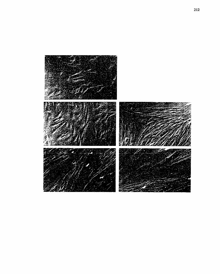
FIGURE 29: L6 Differentiation: Effect of MZBG.
L6 skeletal myoblasts were placed into low-serum a-MEM and differentiation to
myotubes monitored over 4 days in the presence or absence of MIBG (50 PM). Phase-
contrast rnicrographs were used to record the morphology of the cells. Magnification:
105x. The figures are representative photomicrographs of three individual experiments,
al1 of which exhibited the sarne response.
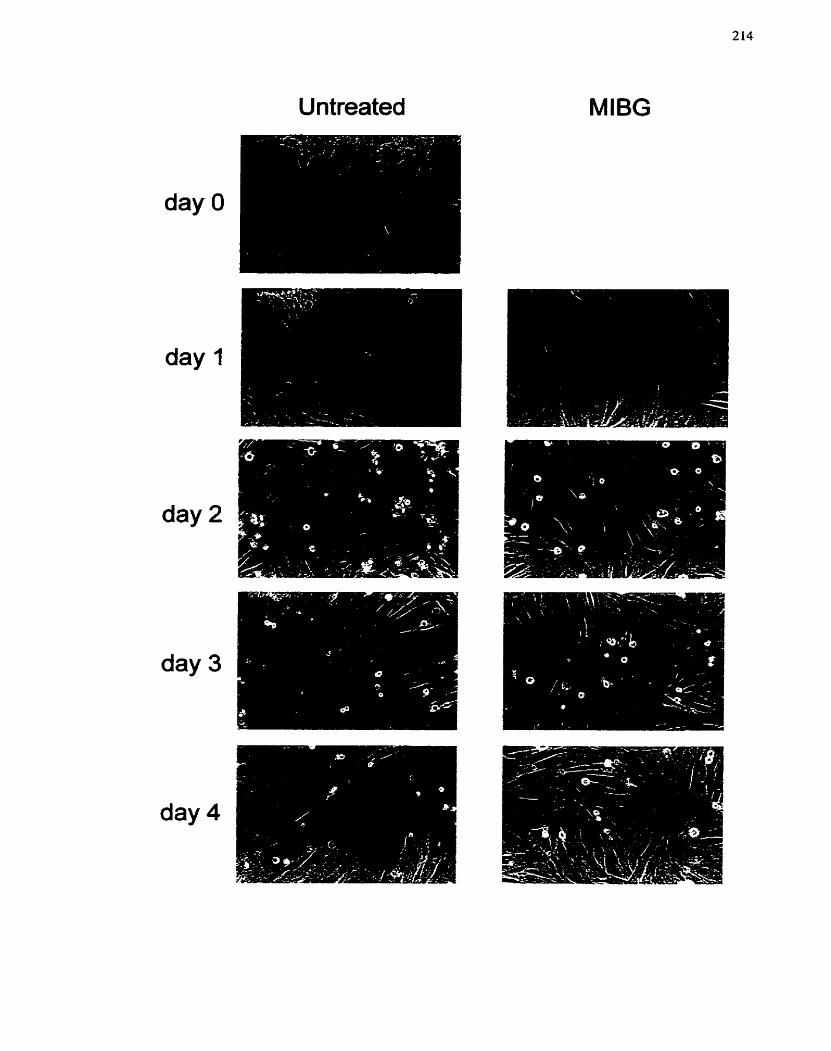
Untreated MlBG
FIGURE 30: Effect of ADP-ribosylation In hibitors on L6 Dijjferentiution.
L6 skeletal myoblasts were placed into low-senun a - m M and differentiation to
myotubes monitored over 4 days in the absence and presence of a variety of ADP-
ribosylation inhibitors. Inhibitors tested include: PD 128763 (10 PM), 3-aminobenzamide
(3-AB) (5 mM), para-aminobenzoic acid (PABA) (5 mM), MIBG (50 pM) and MIE3A
(50 PM). Phase-contrast micrographs were taken at the 4 day mark to record the
morphology of the cells. Note that al1 comparîsons should be made versus the 4 day
untreated panel. Magnification: 138x. The figures are representative photomicrographs of
two individual experiments, both of which exhibited a sirnilar response.
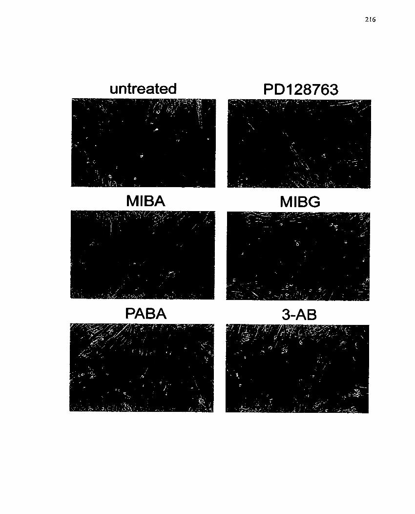
u ntreated
-
MIBA MIBG
PABA
events are not required for differentiation, but that mono(ADP-rïbosy1)ation events rnay
play a role in regulating the differentiation process.
Since MIBG inhibited the differentiation of L6 myoblasts, further studies were
conducted to examine the effect of this arg-mART inhibitor on L6 myoblast
differentiation- To detern~ine how MLBG influences the rnorphological changes
associated with diflerentiation, cells were viewed at daily intervals. This study clearly
established that MEBG inhibited the differentiation of these cells (Figure 29, MIBG
column). Interestingly, MLBG may have slowed the growth of these cells, since a day-by-
day companson between untreated and MIBG treated L6 cells showed fewer cells were
present on both day 1 and day 2. The cells also appeared to be larger, possibly indicating
a block in ce11 division, which would cause hypertrophy after a round of DNA synthesis.
Furtherrnore, the myoblasts appeared to be less organized at day 2, and at day 4 loss of
ce11 borders and fusion of the cells was not observed (Figure 29). Since there was no
fusion, clustering of the nuclei was not seen.
To establish the minimum concentration at which MIBG was inhibiting L6
skeletal myoblast differentiation, a concentration-response study was conducted (Figure
31). Quantitation of myotube fornation was also conducted in parallel, using Giemsa
stain to visualize the ce11 nuclei. This stain perrnits the counting and subsequent
quantification of nuclei in fused and unfused L6 cells (Figure 32A,B). These values were
then used to calcdate the fusion index (number nuclei in fused cells/total number of
nuclei), a numerical transformation that allows a direct comparison of the various
treatments (Figure 32C). MlBG (0.5 to 50 PM) was added to the differentiation media
and myotube formation monitored after 4 days- MIBG at concentrations From 0.5 to 10
FM did not significantly inhibit myotube formation (Figures 3 1, 32C). Only the highest
concentrations were effèctive (20 and 50 pM), with 50 p M having the greatest effect
FIGURE 3 1: Effect of MTBG Concentration on In hibition of L 6 Dr;ffereentiation.
L6 skeletal myoblasts were placed into Iow-sem a-MEM and differentiation to
myotubes monitored over 4 days in the presence of varying concentrations of MIBG (0.5
to 50 FM). Phase-contrast micrographs were taken on day 4 to record morphology of the
cells. MIBG concentrations used in each panel are: (A) 0.5 PM, @) 2 pM, (C) 5 PM, (D)
10 PM, (E) 20 pM and (F) 50 W. Magnification: 132x. The figures are representative
photomicrographs of two experiments, both of which exhibited the same response.
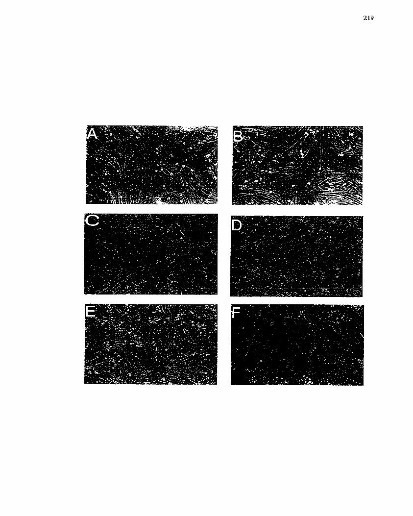
FIGURE 32: Effect of M7BG Concentration un Inhibition of L6 Differentiation.
Quantification of L6 Myotube Formation.
Representative photomicrographs of Giemsa stained L6 cells that were untreated (A) or
treated with 50 p M MIBG (B) over 4 days of differentiation. Magnification: 63x. (C) The
number of nuclei present in fused and unfised L6 cells was determined after addition of
MIBG (5 to 50 PM) for 4 days and presented as the fusion index (fusedhotal nuclei).
Data are presented as mean -t- SE fiom one of two experiments.
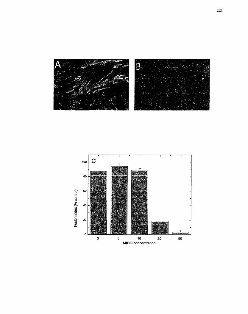
O 5 1 O 20 50
MlBG concentration
(Figure 32C). Thus, 50 pM MIBG was used in al1 subsequent experirnents, This kvas also
the concentration used in the initial studies (Figures 28, 29, 30). Interestingly, this is also
the most effective concentration for inhibiting ce11 growth and proli feration (c hapter 5).
To quantify myotube formation, L6 cells were treated, allowed to differentiate
and visualized with Giernsa stain. Exposure to 3AB (5 mM) decreased the number of
fused cells while treatrnent with MIBG (50 PM) ~ÎgnificantIy decreased the nurnber of
fiised cells (Figure 33A,B). The fusion index for control, untreated L6 cells was
approximately 85%, while 3AB treatment decreased the fusion index to approximately
50% (Figure 33B)- Treatment with MLBG reduced the hsion index to less than 5%
demonstrating very effective inhibition of differentiation (Figure 33B). In this
experiment, a decrease in the total number of nuclei was observed after MIBG treatment,
possibly indicative of an inhibition of cell growth (Figure 33A). As a result, a lirnited
[%I]thyrnidine incorporation experiment was conducted in which growing L6 cells were
treated with MIBG (50 PM) or MiBA (100 pM) over 36 h and incorporation of
['~lthyrnidine used as a rneasure of cell proliferation (Figure 33C). MIBG decreased
DNA synthesis in L6 cells, while MIBA did not, suggesting that MIBG may be inhibiting
events associated with ceIl proliferation in addition to differentiation-specific events in
L6 myoblasts,
To identify the tirne penod during which MIBG inhibits differentiation, a delayed
addition experiment was conducted. In this study, MIBG (50 PM) was added to cells at
varying times (O to 48 h) after placing the myoblasts into differentiation media. Ce11
rnorphology was examined on day 4. Administration of MIBG up to L2 h after the
switching to differentiation media completely blocked differentiation into myotubes
(Figure 34). Even addition of MIBG at 24 and 48 h was able to inhibit the differentiation
event, although the morphology indicated the cells had progresscd to the point at which
F I G U R E 33: Effeci of 3-aminobenzamide and MIBG on L6 Dzfferentiation.
Quantification of L6 Myotube Formation= Fusion h d e m
L6 skeletal myoblasts were placed into low-sem a-MEM and differentiation to
myotubes monitored on day 4 in the absence or presence of 3-arninobenzamide (3AI3, 5
rnM) and MIBG (50 FM). Experiments were carried out in duplicate. (A) The number of
nuclei present in fused and unfused L6 cells and the total number of nuclei in each
treatment was determined and presented as the hsion index (fùsed/total nuclei) (B). Data
are presented as mean I SE fiom one of two individual experiments. (C) Growing L6
skeletal myoblasts were treated with MISG (50 @VI) or MIBA (100 CIM) and
incorporation of [3~]thymidine over 36 1: used as a measure of DNA synthesis. The data
are presented as the mean t SE of experiments conducted in triphcate.
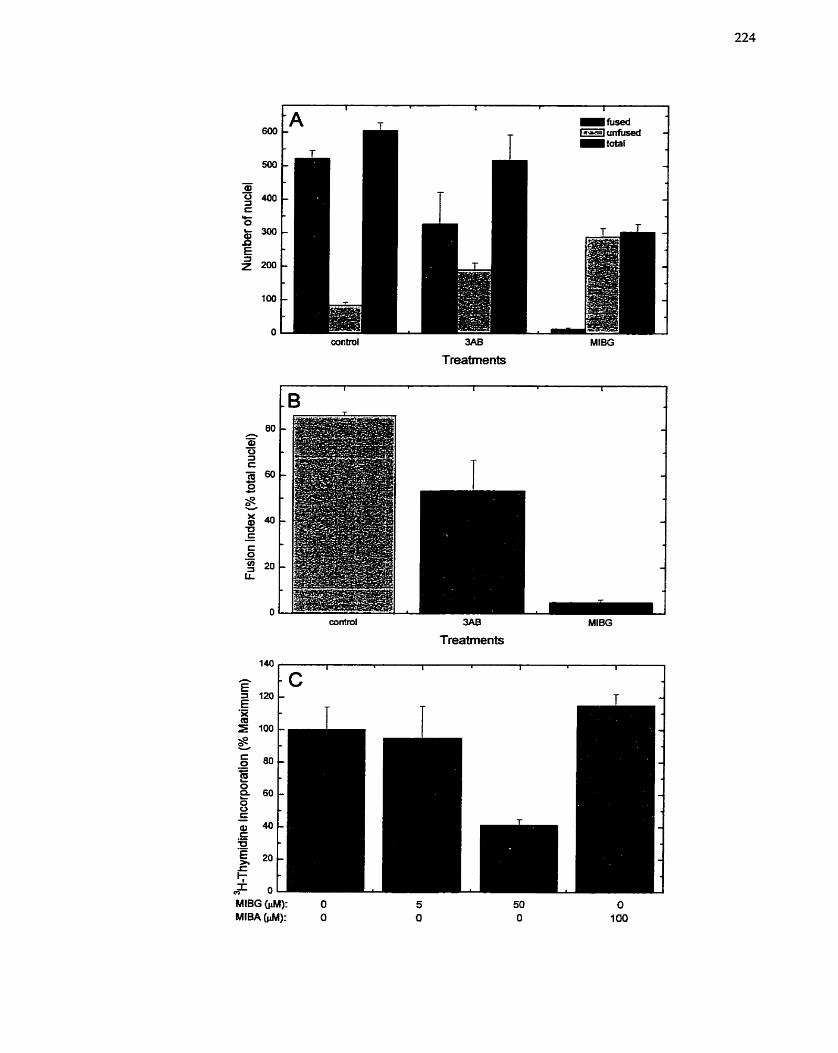
MlBG w): O MlBA W): O
3AB MlBG
Treatments
3AB
Treatments
MlBG
FIGURE 34: Effect of Deïuyed Addition of MTBG on L6 Dzyferentiafion.
L6 skeletal myoblasts were placed into low-serum a-MEM and differentiation to
myohibes monitored on day 4 (A). MIBG (50 ph.l) was added at diEerent tirne points (O
to 48 h) afier the switch to differentiation media. The tirne points at which MIBG was
added were: (B) O h, (C) 3 h, @) 6 h, (E) 12 h, (F) 24 h and (G) 48 h. Phase-contrat
micrographs were taken on day 4 to record the cell morphology. Magnification: 132x.
The figures are representative photomicrographs of two individual expenments, both of
which exhibited a sirnilar response.
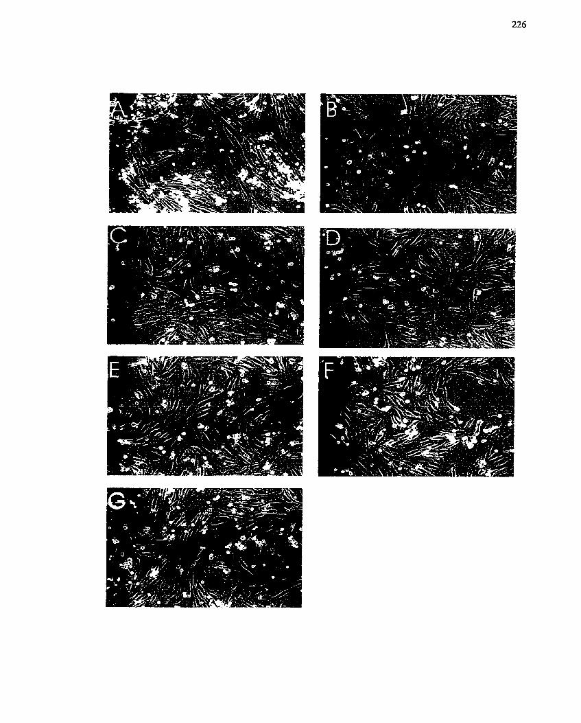
they were lined up and the cell borders had become l e s ~ distinct (Figure 34). Tliese data
indicate MIBG is likely inhibiting a late event in the myogenic program, one that is not
activated until 24 to 48 h after onset of rnyogenesis (possibly ce11 fusion)- Since L6
myoblasts do not initiate the actual fusion process until after 48 h, these data are in good
agreement with the earlier findings in untreated myoblasts (Figures 28, 29). Altematively,
since MIBG prevents both ce11 organization and ce11 fusion events (depending on the time
of addition), MIBG rnay influence myogenesis at multiple points.
To complement the delayed addition expenment, an MIBG removal experiment
was conducted. In this study, MIBG (50 FM) was added with the differentiation media
and then removed at specific time points (30 min to 48 h). The celIs were then maintained
in differentiation media without MIBG for the remainder of the 4 day period. These
experïments were expected to more closely identify the critical time points at which
MLBG exerts its inhibitory effects on differentiation. Both ce11 morphology (data not
shown) and the fusion index depicted a noticeable decrease in myotube formation afier
approxirnately 24 h with MIBG treatment (Figure 35). More dramatic differences were
noted when MIBG was present for 48 and 96 h (Figure 35). Quantification of ce11 fusion
demonstrated a significant decrease in myotube formation had occurred as a result of a 4
h exposure to MIBG (Figure 35). Longer treatments with MIBG result in significantly
greater inhibition of ce11 fusion (Figure 35). These data suggest that the full inhibitory
effect of MIBG requires its presence throughout the entire 4 day period. However, MIBG
may be affecting myogenesis at a point as early as 4 h after the mcdia change. Taken
together, these findings would indicate the MIBG is inhibiting multiple points in the
myogenic program.
To determine the reversibility of the inhibition cause by MIBG, a differentiation
rescue study was conducted, This experiment was dcsigned to also cxaminc whether
FIGURE 35: Effect of Transient MIBG Treatnrent on L6 Differentiation.
L6 skeletal myoblasts were placed into low-serum a-MEM in the presence of MIBG (50
yM) and the degree of differentiation monitored on day 4. MLBG was removed from the
cells at the indicated tirnes: O h, 30 min, 1 h, 2 h, 4 h, 12 h, 24 h, 48 h and 96 h. Control
cells were not exposed to MIE%G, while at 96 h MIBG was not rernoved. The number of
nuclei present in fused and unfused L6 cells was determined after Giemsa staining of the
ceIls (A), and used to calculate the fusion index (füsedhotal nuclei) (B). Data are
presented as mean -t SE. The results were confirmed in two independent experiments.
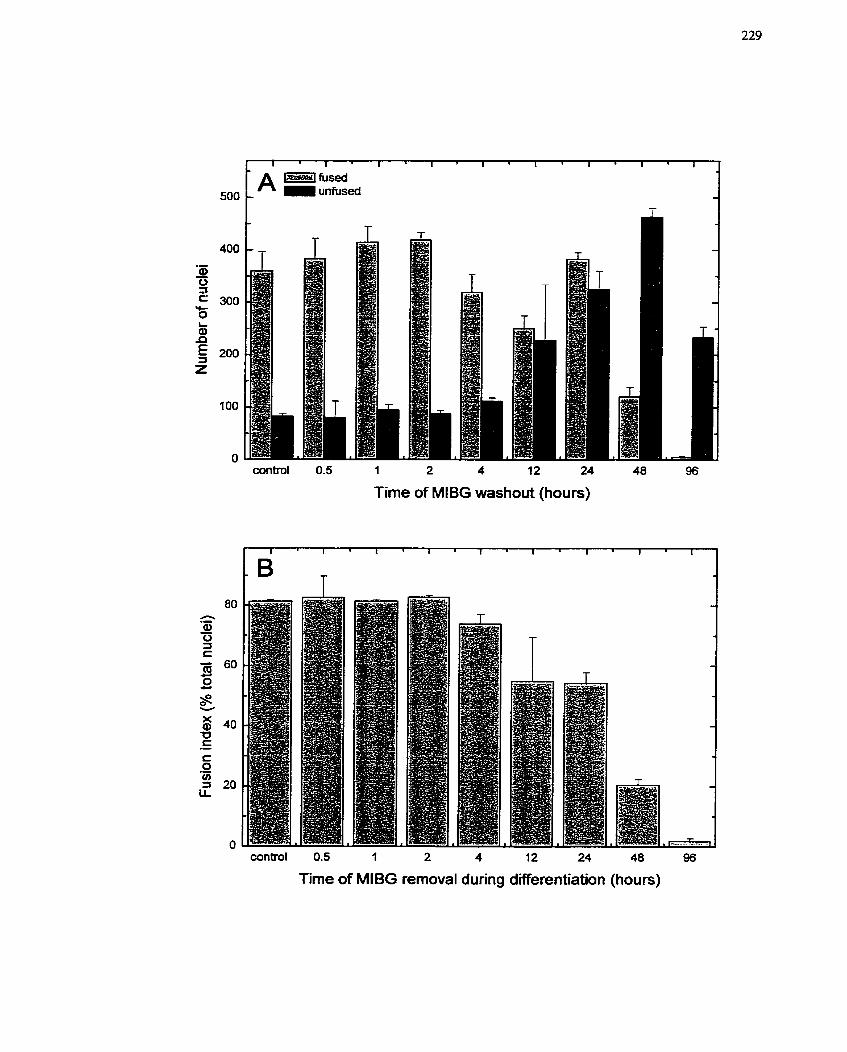
wntrol 0.5 1 2 4 12 24 48 96
Time of MlBG washout (hours)
control 0.5 1 2 4 12 24 48 96
Time of MlBG rernoval during differentiation (hours)
MIBG was tosic to L6 cells or was 'cytostatic' \vit11 rcspect to diffcrentiation. L6
niyoblasts were treated witli MIBG (50 pM) over 4 days. On tlie fourtli day, tlie media
\vas rcmoved and rcplaced with differcntiation media without M [BG. Tlic cells were thcn
incubated for an additional 4 days and differentiation monitored on day S. Cell fusion
\vas observcd on day 8 wliether the ce11 liad been trcated witli MIBG or not. in fact,
niyotube fomiation \vas comparable to those of day 4 untrcated cells (Figure 36).
Nevertlieless. al1 day 8 cells had begun to slio\v signs of necrosis (Figure 36). especially
the untreated cells, since under liigher power rnagnification granulation was observed
within the cells. These data demonstrate that MIBG is not tosic to L6 cells, since the c e h
remained capable of initiating differentiation after renioval of the MIBG. These data
further suggcst that MiBG blocks a specific event in the myogenic program and that this
inhibition is reversible.
6.2.4.3 Expression of myogenic proteins affected by kIIBG
Since it was observed tliat MIBG was capable of inhibiting L6 ceIl diffcrentiation
in a reversible manner, it was of intcrest to identify specific events in the myogenic
program sensitive to inhibition by MIBG. A variety of proteins of the niyogenic program
(muscle regulatory factors) arc cxprcsscd during tlic transition froni myoblast to myotubc.
Many of thcsc forni coniplexes that thcn interacr witli othcr protcins to rcgulate
progression to the diKcrcntiated state. MyoD is thc first major myogenic protein
exprcsscd during differcntiation and opcratcs i n a coniples in association E-protcins and
MEFZ (Lassar el (11. 1991). MEFZ tlicn binds to specific proiiiotcr clciiicnts of othcr
niuscle rcçulatory factors and tnusclc-spccific gcncs (Kauslial c'i ul. 1994; Olson ei ( I I .
1995; Takeda el d. 1995) to cnhancc tlicir cxprcssioii. As a rcsult, it was of intcrcst to
dctcniiinc wlicthcr M IBG influcnccd citlicr of tlicsc proccsscs. Iniiiiunoprccipitaiion of-
E12/17 protciiis I01Iowcd by Wcstcrn blot aiialysis foi- MyoD \vos carricd out to
FIGURE 36: Effect of MBG on L6 Cell Survival: Test of MTBG Cyrotoxicity.
L6 skeletal myoblasts were placed into low-senun a-MEM in the absence or presence of
MIBG (50 pM) for 4 days. On day 4, the differentiation media + MIl3G was removed and
replaced with differentiation media without MIBG. Four days later (day 8), phase-
contrast rnicrographs were used to record ce11 morphology. Mapification: 128 x. The
figures are representative photomicrographs of two experiments, both of which exhibited
the same response. Conditions shown: (A) 4 days without MIBG, (B) 4 days with MIBG,
(C) 8 days without MIBG, @) 4 days with MIBG + 4 days without MIBG.
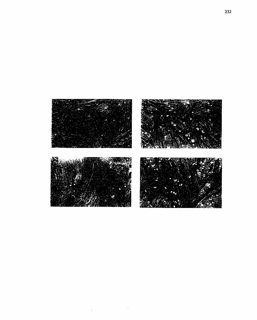
determine if MyoD had associated with E12/47 proteins and to determine if MIBG was
inhibiting formation of this complex. This particular step in the myogenic program occurs
before the formation of the MyoDIMEF2 complex (Lassar et ni. 199 1). Myoblasts did not
contain a pronlinent MyoD/E12/47 complex (Figure 37). In contrast, increased
expression of the complex was observed in rnyotubes. Treatment with MIBG did not alter
the formation of this complex (Figure 37). These data indicate that the formation of the
MyoD/E12/47 complex is not the event affected by MIBG and that MIBG is inhibiting a
downstream event in the differentiation process.
To assess the downstream myogenic event affected by MIBG, extracts of L6
myoblasts/myotubes were prepared at daily intervals over 4 days and Western blot
analysis was used to monitor myogenin, MEFZ and p2 lCrP' expression. Control, untreated
L6 cells showed gradua1 increase in the levels of myogenin and p2 lCrP1 protein over the 4
day time course (Figure 38). MEF2 expression was aIso increased in untreated cells,
however, expression decreased after day 2, reaching near basal levels by day 4 (Figure
l ~ r ~ t 38). In contrast, L6 myoblastç treated with MIBG did not express myogenin or p-
(Figure 38)- Interestingly, the pattern of MEF2 expression was similar to that of untreated
cells, becoming even more prominent on day 2, and remaining elevated on day 3 (Figure
38). These data would suggest that myogenin and p2~C'P ' expression is prevented by
MIBG and that this inhibition blocks progression of the rnyogenic program to the fusion
stage.
The distinct effects of MIBG on MEFZ expression relative to myogenin and
p2 lCrP1 suggested differential regulation of these genes. Since MEFZ lus been reported to
modulate myogenin, an alteration in MEFZ binding to DNA could produce the observed
results (Le. decreased myogenin expression with concomitant decrease in p21CrP'
expression, Figure 38). Nuclear extracts were incubatcd with radiolabelled
FIGURE 37: Association of ElZ/47 wifh MyoD in L6 Skeletai Myotubes.
MyoD was imrnunoprecipitated from L6 ce11 extracts and the presence of El2/47 proteins
complexed with MyoD was determined by Western blot analysis using an antibody
specific to E12/47 proteins. Extracts fkom myoblasts (B), myotubes (T) and MIBG-
treated (50 FM) myotubes (M) were used for this experiment. Similar results were
obtained in two independent experiments.
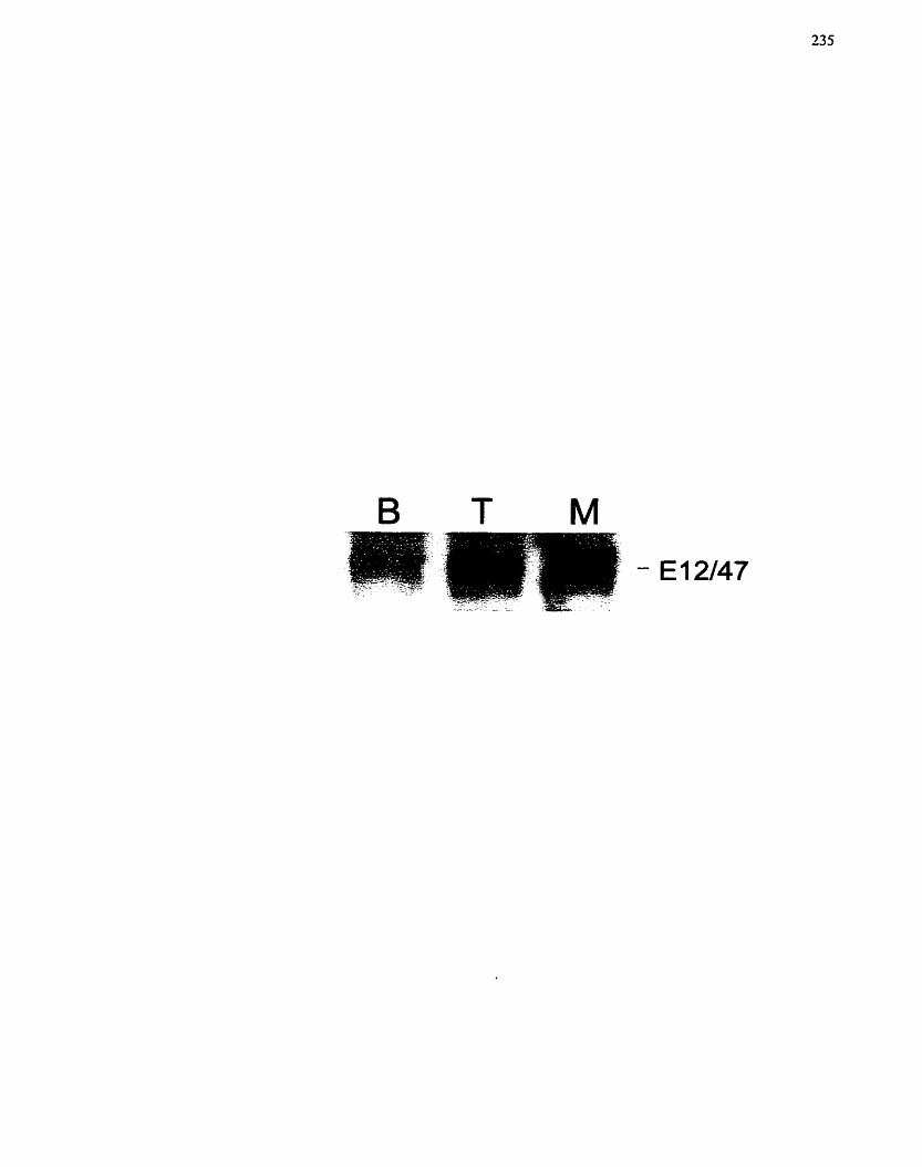
FIGURE 38: Effect of MlBG on Protein Murkers of L6 Cefl Differentiation.
L6 ce11 extracts were prepared at specific tirne points afier transfer of cells to low serum
a-MEM f MIBG (50 FM). The samples were subjected to SDSffAGE (10% geIs) and
Western blot analysis with antibodies for myogenin (diluted 1: 1 OOO), MEF2 (diluted
1 : 1000) and p21 (diluted 1: 1000). Proteins were visualized with the ECL
cherniluminescent detection system. MB denotes myoblast, while 1, 2, 3, 4 denote the
days of differentiation. Specific antibodies are identified on the left of the figure
(rnyogenin, MEF2 and p2 1). One of two independent experiments is shown, both of
which exhibited the same response.
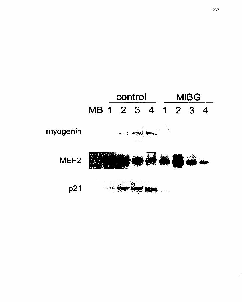
myogenin
MIBG
oligonucleotides containing the MEF2 element, subjected to UV liglit and the proteins
separated by SDSPAGE. The proteins crosslinked to the labelled oligonucleotides were
then visualized by autoradiography. Three bands between the 50- and 84-kDa molecular
mass markers were prominently labelled in the myotube extract (Figure 39). The
myoblast extract displayed two labelled bands. Interestingly, the MIBG treated extracts
showed the sarne banding pattern as the myoblast extract, and were missing the third
labelled band of 50- to 53-Da present in the myotube extracts (Figure 39). These data
suggest that a protein capable of binding to the MEF2 element is either not activated or
not expressed with MlBG treatment. Furthermore, it may be speculated that expression of
this protein is crucial for full expression of the myogenic program.
6-2.4.4 ADP-ribosylation and differentiation
To date, two mART activities (extracellular and intracelIular) have been reported
in n~yoblasts/myotubes (Huang et al. 1993; Huang et al. 1996; Kharadia et aL 1992;
Soman & Graves 1988; Soman et al. 1984a; Zolkiewska & Moss 1993; Zolkiewska et al.
1992). Of these activities, the extracellular mART has been associatcd with a 7 integrin
and desmin (Huang et al. 1996; Zolkiewska & Moss 1995). Both proteins have been
associated witli skeletaI muscle Function and ce11 communication- To deterniine if the
extracelldar cnzyme was thc targct for MIBG action, its activity was evaluated over the
myotube differentiation process and thcn in the presence of MIBG. ADP-ribosylation o fa
surface protein by mART was monitorcd by incubating cells with PINAD- AD-. Labcling
of a single band \vas detcctcd on days 2 and 3 of the differentiation pcriod of untreated
myoblasts (Figure 40A). Sincc the presence of labellcd bands was not greatly altcred by
treatrncnt with MiBG or othcr inhibitors of ADP-ribosylation (Figurc 40B), a rolc for an
cxtracclluIai- (or GPI-Iinkcd) arg-mART in thc diffcrcntiation proccss is not indicatcd.
FIGURE 39: Effect of MTBG un Protein Binding tu the MEF2 Elentent.
Nuclear extracts fiom L6 myoblast (B), L6 myotube (T) and MIBG-treated (50 PM) L6
cells (M) were incubated with radiolabelled oligonucleotide, cross-linked by UV-
irradiation (see Materials and Methods section 4.8.7) and subjected to SDS/PAGE (7.5%
gel). Molecular mass markers used to identi@ the proteins by size are shown on the right
hand side. The Iabelled proteins are indicated on the Ieft-hand side by numerical notation.
One of two independent experiments is shown, both of which exhibited the same
response.
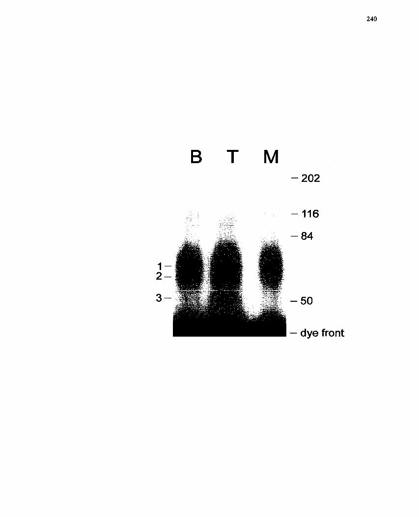
B T M
f
r: . - 84
1 - 2-
3- - 50
- dye front
FIGURE 40: ADP-ribosyiation and L6 Diffeentiation. In situ ADP-ribosylation of
Extracellular Proteins.
L6 myoblasts were placed into differentiation media and ART-dependent modification of
extracellular proteins monitored on a daily basis. Cells were exposed to C~~P]NAD+ and
transfer of label to specific proteins examined by autoradiography after SDSPAGE
(7.5% gel). (A) Separate lanes fiom autoradiographs of untreated L6 cells allowed to
differentiate over 4 days are shown. MB denotes myoblast, while 1, 2, 3 and 4 denote the
days of differentiation. @) Autoradiograph of L6 cells on day 3 that were untreated (O) or
treated with different inhibitors of ADP-nbosylation: para-arninobenzoic acid (PABA, 5
mM), 3-aminobenzamide (3AE3, 5 mM), PD128763 (PD, L CLM), MIBA (50 PM) or
MIBG (50 PM) during the incubation penod with [ 3 2 ~ ] ~ ~ ~ + . One of two independent
experiments is shown, both of which exhibited a similar response. Arrows point to the
most prominent bands detected in these experiments.
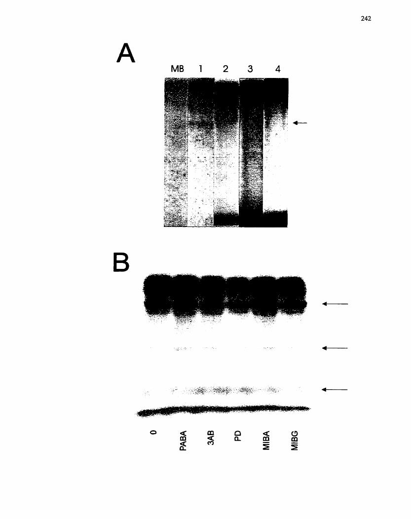
6-2.5 Discussion
The results of this study clearly demonstrated that MIBG is a reversible inhibitor
of myoblast differentiation and theoretically of rnyoblast ce11 profiferation in L6 skeletal
muscle myoblast ce11 Line. This work is in very good agreement of the studies camed out
by Kharadia et al. (1992) and not only confirrns their findings, but also extends their
work with respect to identi&ing the step in the myogenic differentiation program which
MIBG, and presumably an arg-mART, may be influencing.
The initial survey of ADP-ribosylation inhibitors (Figures 30, 33) was in quite
good agreement with the previous work of Shall and associates (Farzaneh et al. 1982),
who demonstrated that the poly(ADP-ribosy1)ation inhibitor 3AB was able to reversibly
inhibit the fiision of myoblasts from primary cultures of chick myogenic cells. These
authors suggested that PARE' was involved in the regulation of myoblast differentiation-
However, as correctly pointed out in the paper by Kharadia et al- (1992), at the
concentrations of 3- that were used for those experiments, both PARP and mART
activities would have been inhibited (Banasik et al, 1992; Duncan et al- 1988; MiIarn &
Cleaver 1984; Rankin et al- 1989). Although a significant inhibition of differentiation
(Figure 30), was not apparent upon visual examination, in contrast to the observations of
Shall and associates (Farzaneh et al. 1982), the quantitative studies demonstrated 3AB
caused a 40% reduction in the fusion index (Figure 33). On the other hand, the reduction
in myotube fusion achieved with 3AB was signifcantly less than the effects obtained
with MIBG treatment (Figares 29, 33, 35). The effective concentrations of MIBG were
between 20 and 50 FM, within the concentration range defined by Kharadia et al. (1992)
and, also interestingly, in agreement with the concentrations that were able to inhibit ce11
proliferation of H41IE cells (and the systems described in chapters 7 and 8). In the studies
of L6 cells, growth inhibition was indicated by a reduction in ce11 nurnber (Figures 28,
29, 33, 3 9 , and the decrease in [3~]thymidine incorporation of growing L6 cells treated
with MIBG (50 PM) (Figure 33C). Furthermore, the cells appear larger, somewhat
suggestive of a block in ceIl proliferation at the G2/M stage (Le. blocked ce11 division).
Since L6 myoblasts constitutively express MyoD (Lognome & Wahrmann 1986), MIBG
must affect progression through the differentiation process as opposed to determination
or cornmitment to the muscle lineage.
4 s discussed in the previous chapter, MiBG could interfere with L6 myoblast
differentiation directly or indirectly. For instance, lack of ce11 fusion could result from
changes in membrane properties independent of myogenic-specific events. As stated by
Kharadia et al. (1992), the effect of MIBG appears to be on the entire differentiation
program, and not just simply the process of ce11 fiision, since both creatine phosphokinase
(CPK) activity and protein accumulation were blocked by MIBG. However, previous
studies have shown that muscle-specific proteins are expressed either in myogenic ce11
cultures in which fusion has been blocked or in fusion-deficient ce11 lines (Devlin &
Konigsberg 1983; Nguyen et ai. 1983). Thus, if only fusion, but not differentiation, was
affected by MIBG treatment, increases in muscle-specific marker proteins such as CPK
activity would be expected. Although neither CPK activity nor other markers of
differentiated muscle were examined in this study, the fact that MIBG-dependent effects
could be seen with only 4 to 12 h exposure early in the differentiation process supports
that fact that MIBG is not just affecting the fusion step. Furthermore, the effect of MIBG
is reversibIe (Figures 35, 36) and thus the short treatment time with MIBG should not be
able to inhibit fusion, a process that occurs 48 to 72 h later. On the other hand, the MIBG
delayed addition studies would suggest that the presence of MIBG may be required at
multiple, temporally separated steps- Altematively, the time frarne for expression of
musclc-specific gencs during differentiation is a Ionger process over thc initial timc
period. Finally, Western blot analysis showed that the expression of muscle-specific
proteins is blocked, which supports the contention that MIBG is affecting specific events
required for differentiation (Figure 38)- Nevertheless, an effect on the fusion and
multinucleation processes cannot be mled out.
According to the findings of this study and information in the literature, the step
at which differentiation is blocked occurs afier determination, since L6 skeletal myoblasts
are already committed myogenic precursor cells (Yaffe 1968). MIBG blocked the
expression of not only myogenin but also p21C1P' (Figure 38). It is well established that
the absence of these proteins prevents myoblast differentiation and fusion into
multinucleated myotubes (Dias et al. 1994; Walsh 1997). Myogenin has been shown to
mediate the differentiation process (Walsh 1997), since it is a marker of myoblast
cornmitment to the differentiation pathway. Subsequently, the expression of p21C'P! is
increased and p2 lCIP1 is a cyclin-dependent kinase inhibitor (Walsh 1997). Presumably it
is the increased expression of this protein that irreversibly withdraws the skeletal
myoblasts from the ce11 cycle and results in terminal differentiation. Interestingly, with
MIBG treatment, since the expression of p21C'PL is decreased, it would suggest that the
myoblasts are still capable of proliferating. On the other hand, MIBG inhibits ce11
proliferation (see chapter 5, Figure 33C and Kharadia et al. 1992). Based on this
information, and the requirement for withdrawal from the ce11 cycle for progression of
differentiation (Lassar et a[. 1994; Molkentin & Olson 1996b), it might have been
expected that MIBG treatment would stimulate differentiation. The ability of MIBG to
abrogate differentiation of L6 cells, in addition to possibly proliferation, suggests that a
scparate mechanism during differentiation may be inhibited. In these studies, although
detailed growth rneasurements were not conducted, it was observed that the number of
myoblasts in the MIBG samples was reduced (Figures 28, 29) and that there was a
decrease in total number of nuclei (Figures 33, 35). This was confirmed by [3~]thymidine
incorporation studies where it was observed that uptake of [3~]thymidine was decreased
with MIBG treatment (Figure 33C).
Since L6 skeletal myoblasts are already commited to the myogenic lineage,
MLBG may delay differentiation by affecting one cruciai step. However, the activation or
expression of this one protein may be required for multiple steps in the cascade. MlBG
could also be toxic to the cells, however, the washout studies would suggest otherwise, as
would the rescue study that was conducted (Figures 35, 36). These findings are in
agreement with those of Kharadia et al. (1992), who also suggested that the inhibition of
differentiation by MBG was not due to the inhibition of ce11 division (Le. proliferation of
myoblasts was blocked before a necessary terminal rnitosis during differentiation). The
inhibitory effects of MIBG were immediate with respect to ce11 fusion (Figures 35, 36),
and removal of MIBG resulted in a rapid increase in ce11 fusion (Figure 36), which might
not have been expected if mitosis was required to initiate the differentiation step
(Kharadia et ul. 1992). The requirement for mitosis was also ruled out by Kharadia et al.
(1992) through the addition of cytosine arabinoside to the refed cultures. if an additional
round of mitosis was required for recovery from the MIBG treatment, blockade of ce11
proliferation with the cytosine arabinoside would have inhibited the differentiation
process, and this was not observed.
The immunoprecipitation studies found that MIBG did not prevent the association
between MyoD and E12/47 proteins (Figure 37). On the other hand, with the cross-
Iinking studies, it was found that an oligonucleotide containing the MEF2 element
associated with a protein in myotube nuclear extracts that was missing in nuclear extracts
from myoblasts and MIBG trcated-myotubes (Figure 39). This protein has a rnolecular
mass of approximately 40-kDa (band s ix of 53-kDa minus the 13-kDa oligonucfeotidc).
[ntcrestingly. the size of tIiis protein docs not corrclatc with thc predoniinant protcin that
bitids to this sequcnce. MEF? (which is 72-kDa), whicli is required to transcriptionally
activate nunierous muscle-specific genes including creatine kinase. myosin Iicavy chain,
desmin and certain MRFs (Ludolpli & Konicczny 1995). For this reason, it is unclear
whethcr the 40-kDa protein has a rolc in the induction of myogenesis-spccik genes. If
this protcin is MEFZ (possibly a breakdown product or novel variant). an impairment in
DNA binding is indicatcd. This conclusion can be reachcd because MEFZ expression is
unaffected by MIBG (Figure 38). Furthemore, MIBG may influence the phosphoylation
state of MEFZ which modulates both DNA binding and protein-protein interactions
(Ferrari et cd. 1997: Molkentin et al. 1996). AIternatively, the 40-kDa protein is unrelated
to MEFî , but it is critical For the cxpression of niuscle-specific genes via the MEFI,
elcnicnt. One such candidate protein niay be SRF (serum response Factor) whicli is also a
part of the MADS box family of transcription factors (Black & Olson 1998; Brand-Saberi
& Christ 1999; Shore &c Sharrocks 1995). OF note, it is entirely possible that the MEF2
oligonuclcotide, which was bascd on a MADS box conscnsus sequcnce, could bind other
MADS box faniily transcription factors. For cxample, SRF is a 62-kDa protcin and splice
variants 11avc bccn found in sinooth and skcletal muscIc of approxiniatcly 67-, 60-, 57-
and 50-kDa (Kcmp & Mctcalfc 2000)- Thcrc is niountinç cvidcnce to suggest that niuscle
spccific transcription niay rcquire functional intcractions of musclc-spccific bHLH
Litciors with othcr rcgulatory pi-otcins sucli as SRF (Duprcy 6r Lcscns 1994; Hautnianri et
ul- 1998; Hautmann er ~ i . 1997; Solway ci al. 1995). Morcovcr, MEF2 DNA biiiding
sitcs arc typically positioncd within closc proxiniity to MRF binding sitcs (i-c. E-boxcs)
in tlic rcgulatory rcçions of' niany niusclc gcncs. Tlicsc sitcs arc rcquircd for thc
transcriptional activation orniyogcnin, MRF4 aiid MyoD (Chcng cr O/. 1993: Naidu el d.
1995; Worig er (il. 1993). Tlic MEF2-binciing silc, but not ttic E-box, is ncccssary for cc11
type-specific expression and activation of the myogenin gene by MyoD in tissue culture
celis (Buchberger et al. 1994). Thus, an effect by MIBG on the expression of myogenin
via the MEF2 site (or a similar MADS box site) would prevent differentiation. Further
study is necessary to determine the exact target through which MIBG has its effect.
Interestingly, if the tarset protein is SRF, then regulation by MIBG may occur through
Rho (Montaner et al. 1999; Poser et al. 2000) which is modulated by ADP-ribosylarion
(Aktories 1997; Lerm et ai. 2000).
The ability of MIBG to inhibit L6 skeletal myoblast differentiation suggests that
an arginine-dependent mART may be involved in one or more of the steps Leading to
differentiation of myoblasts into rnyotubes. Both intracellular and extracelluar arg-
mARTs have been identified in skeletal muscle cells from a variety of species (Okazaki
& Moss 1998; Soinan & Graves 1988; Zolkiewska & Moss 1993; Zolkiewska er al.
1 992). An extracellular GPI-Iinked arg-mART was first demonstrated b y Zolkiewska and
Moss (Zolkiewska & Moss 1993) who originaliy had cloned a rabbit skeletal muscle
NAD:arginine ADP-rhosyltransferase (Zolkiews ka et al. 1 992), and then subsequently
found that it was able to modiQ integrin a7 of differentiated mouse C2C12 and L8 cells.
Intracellular arg-mARTs have been identified in the membrane fraction of rabbit skeletal
muscle (Peterson et ul. 1990; Soman & Graves 1988; Soman et al. 1984a). The
extracellular arg-mART was not involved in modulating differentiation since incubation
with MIBG did not alter the labeling with ["PINAD' (Figure 40). Kharadia et ai. (1992)
had provided evidence that an intracellular arg-mART was inhibited by MIBG.
Furthermore, they concluded that this was the reason for the inhibitory effect of MiBG on
myogenic ce11 differcntiation and proliferation. Their studies demonstrated that a spccific
transfcrasc activity was dctcctablc in thcir primary embryoiiic chick skclctal muscle
culturcs and that thc ADP-ribosylatcd form of MIBG could be dctcctcd in honiogcnatcs
of cultures treated with MIBG (Kharadia et al. 1992). Secondary evidence provided by
these authors include studies showing that MIBG is capable of inhibiting the purified
skeletal muscle arg-mART (Kharadia et al. 1992). Interestingly, the authors also provide
evidence for the specificity of the MIBG effect through its guanidino group, since MIBA
which lacks the guanidino group was unable to inhibit differentiation. The studies
presented in this chapter also show that MIBA does not inhibit fusion of myotubes
(Figure 30).
To this point, the only identified rnechanism by which ADP-ribosylation might
influence the differentiation process is the modification of desmin, an intermediate
filament. ADP-ribosylation results in the inhibition of desmin assembly, and de-ADP-
ribosylation results in assembly (Huang et al. 1993; Zhou et al. 1996). Furthemore,
desmin is one of the earliest myogenic markers and one of the first muscle-specific
proteins to appear during mammalian embryonic development (Babai et al. 1990; Choi et
al. 1990; Furst et al. L989; Lin et al- 1994; Mayo et al. 1992). Thus, interfering with
desmin intermediate filament assembly-disassembly by mono(ADP-ribosy1)ation (Yuan
et al. 1999) may be a possible regulatory rnechanism for rnyogenesis involving fusion
and myofibril organization. However, the block in myogenin and p21C'P' expression is
more applicable to the early stages of myogenesis (although desmin is expressed in pre-
fusion myoblasts (Babai el al. 1990)). Nevertheless, inhibition of desmin organization
could be a part of the regulatory process for differentiation and a part of the multiple
steps that MIBG may be inhibiting. Further investigation of these events and tbeir
regdation by mARTs wi I l be necessary.
6.2.6 Summary
MIBG inhibits the differentiation of L6 skeletal myoblasts into rnyotubes and the
rnechanism is likely mediated by a membrane-associated intracellular arg-mART. The
expression of two key proteins, myogenin and p2 lCtP', for the myogenic lineage appear
to be inhibited by MIBG. Myogenin is an MRF that is critical for the subsequent
expression of other muscle-specific proteins, while expression of p2 1'''' is crucial for the
irreversible withdrawal of the celIs from the ce11 cycle- Furthemore, MIBG also appears
to influence binding of a protein, possibly MEF2, to the MEF2 element, which may
further explain the lack of myogenin protein expression and the abrogation of the
myogenic fusion prograrn.
7.0 SMC as a mode1 of de-differentiation, proliferation and migration
7.1 Introduction
Within the normal vesse1 wall, SMCs are found in the medial layer, and SMCs
within this iayer have been shown to express a range of phenotypes that exist between
two extreme States traditionally referred to as 'contractile' and 'synthetic' (Campbell et
al. 1987; Chamley-Campbell et al. 1979). Contractile SMCs are characterized by the
presence of myofilaments and smooth muscle specific genes that encode contractile
proteins and proteins that regulate contraction (Campbell et al- 1987; Shanahan et al.
1993). On the other hand, synthetic SMCs have a limited myofilament presence and an
abundance of organelies (especially rough endoplasmic reticulum and Golgi apparatus)
for the processing of newly synthesized proteins (Campbell et al. 1987). This concept of
SMC heterogeneity was first identifieci in ce11 culture studies (Chamley-Campbell et al.
1979) and since that time SMC heterogeneity has been found not only in experirnental
animals (Bochaton-Piallat et al. 1996; Orlandi el al. l994a; Pauletto et al 1994; Schwartz
et al. 1995b; Villaschi et al. 1994; Wohrley er al. 1993, but also in the human population
(Glukhova et al. 1991). The relevance of heterogeneity to vascular fibroproliferative
diseases lies with the question of the identification of the SMCs that are responding in a
pathological manner. Were these cells originally heterogeneous and thus only a certain
population was responding? 1s there a spatiotemporal heterogeneity in the expression of
differentiation rnarkers? Or, do SMCs modulate their phenotype and differentiation status
in response to environmental factors? To add to this complexity, evidence is mounting
that committed and differentiated cells can transdifferentiate into another ce11 type
(Arciniegas et al. 2000; Gressner 1996; Lipton et al. 199 1 ; Smith el al. 1999). With the
application of ce11 and molecular biological techniques for the study of development,
differentiation and the pathophysiology of vascular proliferative disorders, distinct
cellular phenotypes involved with SMC myogensis and proli ferative and fi brotic
responses of SMCs in diseased artenes has been identified (McHugh 1995; Owens 1995;
Ross 1993; Sartore et al. 1994; Schwartz et al. 1986).
To define the response of SMCs to various stimuli and growth factors in an
isolated system in vitro, various SMC culture systems have been developed (Chamley-
Campbell et al. 1979). The most commonly studied SMCs are derived from either rabbit
or rat artenes (Birukov et al. 1993; Bcchaton-Piallat et al. 1992; Campbell et al. 1989;
Kocher & Gabbiani 1986; Schwartz et al. 1986; Schwartz et al. 1995a; Skalli et al. 1986;
Thyberg et al. 1990b). In these culture systems, the biological features of SMCs have
been systematically studied, and the identification of distinct SMC phenotypes and
responses to environmental and exogenous stimuli have been described (Birukov et al.
1993; Bochaton-Piallat et al- 1992; Campbell et al. 1987; Campbell et a[. 1989;
Chamley-Campbell et al, 1979; Kocher & Gabbiani 1986; Owens 1995; Schwartz et al.
1995a; Skalli et al. 1986; Thyberg et al. 1990b). However, in these SMC culture systems,
limitations to their use have arisen due to species-specific differences observed in the
response to injury, both in vivo and in vitro, as well as environrnentat factors (Schwartz et
al. 1995b). As a result, many Iarger animal models of restenosis and SMC tissue culture
models derived from these larger animals have been developed. Arnong the most popular
have been models of restenosishtimal thickening employing porcine arteries (Ga1 et al.
1990; Gnnstead et ai. 1994; Schwartz et al. 1993; Zalewski & Shi 1997). With these
models, both pig aortic and coronary SMCs have been studied (Christen et al. 1999;
Gotlieb & Boden 1984; Koo & Gotlielj 1989; Koo & Gotlieb 199 1 ; Koo & GotIieb 1992;
Saward & Zahradka I997b; Wilson et al. 1999). Porcine coronary artery SMCs have
been shown to maintain a high lcvel of differentiation rnarker expression, including
smoothelin (Cliristcn et ul. 1999) and, sincc they behave differcntly in vat-O frorn thc
majority of SMCs previously studied, they may thus represent a better mode1 for the
study of agents influencing SMC behaviour in vitro and in vivo (Christen et al. 1999).
Furthemore, porcine SMCs appear to exhibit features that are more similar to those
observed in human arterial SMCs in culture (Chnsten et al- 1999; Kocan et al. 1980).
One of the most commonly employed methods for the establishment of primary
SMC cultures involves the use of enzymatic dispersion of vessels (Gimbrone & Cotran
1975). However, separation of the different ce11 types present within the vesse1 wall is
much more difficult after a generalized digestion. Additionally, the potential for damage
to cellular membranes and protein receptors bound to the membrane is increased with
time of digestion, thereby affecting the responses of the SMCs to stress, growth stimuli
and other environmental factors. Altematively, microdissection of the vesse1 wall to
remove only the smooth muscle containing media1 layer which can then be cultured by an
explant method has been used to establish prirnary SMC cultures (Ross 1971). This
approach selects for SMCs but the size of the vessel limits the species and vessel type
that can be used. A primary culture system empfoying SMCs derived from porcine
coronary artery explants was developed in this laboratory by a fellow student (Saward &
Zrihradka 1997b). Due to numerous advantages afforded by this systern, including ease of
preparation and homogeneity of the celi population, this well cl-iaracterized system was
employed for the studies described in this chapter.
Using this primary porcine SMC culture system and based on findings from the
previous chapters, a specific hypothesis was proposed: Growtk factor-mediated growth
and prolferafioiz of porcine SMCs will be inhibited by MIBG thi-ozcgh inhibition of a
specific cellular s ignahg pathway that will involve an ADP-ribosylation reaction.
This goal was achieved by i) defining the cellular system, ii) testing the efficacy
of the inhibitor in the systern, and iii) examining the signaling components that might be
invo Ived.
7.2 Mitogenic response of SMC's to growth factors: role of PGEz
7.2.1 Background/rationale
Abnorrnal andlor excessive proliferation of SMCs is believed to play a key role in
the pathogenesis of atherosclerosis and restenosis after revascuIarization (Ross 1995;
Schwartz ef ~ 1 - 1986; Schwartz & Reidy 1996)- Upon injury or exposure to increasing
environmental stress, SMCs within the vesse1 wall become activated and respond to
secreted growth factors and other stimuli by increasing their growth potential. The factors
controling SMC growth in both normal and disease States have therefore becorne the
subject of intensive investigations over the past 10 years. Both positive and negative
stimuli exist for SMCs and small alterations in the baIance of these factors can have
profound effects on SMCs and their growth responsiveness (Liu et al. 1989). Mitogens
such as platelet-derived growth factor (PDGF), angiotensin II (AngII), thrombin and
fibroblast growth factor (bFGF) have been shown to be released by platelets, damaged
endothelial cells and SMCs after injury, and have been demonstrated to influence the
growth and migration of SMCs during the formation of a neointimal thickening (Davies
& Hagen 1994; Neville & Sidawy 1998; Schwartz et al. 1995~). On the other hand,
inhibitors of SMC growth, such as heparin and prostaglandins of the E family, are also
released by ceIls after the injury (Schror & Weber 1997; Scott-Burden & Vanhoutte
1994).
The role of prostaglandins of the E family in the modulation of SMC proliferative
status has been examined in a variety of venues, however, the results rcmain
controversial. PGEI and PGE2 have been shown to operate as both positive and negative
regulators of SMC growth (Owen 1986; Pasricha et al. 1992; See et al. 1987; Shechter et
al. 1997). For example, Shechter et al. (1997) and Urnemura er al. (1997) have shown
PGEi is effective during angioplasty as preventative therapy for coronary restenosis, and
Fan and associates, and others (Fan et ai. 1997; Loesberg el al. 1985; Nilsson & Olsson
1984) have shown that PGEI inhibits vascular SMC growth. PGEi has antiplatelet
aggregation and deposition characteristics, as well as anti-spasm, vasodilation and
cytoprotective effects (Siegel el al. 1984), and is derived from macrophages that may be
populating the injured region. Furthemore, exogenously added PGEz has an
antiproliferative effect on cultured guinea pig tracheal and human airway SMCs (Florio et
al. 1994; Johnson et al. 1995) and rabbit aortic SMCs (Lei & Deng 1989). On the other
hand, Pasricha et al. (1992) have demonstrated that PGE, and PGE2 stimulate the
proliferation of puhnonary artery SMCs. It is possible that the effect of PGEin on the
proliferation of SMCs is dependent on the vascular bed. However, Owen (1986)
dernonstrated that the effect of PGEi on SMCs is dependent on the phase of the ce11 cycle
when it is added. If PGEl was added to a quiescent ce11 population, DNA synthesis was
enhanced. If PGEi was added to asynchronous cycling cells, it functioned as an anti-
proliferative agent. Moreover, Sjolund et al. (1984) have described a phenotype
modulatory action of PGEl in pnmary cultures of arterial SMCs. Tt was thus suggested by
Schror & Weber (1997) that endogenous prostaglandins may serve a duai purpose in
SMCs: maintenance of the differentiated, contractile state and control of proliferation in
the presence of growth factors (Pornerantz & Hajjar 1989). Thus, PGs could stimulate
SMCs to enter the cc11 cycle and to divide in response to tissue injury in order to facilitate
tissue repair whiIc also preventing uncontrolled growth of SMCs by interfcring with the
synthetic phase of the ceIl cycle (i.c. inhibition of proliferation of SMC after stimulation
with growth factors) (Asada et al. 1994; Koh et al. 1993; Owen L986; Pasricha et al.
1992; Schror & Weber 1997; Shirotani et aL 199 1).
Recently, Inagami and colleagues (Ohnaka et al. 2000) demonstrated that AnglI
induced the expression of cyclooxygenase-2 in cultured rat vascular SMCs with
subsequent PGEz production, It has also been dernonstrated by others that AnglI
stimulates the release of PGs in a variety of cellular systems, including SMCs through the
activation of PLA2 (Alexander & Gimbrone 1976; Catalioto et al. 1996; Gimbrone &
Alexander 1975; Schlondorff et al. 1987; Vallotton et ai. 1989). This laboratory has also
demonstrated a link between AngII and PG release in porcine SMCs, that was associated
with a positive growth response (Saward, Yau, Thomas & Zahradka, unpublished
observations). This particular study was therefore initiated to examine the effect of PGE2
on SMC growth and proliferation in relation to SMC growth status.
7.2.2 Specific Airns
1. To compare the growth response of SMCs to a variety of growth factors.
2. To define the contribution of PGEz to SMC growth.
3. To determine the signaling pathways activated by PGEl that are related to the
growth response,
7.2.3 Experimental Design
Primary cultures of porcine SMCs, generated by the explant culture technique,
were incubated in senirn-free supplemented media for 5 days before experimentation.
This was to ensure that the SMCs were in a quiescent, differentiated, contractile state that
is reminiscent of SMCs in the vesse1 wall. The time required for the SMCs to re-enter the
cc11 cycle was monitorcd by [3~]thymidine incorporation. The rates of RNA and DNA
synthesis were then measured in response to serurn and grou-th factor (eg. AngII)
stimulation. A broad spectmm of growth factors were assessed for their ability to
stimulate DNA synthesis. Growth assays were carrïed out to monitor the specific effect of
PGE2, a novel stimulator of SMC growth (Owen 1986; Pasricha el al. 1992). These
assays included lurid ri di ne and [ ' ~ l t h ~ m i d i n e incorporation, MTT assay,
irnmunocytochemistry for PCNA expression and incorporation of bromodeoxyurïdine
(BrdU), and RT-PCR analysis of c-fos gene expression. Moreover, the signaling
pathways associated with PGE2-stimulated growth were assessed by Western blot
analysis. The activation of p21-Ras and MAP kinase was also examined separately.
Finally, the contribution of the PI3-kinase pathway was examined with respect to PGE2-
stimulated SMC growth.
7.2.4 ResuIts
7.2.4.1 Growth characteristics of SMCs in response to serum and other growth
factors
The response of primary porcine coronary artery SMCs to serum withdrawal was
monitored over preset petiods of time. Subconfluent SMCs were placed into serum-free
supplemented D-MEM and the rate of DNA synthesis monitored at specific time points
with 30 min pulses of [3~]thyrnidine. Over a penod of 168 h (7 days), the rate of DNA
synthesis declined considerably, with a basal steady-state level of DNA synthesis being
reached at approximately 48 h (Figure 41A). This is similar ro the findings of Saward &
Zahradka (1 997b), who first characterized this particular model. It was observed that the
cells, even after 7 days under serum-free conditions, did not appear to be dying (data not
shown; Figure 41A). Although ce11 viability assays were not conducted, the ability of the
quiescent SMCs to re-enter the ce11 cycle was taken as evidence that the cells were still
healthy (Figures 41B and 50). Based on this esperirnent and the tïndings O € Saward &
Zahradka (1997b), SMCs were consistently prepared by incubating tl-iem in a scrurn-free
supplemented D-MEM for 120 h (5 days) before initiation of experin-ientation. The
prolonged starve down period ensured that the SMCs had esited the ce11 cycle and
expresscd a contractite plienotype- To characterize the differentiatcd state of these SMCs,
immunocytochemistry with specific antibodies \vas conducted to assess the expression of
the smooth muscle specific markers a-actin and SM-myosin (Figure 42). Hoescht nuclear
stain was applied to identify the location of al1 cells. After 5 days in serurn-free
supplemented D-MEM, SMCs displayed both a-actin and SM-myosin filamentous
staining (Figure 42A, B). Moreover, staining witb a-actin antibody and a stain specific
for globular actin showed that filamentous actin was present in the cytoplasm of the cell,
and that the localization of globular actin was mainly within the nucleus of the ce11
(Figure 42C). These findings are consistent with the expression of a quiescent. contractde
phcnotype for the SMCs.
The abi1ity ofquiescent SMCs to synchronûusly re-enter the ceII cycle after 120 h
in serum-free supplemented media kvas demonstrated by stimulating the SMCs with FBS
(20% v/v) and measuring the rate of DNA synthesis with puIse additions of
[3~]tliymidine (Figure 41B). Addition of the supplcment was conductcd in parallel as thc
negativc control (Figure 41B). It was essential to estabIish that cc11 growth was growth
factor-dependent and did not result from manipulation of the ceIls. FBS was able to
stimulate SMC re-entry into the ceIl cycle with a lag period of approxin-iately 8-10 h
bcforc the onsct of S phase (DNA synthesis) (Figure 41 B). Thesc data indicate that even
aftcr a prolonged pcrïod of scruni starvation, SMCs arc rcsponsivc to growth factor
stiniulation, and ccrtainly indicarivc of cc11 viability. Furtlicrmorc, niicrograplis of scrum-
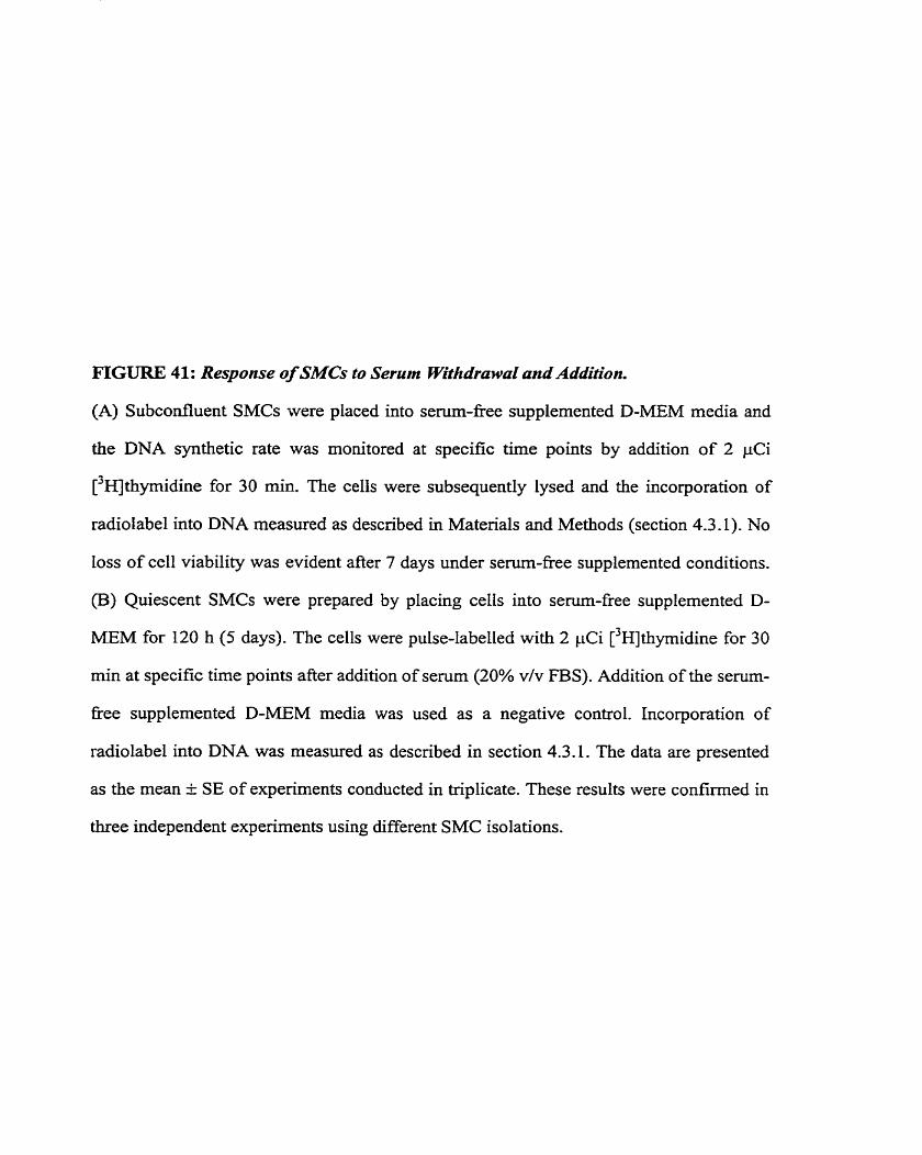
FIGURE 41: Response of SMCs to Serum Withdrawal and Addition.
(A) Subconfluent SMCs were placed into senun-fiee supplemented D-MEM media and
the DNA synthetic rate was monitored at specffic time points by addition of 2 pCi
[3HJthymidine for 30 min. The celis were subsequently lysed and the incorporation of
radiolabel into DNA measured as described in Materials and Methods (section 4.3.1). No
loss of ce11 viability was evident after 7 days under serum-fiee supplemented conditions.
(B) Quiescent SMCs were prepared by placing cells into serurn-fiee supplemented D-
MEM for 120 h (5 days). The cells were pulse-labelled with 2 pCi [3~]thyrnidine for 30
min at specific tirne points after addition of serum (20% v/v FBS). Addition of the serum-
fi-ee supplemented D-MEM media was used as a negative control. Incorporation of
radiolabel into DNA was measwed as described in section 4.3.1. The data are presented
as the mean h SE of experiments conducted in triplicate. These results were confumed in
three independent experiments using different SMC isolations.
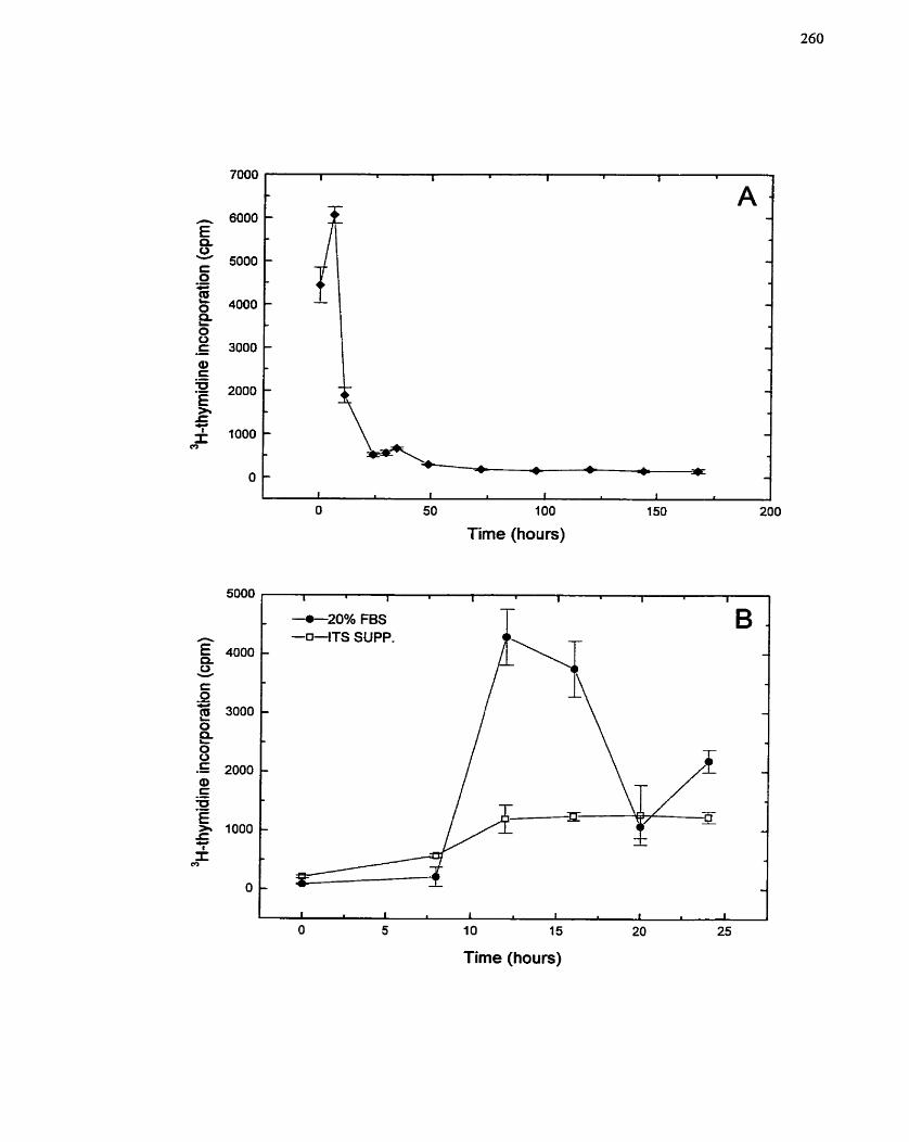
100
Time (hours)
-20% FBS T
Tirne (hours)
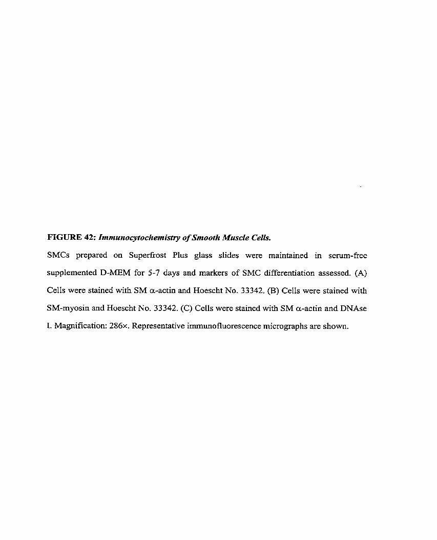
FIGURE 42: Immunocytochernistry of Smooth Muscle Cells.
SMCs prepared on Superfrost Plus glass slides were maintained in serum-free
supplemented D-MEM for 5-7 days and markers of SMC differentiation assessed. (A)
CelIs were stained with SM a-actin and Hoescht No. 33342. (B) Cells were stained with
SM-myosin and Hoescht No. 33342. (C) Cells were stained with SM a-actin and DNAse
1. Magnification: 2 8 6 ~ . Representative immunofluorescence micrographs are shown.
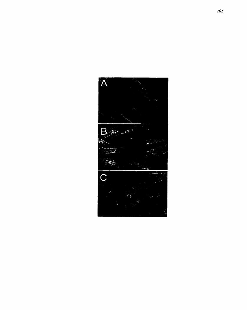
stirnulated (5% v/v FBS) cells (Figure 43) demonstrated a change in the morphology of
the SMCs as they responded to serum and re-entered the ceIl cycle,
To determine the responsiveness of SMCs to individual growth factors, both RNA
and DNA synthesis studies were carried out with AngII (10-" to IO-' M) and FBS (0.5 to
20% v/v). AngIl signifrcantly increased the rate of both RNA and DNA synthesis in
porcine SMCs (Figure 44B,D), although the magnitude of activation was considerably
Iess than the increase in RNA and DNA synthesis observed with FBS stimulation (Figure
44A,C). Interestingly, increasing concentrations of AngII continued to augment the
response (Figure 44B,D), while higher concentrations of FBS (greater than 5%) appeared
to have an inhibitory effect on RNA synthesis (Figure 44A). This may be attributed to
increasing concentrations of inhibitory molecules within the FBS. Finally, other growth
factors and stimuIating agents were tested for their ability to stimulate DNA synthesis in
porcine SMCs. Relative to FBS administration, IGF-1 and PGEz had rnoderate but
significant effects on [3~]thymidine incorporation, while thrombin had positive but not
significant effects (Figure 45). These results demonstrate that porcine SMCs are
responsive to a broad range of growth factors, and that individual growth factors have
distinct efficacy with respect to SMC growth. The potential exists, therefore, for a
synchronous additive effect on SMC growth between growth factors if the appropriate
combination or conditions is achieved.
7.2.4.2 Growth response of SMCs to PGE2
Of the growth factors and stimulating agents tested above, one of the more
interesting and somewhat surprising findings was that an eicosanoid was abIe to increase
DNA synthesis (Figure 45). In a variety of ce11 systems, eicosanoids/prostanoids, in
particular PGE:, and TxA2, have exhibited both inhibitory and stimulatory effects on cell
growth and proiiferation (Florio er al. 1994; Johnson et al. 1995; Lei & Deng 1989;
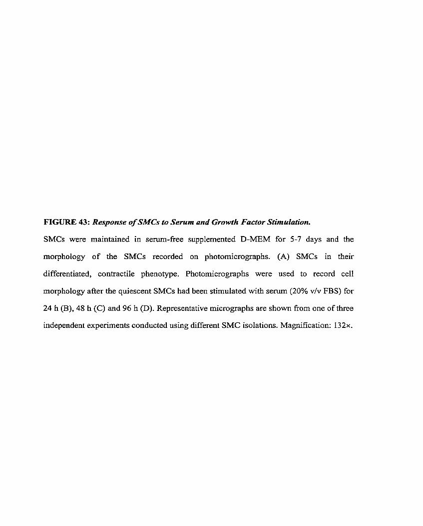
FIGURE 43: Response of SMCs to Serum and Growth Factor Stimulation.
SMCs were maintained in sem-f iee supplernented D-MEM for 5-7 days and the
rnorphology of the SMCs recorded on photornicrographs. (A) SMCs in their
differentiated, contractile phenotype. Photomicrographs were used to record ce11
morphology after the quiescent SMCs had been stimulated with semm (20% v/v FBS) for
24 h (B), 48 h (C) and 96 h @). Representative micrographs are shown fiom one of three
independent experiments conducted using different SMC isolations. Magnification: 1 3 2x.
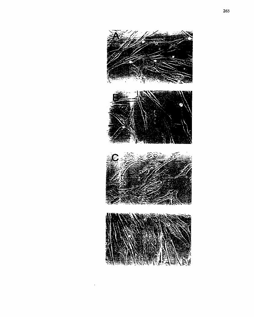
FIGURE 44: Response of SMCs to Serum and Angiotensin II ,
SMCs maintained in sem-£i-ee supplemented D-MEM for 5 days were treated with
either senun (0.5 to 20 % v/v FBS) (A, C) or angiotensin II (AngII) (10-I2 to 10" M) (B,
D) and the growth response monitored. The incorporation of [3131uridine (6 h incubation)
and [3HJthyrnidine (48 h incubation) into trichioroacetate-precipitable matenal after
addition of semm or AngII was used as an indication of RNA (A, B) or DNA (C, D)
synthetic activity. The incorporation rate of untreated cells was set to 100%. The data are
presented as the mean t SE of experiments conducted in triplicate. These results were
confirrned in three independent expenments using different SMC isolations.
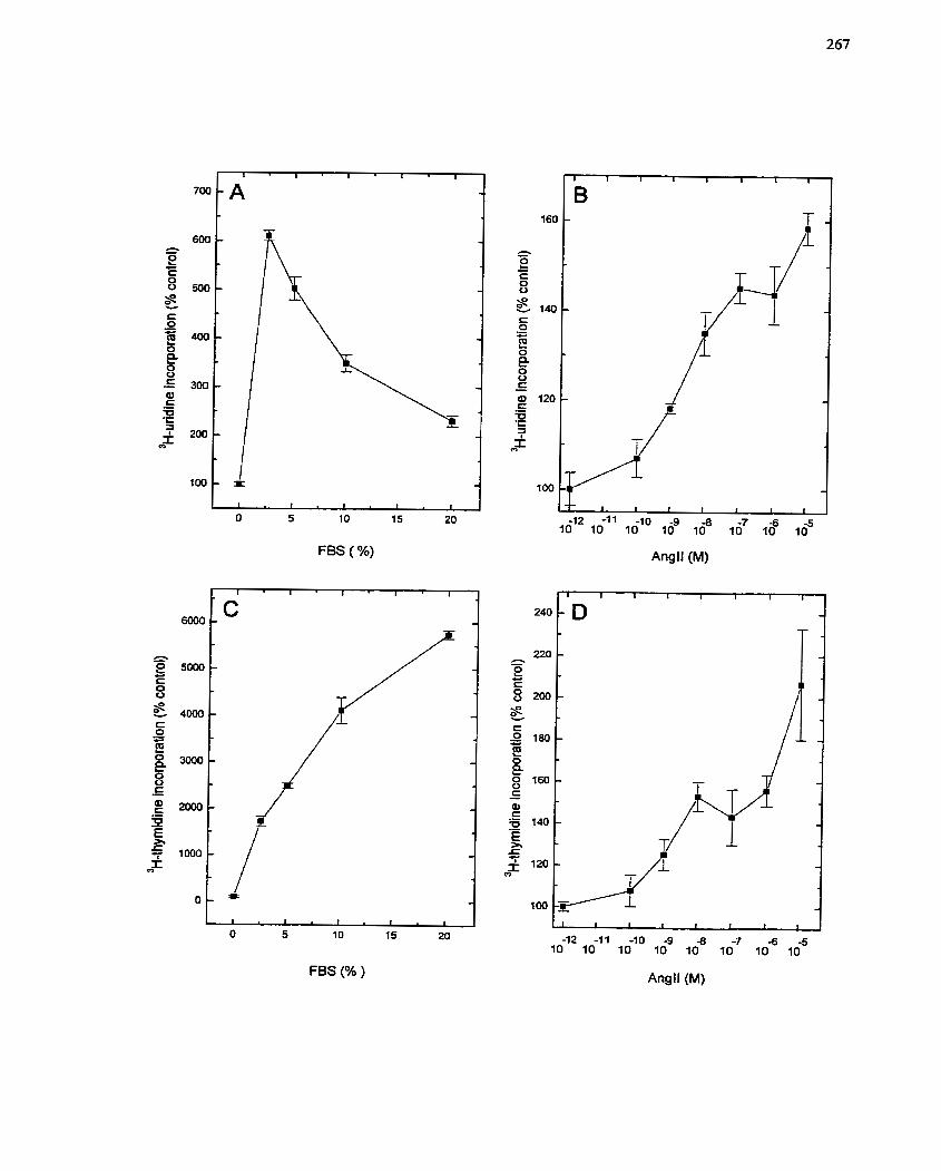
FBS ( %)
O 5 1 O 15 20
FBS (% )
Angll (M)
Angll (M)
FIGURE 45: Response of SMCs tu a Specfrum of Growth Factors and Growth
Srimulafing Agents.
SMCs maintained in serum-fiee supplemented D-MEM for 5 days were treated wiîh a
vaneîy of mitogenic agents and the growth response monitored. The incorporation of
[ '~ l th~mid ine (48 h incubation) into trichloroacetate-precipitable material afier addition
of s e m (5% v/v FBS), IGF- 1 (IO-' M), thrombin (10-~ M) or PGEz (1 0" M), was used
as an indication of DNA synthetic activity. The incorporation rate of untreated cells was
set to 100%- The data are presented as the mean F SE of experiments conducted in
triplicate. These results were confirmed in two independent expenrnents using different
SMC isolations. Cornparisons were made between untreated SMCs and SMCs treated
with growth stimulating agents using the Student's t-test (*, p c 0.05).
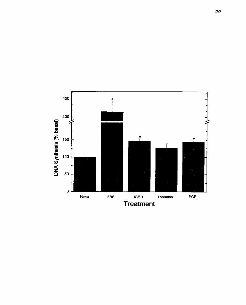
None FBS IGF-1 Thrombin PG E,
Treatment
Morinelli et ai. 1994; Owen 1986; Pasricha et al- 1992; Sachinidis et al. 1995; Sjolund et
al. 1984). For example, TxAl has positive growth effects in SMCs (Morinelli et al. 1994;
Sachinidis et al, 1995), while PGEz has had both stimulatory and inhibitory effects in
SMCs (Owen 1986; f asricha et al. 1992; Sjolund et al- 1984). It kvas o f interest then to
further define the response of SMCs to PGE? considering that previous work in the
Iaboratory had established that PGs were a part of the growth stimulatory pathway of
AngIl (Saward 2000).
To confirm that SMCs were capable of responding to PGE2, PGEz (IO-" to 10~'
M) was administered to SMCs and the rates of EüVA and DNA synthesis monitored at 6 h
and 48 II, respectively. A second PG species, PGIr, was employed in parallel as a
negative control for the growth response. Increasing concentrations of PGE2 were able to
significantly stimulate the SMC up take of [)HI uridine and ['HI thymidine (Figure 46A,B),
with a more abrupt concentration response observed in the DNA synthesis study. In
contrast, if PGEz was administered concurrently with 1ow dose FBS (1% v/v), a decrease
in the growth response was observed (Figure 46C). These data indicate that the growth
state of the SMCs is very important in detemining the response of the celIs to PGE2.
These findings were in agreement of the studies done by Owen (1 986) and the hypothesis
put forth by Schror & Weber (1997).
To verify that the SMC response to PGEl was due to a specific association with
PGE? receptors, Iigand binding was assessed (Figure 47). Unlabelled PGEz was used to
competc for [%~]PGE~. This assay demonstrated that increasing concentrations of PGEz
resutted in increased numbers of PGEl molecules bound to the receptor (Figure 47).
Furthemore, cornpetition with a different PG species (eg. PGI,, PGFz,) did not decrease
PGE2 binding (data not shown). Additionally, a specific receptor antagonist for the EPI
rcccptor (SC5 1322) was uscd to dctcm~ine whcthcr the growth responsc of SMCs to
F I G U R E 46: Response of SMCs to Prostaglandin Er.
Quiescent SMCs were treated with prostaglandin E2 (F'GE2) or prostacyclin/prostaglandin
I2 (PG12) and the growth response monitored. The incorporation of uridine (6 h
incubation) and [3HJthymidine (48 h incubation) into trichloroacetate-precipitable
material after addition of PGEr and PGIÎ (IO-'' to 105 M) was used as an indication of
RNA (A) or DNA (B) synthetic activity. (C) The incorporation of [31Xjthymidine (48 h
incubation) into trichloroacetate-precipitable material after addition 1 % (v/v) FBS and
PGE2 simultaneously over a concentration range (10-'O to IO-' M) was used as an
indication of DNA synthetic activity. The incorporation rate of untreated cells was set to
100%. The data are presented as the mean + SE of experiments conducted in triplkate.
These results were confirmed in three independent expenments using different SMC
isolations. In (C), cornparisons were made relative to FBS treated cells (*, p c 0.05) using
the Student's t-test.
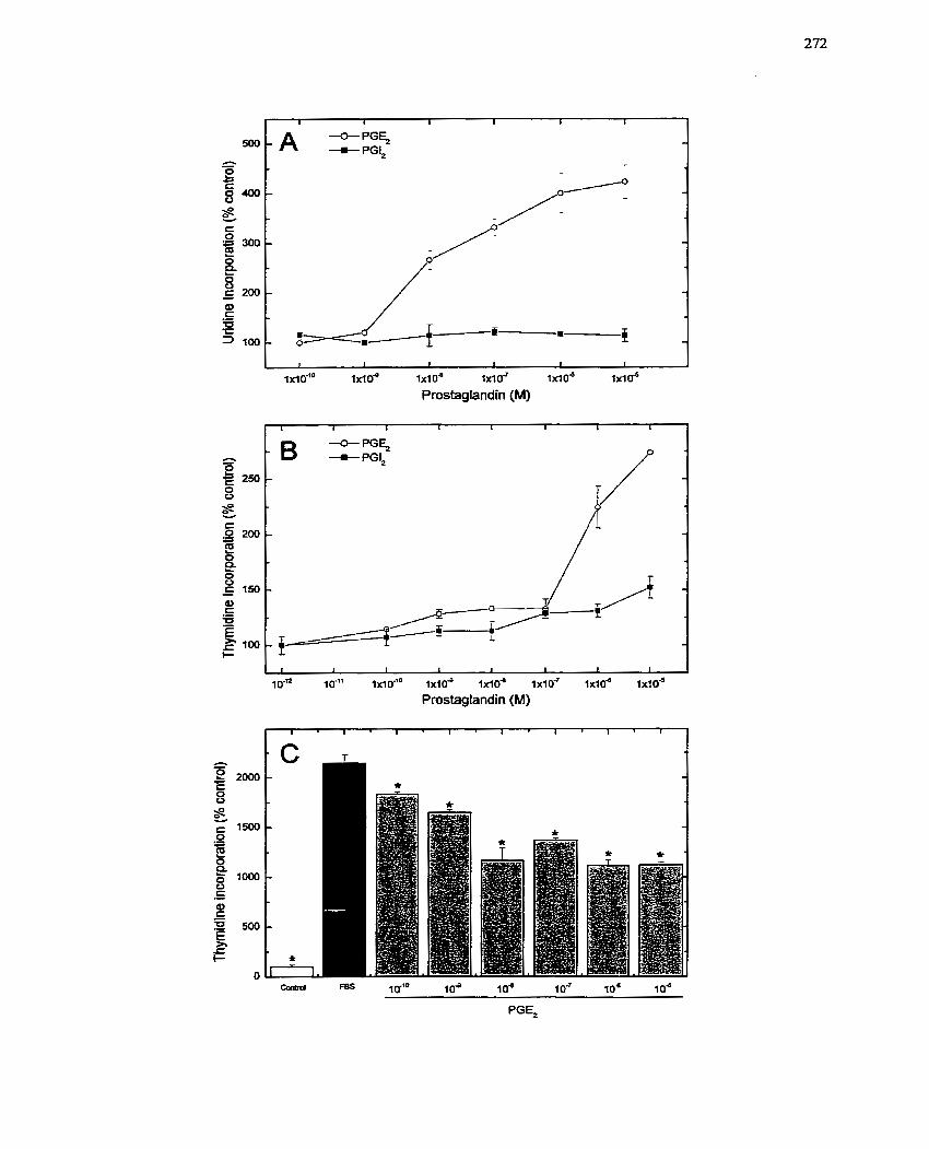
A *PG+ -m- PGI,
i
I 1 1 1 1 1 1
I ~ I O - ~ O 1 x l d 1x1 0" lxluT 1 x 1 0 ~ ixloJ
Prostaglandin (M)
1 I I r 1 1 I 1
-c- PG+ B +Pa,
~ 4 -
% A- ...- ;
1 1 1 l 1 1 1 I
IO-= IO-li 1xio9 1 x 1 0 ~ O - l d o d lxlob
Prostaglandin (M)
PGE,
F I G U R E 47: Prostaglandin E2 Specificity for its own Receptor During the Gro wth
Response
Labelled PGEz (O to 300 nM) was added to quiescent SMCs and subsequently incubated
at room temperature for 20 - 30 min. Non-specific ligand binding was assessed in a
parallel experiment in the presence of 10-~ M unlabelled PGEt. Bound PGEz was then
plotted vs. concentration. The results were confirmed in three independent experiments.
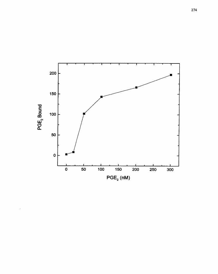
PGE, (nM)
PGEl \vas riiediated by that rcceptor subtypc. RNA and DNA synthesis assays were uscd
to no nit or the rcsponse to PGE2 in the presence o f SC51322 which \vas administered to
the SMCs 1 O niiti prior to PGE2. Only the tiighest concentration o f SC5 1322 tested (10"
M) was able to dccrease PG E2-dependont RNA synthesis ( Figurc 48 A,B), suggesting that
this rcceptor subtype is not likely involvcd in the PGE2 growth response in SMCs.
The hfITT assay was used to confirrn that PGE? activated thc growth response o f
SMCs- FBS was used as a positive control for growth stimulation and other PG species
were used as negative controls. Over 96 h, with assays conducted every 24 h, FBS
significantly increascd the conversion of MTT by SMC mitochondria (Figure 49A). PGE?
also stimulated increasing conversion of MTT, but to a lesser extent (Figure @A).
Neither PGIl nor PGF?, had a stiniulatory effcct, even after 96 h o f incubation. The one
limitation o f tl-ic MTT assay is that it cannot distinguish between hypertrophie and
hyperplastic gowth . As a result, cell number was also quantified by Coulter counting
after treatnient with PGEi (10" M) and FBS (10% v/v). The numbcr o f SMCs was only
increased significantly after 96 h incubation with PGEz compared to the increase seen at
48 h with the FBS stimuIation of SMCs (Figure 49B). The time framc of the PGE2
responsc is siniilar to that obscrvcd for the MTT conversion assay.
Othcr paranictcrs of cc11 growth wcrt? used to confimi t l ~ c findings of the RNA
and DNA synthcsis cxpcrirncnts. inimunostaining of SMC rnonolaycrs trcated with PGE?
rcvcalcd incrcascd PCNA cxprcssion at 48 II (Figurc SOC). Intcrcstingly, not al1 PGEz-
trcated cclls c'cprcsscd PCNA, unlike thc data obtaincd aftcr administration o f FBS (10%
d v ) (Figurc 50B). Immunostaining for BrdU incorporation at 72 h rcvealcd a siniilar
distribution of activatcd cclls betwccn PGE2-trcatcd and FBS-treatcd SMCs (Figure 50D-
F). Thcsc rcsiil~s confirm that PGE, is capüblc of cliciting a growth rcsponse in SMCs,
but also indicatc tliat only a s i ~ b s c ~ ofcclls arc rcsponsivc to tliis agent.
FIGURE 48: In volvement of a Specific Prostaglandin Receptor Subtype in
Prostaglandin-mediated SMC Gm wth.
Quiescent SMCs were treated with PGEz (10" M) in the presence or absence of SC5 1322
(IO-' to 1od5 M). The incorporation of [3~]uridine (6 h incubation) and [3~thymidine (48
h incubation) into trichloroacetate-precipitable rnaterial after addition of both agents was
used as an indication of RNA (A) or DNA (B) synthetic activity. The incorporation rate
of DMSO treated cells was set to 100%. The data are presented as the mean t SE of
experiments conducted in tnplicate. These results were corfiirmed in three independent
experirnents using different SMC isolations. Cornparisons were made relative to PGEz
treated SMCs using the Student's t-test (*, p < 0.05).
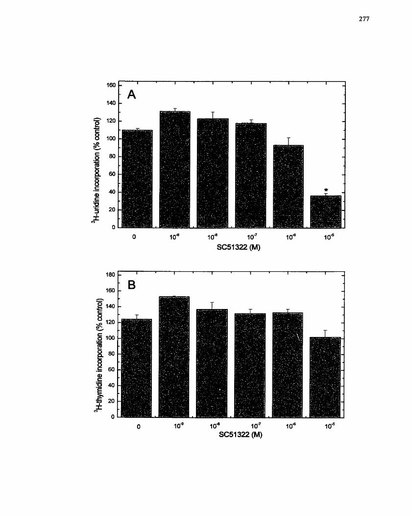
O 1 0" 1 0" 1 O-' 1 O" 1 O"
SC51 322 (M)
O 1 0" 1 O" 1 O-' 1 0" 1 O" SC51 322 (M)
F I G U R E 49: Response of SMCs ro Serum and Prostaglandins: Uypertrophic vs.
Hyperplastic Gro wth.
(A) Ce11 growth was measured using the MTT asaay as described in Materials and
Methods (section 4.3.3). The assay was conducted every 24 h over 4 days after addition
of senun (10% v/v FBS) or prostaglandins (PGE2, PGIz and PGF2,) (10'~ M) to quiescent
SMCs. The data are presented as the mean f SE of at least three separate experiments
conducted in trïplicate. (B) Ce11 number was assessed using a Coulter Counter. Quiescent
SMCs were stimulated with serum (10 % v/v FBS) or PGE2 (10'~ M), and ce11 number
quantified 48 h and 96 h after treatment as described in section 4.3.4. One of two
experiments is show, both of which exhibited a similar response. Each data point was
counted in triplkate fiom samples prepared in duplicate. Cornpansons were made relative
to untreated cells using the Student's t-test (*, p c 0.05).
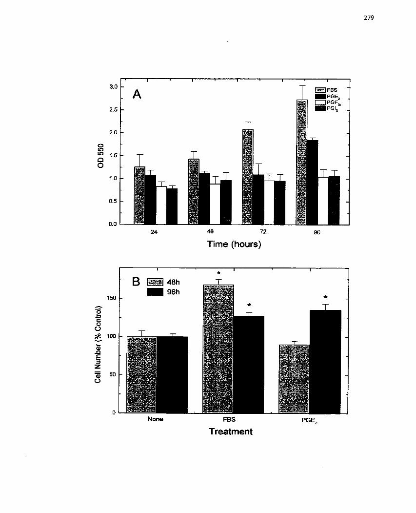
24 48 72
Time (hours)
None FBS
Treatment
FIGURE 50: Response of SMCs to Seruni and Prostaglandins: Other Growth
Parame fers.
(A to C ) Quiescent SMCs were treated with eithcr semm (10 % v/v FBS) or PGEz (10"
M) for 48 h. lmmunocytochernistry was used to monitor PCNA (proliferating ce11 nuclear
antigen) expression as described in section 4.7.3. (A) no treatment, (B) 48 h FBS
treatment and (C) 48 h PGE2 treatment. Representative micrographs are shown.
Magnification: 140x. @ to F) Quiescent SMCs were treated for 72 h with either serum
(10 % V/V FBS) or PGEZ (10'~ M). Before harvest, cells were pulse-labelled for 2 h with
50 p M BrdU. Imrnuno~ytoche~stry was employed to visualize BrdU incorporation as
described in section 4.7.3. (D) no treatrnent; (E) 72 h FBS treatment and (F) 72 h PGE2
treatment. Representative rnicrographs are shown. Magnification: 140x. These results
were confirmed in two and three independent experiments, respectively, using different
SMC isolations, al1 of which exhibited similar patterns of response.

lmmediate early genes (eg. c-fos) are activatcd upon stimulation of cclls witli
growth promoting agents (Rivera & Greenberg 1990; Woodgett 1989). To derennine
whether PGEz could induce c--os gene expression, RT-PCR analysis of c-fos n1RNA
IeveIs was examined over a time course of 2 h, Addition of PGEz to SMCs resulted in a
rapid rise in c-fis mRNA levels that peaked at 30 min and returned to basal levels by 2 11
(Figure 51). These data support the contention that PGEl is a positive modulator of SMC
growth.
7.2.4.3 Activation of MAP kinase and p21-Ras by PGEz
Many intracellular signaling pathways are activated upon stimulation by growth
factors and tyrosine phosphorylation cascades are commonly involved (Ahn et cd. 1992).
As such, the tyrosine phosphorylation of proteins was exarnined after PGE2 ( 1 0 % l )
stimulation. Over a time course of 8 h, PGE2 was able to stiniulate the phosphorylation of
a variety of proteins, most noticeably' proteins of 125-, 85- and 42/44-kDa (Figure QA).
Moreover, imn~unocytochemistry revealed that after 1 5 min of PGEz ( 1 O-' M) treatment
there was an alteration of the pattern of tyrosine phosphorylated proteins with a p a t e r
number of membrane localized associations (Figure 52B,C). Sincc one of the tyrosine
phosphorylated bands obscrved in Figure 52A correIated with the niotecular mass of
MAP kinase, it was of interest to determine whetlier MAP kinasc was activated by PGE2.
Moreover, MAP kinase is one of the s ignahg molecules associatcd with growth evcnts
(Adan1 el ai- 1995; Cowley et ai. 1994; Force & Bonvcntre 1998; KhaIil & Morgan 1993;
Klcmke er al. 1997; Mansour et al, 1994; Nelson et ai. 19%). Witii an in gel MAP kinase
activity assay, it was obscrvcd that PGEz transiently stin-iulates MAP kinase ovcr a tinlc
coursc of 20 min, with pcak activation occurring bctwccn 5-10 niin (Figure 53A). A
parallcl study was conductcd using FBS as thc stiinulus, and a siiiiilar pattern of MAP
kinasc activation was obscrvcd (Figurc 53B), altliough thc niagtiitudc of activation was
FIGURE 51 : R T-PCR Anaiysis of PGErmediated c- fos Gene Expression.
Quiescent SMCs were treated with PGE2 (1U6 M). RNA was extracted at 0, 15, 30, 60
and 120 min after PGE2 addition and c-fos mRNA leveIs monitored by RT-PCR. GAPDH
expression was assessed simultaneously and served to control for RNA loading.
Molecular mass markers ((pX174 and DNA marker VI) were used to confïnn the size of
the PCR products. One representative agarose gel is shown. These results were
reproduced in three independent expenments using different SMC isolations. Appropriate
control reactions were run independently (refer to section 4.10.3).
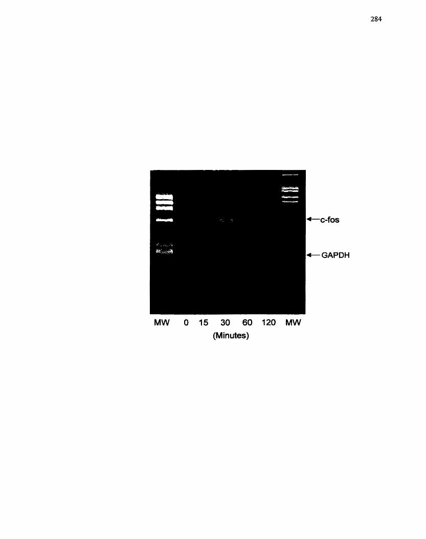
c-fos
- GAPDH
MW O 15 30 60 120 MW
(Minutes)
FIGURE 52 : Tyrosine Ph osphoryiution Stimuiated by Prostuglandin E2.
(A) Quiescent SMCs were treated with PGEz (lod M) over a time course of 8 h, and
extracts prepared according to section 4.8.3. The samples were subjected to SDS/PAGE
and Western blot analysis with an antibody specific for phosphorylated tyrosine (PY20)
(diluted 1:1000). Proteins were visualized with the ECL cherniluminescent detection
systern- MolecuIar mass markers used to identiQ the proteins are shown. The results were
reproduced in three independent experiments using different SMC isolations. 0, C)
Quiescent SMCs were treated for PGEl (10-~ M) for 15 min, and immunocytochemistry
was used to monitor SM a-actin (red) (diluted 1;400) and PY20 (green) (diluted 1: 100)
localization. (B) control SMCs with no treatrnent, (C) SMCs with PGE- 1 treatment .
Representative fluorescence micrographs are shown. Magnification: 122x.
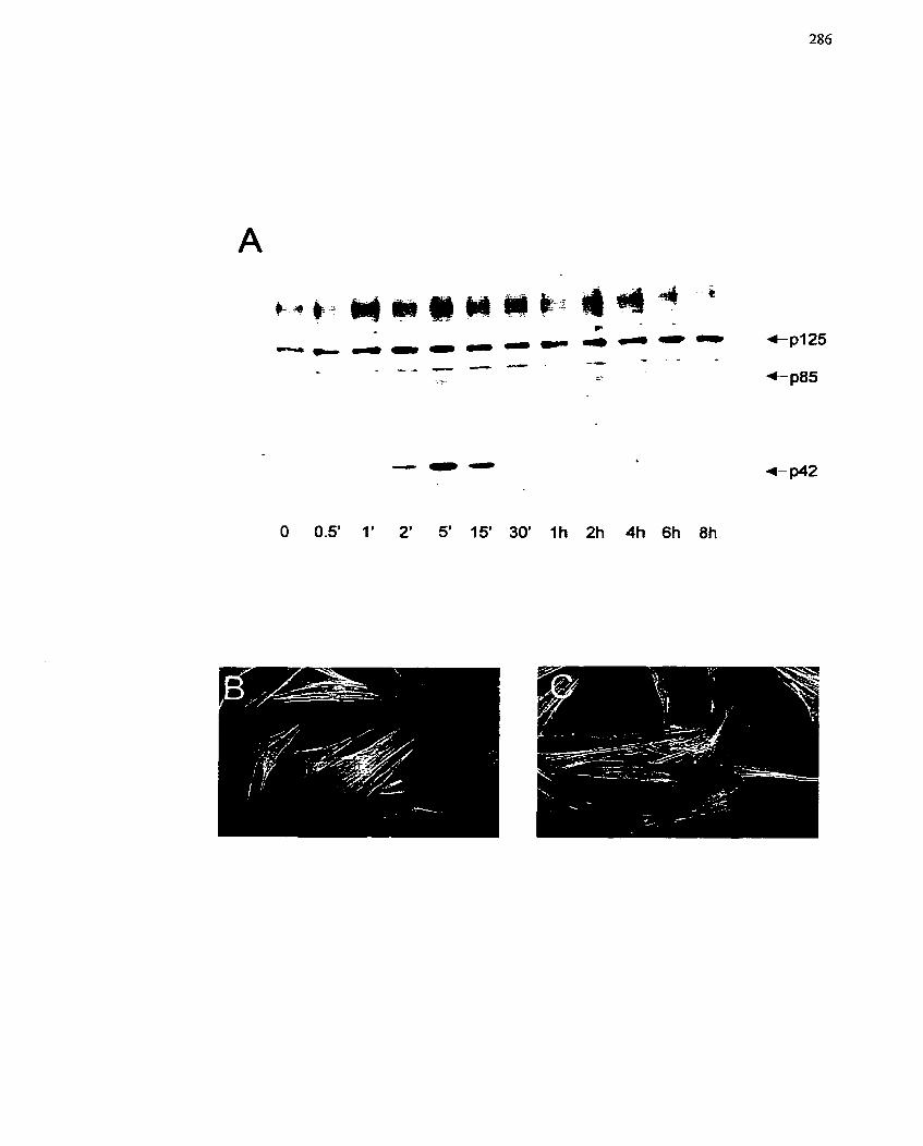
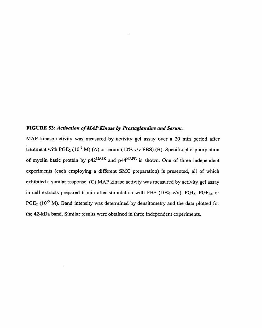
FIGURE 53: Activation of lMAP Kinase by Prostagiundins and Serum.
M A P kinase activity was measured by activity gel assay over a 20 min penod after
treatment with PGE2 (10" M) (A) or serum (10% v/v FBS) (B). Specific phosphorylation
of myelin basic protein by p42MAPK and p44MAPK is shown. One of three independent
experiments (each employing a different SMC preparation) is presented, al1 of which
exhibited a similar response. (C) MAP kinase activity was measured by activity gel assay
in ce11 extracts prepared 6 min after stimulation with FBS (10% v/v), PGI2, PGF2, or
PGEz (106 M). Band intensity was detemiined by densitometry and the data plotted for
the 42-kDa band. Similar results were obtained in three independent experiments.
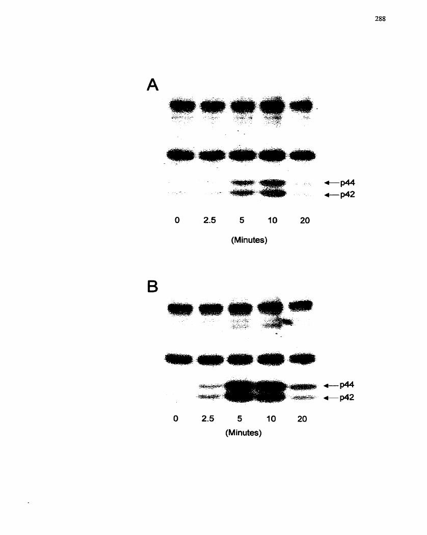
O 2.5 5 10 20
(Minutes)
O 2.5 5 10 20
(M inutes)
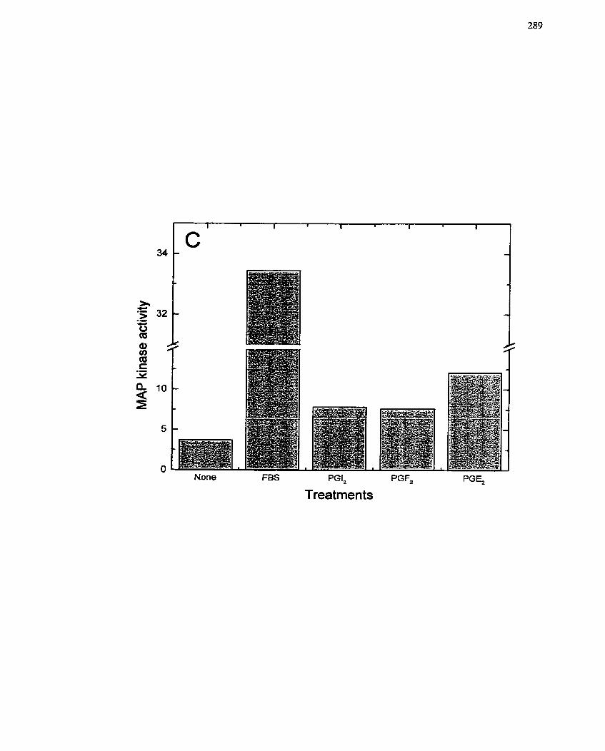
None FBS PGI, PGF,
Treatments
more intense, Interestingly, other PG species tested with respect to MAP kinase
activation, PG12 and PGF2,, were also capable of stimulating MAP kinase activation
(Figure 53C), and yet neither of these PGs was capable of significantly increasing either
RNA or DNA synthesis (Figure 46 and data not shown). These data would suggest that
MAP kinase activation, although potentially linked to growth in the case of PGE2, does
not necessarily lead to growth. This is not surprising since activation of MAP kinase is
also required for other events, such as stress and contractility, that are independent of ce11
growth (Adam et al- 1995; Force & Bonventre 1998; Khalil & Morgan 1993; Klemke et
al. 1997; Nelson et al. 1998; Watts 1998).
As a result of the above data, it was necessary to deterrnine if the PGEz-stimulated
MAP kinase activation correlated with SMC g r o h . An inhibitor of MEK (MAP kinase
kinase), PD98059, was added to SMCs 10 min prior to PGE2 stimulation. PD98059 ( 1 0 - ~
M) decreased PGEz-mediated ce11 growth as measured by RNA and DNA synthesis
(Figure 54). These results confimi the supposition that MAP kinase activation is essential
for PGE2-mediated SMC growth.
-4 second mediator of intracellular signaling that is relevant to growth of a variety
of ce11 types is p2l-Ras (Bos 1995; Maruta & Burgess 1994). This cytosolic GTP-binding
protein was also of interest considering the Iink to mono(ADP-ribosy1)ation. The
contribution of p2 1 -Ras to SMC growth was defined by correlating p2 1 -Ras activation,
specifically an increase in bound GTP, with mitogen stimulation. [3'~]~rthophosphate
labelled SMCs were stimulated with PGE2 (10-~ M), PGIl ( 1 0 ~ ~ M) and TxAl ( 1 0 - ~ M),
and celI lysates were immunoprecipitated with a specific antibody to Ras. The ratio of
GTP:GDP in the immunoprecipitates was calculated after TLC separation. The results of
this assay are prcsented in Table 3, and show that p21-Ras is not activated upon PGE2
stimulation, and neithcr is it activatcd by thc othcr prostanoids PGIî or TxAz. Thesc data
FIGURE 54: Inhibition of Cell Prolifration by the MEal inhibitor PD98059.
PGE2 (10" M)-stimulated RNA ([3~urïdine) and DNA ([3athymidine) synthesis were
rneasured in the presence of the MEK inhibitor PD98059 (1U6 M). Each data point
represents the mean f SE of at least three individual experirnents conducted in triplicate.
Values are presented relative to an untreated control which was set to 100%. These
results were confimed in two independent experirnents using different SMC isolations.
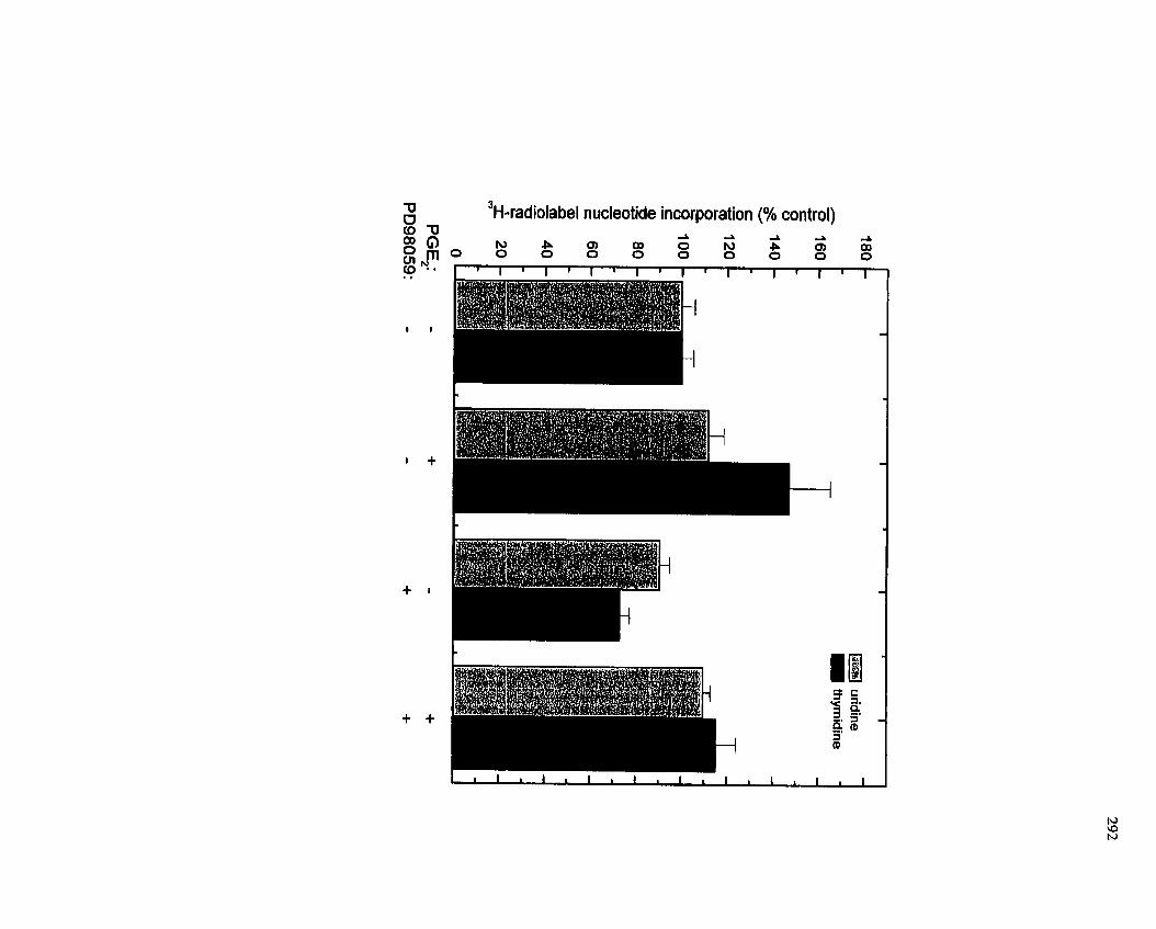
3 H-radiolabel nucleotide incorporation (% CO ntrol)
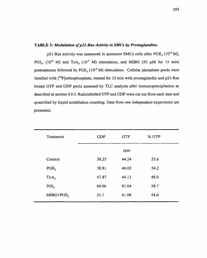
TABLE 3: Modulation of p21-Ras Activity in SMCs by Prostaglarsdins.
p21-Ras activity was measured in quiescent SMCs cells afier PGE, ( lo4 M),
PGI,, (10-~ M) and TxA, (10-~ M) stimulation, and MBG (50 pM for 15 min)
pretreatment followed by PGE, IO-^ M) stimulation. Cellular phosphate pools were
labelled with [32~]orthophosphate, treated for 15 min with prostaglandin and p2 1-Ras
bound GTP and GDP pools assessed by TLC anaIysis after imrnunoprecipitation as
described in section 4.9.5. Radiolabelled GTP and GDP were cut out fiom each lane and
quantified by liquid scintillation counting. Data fiom one independent experiment are
presented,
Treatment GDP GTP % GTP
Control
PGE,
TxA2
PGI,
MIB Gf-PGE,
indicate that p21-Ras rnay not be an important cornponent of PGEz-mediated SMC
growth,
7.2.4-4 Involvement of PI3-kinase in PGEr-dependent SMC growth
The final transducer of intracellular signaling that was exarnined in relation to
PGE2-mediated SMC growth was PI3-kinase. This enzyme has been established as an
- important component of AngII-mediated SMC growth (Saward & Zahradka 1997a).
Since PGs may be important rnediators of AngII-dependent ce11 growth (Saward 2000),
exploration of the PI3-kinase pathway was considered relevant. Furthemore, the
membrane receptor system for AnglI and PGs is similar (Le. G protein-coupled), and
both compounds are able to elicit ce11 growth responses in SMCs. In addition, PD-kinase
is an important cornponent of G protein-coupled receptor activation of MAP kinase
(Della Rocca et al. 1997; Force & Bonventre 1998; Lopez-Ilasaca et al. 1997). An
examination of the importance of this signal rnolecule was initiated by monitoring RNA
and DNA synthesis after a 10 min pretreatment of SMCs with specific inhibitors of PI3-
kinase, LY294002 (LY) and wortmannin (WM), followed by addition of PGE?. Both LY
and WM (IO-'' to IO-' M) inhibited RNA and DNA synthesis stimulated by PGEz, with
increasing concentrations of LY and WM having a significantly greater inhibitory effect
(Figure 55A,B). These data suggest that PI3-kinase is important for PGE-mediated SMC
growth.
To establish that PI3-kinase is indeed activated in SMCs by PGE2, PI3-kinase
assays were can-ied out, both in vivo and Nz vitro. The in vivo assay involved monitoring
PIP pools after ["~]ortho~hos~hate labeling of SMCs and subsequent stimulation with
PG compounds. A spectrum of PGs were assessed in this manner and included PGEz
( 1 0 ~ ~ M), T A 2 ( 1 0 - ~ M), PGll (1 o - ~ M) and PGF2, (10" M). Al1 of the PGs tested were
capable of increasing PIP3 production compared to control, untreated cells extracts
FIGURE 55 : In volvement of Pl3-Kinase irt PGE~mediated SMC Gro wth.
PGE2 (10-~ M)-stimulated RNA (A) and DNA (B) synthesis was measured in îhe
presence of varying concentrations of the PI3-kinase inhibitors wortrnannin (WM) and
LY294002 (LY) (IO-'' to IO-' M). The incorporation of [ 3~u r id ine (6 h incubation) and
[3~thymidine (48 h incubation) into tnchloroacetate-precipitable matenal after addition
of both agents was rneasured as descnbed in section 4.3.1. Values are presented relative
to maximal stimulation with PGE2 (set to 100%). The data are presented as the mean t
SE of experiments conducted in triplkate. These results were confirmed in three
independent experirnents using different SMC isolations.
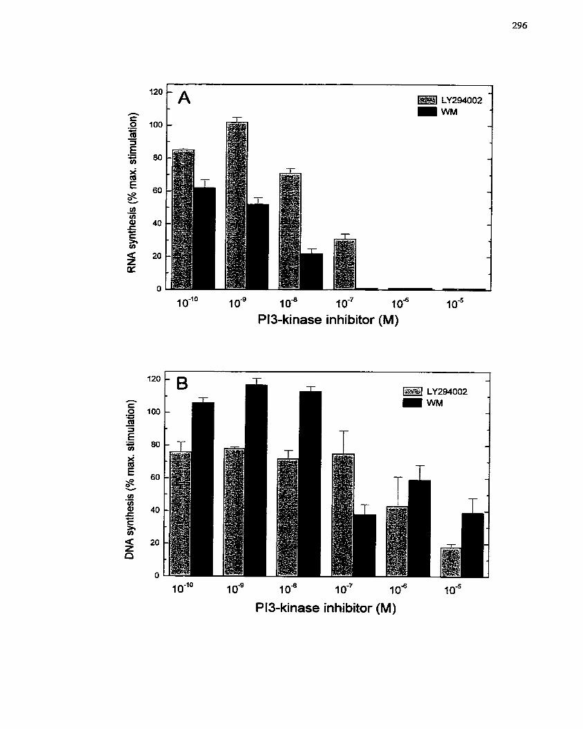
1 O-"' 1 o - ~ 1 o8 1 o - ~ 1 O" 1 0"
Pl3-kinase inhibitor (M)
1 O-Io i O-' i os 1 o - ~ 1 O" 1 O"
PI3-kinase inhibitor (M)
(Figure 56A). To assess the specificity of the P13-kinase response and to ver ib that
wortmannin was indeed inhibiting PI3-kinase, the activation of P13-kinase by PGE- 7 was
tested in the presence of wortmannin (Figure 56B). It was observed that wortmannin
inhibited PIP, generation induced by PGEz (Figure 56B). LY294002 had a sirnilar effect
(data not shoivn). An in vitro PI3-kinase assay was then employed to confimi the
activation of P13-kinase by PGs. In this assay the P13-kinase cornplex was
imrnunoprecipitated with a specific antibody to p85, the regulatory subunit of P13-kinase
and phosphorylation of PI and PIPî was identified by TLC. This experiment
dernonstrated that PGEr (10-~ M), TxA2 (IO-' MM), and PGF2, (lod M) increased the
production of HP3 (Figure 57), albeit to different degrees, and that PGE2 (IO-" to 10" M)
increased the production of PIP3 with higher concentrations having the greatest effect
(Figure 58) . These data clearly established that PU-kinase is activated by PGE2.
The temporal and spatial positioning of PI3-kinase with respect to other signaling
molecules and endpoints activated by PGE2 in SMCs was evaluated with the PI3-kinase
inhibitors, LY294002 and wortmannin. Activation of MAP kinase by PGEr was
diminished by either LY294002 or wortmannin, indicating that PI3-kinase activation is
an event that occurs before MAP kinase (Figure 59). Furthemore, RT-PCR analysis of
c-fos mRNA levels revealed that both LY294002 and wortmannin abrogated PGE2-
mediated c-fos expression (Figure 60), suggesting that activation of P13-kinase is an
important step in the expression of early growth response genes. Overall, these data
confirm that PD-kinase is an important element in PGEz-mediatcd SMC growth.
7.2.5 Discussion
This study has demonstrated that PGEr has growth stimulatory effects in porcine
SMCs. PGE2 was chosen for study because it is released on stimulation or damage of
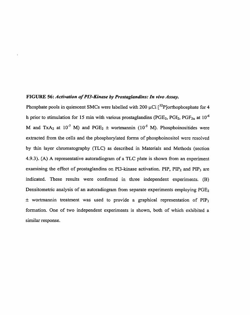
FIGURE 56: Activation of PI3-h'inase by ProstagIundins: In vivo Assny.
Phosphate pools in quiescent SMCs were labeiied with 200 pCi [32~]orthophosphate for 4
h prior to stimulation for 15 min with various prostaglandins (PGE2, PG12, PGF2, at 1 0 - ~
M and TxA2 at IO-' M) and PGE2 t wortmannin (IO-' M). Phosphoinositides were
extracted fiom the cells and the phosphorylated forms of phosphoinositol were resolved
by thin layer chromatography (TLC) as described in Matenals and Methods (section
4.9.3). (A) A representative autoradiogram of a TLC plate is shown fiom an experïment
exarnining the effect of prostaglandins on P13-kinase activation. PP, PIfi and PIP3 are
indicated. These results were confirmed in three independent experiments. (B)
Densitometric analysis of an autoradiogram fiorn separate experiments employing PGEÎ
+: wortrnannin treatment was used to provide a graphical representation of PIP3
formation. One of hvo independent experirnents is shown, both of which exhibited a
sirnilar response,
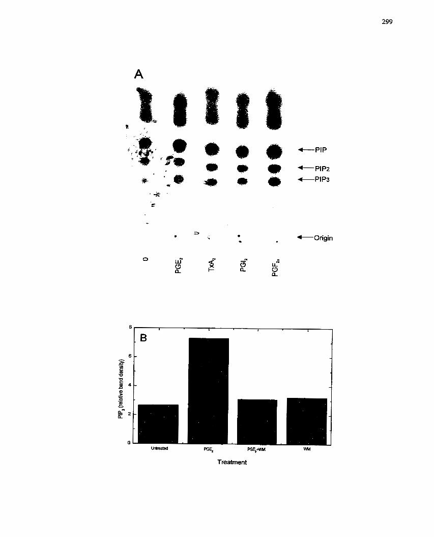
Treatment
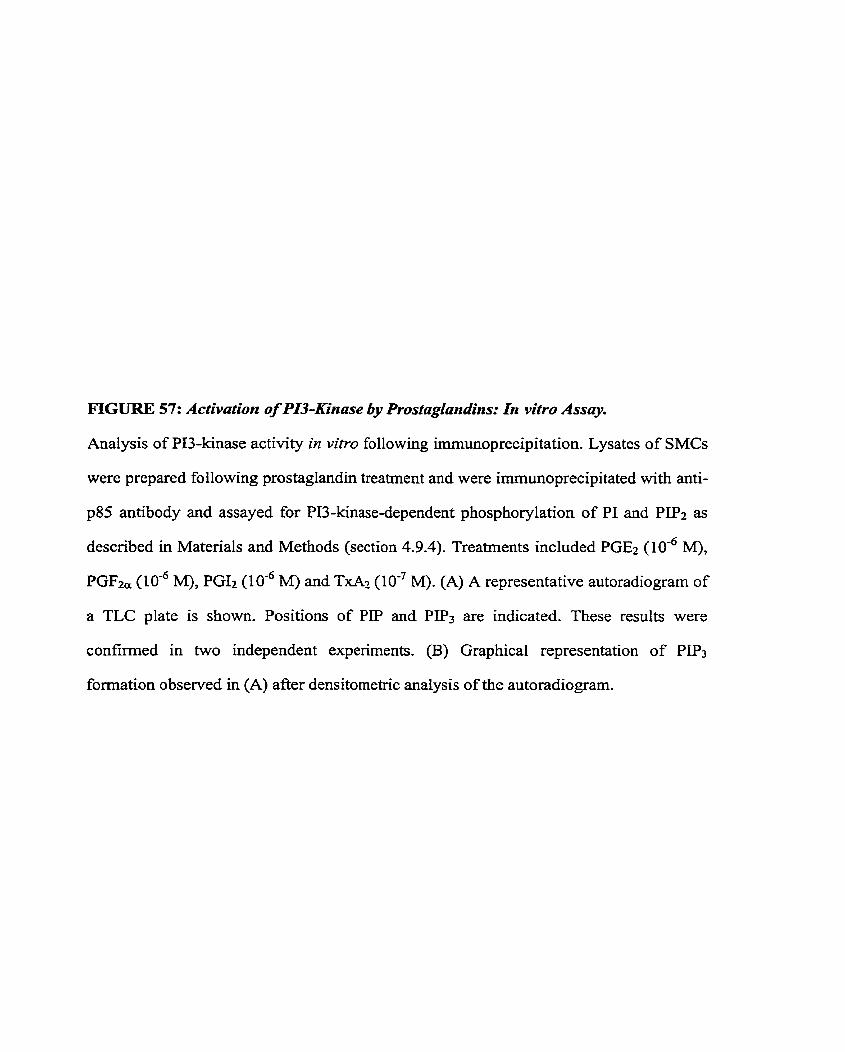
FIGURE 57: Activation of PI3-Kinase by Prostaglandins: In vitro Assay.
Analysis of PU-kinase activity in vitro foliowing imrnunoprecipitation. Lysates of SMCs
were prepared foilowing prostaglandin treatment and were irnmunoprecipitated with anti-
p8S antibody and assayed for PI3-kinase-dependent phosphorylation of PI and PIP2 as
described in Matenals and Methods (section 4.9.4). Treatrnents included PGEz (10" M),
PGFza (10-~ M), PGIÎ M) and TxAz (10'~ M). (A) A representative autoradiogram of
a TLC plate is shown. Positions of PIP and P P 3 are indicated. These results were
confirmed in two independent experiments. (B) Graphical representation of PIP3
formation observed in (A) afier densitometric analysis of the autoradiogram.
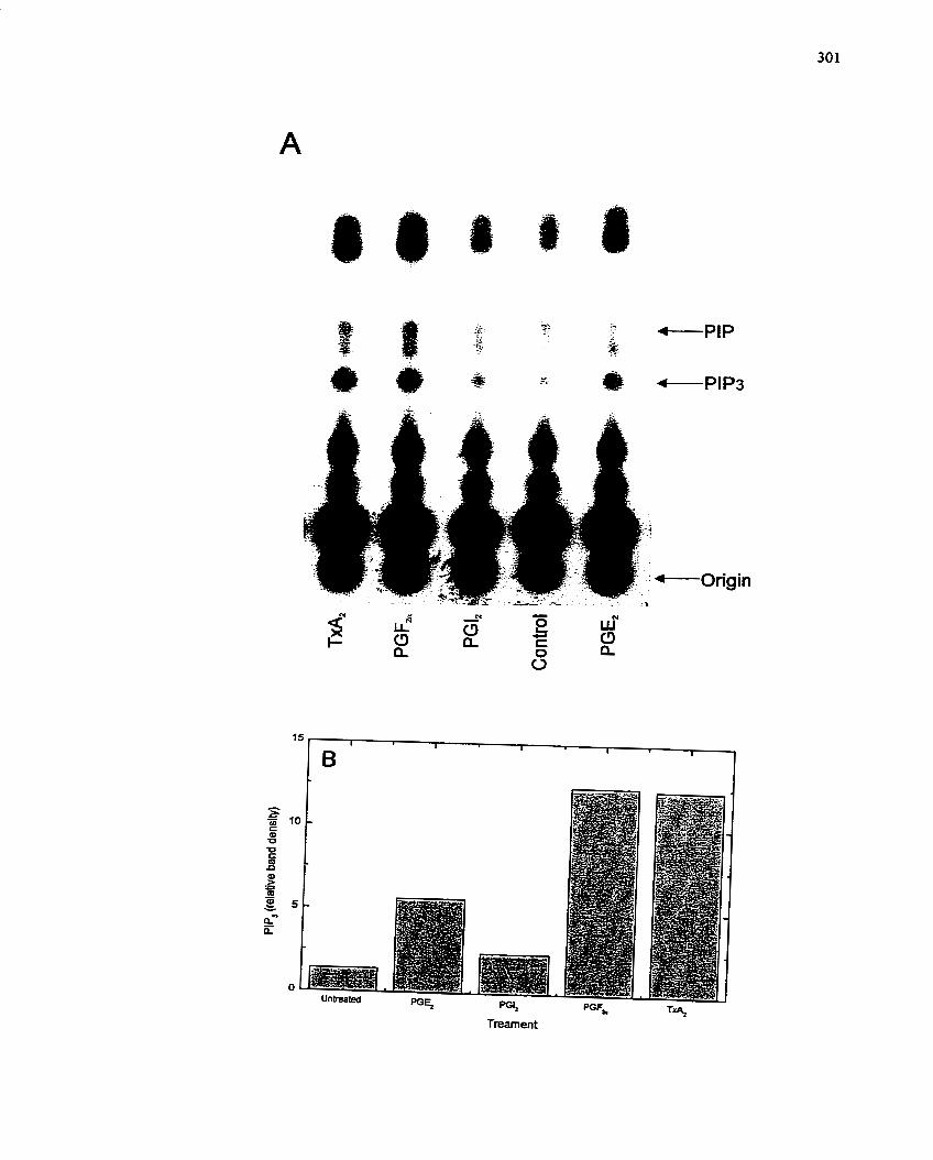
FIGURE 58 : Activrrtion of P I 3 i n a s e by Prostuglundins: Con ceiz tration Effect of
Prostaglandin E2.
Analysis of PI3-kinase activity in vitro following immunoprecipitation. Lysates of SMCs
prepared following prostaglandin treatment were immunoprecipitated with anti-p8 5
antibody and assayed for PI3-kinase-dependent phosphorylation of PI and PIPZ as
described in Materials and Methods (section 4.9.4). Treatment was PGEz (10-l0 to 1 0 - ~
M)- (A) A representative autoradiogram of a TLC plate is shown. Positions of P P and
P P 3 are indicated. These results were c o n f i e d in two independent experirnents- (B)
Graphical representation of PIP3 formation observed in (A) afier densitometric analysis
of the autoradiogram.
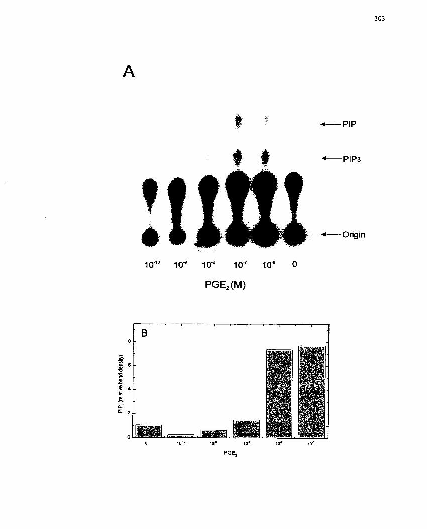
- PIP
FIGURE 59: Effect of PI3-Kinase lnhibitors on PGErme&ated Activation of MAP
Kinase.
MAP kinase activity was measured by activity gel assay after 6 min of stimulation with
PGEz (10-~ M) and pnor pretreatrnent (10 min) with PU-kinase inhibitors wortmannin
(WM) (105 M) or LY294002 (LY) (10" M). (A) An example of the activity gel is shown
with the specific phosphorylation of rnyelin basic protein by p42MAPK and p44 MAPK
identified. (B) Band intensity was determined by densitometry and the data plotted for the
combination of 42- and 44-kDa bands. One of three independent experiments is presented
from different SMC isoIations, al1 of which exhibited the same response.
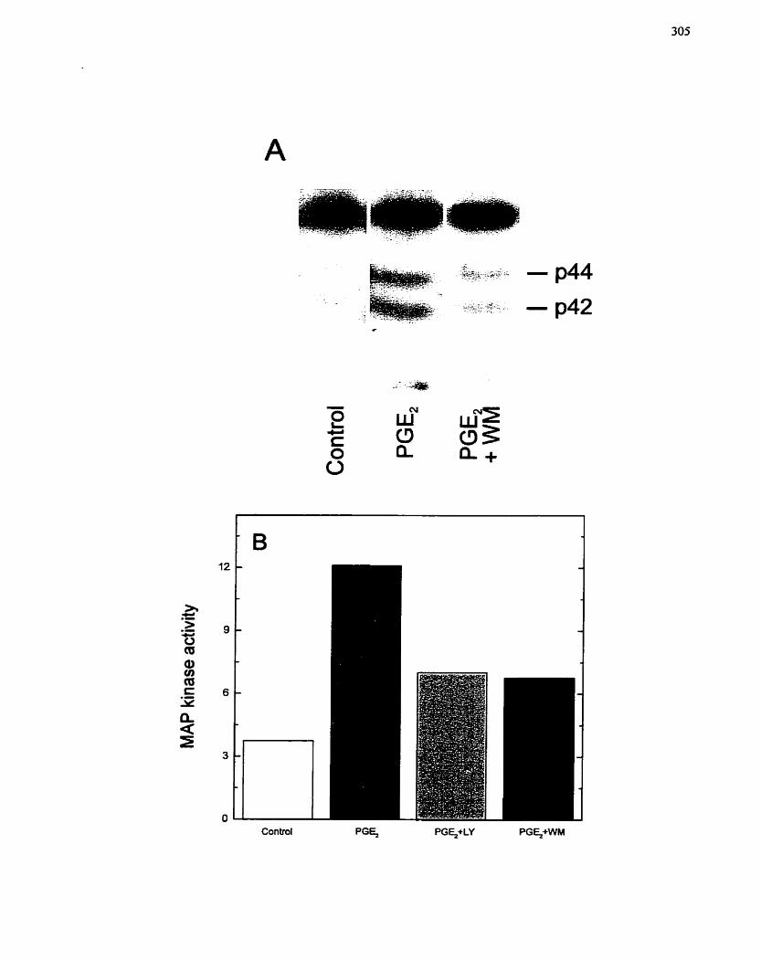
FIGURE 60: Effect of PI3-Kinase Inhibitors on PGEmediated Expression of c-fos.
(A) Quiescent SMCs were pretreated for 10 min with the PU-kinase inhibitor
wortmannin (10" M) and subsequently treated with PGEr (IO" M) for 15 min.
RNA was extracted and c-fos gene expression assessed by RT-PCR. Molecular mass
markers (9x174 and DNA Marker VI) were used to confirm the size of the PCR
products. One representative agarose gel is shown. These results were confimed in two
independent experiments using different SMC isolations. (B) Band intensities h m a
separate experiment using the P13-kinase inhibitor LY294002 (LY) (105 M) were
quantified by densitometry and the data plotted to show the intensity of the c-fos band
relative to treatment. One of two independent expenments is shown, both of which
exhibited a similar response.
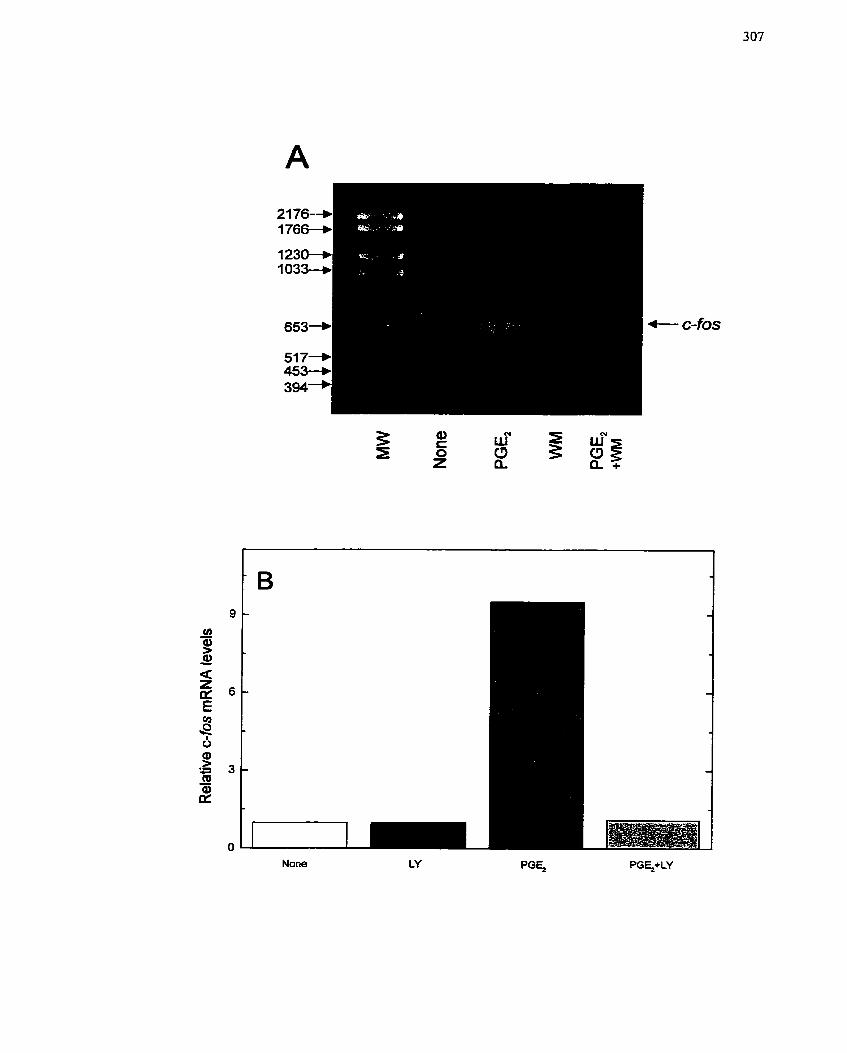
endothelial cells, vascular SMCs and tissue macrophages. and can accumulate during the
inflammatory responses that occur following injury to the vasculature (Kennedy et al.
1982; Leslie & Watkins 1985; Revtyak et al. 1987; Wendling & Harakal 199 1).
Furthemore, this Iaboratory and others have demonstrated that PGEz levels are increased
after stimulation of SMCs and other cells with AnpII (Ohnaka er al. 2000; unpublished
observations from Saward, Yau, Thomas & Zahradka; Saward et al. submitted) and
PDGF (Graves el al. 1996; Weksler 1987), both of which have known migratory and
proliferative effects on SMCs, and both of which have been irnpficated in the formation
of a neointimal lesion after vascdar injury (Casterella & Teirstein 1999; Cercek et a!-
199 1 ; Liu et al- 1989; Wilson et al- 1999)-
Prostanoids are potentially important mediators of normal vascular healing or
abnormal vascular responses following vascular damage (i-e. neointimal thickening
following to balloon injury). Schror & Weber (1997) have postulated that some species of
PGs are bimodal in nature, having both the ability to stimulate SMCs to enter the ce11
cycle and divide in response to tissue injury in order to facilitate tissue repair, and the
ability to prevent uncontrolled growth of SMCs by interfering with the progression of
ceils through the ce11 cycle, especially in S phase. This hypothesis is certainly borne out
by the work of Owen (1986) who demonstrated tliat the effect of PGEl on SMCs is
dependent on the phase o f the ce11 cycle when it is added. In quiescent cells, PGEl
enhanced DNA synthesis, while in growing cells PGEi inhibited ce11 proliferation (DNA
synthesis). That particular study suggested that in growth-arrested SMCs (such as would
be found in the normal vasculature), PGEt may have the ability to stimulate DNA
synthesis (eg. during a possible repair procedure aftcr injury), whereas in growing
vascular SMCs (as would be secn under conditions of athcrosclcrotic plaque formation or
aftcr activation of SMCs by in jury) PGEi can funciion as an inhibitor of DNA synthcsis.
The studies reported liere show PGE? also exhibits growth state-dependent effects on
SMCs, thus supporting and extending the hypothesis postulated by Schror & Weber
(1997). When PGE2 was added to SMCs primed with 1% FBS, PGE? was able to inhibit
the uptake of [3~]thymidine, an indicator of the rate of DNA synthesis (Figure 46C). In
contrast, PGEl added alone to quiescent SMCs stimulated RNA and DNA synthesis
(Figure 46A,B). To support the above finding related to DNA synthesis, PGEz was also
capable of stimulating PCNA expression, BrdU incorporation, increased c-fos mRNA
Levels and increases in ce11 number (Figures 49, 50, 51), al1 indications of cells
undergoing proliferation. However, only a subpopulation of SMCs appeared to respond
to PGE2 based on the observations from BrdU incorporation and PCNA iinmunostaining
(Figure 50). This would certainly account for the restricted mitogenic potential of PGE2
in cornparison to FBS (Figure 49). Whether the SMCs responsive to PGEz represents a
distinct subpopulation of SMCs remains to be determined. In Light of the contrasting
reports in the Iiterature concerning the mode ofregulation of SMC proliferation by PGs
of the E family, it is interesting to note that the majority who reported an inhibitory effect
employed PGEin in conjunction with a stirnuiator of growth (Fan et al. 1997; FIorio et al.
1994; Hayashi el al. 1992; Johnson et al. 1995; Loesberg et al. 1985; Nilsson & Olsson
1984), while those who saw a stimulatory effect, added PGE,,? aIone (Owen 1986;
Pasricha et ai. 1992). Only one publication has to this point examined SMCs in both
growth states (Owen 1986). Thus, although much of the work appears to be contradictory
and somewhat controversial, the findings observed here support evidence for a bimodal
effect by PG's of the E family that is dependent on SMC growth status. The relevance of
PGE2 to intimal lesion formation may therefore be two-fold: i ) during the course of
intimal Iesion formation, PGEz will act first to stimulate tissue rcpair, and ii) foliowing
activation of SMCs andor completion of rcpair, PGEz inhibits cell cycle re-cntry. Thus,
restenosis may occur in those individuals where PGEl does not inhibit SMC proliferation
and consequently a much thickened neointima will form, Alternatively, the bifunctional
effects of PGEz could be the result of the differential expression and function of PGEz
receptors that may be found on populations and subpopulation of SMCs. This possibility
remains to be explored.
PGEz receptors belong to the class of G protein-coupled receptors, and 4 isotypes
(EPi4) have been identified. EP receptors mediate a wide range of biological actions
including contraction and relaxation of SMC, induction and inhibition of neurotransmitter
release and inhibition of inflarnmatory rnediator release. (Coleman et al. 1987; Coleman
et al- 1994). Each receptor subtype is associated with a particular cellular response and
the activation of unique signaling molecules and rnediators (Coleman et al, 1987;
Coleman et al. 1994). The EPr receptor is predominant in smooth muscle of the trachea,
gastrointestinal tract, uterus and bladder where it mediates smooth muscle contraction
(Coleman et al. 1987; Coleman et al. 1994). The EP2 receptor is more widespread and the
response more varied, however, in smooth musde it also mediates relaxation (Coleman el
al, 1987; Colenian et al. 1994). The most ubiquitous of the subtypes is the EP3 receptor
which is present in smooth muscIe of gastrointestinal, uterine and vascular ongin where it
mediates contraction (Coleman et al. 1987; Coleman et a1. 1994). EP4 receptors appear to
primarily have a vascular distribution (Coleman et al. 1987; Coleman et al. 1994;
Lawrence & Jones 1992; Lydford et al. 1996). The signaling pathways associated with
each of the receptor subtypes is vaned and includes: calcium, adenyIate cyclase
stimulation and CAMP generation (Coleman et al. 1994; Creese & Denborough 198 1;
Hardcastle et al. 1982; f umblatt & Paterson 199 1; Reirner et al. 1992; Sonnenburg &
Smith 1988). In this study, an EPI reccptor antagonist was used to asscss the specificity
OF the PGE2-mediatcd growtli responsc. This rcccptor antagonist was apparcntly
incapable of inhibiting PGEz-stimulated growth (Figure 48), implying that this receptor
subtype is not involved. Unfortunately, receptor antagonists for the other EP receptor
isotypes were either not available, or not specific at the time that these studies were
cam-ed out- The advent of newer generations of receptor antagonists and antibodies to the
receptors should heIp to dari@ this issue-
The mechanism by which PGE2 exerts its biological effects is likely through the
activation of adenylate cyclase with elevation of cAMP IeveIs, as shown previously by
Owen (1986) for PGEi. Transient increases in cAMP are required to initiate ce11 cycle
(Franks et al. 1984), but increased CAMP during S phase has the opposite effect, thus
inhibiting DNA synthesis (Franks et al. 1984; Owen 1986). Although activation of
adenylate cyclase and Ievels of CAMP were not investigated in this study, the association
of this signaling molecule with PGE2 receptors (Coleman et al. 1994; HardcastIe er ai.
1982; JumbIatt & f aterson 199 1 ; Reirner el al. 1992; Sonnenburg & Smith l988), and the
dual nature of cAMP with respect to the ce11 cycle (much Iike PGE1j2), strongly suggests
that this may be the case. Interestingly, cAMP has been reported to inhibit SMC
proliferation in response to mitogenic stimulation (Giasson et al. 1997), and agents that
stimulate adenylate cyclase are therefore considered anti-proli feratives. Ho wever, unlike
skeletal myogenesis where the dual contribution of cAMP to cornmitment and
differentiation kas been clanfied (Ball et al. 1979; BaII & Sanwal 1980; Ball et al. 1980;
Kovala et al. 1994), more in depth studies will be required to similarly characterïze the
roIe of cAMP in SMCs and the relationship to different growth stimulating agents.
Mitogenic stimulation by growth factors (eg. PDGF and AngII) results in the
activation of signaling systems commonly associated with ce11 proli feration events. The
best characterized of these signaling pathways include the Ras/M AP kinase cascade and
PI3-kinase. MAP kinase is one component of a signaling pathway that encompasses a
multistep phosphorylation cascade responsible for transmiting cellular signals from ce11
surface receptors to specific targets in the nucleus (Seger & Krebs 1995). Interestingly,
MAP kinase lias been shown to be activated for smooth muscle contraction (Adam et al.
1995), ce11 differentiation (Traverse et al. 1992), ceIl attachment (Chen et al. 1994) and
protein synthesis (Servant et al. 19961, and also the inhibition of ce11 growth/proliferation
(Bornfeldt et al- 1997), depending on the ce11 type and ce11 status.
The studies reported here show that MAP kinase can be activated by PGEz
(Figure 53) which, in Our experïmental system, is required for ce11 proliferation as
demonstrated by use of the MEK inhibitor, PD98059 (Figure 54). This inhibitor has been
used in the past to specifically inhibit the MAP kinase response in other studies involving
prostanoids (Bornfeldt et al. 1997) and has been s h o w to inhibit proliferation and
reverse the transfonned phenotype induced by p21-Ras in specific ce11 lines (Dudley et
al. 1995). Interestingly, other PGs (Le. PG12, PGF?,) were also capable of eliciting an
increased MAP kinase activation even though these PGs did not enhance SMC growth.
This result is not surprising, however, since MAP kinase activation alone is insufficient
for the proliferative response (Gire et al. 2000; Gire et al. 1999; Myers et al. 1994~).
MAP kinase can be activated by both Ras-dependent and Ras-independent
pathways (de Vries-Smits et al. 1992; Thomas et al. 1992). Ras-dependent pathways
leading to MAP kinase activation are usually mediated via activation of tyrosine kinase
receptors (Egan et a/. 1993; Seger & Krebs 1995; Treisman 1995; Treisman 1996; Zhang
el al. 1993b). In contrast, Ras-independent pathways of MAP kinase activation are
usually associated with G protein-coupled receptors (Arai & Escobedo 1996; Hedin et al-
I999a; Ueda et al. 1996; van Biesen et al. 1996). In either case, tyrosine phosphorylation
cvents arc activatcd in rcsponsc to rcccptor Iigands that activatc MAP kinase pathways.
Thus, a rapid clcvation in tyrosine pliosphorylation in rcsponsc io PGE- is not surprising
(Figure 52). In contrast, tlie importance of pZ1-Ras to PGEz-niediated ce11 proliferation
cannot be predictcd. Althougli intuitively PGE? should not be able to activate Ras since
PGE2 activares a G protein-coupled receptor, new evidence linking G protein-coupled
receptors to MAP kinasc niay indicate that this signal molecule (i-e. pZI-Ras) does
function in some ce11 types (Lopez-llasaca 1998; Luttrell et al. 1995; Sugden & Clerk
1997; van Biesen et al. 1995; Zou et al. 1998). In tliis study, it was found that Ras was
not activated by PGEz or any of the other prostanoids (Table 3). in this case, PKA-
dependent Raf-1 activation is a plausible alternative (Wan & Huang 1998), as is PKC
activation of Raf-1 (Takeda el al 1999). However, neither Raf-1 nor PKC was explored
in these experirnents.
In addition to p21-Ras, PI3-kinase has also been linked to MAP kinase activation
by G protein-coupled receptors (Hawes et ui. 1996; Touhara et al. I995). PI3-kinase is a
heterodin-ieric protein composed of two subunits, p85 and pi 10, that catalyzes the
synthesis of 3-pliosphorylated phosphoinositides and is a key intermediate in receptor-
mediated ce11 proliferation (Varticovski er al. 1994). The regulatory p85 subunit of P13-
kinase has no catalytic activity but is charsed with forming complexes witli activated
çrowth factor reccptors as well as signal pathway adaptor protcins such as IRS-1 and Shc
(Kapeller & Cantlcy 1994; Sun et al. 1993). The catalytic p 1 1 O subunit is translocated to
a membrane-associated fraction (KelIy & Rudeman 1993) and thus PI(3)P, P1(3,4)Pr and
PL(3,4,5)P3 are formed. Tliesc lipids subscquently servc as intem~ediatcs for specific
downstream siçnaling evcnts tliat dctern~inc thc ccllular rcsponse to panicular growtli
factors. One key targct for PI(3,4,5)P3 is PDK, a scrine/ttirconinc kinase that controls Akt
pl-iospliorylatioii. wliicli in turn, rcgulatcs p70S"" and tlius proicin syntlicsis.
Espcrirncntal con-ipounds tliat inh i bit PI3-kinase includc LY294002 and
wortmannin (Powis el al. 1994; Sanclicz-Margalct cr al. 1994; Vlahos ci al. 1994; Yano
et al- 1995)- LY294002 exhibits greater specificity for Pi3-kinase (Sanchez-Margalet et
al. 1994; Vlahos et al. 1994; Yano et al. 1995), since wortmannin has also been shown to
have inhibitory effects on unrelated kinases such as myosin light chah kinase (Arcaro &
Wymann 1993) and phospholipases C, D and Az (Bonser et ai. 199 1 ; Cross et al. 1995).
These compounds were therefore utilized in tliis study to determine not only the
specificity of the P13-kinase response to PGEz but also to determine the importance of
this signal molecule in PGEz-mediated SMC growth.
To evaluate the role of PU-kinase in PGErmediated SMC growth, inhibitors of
PI3-kinase were first tested in relation to their ability to inhibit the increase in DNA and
RNA synthesis rnediated by PGE2. Both LY294002 and wortrnannin were capable of
decreasing RNA and DNA synthesis in a concentration-dependent manner (Figure 5 3 ,
confirming the importance of PI3-kinase in the PGEz-mediated SMC growth effect. To
verify that these compounds were indeed inhibiting PI3-kinase, the activation of PI3-
kinase by PGEz was monitored in the presence of wortmannin (Figure 56B). The in vivo
assay, where [3z~]orthophosphate-iabelled lipid pools were separated by TLC analysis,
demonstrated that PGEl and other PGs were capable of increasing the PIP3 production
compared to control cells in SMCs (Figure 56A). Inclusion of wortmannin suggested that
these inhibitor cornpounds were inhibiting PI3-kinase (Figure 56B). In virro assays were
carried out to establish the specificity of the increased PIP3 production observed with the
in vivo assay relative to activation of PI3-kinase and demonstrated that PGEz was capable
of increasing the production of PIP3 above basal tevels in a concentration-dependent
manner (Figures 57, 58), and that other prostanoids were also capable of eliciting
increased PiP3 production, although in smaller quantities (Figure 57). These data suggest
that PI3-kinase is an important component of PGEz-mediatcd SMC growth.
PD-kinase has already been demonstrated to be an important component of
AngII-mediated SMC growth (Saward & Zahradka 1997a). It is also known to link other
G protein-coupled receptors to MAP kinase. To determine whether P13-kinase operated
similarly in this particular ce11 system, the PU-kinase inhibitors were tested for their
ability to abrogate the activation of MAP kinase by PGE2. Addition of either LY294002
or wortmannin inhibited the activation of MAP kinase (Figure 59). The expression of c-
fos in response to PGEz was also prevented (Figure 60). These findings the
placement of the PI3-kinase pathway before the MAP kinase pathway (Lopez-Ilasaca
1998; Luttrell er al. 1997; Sugden & Clerk 1997; Zou el al. 1 998) and also confimi the
importance of this pathway in regulating cellular growth, since activation of c-/os is
considered a hallmark of ce11 proliferation (Rivera & Greenberg 1990; Woodgett 1989).
Interestingly, based on the lack of p2 1-Ras activation by PGE2, the signal molecule that
links P13-kinase to MAP kinase may be an isoform of PKC (Takeda et a[. 1999).
In conclusion, this study demonstrates that PGEz is a positive modulator of SMC
growth, since it can stimulate not only RNA and DNA synthesis, but other markers of
ce11 growth and proliferation, such as PCNA expression, BrdU incorporation and
expression of c'fis mRNA. Moreover, PGEz positively modulates intracellular signaling
pathways associated with ce11 growth including MAP kinase and PI3-kinase. These
studies also suggest that P13-kinase is activated by PGE2 pnor to MAP kinase activation,
but that the activation of MAP kinase is via a Ras-independent mechanism, possibly via
PKA or PKC (Takeda et al. 1999; Wan & Huang 1998). This might suggest an
alternative regulatory pathway for linking PI3-kinase to MAP kinase, and it would be
interesting to investigate the potential for differential involvement of adenylate cyclase
and CAMP Icvels, cspecially in rclation to the dual role for PGE?, both as a stimulator and
inhibitor of SMC growth.
7.3 ADP-ribosylation and SMC proliferation
7.3.1 Background/rationale
The role of ADP-ribosylation in reguIating ce11 differentiation and growth is
becoming increasingly more apparent (Grainger et al. 1992; Kharadia et al. 1992; Miwa
& Sugimura 1990; Thyberg et al. 1995b). For exarnple, as described in previous chapters
(chapters 5 and 6) , ADP-ribosylation reactions are involved in differentiation of skeletal
myoblasts into myotubes and the proliferation of H4IIE cells. ADP-ribosylation rnay also
be involved in tumour ceIl growth, since MIBG, a specific inhibitor of arg-mART, is
capable of inhibiting neuroblastoma tumour progression and metastases (Kuin et al.
1999; Smets et al. 1988b; Taal et al, 1996). Furthemore, a Iink between ADP-
ribosylation reactions and SMC growth and differentiation has been documented by
Thyberg et al. (1995b) and Grainger et al. (1992) who showed that inhibitors of ADP-
ribosylation inhibit the proliferation (andior phenotypic modulation) of rat (and human)
SMCs.
The participtation of SMCs in the formation of a neointimal lesion after vascular
injury is clearly established (Davies & Hagen 1994; Ellis & Muller 1992; Liu et al. 1989;
Schwartz et al. 1995a; Schwartz & Ross 1984). The SMCs located in these vascular
lesions express a modulated phenotype compared to the SMCs in the media of the normal
vesse1 wall (Owens 1995; Schwartz et al. 1986). However, phenotypic modulation is
fully reversible both in vifro and in vivo under the appropriate conditions (Kocher et al.
199 1; Owens 1995; Thyberg 1998; Thyberg et al. 1997). Considering the possible
participation of ADP-ribosylation in the control of ceIl differentiation and growth, a
contribution to the modulation of SMC differentiation status may be postulated. This has
a1ready been indicated by the findings of Thyberg et al. (1995b). Thus, an examination of
ADP-ribosylation in thc regulation of SMC growth was carried out in a primary SMC
culture system with MIBG. The activation of arg-mART was also examined in SMCs
after treatment with two growth stimulatory molecules, AngII and PGE2-
7.3.2 Specific Aims
1- To determine if arg-mART activity is increased following growth factor-
stimulation of SMCs by AnglI and PGE2-
2. To assess the efficacy of the mono(ADP-ribosy1)ation inhibitor, MIBG, to
inhibit SMC proliferation.
7.3.3 Experimental Design
Primary cultures of porcine SMCs will be utilized for al1 experiments after 5 days
in serum-free supplemented D-MEM. To assess the participation of arg-rnART in SMC
growth and proliferation, mitogenic activation of mART will be measured in both
cytosolic and microsomal extracts of AngII- and PGE2-stirnulated SMCs. Subsequently,
the contribution of arg-mART to SMC growth and proliferation will be assessed with
MIBG, an inhibitor of arg-mART. Specifically, RNA and DNA synthesis and ce11
nurnber will be monitored after growth factor stimulation of SMCs. The precursor analog,
MIBA, will be included in the majority of these assays. Finally, specific assays will be
used to ascertain the role of rnitochondria in the cytostatic actions of MIBG.
7.3.4 Results
7.3.4.1 Activation of an arg-mART in porcine SMCs
To detemine if ADP-ribosylation events are responsive to growth factor
treatment of SMCs, arg-mART activity was measured in extracts from SMCs that had
been treated with AngII (10" M). I t was observed that a cytosolic arg-mART activity was
transiently increased, exhibiting a peak at 15 min. In contrast, only a minimal increase in
rnicrosomal arg-mART activity was detected (Figure 61A). Although this appears to be
the converse of the findings with H4IIE cells, it is possible that the arg-mART activity
that is stimulated in SMCs arises €rom a different intracellular source, or that the arg-
mART activity observed with AngII is specific to AngII activation only. On the other
hand, when stimulation of SMCs by AngII and PGEr was compared, it was found that
AngII provoked a much Iarger arg-mART activation in both cytosolic and microsomal
fractions when compared to PGEz (Figure 6 1B), but that the degree of stimulation in both
fractions was equivalent. Interestingly, although the overall magnitude of the arg-mART
activity stimulated by PGE2 was smaller than with AngII, the microsomal fraction
dernonstrated a stronger activation than the cytosolic fraction (Figure 6 1B). These data
indicate that arg-mART is activated in response to AngII and PGE?, and suggests the
possibility that specific growth factors have a defined but distinct arg-mART response.
7.3.4-2 Effect of MIBG on SMC proliferation/growth
Frorn the previous sections, it was evident that SMCs are responsive to mitogens
(eg. AngII, FBS and PGEù, and that two of these compounds, AngII and PGEz can
activate arg-mART. Based on these findings and those from chapter 5 , it was postulated
that growth factor stimulation of SMC arg-mART rnight be invoIved in the signaling
andor activation of SMC growth. Therefore, the arg-mART inhibitor, MIBG, was used
to Further define this relationship, and to confirm mART participation in SMC growth
and proliferation. Increased SMC growth was monitored via incorporation of lurid ri di ne and [ )~ ] th~mid ine . SMCs were pretreated with MIBG for LO min prior to addition of the
growth stirnulating agents (2% v/v FBS, IO-^ M AngII or 1 0 - ~ M PGE2). MIBG (2.5 to 50
PM) inhibitcd both RNA and DNA synthesis in AngII-stimulatcd SMCs in a
concentration-dcpcndcnt manner (Figurc 62A,%). Similarly, MIBG inhibitcd both RNA
FIGURE 61 : Activation of Mono(ADP-ribosyl) haiisferase by Angioterisin II and
Prostagïan di11 E2.
Quiescent SMCs were treated with angiotensin II (AngII) (10" M) or PGEz ( 1 0 ~ ~ M) over
120 min and subcellular fiactions prepared as described in Materials and Methods
(section 4.8.4) at the specified times. (A) Microsomal (membrane) and cytosolic (cytosol)
fiactions of AngII-sthulated SMCs were assayed for arg-mART activity as described in
Matenals and Methods (section 4.9.8). Polyarginine (2 mg/mL) was used as the acceptor
protein for the assay. One of two independent experiments is s h o w , both of which
exhibited a similar response. (ES) An in vitro mono(ADP-ribosy1)ation assay was camed
out using both the rnicrosomal (membrane) and cytosolic (cytosol) fractions, and the
changes in arg-mART activity compared in AngII-treated (30 miri stimulation), PGE2-
treated (30 min stimulation) and untreated cells. Polyarginine (2 mg/rnL) was used as the
acceptor protein for the assay. Untreated ce11 activity was set to 1 .O. Data represent one of
two independent experiments, both of which exhibited a similar response.
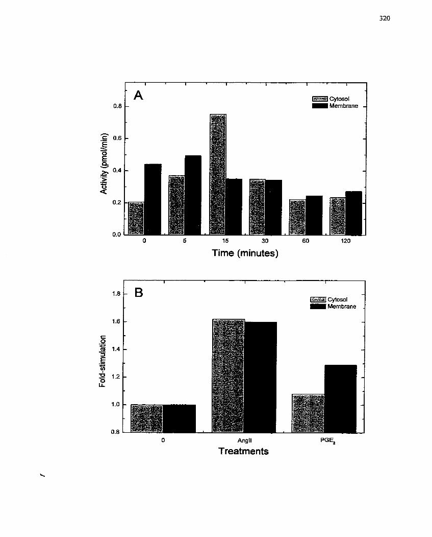
qtomr = Membrane
Time (minutes)
Angll
Treatments PGE,
FIGURE 62: Sensitivity of Mitogert-stimulated SMC Growth to MZï?3G.
Quiescent SMCs were pretreated with MIBG (2.5 to 50 FM) for 10 min prior to addition
of angiotensin II (AngII) (106 M) (A, B) or s e m (2% v/v FBS) (C, D). The
incorporation of [3auridine (6 h incubation) or [3Kjthyrnidine (48 h incubation) into
trichloroacetate-precipitable material after addition of AngII and FBS was used as an
indication of RNA (A, C) and DNA (B, D), respectively. The incorporation rate of
untreated cells uras set to 100%. The data are presented as the mean + SE of at least three
separate experiments conducted in triplicate using different SMC isolations. Cornparisons
were made relative to growth factor treated SMCs with the Student's t-test (*, p c 0.05).
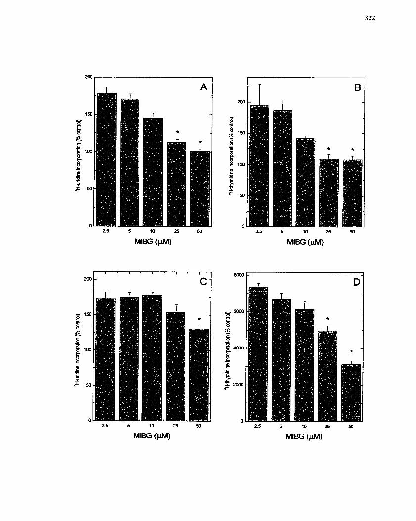
and DNA synthesis in FBS-stimulated SMCs (Figure 62C,D), although the response was
not retumed to basal levels as seen with the h g 1 1 (Figure 62A,B). This is likely due to
the magnitude of the SMC growth response elicited by FBS as a result of the multiple
growth stimulating agents present in FBS. When MIBG was tested with PGE7-stimulated
SMCs, MLBG (2.5 to 50 PM) decreased both RNA and DNA synthesis in a
concentration-dependent manner (Figure 63A,B), and the inhibition was comparable to
that seen with AngII (Figure 62A,B). Moreover, MIBG inhibition of DNA synthesis was
also observed in human SMCs stimulated with FBS (data not shown). To confirm the
specificity of MIBG, the actions of MIBA (2.5 to 50 FM) were a1so tested in FBS-treated
SMCs. MIBA was ineffective at inhibiting FBS-mediated stimulation of DNA synthesis
(Figure 64), even at the highest concentration.
Ce11 number was quantified to determine if MIBG (20 yM) prevented the increase
in SMC ce11 number induced by FBS addition. FBS (2% v/v) essentially doubled ce11
nurnber over a 72 h period (Figure 65). This increase in ceil number was only modestly
inhibited by MIBG (20 FM), and not at al1 by the anaiog, MIBA (20 PM) (Figure 65).
MIBG and MIBA treatment of SMCs in the absence of mitogen did not aitcr ce11 number
relative to untreated control SMCs. These data suggest that MIBG iohibits SMC
proliferation. The small degree of inhibition obsewed in this experiment may be due to
the concentration of MIBG (20 PM) used in the study relative to the strength of the
mitogen being used (Le. FBÇ vs. a single mitogen such as AngII). Furthemore, because
the treatment of SMCs with MIBG or MIBA alone did not decrease thc ceil number, as
was obscrved with H4tIE cells (chapter 5 ) , it is possible that MIBG is less toxic to SMCs.
Cytotoxicity studies werc thcrefore conducted to assess this parameter.
FIGURE 63 : Sensitivity of Prostaglandin E~mediated Cell Growth in SMCs to MIBG.
Quiescent SMCs were pretreated with MIBG (2.5 to 50 pM) for 10 min pnor to addition
of prostaglandin E2 (PGE2) (lod M). The incorporation of lu lu ri di ne (6 h incubation) or
[3~]thymidine (48 h incubation) into trichloroacetate-precipitable material after addition
of PGE2 was used as an indication of RNA (A) and DNA (B) synthetic activity,
respectively. The incorporation rate of untreated cells was set to 100 %. The data are
presented as the mean + SE of at least three separate experirnents conducted in triplicate
using different SMC isolations.
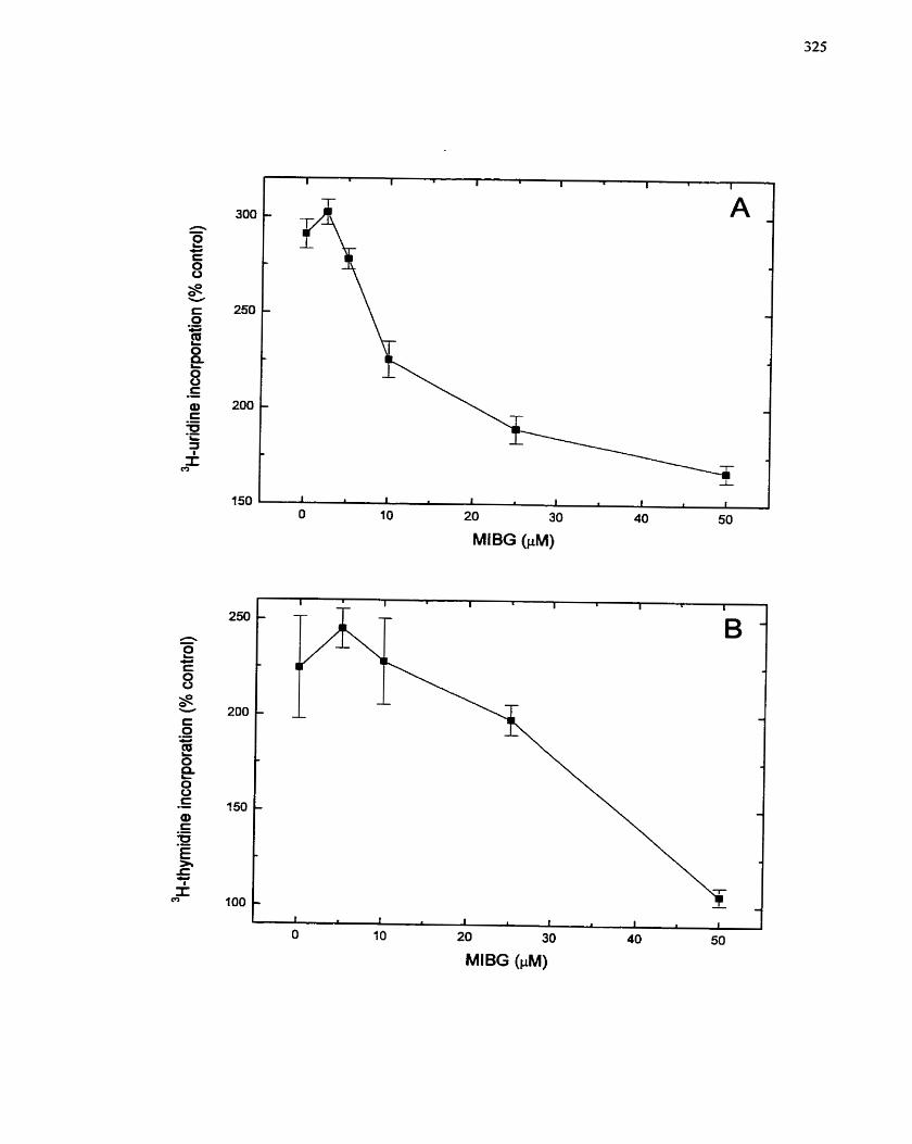
MlBG (FM)
FIGURE 64: Sensitivity of Serum-mediated Ce11 Growth in SMCs to MIBA.
Quiescent SMCs were pretreated with MIBA (2.5 to 50 pA4) for 10 min prior to addition
of semm (2% v/v FBS). The incorporation of [3~thymidine (48 h incubation) into
trichloroacetate-precipitable material afier addition of FBS was used as an indication of
DNA synthetic activity. The incorporation rate of serurn-treated cells was set to 100%.
The data are presented as the mean + SE of three separate experiments conducted in
triplicate using different SMC isolations.
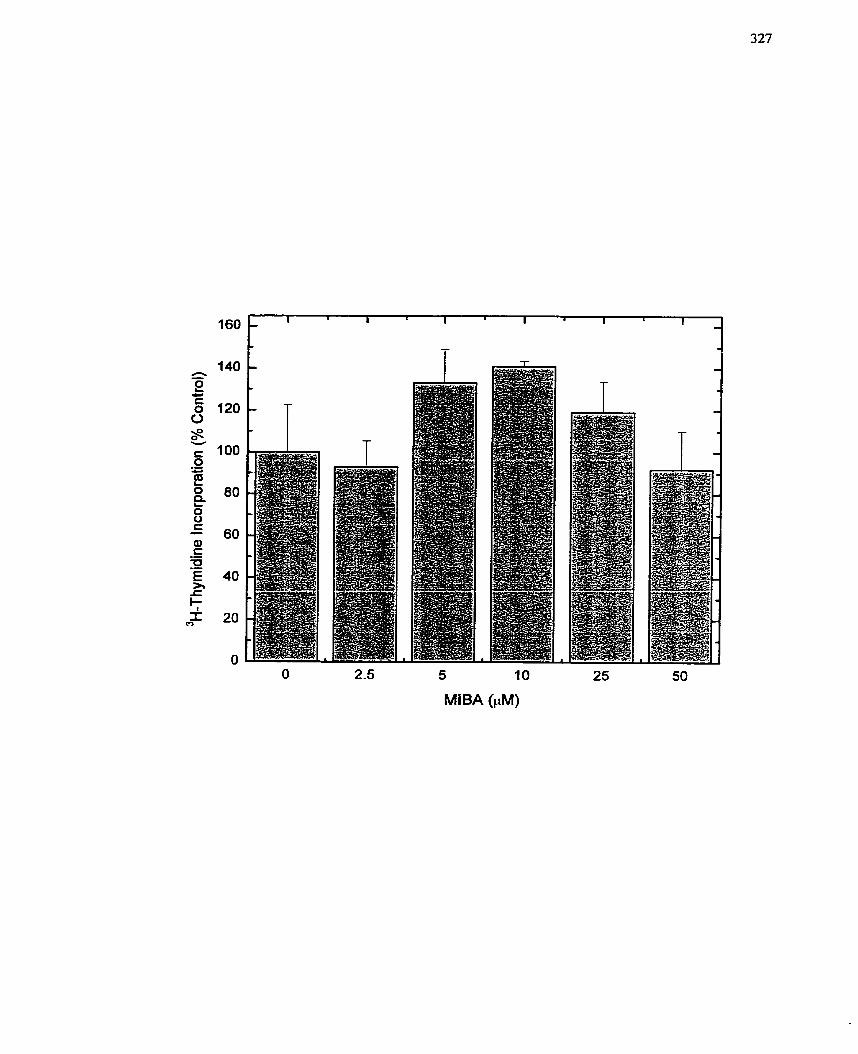
O 2.5 5 10 25 50
MIBA (PM)

FIGURE 65: Effect of MilBG and MTBA on Serum-stimulated SMC Proliferation.
Quiescent SMCs were pretreated with MBG or MIBA (20 PM) for 10 min prior to
addition of senun (2% v/v FBS). The cells were incubated for 72 h and the ce11 number
quantified using a Coulter Counter. Similar results were obtained in two independent
experiments. Each data point was counted in triplicate fiom samples prepared in
duplicate. Data is presented as rnean f SE. Cornparisons were made relative to serum-
treated cells with the Student's t-test (*, p < 0.05).
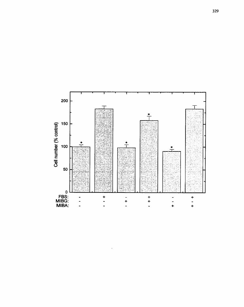
O FBS:
MIBG: MIBA: -
7.3.4.3 Cytotoxic effects of MIBG on SMCs
As observed in chapter 5, H4IIE cells are quite sensitive io the addition of MIBG.
On the other hand, L6 skeletal rnyoblasts, as observed in chapter 6, are not adversely
affected by MIBG- Studies of SMCs (Figure 65) suggest that the sensitivity of SMCs to
MIBG has greater similarity to myoblasts than hepatomas. An MTT assay that assesses
mitochondrîal function was used to obtain information regarding the cellular toxicity of
MIBG, Over a concentration range frorn O to 100 PM, MEBG, even after 73 h, did not
appreciably decrease MTT conversion in quiescent SMCs (Figure 66). Tliese data wouId
suggest that MIBG is not toxic to SMCs, which is in marked contrast to the results with
H4IE cells. Moreover, a comparison of the effect of MIBA and MIBG (O to 500 FM) on
SMC morphology, afier incubation with the compounds for 72 h, dernonstrated that onIy
at the highest concentration was MIBG toxic (Le. 500 PM) (Figure 67). Only in panel 1
was it observed that SMCs had rounded up and lifted off the plate. At the concentration
that effectively inhibited DNA and RNA synthesis and differentiation in the previous
studies (50 PM), no change in ce11 morphology was observed. MIBA did oot appear to
exert toxic effects at any of the concentrations tested (Figure 67). Tlierefore, SMCs may
resernbIe L6 skeletal myoblast cells in their sensitivity and response to MiBG.
7.3.5 Discussion
The results of this study indicate that an arg-mART is activated upon stimulation
of SMCs with growth factors such as AnglI and PGE?, and that MIBG, a specific
inhibitor of arg-mART activity, is capable of inhibiting SMC growth and prolifcration
resulting frorn growth factor stimulation. These findings would thereforc suggest that an
ADP-ribosylation event is important for SMC pro1 iferation and that inhibition of this
cnzymc prcvcnts growth. Thcsc findings imply that an arg-mART is important for thc
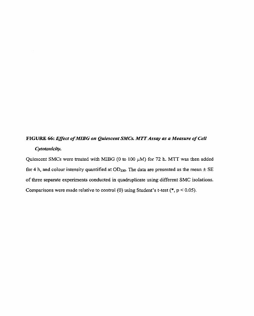
FIGURE 66: Effect of lWBG on Quiescent SMCs. MTT Assay as a Measure of CeCl
Cytotoxicity.
Quiescent SMCs were treated with MIBG (O to 100 jM) for 72 h. MTT was then added
for 4 h, and colour intensity quantified at ODSso. The data are presented as the mean + SE
of three separate experiments conducted in quadruplicate using different SMC isolations.
Cornparisons were made relative to control (O) using Student's t-test (*, p c 0.05).
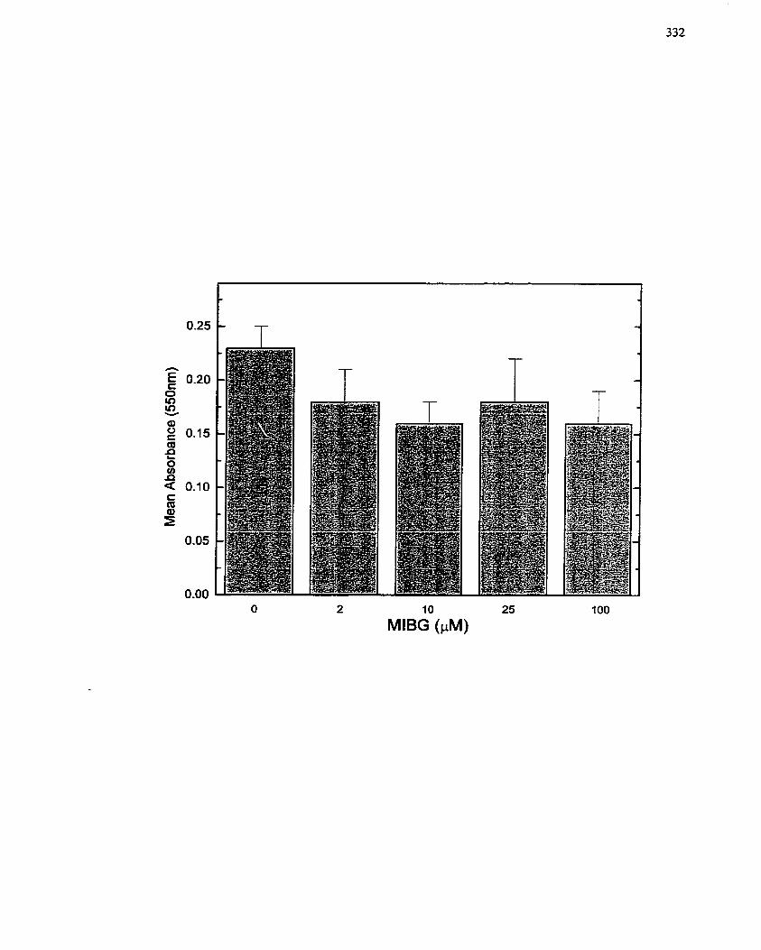
10
MlBG (PM)
FIGURE 67: Effect of M B G and MTBA on the MorphoCogy of Quisecent SMCs.
Quiescent SMCs were treated with MIBG or MIBA (O to 500 pMJ for 72 h.
Photomicrographs were used to record ce11 morphology. Representative micrographs are
shown for untreated SMCs. Specific treatments are: control (A), 50 ph4 MIBA (B) and
MIBG (C), 100 p M MIBA @) and MBG (E), 200 pM MIBA (F) and MIBG (G), and
500 p M MIBA (H) and MIBG (0. Magnification: 128x.
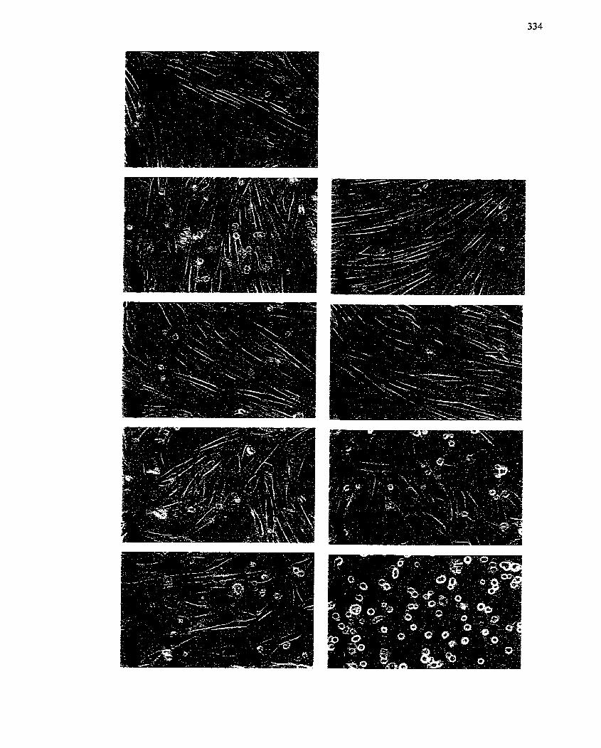
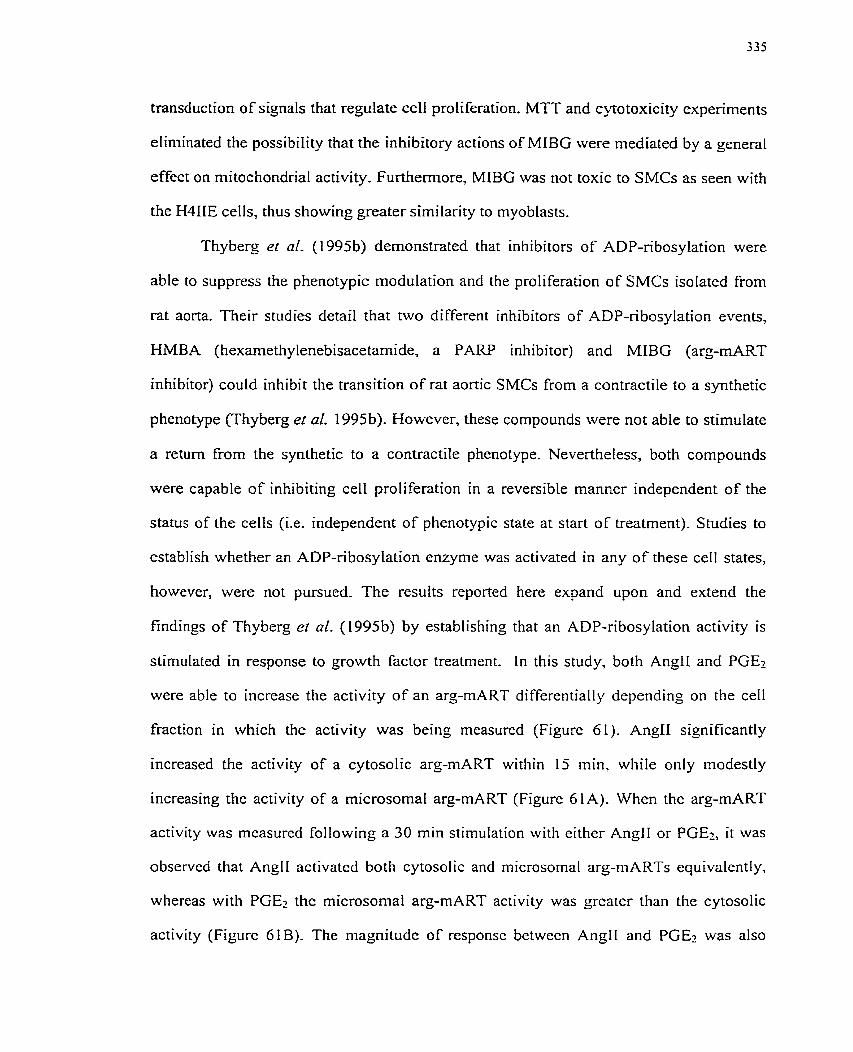
transduction of signais that regulate ce11 proliferation, MTT and cytotoxicity experiments
eliminated the possibility that the inhibitory actions of MIBG were mediated by a general
effect on mitochondrial activity. Furthem~ore, MIBG was not toxic to SMCs as seen with
the H4IIE cells, thus showing greater similarity to myoblasts.
Thyberg et al. (1995b) demonstrated that inhibitors of ADP-ribosylation were
able to suppress the phenotypic modulation and the proliferation of SMCs isolated from
rat aorta. Their studies detail that two different inhibitors of ADP-rïbosylation events,
HMBA (hexamethylenebisacetamide, a PARP inhibitor) and MIBG (arg-mART
inhibitor) could inhibit the transition of rat aortic SMCs from a contractile to a synthetic
phenotype (Thyberg et al. 1995b). Wowever, these cornpounds were not able to stimulate
a return from the synthetic to a contractile phenotype. Nevertheless, both compounds
were capable o f inhibiting ce11 proliferation in a reversible rnanner independent o f the
status of the tells (Le, independent o f phenotypic state at start of treatment). Studies to
establish whether an ADP-ribosylation enzyme was activated in any of these ce11 States,
however, were not pursued. The results reported here expand upon and extend the
findings of Thyberg et al. (1995b) by establishing that an ADP-ribosylation activity is
stimulated in response to growth factor treatment. in this study, both AngIZ and PGEz
were able to incrcase the activity of an arg-mART differentially depending on the ce11
fraction in which the activity was being rneasured (Figure 61). AngIl significantly
increased the activity of a cytosolic arg-mART within 15 min, while only modestly
increasing the activity of a microsomal arg-mART (Figure 6 1 A). Whcn the arg-mART
activity was mcasurcd foIlowing a 30 min stinlulation with eithcr AngII or PGE?, it was
observcd that AnçII activatcd both cytosolic and microsomal arg-mARTs equivalently,
whereas with PGE-, thc microsomal arg-mART activity was grcatcr than thc cytosolic
activity (Figurc 61B)- The magnitudc of responsc bctwccn AngII and PGEz was also
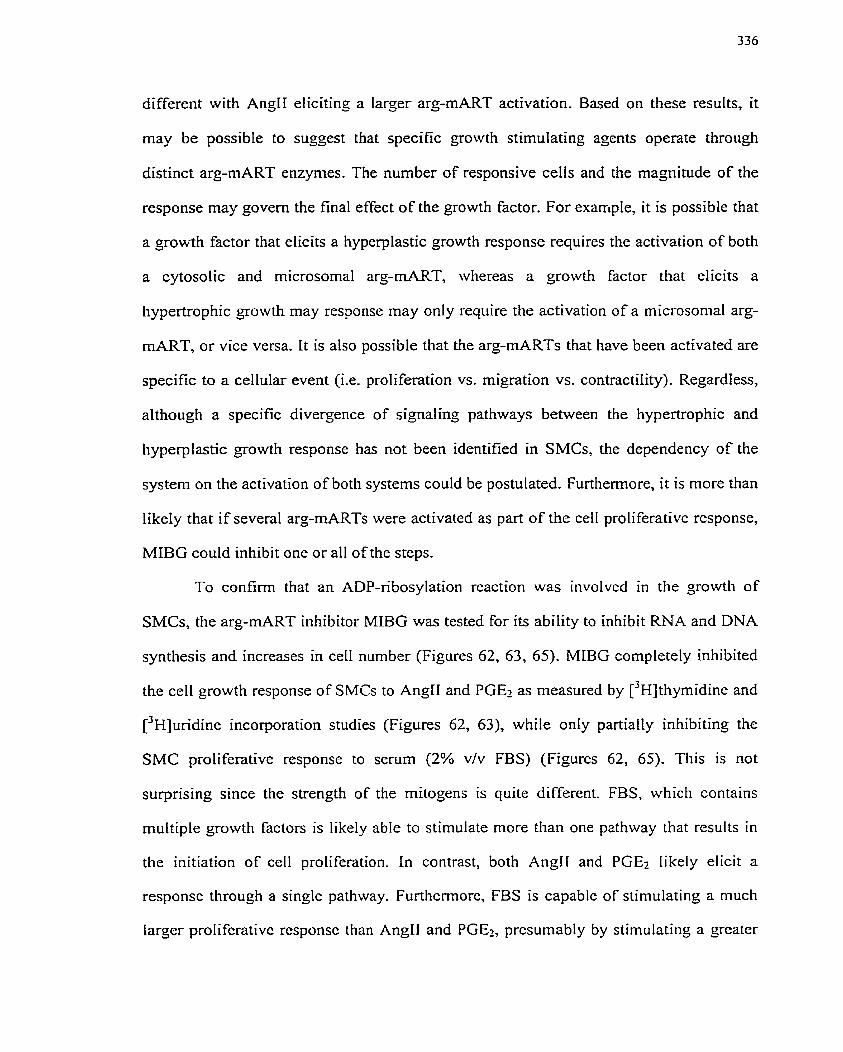
different with AngH eliciting a larger arg-mART activation. Based on these resuIts, it
may be possible to suggest that specific growth stimulating agents operate through
distinct arg-mART enzymes. The number of responsive cells and the magnitude of the
response may govem the final effect of the growth factor. For example, it is possible that
a growth factor that elicits a hyperplastic growth response requires the activation of both
a cytosolic and microsomal arg-rnART, whereas a growth factor that elicits a
hypertrophic growth rnay response rnay only require the activation of a microsoma1 arg-
mART, or vice versa, It is also possible that the arg-mARTs that have been activated are
specific to a cellular event (Le. proliferation vs. migration vs. contractility). Regardless,
although a specific divergence of signaling pathways between the hypertrophic and
hyperplastic growth response has not been identified in SMCs, the dependency of the
system on the activation of both systerns could be postulated. Furthemore, it is more than
likely that if several arg-mARTs were activated as part of the ce11 proliferative response,
MIBG could inhibit one or al1 of the steps.
To confimi that an ADP-ribosylation reaction was involved in the growth of
SMCs, the arg-mART inhibitor MIBG was tested for its ability to inhibit RNA and DNA
synthesis and increases in ce11 number (Figures 62, 63, 65). MIBG completely inhibited
the ce11 growth response of SMCs to AngII and PGEz as measured by [3~]thymidine and
uri uridine incorporation studies (Figures 62, 63), while only partially inhibiting the
SMC proliferative response to serum (2% v/v FBS) (Figures 62, 65). This is not
surprising since the strength of the mitogens is quite different. FBS, which contains
multiple growth factors is likely able to stimulate more than one pathway that results in
the initiation of ceIl proliferation. In contrast, both Angll and PGEz likely elicit a
response throuçh a single pathway. Furthcrmorc, FBS is capable of stimulating a much
Iargcr prolifcrativc responsc than AnglI and PGE?, prcsumably by stimulating a grcatcr
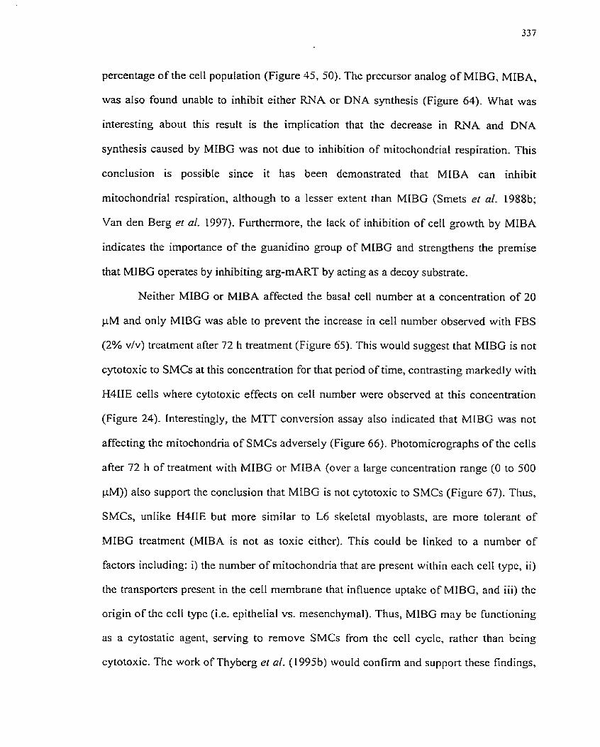
percentage of the cell population (Figure 42, 50). The precursor analog of MIBG, MIBA,
was also found unable to inhibit either RNA or DNA synthesis (Figure 64). What was
interesting about this result is the irnptication that the decrease in RNA and DNA
synthesis caused by MIBG was not due to inhibition of mitochondnal respiration. This
conclusion is possible since it has been demonstrated that MIBA can inhibit
mitochondrial respiration, although to a lesser extent than MIBG (Srnets el al. 1988b;
Van den Berg er al. 1997). Furthemore, the lack of inhibition of ce11 growth by MIBA
indicates the importance of the guanidino group of MIBG and strengthens the premise
that MIBG operates by inhibiting arg-mART by acting as a decoy substrate.
Neither MIBG or MIBA affected the basal ce11 number at a concentration of 20
pM and only MIBG was able to prevent the increase in ce11 number observed with FBS
(2% vlv) treatrnent after 72 h treatment (Figure 65). This would suggest that MIBG is not
cytotoxic to SMCs at this concentration for that penod of time, contrasting markedly with
H41IE cells where cytotoxic effects on ce11 number were observed at this concentration
(Figure 24). Interestingly, the MTT conversion assay also indicated that MIBG was not
affecting the rnitochondna of SMCs adversely (Figure 66). Photomicrograp hs of the cells
after 72 h of treatment with MIBG or MIBA (over a large concentration range (O to 500
PM)) also support the conclusion that MIBG is not cytotoxic to SMCs (Figure 67). Thus,
SMCs, unlike H4IIE but more similar to L6 skeletal myoblasts, are more tolerant of
.MIBG treatment (MIBA is not as toxic either). This could be linked to a number of
factors including: i) the number of mitochondria that are present within each ce11 type, ii)
the transporters prescnt in the ce11 membrane that influence uptake of MIBG, and iii) the
origin of the ce11 typç (Le- epitlielial vs. mesenchymal). Thus, MIBG may be functioning
as a cytostatic agent, serving to remove SMCs from the ceIl cycIe, rather than being
cytotoxic. The work of Thyberg et al. (1995b) would confimi and support thcse findinçs,
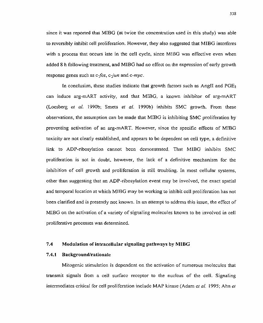
since it was reported that MIBG (at twice the concentration used in this study) was able
to reversibly inhibit ce11 proliferation. However, they afso suggested that MiBG interferes
with a process that occurs late in the ce11 cycle, since MIBG was effective even when
added 8 h following treatment, and MIBG had no effect on the expression o f early growth
response genes such as c-fos, c-jtrrz and c-nryc-
In conclusion, these studies indicate that growth factors such as AngII and PGEî
can induce arg-mART activity, and that MIBG, a known inhibitor of arg-mART
(Loesberg et al. 1990b; Smets et al. 1990b) inhibits SMC growth. From these
observations, the assurnption can be made that MIBG is inhibiting SMC proliferation by
preventing activation of an mg-rnART. However, since the specific effects of MIBG
toxicity are not clearly established, and appears to be dependent on ceII type, a definitive
Iink to ADP-ribosylation cannot been demonstrated. That MIBG inhibits SMC
proliferation is not in doubt, however, the Lack of a definitive mechanism for the
inhibition of ce11 growth and proliferation is still troubling. In most cellular systems,
other than suggesting that an ADP-ribosylation event rnay be involved, the exact spatial
and temporal location at which MIBG may be working to inhibit ce11 proliferation has not
been clarified and is presently not known. In an attempt to address this issue, the effect of
MIBG on the activation of a variety of signaling rnoiecules known to be invoIved in ce11
proliferative processes was deterrnined.
7.4 Moduiation of intracellular signaling pathways by MIBG
7.4.1 Background/rationale
Mitogenic stimulation is dependent on tl-ic activation of numerous molecules that
transmit signals from a ce11 surfacc receptor to the nuclcus of the ccll. Signaling
intermediates critical for ceIl proliferation includc MAP kinase (Adam ct al. 1995; Ahn el
al. 1992; Force & Bonventre 1998; Khalil & Morgan 1993; Klemke et al. 1997; Nelson
et al. 1998), p21-Ras (Bos 1995) and PI3-kinase (Varticovski et ai. 1994). The
expression OF early growth response genes is also essential for the initiation and
maintenance of ce11 growth processes. Thus, since it has already been established that
growth-stimulating agents such as PGEr can activate these signaling systems and that
MIBG inhibits the proliferation of PGEz-stimulated SMCs, the impact of MIBG on early
growth signaling was investigated.
7.4.2 Specific Aims
1. To examine the effect of MIBG on MAP kinase activation,
2. To examine the effect of MIBG on p2 1 -Ras activation.
3. To examine the effect of MIBG on PI3-kinase activation.
4. To examine the effect of MIBG on c-fos gene expression.
7.4.3 Experimentd design
Primary cultures OF quiescent porcine SMCs were pretreated with MIBG for 10
min pnor to addition of PGE2. SMCs were harvested for the evaluation of the specific
signaling elements. An in gel MAP kinase activity assay was used to determine the effect
of MIBG on MAP kinase activation. GTP binding to p2 1-Ras was measured to define the
effect oFMIBG on p21-Ras activation state. The effect of MIBG on PI3-kinase activation
and formation of PIPs Iipid intemediates was ako assessed. As weII, the effect of MIBG
on c-fos gene expression was determined. Each of these investigations allows not only a
prospective spatial and temporal placement for the inhibition by MIBG, but may also
reveal a spatial and temporal placement for arg-mART reactions rcquired for SMC
growth and proliferation.
7.4-4 Results
7.4.4.1 Effect of MIBG on MAP kinase activation
As demonstrated in Figure 53, MAP kinase is activated by a vanety of growth
factors, inchding FBS and PGE2. To determine if the ce11 growth inhibitory effect of
MIBG is due to inhibition of MAP kinase activation, MIBG (50 pM) was added to SMCs
10 min pnor to growth factor stimulation. It was observed that MIBG was not able to
abrogate the MAP kinase response activated by PGE2 ( 1 0 - ~ M) (Figure 68A).
Densitometric anaIysis of a separate experiment confimed thr finding that LMIBG did not
inhibit the MAP kinase activation (Figure 68B). These data demonstrate that MIBG does
not affect this critical ce11 growth pathway and fûrther indicates that an arg-mART does
not participate in the activation cf MAP kinase. Similar results were obtained with H4IIE
celIs in chapter 5.
7.4.4.2 Effect of MIBG on p21-Ras activation
As demonstrated previously (Table 3), p21-Ras is not activated by PGE2.
However, the effect of MIBG on this process was still examined in case MIBG treatment
would activate or potentiate p21-Ras activity. In other words, mART activation could
function as a negative reguIator of p2l-Ras. Addition of MIBG (50 PM) 10 min prior to
PGE2 (10 -~ M) addition revealed that MIBG did not influence p21-Ras stimulation (Table
3). These findings indicate that MIBG does not affect p21-Ras activity and that a mART
does not serve as a negative regulator of this enzyme.
7.4.4.3 Effect of MIBG on PI3-kinase activation
As established in Figures 56 to 58, PI3-kinasc is activated by PGE2. Pretreatment
of SMCs with MIBG (50 FM) for 10 min, did not inhibit P13-kinase-dependent formation
of PIP, stirnulated by PGEz (10~' M), as dctermined by an in vivo P13-kinase assay
(Figure 69A,B). Thcsc findings demonstratc that MIBG docs not inhibit PI3-kinase
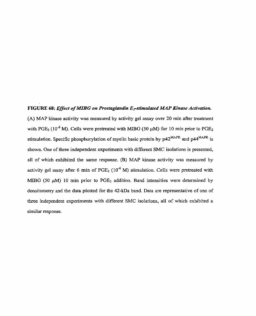
FIGURE 68: Effecr of MIBG on Prostaglandin E?stimuiated MilP Kinase Activation.
(A) MAP kinase activiîy was measured by activity gel assay over 20 min afier treatrnent
with PGEz (106 M). Cells were pretreated with MIBG (50 jM) for 10 min prior to PGEz
stimulation. Specific phosphorylation of myelin basic protein by ~ 1 4 2 ~ ~ and is
shown. One of three independent experiments with different SMC isolations is presented,
al1 of which exhibited the sarne response. (B) MAP kinase activity was measued by
activity gel assay after 6 min of PGEî (10" M) stimulation. Cells were pretreated with
MIBG (50 PM) 10 min pnor to PGE2 addition. Band intensities were determined by
densitometry and the data plotted for the 42-kDa band. Data are representative of one of
three independent experiments with different SMC isolations, al1 of which exhibited a
similar response.
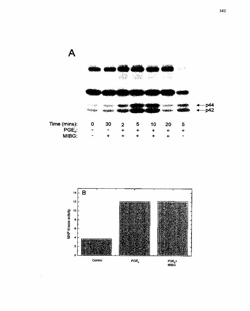
Time (mins): O 30 2 5 10 20 5 PGE,: - - + + + + + MIBG: - + + + + + -
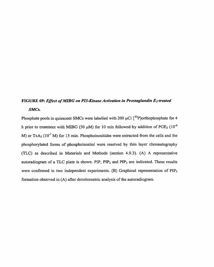
FIGURE 69: Effect of MIBG on PI3-Kinase Activation in Prostaglandin Eptreated
SMCs.
Phosphate pools in quiescent SMCs were labelled with 200 pCi [32~]orthophosphate for 4
h pnor to treatment with MIBG (50 pM) for 10 min followed by addition of PGEz (10'~
M) or TxAz (IO-' M) for 15 min. Phosphoinositides were extracted from the cells and the
phosphorylated forms of phosphoinositol were resolved by thin layer chromatography
(TLC) as descnbed in Materials and Methods (section 4.9.3). (A) A representative
autoradiogram of a TLC plate is shown. PIP, P P 2 and P P 3 are indicated. These results
were confïrmed in two independent experiments. (B) Graphical representation of PIP3
formation observed in (A) after densitometric analysis of the autoradiogram.
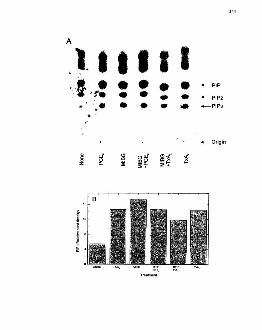
c Origin
directly or any process leading to PI3-kinase activation and that a rnART does not
participate to regulate this s ignahg molecule.
7.4.4.4 Effect of MIBG on c-fos gene expression
As demonstrated in Figure 51, c-fos mRNA levels are transiently increased by
treatment with PGE2- Pretreatment of SMCs with MIBG (50 PM) for 10 min inhibited the
increase in c-fos mRNA levels observed in response to PGEr (10-~ M) (Figure 70A).
Densitometric analysis of a separate experiment confirmed the finding that MLBG
treatrnent inhibited PGEr-dependent c-fos gene expression (Figure 70B). This observation
indicates that a step required for induction of c-fos gene transcription is sensitive to
MLBG treatment and rnay involve regulation by a mART. Although the results presented
in this section suggest that MIBG operates after MAP kinase, p21-Ras and PI3-kinase,
but before the activation of c-fus gene expression, the results may also indicate that
MIBG affects a parallel and independent pathway.
7.4.5 Discussion
The results of this section suggest that MIBG does not have an effect on s ignahg
processes that lead to MAP kinase in SMCs (Figures 68, 69), simiIar to that which was
observed with H4IIE cells in chapter 5. This would indicate that MIBG is not having an
effect on either the PGEz receptor or the G protein to which it is coupled, and also not the
downstream effectors that iead to MAP kinase. Activation of MAP kinase pathways by G
protein-coupled receptors progresses through tyrosine phosphorylated intermediates (Le.
PI3-kinase) which subsequently activate MAP kinase through Ras-dependent or Ras-
independent rnechanisms (Ami & Escobedo 1996; de Vries-Smits et al. 1992; Egan et a!.
1993; Hedin et ai. 1999b; Seger & Krebs 1995; Thomas et al. 1992; Treisman 1995;
Treisman 1996; Ueda et al, 1996; van Biesen et al. 1996; Zhang et al. 1993b). As
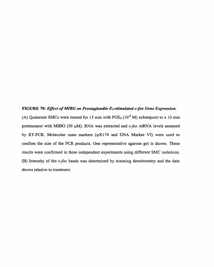
FIGURE 70: EfSect of M?BG on Prostaglandin E~stimulated c-fos Gene Expression.
(A) Quiescent SMCs were treated for 15 min with PGE2 (10'~ M) subsequent to a 10 min
pretrearnent with MIl3G (50 pM). RNA was extracted and c-fos mRNA levels assessed
by RT-PCR. Molecular mass markers (cpX174 and DNA Marker VI) were used to
confîrm the size of the PCR products. One representative agarose gel is shown. These
results were confirmed in three independent experiments using different SMC isolations.
(B) Intensity of the c-fos bands was determined by scanning densitometry and the data
shown relative to treatment.
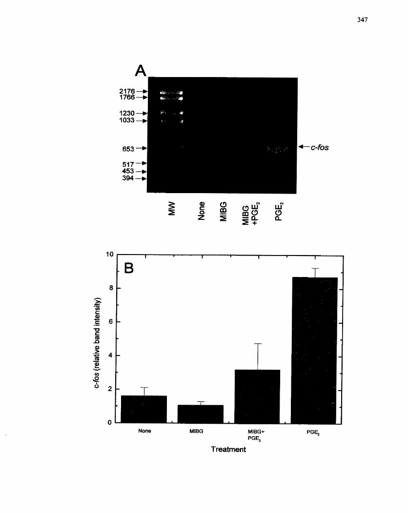
Treatrnen t
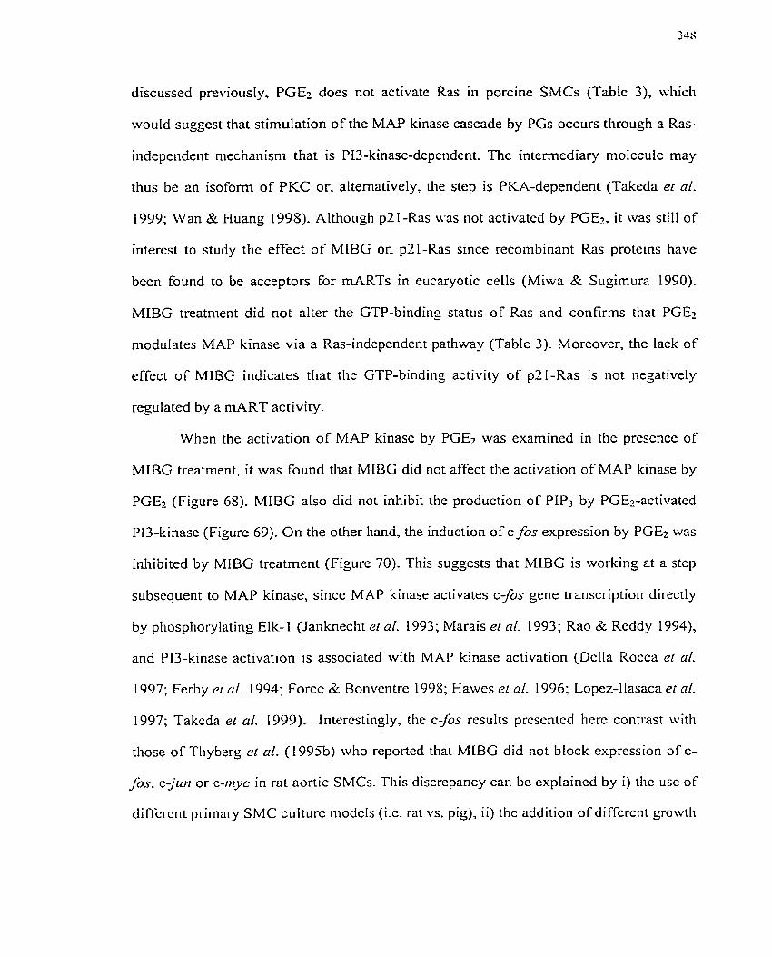
discussed previously, PGE-, does not activate Ras in porcine SMCs (Table 3), which
would suggest that stimulation of the MAP kinase cascade by PGs occurs tlirough a Ras-
independent n-iechanism that is P13-kinase-depetident. The internediary n-iolecule may
thus be an isoform of PKC or, alternatively, the step is PKA-dependent (Takeda er al.
1999; Wan & Huang 1998). Altl-iough p21-Ras \vas not activated by PGEz, it was still of
interest to study the effect of MIBG on p21-Ras since recombinant Ras protcins have
been found to be acceptors For rnARTs in eucaryotic ceHs (Miwa & Sugirnura 1990).
MIBG treatment did not alter the GTP-binding status of Ras and confirms that PGEz
modulates MAP kinase via a Ras-independent pathway (Table 3 ) - Moreover, the lack of
effect of MIBG indicates that tlie GTP-bindinz activity of p21-Ras is not negatively
regulated by a niART activity.
When the activation of MAP kinase by PGE2 was examined in the presence of
MIBG treatment, it was found that MIBG did not affect tlie activation of MAP kinase by
PGEz (Figure 68). MIBG also did not inhibit the production of PLP3 by PGEz-activatcd
PI3-kinase (Figure 69). On the otlier hand, the induction of c-fos expression by PGE2 was
inhibited by MIBG treatment (Figure 70). This suggests that MIBG is working at a step
subsequent to MAP kinase, since MAP kinase activates c-fos gene transcription directly
by pliosphorylating Elk- 1 (Janknecht er ai. 1993; Marais et al. 1993; Rao & Reddy I994),
and PI3-kinase activation is associated with MAP kinase activation (Dclla Rocca et cil.
1997; Ferby et cri- 1994; Force & Bonvcntre 1998; Hawcs er al. 1996; Lopez-Ilasaca er nl.
1997; Takcda er al. 1999). interestingly, the c a s results prcsented here contrast with
thosc of Tliyberç el ul. ( 1995b) who reported that MIBG did not block expression of c-
fis, c-jr~n or c-iuyc in rat aortic SMCs. This discrcpancy can bc cxplaincd by i) tlic usc of
difrcrcnt primary SMC culturc modcls (i.c. rat vs. piç), i i ) tlic addition ofdiffcrcnt çrowtli
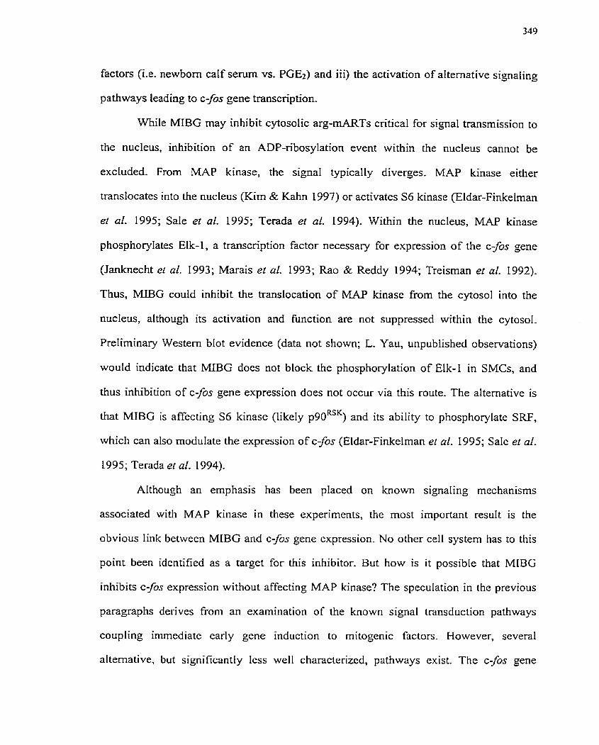
factors (Le. newbom calf serum vs. PGE2) and iii) the activation of alternative signaling
pathways leading to c-fos gene transcription.
While MIBG may inhibit cytosolic arg-mARTs cntical for signal transmission to
the nucleus, inhibition of an ADP-ribosylation event within the nucleus cannot be
excluded. From MAP kinase, the signal typically diverges. MAP kinase either
translocates into the nucleus (Kim & Kahn 1997) or activates S6 kinase (Eldar-Finkelman
et al. 1995; Sale et al- 1995; Terada et al. 1994)- Within the nucleus, MAP kinase
phosphorylates Elk-1, a transcription factor necessary for expression of the c-fos gene
(Janknecht el al. 1993; Marais el al. 1993; Rao & Reddy 1994; Treisman et al. 1992).
Thus, MiBG could inhibit the translocation of MAP kinase from the cytosol into the
nucleus, although its activation and function are not suppressed within the cytosol.
Preliminary Western blot evidence (data not shown; L. Yau, unpublished observations)
would indicate that MIBG does not block the phosphorylation of Elk-1 in SMCs, and
thus inhibition of c-fos gene expression does not occur via this route. The alternative is
that MIBG is affecting S6 kinase (likely p90RSK) and its ability to phosphorylate SRF,
which can also modulate the expression of c-fos (Eldar-Finkelman et al. 1995; Sale et al.
1995; Terada et al. 1994).
Although an emphasis has been placed on known s ignahg mechanisms
associated with MAP kinase in these experiments, the most important resuIt is the
obvious link between MIBG and c-fos gene expression. No other ce11 system has to this
point been identified as a target for this inhibitor. But how is it possible that MIBG
inhibits c-fos expression without affecting MAP kinase? The speculation in the previous
paragraphs derives from an examination of the known signal transduction pathways
coupling immediate early gene induction to mitogenic factors. However, scveral
alternative, but significantly lcss well characterizcd, pathways exist. The c-fos gene
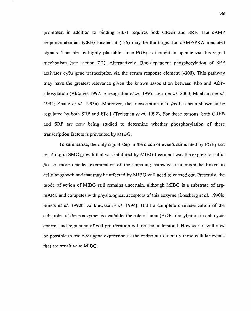
promoter, in addition to binding EIk-1 requires both CREB and SRF. The CAMP
response element (CRE) located at (-56) may be the target for cAMPlPKA mediated
signals. This idea is highly plausible since PGEz is thought to operate via this signal
mechanism (see section 7.2). ALtematively, Rho-dependent phosphorylation of S W
activates c-fos gene trasncription via the serum response element (-300). This pathway
may have the greatest relevance given the known association between Rho and ADP-
nbosylation (Aktories 1997; Ehrengruber et al. 1995; L e m er al. 2000; Maehama et al.
1994; Zhang et al. 1993a). Moreover, the transcription of c-fos has been s h o w to be
regulated by both SRF and Elk-1 (Treisman et al. 1992). For these reasons, both CREB
and SRF are now being studied to determine whether phosphorylation of these
transcription factors is prevented by MIBG.
To sumrnarize, the only signal step in the chain of events stimulated by PGE2 and
resulting in SMC growth that was inhibited by MIBG treatment was the expression of c-
fox A more detailed examination of the signaling pathways that might be Iinked to
cellular growth and that may be affected by MIBG will need to can-ïed out. Presently, the
mode of action of MIBG still remains uncertain, although MIBG is a substrate of arg-
mART and cornpetes with physiological acceptors of this enzyme (Loesberg et al. 1990b;
Smets el al. 1990b; Zolkiewska er al. 1994). Until a complete characterization of the
substrates of these enzymes is available, the role of rnono(ADP-ribosy1)ation in ceII cycle
control and regulation of ce11 prolifcration will not be understood. Mowever, it will now
be possible to use c-fos gene expression as the endpoint to identify those cellular events
that are sensitive to MIBG.
7.5 Effect of MIBG on SMC migration
7.5.1 Background/rationale
Ce11 migration has been extensively studied because it is known to occur during
embryogenesis, wound healing, tumour metastasis and angiogenesis. Although complete
details of ce11 migration are not well understood, ceIl migration is known to involve
regulated attachment and detachment, cytoskeletal plasticity, contraction of non-muscle
myosin and actin, and protein synthesis (Caterina & Devreotes 199 1 ; Kelley et al. 1 99 1 ;
Madn et al. 199 1). Many extracellular signals can stimulate migration, including physical
forces (Ingber & Folkman 1989), vasoactive hormones (Bell & Madn 1989; Bell &
Madri 1990), growth factors (Madri et al. 1991), ions, pH and pO2 (Banai et al. 1990).
Other factors that regulate SMC migration include plasma and platelets, hemodynamics,
macrophages, SMCs and endothelial cells. Unfortunately, the intraceHular signaling
pathways involved in migration and directed ce11 movement are presently not clear.
However, reorganization of the cytoskeleton and alterations in ECM contacts must occur,
but how these events are coordinated is unknown (Stossel 1993).
Migration of cells plays a key role in the formation of a restenotic lesion post-
vascular injury. Normal SMCs in the media1 layer of the vesse1 wall undergo phenotypic
modulation from the contractile state tc a synthetic state after injury. This change in ce11
state renders the SMCs capable of directional movement in response to chemoattractant
factors (Le. PDGF, AngII, TGF-P, bFGF, etc.) and other environmental stimuli, and
involves a marked change in cytoskeletal structure. The typical schema of events
described for neointimal lesion formation in a rat carotid mode1 involves a short burst of
SMC proliferation, followed by migration of the cells from the media to the (neo)intirna
through breaks in the interna1 elastic lamina, with subsequent rounds of ce11 proliferation
and ECM deposition (Casscells t 992; Schwartz ei al. 1995a; Schwartz & Reidy 1996). in
other animal n~odels o f injury, the ccllular events and timelinc of progression lnay not be
similar to that of the rat niodel, but still include both cellular migration from the media of
the vesse1 wall into the (neo)intima and cellular proliferation with deposition of ECM
(Schwartz 1994). Both ce11 migration and ceIl proliferation are equally important for
neointirnal formation, since restenosis can be prevented by inhibitors of either migration
or ce11 proliferation (in many experinlental systems) (Landzbcrg et aL 1997; Lefkovits &
Topo1 1997)-
The progression of tumours is a k o characterized by ce11 proliferation
(tumor~rigenicity) and ceII migration (metastasis). Metastasis involves the rnovement of
cancerous cells from one location to another and involves cellular migration. Many
cancer therapies involve the use of compounds which not only halt the growth of a
tumour but also retard the spreading of tumour cells. MIBG, a known inhibitor of arg-
mART, used as both a radiolabelled and chemotherapeutic agent for the treatment of
neuroendocrine turnours and neuroblastoma, inhibits both tumour growth and metastases
of neuroblastoma type tumours (Hoefnagel et al. 1987; Hoefnagel et al. 199 1; Kuin et al.
1999; Smets et al. 1988b; Taal el al. 1999; Taal et al. 1996; Zuetenhorst et al. 1999).
Moreover, inhibitors of ARTS were found to inhibit thc chernotaxis of human
polyn~orphonuclear neutrophi 1s (PMNs) and inhibition of tlic enzyme activi ty correlated
closely with the inhibition of both chemotaxis and actin asscnibly (Allport et ni. 1996a;
Al Iport et Q I . I996b)- intcrestingly, cheniotactic activity in PMNs has bcen associated
witli ART1 (Kcfalas et al. 1997; Kefalas et al. 1998), a GPi-linked cc11 srirfacc enzyme
also found in skcletal nmscle and lyn~phocytes (Nemoto et al. 1996; Zolkiewska et cd.
1992). In skclctal niusclc, the substrate for thc enzyme appears to bc intcçrin a7
(Zolkicwska 6r Moss 1993). an üdlicsion rcccptor for Ian~inin, whcrcas in T cells thc
siibstrütc appcars to bc lympliocytc Iùnction-activatcd niolccrilc 1 (LFA- 1 ) (Ncinoto er c d .
1996). It may then be reasonable to assume that ADP-ribosylation of integrins on the
sul-face of SMCs could perhaps trigger integrin signaling for cytoskeletal re-alignment or
affect the adhesion of SMCs to the ECM. Saxty et al. (1 998b) have already investigated
the possibility that a mART activity might be found on SMCs. Using the A7r5 SMC ceIl
line derived from embyronic rat thoracic aorta, they found that the chemotactic activity of
A7r5 cells was independent of mART activity, even though a novel panel of mART
inhibitors reduced the ce11 migration- Although a lack of cell surface mART may have
been indicated, this study does not rule out the possibility that an intracerlular mART is
being activated dunng ce11 migration events, particularly since intracellular rnARTs have
already been shown to be associated with desmin filament rearrangement and the
rearrangement of actin cytoskeletal components (Clancy et al. 1995; Huang et al- 1993;
Okazaki & Moss 1996b; Terashima et al. 1992; Terashima et al. 1999; Terashima et al.
1995; Yuan et al. 1999; Zhou et al. 1996)- As a result, it was of interest to determine if
MIBG was capable of inhibiting SMC migration, an event that is associated with intima1
lesion formation-
7.5.2 Specific aims
1, To assess the chemotactic action of AnglI and PGE2.
2. To deterrnine the effect of MIBG on SMC migration.
7.5.3 Experimental design
Growing porcine SMCs were seeded into a Boyden chamber and migration
through a membrane with 5 pM pores assessed with and without chemoattractant in the
lower wells of the chamber, Inclusion of MIBG in the upper weIk of the chamber was
used to measure the effect of MIBG on SMC migration.
7.5.4 Results
754.1 Effect of PGEz and AngII on SMC migration
SeveraI growth factors are known to stirnulate SMC migration, including PDGF,
interIeukins and TNFa (Abedi et al. 1995; Abedi & Zachary 1995; Jang et al. 1993; Yue
et al. 1994). AngII has also been shown to induce chemotaxis (Bell & Madri 1990;
Dubey et al- 1995). It was therefore of interest to determine whether PGEl was also
capable of stimulating SMC migration across a membrane in an artificial setting. Using
the Boyden chamber method, it was observed that PGEz (10 -~ M) was able to stirnulate
SMC migration (Figure 71A). Similady, AngII (IO-' M) could direct SMC migration, as
well as TxAz (IO-' M) and to a lesser extent PGIz ( lod M) (Figure 71A). These data
confim that hg11 is a chemoattractant factor for porcine coronary artery SMCs and also
establish that PGE2 is a chernoattractant for SMCs as are other prostanoids.
7.5.4.2 Effect of MIBG on AngII-rnediated SMC migration
Since MIBG prevents the differentiation of L6 skeletal myoblasts, and inhibits
ce11 growth and proliferation of both H4IIE hepatoma cells and SMCs, it was of interest
to deterrnine if MIBG would also affect SMC migration. Addition of MIBG (50 PM) to
the upper wells of the Boyden chamber demonstrated that MIBG was capable of
significantly decreasing the migratory response of SMCs to AngII (1 O-' M), returning the
levels to almost basal (Figure 7113). These data demonstrate that MIBG inhibits SMC
migration.
7.5.5 Discussion
Migration and proliferation of SMCs are hallmark features of restenotic lesion
formation (Landzbcrg er al. 1997; Schwartz et al. 1995a). Several reports in the Iiterature
have provided evidencc that factors derivcd from platelets, injurcd cndothelia1 cells and
FIGURE 71: Activation of Migration in SMCs by Angioîensin 11 and Prostaglandin
Er. Inhibition by MBG.
Growing SMCs were placed into a Boyden chamber and directional movement of SMCs
through a 5 p membrane towards a chemoattractant over 48 h monitored as described in
section 4.5. SMCs on the underside of the membrane were fured, stained and counted.
(A) Chemoattractants placed into the lower chamber included angiotensin II (AngII) (105
M), PGEt (10" M), PGG (106 M) or TxAz (IO-' m. (B) AngII (10" M) was placed into
the lower chamber and MEBG (50 pM) in the upper chamber for the migration penod (48
h). Data are presented as mean + SE (n = 6). The graphs are representative of one of two
independent experiments using different SMC isolations, both of which exhibited a
sirnilar response. Cornparisons in (A) were made relative to untreated SMCs while
comparkons in (B) were made relative to hg11 treated SMCs with the Student's t-test
(*, p < 0.05). (Data generated by Shawn Thomas, a B.Sc.Med surnrner student, and
shown with his permission).
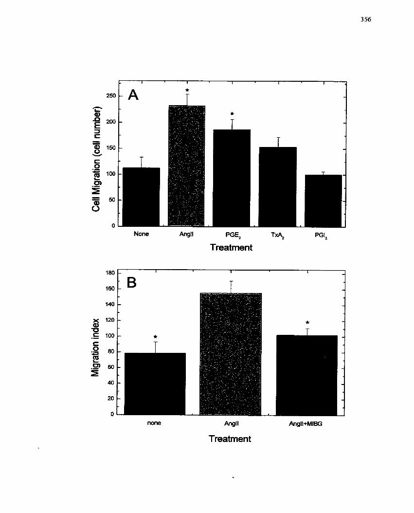
Angll PGE,
Treatment PGI,
Angll
Treatment
SMCs are the pnmary products leading to both migration and prolitèration of SMCs
(Casscells 1992; Golino et al. 1997; Ross et al. 1986). Althougli thc best characterized
factors include PDGF, EGF, FGF, IL-1, IL-6 and TNFa, other agents, including
thrombin and prostanoids such as TxAr and PGE?, may also be involved (Hanasaki et al.
1990; Morinelli et al. 1994; Nagata et al. 1992).
In this study the ability of a peptide growth factor, An& and a nonpeptide
growth factor, PGE2 (as established in section 7.2), to promote SMC migration \\las
studied using a Boyden charnber. AngII is already an established cliemotactic factor for
SMCs (Bell & Madn 1990; Dubey et al. 1995) and was used to determine the degree of
chemotaxis induced by PGE2. The ability of PGEz to cause migration was almost of the
same magnitude as AngII (Figure 7 1A), suggesting a similar efficacy. In comparison to
the proliferative response generated by these cornpounds (Figures 44. 45. 46), the results
of this study would support the conclusion that AngII is a stronger chemotactic agent than
PGE2. Other prostanoids have previously been sliown to have chernotac tic abilities with
respect to a variety of different ceIl types (Hofbauer et al. 2000; 'lie el al. 2000). More
recently, Ratti et al. (1998) indirectly dernonstrated the importance of TxA2 in promoting
SMC migration and proliferation by reporting that picotamide, an anti-thromboxane
agent, inhibitcd both the migration and proliferation of rat arterial myocytes. The ability
of PGEz to promote migration of SMCs may be related to its ability to stimulate the
prolifcration of quiescent SMCs (section 7.2). Lnterestingly, PGEl has also been shown to
decrease collagcn syntliesis, which may be a factor in SiMC migration (Diaz el al- 1993;
Holnies et al. 1997). Wticther the brcakdown of collagcn promotcd by PGEz is due to
incrcascs in MMP activity (Le. MMP-9) remains to bc detcrmined.
MIBC trcatnlcnt inhibited the migration O F SMCs promotcd by AngII (Figure
71 8). Saxty et al. (1998b) had previously dcmonstratcd that a pancl of structurally
unrelated ADP-ribosylation inhibitors prevented chemotaxis in a rat thoracic aorta S MC
ce11 Iine (A7r5)- These inhibitors included Vitamin KLI3, novobiocin, nico tinamide and
small guanidine containing analogues of arginine such as agrnatine (decarboxyl-arginine),
arginine rnethyIester (AME) and DEA-BAG (Banasik et al. 1992; Soman et al. 1986).
Although al1 of the inhibitors were capable of inhibiting chemotaxis, and the efflcacy of
the panel of inhibitors suggested that it might be through inhibition of ART1 (a GPI-
linked extracellular mART discovered by Zolkiewska and col teagues (Zolkiewska &
Moss 1993; Zolkiewska et al. 1992)), it was concluded that these inhibitors could not
operate by this mechanism since ART1 was not present on A7r5 SMCs (Saxty et al.
1998b). Rather, the authors agreed with the findings of Taguchi et al. (1993) that, similar
to L-arginine, the inhibitory effects were Iikely due to an effect on nitric oxide generation
instead of ADP-ribosylation. While it is possible that the SMCs used in this study do
express ART 1, MIBG may alternatively influence an intracellular ADP-ribosylation
reaction. This assumption can be made based on the ability of AnglI to increase an arg-
mART activity in a previous section (section 7.3). Although enzyme activity was
explored with respect to the capacity of AngIi as a mitogen, a similar arg-mART reaction
may be activated for the regulation of migration since both proliferation and migration
are characteristic of the synthetic SMC phenotype. For exampIe, MIBG may be inhibiting
an ADP-ribosylation reaction involved in the assembIy/disassembly of actin filaments
(Clancy et al. 1995; Okazaki & Moss 1996b; Terashima et al. 1992; Terashima et al.
1999; Terashima et al. 1995). Actin is an important protein conlponent of the
cytoskekton and is an important filamentous protein with respect to motiIity and
contraction in SMCs (EIbaum et al. 1999; Takai et al, 1995; Taylor 1986). Altematively,
MIBG inhibits desmin assembly/disassembly (Huang ef al, 1993; Yuan ef ai. 1999; Zhou
et ul, 1996) which can also affect the ability of thc ce11 to migratc (and possibly cvcn
proliferate) (Berna1 & Statiel 1985; Li er al. 1997: Siegel et cil. 1998). It also remains
plausible tliat MIBG niay affect the cytoskeletal rearrangeinents iieccssary for migration
through key GTP-binding proteins such as Rho, Rac and cdc42.
Although a number of potential modes of action have becn supplied, a cytotoxic
effect of MIBG on the SMCs cannot be ruled out. Nevertheles? based on the response of
SMCs in cuIture to MiBG seen in the previous section (section 7.3), it is unlikely that
cytotoxicity is the mechanism of action on ceIl nligration. Pressntly, the niechanism
whereby MIBG inhibits SMC migration remains unknown and will the subject o f future
studies. Additional studies wiil include: i) actin and desmin assembly/disassenibly and its
regulation by arg-rnART/MIBG in SMCs, ii) an examination o f extracellular components
that may be affected by MIBG, and iii) identification and characterization of the mART
involved in SMC niigration.
7.6 Summary
The studies summarïzed in this chapter cover the ce11 proliferative and migratory
activities of SMCs, including the characterization of a novcl stimuhtor of' SMC
prolifcration and migration, PGE?, and an e~amination of how MIBG might inhibit both
o f thesc ccllular events. Furthermore, a rnitogen-activated arg-mART activity associated
with the microsomal Fraction of SMCs was detectcd. While the mcchanism O F MIBG
action was not fully clarified, thc expcrinlcnts tliat wcrc dcscribcd led to the identification
of the c-fos gcnc as a targct for this inhibitor. Morc nicchanistic studics arc now possiblc
givcn this inforniation. Morcovcr, the rcsiilts of tlicsc studics, which inciicatc that MIBG
c m succcssfulIy and crficicntly inhibit botli thc migration and prolifcration of SMCs, two
of tlic critical cvcnts in rcstcnotic lcsion formation, warrant furthcr invcstigation of this
cornpound in niodcls of vascular injury and intimai lcsion formation. Thc ncxt chaptcr

describes the initial studies that were undertaken to examine the suitability of this
compound for the treatment of restenosis.
I
8.0 Models of neointimal lesion formation
8.1 Introduction
Restenosis remains a significant problern following a11 types of revascularization
procedures, including coronary angioplasty, occurring in 30 to 50% of cases (Badimon et
al. 1995; Casterella & Teirstein 1999). Several factors contribute to the formation of an
(neo)intimaI lesion within injured coronary arteries, including thrombus formation,
elastic artery recoii, migration and proliferation of SMCs, and vascular rernodeling (Ellis
& Muller 1992; Glagov 1994; Harker 1987; Lafont et al. 1995; Lau & Sigwart 1995; Liu
et al. 1989; Mintz et al. 1996; Post et al. 1994; Schwartz et al. 1992a; Schwartz et al.
1995a; Schwartz & Reidy 1996; SteeIe el al. 1985; Wilentz et al. 1987). Although al1 of
these factors contribute to lesion formation, the central mechanism appears to involve the
SMC. For this reason, inhibition of S.MC activation (Le. migration and/or proliferation
and/or deposition of ECM) could have a large impact on the pathogenesis on the
morbidity of revascularization procedures. As a result, the behaviour of SMCs has been
studied in a variety of systems in vitro and in vivo, and these cells have been the target of
many studies and therapeutic treatments for the arnelioration of restenosis (Andres 1998;
Landzberg et ai. 1997; Neville & Sidawy 1998; Nikol & Hofling 1995).
Many different types of animal models have been used for the study of restenosis
post-angioplasty. Smaller animals (Le. rats, rabbits and mice) have always been more
popular and, as such, the pathogenesis and physioIogical relevance of injury in these
models is better defined (Schwartz et al. 1995a). The popularity of these models is based
on the Fact that smaller animals are well characterized genetically, less expensive and
more readily available. ,4s well, they are easier to manipulate and, because they are
smaller, the quantities of investigational new drugs required for in vivo screening are Iess
(Handiey 1995; Johnson et al. 1999). On thc other hand, with the use of thcse smaller
animal models, especially for the cesting of therapeutic cornpounds for the treatrnent of
restenosis, the predictive value of Che data is limited (Johnson et al. 1999). Likely this is
the result of differences in pathophysiotogy and physiology of the
themselves (Schwartz 1994). DifEerent portions of the vascular tree
empIoyed, since in the rat. for example, the coronary arteries are too smal
Larger anirnals (Le. pigs,, dogs and primates), though less
nevertheless been utilized for t h e pre-chical evaluation of interven
animal models
would aIso by
popular, have
ions to reduce
restenosis. The disadvantages of t h e larger animal models are primariiy the converse of
the advantages of srnall animal models. These include greatly increased cost and limited
distribution potential, less precise genetic characterization, paucity of transgenic models,
more difficulty in rnanipulatiorm and the requirernent for larger quantities of
investigational new drugs (Johnson el ai. 1999; Schwartz 1994). However, the larger size
of the animals allows for the use mf instrumentation and evaIuation of results much as
would be done in humans. As weell, in the case of pigs, there is a similarity of the
coronary artery anatomy, and the histopathological characteristics of the proliferative
response following deep coronary artery injury closely resemble that seen in humans
(Schwartz el al. 1990; Steele et al. 1985). Thus, large animal models of restenosis,
although less defined, have provided valuable insight into the progression of hurnan
disease. However, both the coronary vasculature and other vascular beds such as the
femoral artery and carotid artery- have been ernployed. Since there are significant
differences in the responses of these vessels, interpretation of results frorn other vascular
beds shouId be applied to the humam coronaries with judicious care (Badimon et ui. 1998;
Pyles et al. 1997).
To this point, al1 studics in tzliis thesis have cmployed cells in culture. It is cvidcnt
from tlicse cxperimcnts that MIBC is an agent that has potcnt anti-prolifcrativc, anti-
migratory and anti-differentiation properties. Thus, MlBG might have application for the
prevention o f restenosis. Although an animal study appears warranted, an organ culture
system was first employed to validate the effect o f MIBG on neointimal formation.
A balloon injury sirnilar to that wbich occurs duting coronary angioplasty can be
easily applied to the coronary vasculature of an ex vivo heart (Koo & Gotlieb 1992).
Studies by Gotlieb and colleagues (Gotlieb & Boden 1984; Koo & Gotlieb 1989; Koo &
Gotlieb 199 1; Koo & Gotlieb 1992) and Wilson et al. (1 999) have established that
intimal proliferation occurs in coronary artenes in organ culture following medial
distention by balloon inflation. Furthermore, with this model Wilson et al. (1999)
demonstrated that the neointirnal formation within the coronary artery segments can be
inhibited by agents that alter the progression of restenosis.
The study that was conducted examined neointimal formation in the presence and
absence of MlBG (O to 50 PM) for 14 days. As reported by Wilson et al. (1999), a
reduction in neointimal formation is indicated by a decrease in neointimal index, a ratio
of intima1 area to media1 area (Figure 72). It was observed that MIBG reduced the
neointimal index to basal levels at and above concentrations of 10 pM (Figure 73)-
Furthemore, there is no evidence of adverse effects upon histological examination
(Figure 73A). Interestingly, migration may have a greater contribution to this mode1 than
ce11 proliferation (Wilson, Wong & Zahradka, unpublished observations). Regardless,
these data clearly establish that MlBG has the potentiaI to prevent resteriosis. Extension
to a live animal model was therefore initiated.
Based on the findings of the previous chapters and the succcss of MIBG to
prevent intimal lesion formation in an organ culture mode1 of restenosis, a specific
hypothesis was proposed: MIBG will redrrce the exteizt of neoitztimal forntaiioiz irtd~tced
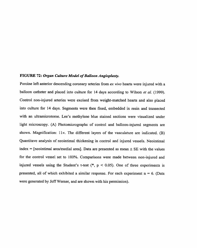
FIGURE 72: Organ Culture Model of Balloon Angioplasty.
Porcine left anterior descending coronary artenes from ex vivo hearts were injured with a
balloon catheter and placed into culture for 14 days according to Wilson et al. (1999).
Control non-injured artenes were excised fiom weight-matched hearts and also placed
into culture for 14 days. Segments were then fixed, embedded in resin and transected
with an ultramicrotome. Lee's methylene blue stained sections were visualized under
light microscopy. (A) Photomicrographs of control and balloon-injured segments are
shown. Magnification: I lx. The different layers of the vasculature are indicated. (B)
Quantitave analysis of neointimal thickening in control and injured vessels. Neointimal
index = [neointimal arealmedial area]. Data are presented as mean I SE with the values
for the control vesse1 set to 100%. Cornparisons were made between non-injured and
injured vessels using the Student's t-test (*, p c 0.05). One of three expenments is
presented, all of which exhibited a similar response. For each experiment n = 6. (Data
were generated by Jeff Werner, and are shown with his permission).
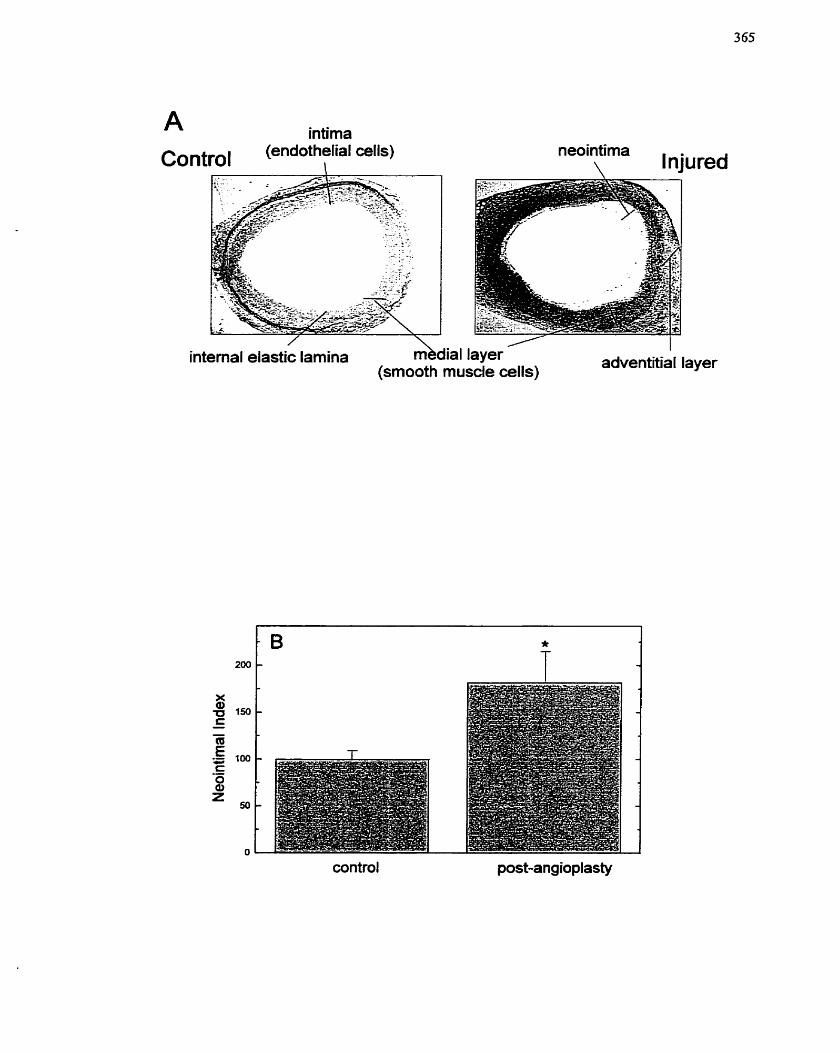
A intima
(smooth muscle cells)
control post-ang ioplasty
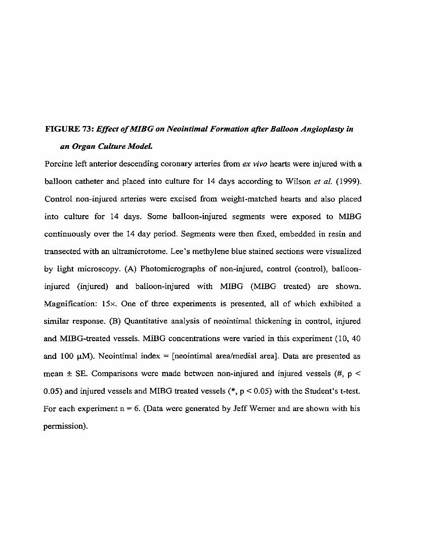
FIGURE 73: Effect of MLBG on Neointimal Formation afier BaCZoon Angiopiasty in
an Organ Culture ModeL
Porcine left anterior descending coronary artenes fiom ex vivo hearts were injured with a
balloon catheter and placed into culture for 14 days according to Wilson et al. (1999).
Control non-injured arteries were excised from weight-matched hearts and also placed
into culture for 14 days. Some balloon-injured segments were exposed to MIBG
continuously over the 14 day period. Segments were then fmed, embeddec! in resin and
transected with an ultramicrotome. Lee's methylene blue stained sections were visualized
by light microscopy. (A) Photomicrographs of non-injured, control (control), balloon-
injured (injured) and balloon-injured with MIBG (MIBG treated) are shown.
Magnification: 15x. One of three experiments is presented, al1 of which exhibited a
similar response. (B) Quantitative analysis of neointimal thickening in control, injured
and MU3G-treated vessels. MIBG concentrations were varied in this experirnent (10, 40
and 100 pM). Neointimal index = [neointimal area/medial area]. Data are presented as
mean t SE. Cornparisons were made between non-injured and injured vessels (#, p <
0.05) and injured vessels and MIBG treated vessels (*, p < 0.05) with the Student's t-test.
For each experiment n = 6. (Data were generated by Jeff Werner and are s h o w with his
permission).
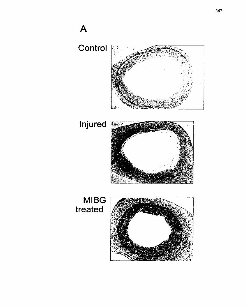
Control
Injured
MIBG treated
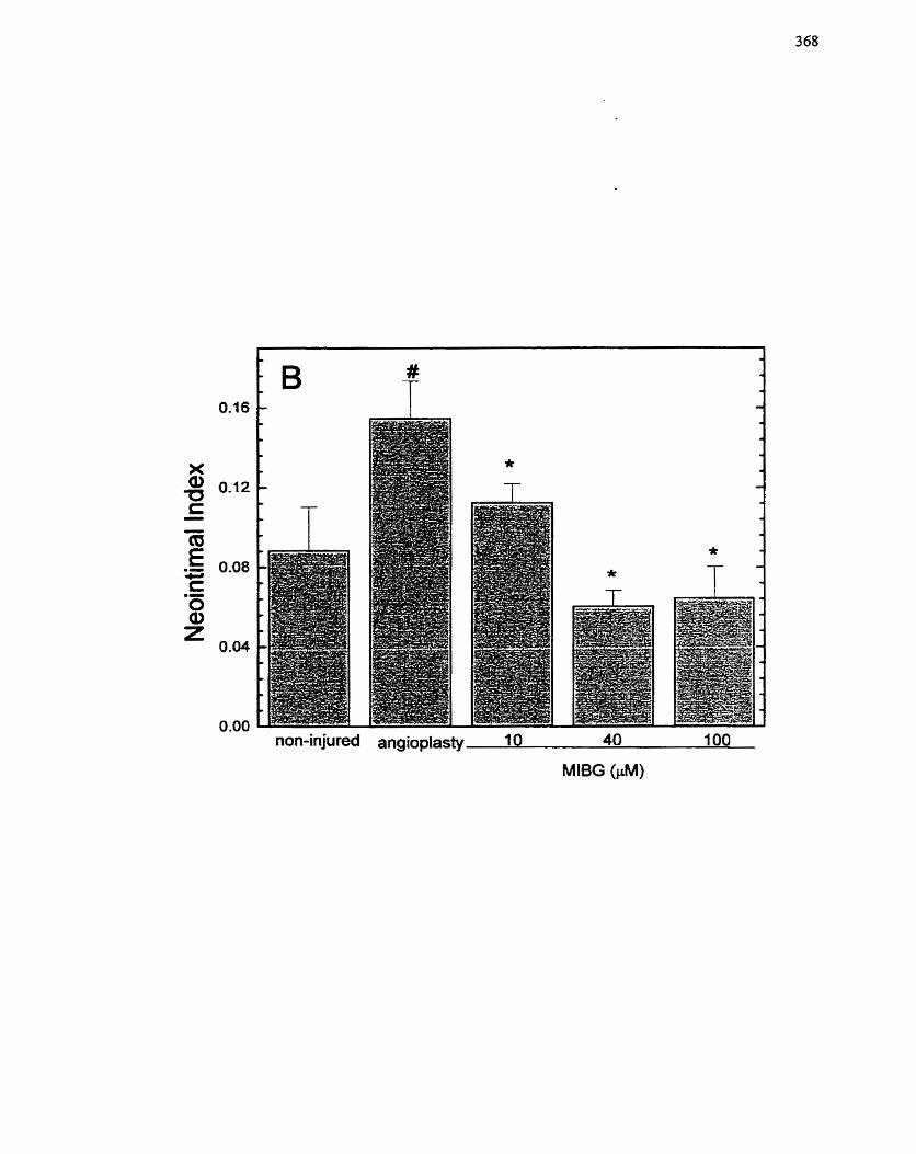
non-injured angioplasty 10 40 100
MlBG (FM)
by balloon mzgioplusty wlzen applied to the perivasctriar sutface wifh a stcrgical fibrin
glue (Tisseel).
8.2 irt vivo porcine femoral angioplasty model of restenosis
8.2.1 Background/rationale
MIBG was tested for its ability to inhibit restenosis with an organ culture model
of neointimal formation. With this system, MIBG was shotvn to be effective under
conditions where injury and repair were simulated in an ex vivo culture environment. To
extend these studies and provide expenmental information that would eventually permit
clinical application, a live animal model of balloon angioplasty was developed- The pig
was chosen because of its greater sirnilarity to humans (Ferrell et al. 1992; Johnson et al.
1999), while the fernoral artery was selected for its accessibility. Atthough this particular
vesse1 is not identical to the coronary artenes in structure and properties, both vessels
nevertheless respond similarly to vascular injury and this perrnits an examination of
events cornmon to both peripheral and coronary vascular disease. Furthemore,
Larnawansa et al- (1997 and 1999) and Yang et al- (1996) demonstrated that balloon
injury of the porcine femoral artery results in the formation of a neointirna, This
particular pig mode1 has a number of advantages: i) it is easy to produce a consistent
injury, ii) it is accessible for angiography and IVUS (intravascular ultrasound), and iii)
the response to injury is similar to human arteries (Ferrell et al. 1992)-
8.2.2 Specific Aims
1. To establish the porcine femoral artery model of balIoon injury as a viable
experimental model for the study OF restenosis.
2. To determine the effect of MIBG on neointimal tesion formation in this
model-
8.2.3 Experimental Design
An NI vivo mode1 of balloon injury will be used to extend the findings generated
using the organ culture mode1 of balloon injury and restenosis. Porcine femoral artet-ies
will be injured, and fibrin glue applied + MIBG in a random, double-blind rnanner. n e
animals will be allowed to recover and the arteries will be harvested after 14 days. Each
animal will serve as its own control (vessel + MIBG), and each vessel will contain both
positive and negative controls (k injury). Each femoral artery will be divided into four
sections (Le. proximal, balloon-injured, distal and arteriotomy). Each region is depicted
on the angiogram presented in Figure 74. Vessels will be cut into segments and frozen in
OCT for cryosectioning. Following staining with Lee's methylene blue, rnorphornetric
analysis will be used to calculate the neointirnal index.
8.2.4 Results
8.2-4.1 Effect of MIBG on femoral artery balloon injury and restenosis
To define the response of the femoral artery to balloon injury, proximal segments
of the vessel were compared to the balloon-injured region of the vessel. Balloon-injury
resulted in a significant thickening of the intima1 region (Figure 75B) compared to '
uninjured regions (Figure 75A) consistent with induction of a lesion. Treatment of the
balloon-injured segment with MIBG significantly inhibited neointimal formation (Figure
75C). Tt was also interesting to note that the media1 layer o r injured segments (Figure
75B) had a more cellular and fibrotic pattern of staining compared to uninjured regions
(Figurc 75A), and injurcd regions treatcd with MIBG (Figurc 7%). Morcover, in the
FIGURE 74: Angiogrum of Porcine Fernoruf Arfery Region.
Porcine femoral arteries were subjected to balloon angioplasty Ni vivo and harvested after
14 days (see Materials and Methods section 4.2.4). Angiography was performed at the
t h e of sacrifice (see Materials and Methods section 4.2.4.1). The four regions of the
femoral artery analyzed in the experiment are identif'ed: proximal region, balloon-injured
region (site of angioplasty), distal region and arteriotomy site.
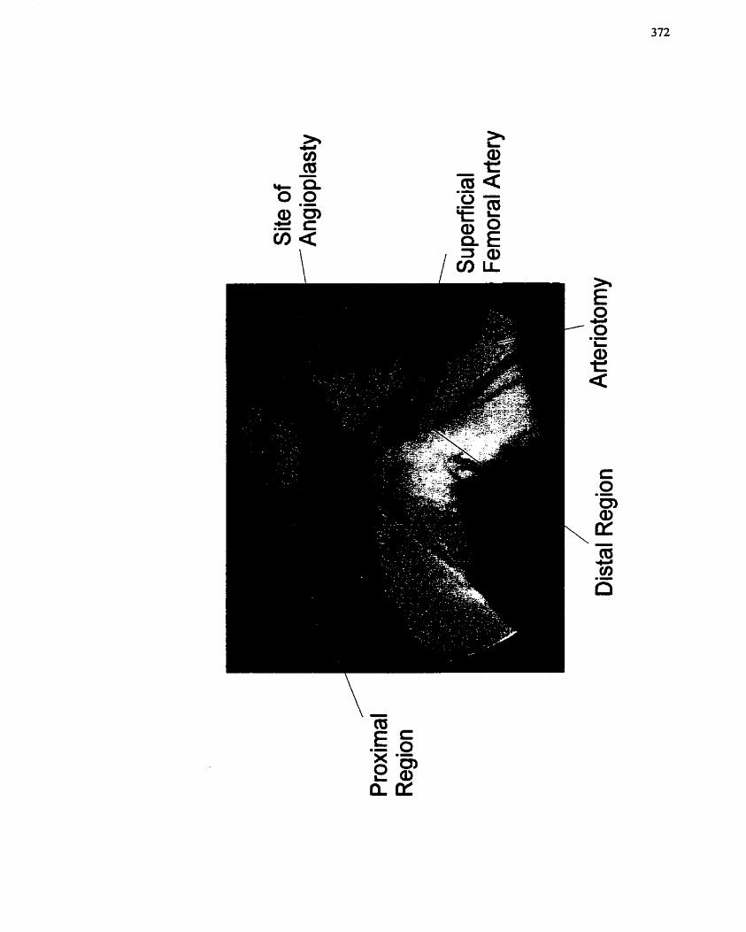
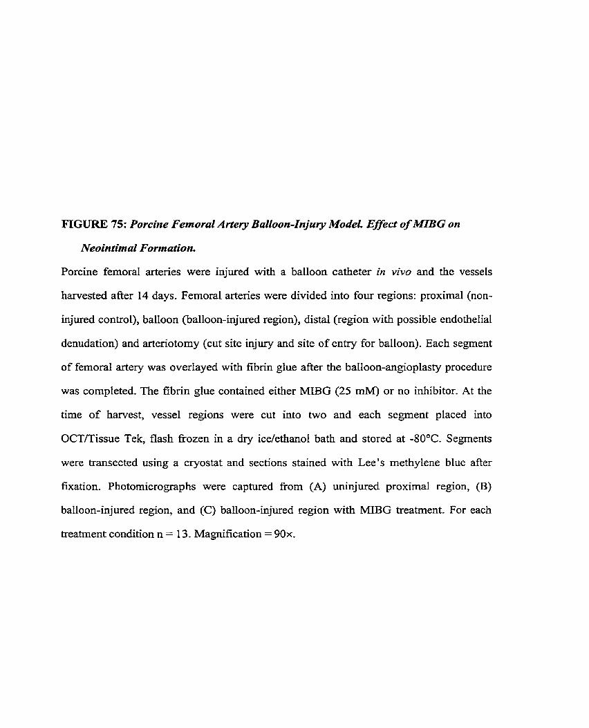
FIGURE 75: Porcine Femoral Artery Balloon-Injury Modei. Effect of MIBG on
Neointimal Formation.
Porcine femoral arteries were injured with a balloon catheter in vivo and the vesseIs
harvested after 14 days. Femoral arteries were divided into four regions: proximal (non-
injured control), balloon (balloon-injured region), distal (region with possible endothelial
denudation) and arteriotomy (cut site injury and site of entry for balloon). Each segment
of femoral artery was overlayed with fibrin glue after the balloon-angioplasty procedure
was completed. The fibrin glue contained either MIBG (25 mM) or no inhibitor. At the
time of harvest, vesse1 regions were cut into two and each segment placed into
OCT/Tissue Tek, flash fkozen in a dry ice/ethanol bath and stored at -80°C. Segments
were transected using a cryostat and sections stained with Lee's methylene blue after
fixation. Photomicrographs were captured fi-orn (A) uninjured proximal region, (B)
balloon-injured region, and (C) balloon-injured region with MIBG treatment. For each
treatrnent condition n = 13. Magnification = 90x.
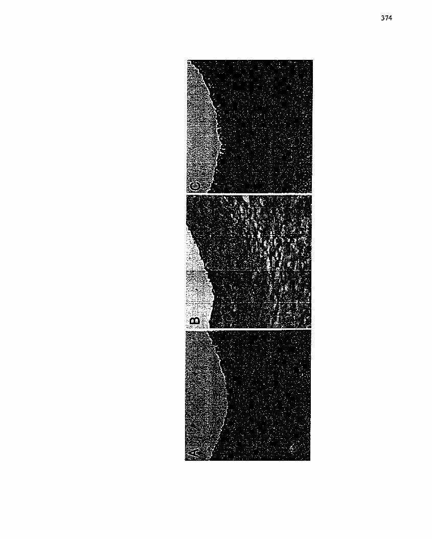
injured segment treated with MIBG, re-endothelialization appears to bc occurring (Figure
75C) when compared to the injured segment without MIBG treatment (Figure 75B). This
observation would need to be confirmed by conducting immunostaining experiments
using antibodies for endothdial ce11 specific markcrs. These data suggest that MIBG is
effective in preventing intimai lesion fom~ation after arterial injury and that it will be an
effective tlierapeutic option for the trcatment of restenosis. The data also indicate that
MIBG in vivo tnay be affecting not only the proliferation of SMCs. but perhaps also SMC
migration, which supports the data presented in chapter 7.
A quantitative measure of neointimal formation revealed that MIBG treatment
significantly decreased the neointimal index of the balIoon injured region cornpared to
the non-MIBG treated balloon injured region, (Figure 76). Moreover, in the distal and
arteriotoniy vesse1 segments, MIBG treatment was able to decrease the degree of
neointima1 formation observed (Figure 76) although statistically significant differences
were not attained. The neointimal index was calculated for al1 13 pigs that were in the
experimental group, however, one pig was exchded from both the graphical and
statistical analysis due to rupture of the femoral artery on one side during the angioplasty
procedure and lack of balloon inflation on the other side. The decision to exclude this pig
from the analysis was made at the time of the initial procedure. The efficacy of MIBG
trcatment for attenuating intima1 lesion formation in the balloon injured region was
confimied by a match-paircd t-test (p = 0.009) (data not shown) that compared thc niean
of the differcnce of the neointimal indiccs in MIBG treated and untrcated balloon injured
regions to zero. Tliese data suggcst that MIBG effectively inhibits restenosis. In addition,
MIBG n-iay in flucncc vesscl rcmodcl ing.
FIGURE 76: Quantitative AnuCyss of Vessel Sections from the Porcine Fernord
Artery Iltjuty Mudei.
Quantitative analysis of whole vesse1 photomicrographs was used to determine
neointimal thickening in al1 segments -Ir MIBG treatment (25 mM). Neointimal index =
[neointimal aredmedial area]. Data are presented as mean + SE (n = 12). One animal was
not included in the statistical anaiysis due to rupture of the vessels during balloon injury.
Cornpansons were made between untreated @aN) and treated (balM) balloon injured
segments (*, p < 0.05) and injured untreated (baIN, distN, arthi) vs. proximal untreated
@roxN) femoral artery segments ($, p < 0.05). Column designations: proximal region =
prox, baIloon-injured region = bal, distal region = dist, arteriotomy site = art; untreated =
N and MIBG-treated = M.
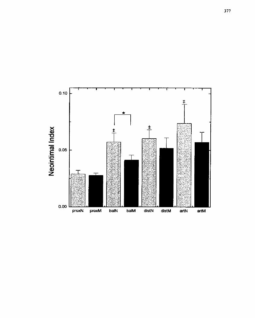
8.2-5 Discussion
Balloon injury o f the porcine femoral artery resulted in a visible intimal
thickening (Figure 75). It was noted that the media1 Iayer had a more cellular and fibrous
appearance than uninjured segments of the femoral artery, possibly indicating that SMCs
from the media1 layer had migrated to the lumen side and initiated the formation of the
intimal lesion. Hypercellularity is also a hallmark of the wound healing response.
Interestingly, the morphological appearance of the MIBG-treated segments was similar to
that of the uninjured controI segments, indicating that MIBG had indeed inhibited
formation of the neointima. In the MBG-treated segment, there appear to be cells that
have partially re-colonized the lumen surface (Figure 75C). Although specific staining for
endothelial cells (ECs) was not done, and is planned for the future, this may suggest that
MIBG is promoting the re-endothelialization of the lumen surface and could potentially
be one of the mechanisms by which MIBG inhibits neointimal formation- The endothelial
layer has been shown to play an important role in maintaining the low rate of SMC
proliferation and migration under normal conditions (Fishrnan et al. 1975).
In the balloon-injured segment, MIBG treatment significantly reduced the
neointimaI index compared to injured segments (Figure 76). MIBG also decreased the
neointimal index in the dista1 and arteriotomy segments, although significance was not
observed (Figure 76). This latter observation may indicate that MIBG is working in part
by promoting positive remodeling of the vesse1 so that the lumen diameter is larger.
Alternatively, MIBG rnay be reducing the sympathetically-mediated vasoconstriction that
occurs following angioplasty (Gregorini et al. 1994; Gregorini et al. 1997). Neuronal
rclease of N E is increased for up to 4 weeks following experimental angiopIasty and
pcrsistcnt liypcrscnsitivity to catccholaniines is scen after angioplasiy (Candipan et ai.
1994). Considcring tl-ic iniportancc of clastic rccoil to constrictive arterial remodeling and
tlius rcstenosia (Ardissino et cd. 1991; Candipan er cd. 1994: Tesfaniariani & Cohen
1988), an effccr by MIBG on this process would provide additional benefits beyond those
specific to cell migration and prdiferation. Studies of the relationship between MIBG
and vasoconstriction sliould be pursued in the future-
The latter tinding from above may also suggest tbat MIBG is capable ofrcducing
intima1 lesion formation regardless of the type of vascular injury that is observed (Figure
76)- The ançioplasty segment is subjected to a stretch/cmsh injury, the distal segment is
primarily an endothdial cell denudation injury and the arteriotomy site contains a cut site
injury witli suture. Regardless of injury type, if SMCs were activated to migrate andor
proliferate, the amount of intima1 tliickening seen at a given site would increase assuming
that intima1 Iiyperplasia has a substantial contribution to restenosis, I f MIBG were
capablc of inhibiting either or botli SMC migation and proliferation, which is shown in
chapter 7, MIBG should effectiveIy decrease the neointimal index.
MlBG was applied to the perivascular surface in Tisseel. TisseeI is a
cornmercially available fibrin glue tlzat is used routinely in the surgicaI suite. The
biochcniical reaction that produces tlic coaguluni involves a rcaction between thrombin
and fibrinogcn. This particular rcaction has been capitalized upon to produce a naturat,
biodegradable adhesivc tliat has since been applied for sustained drug dclivcry (Scnderoff
et al. 199 1)- Intercstingly, duc to the dcpcndency of the reaction on conditions such as pH
and ionic strcngth, tlic microstructure of the fibrin poIynicr can bc manipulatcd to controI
the rciativc availabil ity of cncapsulatcd drug (Scndcro ff cr crl. 1 99 1 ). I n Our cspcrimental
systcni, t l x only conccrns associatcd witli the usc of Tissccl wcrc thc fact that thrombin
lias bccn sho\vn to proniotc f'ibrosis and formation of ncointinia (Gallo et crl. 1998;
Maruyaiiia cr cd. 1997; Pakala & Bciicdict 1999; Salvioni el al. 1998; Stouffcr ef al.
1996) aiid tliat fibrin dcgradation products can proniotc SMC niiçration and prolifct-alion
(Naito et al, 2000). However, considering the results, MIBG apparentfy is suffrciently
potent to overrîde any negative effects that might be attributed to thrombin andor fibrin.
Release of MIBG fkom its depot will Iead to distribution within the adjacent
region of the vessel. MIBG will then enter the cells. The mechanism of entry may involve
either passive diffusion or a NE transporter based uptake and release system. Within
neuroblastoma tumours, the latter mechanism leads to the redistribution of MIBG over a
significant distance from its original point of application (Servidei et al. 1995; Smets et
al. 1989; Weber et al. 1996). In this way, MIBG may be reaching the media1 Iayer and
affecting the migration and proliferation of SMCs. On the other hand, if the adventitia
plays an important role in the formation of neointima (Li et al, 2000; Shi et al. L996a; Shi
et al. 1996b; Zalewski & Shi 19971, then placement of MIBG on the adventitia1 surface
should prevent the migration of myofibroblasts to the luminal surface. Future studies will
examine the distribution and Iocalization of activity for MIBG in preventing neointimal
thickening,
Direct application of MIBG to a vessel sudace avoids the potential probIems
associated with systemic delivery (Taal et al. 1996). However, one of the unknown
factors then becomes the actual concentration of MIBG that reaches the cells/tissue
within the Iocalized area. In these experiments, MIBG at a concentration of 25 rnM was
encapsulated within a 1 .O mL bolus of Tisseel, which was applied to each femoral artery.
It is assumed that 25 mM would not be the concentration that the local tissues are
exposed to after whole body and other local environmental considerations are factored in.
NevertheIess, in any local application, the question as to the amount that reaches the
systemic circulation is a concern. Although the concentration of MIBG encapsultated
within the Tisseel (25 mM) was 3 orders of magnitude higher that than required in Our
ccllular studics for cfficacy (i.c. 20 to 50 PM), the equivalcnt dosage (25 rnM) given
systemicaIIy would be 0.5 mg/kg or 8.9 M . Use of MIBG as a
pharrnacological/chemotherapeutic agent for the suppression of tumour growth (Taal et
al- 1999; Taal et al- 1996; Zuetenhorst er al. 1999) has demonstrated that a non-toxic
single dose bolus in humans is approximately 15 mg/kg. Animal studies of higher dosage
regimens (up to 60 mglkg) have shown that renal function is compromised first (at 40
mg/kg) and is the primary cause of death (Kuin et al. 1999). In these studies, the apparent
systemic dosage would be considerably Iess than the concentrations already employed by
Taal and colIeagues for treatment of carcinoids syndromes (15 mgkg) (Taal et al- 1999;
Taal et al. 1996; Zuetenhorst et al. 1999) and onIy 4 times higher than the concentrations
of MIBG empIoyed for scintillographic detection purposes (Hoefnagel et al. 1987).
Further investigations wiIl be required to determine the local effective concentration of
MIBG after release fiom the Tisseel depot, and the Iength of time that MIBG remains in
the depot and the timc period over which it is released.
MIBG may have an additional effect on inflammation and/or thrombus formation.
Jonsson et al. (L998) demonstrated that MIBG inhibits histamine receptor binding on
endothelial cells and prevents histamine-induced inositol-triphosphate and prostacyclin
production, and arachidonic acid release (Halldorsson et al. 1992). Histamine,
prostanoids and arachidonic acid participatc in activation of pathways for inflammatory
responses (Bach 1982; Hedqvist et al. 2000; Vane & Botting 1987). MLBG may thus be
affecting the progression of restenosis, in part, by inhibiting the inflammatory response.
Moreover, Hauscliildt et al. (1998) have demonstrated that MIBG can prevent the
production of TNF-a and IL-6 at the protein and mRNA level in Iiuman monocytes.
Thus, if MIBG is ablc to inhibit the production of proinflammatory cytokines by
monocytcs, the inflammatory responsc may be considerably reduced and thus any effects
on SMCs dccrcascd. This is highly relcvant to restenosis sincc both TNF-cc and
interleukins have been shown to stimulate migration of SMCs (Shawn Thomas,
unpublished obsenrations; Jang et ai. 1993; Rectenwald et al. 2000; Yue et al. I994), and
the Enflammatory response has been proposed to play a role in intima1 lesion formation
(Biasucci et al. 1999; Forrester et ai. 1991; Nikol & Hofling 1995; Virmani & Farb
1 999).
Longer term studies are now in progress to determine if the inhibitory effects of
MLBG are sustainable beyond 14 days. A 45 day study period also more closely reflects
the time over which patients develop restenosis when relative growth rates are factored
in. Moreover, pathology specimens will enable an exarnination for adverse effects caused
by MIBG with respect to cytotoxicity.
8.3 Summary
This final section of the thesis deals with the use of modeIs of angioplasty and
subsequent restenosis, both in vitro and in vivo, in order to assess the efficacy of MIBG
as an inhibitor of intimal lesion formation. Both the organ culture model of restenosis and
the in vivo femoral artery angioplasty model of restenosis demonstrated significant
formation of neointima in the injured regions of the vasculature in comparison with non-
injured segments. In both models, MIBG treatment was associated with a significant
reduction in intimal lesion size, with a neointimal index (neointimdmedia ratio)
approaching baseline values. These findings suggest that MIBG has potential for use in
the treatment of restenosis post-angioplasty. Furthemore, Tisseel has been identified as
an effective delivery vehicIe. Although a mechanism for the action of MIBG is not yet
known, based on findings in previous chapters of this thesis, it is speculated that MIBG is
affccting an arg-mART that may be important for signaling pathways associatcd with
proliferation and migration of SMCs. Futurc studies will bc dircctcd at proving thc safcty

and effrcacy of MIBG in a porcine mode[ with the goal of initiating clinica1 trials to test
MIBG as a treatment for revascuIanzation-induced restenosis-
9.0 Conclusions and Significance
Twenty years after the first successfûl PTCA procedure (and approximately 30
years after the first successfûl CABG procedure), restenosis remains a significant
complication in the battle against CAD. To date, an effective form of therapy to
neutralize the problem has not been found (Landzberg et al. 1997; Lefkovits & Topo1
1997), although there are promising therapeutic interventions in the early stages of
dinical testing (eg. probuco1, tranilast and radioactive stents)- The time-span covered by
my research has seen many technical, technological, methodological and therapeutic
advances implemented for the treatrnent of CAD and subsequent restenosis. However, the
improvernent in short-term and long-term outcomes has only been satisfactory. Whether
this is due to the increased practice of coronary interventions in the face of lagging
knowIedge of the pathohiology of restenosis remains to be determined. Nevertheless,
advances in our understanding of the biology of the complications resulting from
interventional procedures, coupled with technical improvements, should heIp with the
development of more rational and endunng strztegies for the treatment of restenosis.
Over the past 20 years, much time and effort has been invested in both the
experimental and clinical testing of compounds that target ce11 migration and ce11
proliferation, specifrc growth factor receptors, tlirombus formation and the ensuing
inflammatory process, and, more recently, ECM degradation. Mechanical strategies (Le,
stents) are now favoured in many treatrnent centres, however, upon comparison with
more traditional interventional procedures such as balloon angioplasty, it is apparent that
the rates of restenosis are equivalent. The advent of adenoviral vectors and improvements
in anti-sense strategies coupled with the attractiveness and assumed spccificity of gene
therapy has led many rescarchers to evaluatc this modality of trcatmcnt for restenosis.
Howcvcr, paticnt safcty is still of major conccrn and Iong-tcrm outcomcs arc not provcn.
interestingly, the uniforn~ failure of clinical trials usiilg systeniic thcrapies to prevent
restenosis and the amval of gene therapy sparked a revolution in the field of dmg
delivery systems, since the ability to specifically tarset a region of vascular tissue was
required. lntraluniinal drug and gene delivery systems that could be coupled to the
angioplasty catheter werc developed in quick succession, as were technolo_oics for
polymenc-based, biodegradable perivascular appIication. An assessrnent of thc success of
these delivery systems coupled to pharmacological and gene therapies is still pending,
although controversies about their use and utility have already been reviewed (Chorny et
al. 2000). Nevertheless, the success of therapies applied in these manncrs will be
dependent on drug and vehicle type, dosage, rates of release, length of sustained release,
and ease of cornpliance and integration into the clinical setting.
Restenosis is a niulti-factorial disease process witli a very cornplex pathogenesis-
The Iimited success that has been demonstrated with single modality therapy is Iikely due
to the eficiency at which the body initiates and maintains the repair proccss (and the
myriad of coordinatcd events that are involved therein), and tlic rcdundancy found in the
niany cellular rcsponscs and cellular signaling pathways that are involved. In addition,
the diffcrcnccs in patient-to-patient response, and the diffcrential contribution of
neointimal thickcning, remodeling, elastic recoil, etc., that varies on an individual basis,
add Iayers of complcxity to the treatment of rcstenosis that have yct to be accounted for.
Our prescnt undcrstanding of the progression of artcrial wound rcpaidintimal lcsion
fomiation is liimitcd to the specific ccllular proccsscs (i.c. migration, prolifcration and
(de)-difkrcntiation) that we can nionitor as physical cndpoints in our expcrimental
systcius. On thc othcr Iiand, thc interactions tliat occur bctwcen cclls and thc coordination
of ccllular cvcnts is still rclativcly unknown, as arc rnany of tlic cxtcrnal, cnvironmcntal
and intraccllular hctors and signals,
The studies described within this thesis summarize the efforts to characterize
cornponents of three cellular processes that are critical to the developrnent of restenotic
lesions post-angiopiasty, namely cell proliferation, ce11 migration and phenotypic
plasticity (Le. differentiation). The experimental culture systems that were utilized
provided the capabifity to study distinct endpoints, the contribution of unique factors and
discreet cellular responses, while the animal mode1 that was developed provided a
feasible system for relevant phamacoiogical testing. Using these resources, the work
surnmarized by this thesis is the first to recognize the contribution of post-translational
modification by arginine-dependent mono(ADP-ribosy1)transferases to ce11 proliferation,
ce11 migration and differentiation. Furthemore, MIBG, an inhibitor of arginine-
dependent ADP-ribosylation reactions (Loesberg et al. 1990b; Smets et al. 1990b), was
identified as a potential therapeutic agent for the treatment of restenosis post-angioplasty.
MLBG was previously demonstrated to exhibit anti-proliferative activity and this
property forrned the basis for evaluating its applicability in the treatment of certain
diseases such as cancer. Trials for cancer have been reported (Taal et al. 1999; Taal et al-
1996; Zuetenhorst et al. 1999) and the success rate is moderately good. Application to
other related disease processes, however, has been overlooked. Restenosis is a
multifactonal repair-based disease process that includes a ce11 proliferation component.
Based on this reason alone, MIBG should success h l l y prevent restenosis. However,
highly selective inhibitors of ce11 proliferation have proven unsuccessful in treating
restenosis (Landzberg et al. 1997; Lefkovits & Topo1 1997; Muller et al. 199 1 ; Muller et
ul. 1992b). Nevertheless, we have shown that MIBG should still be able to prevent
restenosis since MIBG is capable of inhibiting celt migration and differentiation in
addition to ce11 proliferation. These muItiple efFccts give MIBG an advantage ovcr
traditional pharmacological intcrvcntions that targct a single ccllular cvcnt (ix,
proliferation or migration or differentiation). Thus. MIBG, and any compound with
similar properties, should be suitable for the purpose of preventing vascular restenosis
following injury regardless of cause (eg. angioplasty, vascular grafting, etc.).
How does MIBG influence al1 three cellular events and inhibit restenosis? The
answer lies with the ability of MIBG to inhibit the activity of arginine-dependent
mono(ADP-ribosyl)transferases. ADP-ribosylation reactions are activated in vanous ce11
types in response to a variety of stimuli, and the activation of these regulatory enzymes
may be an important component of ce11 proliferation, ce11 differentiation and ce11
migration (Kharadia el al. 1992; Saxty et al. 1998a; Terashima el al. 1992; Thyberg er al.
1995b). Oddly enougli, even after having been studied for over thirty years, the precise
regulatory functions of these enzymes are not well known. What is intriguing though, is
that ADP-ribosylation, much like p hosphorylation, is reversible via the action of ADP-
ribosyl-hydrolases (Moss et al. 1997). But how ADP-ribosy lation exerts its regulatory
effects remains a mystery. In large part, this uncertainty is a consequence of our lack of
information regarding the targets of this modification. It is conceivable that ADP-
ribosylation reactions operate tlirough G proteins, which are not typically regulated by
phosphorylation. Integration of ADP-ribosyltransferases as a regulatory component of
cellular signaling patliways would add a further layer of control, and thereby pemiit more
subtlc coordination of ce11 functions. In accordance with this prcmise, dual regulation of
bFGF by both phosphorylation and ADP-ribosylation has becn dcmonstrated (Boulle et
al. 1995; Feige & Baird 1989; Jones & Baird 1997; Vilgrain & Baird 199 1 ; Vilgrain et ( I I .
1993). Ofparticular interest is that fact that this faniily of enzymes appears to be closely
linked to distinct ccllular processcs, namcly prolifcration, migration and differentiation
(Kliaradia et al. 1992; Saxty ef al. 199%; Tcrashima el al. 1992; Thybcrg et al. 1995b).
tliat arc criticai componcnts of rcstcnotic lcsion formation (Büutcrs & Isncr 1997; Davics
& Hagen 1994). Thus, MIBG, an inhibitor of arginine-dependent niono(ADP-
ribosy1)ation reactions, would inhibit restenosis via its effect on multiple and independent
targets. How ADP-ribosyIation functions to regulate each of these distinct cellular
processes (Le. proliferation, migration and differentiation) remains to be detemined and
should be the subject of future studies.
While it is fine to discuss the individual events involved in the pathogenesis of
restenosis post-angioplasty, neointimal formation is actually highly integrated and
coordinated with other systernic and local processes in the whole animal, such as
thrombus formation, vesse1 spasm/elastic recoil, inflammation, and remodehg (Biasucci
et al 1999; CaZiff et al. 199 1; Glagov 1994; Liu et ai. 1989). Whether MIBG also has an
effect on these phenomena, however, remains to be detennined. Based on its structure
and reports in the Iiterature, at Ieast one possibility exists by which it may influence these
processes independent of its effect via arg-rnART. Since MIBG resernbles NE and is
taken into cells via the NE specific uptake- 1, it may modulate adrenergic function, which
has been shown to have detrimental effects towards elastic recoil and the development of
restenosis (Candipan et al. 1994; Gregorini et al. 1994; Gregorini et al. 1997). MIBG
may thus be able to decrease elastic recoil. In this way, the long-term ramifications for
arterial remodeling may be improved, adding yet another beneficial feature to the
restenosis fighting capacity of MIBG.
At the ceIlular level, MtBG does not appear to function by inhibiting traditionai
kinase cascade signaling pathways sincc the cellular intemediates nornîally associatcd
with proliferation, migration and differentiation, specifically MAP kinase, Pt3-kinase and
p2 1-Ras (Coolican et al. 1997; Cowley et al. 1994; Klcmke et al. 1997; Lopez-Ilasacü
1998; Marshall 1996; Pages el al. 1993; Poser et al. 2000; Pyles et al. 1997), arc not
afrcctcd by MIBG. Yct, MIBG is capüblc of intcrfcring with agonist-dcpcndcnt c--JS
mRNA expression. An alternate pathway for thc regutation of c-jos. and its downstreani
effects, by MIBG has tlierefore been proposed. A mode1 has been developcd in which the
traditionat pathways that are initiated by growth factors and other stimuli diverge
intracellularly. but converge at the level of the nuclcus to regutate the transcription of
early growth response genes (Figure 77). Since the induction of c-fos gsne transcription
requires the coordinate activation of several transcription factors including CREB. SRF
and Elk-1 for temary cornplex formation. a contribution by multiple intracelMar
pathways and intemediatcs is not implausible. Furthermore, the activation of at least one
of these transcription factors (SRF) is regulated by a signaling pathway that is linked to
the srna11 GTP-binding protein Rlio. Since the ADP-ribosylation of Rho has already been
documented in the literature (-4ktories 1997; Aktories & Wegner 1992; Lemi er al- 2000:
Schmidt & Aktorics 1998; Tsakiridis er al. 1998), MIBG couId inhibit c-fos gene
expression via this route, indcpendent of MAP kinase.
The research surnmarized in this thesis presents evidence that MIBG will be a
useful tool in the prevention of restenosis post-angioplasty. Further rescarcli will need to
confirm not onIy the efficacy of this conipound, but also to clarify the safety of this
conipound whcn directly applied to vascuIar tissues. Although extensive study of whole
body toxicity has establishcd the limits for systemic appIication, safcty concems for local
delivcry must be addressed before the compound can proceed to clinical trials. In
addition, it will be of utmost importance to detcmine the niode of action of MIBG within
ccllular and cxpcrinicntal animal niodcl systcnis, and to charactcrizc t t~c niodc of
distribution not only within a c d , but also as it pcrtains to local dclivcry. Tlius, future
cspcrirncnts arc rcconmcndcd to procccd in a direction tliat will dcfinc not only tlic
functions of MIBG, but will also providc ncw insight into the pathobiology of rcstcnosis.
FIGURE 77: Model of Signal Intermediates Associaled with c-fos Gene tenduction by a
Growth Stimulus or Injury.
Targets for inhibitors of the traditional kinase cascade leading to c-fos gene transcription
are shown. A plausible site for MIBG action in a parallel signaling pathway is presented.
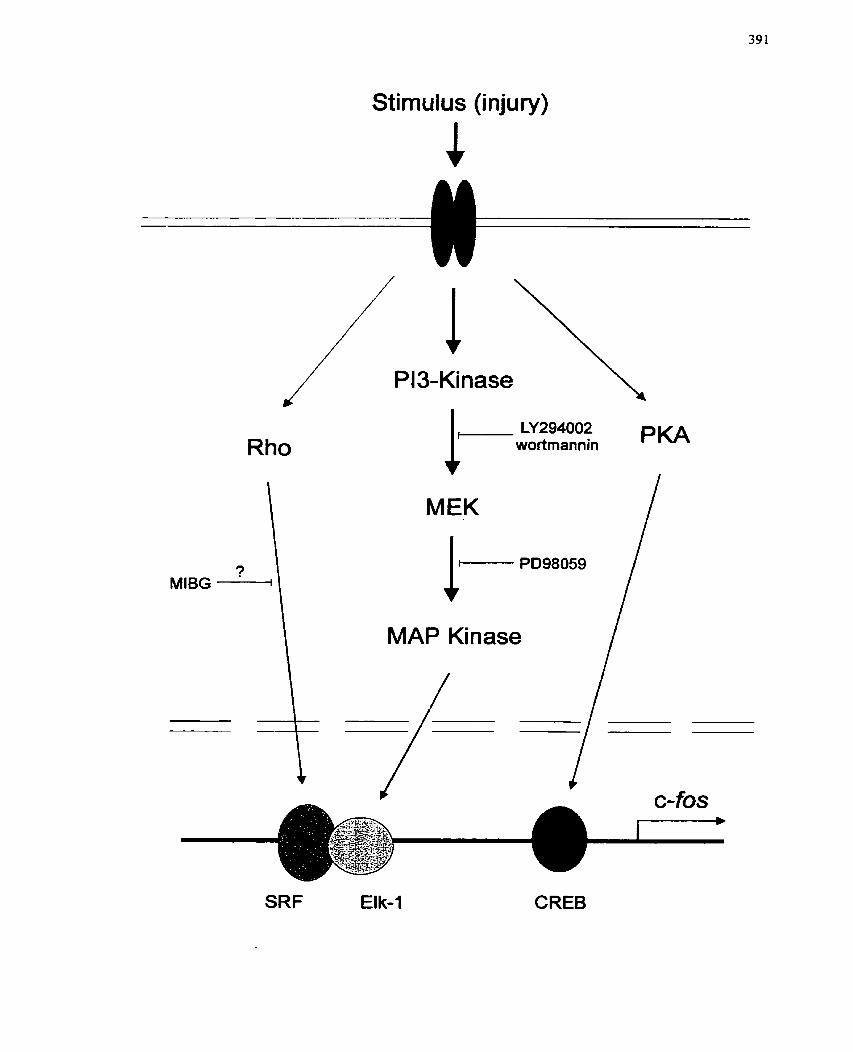
Stimulus (injury)
1- LY294002 wortmannin
v MEK
PKA
MAP Kinase 1
SRF Elk-1 CREB
Research efforts deaIing with the problem or restenosis promise to bring new
modalities of therapy to cIinicaI trial before the end of the next decade. Tt is hoped that
the novel compound descrïbed herein, MIBG, will be thoroughly investigated with
respect to its clinical suitabiIity so that patients undergoing revascularization procedures
for CAD or other peripheral vascular disease could benefit frorn these findings.
Regardless, in the end, it is hoped that the studies reported within this thesis will provide
a broader perspective for understanding the cornplex biology of restenosis and the
coordinated cellular repair process that it represents, and ultimately assist in the rational
design of new compounds for the treatment of restenosis.
10.0 REFERENCES
Abedi, H., Dawes, K.E. & Zachary, 1. (1995). Differentiat eftècts of platelet-derived growth factor BB on pl25 focal adliesion kinasc and pasillin tyrosine phosphorylation and on ceII migration in rabbit aortic vascular smooth muscle cells and Swiss 3T3 fibroblasts. J Biol Ctlerir 270: 1 1367-76-
Abedi, H, & Zachary, 1- (1995)- Signalling mechanisms in the replation of vascular ce11 migration. Cui-clio vasc Res 30: 544-56.
Aboul-Ela, N., Jacobson, E.L. & Jacobson, M.K. (1988). Labeling niethods for the study of poiy- and mono(ADP-ribose) metabolism in cultured cells. Aiial Bioclzeriz 174: 239-50,
Adam, L.P., Franklin, M.T., Raff, G.J. & Hathaway, D.R. ( 1995). Activation of mitogen- activated protein kinase in porcine carotid artenes. Cif-c Res 76: 183-90.
Adarnietz, P, (1987). Poly(ADP-ribose) synthase is the major endogenous nonhistone acceptor for poIy(ADP-ribose) in alkylated rat hepatoma cells. ELU- J Biocher~z 169: 365-72.
Adelman, A.G., Cohen, E.A., Kimball, B.P., Bonan, R., Ricci, D.R., Webb, J.G., Laramee, L., Barbeau, G., Traboulsi, M., Corbett, B.N. & et al, (1993). A companson of directionai atherectomy with balloon angioplasty for lesions of the left anterior dcscending coronary artet-y. N ErzgZ J Med 329: 235-33.
AHA, A.H,A. (1999). Economic Cost of Cardiovascular Disease, Arizericniz Hear-r Associatiorz. www.americanlieart,or~statistics.
Ahn, N-G., Seger, R., Bratlien, R.L. & Krebs, E.G. ( 1992). Growth factor-stiniulated phosphorylation cascades: activation of growth factor-stimuIated MAP kinase. Ciba Fozrrzd Synp 164: 1 13-26.
Akle, C.A., Wardle, D.G., Bomanji, J.B. & Talbot, 1. (1997). mIBG diagnosis and tlierapy in smooth muscle tumours of the srnaII bowel. Ezrr- J Nucl Med 24: 1 196.
Aktories, K. (1994). Clostridial ADP-ribosylating toxins: effects on ATP and GTP- binding proteins. Mol Cell Biocheriz 138: 167-76.
Aktories, K. (1997). Rho proteins: targets for bacterial toxins. Tt-erds k1ic.r-obioi 5: 282-8.
Aktorics, K. & Wcgncr, A. (1992). Mechanisms of thc cytopathic action of actin-ADP- ribosylating toxins. Mol Mkt-obiol6: 2905-8.
Alcssi, DR., Cucnda, A., Cohci~, P., Dudlcy, D.T. & Saltiel, A.R. ( 1 995). PD 098059 is a spccific inhibitor of thc activation of niitogcn- activatcd protcin kinasc kinasc i n vitro and in vivo. J Biol Clzmu 270: 27489-94.
Alexander, Schlant gi. Fustcr. ( 199s). Hurst's The Hcart. . McGraw-Hill, New York.
Alexander, R.W. & Gimbrone. M.A.. Jr. (1976). Stimulation o f prostaglandin E synthesis in cultured hun~an unibilicaI veiti smooth muscle c e l k froc Ncitl Actrci Sci U S il 73: 1617-20.
Ali, N. & Agrawal, D.K. (1993). Guanine nucleotide binding regulatory proteins: tlieir characteristics and identification. J Pllai-irrncol Tmicoi ~ t k r l ~ o d ~ 32: 1 87-96.
Allport. J.R., Donnelly, L.E., Hayes, B.P.. Murray, S., RcndelI, N.B., Ray, K.P. & MacDermot, J. ( 1996a). Reduction by inliibitors of nmno(ADP-ribosy1)transferase of chernotaxis in human neutropliil teucocytes by inhibition o f the assenibly of filamentous actin, BI- J Plzm- nac coi 1 18: 1 1 I 1 -8.
Ailport, J.R-, Donnelly, L.E., KeFalas, P., Lo, G., Nunn, A., Yadollahi-Farsani, M., Rendell, N.B., Murray, S., Taylor, G.W. & MacDernlot, J- (1996b)- A possible role for mono (ADP-ribosyl) transferase in the signalling pathway mediating ncutrophil chemotaxis. Br J C h Plzur-nzacol42: 99- 106.
Althaus, F.R. & Richter, C H (1957). ADP-ribosylation o f proteins: Enzyniology and biological significance. In: . Solioz, M. (edç.). Springer, Berlin. pp 1-237.
Alvarez-Gonzalez, R, & Jacobsen, M.K. (1957). Characterization o f polymers of adenosine diphospliate ribose generated in vitro and in vivo. f3ioclzenzisti~- 26: 32 18-24.
Alvarez-Gonzalez, R., Pacheco-Rodriguez, G. & Mendoza-Alvarez, H. (1994). Enzymology of AD P-ribose polyrner syntIiesis. Mol CelZ Bioclzenr 138: 33-7.
Anle, J-C., RolIi, V., Schreiber, V., Niedergang, C., Apiou, F., Decker, P., Muller, S., Hoger, T., Menissier-de Murcia, J. & de Murcia, G. (1999). PARP-2, A novel rnamrnalian DNA damage-depetident poly(ADP-ribose) polymerase. J Bioi Clzetil 274: 17560-5.
Anistad, P.A., Krupitza, G. & Cerutti, P.A. (1992). Mcchanisin o f c-fos induction by activc oxygcn. Cmcer- Res 52: 3952-60.
Andersen, H.R., Macng, M., Tlionvest, M. & Falk, E. (1996). Rcniodcling rathcr than ncointinial formation cxplains Iuminal narrowing afier dccp vcsscl wall injury: insiglits froni a porcine coronary (rc)stcnosis inodcl. Ci~-c'trlutior~ 93: 17 16-34.
Andrawis, N.S., R~ilcy, EH. & Abcrncthy, D.R. ( 1993)- Ançiotcnsin I I rcsulatcs hr111ia11 vrisculür sniootli inrisclc alpha-actiii gcnc cxpt-cssion. B~OCIIC'IIZ Biopb"; /?es Corw~irr/i 196: 963-8.
Andres, V. (1998). Control of vascular smooth muscle ce11 growth and its implication in atherosclerosis and restenosis- ltrr J Mol Med 2: 8 1-9,
Arai, H. & Escobedo. J.A. (1996). Angiotensin II type 1 receptor signals through Raf-1 by a protein kinase C-dependcnt, Ras-independent mechanism. Mol Plzar-171acol 50: 522-8-
Arcaro, A. & Wymann, M.P, (1993)- Wortmannin is a potent phosphatidylinositol 3- kinase inhibitor: the role of phosphatidylinositol 3,4,5-trisphosphate in neutropliil responses. Biochenz J 296: 297-30 1,
Arciniegas, E., Ponce, L., Hartt, Y., Graterol, A. & Carlini, R.G. (2000). Intima1 thickening involves transdifferentiation of embryonic endothelial cells. Anat Rec 258: 47-57.
Ardissino, D., Barberis, P., De Servi, S., Merlini, P.A., Bramucci, E., Falcone, C- & Specchia, G. (1991). Abnormal coronary vasoconstriction as a predictor of restenosis after successful coronary angioplasty in patients with unstable angina pectoris. N Engl JMed 325: 1053-7.
Arundel-Suto, CM., Scavone, S.V., Turner, W.R., Suto, M.J. & Sebolt-Leopold, J.S- (1991). Effect of PD 128763, a new potent inhibitor of poly(ADP-ribose) polyrnerase, on X-ray-induced cellular recovery processes in Chinese hamster V79 cells. Radiar Res 126: 367-7 1.
Asada, Y., Kisanuki, A., Hatakeyama, K., Takahama, S., Koyaina, T., Kurozumi, S. & Sumiyoshi, A. ( 1994). inhibitory effects of prostacyclin analogue, TFC- 132, on aortic neointimal thickening in vivo and smooth muscle ce11 proliferation in vitro. Pt-ostaglandins Leukor Essent Fatty Acids 51 : 245-8.
Asada, Y., Kisanuki, A., Tsuneyoshi, A,, Manitsuka, K., Hatakeyama, K. & Surniyoshi, A, (1996)- Effects of inflation pressure of balloon cathcter on vascular injuries and subsequent development of intima1 hyperplasia in rabbit aorta. Atherosclei-osis 12 1 : 45-53,
Babai, F., Musevi-Aghdam, J., Schurch, W., Royal, A. & Gabbiani, G. (1990). Coexpression of alpha-sarcomeric actin, alpha-smooth muscle actin and desmin during myogenesis in rat and mouse embryos 1. Skeletal muscle. Dffet-e~ztinfiorr 44: 132-42.
Bach, M.K. (1982). Mediators of anaphylaxis and inflammation. Atznrr Rev Microbiol36: 371-413.
Badimon, J.J., Ortiz, A.F., Meyer, B., Mailhac, A., Fallon, J.T., Falk, E., Badimon, L., Chescbro, J.H. & Fuster, V. (1998). Difkrcnt response to balIoon angioplasty of carotid and coronary artcrics: effccts on acute platclct dcposition and intima1 thickcning. Aht-osclet-osis 140: 307- 14.
Badimon, L., AIfon, J., Royo, T-, Berrozpe, M., Martinez-Gonzalez, J., Vidal, F., Chesebro, J.H., Fuster, V. & Badimon, J.J. (1995). Ce11 biology of restenosis post- angioplasty. Kat-dioz 84: 145-9.
Bail, E-H,, Narindrasorasak, S. & Sanwal, B.D. (1979). The cyclic adenosine monophosphate phosphodiesterases of rnyoblasts, fibrob Iasts, and their somatic ce11 hybrids. Caiz JBiochem 57: 1220-8.
Bail, E.H. & Sanwal, B.D. (1980). A synergistic effect of glucocorticoids and insulin on the differentiation of myoblasts. J Cell Physiol102: 27-36.
Ball, E.H., Seth, P.K- & Sanwal, B.D. (1980). Regulatory mechanisms involved in the control of cyclic adenosine 3':S- monophosphate phosphodiesterases in myoblasts. J Bial Chenr 255: 2962-8.
Banai, S., Haggroth, L., Epstein, S.E. & Casscells, W. (1990). Influence of extracellular magnesium on capillary endothelia1 ce11 proliferation and migration. Circ Res 67: 645-50.
Banasik, M-, Komura, H., Shimoyama, M. & Ueda, K. (1992). Specific inhibitors of poly(ADP-ribose) synthetnse and mono(ADP- ribosy1)transferase. J Biol Chem 267: 1569-75.
Banasik, M, & Ueda, K, (1 994). Inhibitors and activators of ADP-ribosylation reactions. Mol Cell Biochem 138: 185-97.
Banskota, N.K., Taub, R-, Zellner, K. & King, G,L. (1989). Znsulin, insulin-like growth factor 1 and platelet-derived growth factor interact additively in the induction of the protooncogene c-myc and cellular prdiferation in cultured bovine aortic smooth muscle cells. Mol Endocrinol3: 1 183-90.
Barry, W.L., Wiegrnan, P.J., Gimple, L.W., Gertz, S.D., Powers, E.R., Owens, G.K. & Sarembock, IJ. (1997). A new singie-injury model of balIoon angioplasty in cholesterol-fed rabbits: beoeficial effect of hirudin and cornparison with double- injury model. Lab Invest 77: 109- 16.
Bar-Shavit, R., Benezra, M., Eldor, A., Hy-Am, E., Fenton, J.W.d., Wilner, G.D. & Vlodavsky, 1. (1990). Thrombin immobilized to extracellular matrix is a potent mitogen for vascular smooth muscle cells: nonenzymatic mode of action. Cell Regul 1: 453-63.
Baserça, R., Porcu, P., Rubini, M. &: Sell, C. (1994). Ce11 cycle control by the IGF-1 receptor and its ligands. In: Current directions in insulin-like growth factor research. LcRoith, D. & Raizûda, M.K. (cds.). Plenum Press, Ncw York. pp 105- 112.
Battistini, B., Chtailler, P., D'Orleans-Juste, P., Briere, N. & Sirois, P. (1993). Growth regulatory properties of endothelins, Peptides 14: 385-99-
Bauer, P.L, Buki;, K.G-, Hakam, A- & Kun, E. (1990)- Macromolecular association of ADP-ribosyltransferase and its correlation with enzymic activity. Biochenz J 270: 17-26.
Bauters, C. & ismer, J.M. (1997). The biology of restenosis. Pi-og Car-diovcrsc Dis 40: 107-16.
Beale, E., Andreone, T., Koch, S., Granner, M. & Granner, D. (1984). Insulin and glucagon neguIate cytoso!ic phosphoenolpynivate carboxykinase (GTP) mRNA in rat b e r - Diaberes 33: 328-32-
Bell, L. & Madri., LA. (1989). Effect of platelet factors on migration of cultured bovine aortic end*othelial and smooth muscle ce1 1s. Circ Res 65: 1057-65.
Bell, L. & Madrn, J.A. (1990). Influence of the angiotensin system on endothelial and srnooth mr uscle ce11 migration- Anz J Pathol 137: 7- 12.
Beneke, R., Geisen, C., Zevnik, B., Bauch, T., Muller, W.U., Kupper, J.H. & Moroy, T. (2000). D'NA excision repair and DNA damage-induced apoptosis are linked to Poly(ADF-ribosy1)ation but have different requirements for p53. Mol Cell Biol 20: 6695-703,
Berger, N.A. (1985). Poly(ADP-ribose) in the cellular response to DNA damage. Ra&& Res 101: 4-15.
Berk, B.C. & ALexander, R.W. (1989). Vasoactive effects of growth factors. Biochertz Phat-nzacml38: 2 19-25.
Bemal, S.D. & Stahel, R.A. (1985). Cytoskeleton-associated proteins: their role as cellular in-tegrators in the neoplastic process. Crir Rev Oizcul He,natoI 3: 19 1-204.
Bernstein, L.R., Antoniades, H. & Zetter, B.R. (1982). Migration of cultured vascular cells in response to plasma and platelet- derived factors. J CeII Sci 56: 7 1-82.
Biasucci, L.M., Liuzzo, G., Buffon, A. & Maseri, A. (!999). The variable roIe of inflammattion in acute coronary syndromes and in restenosis. Senzin itlrerv Curdiol4= 105- 1 O.
Birukov, KG., F:rid, M.G., Rogers, J.D., Shirinsky, V.P., Koteliansky, V.E., Campbell, J.H. & Campbell, G.R. (1993). Synthesis and expression of smootli muscle phenotype markers in primary culture of rabbit aortic smooth muscle cells: influence of seeding dcnsity and media and relation to ce11 contractility. Exp Cell Res 204: 46-53.
Black, B.L. & Olsoii, E X . (1998). Transcriptional control of iiiuscle development by myocyte cnhancer factor-, (MEFZ) proteins. A n m Rev Cell Dev Bi01 14: 167-96.
Blake, G.M., Lewington, V.J., Zivanovic, M.A. & Ackery, D.M. (1989). Glomerular filtration rate and the kinctics of 123 1- metaiodobenzylguanidine. Etri- J MrcZ Med 15: 618-23.
Blank, R.S. & Owens, G.K. (1990). Platelet-derived growtli factor regulates actin isoform expression and growth state in cultured rat aortic sniooth muscle cells. J Cell Plzysiol 142: 635-42.
Blank, R.S., Swartz, E.A., Tliompson. M.M.. Olson, E.N. 8( Owens, G.K. (1995). A retinoic acid-induced clona1 ce11 line derived from multipotential P 19 embryonal carcinoma cells expresses smoo th muscle c haracteristics. Cfi-c Res 76: 742-9.
Bobik, A. & Campbell, J.H. (1993). Vascular derived growtli factors: ce11 biology, pathophysiology, and pharrnacology. Phal-nzacol Rev 45: 1-12.
Bobik, A., Grooms, A., Millar, .LA., Mitchell, A. & Grinpukel, S. (1990). Growth factor activity of endothelin on vascular smooth muscle. Atn J P/zji;iol258: C4O8- 15.
Bochaton-Piallat, M.L., Gabbiani, F. & Gabbiani, G. (1994). Heterogeneity of rat aonic smooth muscle ce11 replication during development: correlation with replicative activity after experirnental endothelial denudation in adults. J Subtnict-osc Cylol Patlzol26: 1-8-
Bochaton-Piailat, M.L., Gabbiani, F., Ropraz, P. & Gabbiani, G. (1992). Cultured aortic smooth muscle cclls from newborn and adult rats show distinct cytoskeletal features. D$fel-mticliio~~ 49: 175-85.
Bochaton-Piallat, M.L., Gabbiani, F., Ropraz, P. & Gabbiani, G. (1993). Age influences the replicative activity and the differentiation features of cultured rat aortic smooth muscle cell populations and clones. Artel-iosclet- Tlli'o~nb 13 : 1449-55.
Bochaton-Piallat, M.L., Ropraz, P., Gabbiani, F. & Gabbiani, G. (1996). Phenotypic heterogeneity of rat arterial sn-iooth muscle ce11 clones. Implications for the development of expcrirncntal intima1 thickening. AI-teriosclel- Th-o t~b V m c Biol 16: 8 15-20.
Bonin, P.D., Fici, G.J. & Sinçli. J.P. ( 1989). Interlcukin- 1 proiiiotcs prolifcration of vascular smootli inusclc cells in coordination with PDG F or a n-ionocytc deri vcd growth factor. Esp Cdl Res 18 1: 475-82.
Bonser, R.W.. Thoiiipson, NT., Randall. R.W., Tatcson, J.E., Spaccy, GD., Hodson, H.F. & Gai-land, L.G. ( 199 1 ). Dcmctlioxyviridin and wortniai-inin block phospliolipasc C and D activation in tlic Iiuiuan iicutropliil. Bi- J Pliril-~izucol 103: 1237-4 1 .
Bornfeldt, K.E., Campbell, J-S., Koyama, H., Argast, G.M., Leslie, C C , Raines, E.W., Krebs, E.G. & Ross, R. (1997). The mitogen-activated protein kinase pathway can mediate growth inhibition and proliferation in smooth muscle c e k . Dependence on the availability of downstream targets. J Clin Invesr 100: 875-85.
Bos, J.L. (1995). p21ras: an oncoprotein functioning in growth factor-induced signal transduction. Eztr- J Cartcer 3 1 A: 105 1-4,
Boulikas, T. (1988). At least 60 ADP-ribosylated variant histones are present in nuclei from dimethylsulfate-treated and untreated cells. EMBO J 7: 57-67.
Boulikas, T. (1990). PoIy(ADP-ribosylated) histones in chromatin replication. J Bi01 Chem 265: 14638-47.
Boulle, N., Jones, E.M., Auguste, P. & Baird, A. (1995). Adenosine diphosphate ribosylation of fibroblast growth factor-2. Mol Endocrinol 9: 767-75.
Bourcier, T., Dockter, M. & Hassid, A. (1995). Synergistic interaction of interleukin-1 beta and growth factors in primary cultures of rat aortic smooth muscle cells. J Cell Physiol164: 04-4-57.
Bowen-Pope, D.F. & Ross, R. (1982). Platelet-derived growth factor. II. Specific bindin; to cultured cells- J Biol Chem 257: 5 16 1-7 1.
Bowen-Pope, D.F., Ross, R. & Seifert, R.A. (1985). Locally acting growth factors for vascular sn~ooth muscle cells: endogenous synthesis and release from platelets. Circulation 72: 735-40.
Bowes, J., McDonald, MC., Piper, J. & Thiemermann, C. (1999). Inhibitors of poly (ADP-ribose) synthetase protect rat cardiomyocytes against oxidant stress. Caudiovasc Res 41: 126-34,
Bowes, J., Piper, J. & Thiememlann, C. (1998). Inhibitors of the activity of poly (ADP- ribose) synthetase reduce the ce11 death caused by hydrogen peroxide in human cardiac myoblasts. Br J Phar-macol 124: 1760-6.
Brand-Saberi, B. & Christ, B. (1999). Genetic and epigenetic control of muscle developmcnt in vertebratcs, Cell Tissue Res 296: 199-2 12.
Bravo, R. (1990). Genes induced during the GO/G 1 transition in nlouse fibroblasts. Serzzirz Curzcer- Blol 1: 37-46.
Brizzi, M-F., Dcfilippi, P., Rosso, A., Vcnturino, M., Garbarino, G., Miyajima, A., Silengo, L., Taronc, G. & Pcgoraro, L. (1999). Integrin-mcdiatcd adhesion of endothelial cclls induces JAK2 and STATSA activation: roIe in thc control of c- fos genc cxprcssion. Mol Biol Cell 10: 3463-7 1 .
Brogan, W.C.d., Poprna, J.J., Pichard, AD., Satler, L.F., Kent, KM., Mintz, G.S. & Leon, M,B. (1993). Rotational coronary atherectomy after unsuccessfu1 coronary balloon angioplasty. Am J Car-di01 71: 794-8-
Brown, C., Clark, L., Williams, L., Gallagher, S-, Levesque, M. & Silva, J. (1996). PearIs for Practice: Coronary Restenosis. Journal of the Americnn Academy of Nurse Practitioners 8: 28 3-28 8 -
Brune, B., DimmeIer, S., Molina y Vedia, L. & Lapetina, E.G. (1994). Nitric oxide: a signal for ADP-ribosylation of proteins. Lzye Sci 54: 6 1-70.
Brune, B. & Lapetina, E.G. (1989). Activation of a cytosolic ADP-ribosyltransferase by nitric oxide- generating agents. J Bi02 Chem 264: 8455-8.
Buchberger, A., Ragge, K. & Arnold, K.H. (1994). The rnyogenin gene is activated dunng myocyte differentiation by pre- existing, not newly synthesized transcription factor MEF-2. JBiol Chenz 269: 17289-96-
Bucher, M.L. & Swaffield, MN. (1975). Regulation of hepatic regeneration in rats by synergistic action of insulin and glucagon. Proc Nat1 Acad Sci U S A 72: 1 157-60.
Burkart, V., Blaeser, K. & Kolb, H. (1999). Potent beta-ce11 protection in vitro by an isoquinolinone-derived PARP inhibitor. Horiii Melab Res 3 1 : 64 1-4.
Burke, J.M. & Ross, R. (1979). Synthesis of connective tissue macromolecules by smooth muscle. Int Rev Connect Tisszte Res 8: 1 19-57.
Burkle, A., Muller, M., Wolf, I. & Kupper, J.H. (1994). Poly(ADP-ribose) polyrnerase activity in intact or permeabilized leukocytes fiom mammalian species of different longevity. Mol Cell Biochenz 138: 85-90.
Califf, R.M., Fortin, D.F., Fnd, D.J., Harlan, W.R.d., Ohman, E.M., Bengtson, J.R., Nelson, CL., Tcheng, .LE-, Mark, D.B. & Stack, R.S. (199 1). Restenosis after coronary angioplasty: an overview. JAin C d Cardiol 17: 2B-13B.
Campbell, J.H. & Campbell, G.R. (1997). The ce11 biology of atherosclerosis--new developments. Atisr N Z J Med 27: 497-500.
Campbell, J.H., Campbell, G.R., Kocher, O. & Gabbiani, G. (1987). Ce11 biology of smooth muscle in culture: implications for atherogenesis. [nt Aizgiol6: 73-9.
Campbell, J.H., Kocher, O., Skalli, O., Gabbiani, G. & Campbell, G.R. (1989). Cytodiflerentiation and expression of alpha-smooth muscle actin mRNA and protein during primary culture of aortic smooth muscle cells. Correlation with ce11 density and pro1 i ferativc state. A~-terioscIerosis 9: 53 3-43.
Canada, H. (1995). Heart Disease and Stroke in Canada, Heahlz Canada. www-hc- sc.gc/hpb/lcdc/bcrdd/hdsc97.
Candipan, R.C., Hsiun, P.T., Pratt, R. & Cooke, J.P. (1994). Vascular injury augments adrenergic neurotransmission. Ciradation 89: 777-84.
Carey, D.J. (1991). Control of growth and differentiation of vascular cells by extracelIular matrix proteins. Annu Rev Physiol53: 16 1-77.
Carson, D.A., Seto, S., Wasson, D.B. & Carrera, C L (1986). DNA strand breaks, NAD metabolism, and programmed ce11 death. E-rp Cell Res 164: 273-8 1.
Carter, A.J., Laird, J.R., Farb, A-, Kufs, W-, Wortham, D.C. & Virmani, R. (1994). Morphologie characteristics of lesion formation and time course of smooth muscle ce11 proliferation in a porcine proliferative restenosis model. J Anz COU Cardiol24: 13 98-405.
Casscells, W. (1992). Migration of smooth muscle and endothelial cells. Critical events in restenosis. Circulation 86: 723-9.
Cassel, D. & Pfeuffer, T. (1978). Mechanism of cholera toxin action: covalent modification of the guanyl nucleotide-binding protein of the adenylate cyclase system. Proc Nati Acad Sci U S A 75: 2669-73.
Casterella, P.J. & Teirstein, P.S. (1999). Prevention of coronary restenosis. Cardiol Rev 7: 219-3 1.
Catalioto, R-M,, Renzetti, A.R., Criscuoli, M., Morbidelli, L. & Subissi, A. (1996). Role of calcium in angiotensin II-induced prostaglandin release and DNA synthesis in rat vascular smooth muscle cells. J Cardiovasc Plzarmacoi27: 2 95-200.
Caterina, M.J. & Devreotes, P.N. (199 1). Molecular insights into eukaryotic chernotaxis. FASEB J 5: 3078-85,
Cercek, B., Sharifi, B., Barath, P., Bailey, L. & Forrester, J.S. (199 1). Growth factors in pathogenesis of coronary arterial restenosis. Am J Cardiol68: 24C-33C.
Cesarone, CF., Scarabelli, L., Scovassi, A.[., Izzo, R., Menegaui, M., Carcereri De Prati, A., Orunesu, M. & Bertazzoni, U. (1990). Changes in activity and mRNA levels of poly(ADP-ribose) polymerase during rat liver regeneration. Biochiirz Biophys Acla 1087: 241-6-
Chamley, J.H., Campbell, G.R., McConnell, J.D. & Groschel-Stewart, U. (1977). Cornparison of vascular smooth muscle cells from adult human, monkey and rabbit in primary culture and in subcuIture+ Cell Tissue Res 177: 503-22.
Charnley-Campbell, J., Campbell, G.R. & Ross, R. (1979)- The smooth muscle ce11 in culture, Physiol Rev 59: 1-6 1.
Charnley-Campbell, J.H-, Campbell, G.R. & Ross, R. ( 198 1)- Phenotype-dependent response of cultured aortic smooth muscle to serum rnitogens. J Cell BioZ89: 379- 83.
Chang, Y.C., Sornan, G. & Graves, D.J. (1986). Identification of an enzymatic activity that hydrolyzes protein-bound ADP-ribose in skeletal muscle. Biocltern Biophys Res Corizrnzuz 139: 932-9.
Chao, D., Severson, D.L., Zwiers, H. & Hollenberg, M.D. (1994). Radiolabelling of bovine rnyristoylated aIanine-rich protein kinase C substrate (MARCKS) in an ADP-ribosyIation reaction- Biochern CeZl Biol72: 39 1-6.
Cheatham, B., Vlahos, C.J., Cheatham, L., Wang, L., Blenis, J. & Kahn, C.R. (1994). PhosphatidyIinositol 3-kinase activation is required for insulin stimulation of pp70 S6 kinase, DNA synthesis, and glucose transporter translocation. Mol Cell Bioi 14: 4902- 1 1,
Chen, Q., Kinch, M.S., Lin, T.H., Bum-dge, K. & Juliano, R.L. (1994). Integrin-mediated cell adhesion activates mitogen-activated protein kinases. J Biol Chem 269: 26602-5.
Cheng, T C , Wallace, M C , Merlie, J.P. & Olson, E.N. (1993). Separable regulatory elements governing myogenin transcription in mouse embryogenesis. Science 261: 215-8.
Choi, J., Costa, M.L., Mermelstein, CS., Chagas, C., Holtzer, S. & Holtzer, H. (1990). MyoD converts primary derrnal fibroblasts, chondroblasts, smooth muscle, and retinal pigmented epithelial cells into striated mononucleated myoblasts and multinucleated myotubes. Proc Natl Acad Sci U S A 87: 7988-92.
Chomy, M., Fishbein, 1. & Golornb, G. (2000). Drug delivery systems for the treatment of restenosis. Cid Rev Ther Drug Carrier Syst 17: 249-84.
Chougule, P.B. (2000). Vascular radiation therapy to reduce coronary artery restenosis. Surg Oncol Clin N Anz 9: 577-84, x.
Christ, B., Nath, A., Bastian, H. & Jungermann, K. (1988). Regulation of the expression of the phosphoenolpymvate carboxykinase gene in cultured rat hepatocytes by glucagon and insuIin. Ertr J Biochern 178: 373-9-
Christen, T., Bochaton-Piallat, M.L., Ncuville, P., Rensen, S., Redard, M., van Eys, G. & Gabbiani, G. (1999). Cultured porcine coronary artery smooth muscle cells. A ncw mode1 with advanced differentiation- Cit-c Res 85: 99-1 07-
Civen, M. & Knox, W.E. (1960). The specificity of tryptophan analogues as inducers, substrates, inhibitors and stabilizers of liver tryptophan pyrrolase. J BioZ Chem 235: 1716-1718,
Clancy, R., Leszczynska, J., Amin, A., Levartovsky, D. & Abramson, S.B. (1995). Nitnc oxide stimulates ADP ribosylation of actin in association with the inhibition of actin polymerization in human neutrophils. J Leztkoc Bid 58: I 96-202,
Clemmons, D R (1 984). Interaction of circulating cell-derived and plasma growth factors in stimulating cultured smooth muscle ce11 replication. J Cell Physiol 121: 425- 30.
Clemmons, D.R. & Van Wyk, J-J. (1985). Evidence for a functional role of endogenously produced sornatomedinlike peptides in the regdation of DNA synthesis in cultured human fibroblasts and porcine smooth muscle cells. J Clin invest 75: 19 14-8.
Clowes, A.W., Clowes, M.M., Fingerle, J. & Reidy, M.A. (1989). Regulation of smooth muscle ce11 growth in injured artery. J Cardiovasc Pharmacol 14: S 12- 15.
Clowes, A.W., Clowes, M.M., Kocher, O., Ropraz, P., Chaponnier, C . & Gabbiani, G. (1988). Arterial smooth muscle cells in vivo: relationship between actin isoform expression and mitogenesis and their modulation by heparin. J Cell Biol 107: 1939-45.
Clowes, A.W. & Reidy, M.A. (1991). Prevention of stenosis after vascular reconstruction: pharmacologie control of intima1 hyperplasia--a review- J Vasc Surg 13: 885-9 1.
Clozel, J.P., Hess, P., Michael, C., Schietinger, K. & Baumgartner, H.R (1991). Inhibition of converting enzyme and neointima formation after vascular injury in rabbits and guinea pigs. Hypertension 18: 1155-9.
Cohick, W.S. & Clemmons, D.R. (1993). The insulin-like growth factors. Annzt Rev Physiol55: 13 1-53.
Coleman, R.A., Kennedy, 1. & Sheldrick, R.L. (1987). New evidence with selective agonists and antagonists for the subclassification of PGE2-sensitive (EP) receptors. Adv Prostaglandin Thrornboxane Leltkot Res 17A: 467-70.
Coleman, R.A., Smith, W.L. & Nammiya, S. (1994). International Union of Pharmacology classification of prostanoid receptors: properties, distribution, and structure of the receptors and their subtypes. Pharmacol Rev 46: 205-29.
Collier, R.J. (1967). Effect of diphtheria toxin on protein synthesis: inactivation of one of the transfer factors. JMol Biol25: 83-98,

Collier, R.J. (1975)- Diphtheria toxin: mode of action and structure. Bacter-iol Rev 39: 54- 85 -
Cornbettes-Souverain, M, & Issad, T. (1998)- Molecular basis of insulin action. Diabetes Metab 24: 477-89.
Cook, S.J., Balmanno, K-, Garner, A-, Millar, T,, Tavemer, C. & Todd, D. (2000). Regulation of ce11 cycle re-entry by growth, survival and stress signalling pathways. Biochem Soc Tram 28: 233-40.
Coolican, S.A., Samuel, DS,, Ewton, D.Z., McWade, FJ- & Florini, J.R. (1997). The mitogenic and myogenic actions of insulin-like growth factors utilize distinct s ignahg pathways- J Biol Chem 272: 6653-62.
Corjay, M.H., Thompson, M.M., Lynch, K.R. & Owens, G.K. (1989). Differential effect of platelet-derived growth factor- versus serum- induced growth on smooth muscle alpha-actin and nonrnuscIe beta-actin mRNA expression in cultured rat aortic smooth muscle cells. J Biol Cheriz 264: 1050 1-6.
Comelissen, J., Van Kuilenburg, A-B., Voute, P.A. & Van Gennip, A.H. (1997a). The effect of the neuroblastorna-seeking agent meta-iodobenzylguanidlne (MIBG) on NADH-driven superoxide formation and NADH-driven Iipid peroxidation in beef heart submitochondriaI particles. Errr- J Cmîcer- 33: 42 1-4.
Cornelissen, J., Van Kuilenburg, A.B., Voute, P.A. & Van Gennip, A.H. (1997b). MIBG causes oxidative stress and up-regulation of anti-oxidant enzymes in the human neuroblastorna ce11 line SK-N-BE(2c). Inî J Cancer- 72: 486-90,
Cornelissen, J., Wanders, R.J., Van den Bogert, C., Van Kuilenburg, AB. , Elzinga, L., Voute, P.A. & Van Gennip, A.H. (1995a). Meta-iodobenzylguanidine (MIBG) inhibits malate and succinate driven mitochondrial ATP synthesis in the human neuroblastorna ce11 Iine SK-N- BE(2c). Ezrr J Cancer 4: 582-6.
Comelissen, J-, Wanders, R.J., Van Gennip, A.H., Van den Bogert, C-, Voute, P.A. & Van Kuilenburg, A.B. (1995b). Meta-iodobenzylguanidine inhibits compIex 1 and III of the respiratory chain in the liuman ce11 line Molt-4. Bioclzenz Phaimacol 49: 47 1-7.
Cornwell, D.G., Huttner, J.J., Milo, G.E., Panganamala, R-V., Sharrna, K M . & Geer, J-C. (1979)- Polyunsaturated fatty acids, vitamin E, and thc proliferation of aortic smooth muscIe cells, Lipids 14: 194-207,
Cornwell, T.L,, Arnold, E,, Boerth, N.J. & Lincoln, T.M. (1994)- Inhibition of smooth musclc ce11 growth by nitric oxide and activation of CAMP-dcpcndent protcin kinase by cGMP- Am J Plzysiol267: C 1405- 1 3.
Corson, M.A. & Berk, B.C. (1993). Growtli factors and the vesse1 wall. Heai-r Dis Stroke 2: 166-70.
Cote, G., Tardif, J-C., Lesperance, J., Lambert, J., Bourassa, M., Bonan. R., Gosselin, G., Joyal, M., Tanguay, J-F., Nattel, S-, Gallo, R. & Crepeau, J. (1999). Effects of probucol on vascular remodeling after coronary angioplasty. Multivitarnins and Protocol Study Group- Circztlutiorz 99: 30-5.
Cowley, S., Paterson, H., Kemp, P. & Marshall, C.J. (1994). Activation of MAP kinase kinase is necessary and sufficient for PCI2 differentiation and for transformation of NIH 3T3 cells- Cell77: 84 1-52,
Creese, B.R. & Denborough, M.A. ( 198 1 ). Sources of calcium for contraction of guinea- pig isolated tracheal smooth muscle. C h Exp Phai-macol Physiol8: 175-82.
Creissen, D. & Shall, S. (1982). Regdation of DNA ligase activity by poly(ADP-ribose). Natzwe 296: 27 1-2.
Cross, M.I., Stewart, A., Hodgkin, M.N., Kerr, D.J. & Wakelam, M.J. (1995). Wortmannin and its structural analogue dernethoxyviridin inhibit stimuIated phospholipase A2 activity in Swiss 3T3 cells. Wortmannin is not a specific inhibitor of phosphatidylinositol3-kinase. J Biot Chenz 270: 253 52-5.
Cserjesi, P., Lilly, B., Bryson, L., Wang, Y., Sassoon, D.A. & Olson, E.N. (1992). MHox: a n~esodermally restricted homeodomain protein tliat binds an essential site in the muscle creatine kinase enhancer. Devehpnzent 1 15: 1087- 10 1.
Czech, M.P. (1977). MoIecular basis of insulin action. Annri Rev Btocheitz 46: 359-84.
Czech, M.P. (1995). Molecular actions of insulin on glucose transport. Anmi Rev Nutr 15: 441-71.
Dalmau, M., Joaquin, M., Nakamura, T., Bartrons, R. & Gil, J. (1996). Nitric oxide inhibits DNA syntliesis and induces activation of poly(ADP- ribose) polymerasc in cultured rat hepatocytes. Exp Celf Res 228: 14-8.
Dalsgaard, CL, Hultgardh-Nilsson, A., Haegerstrand, A. & Nilsson, J. ( 1 989). Neuropcptides as growth factors. Possible roles in human diseases. Regd Pepr 25: 1-9.
D'Amours, D., Desnoyers, S., D'Silva, 1. & Poirier, G.G. (1999). Poly(ADP-ribosy1)ation rcactions in the regulation of nuclear functions. Biochem J 342: 249-68.
Dangas, G. & Fuster, V. (1996). Managcment of restcnosis after coronary intcrvcntion. Attz Heurt J 132: 428-36.
Dartsch, P.C., Voisard, R., Baurïedel, G., Hofling, B. & Betz, E (1990a). Growth characteristics and cytoskeIeta1 organization of cuItured smooth muscle cells from human prïmary stenosing and restenosing lesions- Arter-iosclei-osis 10: 62-75.
Dartsch, P.C., Weiss, H.D. & Betz, E. (1990b). Human vascular smooth muscle cells in culture: growth characteristics and protein pattern by use of seruni-free media supplements. Ettr J Ce21 Biot 51: 285-94.
Davies, M.G. & Hagen, P.O. (1994). Pathobiology of intima1 hyperplasia. B r J Sui-g 81: 1254-69-
Davies, P.F, (1990). Coculture of endothelial and smooth muscle cells, In: Ce11 culture techniques in heart and vessel research. Piper, H.M. (eds.). Springer-Verlag, Berlin, pp 290-298.
Davies, P.F. & O'Connor, S.E. (1990). Methods of culturing vascular smooth muscle ceiIs on microcarriers- In: Ce11 culture techniques in heart and vessel research. f iper, H.M. (eds.). Springer-Verlag, Berlin. pp 334-342.
Davis, R.L., Weintraub, H. & Lassar, A.B. (1987). Expression of a single transfected cDNA converts fibroblasts to myoblasts. Cell51: 987-1000.
de Crombrugghe, B., Lefebvre, V., Behnnger, R.R., Bi, W., Murakami, S. & Huang, W. (2000). Transcriptional mechanisms of chondrocyte differentiation. Matrix Biol 19: 389-94.
De Meyts, P., Wallach, B., Christoffersen, C.T., Urso, B., Gronskov, K., Latus, LJ., Yakushiji, F., Ilondo, M.M. & Shymko, R.M. (1994). The insulin-like growth factor-1 receptor. Structure, ligand-binding mechanism and signal transduction. H o m Res 42: 152-69.
de Murcia, G., Huletsky, A- & Poirier, G.G. (1988). Modulation of chrornatin structure by poly(ADP-ribosy1)ation. Biochem CeZl Biol66: 626-3 5 .
de Murcia, G., Jongstra-Bilen, J., Ittel, M.E., Mandel, P. & Delain, E. (1983). Poly(ADP- ribose) polymerase auto-modification and interaction with DNA: eIectron rnicroscopic visualization. EMBO J 2: 543-8.
de Murcia, G., Schreiber, V-, Molinete, M,, Saulier, B., Poch, O., Masson, M., Niedergang, C. & Menissier de Murcia, J. (1994). Structure and function of poly(ADP-ribose) polymerase. Mol Cell Biochem 138: 15-24.
de Murcia, J.M., Niedergang, C., Trucco, C., Ricoul, M., Dutrillaux, B., Mark, M., Oliver, F.J., Masson, M., Dierich, A., LeMeur, M., Walztinger, C., Chambon, P. & de Murcia, G . (1 997). Requirement of poly(ADP-ribose) polymcrase in recovery from DNA damage in rnice and in cells. Proc Abd Acad Sci U S A 94: 7303-7-
de Smet, B.J., Kuntz, R.E., van der Helm, Y.J., Pasterkarnp, G., Borst, C. & Post, M J. (1997). Relationship between plaque mass and neointimal hyperplasia after stent placement in Yucatan micropigs. Radiology 203: 484-8.
de Vries-Smits, A.M., Burgering, B.M., Leevers, S.J., Marshall, C.J. & Bos, J.L. (1992). Involvement o f p2lras in activation of extracellular signa[-regulated kinase 2. Natztre 357: 602-4.
Delafontaine, P., Lou, H. & Alexander, R.W. (199 1). Regulation of insulin-like growth factor 1 messenger RNA levels in vascular smooth muscle cells. Hypertension 18:
Della Rocca, G.J., van Biesen, T., Daaka, Y., Luttrell, D K , Luttrell, L.M. & Lefkowitz, R.J- (1 997). Ras-dependent mitogen-activated protein kinase activation by G protein- coupled receptors. Convergence of Gi- and Gq-mediated pathways on calcium/calrnodulin, Pym, and Src kinase. J Biol Chenz 272: 19 125-32.
DeLuca, C., Massaro, E.J. & Cohen, M.M. (1972). Biochemical and cytogenetic characterization of rat hepatoma ce11 lines in vitro, Cancer Res 32: 2435-40.
Denizot, F. & Lang, R. (1986). Rapid colorimetric assay for ce11 growth and survival: Modifications to the tetrazolium dye procedure giving improved sensitivity and reliability. J h m u n o l Methods 89: 27 1-77.
Desmet, W., Vrolix, M., De Scheerder, I., Van Lierde, J., Willems, J.L. & Piessens, J. (1994). Angiotensin-converting enzyme inhibition with fosinoprïl sodium in the prevention of restenosis after coronary angioplasty. Civctchiion 89: 385-92.
Desnoyers, S., Shah, G.M., Brochu, G., Hoflack, J-C., Verreault, A. & Poirier, G.G. (1995). BiochemicaI properties and function of poly(ADP-ribose) glycohydrolase. Biochimie 77: 433-8.
Devlin, B.H. & Konigsberg, I.R. (1983). Reentry into the ceIl cycle of differentiated skeletal myocytes. Dev Biol 95: 175-92.
Di Cola, G., Cool, M.H. & Accili, D. (1997). Hypoglycemic effect of insulin-like growth factor-1 in mice lacking insulin receptors. J Clin hvest 99: 2538-44.
Di Girolamo, M., Lupi, R., Silletta, M-G., Turacchio, S., lurisci, C., Luini, A. & Corda, D. (1997). Modulatory role of GTP-binding proteins in the endogenous ADP- ribosyiation of cytosolic proteins. Adv Exp Med Biol419: 343-7.
Di Salvo, J., Nelson, S.R. & Kaplan, N- (1997). Protein tyrosine phosphorylation in smoo th muscle: a potential coupling mechanism between receptor activation and intraceIIular calcium. Proc Soc Exp Biol Med 214: 285-30 1.
Dias, P,, Dilling, M. & Houghton, P. (1994). The molecular basis of skeletal muscIe differentiation, Sernin Diagn Pathol 11: 3- 14.
Diaz, A., Munoz, E., Johnston, R., Korn, J.H. & Jimenez, S.A- (1993). Regdation of human Iung fibroblast aIpha l(1) procollagen gene expression by tumor necrosis factor alpha, interleukin-1 beta, and prostaglandin ES. J Biol Chem 268: 10364- 71,
Dike, L.E. & Ingber, D.E. ( 1996). Integnn-dependent induction of early growth response genes in capillary endotheiial cells. J Cell Sci 109: 2855-63.
Domenighini, M. & Rappuoli, R. (1996)- Three conserved consensus sequences identify the NAD-binding site of ADP-ribosylating enzymes, expressed by eukaryotes, bacteria and T-even bacteriophages- Mol Mcrobiol21: 667-74.
Donadoni, M.L., Gavezzotti, R., BorelIa, F., Di GiuIio, A.M. & Gorio, A. (1995). Experimental diabetic neuropathy. Inhibition of protein mono-ADP- nbosylation prevents reduction of substance P axonal transport. J Phamacol Exp Ther 274: 570-6-
Donnelly, L.E., Boyd, R.S. & MacDerrnot, J. (1992)- Gs alpha is a substrate for rnono(ADP-ribosy1)transferase of NG 108- 15 cells. ADP-ribosylation regulates Gs alpha activity and abundance. Biachein J 288: 33 1-6.
DorIand, W.A.N. (1988). Dorland's illustrated medical dicrionaiy.W,B. Saunders, Philadelphia. xxxii, 1888.
Duband, J.L., Gimona, M., Scatena, M., Sartore, S. & Small, J.V. (1993). Calponin and SM 22 as differentiation markers of smooth muscle: spatioternporal distribution during avian embryonic development- D~yferentiation 55: 1- 1 1.
Dubey, R.K., Jackson, E.K. & Luscher, T.F. (1995)- Nitric oxide inhibits angiotensin II- induced migration of rat aortic smooth rnuscfe cell. RoIe of cyclic-nucleotides and angiotensin f receptors, J Clin Invesr 96: 14 1-9.
Dudley, D.T., Pang, L., Decker, SJ., Bridges, A.J. & Saltiel, A.R. (1 995). A synthetic inhibitor of the mitogen-activated protein kinase cascade. Proc Natl Acad Sci C/ S A 92: 7686-9.
Duman, R S , Tenvilliger, R.Z. & Nestler, E.J. (199 1). Endogenous ADP-ribosylation in brain: initial characterization of substrate proteins. J Neut-ochein 57: 2 124-32.
Duncan, M.R., Rankin, P.R., King, R.L., Jacobsen, M.K. & Dell'Orco, R.T. (1 988). Stimulation of mono (ADP-ribosy1)ation by reduced extracelIular calcium Ievels in human fibroblasts. J Cell Physiol 134: 16 1-5.
Duprey, P. & Lesens, C . (1994). Control of skeletal muscle-specific transcription: involvement of paired homeodomain and MADS domain transcription factors. Int J Dev Biol38: 59 1-604-
Eber, B., Schumacher, M., Auer-Gmmbach, P., Toplak, H. & Klein, W. (1992). Increased IgM-anticardiolipin antibodies in patients with restenosis after percutaneous translumina1 coronary angiopIasty. Anz J Cardiol69: 1255-8.
Egan, S.E., Giddings, B.W., Brooks, M.W.. Buday, L., Sizeland, A.M. & Weinberg, ELA. (1993). Association of SOS Ras exchange protein with Grb2 is implicated in tyrosine kinase signal transduction and transformation. Nature 363: 45-5 1.
Ehrengruber, MN., Boquet, P., Coates, T.D. & Deranleau, D.A. (1995). ADP- ribosylation of Rho enhances actin polymerization-coupled shape oscillations in human neutrophils. FEBS Lett 372: 16 1-4-
Elbaum, M., Chausovsky, A., Levy, E.T., Shtutrnan, M. & Bershadsky, A.D. (1999). Microhibule involvement in regulating ce11 contractility and adhesion- dependent signalling: a possible rnechanism for polarkation of ce11 motility. Bioclzem Soc Symp 65: 147-72.
Eldar-Finkelman, H., Seger, R., Vandenheede, J.R. & Krebs, E.G. (1995). Inactivation of glycogen synthase kinase-3 by epidermal growth factor is mediated by mitogen- activated protein kinase/p90 nbosomal protein S6 kinase signaling pathway in NIH/3T3 cells. JBiol Chem 270: 987-90.
Ellis, S.G. & Muller, D.W. (1992). Arterial injury and the enigma ofcoronary restenosis [editorial; comment]. J Am COU Car-diol 19: 275-7.
Ellis, S.G., Savage, M., Fischman, D., Baim, D.S., Leon, M., Goldberg, S., Hirshfeld, J.W., Cleman, M.W., Teirstein, P.S., Walker, C . & et al. (1992). Restenosis after pIacement of Palmaz-Schatz stents in native coronary arteries. Initial results of a multicenter experience. Cii-culation 86: 1830-44.
Evans, M.J. & Kovacs, C.J. (1977). Properties of the H-4-II-E turnor ce11 system. 1. Growth and ce11 proliferation kinetics of an experimental hepatoma. Cell Tissue Kinet 10: 233-43.
Fan, Y.Y., Ramos, K.S. & Chapkin, R.S. (1997). Dietary gamma-linolenic acid enhances mouse macrophage-derived prostaglandin El which inhibits vascular smooth muscle ce11 proIiferation. J Nzitl- 127: 1765-7 1.
Farzaneh, F., Zalin, R., Brill, D. & Shall, S . (1982). DNA strand breaks and ADP-ribosyl transferase activation during ce11 differentiation. Narure 300: 362-6.
Favaloro, R.G. (1994). Current status of coronary artery bypass graft (CABG) surgery. Senzin Thorac Cal-ciiovasc Snrg 6: 67-7 1.
Faxon, D.P., Coats, W. & Cumer, J. (1997). Remodeling of the coronary artery after vascular injury. Prog Cardiovasc Dis 40: 129-40,
Feige, J.J. & Baird, A. (1989). Basic fibroblast growth factor is a substrate for protein phosphorylation and is phosphorylated by capillary endothelial cells in culture. PI-oc Nat2 Acad Sci U S A 86: 3 174-8.
Feigelson, P. & Greengard, 0. (1961). a microsomal iron-porphyrin activator of rat liver tryptophan pyrrolase- J Bi01 Chem 236: 153- 157.
Ferby, LM., Waga, I., Sakanaka, C., Kume, K* & Shimizu, T. (1994). Wortmannin inhibits mitogen-activated protein kinase activation induced by platelet-activating factor in guinea pig neutrophils. JBiol Chenz 269: 30485-8-
Ferguson, M.A. & Williams, A.F. (1988). Cell-surface anchoring of proteins via glycosyl-phosp hatidylinositol structures. Annu Rev Biochern 57: 285-320.
Fernandez-Alfonso, M X , Martorana, P.A., Licka, I., van Even, P., Trobisch, D., Scholkens, B.A. & Paul, M. (1 997). Early induction of angiotensin 1-converting enzyme in rat carotid artery after balloon injury. Hypertension 30: 272-7.
Ferrari, S., Molinari, S., Melchionna, R., Cusella-De Angelis, M.G., Battini, R., De Angelis, L., Kelly, R. & Cossu, G. (1997). Absence of MEFZ binding to the AIT- rich element in the muscle creatine kinase (MCK) enhancer correlates with lack of early expression of the MCK gene in embryonic rnarnmalian muscle. Cell Growth Dger 8: 23-34,
FerreII, M., Fuster, V., Gold, H.K. & Chesebro, J.H. (1992). A dilemma for the 1990s. Choosing appropriate experimental animal model for the prevention of restenosis. Circulation 85: 1630-1.
Ferro, A.M. & Olivera, B.M. (1984). Poly(ADP-ribosylation) of DNA topoisornerase I from calf thymus. J Bi02 Chem 259: 547-54.
Field, M. (1976). Regulation of active ion transport in the srnaII intestine. Ciba Found Symp 42: 109-27.
Finking, G. & Hanke, H. (1997). Nikolaj Nikolajewitsch Anitschkow (1885-1964) established the cholesterol-fed rabbit as a model for atherosclerosis research. A&herosclerosis 135: 1-7.
Fischell, TA., Derby, G., Tse, T.M. & Stadius, M.L. (1988). Coronary artery vasoconstriction routinely occurs after percutaneous transluminal coronary angioplasty. A quantitative artenographic analysis. Circulation 78: 1323-34.
Fischman, D.L., Leon, M.B., Bairn, D.S., Schatz, R.A., Savage, M-P., Penn, I., Detre, K., Veltn, L., Ricci, D., Nobuyoshi, M. & et al. (1994). A randomized cornparison of coronary-stent placement and balloon angioplasty in the treatment of coronary artery disease. Stent Restenosis Study Investigators. NEngl J Med 33 1: 496-50 1.
Fishman, J.A., Ryan, G.B. & Karnovsky, M.J. (1975). Endothelia1 regeneration in the rat carotid artery and the significance of endotheliai denudation in the pathogenesis of myointirnal thickening- Lab Invest 32: 339-5 1.
Fletcher, L.M. & Tavare, LM. (1999)- Divergent signalling mechanisrns involved in insulin-stimulated GLUT4 vesicle trafficking to the plasma membrane. Biochem Soc Tram 27: 677-83,
Florio, C., Martin, J.G., StyhIer, A. & Heisler, S. (1994)- Antiproliferative effect of prostaglandin E2 in cultured guinea pig tracheal smooth muscle cells. Am J Plzysiol266: L 13 1 -7.
Force, T. & Bonventre, J.V. (1998). Growth factors and mitogen-activated protein kinases. Hyperrension 3 1 : 152-6 1.
Forrester, J.S., Fishbein, M., Helfant, R. & Fagin, J. (1991). A paradigm for restenosis based on ce11 biology: clues for the development of new preventive therapies. J Anz Coll Cardiol 17: 758-69.
Franks, D.J., Plamondon, J. & Hamet, P. (1984). An increase in adenylate cyclase activity precedes DNA synthesis in cultured vascular smooth muscle cells. J Gel[ Physiol 119: 41-5.
Frattali, A.L., Treadway, I.L. & Pessin, LE. (1992). Transmembrane signaling by the human insulin receptor kinase. Relationship behveen intramolecular beta subunit trans- and cis- autophosphorylation and substrate kinase activation, J Biol Chem 267: 1952 1-8.
Freemark, M., D'Ercole, A.J- & Handwerger, S. (1985). Somatomedin-C stimulates glycogen synthesis in fetal rat hepatocytes. Endocrinology 116: 2578-82.
Frid, M.G., Moiseeva, E.P. & Stenmark, KR. (1 994). Multiple phenotypically distinct smooth muscle ceIl populations exist in the adult and developing bovine puImonary arterial media in vivo. Circ Res 75: 669-8 1.
Frishman, W.H., Chiu, R., Landzberg, BR. & Weiss, M. (1998). Medical therapies for the prevention of restenosis after percutaneous coronary interventions. C w r Probl Cardiol23: 534-635.
Fritz, K.E., Jarmolych, J. & Daoud, AS. (1970). Association of DNA syntliesis and apparent dedifferentiation of aortic smooth muscle cells in vitro. E.rp Moi Parhoi 12: 354-62.
Fujita, H., Okamoto, H. & Tsuyama, S. (1995). ADP-nbosylation in adrenal glands: purification and characterization of mono-ADP-ribosyltrans ferases and ADP- ribosylhydrolase affecting cytoskeletal actin- Int J Biochenz Ce11 Biol27: 1065-78.
Fukuyama, J., Ichikawa, K., Hamano, S. & Shibata, N. (1996). Tranilast suppresses the vascular intima1 hyperplasia after balloon injury in rabbits fed on a high- cholesterol diet- Eztr J Pharmacol3 18: 327-3 2.
Furneaux, K.M. & Pearson, C-K- (1980). 1ntracelluIar NAD+ content and ADP-ribose polymerase activity of serurn- stirnulated baby hamster kidney fibroblasts. J CeZZ Physiol105: 401-7.
Furst, D.O., Osbom, M. & Weber, K. (1989). Myogenesis in the mouse embryo: differential onset of expression of rnyogenic proteins and the involvement of titin in myofibril assembly. J CeZl Biol 109: 5 17-27.
Fuster, V., Badimon, L., Badimon, J.J., Ip, J.H. & Chesebro, J.H. (199 1). The porcine model for the understanding of thrombogenesis and atherogenesis. Mayo Clin Proc 66: 8 18-3 1.
Gal, D., Rongione, A.J., Slovenkai, G.A., DeJesus, S.T., Lucas, A., Fields, C.D. & Isner, J.M. (1990). Atherosclerotic Yucatan microswine: an animal model with high- grade, fibrocalcific, nonfatty lesions suitable for testing catheter-based interventions- Am Heart J 119: 29 1-300.
Galbut, D.L., Traad, E.A., Dorman, M.J., DeWitt, P.L., Larsen, P.B., Kurlansky, P.A., Button, J.H., Ally, J.M. & Gentsch, T.O. (1990). Seventeen-year experience with bilateral interna1 mammary artery grafts. Ann Thot-ac Szwg 49: 195-20 1 .
Gallo, R., Padurean, A., Toschi, V., Bichler, J., Fallon, J.T., Chesebro, J.H., Fuster, V. & Badimon, J.J. (1998). Prolonged thrombin inhibition reduces restenosis after balloon angioplasty in porcine coronary arteries. Circulation 97: 58 1-8.
Gasnier, B., Roisin, M.P., Scherrnan, D., Coornaert, S., Desplanches, G. & Henry, J.P. (1 986). Uptake of meta-iodobenzylguanidine by bovine chromaffin granule membranes. Md Pharmacol 29: 275-80-
Gawler, D., Milligan, G., Spiegel, A.M., Unson, C G . & Houslay, M.D. (1987). Abolition of the expression of inhibitory guanine nucleotide regulatory protein Gi activity in diabetes, Natztre 327: 229-32.
Gay, C G . & Winkles, I.A. (199 1). Interleukin 1 regulates heparin-binding growth factor 2 gene expression in vascular smooth muscIe cells. Proc Natl Acad Sci U S A 88: 296-300.
Gaze, M X . , Huxham, LM,, Mairs, R.J. & Barrett, A- (1991). Intracellular iocalization of metaiodobenzyl guanidine in human neuroblastoma cells by electron spectroscopic imaging. [nt J Cancer 47: 875-80.
Gershlick, A.H- & Baron, J. (1998)- Dealing with in-stent restenosis. B a r i 79: 3 19-23.
Ghani, Q.P., Hussain, M.Z., Zhang, J, & Hunt, K.H. (1992). ControI of procollagen gene transcription and prolyl hydroxylase activity by poly(ADP-ribose). In: ADP- ribosylation reactions. Poirier, g.G. & Moreau, P. (eds.). Springer-Verlag, New York. pp 21 1-217.
Giasson, E., Servant, M.J. & MeIoche, S. (1997). Cyclic AMP-mediated inhibition of angiotensin U-induced protein synthesis is associated with suppression of tyrosine phosphorylation signaling in vascular smooth muscle cells. J Biol Chem 272: 26879-86.
Gibbons, G.H. & Dzau, V.J. (1994). The emerging concept of vascular remodeling. N Etzgl J Med 330: 143 1-8.
Gierschik, P. (1 992). ADP-ribosylation of signal-transducing guanine nucleotide-binding proteins by pertussis toxin. Curr Top Microbiol Irnmunoll75: 69-96.
Gill, D.M. & Meren, R (1978). ADP-ribosylation of membrane proteins catalyzed by cholera toxin: basis of the activation of adenylate cyclase. Pvoc Nat1 Acad Sci U S A 75: 3050-4.
Gille, H., Sharrocks, AD. & Shaw, P.E. (1992). Phosphorylation of transcription factor p62TCF by MAP kinase stimulates ternary complex formation at c-fos promoter. ..Varure 358: 414-7,
Girnbrone, M.A., Jr. & Alexander, R.W. (1975). Angiotensin II stimulation of prostaglandir? production in cultured human vascular endothelium. Science 189: 2 19-20.
Gimbrone, M.A., Jr. & Cotran, R.Sr (1975). Human vascular smooth muscle in culture. Growth and ultrastructure. Lab Invest 33: 16-27.
Gire, V., Marshall, C. & Wynford-Thomas, D. (2000). PI-3-kinase is an essential anti- apoptotic effector in the proliferative response of primary human epithelial ceIIs to mutant RAS. Oncogene 19: 2269-76-
Gire, V., Marshall, C.J. & Wynford-Thomas, D. (1 999). Activation of mitogen-activated protein kinase is necessary but not sufficient for proliferation of human thyroid epithelial cells induced by mutant Ras. Oncogene 18: 48 19-32.
Glagov, S. (1994). Intima1 hyperplasia, vascular modeling, and the restcnosis problem. Circulatiorz 89: 2888-9 1.
Glagov, S., Weisenberg, E., Zarins, C.K., Stankunavicius, R. & Kolettis, G.J. (1987). Cornpensatory enlargement of human atherosclerotic coronary arteries. N Engl J Med 316: 137 1-5-
GIowniak, J.V., KiIty, J E , Amara, S.G., Hoffman, B.J. & Turner, F.E. (1993). Evaluation of rnetaiodobenzylguanidine uptake by the norepinephnne, dopamine and serotonin transporters. J Mccl Med 34: 1 140-6.
Gtukhova, M.A., Frid, MG. & Koteliansky, V.E. (199 1). Phenotypic changes of human aortic smootli muscle cells during development and in the adult vessel. Ain J Phy~iol261: 78-80.
Godeau, F., Belin, D. & Koide, S.S. (1984). Mono(adenosine diphosphate nbosyl) transferase in Xenopus tissues. Direct dernonstration by a zyrnographic localization in sodium dodecyl sulfate--polyacrylamide gels. Anal Biochem 137: 287-96,
Golino, P., Ambrosio, G., Ragni, M., Cirillo, P., Esposito, N., Willerson, J.T., Rothlein, R., Petrucci, L., Condorelli, M., Chiariello, M. & Buja, L.M. (1997). Inhibition of leucocyte and platelet adhesion reduces neointimal hyperplasia after artenal injury. Thromb Haemost 77: 783-8.
Gordon, D., Mohai, L.G. & Schwartz, S.M. (1986)- Induction of polyploidy in cultures of neonatal rat aortic smooth muscle cells. Circ Res 59: 633-44.
Gordon, D., Reidy, M.A., Benditt, E.P. & Schwartz, S.M. (1990). Ce11 proliferation in hurnan coronary arteries. Pt-oc N d Acad Sci U S A 87: 4600-4.
Gorski, D-H., LePage, D.F., Patel, C.V., Copeland, N.G., Jenkins, N.A. & Walsh, K. (1993). Molecular cloning of a diverged homeobox gene that is rapidly down- regulated during the GO/G1 transition in vascular smooth muscle cells. Mol Ceil Bi02 13: 3722-33.
Gospodarowicz, D., Hirabayashi, K., Giguere, L. & Tauber, J.P. (1981). Factors controlling the proliferative rate, final ce11 density, and life span of bovine vascular smooth niuscIe cells in culture. J Celi Biol89: 568-78.
Gotlieb & Havenith. (199 1). Atherosclerosis lesions and pathogenesis. In: Cardiovascular pathology. Silver, M.D. (eds.). Churchill Livingstone, New York. pp 225-265.
Gotlieb, A.I. & Boden, P. (1984). Porcine aortic organ culture: a mode1 to study the cellular response to vascular injury. riz V i m 20: 535-42.
Grainger, D.J., Hesketh, T.R., Weissberg, P.L. & Metcalfe, J-C. (1992). Hexamethylenebisacetamide selectively inhibits the proliferation of human and rat vascular smooth-muscle celis, Biochent J 283: 403-8.
Grainger, D L , Witchell, C.M., Weissberg, P.L. & Metcalfe, J-C. (1994). Mitogens for adult rat aortic vascular smooth muscle cells in serurn- free primary culture. Cardiovasc Res 28: 1238-42.
Graves, D.J., Huiatt, T.W., Zhou, H., Huang, H.Y., Sernett, S.W., Robson, R.M. & McMahon, ECK- (1997). Regulatory role of arginine-specific mono(ADP- nbosy1)transferase in muscle cells. Adv Exp Med Biol419: 305-13.
Graves, J.D. & Krebs, E-G. (1999). Protein phosphorylation and signal transduction. Pharmacol Ther 82: 1 1 1-2 1.
Graves, L-M., Bomfeldt, K.E., Raines, E.W., Potts, B-C-, Macdonald, S.G., Ross, R- & Krebs, E.G. (1993). Protein kinase A antagonizes platelet-derived growth factor- induced signaling by mitogen-activated protein kinase in human artenal smooth muscle ceIls. P roc Nat1 Acad Sci U S A 90: 10300-4.
Graves, L.M., Bornfeldt, K.E., Sidhu, J.S., Argast, G.M., Raines, E.W., Ross, R., Leslie, CC. & Krebs, E.G. (1996). Platelet-denved growth factor stimulates protein kinase A through a mitogen-activated protein kinase-dependent pathway in hurnan artenal smooth muscle cells, JBiol Chern 271: 505- 1 1.
Green, G.E. (1989). Use of interna1 thoracic artery for coronary artery grafting. Circulation 79: 130-3.
Gregorini, L., Fajadet, J., Robert, G., Cassagneau, B., Bernis, M. & Marco, .J. (1994). Coronary vasoconsniction after percutaneous transluminal coronary angioplasty is attenuated by antiadrenergic agents. Circulation 90: 895-907.
Gregorini, L., Marco, J., Bemies, M., Cassagneau, B., Pomidossi, G., Anguissola, G.B. & Fajadet, J. (1997). The alpha4 adrenergic blocking agent urapidil counteracts postrotational atherectomy "elastic recoil" where nitrates have failed. Am J Cardiol79: 1 100-3.
Grcssner, A.M. (1996). Transdifferentiation of hepatic stellate cells (Ito cells) to myofibroblasts: a key event in hepatic fibrogenesis. Kidney Int Suppl54: S3945.
Gressner, A.M., Lahme, B. & Brenzel, A. (1995). Molecular dissection of the mitogenic effect of hepatocytes on cultured hepatic stellate cells. Hepalology 22: 1507-1 8.
Grinstead, W.C., Rodgers, G.P., Mazur, W., French, B.A., Cromeens, D., Van Pelt, C., West, S.M. & Raizner, A.E. (1994). Comparison of three porcine restenosis models: the relative importance of hypercholesterolemia, endotheliat abrasion, and stenting. Cor-orz A ~ e y Dis 5: 425-34.
Grotendorst, G.R, Chang, T., Seppa, H.E., Kleinman, H.K. & Martin, G.R. (1982). Platelet-derived growth factor is a chemoattractant for vascular smooth muscle cells. J Cell Physiol 113: 26 1-6.
Grotendorst, G-R-, Seppa, H.E., Kleinman, H.K. & Martin, G.R. (198 1). Attachment of smooth muscle cells to collagen and their migration toward platelet-derived growth factor. Proc Natl Acad Sci U S A 78: 3669-72.
Grube, K. & Burkle, A. (1992). Poly(ADP-ribose) polymerase activity in mononuclear leukocytes of 13 rnammalian species correlates with species-specific life span. Proc Nat1 Acad Sci U S A 89: 1 1759-63 -
Gmndy, S .M. ( 1 990). Cholesterol and A therosclerosis: Diagnosis and Treatment. J. B. Lippincott Co., PhiIadelphia,
Gruntzig, A.R., Senning, A. & Siegenthaler, W.E. (1979). Nonoperative dilatation of coronary-artery stenosis: percutaneous transluminal coronary angioplasty. N Epzgl JMed301: 61-8.
Grunwald, J., Chobanian, A.V. & Haudenschild, CC. (1987). Smooth muscle ce11 migration and proliferation: atherogenic mechanisms in hypertension. A tlzerosclerosis 67: 2 1 5-2 1.
Guertner, C., Klepzig, H., Jr., Maul, F.D., Hartmann, A., LeIbach, S., Hellmann, A. & Hoer, G. (1 993). Noradrenaline depletion in patients with coronary artery disease before and after percutaneous transluminal coronary angioplasty with iodine- 123 metaiodobenzy lguanidine and single-photon emission tornography. E~rr J Nucl Med 20: 776-82,
Hadrava, V., Tremblay, J. & Hamet, P. ( 1989). Abnormalities in growth characteristics of aortic smooth muscle cells in spontaneously hypertensive rats. Nypertension 13: 589-97.
Haggerty, D.F., Young, P-L., Buese, J.V. & Popjak, G. (1975). Effect of semm on phenylalanine hydroxylase levels in cultured hepatoma cells. J Biol Chem 250: 8428-37.
Hahn, A.W., Resink, T.J., Bernhardt, J-, Ferracin, F- & Buhler, F R . (1991). Stimulation of autocrine platelet--derived growth factor AA-homodimer and transforming growth factor beta in vascular smooth muscle cells. Biochenz Biophys Res Commun 178: 1451-8.
Hahn, A.W., Resink, T.J., Scott-Burden, T., Powell, J., Dohi, Y. & Buhler, F.R. (1990). Stimulation of endothelin mRNA and secretion in rat vascular smooth muscle cells: a noveI autocrinc function- Ccil Regd 1: 649-59.
Hajjar, D.P. & Pomerantz, K.B. (1992). Signal transduction in atherosclerosis: integration of cytokines and the eicosanoid network- FASEB J 6: 2933-4 1.
Hall, A. (1993). Ras-related proteins. C m Opin Cell Biol5: 265-8.
Halldorsson, H., Bodvarsdottir, T., Kjeld, M. & Thorgeirsson, G. (1992). Role of ADP- ribosylation in endothelial signal transduction and prostacyclin production. FEBS Lett 314: 322-6.
Hanasaki, K., Nakano, T. & Anta, H. (1990). Receptor-mediated mitogenic effect of thromboxane A2 in vascular smooth muscle cells- Biochem Pharmacol 40: 2535- 42.
Handley, D.A. (1995). Experimental therapeutics and clinical studies in (re)stenosis. Micron 26: 5 1-68.
Hara, N., Mishima, K., Tsuchiya, M., Tanigawa, Y. & Shimoyama, M. (1987). Mono(ADP-ribosy1)ation of Ca2+-dependent ATPase in rabbit skeletal muscle sarcoplasmic reticulum and the effect of poly L-lysine. Biochem Biophys Res Commun 144: 856-62,
Hara, S., Morishita, R., Tone, Y., Yokoyama, C., houe, H., Kaneda, Y., Ogihara, T. & Tanabe, T. (1995). Overexpression of prostacyclin synthase inhibits growth of vascular smooth muscle cells. Biochem Biophys Res Commun 216: 862-7.
Hardcastle, J., Hardcastle, P.T. & Redfem, I.S. (1982). Morphine has no direct effect on PGE2-stimulated cyclic AMP production by rat isolated enterocytes. J P h a m Pharmacol 34: 68.
Hardy, S., Kong, Y. & Konieczny, S.F. (L993). Fibroblast growth factor inhibits M W 4 activity independently of the phosphorylation status of a conserved threonine residue within the DNA- binding domain. Mol Cell Biol13: 5943-56.
Harker, L.A. (1987). Role of platelets and thrombosis in mechanisms of acute occlusion and restenosis after angioplasty. Am J Cardiol60: 20B-28B.
Hartikainen, J., Mustonen, J., Kuikka, J., Vanninen, E. & Kettunen, R. (1997). Cardiac sympathetic denervation in patients with coronary artery disease without previous myocardial infarction. Am J Cardiol80: 273-7.
Hassa, P.O. & Hottiger, M.O. (1 999). A role of poly (ADP-ribose) polyrnerase in NF- kappaB transcriptional activation. Biol Cheriz 380: 953-9.
Haudenschild, C.C. & Grunwald, J. (1 985). Proliferative heterogeneity of vascular smooth muscle cells and its alteration by injury. Exp Cell Res 157: 364-70.
Hauschildt, S-, Ulmer, AJ-, Flad, H.D., Heyden, T- & Heine, H. (1998). ADP- ribosylation: role in LPS-induced phosphorylation of two cytosoiic proteins (p36/38) in monocytes. Prog Clin Biol Res 397: 147-55.
Hautmam, M.B., Madsen, CS., Mack, C.P. & Owens, G.K. (1998). Substitution of the degenerate smooth muscle (SM) alpha-actin CC(AlT- rich)6GG elcments with c- fos semm response elemerits results in increased basal expression but relaxed SM cell specificity and reduccd angiotensin II inducibiIity. J Biol Chem 273: 8398- 406.
Hautmann, M.B., Madsen, CS. .& Owens, G.K. (1997)- A transforming growth factor beta (TGFbeta) control eiement drives TGFbeta-induced stimulation of smooth muscle alpha-actin gene expression in concert with two CArG elements. J Bi02 Chem 272: 10938-56-
Hawes, B,E-, LuttrelI, L.M., van Biesen, T- & Lefkowitz, R.J. (1996)- Phosphatidylinositol 3-kinase is an early intermediate in the G beta gamma- mediated mitogen-activated protein kinase signaling pathway, J Biol CFzem 271: 12133-6.
Hayaishi, 0- & Ueda, K. (1977). Poly(ADP-ribose) and ADP-ribosylation of proteins. Annu Rev Biochem 46: 95-1 16-
Hayashi, K., Nishio, E., Nakashima, K., Amioka, H., Kurokawa, J. & Kajiyama, G. ( 1992). Role of cholesterol-accumulating macrophages on vascular smooth muscle ce11 proliferation. Clin Biochenz 25: 345-9.
Heaton, J.H., Schilling, E.E., Gelehrter, T.D., Rechler, M.M,, Spencer, C.J. & NissIey, S.P. (1 980). Induction of tyrosine aminotransferase and arnino acid transport in rat hepatoma cells by insulin and the insulin-like growth factor, multiplication- stimulating activity. Mediation by insulin and multiplication-stimulating activity receptors. Biochim Biophy-s Acta 632: 192-203.
Hedin, K.E., Bell, M.P., Huntoon, C.J., Kamitz, L-M. & McKean, D.J. (1999a). Gi proteins use a novel beta gamma- and Ras-independent pathway to activate extracellular signal-regulated kinase and rnobilize AP-1 transcription factors in Jurkat T lymphocytes- J Biol Chem 274: 19992-2000 1.
Hedin, U., Roy, J., Tran, P.K., Lundmark, K. & Rahman, A. (1999b). Control of smooth muscle cell proliferation--the role of the basement membrane. Thromb Haemost 82 (Suppl 1): 23-6.
Hedqvist, P., Gautam, N. & Lindbom, L. (2000). Interactions between leukotrienes and other inflammatory mediators/modulators in the microvasculature- Am J Respir Crir Care Med 161: SI 17-9.
Herity, NA., Ward, M.R., Lo, S. & Yeung, A C (1999). Review: Clinical aspects of vascular remodeling, J Cardiovasc Electrophysiol10: 10 16-24.
Hilz, H. (1997). ADP-nbose. A historicai overview. Adv Exp Med Biol419: 15-24.
Hirata, Y., Takagi, Y., Fukuda, Y. & Marumo, F. (1989). Endothelin is a potent mitogen for rat vascufar smooth muscle cells. Atherosclerosis 78: 225-8.
Woefnagel, C.A. (1994)- Metaiodobenzylguanidine and somatostatin in oncology: role in the management of neural crest tumours. Eur J Nucl Med 21 : 56 1-8 1.
Hoefnagel, C.A., den Hartog Jager, F.C., Taal, B.G., Abeling, N.G. & Engelsman, E-E. (1987). The role of 1-13 1-MLBG in the diagnosis and therapy of carcinoids. Ezir J Nucl Med 13: 187-91.
Hoefnagel, C A Schornagel, J & Valdes Olmos, R.A. (1991). [13 1 I]metaiodobenzylguanidine therapy of malignant pheochromocytoma: interference of medication. JNucl Biol Med 35: 308-12.
Hofbauer, R., Moser, D., Kaye, A.D., Knapp, S., Gmeiner, B., Kapiotis, S., Wagner, O. & Frass, M. (2000). Prostaglandin E(l) is able to increase migration of leukocytes through endothelial ce11 monolayers. Microvasc Res 59: 354-60.
Hofstadter, F. (1 995). [Pathology of wound healing]. Chinrrg 66: 174-8 1.
Holmes, D.R., Jr., Vlietstra, R.E., Smith, H.C., Vetrovec, G.W., Kent, KM., Cowley, M.J., Faxon, D.P-, Gmentzig, A.R., Kelsey, S.F., Detre, K.M. & et al. (2984). Restenosis afier percutaneous transluminal coronary angioplasty (PTCA): a report from the PTCA Registry of the National Heart, Lung, and BIood Institute. Am J Cardiol53: 77C-8 1C.
Holmes, D.R., Wester, W., Thornpson, R.W. & Reilly, J.M. (1997). Prostaglandin E2 synthesis and cyclooxygenase expression in abdominal aortic aneurysms. J Vase Sztrg 25: 8 10-5,
Holycross, B.J., Blank, R.S., Thompson, M.M., Peach, M.J. & Owens, G.K. (1992). Platelet-derïved growth factor-BB-induced suppression of smooth muscle ce11 differentiation. Circ Res 71: 1525-32.
Honjo, T., Nishizuka, Y. & Hayaishi, 0. (1968). Diphtheria toxin-dependent adenosine diphosphate ribosylation of aminoacyl transferase II and inhibition of protein synthesis. J Biol Chenz 243: 3553-5.
Hoshi, H., Kan, M., Chen, J.K. & McKeehan, W.L. (1988). Comparative endocrinology- paracrinology-autocrinoIogy of human adult large vesse1 endothelial and smooth muscle cells. In Vitro Cell Dev Biol24: 309-20.
Huang, KY., Graves, DL, Robson, RM. & Huiatt, T.W. (1993). ADP-ribosylation of the intermediate filament protein desmin and inhibition of desmin assembly in vitro by muscle ADP-ribosyltransferase. Biochern Biophys Res Cornlnun 197: 570-7.
Huang, H.Y., Zhou, H., Huiatt, T.W. & Graves, D.J. (1996). Target proteins for arginine- specific mono(ADP-ribosyl) transferase in membrane fractions from chick skeletal muscle cells, Exp Cell Res 226: 147-53.
Huletsky, A., Niedergang, C., Frechette, A., Aubin, R., Gaudreau, A- & Poirier, G.G. (1985). Sequential ADP-nbosylation pattern of nucleosomal histones. ADP- ribosylation of nucleosomal histones. Etw J Biochem 146: 277-85.
Hwang, D.L., Latus, L.J. & Lev-Ran, A. (1992). Effects of platelet-contained growth factors (PDGF, EGF, IGF-1, and TGF- beta) on DNA synthesis in porcine aortic srnooth muscle ceIIs in culture. Exp Cell Res 200: 358-60.
Iavarone, A., Lasorella, A., Servidei, T., Riccardi, R. & Mastrangelo, R. (1993). Uptake and storage of m-iodobenzylguanidine are fiequent neuronal fûnctions of human neuroblastoma ce11 lines. Cancer Res 53: 304-9-
Iglewski, W.J. (1994). Cellular ADP-ribosylation of elongation factor 2. Mol Cell Biochern 138: 13 1-3.
Inagami, T, & Eguchi, S. (2000). Angiotensin II-mediated vascular smooth musde ce11 growth signaling- Braz J Med Biol Res 33: 6 19-24.
Ingber, D.E. & Folkman, 1. (1989). How does extracellular matrix control capillary morphogenesis? Cell58: 803-5,
Ip, J-H., Fuster, V., Badimon, L., Badimon, J., Taubman, M.B. & Chesebro, J.H. (1990). Syndromes of accelerated atherosc1erosis: role of vascular injury and srnooth muscle ce11 proliferation. J Am Coll Cardiol15: 1667-87.
Isogaya, M., Yamada, N., Koike, H., Ueno, Y., Kumagai, H., Ochi, Y., Okazaki, S. & Nishio, S. (1995). Inhibition of restenosis by beraprost sodium (a prostaglandin 12 analogue) in the atherosclerotic rabbit artery afier angioplasty. J Cardiovasc Pltarnzacol25: 947-52.
Itoh, H., Mukoyama, M., Pratt, R.E., Gibbons, G.H. Br Dzau, V.I. (1993). Multiple autocrine growth factors modulate vascular smooth muscle ceIl growth response to angiotensin II. J Clin Invest 91: 2268-74.
Iversen, L.L., Jarrott, B. & Simmonds, M.A. (1971). Differences in the uptake, storage and metabolism of (+)- and (-)- noradrenaiine. Br J Pharmacol 43: 845-55.
Ive% T., Huminen, K., Landou, C. & Bjork, V.O. (1988). Angiographie studies of interna1 mammary artery grafts 11 years after coronary artery bypass grafting. J Thorac Cardiovasc Surg 96: 1- 12.
Jackson, CL. & Schwartz, S.M. (1992). Pharmacology of smooth muscle ce11 replication. Hypertension 20: 7 13-36.
Jacobson, E.L., Cervantes-Laurean, D. & Jacobson, M.K. (1994). Glycation of proteins by ADP-ribose- Mol Cell Biochem 138: 207- 12.
Jacquemin, C., Thibout, H., Lambert, B. & Correze, C. (1986). Endogenous ADP- nbosylation of Gs subunit and autonornous regulation of adenylate cyclase. Nature 323: 182-4.
Janakidevi, K., Fisher, M.A., Del Vecchio, P.J., Timppathi, C., Figge, J. & Malik, A.B. (1992)- Endothelin-l stimulates DNA synthesis and proliferation of pulmonary artery smooth muscle cells. Am J Physiol263: C 1295-30 1 -
Janat, M.F. & Liau, G. (1992). Transforming growth factor beta 1 is a powerful modulator of platelet- derived growth factor action in vascular smooth muscle celIs- J CelI Physiol150: 232-42.
Jang, I.K., Lassila, R. & Fuster, V. (1993). Atherogenesis and inflammation. Eut- Kear-t J 14 (Suppl K): 2-6.
Janknecht, R., Ernst, WH., Pingoud, V. & Nordheim, A. (1993). Activation of temary complex factor Elk- 1 by MAP kinases. EMBO J 12: 5097- 104.
Jaques, S., Jr., Tobes, M.C. & Sisson, J-C. (1987). Sodium dependency of uptake of norepinephrine and m-iodobenzylguanidine into cultured hurnan pheochromocytoma cells: evidence for uptake-one. Cancer Res 47: 3920-8.
Jaques, S., Jr., Tobes, M C , Sisson, J-C., Baker, LA. & Wieland, D.M. (1984). Cornpanson of the sodium dependency of uptake of rneta- lodobenzylguanidine and norepinephrine into cultured bovine adrenomedullary cells. Mol Pharmacol 26: 539-46.
Jarmolych, I., Daoud, AS., Landau, I., Fritz, K.E. & McElvene, E. (1968). Aortic media explants. Ce11 proliferation and production o f mucopolysaccharides, collagen, and elastic tissue. Exp Mol Pathol9: 17 1-88.
Johansson, M. (1999). A hurnan poly(ADP-ribose) polymerase gene family (ADPRTL): cDNA cloning of two novel poly(ADP-ribose) polymerase homologues. Genomics 57: 442-5.
Johnson, G.J., Griggs, T.R. & Badimon, L, (1999). The utility of animal models in the preclinical study of interventions to prevent human coronary artery restenosis: analysis and recommendations, On behalf of the Subcommittee on Animal, Cellular and Molecular Models of Thrombosis and Haemostasis of the Scientific and Standardization Cornmittee of the International Society on Thrombosis and Haemostasis- Thronzb Haemost 8 1 : 835-43.
Johnson, P-R-, Armour, CL., Carey, D. & Black, J.L. (1995). Heparin and PGE2 inhibit DNA synthesis in human airway smooth muscle cells in culture. Anz J Plzysiol 269: LS 14-9.
Jones, E.M. & Baird, A. (1997). Cell-surfàce ADP-ribosylation of fibroblast growth factor-2 by an arginine-specific ADP-ribosyltransferase. Biochenz J 323: L 73-7.
Jonsson, O., Halldorsson, H., Kjeld, M. & Thorgeirsson, G. (1998). Inhibitory effects of meta-iodo-benzylguanidine (MLBG) on endothelial histamine receptor binding. Biochim Biophys Acta 1379: 143-50.
Jurnblatt, M.M- & Paterson, C.A. (199 1). Prostaglandin E2 effects on corneal endothelial cyclic adenosine monophosphate synthesis and ce11 shape are mediated by a receptor of the EP2 subtype. invest Ophfhalmol Vis Sci 32: 360-5.
Kadowaki, T., Tobe, K., Honda-Yamamoto, R-, Tamemoto, H., Kaburagi, Y., Momomura, K., Ueki, K., Takahashi, Y., Yamauchi, T., Akanuma, Y. & Yazaki, Y. (1996). Signal transduction mechanism of insulin and insulin-like growth factor- 1. Endocr J 43 (Suppl): S33-41.
Kahn, B.B, (1996). Lilly lecture 1995. Glucose transport: pivotal step in insulin action. Diabetes 45: 1644-54.
Kakuta, T., Cumer, J-W-, Haudenschild, C.C., Ryan, T.J. & Faxon, D.P. (1994). Differences in compensatory vesse1 enlargement, not intima1 formation, account for restenosis after angioplasty in the hypercholesterolemic rabbit model, Circtrlation 89: 2809- 15.
KalImeier, R.C., Somasundaram, C. & Babij, P. (1995). A novel smooth muscle-specific enhancer regulates transcription of the smooth muscle myosin heavy chain gene in vascular smooth muscle cells. J Biol Chem 270: 30949-57.
Kameoka, M., Ota, K., Tetsuka, T., Tanaka, Y., Itaya, A., Okamoto, T. & Yoshihara, K, (2000). Evidence for regulation of NF-kappaB by poly(ADP-ribose) polymerase. Bioclzenz J 346: 641 -9.
Kanagy, N.L., Charpie, J.R- & Webb, R-C. (1995). Nitric oxide regulation of ADP- ribosylation of G proteins in hypertension. Med Hypotlzeses 44: 159-64.
Kapasi, A.A. & Singhal, P.C. (1999). Aging splenocyte and thyrnocyte apoptosis is associated with enhanced expression of p53, bax, and caspase-3. Mol Cell Biol Res Conznzun 1: 78-8 1.
Kapeller, R. & Cantley, L.C. (1994). Phosphatidylinositol 3-kinase. Bioessays 16: 565- 76.
Karas, S.P., Gravanis, M.B., Santoian, E.C., Robinson, KA., Anderberg, K.A. & King, S.B.d. (1992). Coronary intima1 proliferation after balloon injury and stenting in swine: an animal mode1 of restenosis. J A m Col[ Cardiol20: 467-74.
Karin, M. (1996). The regulation of AP-1 activity by mitogen-activated protein kinases. Philos Tram R Soc Lond B Biol Sci 351: 127-34.
Kasid, U.N., Halligan, B., Liu, L.F., Dntschilo, A. & Srnulson, M. (1989). PoIy(ADP- ribose)-mediated post-translational modification of chromatin- associated hurnan topoisornerase 1. Inhibitory effects on catalytic activity. J Biol Chem 264: 18687- 92,
Kastrati, A., Schomig, A., Dietz, R., Neumann, F.J. & Richardt, G. (1993). Time course of restenosis during the first year after emergency coronary stenting. Circulation 87: 1498-505.
Kato, H., Faria, T.N., Stannard, B., Levy-Toledano, R., Taylor, S.I., Roberts, C.T., Jr. & LeRoith, D. (1993). Paradoxical biological effects of overexpressed insulin-like growth factor- 1 receptors in Chinese hamster ovary cells. J CelZ Physiol156: 145- 52.
Katoh, Y. & Periasamy, M. (1996). Growth and differentiation of smooth muscle cells during vascular development. Trends in Cardiovascular Medicine 6: 100- 106.
Kaushal, S., Schneider, J.W., Nadal-Ginard, B. & Mahdavi, V- (1994). Activation of the myogenic lineage by MEF2A, a factor that induces and cooperates with MyoD. Science 266: 1236-40.
Kawaichi, M., Ueda, K. & Hayaishi, 0. (198 1). Multiple autopoly(ADP-ribosy1)ation of rat liver poly(ADP-ribose) synthetase. Mode of modification and properties of automodified synthetase. J Biol Chem 256: 9483-9-
Kefalas, P., Allport, SR., Donnelly, L.E., Rendell, N.B., Murray, S., Taylor, G.W., Lo, G., Yadollahi-Farsani, M. & MacDermot, J. (1997). Arginine-specific mono(ADP-ribosy1)transferase activity in human neutrophil poIymorphs. A possible link with the assembly of filamentous actin and chemotaxis. Adv Exp Med Biol419: 24 1-4.
Kefalas, P., Yadollahi-Farsani, M., Saxty, B.A. & MacDemot, J. (1998). Arginine- speci fic ADP-ribosyltransferase of human polymorphonuclear neutrophil
leucocytes: cDNA cloning and identification of a polymorphic site in the coding region- Biochetn Soc Tram 26: S8 1,
Kelley, C.A., Kawamoto, S.. Conti, M.A. & Adelstein, R.S. (1991). Phosphorylation of vertebrate smooth muscle and nonmuscle myosin heavy chains in vitro and in intact cells. J Cell Sci Slippl 14: 49-54.
Kelly, K.L. & Ruderman, N.B. (1993). Insulin-stimulated phosphatidylinositol 3-kinase. Association with a 185-kDa tyrosine-phosphorylated protein (IRS- 1) and localization in a Iow density membrane vesicIe. J Biol Chem 268: 439 1-8.
Kemp, P.R. & Metcalfe, J-C. (2000). Four isoforms of semm response factor that increase or inhibit smooth- muscle-specific prornoter activity. Biochern J 345: 445-5 i.
Kennedy, 1-, Coleman, R.A., Humphrey, P-P., Levy, G.P. & Lumley, P. (1982). Studies on the characterisation of prostanoid receptors: a proposed classification. Pt-ostaglatzdins 24: 667-89.
Khalil, R.A. & Morgan, KG. (1993). PKC-mediated redistribution of mitogen-activated protein kinase dunng smooth muscle ce11 activation. Am JPhysiol265: C406-11.
Kharadia, S.V., Huiatt, T.W., Huang, H.Y., Peterson, J.E. & Graves, D.J. (1992). Effect of an arginine-specific ADP-ribosyltransferase inhibitor on differentiation of embryonic chick skeIetal muscle cells in culture. Esp CeU Res 201 : 33-42.
Kibbe, M.R., Billiar, T.R. & Tzeng, E. (2000). Gene therapy for restenosis. Circ Res 86: 829-33.
Kiehlbauch, CC., Aboul-Ela, N., Jacobson, E.L., Ringer, D.P. & Jacobson, M.K. (1993). High resolution fractionation and characterization of ADP-ribose polymers. Anal Biocheln 208: 26-34,
Kim, D.K., Zhang, L., Dzau, V.J. & Pratt, R.E. (1994). H19, a developmentalIy regulated gene, is reexpressed in rat vascular smooth muscle cells after injury. J Cik lizvesr 93: 355-60.
Kim, S.J. & Kahn, C R - (1997). Insulin regulation of rnitogen-activated protein kinase kinase (MEK), mitogen-activated protein kinase and casein kinase in the ceIl nucleus: a possible role in the regulation of gene expression. Biochetn J 323: 62 1-
Kimball, B.P., Bui, S., Cohen, E.A., Carere, R.G. & Adelman, A.G. (1992). Cornparison of acute elastic recoil after directional coronary atherectomy versus standard baIloon angiopiasty. Atn Hem-t J 124: 1459-66.
Kiincs, B.W. & Brandt, R.L. (1976). Charactcrization of two putativc smooth nlusclc cc11 lincs from rat thoracic aorta, Esp Ccl( Res 98: 349-66.
Kirnura, T., Kaburagi, S-, Tamura, T., Yokoi, H., Nakagawa, Y., Hamasaki, N., Nosaka, H., Nobuyoshi, M., Mintz, G.S., Popma, J.J. & Leon, M.B. (1 997). Remodeling of human coronary artenes undergoing coronary angioplasty or atherectomy. Cir-cztlation 96: 475-83.
Kimura, T., Miyauchi, K-, Yamagami, S., Daida, H. & Yamaguchi, H, (1998). Local delivery infusiom pressure is a key deterrninant of vascular damage and intima1 thickening. Jpn Circ J 62: 299-304-
King, SB., 3rd, Williams, D.O., Chougule, P., Klein, J.L., Waksman, R,, Hilstead, R-, Macdonald, J., Anderberg, K. & Crocker, I.R. (1998). Endovascular beta- radiation to redrrice restenosis after coronary balloon angiopIasty: results of the beta energy restenosis trial (BERT). Circulalion 97: 2025-30.
Kirkland, J.B. (199 1). Cipid peroxidation, protein thiol oxidation and DNA damage in hydrogen peroxide-induced injury to endothelial cells: role of activation of poly(ADP-ribose)poIymerase. Biochim Biophys Acta 1092: 3 19-25.
Kishi, K., Sasaki, T,, Kuroda, S., Itoh, T. & Takai, Y. (1993). Regulation of cytoplasmic division of Xenopus embryo by rho p21 and its inhibitory GDP/GTP exchange protein (rho GD1 ) - J CeZl Biol 120: 1 187-95-
KIeczkowska, H.E. & Althaus, F.R. (1996). The role of poly(ADP-ribosy1)ation in the adaptive response. Murat Res 358: 2 15-2 1.
KIemke, R-L., Cai, S., Giannini, A.L., Gallagher, P.J., de LaneroIIe, P. & Cheresh, D.A. (1997). Regdat ien of cell motility by mitogen-activated protein kinase. J Cell Biol137: 48 1-92 .
Kline, R.C., Swanson, D-P,, Wieland, D-M,, Thrall, J.H., Gross, M.D., Pitt, B. & Beienvaltes, W-H, (1981). Myocardial imaging in man with 1-123 meta- iodobenzyiguanidine. JNucl Med 22: 129-32.
Knebel, B., KeIIner, S., Kotzka, J., Siemeister, G., Dreyer, M-, Streicher, R., Schiller, M., Rudiger, H.W., Seemanova, E., Krone, W, & Muller-Wieland, D. (1997). Defects of insulin and IGF-1 action at receptor and postreceptor level in a patient with type A syndrome of insulin resistance. Biochern Biophys Res Comnzttn 234: 626- 30.
Knox, W.E. (1951). T ~ v o mechanisms which increase in vivo the Iiver tryptophan peroxidase activ-ity: Specific enzyme adaptation and stin-iulation of pituitary- adrenal system. BI-ir J Exp Path 32: 462-469.
Knox, W-E., Auerbach, V .H. & Lin, E.C.C. (1956). Enzymatic and metabolic adaptations in aninials. PlysEol Rcv 36: 164-254,
Kocan, KM., Moss, N.S. & Benditt, E.P. (1980). Human arterial wall cells and tissues in culture. Methods CeiZ Biol: 153-66.
Kocher, O., Gabbiani, F., Gabbiani, G., Reidy, M.A., Cokay, M.S., Peters, K. & Huttner, 1. (1991). Phenotypic features of smooth muscle cells during the evolution of experimental carotid artery intimal thickening, Biochemical and morphologic studies, Lab Invest 65: 459-70.
Kocher, O. & Gabbiani, G. (1986). Expression of actin mRNAs in rat aortic smooth muscle cells during development, expenmental intimal thickening, and culture. Diffemztiation 32: 245-5 1.
Kocher, O. & Madri, J.A. (1989). Modulation of actin rnRNAs in cultured vascular cells by matrix components and TGF-beta 1, In Vitro Cell Dev Biol25: 424-34,
Koh, E-, Morimoto, S., Jiang, B., Inoue, T., Nabata, T-, Kitano, S., Yasuda, O., Fukuo, K- & Ogihara, T. (1993). Effects of beraprost sodium, a stable anarogue of prostacyclin, on hyperplasia, hypertrophy and glycosaminoglycan synthesis of rat aortic smooth muscle cells. Artery 20: 242-52,
Kolpakov, V., Gordon, D. & Kulik, T.J. (1995). Nitnc oxide-generating compounds inhibit total protein and collagen synthesis in cultured vascular smooth muscle c e k . Circ Res 76: 305-9.
Komuro, I., Kurihara, H., Sugiyama, T., Yoshizumi, M., Takaku, F. & Yazaki, Y. ( 1988). Endothelin stimulates c-fos and c-myc expression and proliferation of vascular smooth muscle cells. FEBS Lett 238: 249-52,
Koo, E.W. & Gotlieb, A.I. (1989). Endothelial stimulation of intimal ce11 proliferation in a porcine aortic organ culture. Am J Pathol134: 497-503.
Koo, E.W. & Gotlieb, A.I. (1991). Neointimal formation in the porcine aortic organ culture, 1. Cellular dynamics over 1 month. Lab ïnvest 64: 743-53.
Koo, E.W. & Gotlieb, A.I. (1992). The use of organ cultures to study vesse1 wall pathobiology. Scanning Microsc 6: 827-34.
Koontz, LW. (1984)- The role of the insulin receptor in mediating the insulin-stimulated growth response in Reuber H-35 cells. Mol Cell Biochem 58: 139-46.
Koopmann, CF., Jr. (1995). Cutaneous wound healing. An overview. Ototaryngol Clin North Am 28: 835-45,
Kovacs, C.J., Evans, M.J. & Hopkins, H.A. (1977). Properties of the H-4-II-E tumor ce11 system. II. In vitro characteristics of an experimental tumor ceIl line. Cell Tissue Kinet 10: 245-54.
Kovala, T., Lorimer, I.A., Brickenden, A.M., Ball, E.H. & Sanwal, B.D. (1994). Protein kinase A regulation of CAMP phosphodiesterase expression in rat skeletal myoblasts. J Biol Chem 269: 8680-5-
Kreimeyer, A., Wielckens, K., Adamietz, P. & Hilz, H. (1984). DNA repair-associated ADP-ribosylation in vivo. Modification of histone HZ differs from that of the principal acceptor proteins. JBiol Chern 259: 890-6.
Krook, A., Moller, D.E., Dib, K. & OrRahiIly, S. (1996). Two naturally occurring mutant icsulin receptors phosphorylate insulin receptor substrate-Z (IRS-l) but fail to mediate the biological effects of insulin. Evidence that IRS-1 phosphorylation is not sufficient for normal insulin action, JBiol Chem 271: 7 134-40.
Kudravi, S-A- & Reed, M.J. (2000). Aging, cancer, and wound healing. In Vivo 14: 83- 92.
Kuin, A., Aalders, M., van der Valk, M.A., Frey, A., Schmidt, H.H. & Smets, L.A. (1998). Renal toxicity of the neuron-blocking and mitochondriotropic agent m- iodobenzylguanidine. Cancer Chenzother Phannacol 42: 3 7-45.
Kuin, A., Rutgers, M., van der Valk, M.A., Beijnen, J.H. & Smets, L.A. (1999). Bioavailability and toxicity after oral administration of m- iodobenzylguanidine (MIBG). Br J Cancer 79: 802-6.
Kuin, A., Smets, L., Volk, T., Paans, A., Adams, G., Atema, A,, Jahde, E., Maas, A., Rajewsky, M.F., Visser, G. & et al. (1994). Reduction of intratumoral pH by the mitochondrial inhibitor m-iodobenzylguanidine and moderate hyperglycemia. Cancer Res 54: 3785-92,
Kumagal, N., Morii, N., Fujisawa, K., Nemoto, Y. & Narumiya, S. (1993). ADP- nbosylation of rho p2 1 inhibits lysophosphatidic acid-induced protein tyrosine phosphorylation and p hosphatidylinositol 3-kinase activation in cultured Swiss 3T3 cells. J Biol Chem 268: 24535-8.
Kuntz, R.E. & Baim, D.S. (1993). Defining coronary restenosis. Newer clinical and angiograp hic paradigms. Circulation 88: 1 3 10-23.
Kuntz, R.E. & Baim, D.S. (2000). Prevention of coronary restenosis : the evolving evidence base for radiation therapy. Circulation 101 : 2 130-3.
Kuo, M.L., Chau, Y.P., Wang, J.H. & Shiah, S.G. (1996). Inhibitors of poly(ADP-ribose) polymerase block nitric oxide-induced apoptosis but not differentiation in human leukemia HL-60 cells, Biochem Biophys Res Commun 219: 502-8.
Lafont, A., Guzman, L-A., WhitIow, P.L., Goormastic, M,, Comhill, J.F- & Chisolm, G.M- (1995). Restenosis after experirnental angioplasty. Intirnal, medial, and adventitial changes associated with constrictive remodeling- Circ Res 76: 996- 1002.
Lagueux, J., Shah, G.M., Menard, L., Thornassin, H., Duchaine, C., Eiengartner, C. & Poirier, G.G. (1994)- Poly(ADP-ribose) catabolism in mammalian c e k Mol Cell Biochern 138: 45-52.
Lamawansa, M.D., Wysocki, S.J., House, A.K. & Norman, P.E. ( 1997)- Morphometric changes seen in baIloon-injured porcine iliac arteries: the influence of sympathectomy on intima1 hyperplasia and remodelling. Eur- J Vasc Endovasc Surg 13: 43-7.
Lamawansa, M.D., Wysocki, S L , House, A-K. & Norman, P.E. (1999)- The changes seen in balloon-injured porcine femoral arteries following syrnpathectomy. Cardiovasc Surg 7: 526-3 1.
Landzberg, B.R-, Frishman, W.H. & Lemck, K. ( 1997). Pathophysiology and phamacological approaches for prevention of coronary artery restenosis following coronary artery balloon angioplasty and related procedures. Prog Car-diovasc Dis 39: 36 1-98.
Lashford, L.S., Hancock, J.P. & Kemshead, J.T. (1 99 1). Meta-iodobenzylguanidine (mIBG) uptake and storage in the human neuroblastoma ce11 line SK-N-BE(2C). In& J Cancer 47: 105-9.
Laskey, W.K., Brady, S.T., Kussmaul, W-G., Waxler, A.R., Krol, J., Hemnann, H.C., Hirshfeld, J.W., Ir. & Sehgal, C. (1993). Intravascular ultrasonographic assessrnent of the results of coronary artery stenting. Am Heart J 125: 1576-83.
Lassar, A. & Munsterberg, A. ( 1994). Winng diagrams: regulatory circuits and the control of skeletaI myogenzsis. Curr Opin Cell Biol6: 432-42,
Lassar, A.B., Davis, R.L., Wright, W.E., Kadesch, T., Murre, C., Voronova, A., Baltimore, D. & Weintraub, H. (1991). Functional activity of myogenic HLH proteins requires hetero- oligomerization with E 12/E47-like proteins in vivo. Cell 66: 305-15.
Lassar, A.B., Skapek, S.X, & Novitch, B. (1994)- Regulatory mechanisms that coordinate skeletal muscle differentiation and ce11 cycle withdrawal. Curr Opriî Cet1 Biol 6: 788-94.
Lau, K-W. & Sigwart, U. (1992). Novel coronary interventional devices: an update. A172
Heur& J 123: 497-506-
Lau, K.W- & Sigwart, U. (1993). AngiopIasty, stenting, atherectomy, and Iaser treatment after c o r o n q artery bypass grafting. Czcrr Opin Cardrol8: 95 1-62.
Lau, K.-W. & Sigwart, U- (1995). Restenosis-an accelerated arteriopathy: pathophysiology, preventative strategies and research horizons. In: Molecular Interventions and Local Dmg Delivery. Edelman, ER. (eds.). W.B. Saunders Co. Ltd., London. pp 1-27.
Laughon, A. (1991). DNA binding specificity of homeodomains. Biochenzist~y 30: 1 1357-67.
Lauris, V., Crettaz, M. & Kahn, C R . (1986). Coordinate roles of insulin and glucose on the growth of hepatoma cells in culture. Endocr-inology 1 18: 25 19-24.
Lautier, D., Lagueux, J., Thibodeau, J., Menard, L- & Poirier, G-G. (1993). Molecular and biochemical features of poly (ADP-ribose) metabolism. Mol CeIl Bioc?zenz 122: 171-93-
Law, R.E., Meehan, W.P., Xi, X.P., Graf, K., Wuthrich, D.A., Coats, W., Faxon, D. Br Hsueh, W.A. (1996). Troglitazone inhibits vascular smooth muscle ce11 growth and intima1 hyperplasia. J Clrn Invest 98: 1897-905.
Lawrence, M., Watson, L., Pettijohn, T., Doyle, T-, Tasca, S.I. & Stoica, G. (1995). Immunohistochemical localization of proliferating cells, basic fibroblast growth factor and Fos protein following iIiac artery balIoon angioplasty in hypercholesterolemic rabbits. In Vivo 9: 27-34.
Lawrence, R.A. & Jones, R.L. (1992). Investigation of the prostaglandin E (EP-) receptor subtype mediating relaxation of the rabbit jugular vein. Br J Pharmzcol 105: 8 17- 24.
Lazar, D.F., Wiese, R I , Brady, M L , Mastick, C.C., Waters, S.B., Yamauchi, K., Pessin, J.E., Cuatrecasas, P. & Saltiel, A.R. (1995). Mitogen-activated protein kinase kinase inhibition does not block the stimulation of glucose utilization by insulin. J Biol Chem 270: 2080 1-7.
Lazebnik, Y.A., Kaufmann, S.H., Desnoyers, S., Poirier, G.G. & Earnshaw, W.C. (1994). Cleavage of poly(ADP-ribose) polymerase by a proteinase with properties like ICE. Nature 371: 346-7.
Ledford, B.E. & Leno, G.H. (1994). ADP-ribosylation of the molecular chaperone GRP78/BiP. Mol CelZ Biochem 138: 141-8.
Lee, D.J., Rosenfeldt, H- & Grinnell, F. (2000). Activation of ERK and p38 MAP kinases in human fibroblasts during coliagen rnatrix contraction. Exp C d Res 257: 190-7.
Lee, N-D. (1956). The induced increase in the tryptophan peroxidase-oxidase activity of rat liver: Time studies. JBiol Chenz 219: 21 1-220.
Lefkovits, J. & TopoI, E L (1997). Pharmacological approaches for the prevention of restenosis afier percutaneous coronary intervention. Prog Cal-diovuc Dis 40: 141-58.
Lei, Z.Z. & Deng, Y.P. (1989). [The effect of prostaglandin E2 (PGE2) on growth, proliferation, morphology and protein synthesis of aortic smooth muscle cells (SMC) in vitro], Chung Hua PNzg Li Hsrreh Tsa Chih 18: 188-90.
Lemire, J.M., Covin, C.W., White, S., Giachelli, C.M. & Schwartz, S.M. (1994). Characterization of cloned aortic srnooth muscle cells from young rats. Am J Pathol144: 1068-8 1.
Leno, G.H. & Ledford, B.E. (1989). ADP-ribosylation of the 78-kDa glucose-regulated protein during nutritional stress. Eztr J Biochern 186: 205-1 1.
Lem, M., Schmidt, G. & Aktones, K. (2000). Bacterial protein toxins targeting rho GTPases. FEMS Microbiol Lett 188: 1-6.
LeRoith, D., Werner, H., Beitner-Johnson, D. & Roberts, C.T., Jr. (1995). Molecular and cellular aspects of the insulin-like growth factor I receptor. E~zdocr Rev 16: 143- 63.
Leslie, I.B. & Watkins, W.D. (1985). Eicosanoids in the central nervous system. J Neurosurg 63: 659-68-
Leverence, R.R., Beale, E.G. & Granner, D.K. (1988). 3-Aminobenzamide inhibits poly(ADP ribose) synthetase activity and induces phosphoenolpyruvate carboxykinase (GTP) in H4IIE hepatoma ceils. Al-ch Biochern Bioplrys 260: 667- 73.
Levin, E.R. (1995). Endothelins. N Engl J Med 333: 356-63.
Li, C. & Xu, Q. (2000). Mechanical stress-initiated signal transductions in vascular smooth muscle cells. Cell Signal 12: 435-445.
Li, G., Chen, S.J., Oparil, S., Chen, Y.F. & Thompson, J.A. (2000). Direct in vivo evidence demonstrating neointimal migration of adventitial fibroblasts after balloon injury of rat carotid arteries- Circulation 101: 1362-5.
Li, G., D'Souza-Schorey, C., Barbieri, M.A., Roberts, R.L., Klippel, A., Williams, L.T. & Stahl, P.D. (1995). Evidence for phosphatidylinositol )-kinase as a regulator of endocytosis via activation of RabS. PI-oc Natl Acad Sci U S A 92: 10207- I 1.
Li, P.L., Chen, C L , Bortell, R. & Campbell, W.B. (1999). 11.12-Epoxyeicosatnenoic acid stimulates endogenous mono-ADP- ribosylation in bovine coronary arterial smooth muscle. CCI-c Res 85: 349-56.
Li, Z., Cheng, H-, Lederer, WJ., Froehlich, 1. & Lakatta, E-G. (1997). Enhanced proliferation and migration and altered cytoskeletal proteins in early passage smooth muscle cells from young and old rat aortic explants. Eip Mol Pathol 64: 1-1 1.
Libby, P. & Hansson, G.K. (1991). Involvement of the immune system in human atherogenesis: current knowledge and unanswered questions. Lnb imesi 64: 5- 1 5.
Libby, P., Ordovas, J.M., Birinyi, L-K., Auger, K R - & DinareIlo, C.A. (1986). Inducible interleukin-1 gene expression in human vascular smooth muscle cells. J Clin lizvest 78: 1432-8.
Libby, P. & Tanaka, H. (1 997). The molecular bases of restenosis. Prog Cardiovasc Dis 40: 97-106,
Libby, P., Wamer, S.J. & Friedman, G.B. (1988). Interleukin 1: a mitogen for human vascular smooth muscle ceIIs that induces the release of growth-inhibitory prostanoids, J Clin hvest 81: 487-98.
Lim, L., Manser, E., Leung, T. & Hall, C . (1996). Regulation of phosphorylation pathways by p21 GTPases. The p21 Ras- related Rho subfamily and its role in phosphorylation signalling pathways. Eur JBiochem 242: 17 1-85.
Lim, R.W., Zhu, C.Y. & Stringer, B. (1995). Differential regulation of primary response gene expression in skeletal muscle cells through multiple signal transduction pathways. Biochim Biophys Acta 1266: 9 1-100.
Lin, L.L., Wartmann, M., Lin, A.Y., Knopf, J.L., Seth, A. & Davis, R.J. (1993). cPLA2 is phosphorylated and activated by M M kinase. CelZ 72: 269-78.
Lin, Z., Lu, M.H., Schultheiss, T., Choi, J., Holtzer, S., DiLullo, C., Fischman, D.A. & Holtzer, H. (1994). Sequential appearance of muscle-specific proteins in myoblasts as a function of time after ce11 division: evidence for a conserved myoblast differentiation program in skeletal muscle. Cell Motil Cytoskeleion 29: 1-19,
Lincoff, A.M., Topol, E.J. & Ellis, S.G. (1994). Local drug delivery for the prevention of restenosis. Fact, fancy, and future. Circulation 90: 2070-84.
Lindahl, T., Satoh, M.S., Poirier, G.G. & Klungland, A. (1995). Post-translational modification of poly(ADP-ribose) polymerase induced by DNA strand breaks. Trends Biochern Sci 20: 405- 1 1.
Lindner, V., Lappi, D.A., Baird, A,, Majack, X A , & Reidy, M.A. (1991). RoIe of basic fibroblast growth factor in vascular leslion formation, Circ Res 68: 106- 13.
Lindner, V., Majack, R.A. & Reidy, M.A-. (1990). Basic fibroblast growth factor stimulates endothelial regrowth and proliferation in denuded arteries- J Clin hvest 85: 2004-8.
Lipton, B K , Bensch, K-G- & Karasek, M.A. (1991). Microvessel endothelial ce11 transdifferentiation: phenotypic charac~erization. Differetztiution 46: 1 17-33.
Litvack, F., Eigler, N., Margolis, J., Rothbaum, D., Bresnahan, J-F., Holrnes, D., Untereker, W., Leon, M., Kent, K,, Pichard, A, & et al. (1994)- Percutaneous excimer laser coronary angiopIasty: resuits in the k t consecutive 3,000 patients. The ELCA Investigators- J A m COU C ~ r d i o l 2 3 : 323-9.
Liu, M.W., Roubin, GS- & King, S.B.d- (1989). Restenosis after coronary angioplasty. Potential biologic determinants and rade of intima1 hyperplasia. Ciradation 79: 1374-87.
Livingstone, C., MacDonald, C., Willett, B. & Houslay, M.D. (1994). Analysis of the adenylate cyclase signalling system, and alterations induced by culture with insulin, in a novel SV40-DNA-imrnmrtalized hepatocyte ce11 line (P9 cells). Biochern J 300: 835-42-
Lode, H.N., Bruchelt, G., Seitz, G., Gebhardt, S., Gekeler, V., Niethammer, D, & Beck, J. (1995). Reverse transcriptase-polymerase chain reaction (RT-PCR) analysis of rnonoamine transporters in neuroblastorna ce11 lines: correIations to meta- iodobenzylguanidine (MIBG) uptake a n d tyrosine hydroxylase gene expression. Eur J Cancer 4: 586-90.
Loesberg, C., Van Rooij, H., Nooijen, W-JW., Meijer, A.J. & Smets, L.A- (1990a). Impaired mitochondrial respiration and stimulated glycolysis by m- iodobenzylguanidine (MIBG), ïnt J Camcer 46: 276-8 l .
Loesberg, C., van Rooij, H. & Smets, L.A. (1990b). Meta-iodobenzylguanidine (MIBG), a novel high-affinity substrate for cholera toxin that interferes with cellular mono(ADP-ribosylation). Biochim Biophys Acra 1037: 92-9.
Loesberg, C., Van Rooji, H., Romijn, J-C. & Smets, L.A. (1 99 1). Mitochondrial effects of the guanidino group-containing cytostatic drugs, m-iodobenzylguanidine and methylglyoxal bis (guanylhydrazone). Biochern Pharmacol 42: 793-8.
Loesberg, C., van Wijk, R., Zandbergen, J., -van Aken, W.G., van Mourik, J.A. & de Groot, P.G. (1985). Ce11 cycle-dependlent inhibition of human vascular srnooth muscle ce11 proliferation by prostaglandin El. Ekp Cell Res 160: 1 17-25

Lognonne, J.L. & Wahrmann, J.P. (1986). Spontaneous myoblast fusion is rnediated by ceIl surface Ca2-t-dependent protein kinase(+ Exp Cell Res 166: 340-56.
Loop, F.D., Lytle, B.W., Cosgrove, D.M., Stewart, R.W., Goormastic, M., Williams, G.W., Golding, L.A., Gill, CC,, Taylor, P.C., SheIdon, W.C. & et al. (1986). Influence of the intemal-mammary-artery graft on 10-year survival and othei- cardiac events- N Engl J Med 3 14: 1-6.
topez-IIasaca, M. (1 998). Signaling From G-protein-coupled receptors to mitogen- activated protein (MAP)-kinase cascades- Bioche~n Phamiacol56: 269-77.
Lopez-ltasaca, M., Crespo, P., Pellici, P.G., Gutkind, J.S. & Wetzker, R. (1997). Linkage of G protein-coupled receptors to the MAPK signaling pathway through PI 3- kinase gamma. Science 275: 394-7.
Loppnow, H. & Libby, P. (1992). Functional significance of human vascular smooth muscle cell-derived interleukin 1 in paracrine and autocrine regulation pathways. Exp Cell Res 198: 283-90.
Low, M.G. (1989). Glycosyl-phosphatidylinositol: a versatile anchor for ceII surface proteins. FASEB J 3: 1600-8-
Ludolph, D.C. & Konieczny, S.F. (1995). Transcription factor families: muscling in on the myogenic program. FASEB J 9: 1595-604.
Lupi, R., Corda, D. & Di Girolarno, M. i2000). Endogenous ADP-ribosylation of the G protein beta subunit prevents the inhibition of type 1 adenylyl cyclase. J Bi01 Chem 275: 94 1 8-24.
Luttrell, L.M., van Biesen, T., Hawes, B.E., Koch, W.J., Krueger, KM., Touhara, K. & Lefkowitz, R.J. (1997). G-protein-coupled receptors and their regulation: activation of the MAP kinase signaling pathway by G-protein-coupled receptors- Adv Second Messenger Phosphoprotein Res 3 1 : 263-77.
Luttrell, L.M., van Biesen, T., Hawes, B.E., Koch, W.J., Touhara, K. Lk Lefkowitz, R.J. (1995)- G beta gamma subunits mediate mitogen-activated protein kinase activation by the tyrosine kinase insulin-Iike growth factor 1 receptor. J Biol Chem 270: 16495-8.
Lydford, S.J., McKechnie, K.C. & Dougall, I.G. (1996). Phamacological studies on prostanoid receptors in the rabbit isolated saphenous vein: a cornparison with the rabbit isolated ear artery, Br J Pharrnacol 117: 13-20,
Lyon, R.T., Zarins, C-K., Lu, C-T., Yang, CF, & Glagov, S. (1987)- VesseI, plaque, and lumen morphology after transIumina1 balloon angioplasty. Quantitative study in distended human arteries. Arteriosclerosis 7: 306- 14.
Lytle, B.W., Loop, F.D., Cosgrove, D.M., Ratliff, NB., Easiey, K. & Taylor, P.C. (1985)- Long-term (5 to 12 years) serial studies of interna1 marnmary artery and saphenous vein coronary bypass grafis. J Thorac Cardiovasc Surg 89: 248-58.
Lytle, B.W., Loop, F.D., Cosgrove, D.M., Taylor, P.C., Goormastic, M., Peper, W., Gill, C C , Golding, L.A. & Stewart, R.W. (1987). Fifteen hundred coronary reoperations. Results and determinants of early and late survival. J Tlzorac Cardiovasc Surg 93: 847-59.
Madn, LA,, Bell, L., Marx, M., Menvin, 1.R-, Basson, C. & Prinz, C, (1991). Effects of soluble factors and extraceIluIar matrix components on vascular ce11 behavior in vitro and in vivo: models of de- endotheIialization and repair. J CeM Biochein 45: 123-30.
Maehama, T,, Sekine, N., Nishina, H., Takahashi, K. & Katada, T. (1994). Characterization of botulinum C3-catalyzed ADP-ribosyIation of rho proteins and identification of mammalian C3-Iike ADP-ribosyltransferase. Mol Cell Biochenz 138: 135-40.
Majesky, M.W., Giachelli, CM,, Reidy, M-A. & Schwartz, S.M. (1992). Rat carotid neointimal smooth muscle cells reexpress a developmentally regulated mRNA phenotype during repair of arterial injury. Circ Res 71 : 759-68.
Malanga, M., Pleschke, S.M., Kleczkowska, H.E. & Althaus, F.R. (1998). Poly(ADP- ribose) binds to specific domains of p53 and alters its DNA binding functions. J Biol Chem 273: 1 1839-43.
Malarkey, K., Chilvers, E.R., Lawson, M.F. & Plevin, R. (1995). Stimulation by endothelin-1 of mitogen-activated protein kinases and DNA synthesis in bovine tracheal smooth muscle cells. Br J Phai-rnacol 116: 2267-73.
Malomi, W., Rivabene, R,, Straface, E., Rainaidi, G., Monti, D., Salvioli, S-, Cossarizza, A. & Franceschi, C. (1995). 3-Aminobenzamide protects cells from UV-B- induced apoptosis by acting on cytoskeleton and substrate adhesion. Biochem Biophys Res Commun 207: 7 15-24.
Mangner, T.J., Tobes, M.C., Wieland, D.W., Sisson, J-C. & Shapiro, B. (1986). Metabolism of iodine- 13 1 metaiodobenzylguanidine in patients with metastatic pheochrornocytoma~ J Nucl Med 27: 37-44.
Mansour, S.J., Matten, W.T., Hermann, AS., Candia, J.M., Rong, S., Fukasawa, K., Vande Woude, G.F. & Ahn, N.G. (1994). Transfomiation of mammalian cells by constitutively active MAP kinase kinase. Science 265: 966-70.
Marais, R,, Wynnc, J- & Treisman, R. (1993). The SRF accessory protein EIk-1 contains a growth factor-regulatcd transcriptional activation domain. CeIl 73: 38 1 -93.
Maresta, A., BalduceIli, M., Cantini, L-, Casari, A-, Chioin, R., Fabbri, M., Fontanelli, A-, Monici Preti, P.A., Repetto, S., De Senri. S. & et al. (1994). Trapidil (trïazolopyrimidine), a platelet-decived growth factor antasonist, reduces restenosis after percutaneous transluminal coronary angioplasty. Resutts o f the randornized, double-blind STARC study. Studio Trapidil versus Aspirin nella Restenosi Coronanca. Circrrlation 90: 27 10-5.
Margolis, S.R. & Mehta, S. ( 1992). Excimer laser coronary angioplasty. .-lin J Cardiol69: 3F-I IF.
Marshall, C. (1 999). How do smal l GTPase signal transduction pathways regulate cell cycle entry? Czrrr Opin Cell Biol 1 1 : 732-6-
Marshall, C.J. ( 1995). Specificity of receptor tyrosine kinase signaling: transient versus sustained extracellular signal-regulated kinase activation. CeU80: 179-85.
Marshall, C.J. (1996). Ras effectors. Cru-r Opiiz Cell Biol8: 197-204.
Maruta, H. & Burgess, A.W. (1994). Regulation of the Ras signalling network. Bioessays 16: 489-96.
Maruyama, I., Shigeta, K., Miyahara, H., Nakajima, T., Shin, M.: Ide, S. & Kitajirna, 1. (1 997). Thrombin activates NF-kappa B through thrombin receptor and results in proliferation of vascular smooth muscle cells: role of thrombin in atherosclerosis and restenosis. Ann N Y Acad Sci 81 1: 429-36.
Massague, J. & Czech, M.P. (1982). The subunit structures of two distinct receptors for insulin-like growth factors 1 and II and their relationship to the insulin receptor. J Biol Chern 257: 5038-45.
Masutani, M., Suzuki, H., Kamada, N., Watanabe, M., Ueda, O., Nozaki, T., Jishage, K., Watanabe, T., Sugimoto, T., Nakagama, H., Ochiya, T. & Sugimura, T. (1999). Poly(ADP-nbose) polymerase gene disruption conferred mice resistant to streptozotocin-induced diabetes- Pr-oc Natl Acad Sci U S A 96: 230 1-4.
Matsuyama, S. & Tsuyama, S. (1 99 1). Mono-ADP-ribosylation in brain: purification and characterîzation of ADP-ribosyltransferases affecting actin from rat brain. J Neurochern 57: 1 380-7.
Mayo, M.L., Bringas, P., Jr-, Santos, V,, Shum, L. & Slavkin, H.C. (1992)- Desmin expression during earIy mouse tongue morphogenesis. Iut J Dev Biol36: 255-63.
McBride, W., Lange, R.A. & Hillis, L.D. (1 988). Restcnosis after succcssful coronary angioplasty. Pathophysiology and prevcntion. N Engl J Mecl 3 18: 1734-7.
McCIain, D.A., Maegawa, H., Thies, R.S- & Olefsky, J.M- (1990). Dissection of the growth versus metabolic effects of insulin and insulin-like growth factor4 in transfected cells expressing kinase- defective human insulin receptors. J Biol Chem 265: 1678-82.
McDonald, LJ,, Wainschel, L.A., Oppenheimer, N.J- & Moss, J. (1992). Arnino acid- specific ADP-ribosylation: structural characterization and chernical differentiation of ADP-ribose-cysteine adducts forrned nonenzymatically and in a pertussis toxin-catalyzed reaction. Bioclzernist~y 3 1: 1 188 1-7.
McHugh, K M - (1995). Molecular analysis of smooth muscle development in the mouse. Dev Dyn 204: 278-90.
McKenna, K-T., McEniery, P.T., Maas, F., Aroney, C.N., Bett, J.H., Cameron, J., Holt, G. & Hossack, K.F. (1994). Percutaneous transluminal coronary angioplasty: clinical and quality of life outcomes one year later. Az~sf N Z J Med 24: 15-2 1.
McKillop, I.H., Wu, Y., Cahill, P.A. & Sitzmann, J.V. (1998). Altered expression of inhibitory guanine nucleotide regulatory proteins (Gi-proteins) in experimental hepatocellular carcinoma, J Cell Plzysiol 175: 295-304,
McNamara, C.A., Sarembock, I.J., Gimple, L.W., Fenton, LW-d., CoughIin, S.R. & Owens, G.K. (1993). Thrombin stimulates proliferation of cultured rat aortic smooth muscle cells by a proteolytically activated receptor. J Clin Invest 91: 94-8.
Meerkin, D., Bonan, R., Crocker, I.R., Arsenault, A,, Chougule, P., Coen, V., Williams, D.O., Sermys, P. & King, SB., 3rd. (1998). Efficacy of beta radiation in prevention of post-angioplasty restenosis. An interim report frorn the beta energy restenosis t d . Herz 23: 356-6 1.
Mendoza-Alvarez, H- & Alvarez-Gonzalez, R. (1993)- Poly(ADP-ribose) polymerase is a cataIytic dimer and the automodification reaction is intemolecular. J Biol Chem 268: 22575-80.
MERCATOR, S.G. (1992). Does the new angiotensin converting enzyme inhibitor cilazapril prevent restenosis after percutaneous transluminal coronary angioplasty? Results of the MERCATOR study: a multicenter, randomized, double-bIind placebo-controIIed trial. Multicenter European Research Trial with Cilazapril after Angiop lasty to Prevent Transluminal Coronary Obstruction and Restenosis (MERCATOR) Study Group. Cil-czclafion 86: 100- 10.
Miano, J.M., Vlasic, N., Tota, R.R. & Sterneman, M.B. (1993). LocaIization of Fos and Jun proteins in rat aortic smooth musde cells after vascular injury. AIIZ J Pathol 142: 7 15-24.
Mii, S., Ware, J.A., Mallette, S.A. & Kent, K.C. ( 1994)- Effcct of angiotensin I I on human vascular smooth muscle ceil growth. J Surg Res 57: 174-8.
Milam, K.M. & Cleaver, J.E. (1984). Inhibitors of poly(adenosine diphosphate-ribose) synthesis: effect on other metabolic processes. Science 223: 589-9 1.
Miles, K. & Wagner, M. (2000). Overexpression of nPKC theta is permissive for rnyogenic differentiation. J Cell Biochem 79: 7 1-9.
Miller, D.A., Varthilingam, G.D., Borek, C. & Miller, O.J. (1972). The quinacine fluorescent and giemsa banding karyotype of the rat, Rattus norvegicus, and banded chromosome analyses of transformed and malignant rat liver ce11 lines. Cancer Res 32: 2375.
Milligan, G. & Grassie, M.A. (1997). How do G-proteins stay at the plasma membrane? Essays Biochem 32: 49-60.
Mintz, G.S., Hoffmann, R., Mehran, R., Pichard, A.D., Kent, K.M., Satler, L.F., Popma, J.J. & Leon, M.B. (1998). In-stent restenosis: the Washington Hospital Center expenence. Am J Cardiol81: 7E-13E.
Mintz, G.S., Kent, KM., Pichard, A.D., Satler, L.F., Popma, J.J. & Leon, M.B. (1997). Contribution of inadequate artenal remodeling to the development of focal coronary artery stenoses. An intravascular ultrasound study- Circzdation 95: 179 1- 8.
Mintz, G.S., Popma, J.J., Pichard, A.D., Kent, K.M., Satler, L E , Wong, C., Hong, M.K., Kovach, J.A. & Leon, M.B. (1996). Artenal remodeling after coronary angioplasty: a serial intravascular ultrasound study. Circtdation 94: 35-43.
Miwa, M. & Sugirnura, T. (1990). ADP-ribosylation and carcinogenesis. In: ADP- ribosylating toxin and G proteins: Insights into signal transduction. Moss, 1. & Vaughan, M. (eds.). Amencan Society of Microbiology, Washington. pp 543-560.
Mohn, K-L., Laz, T.M., Hsu, J-C., Melby, A.E., Bravo, R. & Taub, R. (1991). The immediate-early growth response in regenerating liver and insulin- stimulated H- 35 cells: cornparison with serum-stimulated 3T3 cells and identification of 41 novel immediate-early genes. Mol Cell Biol 11: 3 8 1-90.
Molina y Vedia, L., Nolan, R-D. & Lapetina, E.G. (1989). The effect of iloprost on the ADP-ribosylation of Gs alpha (the alpha- subunit of Cs). Biochem J 261: 841-5.
Molkentin, J.D., Li, L. & Olson, E.N. (1996). Phosphorylation of the MADS-Box transcription factor MEF2C enhances its DNA binding activity. J Biol Chem 271: 17 199-204.
Molkentin, J.D. & Olson, E.N. (1996a). Combinatorial controt of muscle development by basic helix-loop-helix and MADS-box transcription factors. froc Nufl Acad Sci C/ S A 93: 9366-73.
Moikentin, J.D. & Oison, E.N. (1996b). Defining the regulatory networks for muscle development. Cum OpCn Genet Dev 6: 44-53.
Montaldo, P.G., Carbone, R., Lanciotti, M., Ponzoni, M. & Comaglia-Ferraris, P. (199 1). In vitro pharmacology of metaiodobenzylguanidine uptake, storage and release in human neuroblastorna cells. J Nucl Biol Med 35: 195-8.
Montaldo, P.G., Raffaghello, L., Guarnaccia, F., Pistoia, V., Garaventa, A. & Ponzoni, M. (1 996). Increase of metaiodobenzylguanidine uptake and intracellular haIf-life during differentiation of human neuroblastoma cells. [nt J Cancer 67: 95- 100.
Montaner, S., Perona, R., Saniger, L. & Lacal, J.C. (1999). Activation of serum response factor by RhoA is mediated by the nuclear factor-kappaB and CEBP transcription factors. JBiol Chem 274: 8506-15.
Monnelli, T.A., Zhang, L.M., Newman, W.H. & Meier, K.E. (1994). Thromboxane AZprostaglandin HZ-stimulated mitogenesis of coronary artery smooth muscle cells involves activation of rnitogen-activated protein kinase and S6 kinase. J Biol Chem 269: 5693-8.
Monsaki, N., Kanzaki, T., Motoyama, N., Saito, Y. & Yoshida, S. (1988). Ce11 cycle- dependent inhibition of DNA synthesis by prostaglandin 12 in cultured rabbit aortic smooth muscle cells. Atherosclerosis 71: 165-7 1.
Morse, P.A., Gentry, G.A. & Potter, V.R. (1961). Pyrimidine metabolisrn in tissue culture cells derived from rat hepatomas. IV. Stationary ce11 cultures derived frorn minimal deviation hepatomas. Cancer Res.
Moss, J. (1987). Signal transduction by receptor-responsive guanyl nucleutide-binding proteins: modulation by bacterial toxin-catalyzed ADP-ribosylation. Clin Res 35: 45 1-8.
Moss, I. & Stanley, S.J. (198 la). Amino acid-specific ADP-ribosylation. Identification of an arginine- dependent ADP-ribosyltransferase in rat liver. J Bid Chein 256: 7830-3.
Moss, J. & Stanley, S.J. (198 I b). Histone-dependent and histone-independent forms of an ADP- ribosyltransferase from human and turkey erythrocytes. Proc Natl Acad Sci U S A 78: 4809-12.
Moss, J., Stanley, S.J., Nightingale, M.S., Murtagh, J.J., Jr., Monaco, L., Mishima, K., Chen, H.C., Williamson, K.C. & Tsai, S C . (1992). Molecular and immunological characterization of ADP-ribosylarginine hydrolases. J Biol Cltern 267: IO48 1-8.
Moss, J., Stanley, S.J. & Watkins, P.A. (1980). Isolation and properties of an NAD- and guanidine-dependent ADP- ribosyltransferase €rom turkey erythrocytes. J Bi01 Chenz 255: 5838-40.
Moss, J., Tsai, S C . , Adamik, R., Chen, H.C. & Stanley, S.J. (1988). Purification and characterization of ADP-ribosylarginine hydrolase from turkey erythrocytes. Biochenzistry 27: 58 19-23.
Moss, J. & Vaughan, M. (1977). Mechanism of action of choleragen. Evidence for ADP- nbosyltransferase activity with arginine as an acceptor. J Biol Chein 252: 2455-7.
Moss, J. & Vaughan, M. (1988). ADP-ribosylation of guanyl nucleotide-binding regdatory proteins by bacterial toxins. Adv E-ymol Relat Areas Mol Biol 61: 303-79.
Moss, J., Zolkiewska, A. & Okazaki, 1. (1997). ADP-ribosylarginine hydrolases and ADP-ribosyltransferases. Partners in ADP-ribosylation cycles. Adv Exp Med Biol 419: 25-33-
Mossman, T. (1983). Rapid colorïmetric assay for cellular growth and survival: Application to proliferation and cytotoxicity assays. J Immun01 Methods 65: 55- 63.
Mudra, H., Regar, E., Klauss, V., Werner, F., Henneke, K.H.. Sbarouni, E. & Theisen, K. (1997). Serial follow-up after optimized ultrasound-guided deployment of Palmaz- Schatz stents. In-stent neointimal proliferation without significant reference segment response. Circularion 95: 363-70.
Mueckler, M. (1994). Facilitative glucose transporters. Eur J Biochem 219: 713-25.
Muller, D.W., Ellis, S.G. & Topol, E.J. (1 99 1). Colchicine and antineoplastic therapy for the prevention of restenosis after percutaneous coronary interventions. J Am Coll Cardi01 17: l26B- 13 1 B,
Muller, D.W., Ellis, S.G. & Topol, E.J. (1992a). Experîmental models of coronary artery restenosis. J Am Coll Cardiol 19: 4 18-32.
Muller, D.W., Topol, EL, Abrams, G.D., Gallagher, K-P. & Ellis, S.G. (1992b). intramural methotrexate therapy for the prevention of neointimal thickening after balloon angioplasty. J Anz Colt Cardiol20: 460-6.
Murray, M.R. & Kopech, G. (1953). A bibliography o f the research in tissue culture. Academic Press, Inc., New York.
Mutsaers, S.E., Bishop, J.E., McGrouther, G. & Laurent, G.J. (1997). Mechanisms of tissue repair: from wound healing to fibrosis. Lnf J Biochem Cell Bid 29: 5-1 7.
Myers, M.G., Jr., Grammer, TC., Wang, L.M., Sun, X.J., Pierce, J.H., Blenis, J. & White, M.F. (1994a). Insulin receptor substrate-1 mediates phosphatidyIinosito1 3'-kinase and p70S6k signaling during insulin, insulin-like growth factor-1, and interleukin-4 stimulation- J Biol Chet~z 269: 28783-9.
Myers, M.G., Sr., Sun, X.J. & White, M.F. (1994b). The IRS-1 signaling system. Tt-ends Biocltenî Sci 19: 289-93.
Myers, M G , Ir., Wang, L.M., Sun, X.J., Zhang, Y., Yenush, L., Schlessinger, J., Pierce, J.H. & White, M.F. (1994~). Role of IRS-1-GRB-2 complexes in insulin signaling. Mol CelI Biol 14: 3577-87.
Myster, D.L- & Duronio, R I (2000)- To differentiate or not to differentiate? Curr Biol 10: R302-4.
Naftilan, A.J., Pratt, R.E. & Dzau, V.J. (1989). Induction of platelet-derived growth factor A-chain and c-myc gene expressions by angiotensin II in cultured rat vascular srnooth muscle cells. J C h Invest 83: 1419-24,
Nagata, S., Kebo, D.K., Kunkel, S. & Glovsky, M.M. (1992). Effect of adenylate cyclase activators on CSa-induced human neutrophil aggregation, enzyme release and superoxide production. Int Arch Aliergy i1nrnrtnol97: 194-9.
Naidu, P.S., Ludolph, D.C., To, R.Q., Hinterberger, T.J. & Konieczny, S.F. (1995). Myogenin and MEF2 function synergistically to activate the M W 4 promoter during myogenesis. Mol CeZZ Bi02 15: 2707-1 8.
Naito, M., Stirk, CM., Smith, E.B. & Thompson, W.D. (2000). Srnooth muscle ce11 outgrowth stimulated by fibrin degradation products. The potential role of fibrin fragment E in restenosis and atherogenesis. Thrornb Res 98: 165-74.
Nakaki, T., Nakayama, M. & Kato, R. (1990). Inhibition by nitric oxide and nitric oxide- producing vasodilators of DNA synthesis in vascuIar smooth muscle cells. Ertr J Pharmacol 189: 347-53.
Nakata, T., Nagao, K., Tsuchihashi, K., Hashirnoto, A., Tanaka, S. & Iimura, 0. (1996). Regional cardiac sympathetic nerve dysfunction and the diagnostic efficacy of rnetaiodobenzylguanidine tomography in stable coronary artery disease. Am J Cardiol78: 292-7.
Narayanaswamy, M., Wright, K.C. & Kandarpa, K. (2000). Anima1 rnodels for atherosclerosis, restenosis, and endovascular graft research. J Vasc lnteiv Radio! 11: 5-17.
Negri, C., Donzelli, M., Bemardi, R., Rossi, L., Burklc, A. & Scovassi, A.I. (1997). Multiparametric staining to identify apoptotic hurnan cells. Exp Ceil Res 234: 1 74-7.
Nelson, P.R., Yamamura, S., Mureebe, L., Itoh, H. & Kent, K.C. (1998). Smooth muscle ce11 migration and proliferation are mediated by distinct phases of activation of the intracellular messenger mitogen-activated protein kinase. J Vasc Swg 27: 1 17-25.
Nemenoff, R.A., Winitz, S., Qian, N.X., Van Putten, V., Johnson, G.L. & Heasley, L.E. (1993). Phosphorylation and activation of a high molecular weight form of phospholipase A2 by p42 microtubule-associated protein 2 kinase and protein kinase C. J Biol Chenz 268: 1960-4.
Nemoto, E., Stohlman, S. & Dennert, G. (1996). Release o f a glycosylphosphatidylinositol-anchored ADP- ribosyltransferase from cytotoxic T cells upon activation. JImrnunol156: 85-92.
Neville, R.F. & Sidawy, A.N. (1998). Myointimal hyperplasia: basic science and clinical considerations. Senrin Vasc Surg 11: 142-8.
Newby, AC. & George, SJ. (1993). Proposed roles for growth factors in mediating smooth muscle proliferation in vascular pathologies. Cal-diovasc Res 27: 1 173-83.
Nguyen, H.T., Medford, R.M. & Nadal-Ginard, B. (1983). Reversibility of muscle differentiation in the absence of comrnitment: analysis of a myogenic ce11 line temperature-sensitive for cornmitment. Cell34: 28 1-93.
Nie, D., Lamberti, M-, Zacharek, A., Li, L., Szekeres, K., Tang, K., Chen, Y. & HOM, K.V. (2000). Thromboxane A(2) regdation of endotheIial ce11 migration, angiogenesis, and tumor metastasis. Bîochem Biophys Res Cornnzzrn 267: 245-5 1.
Nikol, S. & Hofling, B. (1995). Regulation of smooth muscle ce11 proliferation and its possible role in preventing restenosis post-angioplasty. Wett K h Wochenschr 107: 379-89-
Nilsson, J, (1993). Cytokines and smooth muscle cells in atherosclerosis. Cardiovasc Res 27: 1 184-90.
Nilsson, J,, Ksiazek, T., Heldin, C.H. & Thyberg, J. (1983). Demonstration of stimulatory effects of platelet-denved growth factor on cuttivated rat arterial smooth muscle cetls, Differences between cells from young and adult animals. Exp Cell Res 145: 23 1-7,
Nilsson, J. & Oisson, A.G. (1984). Prostaglandin El inhibits DNA synthesis in artenal smooth muscle celk stimulated with platelet-derived growth factor. A therosclerosis 53: 77-82,
Nishiki, T., Narurniya, S., Morii, N., Yamamoto, M., Fujiwara, M., Kamata, Y., Sakaguchi, G. & Kozaki, S. (1990). ADP-ribosylation of thc rho/rac proteins
induces growth inhibition, neurite outgrowth and acetylcholine esterase in cultured PC- 12 cells, Biochenz Biophys Res Cornnzun 167: 265-72,
Norgauer, J-, Kownatzki, E., Seifert, R, & Aktories, K. (1988). Botulinum C2 toxin ADP- ribosylates actin and enhances 02- production and secretion but inhibits migration of activated human neutrophils. J Clin invest 82: 1376-82-
Novikoff, A-B- (1961). Enzyme Iocalization in turnour cells. In: Ce11 physiology of neoplasia. Hsu, T C (eds.). Univ Texas Press, Austin. pp 2 19-268.
Obara, S., Mishima, K., Yamada, K., Taniguchi, M. & Shimoyama, M. (1989)- DNA- regulated arginine-specific mono(ADP-ribosy1)ation and de-ADP-ribosylation of endogenous acceptor proteins in human neutrophils. Biochem Biophys Res Cornnzuïz 163: 452-7.
Obara, S., Yarnada, K., Yoshirnura, Y. & Shimoyama, M. (1991). Evidence for the endogenous GTP-dependent ADP-rïbosylation of the alpha-subunit of the stimulatory guanyl-nucleotide-binding protein concomitant with an increase in basal adenylyl cyclase activity in chicken spleen ce11 membrane. Eur J Biochem 200: 75-80.
O'Brien, E.R., Alpers, C.E., Stewart, D.K., Ferguson, M., Tran, N., Gordon, D., Benditt, E.P., Hinohara, T., Simpson, J.B. & Schwartz, S.M. (1993). ProIiferation in primary and restenotic coronary atherectomy tissue. Implications for antiproliferative therapy. Cire Res 73: 223-3 1.
O'Brien, E.R., Urieli-Shoval, S., Garvin, M.R., Stewart, D.K., Hinohara, T., Simpson, J.B., Benditt, E.P. & Schwartz, S.M. (2000). Replication in restenotic atherectomy tissue. Atherosclerosis 152: 1 17-26.
O'Brien, T. & Simari, R.D. (2000). Gene therapy for atherosclerotic cardiovascular disease: a time for optimism and caution. Mayo Clin Proc 75: 83 1-4.
Oei, S.L-, Griesenbeck, J., Buchiow, G., Jorcke, D., Mayer-Kuckuk, P., Wons, T. & Ziegler, M. (1996). NAD+ analogs substituted in the purine base as substrates for poly(ADP- ribosyl) transferase- FEBS Lett 397: 17-2 1.
Oesterle, S.N., Whitboum, R., Fitzgerald, P.J., Yeung, A C , Stertzer, S.H., Dake, M.D., Yock, P.G. & Virrnani, R. (1998). The stent decade: 1987 to 1997. Stanford Stent Summit faculty. Arn Heart J 136: 578-99.
Ohnaka, K,, Numaguchi, K., Yarnakawa, T. & Inagami, T. (2000). Induction of cycIooxygenasc-2 by angiotensin II in cultured rat vascular smooth muscle cells. Hyperkmion 35: 68-75.
Okazaki, I.J. & Moss, J. (1996a). Mono-ADP-ribosylation: a revcrsible posttranslational modification of protcins. Adv Phut-nzacol35: 247-80.
Okazaki, I.J. & Moss, J. (1996b). Structure and function of eukaryotic mono-ADP- ri bosyltrans ferases. Rev Physiol Biochern Pharmacol 129: 5 1 - 1 04,
Okazaki, I.I. & Moss, J. (1 998). Glycosylphosphatidylinositol-anchored and secretory isoforms of mono- ADP-ribosyltransferases- J Biol Cheni 273: 236 17-20.
Okazaki, I.J. & Moss, J. (1999). Characterization of glycosylphosphatidylinositiol- anchored, secreted, and intracellular vertebrate mono-ADP-ribosyltransferases. A m u Rev Ntrtr* f 9: 485-509.
Oliver, F.J., Menissier-de Murcia, J-, Nacci, C., Decker, P., Andriantsitohaina, R., MulIer, S., de Ia Rubia, G., Stoclet, J-C. & de Murcia, G. (1999). Resistance to endotoxic shock as a consequence of defective NF-kappaB activation in poly (ADP-ribose) polymerase-1 deficient mice. EMBO J 18: 4446-54.
Olson, A.L. & Pessin, J.E. (1996). Structure, function, and regulahon of the marnrnalian facilitative glucose transporter gene family. Annu Rev Nutr 16: 235-56.
Olson, E.N., Peny, M. & Schulz, R.A. (1995). Regulation of muscle differentiation by the MEF2 family of MADS box transcription factors. Dev Biol 172: 2-14,
Olson, M.F. & Marais, R. (2000). Ras protein signalling. Semin i1nrnunol12: 63-73.
O'Neill, B.J. (2000). Clinical data: AVERT and QUO VADIS. Can J Cardiol 16 (Suppl E): 32E-35E.
Orlandi, A., Ehrlich, H-P-, Ropraz, P., Spagnoli, L.G. & Gabbiani, G. (1994a). Rat aortic smooth muscle cells isolated fiom different layers and at different times afier endothelial denudation show distinct biological features in vitro. Arterioscler Thromb 14: 982-9.
Orlandi, A., Ropraz, P. & Gabbiani, G. (1994b). Proliferative activity and alpha-smooth muscle actin expression in cultured rat aortic smooth muscle celIs are differently modulated by transforming growth factor-beta 1 and heparin. Exp C d Res 214: 528-36.
Owen, N.E. (1986). Effect of prostaglandin El on DNA synthesis in vascular smooth muscle cells. Am J Physiol250: C584-8.
Owens, G.K. (1989). Control of hypertrophie versus hyperplastic growth of vascular smooth muscle cells. Am J PhysioZ257: H 1755-65.
Owens, G.K. (1995). Regulation of differentiation of vascular smooth muscle cells. Plzysiol Rev 75: 487-5 1 7.
Owens, G.K. (1998). MolecuIar control of vascular smooth muscle ce11 differentiation- Acra Physiol Scaizd 164: 623-3 5.
Owens, G-K., Vernon, S-M- & Madsen, C S (1996)- Molecular regulation of smooth muscle ce11 differentiation. JHyperteizs Suppl14: S55-64.
Owens, G.K. & Wise, G. (1997). Regulation of differentiatiodmaturation in vascular smooth muscle cells by hormones and growth factors. Agents Actiorzs Srlppi 48: 3- 24.
Pages, G., Lenormand, P., L'Allemain, G., Chambard, J-C., Meloche, S. & Pouyssegur, J. (1993). Mitogen-activated protein kinases p42rnapk and p44rnapk are required for fibroblast proliferation. Proc Nati Acad Sci U S A 90: 83 19-23.
Pakala, R. & Benedict, C. (1999). Synergy between thrombin and serotonin in inducing vascular smooth muscle cell proliferation. JLab Clin Med 134: 659-67.
Papkoff, J., Chen, R.H., Blenis, J. & Forsman, J. (1994). p42 mitogen-activated protein kinase and p90 ribosomal S6 kinase are selectively phosphorylated and activated dunng thrombin-induced platelet activation and aggregation. Mol Cell Biol 14: 463-72-
Parrado, J-, Bougria, M., Ayala, A. & Machado, A. (1999). Induced mono-(ADP)- ribosylation of rat liver cytosolic proteins by lipid peroxidant agents. Ft-ee Radic Biol Med 26: 1079-84.
Pasricha, P.J., Hassoun, P.M., Teufel, E., Landman, M.J. & Fanburg, B.L. (1992). Prostaglandins E l and E2 stimulate the proliferation of pulmonary artery smooth muscle cells. Prostaglandins 43: 5- 19.
Pasterkamp, G., Schoneveld, A.H., van Wolferen, W., Hillen, B., CIarijs, R.J., Haudenschild, C.C. & Borst, CI (1997). The impact of atherosclerotic arterial remodeling on percentage of luminal stenosis varies widely within the arterial system- A postmortem study. Artet-iosclet- Thi-oinb Vasc Biol 17: 3057-63.
Patei, C.V., Gorski, DM., LePage, D.F., Lincecum, J. & Walsh, K. (1992). Molecular cloning of a homeobox transcription factor from adult aortic srnooth muscle. J Biol Chern 267: 26085-90.
Paterson, H.F-, Self, A.J., Garrett, MD., Just, I., Aktories, K. & Hall, A. (1990). Microinjection of rccombinant p2lrho induces rapid changes in cell morphology. J CelI Biol 1 1 1 : 1 00 1 -7.
Pauletto, P., Chiavcgato, A-, Giuriato, L-, Scatena, M., Faggin, E., Grisenti, A., Sarzani, R-, Paci, M.V., Fulçcri, P.D., Rappclli, A. & ct al. (1994). Hyperplastic çrowth of aortic smooth 1nusc1c cc1 1s in rcnovascular tiypertcnsivc rabbits is charactcrizcd by thc cspansion of an inimaturc cc11 plicnotypc. Circ Res 74: 774-88.
Payne, D.M., Rossomando, A.J., Martïno, P., Enckson, A.K., Her, J.H-, Shabanowitz, J., Hunt, D.F., Weber, M L & SturgilI, T.W. (199 1). Identification of the regulatory phosphorylation sites in pp42/mitogen- activated protein kinase (MAP kinase). EMBO J 10: 885-92,
Pero, R.W., Axelsson, B., Siemann, D., Chaplin, D. & Dougherty, G. (1999). Newly discovered anti-inflammatory properties of the benzamides and nicotinamides. Mol Cell Biochern 193: 1 19-25,
Peterson, J.E., Larew, J.S. & Graves, D.J. (1990). Purification and partial characterization of arginine-specific ADP- ribosyltransferase from skeletal muscle microsornai membranes. J Biol Chern 265: 17062-9,
Petntsch, C., Woscholski, R., Edelmann, H-M,, Parker, P.J. & Ballou, L.M. (1995). Selective inhibition of p70 56 kinase activation by phosphatidylinositol 3-kinase inhibitors- Eur J Biochern 230: 43 1-8.
Pfeifle, B., Boeder, H. & Ditschuneit, H. (1987a). Interaction of receptors for insulin-like growth factor 1, platelet- derived growth factor, and fibroblast growth factor in rat aortic cells. Endocrinology 120: 225 1-8.
Pfeifle, B., Hamann, H., Fussganger, R- & Ditschuneit, H. (1987b). Insulin as a growth regulator of arterial smooth muscle cells: effect of insulin of I.G.F.I. Diabete Metab 13: 326-30.
Pfitzer, G. & Amer, A. (1998). Involvement of small GTPases in the regdation of smooth muscle contraction. Acta Physiol Scmd 164: 449-56.
Pieper, A.A., Verma, A., Zhang, J. & Snyder, S.H. (1999). Poly (ADP-ribose) polymerase, nitric oxide and ce11 death. Trends Phar-macol Sci 20: 17 1-8 1.
Pietila, K., Moilanen, T. & Nikkari, T. (1980). Prostaglandins enhance the synthesis cf glycosaminoglycans and inhibit the growth of rabbit aortic smooth muscle cells in culture- Artery 7: 509- 18.
Piron, K.J. & McMahon, K.K. ( 1990). Localization and partial characterization of ADP- ribosylation products in hearts from adult and neonatal rats. Bioclzenz J 270: 59 1- 7.
Pitot, H.C. (1959). Studies on the control of protein synthesis in nomial and neopIastic rat liver, Ph.D. Dissertation. Tulane University, Tulane.
Pitot, H.C. (1 962). Molecular pathogenesis of experimental liver cancer. Fed Proc 21 : 1124-1 129.
Pitot, H.C., Peraino, C., Morse, P.A.J. & Potter, V.R (1964). Hepatomas in tissue culture compared with adapting liver in vivo. National Cancer Institute Monograph 13: 229-245.
Pleschke, J.M., Kleczkowska, H.E., Strohm, M. & Althaus, F.R- (2000). Poly(ADP- ribose) binds to specific domains in DNA checkpoint proteins. JBiol Chem.
Pomerantz, K.B. & Hajjar, D.P. (1989). Eicosanoids in regulation of arterial smooth muscle ce11 phenotype, proliferative capacity, and cholesterol metabolism. Arter-iosclerosis 9 : 4 1 3 -29,
Popoff, M.R. (1998). Interactions between bacterial toxins and intestinal cells. Toxicon 36: 665-85.
Poser, S., Impey, S., Trinh, K., Xia, 2. & Storm, D.R. (2000). SRF-dependent gene expression is required for PU-kinase-regulated ceIl proliferation. EMBO J 19: 4955-4966.
Post, M.J., Borst, C. & Kuntz, R.E. (1994). The relative importance of artenal rernodeling compared with intima1 hyperplasia in lumen renarrowing after balloon angioplasty. A study in the normal rabbit and the hypercholesterolernic Yucatan micropig. Circrriation 89: 28 1 6-2 1.
Post, M.J., de Smet, B.J., van der Helm, Y., Borst, C. & Kuntz, R.E. (1997)- Arterial remodeling after balloon angioplasty or stenting in an atherosclerotic experimental model. Circulation 96: 996- 1003.
Potter, V.R., Watanabe, M., Becker, J.E. & Pitot, H.C- (1967). Hormonal effects on enzyme activities in tissue culture and in whoIe animals. Adv Enzyme Re@ 5: 303-1 6.
Powell, J.S., Muller, R.K. & Baumgartner, H.R. (1 99 1). Suppression of the vascular response to injury: the role of angiotensin- converting enzyme inhibitors. J Am Coli Cardiol17: 137B-142B.
Powis, G., Bonjouklian, R., Berggren, M.M., Gallegos, A., Abraham, R., Ashendel, C., Zalkow, L., Matter, W.F., Dodge, J., Grindey, G. & et al. (1994). Wortrnannin, a potent and selective inhibitor of phosphatidylinositol-3- kinase. Carzcer Res 54: 24 19-23.
Prager, D. & Melmed, S. (1993). Insulin and insulin-like growth factor 1 receptors: are there functional distinctions? Elzdocrinology 132: 14 19-20.
Pwulovich, E.M., Stein, R.C., Bomanji, J-B., Lederrnann, J.A., Taylor, 1. & E11, P.J. (1 998). todine-13 1 -MIBG therapy o f a patient with carcinoid liver metastases. J Nucl Med 39: 1 743-5.
Pumell, M.R., Stone, P.R. & Whish, W.J- (1980)- ADP-ribosylation of nuclear proteins. Biochenz Soc Tram 8: 2 15-27,
Pyles, J.M., March, K-L., Franklin, M., Mehdi, K., Wilensky, R.L. & Adam, L.P. (1997). Activation of MAP kinase in vivo follows balloon overstretch injury of porcine coronary and carotid arteries- Cire Res 8 1 : 904- 10.
Quesada, P., d'Erme, M., Parise, G-, Faraone-MennelIa, M-R-, Caiafa, P. & Farina, B. (1994). Nuclear matrix-associated poly(ADPribosy1)ation system in rat testis chromatin. Eip Cell Res 214: 351-7-
Quesada, P., Tramontano, F., Faraone-Mennella, M.R. & Farina, B. (2000). The analysis of the poly(ADPR) polyrnerase mode of action in rat testis nuclear fractions defines a specific poly(ADP-ribosy1)atioB system associated with the nuclear matrix. Mol Cell Biochem 205: 9 1-9.
Raines, E.W., Bowen-Pope, D.F. & Ross, R. (1990). Platelet-denved growth factor. In: Handbook of Experimental Pharmacology. Sporn. M.B. & Roberts, A.B. (eds.). Springer-Verlag, Berlin- pp 173-262-
Raines, E.W., Dower, S.K. & Ross, R- (1989). Znterleukin-1 mitogenic activity for fibroblasts and smooth muscle cells is due to PDGF-AA. Science 243: 393-6.
Raines, E.W. Bc Ross, R. (1993). Smooth muscle cells and the pathogenesis of the lesions of atherosclerosis. Br Heart J 69: S30-7.
Rakhit, S., Pyne, S. & Pyne, N.J. (2000). The phelet-derived growth factor receptor stimulation of p42/p44 mitogen-activated protein kinase in ainvay smooth muscle involves a G- protein-mediated tyrosine phosphoryiation of Gab 1. Mol Pharniacol 58: 4 13-20.
Ramage, J.K., Catnach, S.M. & WiIliams, R. (1995). Overview: the management of metastatic carcinoid tumors, Liver Tt-anspl Stwg 1: 107- 10.
Rameli, L.E., Chen, CS. & Cantley, L.C. (1 995). Phosphatidylinositol (3,4,5)P3 interacts with SH2 domains and modulates PI 3-kinase association with tyrosine- phosphorylated proteins. Ce12 83: 82 1-30.
Rankin, P.W., Jacobson, E.L., Benjamin, R.C., Moss, J. & Jacobson, M.K. (1989)- Quantitative studies of inhibitors of ADP-ribosylation in vitro and in vivo. J Biol Cl~enz 264: 43 1 2-7.
Rao, V.N. & Rcddy, E S . (1994). eik-1 proteins interact with MAP kinases. Oncogerze 9: 1855-60.
Ratti, S., Quarato, P., Casagrande, C., Fumagalli, R & Corsini, A. (1998). Picotamide, an antithromboxane agent, inhibits the migration and proliferation of arterïal myocytes. Eur J Pharmacol 355: 77-83.
Rectenwald, J.E., Moldawer, L.L., Huber, T.S., Seeger, J.M. & Ozaki, C.K. (2000). Direct evidence for cytokine involvement in neointirnal hyperplasia. Circuhtion 102: 1697-702.
Reel, J.R. & Kenney, F.T. (1968). "Superinduction" of tyrosine transaminase in hepatoma ce11 cultures: differential inhibition of synthesis and turnover by actionomycin D. Proc Nad Acad Sci U S A 61: 200-6,
Regoli, D,, D'Orleans-Juste, P., Rouissi, N. & Rhaleb, N.E- (1993). Vasoactive peptides and characterization of their receptors. Regul Pept 45: 323-40.
Reirner, R, Heim, H K , Muallem, R., Odes, H.S. & Sewing, K.F. (1992). Effects of EP- receptor subtype specific agonists and other prostanoids on adenylate cyclase activity of duodenal epithelial cells. Prostaglandfis 44: 485-93.
Reis, G.J,, Porneranl, R.M., Jenkins, R.D-, Kuntz, R.E., Baim, DS., Diver, D,J-, Schnitt, S.J. & Safian, R.D. (1991). Laser bailoon angioplasty: clinical, angiographic and histologie results. J Am Coll Cardiol18: 193-202.
Rensing, B.J., Hemans, W.R., Deckers, J.W., de Feyter, P.J., Tijssen, J.G. & Sermys, P.W. (1992). Lumen narrowing after percutaneous transluminal coronary balIoon angioplasty follows a near gaussian distribution: a quantitative angiographic study in 1,445 successfully dilated lesions. J A m Coll Cardiol 19: 939-45.
Reuber, M.D. (1 96 1). A transplantable bile-secreting hepatocelIular carcinoma in the rat. J Natl Cancer hstirute 26: 89 1 -899,
Revtyak, GE., Johnson, A.R. & Campbell, W.B. (1987). Prostaglandin synthesis in bovine coronary endothelial cells: comparison with other commonly studied endothelial cells. Thromb Res 48: 67 1-83.
Richter, C. (1990). The prooxidant-induced and spontaneous mitochondrial calcium release: inhibition by meta-iodo-benzylguanidine (MIBG), a substrate for mono (ADP-ribosyIation). Free Radic Res Commun 8: 329-34.
Ridley, A.J. & Hall, A. (1992). The small GTP-binding protein rho regulates the assembly of focal adhesions and actin stress fibers in response to growth factors. Cell70: 389-99.
Ridley, A.J., Paterson, H.F., Johnston, C.L., Diekrnann, O. & Hall, A. (l992). The small GTP-binding protein rac regulates growth factor-induced membrane niffling. CeIl 70: 401-10.
Rivera, V.M. & Greenberg, M.E. (1990). Growth factor-induced gene expression: the ups and downs of c-€os regulation. New Biol2: 75 1-8.
Rizzo, M.A. & Romero, G. (1998). The role of G proteins in insulin signalling. J Basic Clin Physiol Phannacol 9: 167-95.
Robbins, D.J., Cheng, M., Zhen, E., Vanderbilt, C.A., Feig, L.A. & Cobb, M.H. (1992). Evidence for a Ras-dependent extracellular signal-regulated protein kinase ( E K ) cascade, Proc Natl Acad Sei U S A 89: 6924-8 -
Rodbell, M. (1997). The complex regulation of receptor-coupled G-proteins. Adv Enry>ne Regd 37: 427-35.
Rosen, E.M., Goldberg, LD., Shapiro, H.M., Zoller, L.C., Myrick, K.V., Levenson, S.E. & Halpin, P.A. (1985). Growth kinetics as a function of ploidy in diploid, tetraploid, and octaploid srnooth muscle c e h derived fiorn the normal rat aorta. J CeZZ Physiol125: 5 12-20.
Rosenthal, D.S., Ding, R., Simbulan-Rosenthal, CM., Vaillancourt, I.P., Nicholson, D.W. & Srnulson, M. (1997). Intact ceIl evidence for the early synthesis, and subsequent late apopain-mediated suppression, of poly(ADP-nbose) during apoptosis. Exp Cell Res 232: 3 13-2 1 -
Ross, R. (1971). The smooth muscle cell. II. Growth of smooth muscle in culture and formation of elastic fibers. J Cell Biol50: 172-86.
Ross, R. (1993). The pathogenesis of atherosclerosis: a perspective for the 1990s. Nature 362: 801 -9.
Ross, R. (1995). Cet1 biology of atherosclerosis. Annu Rev Physiol57: 79 1-804.
Ross, R. & Fuster, V. (1996). The pathogenesis of atheroscierosis. In: Atherosclerosis and coronary artery disease. Fuster, V., Ross, R. & Topol, E.J. (eds.). Lippincott- Raven, Philadel phia. pp 44 1-460.
Ross, R., Glomset, J., Kariya, B. & Harker, L. (1974). A platelet-dependent serum factor that stimulates the proliferation of arterial smooth muscle cells in vitro. Proc Nad AcadSci U S A 71: 1207-10.
Ross, R., Nist, C., Kariya, B., Rivest, M.J., Raines, E. & Callis, 1. (1978). Physiological quiescence in plasma-derived serum: influence of platelet- det-ived growth factor on ceIl growth in culture. J CelZ Plzysiol97: 497-508.
Ross, R., Raines, E.W. & Bowen-Pope, D.F. (1986). The biology of platelet-derived growth factor. CeZl 46: 155-69-
Rudnicki, M.A., Schnegelsberg, P.N., Stead, R.H., Braun, T., Arnold, H.H. & Jaenisch, R (1993). MyoD or Myf-5 is required for the formation of skeIetal muscle. CeZl 75: 1351-9-
Rutgers, M., Tytgat, G.A., Verwijs-Janssen, M,, Buitenhuis, CI , Voute, P.A, & Smets, LA. (1993). Uptake of the neuron-blocking agent meta-iodobenzylguanidine and serotonin by human platelets and neuro-adrenergic tumour cells, Int J Cancer- 54: 290-5.
Sabate, M., Semys , P.W., van der Giessen, W.J., Ligthart, J-M., Coen, V.L,, Kay, 1.P-, Gijzel, A.L., Wardeh, A.J., den Boer, A, & Levendag, P.C. (1999). Geometnc vascuIar remodeling after balloon angioplasty and beta- radiation therapy: A three-dimensional intravascular ultrasound study, Ciradation 100: 1 182-8-
Sachinidis, A,, Flesch, M., Ko, Y., Schror, K., Bohrn, M-, Dusing, R. & Vetter, K. (1995)- Thromboxane A2 and vascular smooth muscle ce11 proliferation. Hypertension 26: 77 1-80.
Sadoshima, J,, Qiu, Z., Morgan, I.P. & Izumo, S . (1995). Angiotensin II and other hypertrophie stimuli mediated by G protein- coupled receptors activate tyrosine kinase, rnitogen-activated protein kinase, and 90-kD S6 kinase in cardiac myocytes. The critical role of Ca(2+)-dependent signaling. Circ Res 76: 1-15
Sale, E.M., Atkinson, P.G. & Sale, G.J. (1995). Requirement of MAP kinase for differentiation of fibroblasts to adipocytes, for insulin activation of p90 S6 kinase and for insulin or semm stimulation of DNA synthesis. EMBO J 14: 674-84,
Saltis, J. & Bobik, A. (1992). Vascular smooth muscle growth in genetic hypertension: evidence for multiple abnormalities in growth regulatory pathways. J Hypertens 10: 635-43.
Saltis, J., Thomas, A.C., Agrotis, A., Campbell, J.H., Campbell, G-R. & Bobik, A. (1995)- Expression of growth factor receptors in arterial smooth muscle cells. Dependency on cell phenotype and serum factors. Atherosclerosis 118: 77-87.
Salvioni, A., Galli, S., Marenzi, G., Lauri, G., Perego, G.B., Assanelli, E. & Guazzi, M.D. (1998). Thrombin activation and late restenosis after percutaneous translurninal coronary angioplasty, Am Heart J 135: 503-9.
Sanchez-Margalet, V., Goldfine, ID., Vlahos, C.J. & Sung, C.K. (1994). Role of phosphatidylinositol-3-kinase in insuIin receptor signaling: studies with inhibitor, LY294002. Biochern Biophys Res Commun 204: 446-52.
Sanchez-Margalet, V., Gonzalez-Yanes, C., Santos-Alvarez, J. & Najib, S. (1999). Insulin activates G alpha i1,2 protein in rat hepatoma (HTC) ce11 membranes. Cell Mol Lr'jZe Sci 55: 142-7.
Sam-Gonzalez, SM., Poch, E., Perez-Roger, I., Diez-Juan, A., Ivorra, C. & Andres, V. (2000). Control of vascuIar smooth muscle ce11 growth by cyclin-dependent kinase inhibitory proteins and its implication in cardiovascular disease- Front Biosci 5: D6 19-28-
Sartore, S-, Franch, R., Roelofs, M- & Chiavegato, A. (1999). Molecular and ceIlular phenotypes and their regulation in srnooth muscle. Rev Physiol Biochertz Phurinacol 134: 235-320.
Sartore, S., Scatena, M., Chiavegato, A., Faggin, E., Giuriato, L. & Pauletto, P. (1994). Myosin isoform expression in smooth muscle cells dunng physiological and pathological vascular remodeling. J Vasc Res 3 1: 0 1-8 1.
Sasaoka, T., Ishiki, M., Sawa, T., Eshihara, H., Takata, Y., Imamura, T,, Usui, I,, Olefsky, J.M. & Kobayashi, M. (1996). Comparison of the insulin and insulin-like growth factor 1 mitogenic intracellular signaling pathways, Endocvinology 137: 4427-34.
Sassoon, D.A. (1993). Myogenic regulatory factors: dissecting their role and regdation dunng vertebrate embryogenesis. Dev Biot 156: L 1-23.
Satoh, M.S. & Lindahl, T. (1992). Role of poly(ADP-ribose) formation in DNA repair. Nature 356: 356-8.
Sawada, H., Kan, M- & McKeehan, W-L. (1990). Opposite effects of monokines (interleukin- 1 and tumor necrosis factor) on proliferation and heparin-binding (fibroblast) growth factor binding to human aortic endothelial and smooth muscle cells. In Vitro Cell Dev Biol26: 213-6.
Saward, L. (2000). Both the AT1 and AT2 receptor subtypes rnediate angiotensin II- stimulated growth in vascular srnooth muscle cells, Doctor of Philosophy. University of Manitoba, Winnipeg,
Saward, L. & Zahradka, P- (1997a). Angiotensin 11 activates phosphatidylinositol 3- kinase in vascular smooth musck cells. Circ Res 81: 249-57.
Saward, L. & Zahradka, P. (1997b). Coronary artery smooth muscle in culture: migration of heterogeneous ce11 populations from vesse1 wall. Mol Cell Biochent 176: 53-9.
Saxty, B., YadoIlahi-Farsani, M., Kefalas, P., Paul, S. & MacDemot, J. (1998a). ADP- ribosyltransferase inhibitors and pseudo-substrates inhibit rat smooth muscle ce11 migration in vitro. Biochem Soc Tt-ans 26: S 130.
Saxty, B.A. & van Heyningen, S. (1995). The purification of a cysteine-dependent NAD+ glycohydrolase activity from bovine erythrocytes and evidence that it exhibits a novel ADP- ribosyltransferase activity. Biochenz J 3 10: 93 1-7.
Saxty, B.A., Yadollahi-Farsani, M., Kefalas, P., Paul, S. & MacDermot, J. (1998b). Inhibition of chernotaxis in A7r5 rat smooth muscle cells by a novel panel of inhibitors. Br J Pharmacol 125: 152-8.
Scaife, R.M., Wilson, L. & Punch, D.L. (1992). Microtubule protein ADP-rïbosylation in vitro leads to assembly inhibition and rapid depolymerization. Bioclzeinistry 3 1 : 3 10-6.
Schlondorff, D., Decandido, S. & Satriano, J.A. (1987). Angiotensin II stimulates phospholipases C and A2 in cultured rat mesangial celis. Ain J Physiol253: C 1 13- 20.
Schmidt, G. & Aktories, K. (1998). Bacterial cytotoxins target Rho GTPases. Naturwissenschaflen 85: 253-6 1 .
Schnell, O., Muhr, D., Weiss, M., Dresel, S., Haslbeck, M. & Standl, E. (1996). Reduced myocardial 123 1-metaiodobenzylguanidine uptake in newly diagnosed IDDM patients. Diabetes 45: 80 1-5.
Schofer, J., SpieImann, R., Schuchert, A., Weber, K. & Schluter, M. (1988). Iodine-123 meta-iodobenzylguanidine scintigraphy: a noninvasive method to demonstrate myocardial adrenergic nervous system disintegrity in patients with idiopathic dilated cardiomyopathy. JAm Col1 Cardiol12: 1252-8.
Schraufstatter, LU., Hyslop, P.A., Hinshaw, D.B., Spragg, R.G., Sklar, L.A. & Cochrane, C.G. (1986). Hydrogen peroxide-induced injury of celis and its prevention by inhibitors of poly(ADP-ribose) polymerase- Proc Nad Acad Sci W S A 83: 4908- 12,
Schror, K. & Weber, A.A. (1997). Roles of vasodilatocy prostaglandins in mitogenesis of vascular smooth muscle cells. Agents Actions Suppl48: 63-9 1.
Schubert, D., Hams, A.J., Devine, C.E. & Heinemann, S. (1974). Characterization of a unique musde ce11 line. J Cell Biol 61 : 398-4 13,
Schulz, C., Hemnann, R.A., Beilharz, C., Pasquantonio, J. & Alt, E. (2000). Coronary stent symmetry and vascular injury determine experimental restenosis. Heart 83: 462-7.
Schuman, E.M., Meffert, M.K., Schulman, H. & Madison, D.V. (1994). An ADP- ribosyltransferase as a potential target for nitnc oxide action in hippocampal long- term potentiation, Proc Nad Acad Sci U S A 91: 1 1958-62.
Schwartz, R.S. (1994). Neointima and arteriar injury: dogs, rats, pigs, and more. Lab Ivzvesf 71: 789-9 1,
Schwartz, R.S., Chu, A,, Edwards, W-D-, S r k a t s a SS-, Simari, R.D., Isner, LM, & Holmes, D.R., Jr. (1 996). A pr~liferatio~n analysis of arterial neointimal hyperplasia: lessons for antiproliferative restemosis therapies. ïnt J C a r d a 53: 7 1 - 80.
Schwartz, R.S., Edwards, W.D., Bailey, KR., Camrud, A.R., Jorgenson, M.A. & Holmes, D.R., Ir. (1994). Differential neoimtimal response to coronary artery injury in pigs and dogs. Implications for restb-enosis models. Arterioscler Throrlzb 14: 395-400.
Schwartz, RS., Edwards, W.D., Huber, K.C., Antomiades, L.C., Bailey, KR., Camrud, A-R-, Jorgenson, M.A. & Holmes, D.R., Jr. (1 993). Coronary restenosis: prospects for solution and new perspectives from a porcine model. Mayo Clin Proc 68: 54- 62-
Schwartz, R.S., Holrnes, D.R., Ir. & Topol, E.J. (1992a). The restenosis paradigm revisited: an alternative proposa1 for cellular rmechanisms. J Am Col1 Cardial 20: 1284-93.
Schwartz, R.S., Huber, K.C., Murphy, J.G., Edwards, W.D., Camrud, A.R., Vlietstra, R-E, & Wolmes, D.R. (1992b). Restenosis and the proportional neointimal response to coronary artery injury: results in ta porcine rnodel, J Am Coll Cardiol 19: 267-74.
Schwartz, R.S., Murphy, J.G., Edwards, W.D., Camnud, A.R-, Vliestra, R.E. & Holmes, D.R. (1990). Restenosis after balloon angioplasty. A practical proliferative mode1 in porcine coronary arteries. Circulation 82: 2: 190-200.
Schwartz, S.M. (1997). Srnooth musde migraticon in vascular development and pathogenesis. Transpl hrnunol5: 255-60.
Schwartz, S.M., Campbell, G.R. & Campbell, J.H. (L986). Replication of smooth muscle cells in vascular disease. Circ Res 58: 427-44-
Schwartz, S.M., deBlois, D. & O1Bx-ien, E-R. (1995a)- The intima. Soi1 for atherosclerosis and restenosis. Civc Res 77: 445-65.
Schwartz, S.M., O'Brien, E.R., de Blois, D. & GiawcheIli, C.M. (1995b). Relevance of srnooth muscle replication and development t a vascu1ar disease. In: The vascular smooth muscle c d : Molecular and bio1og;icaI responses to the extracellular matrix- Schwartz, S.M. & Mecham, R.P. (eds.). Academic Press Inc., San Diego. pp 81-140-
Schwartz, S.M. & Reidy, M.A. (1996). Restenosis: assessrnent of factors important in artenal occlusion. In: Atherosclerosis and ccoronary artery disease. Fuster, V-, Ross, R. & Topol, E.J- (eds.). Lippincott-Raven, Philadelphia. pp 70 1-7 14.
Schwartz, SM,, Reidy, M.A, & O'Brien, E-R- (1995~). Assessrnent of factors important in atherosclerotic occlusion and restenosis- Thronrb Haentost 74: 54 1-5 1,
Schwartz, S-M- & Ross, R. (1984). Cellular proliferation in atherosclerosis and hypertension. Prog Curdiovasc Dis 26: 355-72-
Scott-Burden, T- & Vanhoutte, P.M. (1994). Regulation of smooth muscle ce11 growth by endothelium-derived factors. Ter Heart Inst J 21: 9 1-7.
Scovassi, A.1-, Denegri, M., Donzelli, M., Rossi, L., Bernardi, R., Mandarino, A., Frouin, 1. & Negri, C . (1998)- Poly(ADP-ribose) synthesis in cells undergoing apoptosis: an attempt to face death before PARP degradation. Errr J Histochenz 42: 25 1-8.
Scovassi, A.1. & Poirier, G.G. (1999). Poly(ADP-ribosylation) and apoptosis. Mol Cell Biochern 199: 125-37,
Scrimgeour, A.G., Blakesley, V.A., Stannard, B.S. & LeRoith, D. (1997). Mitogen- activated protein kinase and phosphatidylinositol 3-kinase pathways are not sufficient for insulin-like growth factor 1-induced mitogenesis and tumorigenesis. Endocrinology 138: 2552-8-
See, J., Shell, W., Matthews, O., Canizales, C., Vargas, M., Giddings, J. & Cerrone, J. (1987). Prostaglandin El infusion after angioplasty in humans inhibits abrupt occlusion and early restenosis. Adv Prostaglandin Th-orrzboxane Leztkot Res: 266- 70.
Seely, B.L., Reichart, D.R., Staubs, P.A., Jhun, B-H,, Hsu, D., Maegawa, H., Miiarski, K.L., Saltiel, A.R. & Olefsky, J.M. (1995). Localization of the insulin-like growth factor I receptor binding sites for the SH2 domain proteins p85, Syp, and GTPase activating protein, J Biol Chem 270: 19 15 1-7.
Seger, R. & Krebs, E.G. (1995)- The MAPK signaiing cascade. FASEB J 9 : 726-35.
Senderoff, RA., Sheu, M.T. & SokoIoski, T.D. (1991). Fibrin based drug delivery systems. J Parenter Sci Technol45: 2-6.
Serruys, P.W., de Jaegere, P., Kiemeneij, F-, Macaya, C., Rutsch, W-, Heyndrickx, G., Emanuelsson, H., Marco, J., Legrand, V-, Materne, P. & et al. (1994). A cornparison of balloon-expandable-stent implantation with baIloon angioplasty in patients with coronary artery disease. Benestent Study Group [see comments]. N Engl J Med 331 : 489-95.
Servant, M.J., Giasson, E. & Meloche, S. (1996). Inhibition of growth factor-induced protein synthesis by a selective MEK inhibitor in aortic smooth musde cells. J Biol Chenz 271: 16047-52.
Servidei, T., Eavarone, A., Lasorella, A-, Mastrangelo, S. & Riccardi, R. (1995). Release mechanisms of [ 125Ilmeta-iodobenzylguanidine in neuroblastoma cells: evidence of a camer-mediated efflux, Eur J Cancer 4: 59 1-5,
Seto, K., Shimizu, M., Nozawa, T., Yoshida, N., Inagaki, S. , Kageyama, M. & Kakishita, M. (1 996). Simultaneous assessrnent of regional adrenergic activity and perfusion with 1231-MIBG and 201Tl in congestive heart failure. M ~ c l Med Cotntmrn 17: 225-30.
Seuwen, K., Lagarde, A. & Pouyssegur, J. (1988). Deregulation of hamster fibroblast proliferation by mutated ras oncogenes is not mediated by constitutive activation of phosphoinositide-specific phospholipase C. EMBO J 7: 16 1-8.
Shall, S. (1994). The hnction of poly (ADP-nbosylation) in DNA breakage and rejoining. Mol CelZ Biochem 138: 7 1-5.
ShalI, S. & de Murcia, G. (2000). Poly(ADP-ribose) polyrnerase-1: what have we learned from the deficient mouse model? Mutat Res 460: 1-15.
Shanahan, CM. & Weissberg, P.L. (1998). Smooth muscle ce11 heterogeneity: patterns of gene expression in vascular srnooth muscIe cells in vitro and in vivo. Arterioscler Thromb Vasc Biol18: 333-8.
Shanahan, CM., Weissberg, P.L. & Metcalfe, J.C. (1993). Isolation of gene markers of differentiated and proliferating vascular smooth muscle cells. Circ Res 73: 193- 204.
Sharp, G.W. (1973). Action of cholera toxin on fluid and electrolyte movement in the small intestine. Annu Rev Med 24: 19-23.
Shechter, M., Agranat, O., Har-Zahav, Y., Rath, S., Kaplinsky, E. & Rabinowitz, B. ( 1997). Prostaglandin E 1 during angioplasty as preventative therapy for coronary restenosis. Am J Ther- 4: 395-400.
Shepherd, P.R., Reaves, B.J. & Davidson, H.W. (1996). Phosphoinositide 3-kinases and membrane traffic. Tt-ends Cell Bi01 6: 92-97,
Sherris, D.A., Larrabee, W.F., Jr. & Murakami, C.S. (1995). Management of scar contractures, hypertrophie scars, and kdoids. Otolaryngol Clin North AM 28: 1057-68.
Shi, Y., Kornovski, B.S., Savani, R. & Turley, E.A. (1993). A rapid, multiwell colorimetric assay for chemotaxis. J inzrnunol Methods 164: 149-54.
Shi, Y., OIBricn, J.E., Fard, A., Mannion, J.D-, Wang, D. & Zalewski, A. (1996a). Adventitial myo fibro blasts contribute to neointirnal formation in injured porcinc coronary arterics. Cit-culation 94: 1655-64.
Shi, Y., Pieniek, M., Fard, A., O'Brien, J., Mamion, J.D. & Zalewski, A. (1996b). Adventitial rernodeling after coronary arterial injury. Cir-czttation 93: 340-8.
Shieh, W.M., Arne, J-C., Wilson, M X , Wang, Z.Q., Koh, D.W., Jacobson, M.K- & Jacobson, E.L. (L998). Poly(ADP-ribose) polymerase nul1 mouse cells synthesize ADP-ribose polyrners. JBiot Chenz 273: 30069-72.
Shirotani, M., Yui, Y., Hattori, R. & Kawai, C. ( 199 1). U-6 1,43 1 F, a stable prostacyclin analogue, inhibits the proliferation of bovine vascular srnooth muscle cells with little antiproliferative effect on endotheIia1 cells. P.rostaglandins 41: 97-1 10.
Shore, P. & Sharrocks, A.D. (1995). The MADS-box family of transcription factors. Etu- JBiochem 229: 1-13-
Short, J.H. & Darby, T.D, (1967). Sympathetic nervous system blocking agents. 3. Derivatives of benzylguanidine. J Med Chem 10: 833 -40.
Shulkin, B.L., Shapiro, B., Tobes, MC., Shen, S-W., Wieland, D.M., Meyers, L.J., Lee, KT-, Petry, N.A., Sisson, J-C. & Beiewaltes, W.H. (1 986). lodine- 123-4-amino- 3 -iodobenzylguanidine, a new s ympathoadrenal imaging agent: cornparison with iodine- 123 metaiodobenzylguanidine. J Nucl Med 27: 1 1 3 8-42.
Siegel, G., Malmsten, M. & Klussendorf, D. (1998). Tumor ce11 locomotion and metastatic spread. Microsc Res Tech 43: 276-82,
Siegel, J.A. (1997). Medical management of coronary artery disease revisited: the endothelial factor. J S C Med Assoc 93: 333-5.
Siegel, R.J., Shah, P.K., Nathan, M., Rodriguez, L. & Shell, W.E. (1984)- Prostaglandin El infusion in unstable angina: effects on anginal frequency and cardiac function. Am Heart J 108: 863-8.
Simbulan, C.M., Suzuki, M., Izuta, S., Sakurai, T., Savoysky, E., Kojima, K., Miyahara, K., Shizuta, Y. & Yoshida, S. (1993). Poly(PLDP-ribose) polymerase stimulates DNA polymerase alpha by physical association. J Biot C'zem 268: 93-9.
Simbulan-Rosenthal, C.M., Rosenthal, DS., BouIares, A.H., Hickey, R.J., Malkas, L.H., ColI, J.M. & Smulson, M.E. (1998). Regulation of the expression or recruitment of components of the DNA synthesome by poly(ADP-ribose) polymerase. Bioclzemistry 37: 9363-70.
Simbulan-Rosenthal, C.M., Rosenthal, D.S., Luo, R. & Smulson, M.E. (1999). Poly(ADP-ribosy1)ation of p53 during apoptosis in human osteosarcorna cells. Cancer Res 59: 2 190-4.
Sims, F.H. (1989). A cornparison o f structural features of the walls o f coronary arterïes frorn 10 different species. Pnrhology 21: 1 15-24.
Sims, I.L., Sikorski, G.W., Catino, D.M., Berger, S.S. & Berger, N.A. (1982). Poly(adenosinediphosphoribose) polyrnerase inhibitors stimulate unscheduled deoxyribonucleic acid synthesis in normal hurnan lymphocytes. Bioclienzisfïy 21: 1813-21.
Singer, A.J. & Clark, R.A. (1999). Cutaneous wound healing. N Engi J Med 341: 735-46.
Sisson, J-C., Wieland, D.M., Sherman, P., Mangner, T.J., Tobes, M C & Jacques, S., Jr. (1987). Metaiodobenzylguanidine as an index of the adrenergic nervous system integrity and fünction. J Nzd Med 28: 1620-4.
Sjolund, M., Nilsson, J., Palrnberg, L. & Thyberg, J. (1984). Phenotype modulation in primary cultures of arterial smooth-muscle cells. Dual effect of prostaglandin E 1. Dgferentiation 27: 158-62.
Skalli, O., Bloom, W.S., Ropraz, P., Azzarone, B. & Gabbiani, G. (1986). Cytoskeletal remodeling of rat aortic smooth muscle cells in vitro: relationships to culture conditions and analogies to in vivo situations. JSzibnzicrosc Cytol 18: 48 1-93.
Slosman, D.O., Morel, D.R., Mo Costabella, P.M. & Donath, A. (1988). Lung uptake of 13 11-metaiodobenzylguanidine in sheep. An in vivo measurement of pulmonary metabolic function, Ezrr JNucl Med 14: 65-70.
Slosman, D.O., Pittet, N., Donath, A. & Polla, B.S. (1993). Fluorodeoxyglucose ce11 incorporation as an index of ce11 proliferation: evaluation of accuracy in ce11 culture. Ezrr- J Micl Med 20: 1084-5.
Smets, L., Metwally, E.A., b o l , E. & Martens, M. (1988a). Potentiation of glucocorticoid-induced Iysis in refractory and resistant leukemia cells by inhibitors of ADP-ribosylation. Leuk Res 12: 737-43.
Smets, L.A., Bout, B. & Wisse, J. (1988b). Cytotoxic and antitumor effects of the norepinephrine analogue meta-iodo-benzylguanidine (MIBG). Cancer Chemother Plzar-macol2 1 : 9- 13.
Smets, L.A., Janssen, M., Metwally, E. & Loesberg, C. (1990a). Extragranular storage of the neuron blocking agent meta-iodobenzylguanidine (MLBG) in human neuroblastorna cells. Biochenz Pharmacol 39: 1959-64.
Smets, L.A., Janssen, M., Rutgers, M., Ritzen, K. & Buttcnhuis, C . (1991). Pharmacokinctics and intracellular distribution of the tumor-targeted radiopharrnaceutical m-iodo-benzylguanidine in SK-N-SH neurobiastoma and PC- 12 pheochromocytoma cells. /nt J Cancer 48: 609- 15.
Smets, L.A-, Loesberg, C., Janssen, M-, Metwally, E.A. & Huiskamp, R. (1989). Active uptake and extravesicular storage of m-iodobenzylguanidine in human neuroblastoma SK-N-SH cells- Cancer Res 49: 2941-4-
Smets, L.A., Loesberg, C., Janssen, M. & Van Rooij, K. (1990b). Intracellular inhibition of mono(ADP-ribosylation) by meta-iodobenzylguanidine: specificity, intracellular concentration and effects on glucocorticoid-mediated ce11 lysis. Biochinz Biophys Acta 1054: 49-55.
Smith, I.D., Bryant, S.R., Couper, L.L., Vary, C.P., Gotwals, P.J.? Koteliansky, V.E. & Lindner, V. (1999). Soluble transforming growth factor-beta type II receptor inhibits negative remodeling, fibroblast transdifferentiation. and intima1 lesion formation but not endothelial growth. Circ Res 84: 12 12-22.
Smith, K.P., Benjamin, R.C., Moss, J. & Jacobsen, M.K. (1985). Identification of enzyrnatic activities which process protein bound mono(ADP-ribose). Biochem Biophys Res Commutz 126: 136-42.
Smith, R.C. & Walsh, K. (2000)- Gene Therapy for Restenosis. Crit-r Cat-di01 Rep 2: 23- 23.
Smith, T.H., Block, NE., Rhodes, S.J., Konieczny, S.F. & Miller, J.B. (1993). A unique pattern of expression of the four muscle regdatory factor proteins distinguishes somitic from embryonic, fetal and newbom rnouse rnyogenic cells. Developrnent 117: 1125-33,
Srnulson, M.E., Pang, D., Jung, M., Dimtchev, A,, Chasovskikh, S., Spoonde, A., Simbulan-Rosenthal, C,, Rosenthal, D-, Yakovlev, A. & Dritschilo, A. (1998)- Irreversible binding of poly(ADP)ribose polymerase cleavage product to DNA ends revealed by atomic force microscopy: possible role in apoptosis. Cancer Res 58:3495-8.
Srnulson, M.E. (1994). Poly(ADP-ribose) polymerase gene on chromosome lq: early role in differentiation linked replication; gene on human chromosome 13q: marker of carcinogenesis. Mol Cell Biochetzz 138: 77-84.
Snow, M.H. (1977a). Myogenic ce11 formation in regenerating rat skeletal muscle injured by mincing. 1. A fine structural study, Anat Rec 188: 18 1-99.
Snow, M.H. (1977b). Myogenic ceIl formation in regenerating rat skeletal muscle injured by mincing, II. An autoradiographic study. Anat Rec 188: 20 1 - 17.
Sobue, K., Hayashi, K. & Nishida, W. (1998). Molecular mechanisrn of phenotypic modulation of smooth muscle c e k Horm Res 50: 15-24.
Sobue, K., Hayashi, K. & Nishida, W. (1999). Expressional regulation of smooth muscle cell-specific genes in association with phenotypic modulation. Mol CeIl Biochem 190: 105-18.
Soltoff, S.P., Kaplan, D.R. & Cantley, L.C. (1993). Phosphatidylinositol 3-kinase. Merlzods in Neurosciences 18: 1 00- 1 1 2.
Solway, J., Seltzer, J., Samaha, F.F., Kim, S., Alger, L.E., Niu, Q., Momkey, E.E., Ip, H.S. & Parmacek, M.S. (1995). Structure and expression of a smooth muscle cell- specific gene, SM22 alpha. J B b l Chem 270: 13460-9.
Soman, G. & Graves, D.J. (1988). Endogenous ADP-ribosylation in skeletal muscle membranes. Ar& Biochem Biuphys 260: 56-66.
Soman, G., Haregewoin, A., Hom, R.C. & Finberg, R.W. (1991). Guanidine group specific ADP-ribosyltransferase in murine cells. Biochenz Bioplzys Res Co~nmun 176: 301-8.
Soman, G., Mickelson, J.R., Louis, C.F. & Graves, D.J. (1984a). NAD: guanidino group specific mono ADP-ribosyltransferase activity in skeletal muscle. Biochem Biophys Res Commun 120: 973-80.
Soman, G., Narayanan, J., Martin, B.L. & Graves, D.J. (1986). Use of substituted (benzy1idineamino)guanidines in the study of guanidino group specific ADP- ribosyltransferase. Biochemistry 25: 41 13-9.
Soman, G., Tomer, K.B. & Graves, D.J. (1983). Assay of mono ADP-ribosyltransferase activity by using guanylhydrazones. Anal Biochem 134: 10 1 - 10.
Sornlyo, A.P. & Somlyo, A.V. (I994a). Signal transduction and regulation in smooth muscle. Nature 372: 23 1-6.
Somlyo, A.P. & Somlyo, A.V. (1994b). Smooth muscle: excitation-contraction coupling, contractile regulation, and the cross-bridge cycle. Aicohol Clin Exp Res 18: 138- 43.
Somsen, G.A., van Vlies, B., de Milliano, P.A., Borrn, LJ., van Royen, E.A., Endert, E. & Lie, K.I. (1 996). Increased myocardial [l23 Il-metaiodobenzylguanidine uptake after enalapril treatrnent in patients with chronic heart failure. H e m 76: 21 8-22.
Sonnenburg, W.K. & Smith, W.L. (1988). Regulation of cyclic AMP metabolism in rabbit cortical collecting tubule cells by prostaglandins. J Bi01 Chem 263: 6 155- 60.
Spahr, R. & Piper, H.M. (1990). Microcarrier cultures of endotheliai cells. In: Ce11 culture techniques in heart and vesse1 research. Piper, H.M. (cds.). Springer- Verlag, Bcrlin. pp 220-229.
Spears, J.R., Reyes, V.P., Wynne, J., Fromm, B.S., Sinofsky, E.L., Andrus, S., Sinclair, LN,, Hopkins, B.E-, Schwartz, L., Aldridge, H-E. & et al, (1990)- Percutaneous coronary laser balloon angiopiasty~ initial results of a multicenter experience. J Am Col1 Cardiol16: 293-303,
Speir, E., Modali, R,, Huang, ES., Leon, M-B., Shawl, F., FinkeI, T. & Epstein, S-E- (1994). Potential role of human cy~omegalovirus and p53 interaction in coronary restenosis, Science 265: 39 1-4,
Spray, T.L- & Roberts, W.C. (1977). Marphologic observations in biologic conduits between aorta and coronary artery. Cardiovasc Clin 8: 1 1-40.
Stary, H.C., Blankenhorn, D.H., Chandler,, A.B., Glagov, S., Insull, W., Jr., Richardson, M., Rosenfeld, M.E., Schaffer, S-A., Schwartz, C.J., Wagner, W.D. & et al. (1992). A definition of the intima of human arteries and of its atherosclerosis- prone regions. A report from the Committee on Vascular Lesions of the Council on Arteriosclerosis, American H e m Association. Cir-czilaiion 85: 39 1-405.
Stary, H.C., Chandler, A.B., Dinsmore, R.E., Fuster, V., Glagov, S., Insull, W., Jr., Rosenfeld, M.E., Schwartz, C.J-, Wagner, W.D. & Wissler, R.W. (1995)- A definition of advanced types o f athei-osclerotic Iesions and a histological classification of atherosclerosis. A, report from the Committee on Vascular Lesions of the Council on Arterioscierosis, American Heart Association. Circulation 92: 1355-74.
Stary, H.C., Chandler, A.B., Glagov, S-, Guyton, J.R., Insull, W., Jr., Rosenfeld, M.E., Schaffer, S-A., Schwartz, C.J., Wagner, W.D- & Wissler, R.W. (1994). A definition of initial, fatty streak, a d intemediate lesions of atherosclerosis. A report from the Committee o n Vascular Lesions of the Council on Artenosclerosis, Amencan Heart Association. Circztlation 89: 2462-78.
Stasia, M L , Jouan, A., Boumeyster, N,, Boquet, P. & Vignais, P.V. (1991). ADP- ribosylation of a small size GTP-binding protein in bovine neutrophils by the C3 exoenzyme of Clostridium botulinmm and effect on the ce11 rnotility, Biochetn Biophys Res Commun 180: 615-22.
Steele, P.M., Chesebro, J.H., Stanson, A.W., Holmes, D.R., Jr., Dewanjee, M.K., Badimon, L. & Fuster, V. (1985)- BalIoon angioplasty. Natural history of the pathophysiological response to injury in a pig model. Circ Res 57: 105- 12.
Stossel, T.P. (1993). On the crawling of animal cells. Science 260: 1086-94.
Stouffer, G.A. & Owens, G.K. (1992). Angiotensin II-induced mitogenesis of spontancously hypcrtensive rat- derived cultured smooth muscie cclls is dependent on autocrinc production of transfom-iing growth factor-bcta. Circ Res 70: 820-8.
Stouffer, G.A., Schmedtje, J.F., Gulba, D., Huber, K., Bode, C., Aaron, 5. & Runge, M.S. (1996). Restenosis following percutaneous revascularization-the potential role of thrombin and the thrombin receptor. Ann Hematol 73: S39-4 1.
Stubberfield, C.R. & Cohen, G.M. (1988). NAD+ depletion and cytotoxicity in isolated hepatocytes. Biochenr Plzarnzacol37: 3967-74.
Sturgill, T-W., Ray, L.B., Erikson, E- & Maller, J.L. (1 988). Insulin-stimulated MAP-2 kinase phosphorylates and activates nbosornal protein S6 kinase II. Natrïre 334: 7 15-8,
Sugden, P.H. & Clerk, A. (1997). Regulation of the ERK subgroup of MAP kinase cascades through G protein- coupled receptors. Cell Signal 9: 337-5 1.
Suggs, W.D., Olson, S C , Madnani, D., Patel, S., Veith, F-J- & Mandani, D. (1999). Antisense oligonucleotides to c-fos and c-jun inhibit intima1 ihickening in a rat vein graft model. Szwgery 126: 443-9.
Sun, X.J., Crimmins, DL., Myers, M.G., Jr-, Miralpeix, M. & White, M.F. (1993). Pleiotropic insulin signals are engaged by multisite phosphorylation of IRS-1. Mol Cell Biol13: 74 18-28.
Sun, Y., Yamauchi, J., Kaziro, Y. & Itoh, H. (1999). Activation of c-fos promoter by Gbetagarnma-mediated signaling: involvement of Rho and c-jun N-terminal kinase. J Biochem (Tobo) 125: 5 15-2 1.
Sutherland, C., Waltner-Law, M., Gnudi, L., Kahn, B.B. & Granner, D.K. (1998). Activation of the ras mitogen-activated protein kinase-ribosomal protein kinase pathway is not required for the repression of phosphoenolpynivate carboxykinase gene transcription by insulin. J Biol Chem 273: 3 198-204.
Szabo, C-, Cuzzocrea, S., Zingarelli, B., O'Connor, M. & Sakman, A.L. (1997). EndotheIial dysfunction in a rat model of endotoxic shock. Importance of the activation of poly (ADP-ribose) synthetase by peroxynitrite. J Clin Irzvest 100: 723-35.
Taal, B.G., Hoefnagel, C., Boot, H., de Jong, D. & Rutgers, M. (1999). [Carcinoid tumors of the intestines: developments in the Netherlands for diagnosis and palliative treatment (published erratum appears in Ned Tijdschr Geneeskd 1999 Apr 3; I43(14):768)]. Ned Tijdschr Geneeskd 143: 445-5 1.
Taal, B.G., HoefnageI, C.A., Valdes Olrnos, R.A., Boot, H. & Beijnen, J.H. (1996)- Palliative effect of rnetaiodobenzylguanidine in metastatic carcinoid turnors. J Clin Oncol 14: 1829-38.
Taguchi, J., Abe, J., Okazaki, H., Takuwa, Y. & Kurokawa, K. (1993). L-arginine inhibits neointimal formation following balloon injury. Lrye Sei 53: L387-92.
Takada, T-, Iida, K. & Moss, J, (1995). Conservation of a common motif in enzymes catalyzing ADP-ribose transfer. identification of domains in mamrnalian transferases, J Biol Chem 270: 54 1-4.
Takada, T., Okazaki, I.J. & Moss, J, (1994)- ADP-ribosylarginine hydrolases. Mol Cell Biochem 138: 119-22.
Takai, Y., Sasaki, T., Tanaka, K. & Nakanishi, H. (1995). Rho as a regulator of the cytoskeleton. Trends Biochein Sei 20: 227-3 1.
Takata, Y., Imamura, T., Haruta, T,, Sasaoka, T., Morioka, H., Ishihara, FI., Sawa, T,, Usui, I., Ishiki, M. & Kobayashi, M. (1996)- The dominant negative effect of a kinase-defective insulin receptor on insulin-like growth factor-1-stimulated signaling in Rat-1 fibroblasts. Mefabolisnz 45: 1474-82.
Takeda, H., Matozaki, T., Takada, T., Noguchi, T., Yamao, T., Tsuda, M., Ochi, F., Fukunaga, K., Inagaki, K. & Kasuga, M. (1999). PI 3-kinase gamma and protein kinase C-zeta mediate RAS-independent activation of MAP kinase by a Gi protein-coupled receptor. EMBO J 18: 386-95.
Takeda, S., North, D.L., Diagana, T., Miyagoe, Y., Lakich, M.M. & Whalen, R.G. (1995). Myogenic regulatory factors can activate TATA-containing promoter elernents via an E-box independent mechanism. J Biol Chem 270: 15664-70.
Takeishi, Y., Atsumi, H., Fujiwara, S., Takahashi, K. & Tomoike, H. (1997). ACE inhibition reduces cardiac iodine-123-MIBG release in heart failure. J Nucl Med 38: 1085-9.
Takishima, K., Griswold-Prenner, I., hgebritsen, T. & Rosner, M.R. (1991). Epidermal growth factor (EGF) receptor T669 peptide kinase from 3T3-L1 cells is an EGF- stimulated " M M " kinase. Pt-oc Natl Acad Sei U S A 88: 2520-4.
Tanaka, H., Sukhova, G-K., Swanson, S.J., Clinton, S.K., Ganz, P-, CybuIsky, M.I. & Libby, P. (1993). Sustained activation of vascular cells and leukocytes in the rabbit aorta after balloon injury. Circulation 88: 1788-803.
Tanaka, Y., Yoshihara, K., Itaya, A., Kamiya, T. & Koide, S.S. (1984). Mechanism of the inhibition of Ca2+, Mg2+-dependent endonuclease of bu11 seminal plasma induced by ADP-ribosylation. J Biol Chem 259: 6579-85.
Tanigawa, Y., Tsuchiya, M., [mai, Y. & Shimoyama, M. ( 1 984). ADP-ribosyltransferasc from hcn livcr nuclei. Purification and charactcrization. J Biol Clzern 259: 2022-9.
Taniguchi, M., Tsuchiya, M. & Shimoyama, M. (1993). Cornparison of acceptor protein specificities on the formation of ADP- ribose-acceptor adducts by arginine- specific ADP-ribosyltransferase from rabbit skeletal muscle sarcoplasmic reticulum with those of the enzyme from chicken peripheral polymorphonuclear cells. Biochim Biophys Acta 1161: 265-7 1.
Taniguchi, T., Suzuki, S. & Shizuta, Y. (1985). Poly (ADP-ribosy1)ation of RNA polymerase II from wheat germ. Biochenz Biophys Res Coini~z~~rz 127: 526-32-
Tanizawa, A., Kubota, M., Hashimoto, H., Shimizu, T., Takimoto, T., Kitoh, T., Akiyama, Y. & Mikawa, H. (1989). VP-16-induced nucleotide pool changes and poly(ADP-ribose) synthesis: the role of VP-16 in interphase death. E-rp Cell Res 185: 237-46.
Tanuma, S. & Johnson, G.S. (1983). ADP-ribosylation of nonhistone high mobility group proteins in intact cells. J Biol Chenz 258: 4067-70.
Tanuma, S., Kawashima, K. & Endo, H. (1988). Eukaryotic mono(ADP- ribosy1)transferase that ADP-ribosylates GTP- binding regulatory Gi protein. J Biol Chem 263: 5485-9.
Tanuma, S., Yagi, T. & Johnson, G.S. (1985). Endogenous ADP ribosylation of high mobility group proteins 1 and 2 and histone Hl following DNA damage in intact ceIIs. At-ch Biochem Biophys 237: 38-42.
Taub, R., Roy, A., Dieter, R. & Koontz, J. (1987). Insulin as a growtli factor in rat hepatoma cells. Stimulation of proto-oncogene expression. J Biol Chem 262: 10893-7.
Taylor, E.W. (1986). Ce11 motility. J CeZl Sci Suppl4: 89-102.
Tenaglia, A.N., Buller, C.E., Kisslo, K.B., Stack, R.S. & Davidson, C.J. (1992). Mechanisms of balloon angioplasty and directional coronary atherectomy as assessed by intracoronary ultrasound- J Am Coll Car-di01 20: 685-9 1 .
Terada, Y., Tomita, K., Homma, M.K., Nonoguchi, H., Yang, T., Yamada, T., Yuasa, Y., Krebs, E.G., Sasaki, S. & Marumo, F. (1994). Sequential activation of Raf-1 kinase, mitogen-activated protein (MAP) kinase kinase, MAP kinase, and SG kinase by hyperosmolality in renal cells. J Biol Chem 269: 3 1296-30 1.
Terashima, M., Mishima, K., Yamada, K., Tsuchiya, M., Wakutani, T. & Shimoyama, M. ( 1 992). ADP-ri bosylation of actins by arginine-specific ADP-ribosyltransferase purified from chicken heterophils. Errr J Biochent 204: 305- 1 1.
Terashima, M,, Shimoyama, M. & Tsuchiya, M. (1999)- Introduction of NAD decreases MLP-induced actin polyrnerization in chicken polymorphonucIear leukocytes-- the role of intracellular ADP- ribosylation of actin for cytoskeIeta1 organization. Biochenz Mol Biol h t 47: 6 1 5-20.
Terashima, M., Yamamori, C. & Shimoyama, M. (1995). ADP-ribosylation of Arg28 and Arg206 on the actin molecule by chicken arginine-specific ADP- ribosyltransferase, Ezrr J Biochenz 23 1 : 242-9.
Tesfamariam, B. & Cohen, R.A. (1988). Inhibition of adrenergic vasoconstriction by endothelial ce11 shear stress. Cire Res 63: 720-5.
Thiemerrnann, C., Bowes, J., Myint, F.P. & Vane, J-R- (1997). inhibition of the activity of poly(ADP ribose) synthetase reduces ischemia-reperfusion injury in the heart and skeletal muscle. Proc Nat1 Acad Sei U S A 94: 679-83.
Thies, R.L. & Autor, A.P. (1991). Reactive oxygen injury to cultured pulmonary artery endothelial cells: mediation by poly(ADP-ribose) polymerase activation causing NAD depletion and altered energy balance. Ai-ch Biochenz Bioplzys 286: 353-63.
-Thomas, S-M-, DeMarco, M., DtArcangeIo, G., Halegoua, S. & Brugge, J S , (1992). Ras is essential for nerve growth factor- and phorbol ester-induced tyrosine phosphorylation of MAP kinases. Cell68: 103 1-40.
Thomson, J.F. & Moss, E.M. (1955). Effect of adrenalectorny on tryptophan peroxidase, adenosine deaminase, and arginase content of regenerating rat liver. Proc Soc Exp Biol Med 89: 230-233.
Thorens, B. (1992). MolecuIar and cellular physiology of GLUT-2, a high-Km facilitated diffusion glucose transporter. Inr Rev Cytol 137: 209-38.
Thorgeirsson, G., Robertson, A.L., Jr. & Cowan, D.H. (1979). Migration of human vascular endothelial and smooth muscle celis- Lab lnvest 41: 5 1-62.
Thyberg, J. (1996). Differentiated properties and proliferation of' arterial smooth muscle cells in culture. Int Rev Cytol169: 183-265.
Thyberg, J . (1998). Phenotypic modulation of smooth muscle cells during formation of neointimal thickenings following vascular injury. Histol Histopathol13: 87 1-9 1.
Thyberg, J., Blomgren, K-, Hedin, U. & Dryjski, M. (1995a). Phenotypic modulation of smooth muscle cells during the formation of neointimal thickenings in the rat carotid artery after balloon injury: an electron-microscopie and stercological study. CeII Tissue Res 281: 42 1-33.
Thyberg, J., Blomgren, K., Roy, J., Tran, P.K. & Hedin, U. (1997). Phenotypic modulation of smooth muscle cells after arterial injury is associated with changes in the distribution of laminin and fibronectin, JHistochertr Cytocltern 45: 837-46.
Thyberg, J., Hedin, U- & Bottger, B.A. (1990a). Attachment substrates for smooth muscle cells. In: Cell culture techniques in heart and vesse1 research. Piper, H.M. (eds.). Springer-Verlag, Berlin. pp 3 15-333.
Thyberg, J., Hedin, U., Sjolund, M., Palmberg, L. & Bottger, B.A. (19906). Regulation of differentiated properties and proliferation of arterial smooth muscle cells. A rteriosclerosis 10 : 966-90 -
Thyberg, J., Hultgardh-Nilsson, A. & KaIlin, B. (1995b). lnhibitors of ADP-ribosylation suppress phenotypic modulation and proliferation of smooth muscle cells cultured fiorn rat aorta. Dgferenriation 59: 243-52.
Thyberg, J., Palmberg, L., Nilsson, J., Ksiazek, T. & Sjolund, M. (1983). Phenotype modulation in primary cultures of arterial smooth muscle cells, On the role of platelet-derived growth factor. DiJfeerentiation 25: 156-67.
Tio, R.A., Isner, LM- & Walsh, K. (1998). Gene therapy to prevent restenosis, the Boston experience. Semin Inrerv Cardiol3: 205- 10.
Tobes, M C , Jaques, S., Jr., Wieland, D.M. & Sisson, J-C. (1985). Effect of uptake-one inhibitors on the uptake of norepinephrine and metaiodobenzylguanidine. J Mrcl Med 26: 897-907.
Topol, E.J., Leya, F., Pinkerton, C.A., Whitlow, P.L., Hofling, B., Simonton, C.A., Masden, R.R., Serruys, P.W., Leon, M.B., Williams, D.O. & et al. (1993). A comparison of directional atherectomy with coronary angioplasty in patients with coronary artery disease. The CAVEAT Study Group. N Engl J Med 329: 22 1-7.
Touhara, K., Hawes, B.E., van Biesen, T. & Lefkowitz, R.J. (1995). G protein beta gamma subunits stimulate phosphorylation of Shc adapter protein. Proc Nat1 Acad Set U S A 92: 9284-7.
Traverse, S., Goniez, N., Paterson, W., Marshall, C. & Cohen, P. (1992). Sustained activation of the mitogen-activated protein (MAP) kinase cascade may be required for differentiation of PC12 cells- Comparison of the effects of nerve growth factor and epiderrnal growth factor. Biochem J 288: 35 1-5.
Treisman, R. (1995). Joumey to the surface of the cell: Fos regulation and the SRE. EMBO J 14: 4905- 13,
Treisman, R. (1996). Regulation of transcription by MAP kinase cascades. Crtrr Opbz Cell Biol 8: 205- 15.
Treisman, R., Marais, R. & Wynne, J. (1992). Spatial flexibility in temary complexes behveen SRF and its accessory proteins. EMBO J 11: 463 1-40.
Trucco, C., Oliver, F.J., de Murcia, G. & Menissier-de Murcia, J. (1998). DNA repair defect in poly(ADP-ribose) polyrnerase-deficient ce11 lines- Nucleic Acids Res 26: 2644-9 -
Tsakindis, T., Bergman, A., Somwar, R., Taha, C., Aktories, K., Cruz, T.F., Klip, A. & Downey, G.P. (1998). Actin filaments faditate insulin activation of the src and collagen homologous/rnitogen-activated protein kinase pathway leading to DNA synthesis and c-fos expression. JBiol Clzern 273: 28322-3 1.
Tsuchiya, M. & Shimoyama, M. (1994). Target protein for eucaryotic arginine-specific ADP-ribosyltransferase. Mol Cell Biochenz 138: 1 13-8.
Tuan, T.L. & Nichter, L.S. (1998). The molecular basis of keloid and hypertrophic scar formation. Mol Med Today 4: 19-24.
Turpeinen, A.K., Vanninen, E,, Kuikka, J.T. & Uusitupa, M.I. (1996). Demonstration of regional sympathetic denervation of the hean in diabetes. Comparison between patients with NIDDM and IDDM. Diabetes Car-e 19: 1083-90.
Tytgat, G.A., Voute, P.A., Takeuchi, S., Miyoshi, 1. & Rutgers, M. (1995). Meta- iodobenzylguanidine uptake in platelets, megakaryoblastic leukaemia ce11 lines MKPL- 1 and CHRF-28- 1 1 and erythroleukaemic ce11 line HEL. Eut- J Cancer 4: 603-6.
Ueda, K. & Hayaishi, 0. ( 1985). ADP-ribosylation. Antzu Rev Biochenz 54: 73- 100.
Ueda, M., Becker, A.E., Tsukada, T., Numano, F- & Fujimoto, T. (1991). Fibrocellular tissue response after percutaneous translurninal coronary angioplasty. An immunocytochemical analysis of the cellular composition. CSrczclation 83: 1327- 3 2-
Ueda, Y., Hirai, S., Osada, S., Suzuki, A., Mizuno, K. & Ohno, S. (1996). Protein kinase C activates the MEK-ERK pathway in a manner independent of Ras and dependent on Raf. JBiol Chem 271: 235 12-9.
Uehara, T., Tokumitsu, Y, & Nomura, Y. (1999). Pertussis toxin-sensitive and insensitive intracellular signalling pathways in undifferentiated 3T3-L 1 cells stirnulated by insulin converge with phosphatidylinositol 3-kinase upstream of the Ras mitogen- activated protein kinase cascade. Ezrr J Biochet?~ 259: 80 1-8.
Uehara, Y., Ishimitsu, T., Kirniira, K., Ishii, M., Ikeda, T. & Sugimoto, T. (1988). Rcgulatory effects of eicosanoids on thymidine uptakc by vascular smootli muscle cclls of rats. Proscuglandim 36: 847-57.
Ui, M. & Katada, T. (1990). Bacterial toxins as probe for receptor-Gi coupling. Adv Second Messet~ger Phosphopt-oteilz Res 24: 63-9.
Umans, V.A., Hermans, W., Foley, D.P., Strikwerda, S., v a n den Brand, M., de Jaegere, P., de Feyter, P.J. & Serruys, P.W. (1993). Restenosis after directional coronary atlierectomy and balloon angioplasty: comparatiwe analysis based on matched lesions. J Atir Cofl Cardiol21: 1382-90,
Umemura, K., Watanabe, S., Kondo, K., Hashimoto, H. & Kakashima, M. (1997). lnhibitory effect of prostaglandin El on imtimal rhickening following photochemically induced endothelial injury Zn the rat femoral artery. Atherosclerosr's 130: 1 1-6.
Urioste, S.S., Arndt, K.A. & Dover, J.S, (1999). Keloids a n d hypertrophie scars: review and treatment strategies. & F ? z ~ ~ z Cz~tan Med Szirg 18 : 159-7 1.
Valdes Olmos, R.A., HoefnageI, C.A. & van der Schoot, J . 3 . (1993)- NucIear medicine in the monitoring of organ function and the detectiaon of injury related to cancer therapy. Eu)- JNzrcl Med 20: 5 15-46.
Valdez, M.R., Richardson, J.A., KIein, W.H. & Olson, L N . (2000). Failure of Myf5 to support myogenic differentiation without myogenin, MyoD, and MRF4. Dev Biof 219: 287-98.
Vallotton, M-B-, Gerber-Wicht, C., Dolci, W. & Wuthrich, R.P. (1989). Interaction of vasopressin and angiotensin II in stimulation of prostacycl in synthesis in vascular smooth muscte cetls. At11 J Physiol257: E6 17-24,
van Biesen, T., Hawes, B.E., Luttrell, D.K., Krueger, K M . , Touhara, K., Porfiri, E., Sakaue, M., Luttrell, L.M, & Lefkowitz, R.J. (19'95). Receptor-tyrosine-kinasc- and G beta gamma-mediated MAP kinase activation by a common signalling pathway. Natrit-e 376: 78 1-4.
van Biesen, T., Hawes, B.E., Raymond, J.R., LuttreII, L- M., Koch, W.J. & Lekowitz, R.J. (1 996)- G(o)-protein alpha-subunits activate m: itogen-activated protein kinase via a novel protein kinase C-dependent mechanism. J Bi01 Clleni 271: 1266-9.
Van den Berg, J.D., Smets, L.A., Rutgers, M., Grumrnels, A., Fokkcns, R., Jonkergouw, P. & van Rooij, H. (1997). Chernical characterization and comparative cellular effects of meta- iodobenzyl guanidine and bcnzyl guanidinc. Cmcei- Clientotl~er- Phartnacol 40: 1 3 1-7,
van Neck, J.W., Medina, J.J., Onnekink, C., Schwartz, S-M. & Blocmers, H.P. (1995). Expression of basic fibroblast growth factor and fibrobIast growth factor rcceptor genes in culturcd rat aortic smooth musclc ccIIs. Bioclziru B iop j z~~ Acta 1261: 2 10-4.
Van Putten, V., Li, X- , Maselli, .i- & Nemenoff, R.A. (1994). Regulation of smooth muscle alpha-actin promoter by vasopressin and platelet-derived growth factor in rat aortic vascular smooth muscle cells- Circ Res 75: 1 126-30,
vanderspek, J., Cosenza, L., Woodworth, T., Nichols, J-C. & Murphy, J.R- (1994). Diphtheria toxin-related cytokine fusion proteins: elongation factor 2 as a target for the treatment of neoplastic disease. Mol Cell Biochem 138: 151-6-
Vane, J. & Botting, R. (1987). Inflammation and the mechanism of action of anti- inflammatory dmgs. FASEB J 1 : 89-96.
Varticovski, L,, Hamson-Findik, D,, KeeIer, M.L. & Susa, M, (1994). Role of PI 3- kinase in mitogenesis. Biochinz Biophys Acta 1226: 1 - 1 1,
Venuti, J.M. & Cse rjesi, P. (1996). Molecular embryology of skeletal myogenesis. Curr Top Dev Biol 34: 169-206.
Vernon, S.M., Sarembock, I.J. & Owens, G.K. (1993). Thrombin-induced proliferation in cultured srnooth muscle cells is not associated with downregulation of smooth muscle alpha-actin gene expression (Abstract). Cil-culation 86: 1- 189.
Verspohl, E.J-, Roth, R.A., Vigneri, R. & Goldfine, I.D. (1984). Dual regulation of glycogen metabolism by insulin and insulin-like growth factors in hurnan hepatoma cells (HEP-G2). Analysis with an anti- receptor monoclonal antibody. J Clin Invest 74: 143 6-43.
Vilgrain, 1. & Baird, A. (1991). Phosphorylation of basic fibroblast growth factor by a protein kinase associated with the outer surface of a target cell. Mol Endocrinol5: 1003-12.
Vilgrain, I,, Gonzalez, A.M. & Baird, A. (1993). Phosphorylation of basic fibroblast growth factor (FGF-2) in the nuclei of SK-Hep4 cells. FEBS Lett 331: 228-32.
Villaschi, S., Nicosia, R.F. & Smith, M.R. (1994). Isolation of a morphologically and functionally distinct smooth muscle ce11 type from the intimai aspect of the normal rat aorta. Evidence for smooth muscle ce11 heterogeneity. h Vitro Cell Dev Biol Anim 30A: 589-95.
Virmani, R. & Farb, A. (1999). Pathology of in-stent restenosis. Ctm- Opin Lrjlidol 10: 499-506.
Vlahos, C.J., Matter, W.F., Hui, K.Y. & Brown, R.F. (1994). A specific inhibitor of phosphatidylinositol 3-kinase, 2-(4- morphoiiny1)-8-phenyl-4H-1-benzopyran-4- one (LY294002). J Biol Chenz 269: 5241-8.
Voisard, R., von Eicken, J., Baur, R., Gschwend, LE-, Wenderoth, U., Kleinschrnidt, K., Hombach, V. & Hoher, M. (1999). A human artenal organ culture mode1 of postangioplasty restenosis: results up to 56 days after ballooning. Atherosclerosis 144: 123-34.
Wafelman, A.R., Hoefnagel, C.A., Maes, R.A. & Beijnen, J.H. ( 1994a). Radioiodinated metaiodobenzylguanidine: a review of its biodistribution and pharmacokinetics, dmg interactions, cytotoxicity and dosimetry. Erïr J Mrcl Med 2 1 : 545-59.
Wafelman, A.R., Konings, M C , Hoefnagel, C.A., Maes, R.A. & Beijnen, J.H- (1994b). Synthesis, radiolabelling and stability of radioiodinated m- iodobenzylguanidine, a review. Appl Radiat isot 45: 9YMO7.
Wakabayashi, Y., Kurata, C., Mikami, T., Shouda, S., Okayama, K. & Tawarahara, K. (1 997). Effects of cilazapril and verapamil on myocardial iodine- 125- metaiodobenzylguanidine accumuiation in cardiornyopathic BI0 53.58 hamsters. J NucZ Med 38: 1540-5-
Waller, B.F- (1985). Coronary luminal shape and the arc of disease-free wall: morphologic observations and clinical relevance. J AIIZ COU CardioZ6: 1 100- 1.
Waller, B.F., Pinkerton, C.A., OIT, CM., Slack, J.D., VanTassel, J.W. & Peters, T. (1991). Restenosis 1 to 24 months after clinically successful coronary balloon angioplasty: a necropsy study of 20 patients. J Am CoZZ Cardiol 17: 58B-70B.
Walsh, K. (1997). Coordinate regulation of ce11 cycle and apoptosis dunng myogenesis. Pmg CeZi Cycle Res 3: 53-8.
Wan, Y. & Huang, X.Y. (1998). Analysis of the Gshitogen-activated protein kinase pathway in mutant S49 cells. JBiol Chem 273: 14533-7.
Wang, J., Nemoto, E. & Dennert, G. (1996). Regulation of CTL by ecto-nictinamide adenine dinucleotide (NAD) involves ADP-ribosylation of a p56Ick-associated protein. J irnmuml 156: 28 19-27.
Wang, Y., Falasca, M., Schlessinger, J., Malstrom, S., Tsichlis, P., Settleman, J., Hu, W., Lim, B. & Prywes, R. (1998). Activation of the c-fos serum response element by phosphatidyl inositol 3-kinase and rho pathways in HeLa cells. CeZZ Growth Dgfer 9: 5 13-22.
Wang, Y., Simonson, M.S., Pouyssegur, J. & Dunn, M.J. (1992). EndotheIin rapidly stimuIates mitogen-activated protein kinase activity in rat mesangial cells. Biochem J 287: 589-94.
Wang, Z.Q., Auer, B., Stingl, L., Berghammer, H., Haidacher, D., Schweiger, M. & Wagner, E.F. (1995). Mice lacking ADPRT and poly(ADP-ribosy1)ation develop norrnaILy but are susceptible to skin disease- Genes Dev 9: 509-20.
Wamer, S.J., Auger; KR. & Libby, P. (1987). Human interleukin 1 induces interleukin 1 gene expression in human vascular smooth muscle cells. J Eip Med 165: 13 16-3 1.
Wamer, S.J. & Libby, P. (1989). Human vascular smooth muscle cells. Target for and source of turnor necrosis factor- J lr7z11zzttzol142: 100-9.
Waters, S.B., Yamauchi, K. & Pessin, J.E, (1993). Functional expression of insulin receptor substrate-1 is required for insulin-stirnulated mitogenic signaling, J Biol Chem 268: 2223 1-4.
Watts, S.W. (1998). Activation of the mitogen-activated protein kinase pathway via the 5- HT2A receptor. Ann N Y Acad Sci 861 : 162-8.
Weber, K., TayIor, D.S. & Molloy, C.J. (1994a). Angiotensin iE induces delayed mitogenesis and cellular proliferation in rat aortic smooth muscle cells. Correlation with the expression of specific endogenous growth factors and reversa1 by suramin. J Clin Invest 93: 788-98.
Weber, H., Webb, M.L., Serafino, R., Taylor, D.S., Moreland, S., Norman, J. & MoIIoy, C.J. (1994b). Endothelin-1 and angiotensin-II stimulate delayed mitogenesis in cultured rat aortic smooth muscle cetls: evidence for common signaling rnechanisms. Mol Etzdocrinol8: 148-58.
Weber, W., Weber, J. & Senekowitsch-Schmidtke, R. (1996). Therapeutic effect of m- [13 111- and m-[125L]iodobenzylguanidine on neuroblastoma rnulticellular tumor spheroids of different sizes. Cancer Res 56: 5428-34.
Weinstein, R., Stemerman, M.B. & Maciag, T. (1981). Hormonal requirernents for growth of arterial smooth muscle ceIls in vitro: and endocrine approach to atherosclerosis. Science 212: 8 18-20.
Weintraub, H., Dwarki, V.J., Verma, I., Davis, R., Hollenberg, S., Snider, L., Lassar, A. & Tapscott, S.J. (1991). Muscle-specific transcriptional activation by MyoD. Genes Dev 5: 1377-86.
Weksler, B.B. (1 987). Regulation of prostaglandin synthesis in human vascular cells. A m N Y Acad Sci 509: 142-8.
Wendling, W.W. & Harakal, C . (1991). Effects of prostaglandin F2 alpha and thromboxane A2 analogue on bovine cerebral arterial tone and calcium fluxes. Stroke 22: 66-72.
Weng, B., Thompson, W.C., Kim, H.J., Levine, R.L. & Moss, J. (1999). Modification of the ADP-ribosyltransfcrase and NAD glycohydrolasc activities of a mammalian transfcrase (ADP-ribosyl~ransfcrasc 5) by auto-ADP-ribosylation. J Biol Chenl 274: 3 1797-803.
White, M.F. & Kahn, C R . (1994)- The insulin signaling system. J Biol Chem 269: f -4,
Wichter, T., Hindrïcks, G., Lerch, H., Bartenstein, P,, Borggrefe, M., Schober, O- & Breithardt, G. (1994). Regional myocardial sympathetic dysinnervation in arrhythmogenic right ventricular cardiomyopathy. An analysis using 123 I-meta- iodobenzylguanidine scintigraphy. Circzilation 89: 667-83 -
Wick, M.J., Frank, D.W., Storey, D.G. & Iglewski, B.H. (1990). Structure, function, and regdation of Pseudornonas aentginosa exotoxin A. Annu Rev Mr'cro5iol44: 335- 63.
Wieland, D.M., Brown, L.E., Rogers, W.L., Worthington, K.C., Wu, J.L., Clinthorne, N.H., Otto, C.A., Swanson, D.P. & Beienvaltes, W.H. (1981). Myocardial imaging with a radioiodinated norepinephrine storage analog. J Nztcl Med 22: 22- 3 1.
Wieland, D.M., Mangner, T.J., Inbasekaran, M-N., Brown, L E & Wu, J.L. (1984)- Adrenal medulla imaging agents: a structure-distribution relationship study of radiolabeled aralkylguanidines, J Med Chem 27: 149-55.
Wieland, D.M., Wu, J., Brown, L.E., Mangner, T.J., Swanson, D.P. & Beierwaltes, W.H. (1 980). Radiolabeled adrenergi neuron-blocking agents: adrenomedullary imaging with I 3 1 I]iodobenzylguanidine. JNucl Med 21: 349-53.
Wilcox, J,N. (1993). Molecular biology: insight into the causes and prevention of restenosis after arterial intervention. Am J Cardiol72: 88E-95E.
Wilden, P.A., Siddle, K., Haring, E., Backer, J.M., White, M.F. & Kahn, C.R. (1992). The roIe of insulin receptor kinase domain autophosphorylation in receptor- mediated activities. Analysis with insuIin and anti-receptor antibodies. J Biol Clzem 267: 137 19-27.
Wilentz, J.R., Sanborn, T.A., Haudenschild, CC., Valeri, CR- , Ryan, T.J. & Faxon, D.P. (1987). Platelet accumulation in experimental angioplasty: time course and relation to vascular injury. Circztlation 75: 636-42.
Willard, FS. & Crouch, M.F. (2000). Nuclear and cytoskeletal translocation and IocaIization of heterotrimeric G-proteins. Immunol Cell Biol78: 387-94.
Williamson, K-C. & Moss, J. (1990). Mono-ADP-ribosyItransferases and ADP- ribosyIarginine hydrolases: a Mono-ADP-ribosylation cycle in animal ceIIs. In: ADP-ribosylating toxins and G proteins: Insights into signal transduction. Moss, J . & Vaughan, M. (eds.). American Society for Microbiology, Washington, D.C. pp 493-5 10.
Wilson, D.P., Saward, L., Zahradka, P. & Cheung, P.K. (1 999). Angiotensin II receptor antagonists prevent neointimal proliferation in a porcine coronary artery organ culture model. Cardiovasc Res 42: 76 1-72.
Wintersberger, U. & Wintersberger, E. (1985). Poly ADP-ribosylation-a cellular emergency reaction? FEBS Lett 188: 189-9 1.
Wohrley, J.D., Frid, MG., Moiseeva, E.P., Orton, E.C., BeIknap, J.K. & Stenmark, K-R- (1995). Hypoxia selectively induces proliferation in a specific subpopulation of smooth muscle cells in the bovine neonatal pulrnonary artenal media- J Clin hvest 96: 273-8 1,
Wong, M.W., Pisegna, M., Lu, M.F., Leibham, D- & Perry, M- (1994). Activation of Xenopus MyoD transcription by members of the MEF2 protein family. Dev Biol 166: 683-95.
Wood, K-W., Sarnecki, C., Roberts, T.M. & Blenis, J. (1992). ras mediates nerve growth factor receptor modulation of three signal- transducing protein kinases: MAP kinase, Raf- 1, and RS K. Cell68: 1 O4 1-50.
Woodgett, J.R. (1 989). Early gene induction by growth factors- BI- Med Bull 45: 529-40.
Woolf, N. (1982). Pathology of atherosclerosis. Postgraduate pathology series. Buttenvorth Scientific, London. xv, 322.
Wren, F.E-, Schor, A.M., Schor, S.L. & Grant, M.E. (1986). Modulation of smooth muscle ce11 behaviour by platelet-derived factors and the extracellular matrix. J Cell Physiol127: 297-302,
Wu, ECK, (1997). Prostacyclin and nitric oxide-related gene transfer in preventing artenal thrombosis and restenosis. Agents Actions Suppl48: 107-23.
Xu, Y.J., Yau, L., Yu, L.P., Elimban, V., Zahradka, P. & Dhalla, NS . (1996). Stimulation of protein synthesis by phosphatidic acid in rat cardiomyocytes. Biochem Pharmacol 52: 1735-40.
Yaffe, D. (1968). Retention of differentiation potentialities during prolonged cultivation of myogenic cells. Proc Nat1 Acad Sci U S A 61: 477-83.
Yamakawa, T., Tanaka, S., Numaguchi, K., Yamakawa, Y., MotIey, E.D., ichihara, S. & Inagami, T. (2000). Involvement of Rho-kinase in angiotensin II-induced hypertrophy of rat vascular smooth muscle celis. Hyperiet~sioti 35: 3 13-8.
Yamamoto, K., Tsukidate, K. & Farber, J.L. (1993a). Differing effects of the inhibition of poly(ADP-ribose) polymerasc on the course of oxidative ce11 injury in hcpatocytcs and fibrob1asts. Bioclzenz Plint-tiracol46: 483 -9 1 .
Yamamoto, M., Yamamoto, K. & Noumura, T. (1993b). Type 1 collagen promotes modulation of cuitured rabbit arterial smooth muscle cells from a contractile to a synthetic phenotype. Exp CeII Res 204: 12 1-9.
Yarnashita, A., Sato, E., Yasuda, K., Kurokawa, T. & Ishibashi, S- (1991). Reduction of mono(ADP-ribosy1)ation of 20 kDa protein with maturation in rat testis: involvement of guanine nucleotides. Biochim Biophys Acta 1091: 46-50.
Yang, Z.Y., Simari, R.D., Perkins, N.D., San, H., Gordon, D., Nabel, G.J. & Nabel, E.G. (1996). Role of the p2I cyclin-dependent kinase inhibitor in limiting intimal ce11 proliferation in response to arterial injury. froc Natl Acad Sci U S A 93: 7905- 10.
Yano, H., Agatsuma, T., Nakanishi, S., Saitoh, Y., Fukui, Y., Nonomura, Y. & Matsuda, Y. (1995). Biochemical and pharmacological studies with KT7692 and LY294002 on the role o f phosphatidylinositol 3-kinase in Fc epsilon RI-mediated signal transduction, Biochern J 3 12: 145-50.
Yau, L., Elliot, T., Lalonde, C. & Zahradka, P. (1998). Repression of phosphoenolpyruvate carboxykinase gene activity by insulin is blocked by 3- aminobenzamide but not by PD128763, a selective inhibitor of poly(ADP-ribose) polymerase- Eur J Biochem 253: 9 1- 100.
Yau, L-, Lukes, H,, McDiarmid, H., Werner, J. & Zahradka, P. (1999). Insulin-like growth factor-l (IGF-1)-dependent activation of pp42/44 mitogen-activated protein kinase occurs independently of IGF-I receptor kinase activation and IRS-1 tyrosine phosphorylation. Eur JBiochem 266: 1 147-57.
Yau, L. & Zahradka, P. (1997). Immunodetection of activated mitogen-activated protein kinase in vascuIar tissues. Mol Cell Biochern 172: 59-66.
Yokoi, H-, Daida, K., Kuwabara, Y., Nishikawa, H., Takatsu, F., Tomihara, H., Nakata, Y., Kutsumi, Y., Ohshima, S., Nishiyama, S., Seki, A., Kato, K., Nishimura, S., Kanoh, T. & Yamaguchi, H. (1997). Effectiveness of an antioxidant in preventing restenosis after percutaneous transluminal coronary angioplasty: the Probucol Angioplasty Restenosis Trial. J A m CON Cardiol30: 855-62.
Yoshida, S. & Simbulan, C.M. (1994). Interaction of poly(ADP-rïbose)poIyrnerase with DNA polymerase alpha. Mol Cell Biochenz 138: 39-44.
Yoshida, Y., Mitsurnata, M., Yamaiie, T., Tomikawa, M. & Nishida, K. (1988). Morphology and increased growth rate of atherosclerotic intimal smooth- muscle cells. Arch Pathol La& Med 112: 987-96.
Yoshihara, K., Hashida, T., Yoshihara, H., Tanaka, Y. & Ohgushi, H. (1977). Enzyme- bound early product of punfied poly(ADP-ribose) polymerase. Biocheriz Biophys Res Conzmun 78: 128 1-8.
Yuan, J., Huiatt, T.W., Liao, C.X., Robson, R.M. & Graves, D L (1999). The effects of mono-ADP-ribosylation on desmin assembly-disassembly. Arch Biochern Biophys 363: 3 14-22.
Yue, T.L., Wang, X-, Sung, C.P-, Olson, B., McKema, P.J., Gu, J.L. & Feuerstein, G.Z. (1 994). Interleukin-8. A mitogen and chemoattractant for vascular smooth muscle cells, Cii-c Res 75: 1-7.
Yun, K. & Wold, B. (1996). Skeletal muscle determination and differentiation: story of a core regdatory network and its context. Curr Opin Cell Biol8: 877-89.
Yutani C., Imakita, M., Ishibashi-Ueda, H-, Tsukarnoto, Y., Nishida, N. & Ikeda, Y, (1999). Coronary atherosclerosis and interventions: pathological sequences and restenosis. Paîlrol Int 49: 273-90.
Zahradka, P. & Ebisuzaki, K. (1982). A shuttle mechanism for DNA-protein interactions. The regulation of poly(ADP-ribose) polymerase- Eur J Biochern 127: 579-85.
Zahradka, P., Larson, D.E. & Sells, B.H. (1989). Post-transcriptional regulation of 5 s rRNA synthesis following myoblast differentiation. Ezir JBiochem 184: 26 1-6.
Zahradka, P., Werner, J. & Yau, L. (1998). Expression and regulation of the insulin-like growth factor- 1 receptor by growing and quiescent H4IIE hepatoma. Biochinz Biophys Acta 1375: 13 1-9.
Zahradka, P. & Yau, L. (1994). ADP-nbosylation and gene expression. Mol CeZZ Biocheizr 138: 9 1-8.
Zalewski, A. & Shi, Y. (1997). Vascular myofibroblasts. Lessons from coronary repair and remodeling. Arterioscler Thromb Vasc Biol 17: 4 17-22.
Zappelli, F., Willems, D., Osada, S., Ohno, S., Wetsel, W.C., Molinaro, M-, Cossu, G- & Bouche, M. (1996). The inhibition of differentiation caused by TGFbeta in fetal myoblasts is dependent upon selective expression of PKCtheta: a possible molecular basis for myoblast diversification during limb histogenesis- Dev Biol 180: 156-64.
Zavitz, K.H. & Zipursky, S.L. (1997). ControlIing ce11 proliferation in differentiating tissues: genetic analysis of negative regulators o f G 1 -->S-phase progression. Curr Opin Ceil Biol9: 773-8 1 .
Zeff, R.H., Kongtahwom, C., Iannone, L.A., Gordon, D.F., Brown, T.M., Phillips, S.J., Skinner, J.R. & Spector, M. (1988). Interna1 mammary artery versus saphenous vein graft to the left anterior descending coronary artery: prospective randomized study with 10-year follow-up. Arzn Tlzorac Szrr-g 45: 533-6.
Zhang, J., King, W.G., Dillon, S., Hall, A-, Feig, L- & Rittenhouse, S.E. (1993a). Activation of platelet phosphatidylinositide 3-kinase requires the small GTP- binding protein Rho- J Biol Chem 268: 2225 1-4.
Zhang, J. & Snyder, S.H. (1992). Nitric oxide stimulates auto-ADP-ribosylation of glyceraldehyde-3- phosphate dehydrogenase. Pi-oc Nat[ Acad Sci U S A 89: 9382- 5 -
Zhang, X.F., Settlernan, J., Kyriakis, J.M., Takeuchi-Suzuki, E., Elledge, S.J., Marshall, M.S., Bruder, JX., Rapp, U.R. & Avruch, J. (1993b). Normal and oncogenic p2 1 ras proteins bind to the amino-terminal regulatory domain of c-Raf- 1. Nattrr-e 364: 308-L3.
Zhao, Y., Bjorbaek, C., Weremowicz, S., Morton, C C & MoIIer, D.E. (1995). RSK3 encodes a novel pp90rsk isoform with a unique N-terminal sequence: growth factor-stimulated kinase function and nuclear translocation. Mol Ce21 Biol 15: 4353-63 -
Zhou, H., Huiatt, T.W., Robson, R.M-, Sernett, S-W. & Graves, D.J. (1996)- Characterization of ADP-ribosylation sites on desmin and restoration of desrnin intemediate filament assembly by de-ADP-ribosylation. Arch Bioclzein Biopltys 334: 2 14-22.
Zolkiewska, A. & Moss, J. (1993). Integrin alpha 7 as substrate for a glycosylpliosphatidylinositol- anchored ADP-ribosyltransferase on the surface of skeletal muscle ceiis. J Biol Clzent 268: 25273-6.
Zolkiewska, A. & Moss, J. (1995). Processing of ADP-ribosylated integrin alpha 7 in skeletal muscle rnyotubes, J Bi01 Chenz 270: 9227-33.
Zolkiewska, A. & Moss, J. (1997). The alpha 7 integrin as a target protein for ce11 surface mono-ADP- ribosylation in muscle cells. Adv Exp Med Biol419: 297-303.
Zolkiewska, A., Nightingale, M.S. & Moss, J. (1992)- Molecular characterization of NAD:arginine ADP-ribosyltransferase from rabbit skeletaI muscle. Proc Natl Acad Sci W S A 89: I 1352-6-
Zolkiewska, A., Okazaki, I.J. tk Moss, J, ( 1994). Vertebrate mono-ADP- ribosyltransferases. Mol Cell Biochein 138: 107-1 2.
Zolkiewska, A., Thompson, W.C. & Moss, J. (1998). Interaction of integrin alpha 7 beta 1 in C2C12 myotubes and in solution with laminin- Exp Celi Res 240: 86-94.
Zou, Y., Komuro, I., Yamazaki, T., Kudoli, S., Aikawa, R., Zhu, W., Shiojima, I., Hiroi, Y., Tobe, K., Kadowaki, T. & Yazaki, Y, (1998). Ce11 type-specific angiotensin II-evokcd signal transduction pathways: critical roles of Gbetagamma subunii, Src family, and Ras in cardiac fibroblasts. Cit-c Res 82: 337-45.

Zuetenhorst, H., Taal, B.G., Boot, H,, Valdes Olmos, R. & HoeFnagel, C- (1999). Long- term palliation in metastatic carcinoid tumours with various applications of meta- iodobenzylguanidin (MIBG): pharmacological MIBG, 13 11-labelled MIBG and the combination, Eur J Gas~oenteroC Hepatol 11: 1 L57-64.


