Embed Size (px)
Citation preview

Anti-Bacterial Effects of Poly-N-Acetyl-GlucosamineNanofibers in Cutaneous Wound Healing: Requirementfor Akt1Haley Buff Lindner1, Aiguo Zhang3, Juanita Eldridge1, Marina Demcheva2, Philip Tsichilis4, Arun Seth3,
John Vournakis2, Robin C. Muise-Helmericks1*
1 Department of Regenerative Medicine and Cell Biology, Medical University of South Carolina, Charleston, South Carolina, United States of America, 2 Marine Polymer
Technologies, Danvers, Massachusetts, United States of America, 3 Sunnybrook Research Institute, University of Toronto, Toronto, Ontario, Canada, 4 Molecular Oncology
Research Institute, Tufts University, Medford, Massachusetts, United States of America
Abstract
Background: Treatment of cutaneous wounds with poly-N-acetyl-glucosamine nanofibers (sNAG) results in increasedkinetics of wound closure in diabetic animal models, which is due in part to increased expression of several cytokines,growth factors, and innate immune activation. Defensins are also important for wound healing and anti-microbial activities.Therefore, we tested whether sNAG nanofibers induce defensin expression resulting in bacterial clearance.
Methodology: The role of sNAG in defensin expression was examined using immunofluoresence microscopy,pharmacological inhibition, and shRNA knockdown in vitro. The ability of sNAG treatment to induce defensin expressionand bacterial clearance in WT and AKT12/2 mice was carried out using immunofluoresent microscopy and tissue gramstaining. Neutralization, using an antibody directed against b-defensin 3, was utilized to determine if the antimicrobialproperties of sNAG are dependent on the induction of defensin expression.
Conclusions/Findings: sNAG treatment causes increased expression of both a- and b-type defensins in endothelial cells andb-type defensins in keratinocytes. Pharmacological inhibition and shRNA knockdown implicates Akt1 in sNAG-dependentdefensin expression in vitro, an activity also shown in an in vivo wound healing model. Importantly, sNAG treatment resultsin increased kinetics of wound closure in wild type animals. sNAG treatment decreases bacterial infection of cutaneouswounds infected with Staphylococcus aureus in wild type control animals but not in similarly treated Akt1 null animals.Furthermore, sNAG treatment of S. aureus infected wounds show an increased expression of b-defensin 3 which is requiredfor sNAG-dependent bacterial clearance. Our findings suggest that Akt1 is involved in the regulation of defensin expressionand the innate immune response important for bacterial clearance. Moreover, these findings support the use of sNAGnanofibers as a novel method for enhancing wound closure while simultaneously decreasing wound infection.
Citation: Lindner HB, Zhang A, Eldridge J, Demcheva M, Tsichilis P, et al. (2011) Anti-Bacterial Effects of Poly-N-Acetyl-Glucosamine Nanofibers in CutaneousWound Healing: Requirement for Akt1. PLoS ONE 6(4): e18996. doi:10.1371/journal.pone.0018996
Editor: Christophe Egles, Universite de Technologie de Compiegne, France
Received October 29, 2010; Accepted March 23, 2011; Published April 29, 2011
Copyright: � 2011 Lindner et al. This is an open-access article distributed under the terms of the Creative Commons Attribution License, which permitsunrestricted use, distribution, and reproduction in any medium, provided the original author and source are credited.
Funding: This work was supported in part by grants from Marine Polymer Technologies and R01HL084565-02 to RM-H and 5T32DE017551 to HBL (http://syvek.com/). The funders had a role in study design, data collection and analysis, decision to publish, and preparation of the manuscript.
Competing Interests: Vournakis and Demecheva are employed by Marine Polymer Technologies and supplied the materials used in this study. This does notalter the authors’ adherence to all the PLoS ONE policies on sharing data.
* E-mail: [email protected]
Introduction
Wound infection is a major complication especially in patients
with chronic disease such as diabetes or during immunosuppres-
sion. Such patients have disruptions in appropriate inflammatory
responses, including the migration and recruitment of neutrophils
and macrophage, which predisposes them to increased infection
[1]. In addition, bacterial infection can lead to impairment of
wound healing and sepsis. Given the ineffectiveness of many
current antibiotic treatments and the increased prevalence of
antibiotic resistant bacteria such as MRSA (Methycillin-resistant S.
aureus), new clinical treatments are in high demand.
Defensins are small (3–4 kDa), cysteine-rich cationic peptides
found in mammals, insects, and plants that are classified into different
families (a, b, and h) based on their pattern of disulfide bonding.
These small peptides are important effectors of innate immunity;
possessing antimicrobial properties that are active against gram
positive and negative bacteria, fungi, and many viruses. Most
defensins are amphipathic molecules that have positively charged and
hydrophobic amino-acid side chains allowing them to directly
interact with microbial membranes [2]. The most plausible
mechanism for bactericidal activity is the ability of defensins to
interact with the bacterial membranes and form pores [3]. a-
defensins are thought to be specific to neutrophils, are found in very
high concentrations (comprising approximately 5–7% of the total
cellular protein) [4], and are secreted during anti-microbial responses
[5]. b-defensins are found in epithelial cell types such as keratinocytes,
mucosal epithelial cells [6,7], oral cavity tissues and salivary secretions
[8], and kidney where they can be up-regulated in response to
infectious or inflammatory stimuli [4]. Interestingly, it has also been
PLoS ONE | www.plosone.org 1 April 2011 | Volume 6 | Issue 4 | e18996

shown that rabbit alveolar macrophages possess a-defensins in levels
comparable to rabbit neutrophils [9]. Given that defensins are part of
the innate immune system, activation of pathways resulting in
defensin expression and secretion will preclude the generation of
resistant organisms as well as allow for the antibiotic-independent
clearance of bacterial infection.
Highly pure and homogenous poly-N-acetyl glucosamine
nanofibers (pGlcNAc) isolated from a marine diatom are presently
used as a hemostatic agent in the clinical arena [10,11]. Although
the mechanism of action is not completely defined, recent data
show that pGlcNAc fiber treated platelets are fully activated. The
consequence of this activation is a marked increase in the
formation of a fibrin matrix [12]. Recent findings show that
treatment of cutaneous wounds with a short, biodegradable form
of pGlcNAc nanofibers (referred to as sNAG) results in an
increased kinetics of wound healing [13] due, at least in part, to
increased angiogenesis, cell migration and proliferation [14].
Interestingly, studies have shown that sNAG specifically
interacts with integrins and mediates integrin dependent signal
transduction. It is this interaction that accounts for it’s activation of
cellular signal transduction [15,16]. Our published data shows that
treatment of primary endothelial cells with sNAG results in an
increased cell migration, which is due to an integrin-dependent
up-regulation of the Ets1 transcription factor. Ets1 regulates
numerous processes such as immune function and embryonic
development by the transcriptional regulation of genes involved in
cell migration, proliferation and survival. Indeed, sNAG stimula-
tion of endothelial cells results in the increased expression of
several cytokines and growth factors such as IL-1 and VEGF that
are imperative for proper wound healing [17].
Given that sNAG stimulation results in increased secretion of
cytokines important for immune function [17]; we sought to
determine if sNAG treatment would result in increased expression
of defensins and would therefore have antibacterial activity in
addition to its wound healing activities.
Results
Keratinocytes and endothelial cells express and secretedefensins when stimulated with sNAG
Our previously published results show that sNAG treatment of
cutaneous wounds results in increased wound closure in a
diabetic mouse model that is due at least in part to increased
angiogenesis and keratinocyte proliferation and migration [14].
Given that we find increased expression of secreted factors that
affect immune regulation, such as IL-1, we sought to determine if
sNAG treatment would modulate the expression of defensins,
small anti-microbial peptides that are part of the innate immune
response. To investigate the affect of sNAG treatment on defensin
expression in vitro, we used primary human umbilical vein
endothelial cells in culture. Here, we show that endothelial cells
express both a-type and b-type defensins when stimulated with
sNAG. As shown in Figure 1A endothelial cells treated with
sNAG show an up-regulation of b-defensin 3 and a-defensin 1
mRNA expression within 1 hour of stimulation. Similar upre-
gualtion of a-defensin 4 and 5 by sNAG treatment was also
observed (data not shown). Custom gene arrays containing over
25 different defensin genes were used to confirm the expression of
the a-type defensins in primary endothelial cells and the b-type
defensins in keratinocytes. sNAG stimulation of endothelial cells
was shown to increase the expression specifically of a-defensins 1,
4 and 5 and b-defensin 3. Additionally, sNAG stimulation of
human keratinocytes increased expression of b-defensin like
genes, several of which are listed in Table 1. These findings
suggest that at least three a-defensin genes and b-defensin 3 are
expressed in primary endothelial cells and multiple b-defensin
genes are expressed in primary keratinocytes in response to
sNAG stimulation.
To test whether the sNAG-dependent defensin expression also
occurred on the protein level, sNAG stimulated endothelial cells
were subjected to immunofluorescence using antibodies directed
against both a and b defensins. As shown in Figure 1B, both b-
defensin 3 and a-defensin 5 are up-regulated upon sNAG
stimulation in this cell type. However, stimulation of primary
human keratinocytes (HaCat) with sNAG did not cause increased
expression of a-defensin but does cause an increase in the
expression of b-defensin 3 (Fig. 1C). Taken together, these
experiments suggest that sNAG stimulation results in an up-
regulation of defensin peptides in both primary keratinocytes and
primary endothelial cells.
sNAG-dependent defensin expression requires Akt1Previously published data show that sNAG stimulation of
primary endothelial cells results in increased integrin activation,
Ets1 expression and MAP kinase activation [17]. Findings from
our laboratory position Akt1 upstream of Ets1 in endothelial cells
and in Drosophila [18]. To begin to determine the signaling
pathway responsible for the expression of defensins, endothelial
cells were serum starved and pre-treated with pharmacological
inhibitors directed against PI3K (wortmannin) or MAP kinase
(PD098059) prior to sNAG stimulation. Quantitative real time
PCR analysis shows that a-defensin 1 mRNA levels are greatly
diminished after inhibition of either the PI3K/Akt pathway or
the MAP kinase pathway (Fig. 2A). RT-PCR analysis of b-
defensin 3 also shows that levels are decreased by the inhibition of
these pathways as well (Fig. 2B). sNAG treatment of endothelial
cells for a short time course leads to phosphorylation of Akt1, a
standard indicator of its activation (Fig. 2C). To confirm that
Akt1 is indeed required for defensin expression, lentiviral delivery
of shRNA directed against Akt1 was used. Quantitative RT-PCR
of serum starved endothelial cells infected with scrambled (SCR)
control or Akt1 shRNA followed with sNAG treatment confirms
that Akt1 expression is required for sNAG-dependent a-defensin
expression (Fig. 2D). Since b-defensins are known to be
expressed in epithelial cells, lentiviral delivery of shRNA directed
against Akt1 was used in human keratinocytes (HaCat). sNAG
treatment of serum starved keratinocytes infected with scrambled
(SCR) control leads to a significant increase in b-defensin 3
expression that is abrogated by Akt1 knockdown (Fig. 2E). These
results illustrate that sNAG treatment activates Akt1 in endothe-
lial cells and strongly suggest that sNAG-dependent defensin
expression requires Akt1 in both endothelial cells and keratino-
cytes.
sNAG treatment of cutaneous wounds increase defensinexpression in vivo
To confirm the dependence of Akt1 for the expression of
defensins in vivo, wild type and Akt1 null animals were used in an
excisional wound healing model. Although most mammalian
leukocytes express a-defensins (human, rabbit, rat, and hamster),
mouse leukocytes do not express a-defensins. We therefore focused
on b-defensin expression in these mouse models. Treatment of
cutaneous wounds with a dried form of sNAG, a thin
biodegradable membrane, for three days results in a statistically
significant increase in b-defensin 3 expression in keratinocytes of
wild type animals (Fig. 3A). Involucrin [19] staining (red) was used
to mark the keratinocyte cell layers and show that the expression of
b-defensin 3 is confined to the epidermal layer. To assess if sNAG-
Akt1 Controls Defensin Expression
PLoS ONE | www.plosone.org 2 April 2011 | Volume 6 | Issue 4 | e18996

dependent defensin expression is dependent on Akt1, a similar
assay was performed using an Akt1 null animal model. Wounds
from Akt1 null mice treated with sNAG membranes show a
markedly reduced induction of b-defensin 3 expression (Fig. 3A).
To better visualize the epidermal layers that are expressing b-
defensin 3, Figure 3B shows a representative image of a sNAG
treated wild type wound harvested on day 3. sNAG treatment of
cutaneous wounds induced b-defensin 3 expression mainly in the
suprabasal layers of skin (Fig. 3B). Quantitative analyses shown in
Figure 3C shows an approximate 5-fold increase in b-defensin 3
expression in sNAG treated wild type animals and that Akt1 is
required for this increase.
Figure 1. sNAG treatment results in expression and secretion of defensins in vitro. (A) RTPCR analysis of serum starved (SS) primaryendothelial cells treated with sNAG (50 mg/ml) for the times indicated and assessed for expression of b-defensin 3 and a-defensin 1. (B)Immunofluorescent labeling of endothelial cells either serum starved (untreated) or treated with sNAG nanofibers (10 mg/ml for 5 hrs). Antibodies aredirected against a-defensin 5 (Green, FITC), b-defensin 3 (Red, Texas Red). Nuclei are stained with TOPRO-3 (Blue). Lower right hand corner representstriple overlay. (C) Immunofluorescent labeling of keratinocytes (HaCat) that are either serum starved (untreated) or treated with sNAG nanofibers(10 mg/ml for 5 hours). Antibodies are directed against a-defensin 5 (Green, FITC), b-defensin 3 (Red). Nuclei are stained with TOPRO-3 (Blue).doi:10.1371/journal.pone.0018996.g001
Akt1 Controls Defensin Expression
PLoS ONE | www.plosone.org 3 April 2011 | Volume 6 | Issue 4 | e18996

sNAG treatment increases the kinetics of wound closurein WT animals
Previous results have shown an increased kinetics of wound
closure in diabetic mouse models in response to sNAG treatment.
We tested whether sNAG had a similar affect in wild type animals.
Excisional wounds were created in wild type animals which were
either treated with the membrane form of sNAG or left untreated.
Tissue sections were taken at 1, 3 and 5 days post wounding and
subjected to H&E staining. As shown in Figure 4, sNAG treatment
of wild type wounds results in complete closure, as visualized by the
solid line, at day 3 post wounding. This occurs two days earlier than
in the control wounds. Akt1 null animals display a delay in wound
closure; these animals do not fully close the wound until 7 days post
wounding. The delay in wound closure in the Akt1 null animals is
not rescued by sNAG treatment (data not shown). These findings
suggest that sNAG not only induces defensin expression but also
increases wound healing kinetics in wild type mice and may be a
novel and effective therapeutic.
sNAG is an effective antimicrobial against S. aureusIt is well known that defensins are members of a large family of
antimicrobial peptides. Since we show that treatment of
endothelial cells with sNAG increases defensin expression (both
a- and b-type) and that treatment of cutaneous wounds with
sNAG dramatically increases b-defensin 3 expression in vivo, we
next assessed the antimicrobial efficacy of sNAG treatment in
bacterially infected wounds. Wild type and Akt1 null animals were
subjected to cutaneous wound healing, followed by infection with
Staphylococcus aureus. Infected wounds were either treated with
sNAG or left untreated for 3 and 5 days post infection. As shown
by the tissue gram staining in Figure 5A, wild type animals
treated with sNAG show a significant reduction in gram positive
staining by day 5 post wounding as compared with untreated
wounds. In contrast, gram stained tissue derived from untreated
wounds in Akt1 null animals at 5 days post wounding show an
accumulation of neutrophils which stain gram positive, indicating
a potential lack of bacterial clearance in these animals that is not
rescued by sNAG treatment. These findings suggest that Akt1 null
animals have a defect in immune clearance mechanisms which is
not rescued by sNAG treatment.
To quantitate sNAG-specific bacterial changes in colony
forming units (CFU), infected wounds from both wild type and
Akt1 null mice either sNAG treated or untreated were harvested
and cultured. As shown in Figure 5B, at 5 days post wounding
bacterial number is markedly reduced (10-fold) in wild type
animals treated with sNAG. However, although the number of
bacteria detected in the Akt1 null animals is reduced in
comparison to wild type, sNAG treatment had a little effect on
absolute bacterial number in the Akt1 null animals. At 3 days post-
infection (Fig. 5C), there is a similar 10-fold decrease in CFU in
sNAG treated wild type mice as compared to untreated controls.
The sNAG treated Akt1 null animals show a 2-fold decrease in
CFU as compared to untreated Akt1 null animals. In general, the
Akt1 null animals have a lower bacterial load per wound which
may be reflective of an Akt1-dependent effect on other processes in
addition to defensin expression. These findings suggest that sNAG
treatment results in a marked reduction in bacterial load in
infected cutaneous wounds in wild type mice but not in Akt1 null
mice, suggesting the possibility that defensins are mediating the
anti-bacterial response. To show that the antibacterial effect of
sNAG treatment is not due to a direct effect of the nanofibers on
bacterial growth or on their survival, S. aureus bacterial cultures
were treated in solution with different amounts of sNAG, for
3 hours and colony forming units were determined. As shown in
Figure 5D, sNAG treatment had no direct effect on the growth of
S. aureus, indicating that sNAG is not directly inhibiting bacterial
growth and may then be working via the up-regulation of
defensins.
Application of defensin peptide mimics the sNAGantibacterial effect
To determine whether addition of defensin peptide can block
bacterial infection similarly to that shown for sNAG treatment,
wild type mice were wounded and inoculated with S. aureus as
described above and then treated with biologically active human
b-defensin 3 peptide (1.0 mm) for three days. Tissue biopsies were
stained using a tissue gram stain and CFU was quantitated.
Figure 5E–5F shows the results of these experiments. Infected
mice treated with b-defensin 3 peptide have a decreased bacterial
load, an approximate 7.5 fold decrease in viable bacteria (Fig. 5F),
similar to that shown in wild type mice treated with sNAG.
One of the mechanisms by which defensin expression is induced
is through stimulation by bacterial LPS, possibly through the
activation of Toll like receptors [20]. To test whether bacterial
infection alone is able to induce b-defensin expression within the
time periods tested, expression of b-defensin was assessed in
infected wounds from wild type animals after three days post
wounding. As shown in Figure 6A, bacterial infection alone does
not induce the expression of b-defensin within 3 days of infection,
as is shown with sNAG treatment. However, in wild type animals,
sNAG treatment of infected wounds causes approximate 3- to 5-
fold increase in the expression of b-defensin within a similar time
period (Fig. 6B). These findings suggest that sNAG treatment
rapidly induces the expression of defensin expression resulting in
marked bacterial clearance in S. aureus infected wounds.
Antibodies directed against b-defensin 3 block theantibacterial effect of sNAG
Since defensins are secreted proteins, we reasoned that
antibodies directed against b-defensin 3 may be able to block
Table 1. Gene array analysis reveals numerous defensin genes upregulated by sNAG.
HUVEC Gene Name Fold Change Keratinocyte Gene Name Fold Change
a-defensin 1 +1.36 b-defensin 1 +1.4
a-defensin 4 +2.74 b-defensin 126 +1.73
a-defensin 5 +2.46 b-defensin 105B +2.55
b-defensin 1 +2.19 b-defensin 123 +1.65
b-defensin 4 +3.06 b-defensin 129 +1.46
doi:10.1371/journal.pone.0018996.t001
Akt1 Controls Defensin Expression
PLoS ONE | www.plosone.org 4 April 2011 | Volume 6 | Issue 4 | e18996

the antibacterial activities. To test this hypothesis, wounds were
created, infected with S. aureus and treated with sNAG as described
above. The wounds were either treated with a b-defensin 3
antibody or an isotype control; one application each day for three
days. Wound sections were obtained and stained for gram positive
bacteria. As shown in Figure 7A, sections derived from wounds
treated with b-defensin antibody have more gram positive bacteria
than those treated with isotype control antibodies. Each section
Figure 2. sNAG induced defensin expression is dependent on Akt1. (A) Quantitative RT-PCR analyses using primers directed against a-defensin 1 from total RNA isolated from serum starved endothelial cells treated with or without sNAG for 3 hours, with or without pretreatment withPD098059 (50 mM), wortmannin (100 nm). Quantitation is relative to the S26 rprotein subunit. (B) Quantitation of b-defensin 3 expression from totalRNA isolated from serum starved endothelial cells treated with or without sNAG for 3 hours, with or without PD98059 (50 mm), wortmannin (100 nm)and shown as relative to S26. (C) Western Blot analysis of phospho-Akt in serum starved endothelial cells (SS) stimulated with sNAG for the timesindicated. Line indicates where lanes have been removed (D) Quantitative RT-PCR analyses of serum starved endothelial cells infected with ascrambled control (SCR) or Akt1 shRNA lentiviruses, treated with or without sNAG and assessed for a-defensin 4 expression. Quantitation is shownrelative to S26. (E) Quantitation of b-defensin 3 expression from total RNA isolated from serum starved endothelial cells infected with a scrambledcontrol (SCR) or Akt1 shRNA lentiviruses, treated with or without sNAG. Quantitation is shown relative to S26. All experiments were done in at leasttriplicate and repeated at least three independent times and p values are shown.doi:10.1371/journal.pone.0018996.g002
Akt1 Controls Defensin Expression
PLoS ONE | www.plosone.org 5 April 2011 | Volume 6 | Issue 4 | e18996

Akt1 Controls Defensin Expression
PLoS ONE | www.plosone.org 6 April 2011 | Volume 6 | Issue 4 | e18996

shown was derived from the wound area directly under the scab.
Quantitation of CFU in these wounds shows that neutralization of
b-defensin 3 prior to sNAG treatment in S. aureus infected wounds
results in a significant increase in bacteria. Animals that were
treated with an IgG isotype control show an approximate 5-fold
reduction in viable bacteria (Fig. 7B). Taken together, our results
suggest that sNAG treatment not only results in the increased
kinetics of wound healing but also promotes an endogenous anti-
bacterial response and supports the use of this nanofiber as novel
therapy to enhance wound healing while concurrently decreasing
wound infection.
Discussion
The findings presented here suggest the use of a marine diatom
derived nanofiber, sNAG as a novel and effective method to
enhance wound healing while concurrently decreasing wound
infection. We show that this FDA approved material presently used
for hemostasis, stimulates the expression of both a-type and b-type
defensins in primary endothelial cells and an up-regulation of the b-
type in primary keratinocytes. We also show both in vitro and in vivo
that Akt1 is required for defensin expression. sNAG treatment
decreases Staph aureus infection of cutaneous wounds in wild type
control animals but not in similarly treated Akt1 null animals. It is
also important to note that sNAG stimulation of wild type cutaneous
wounds results in an increased kinetics of wound closure. Antibody
blockade of b-defensin results in a reduction in the sNAG-
antibacterial activity. Taken together our findings suggest a central
role for Akt1 in the regulation of defensin expression that is
responsible for the clearance of bacterial infection and that sNAG
treatment activates these pathways in wild type animals.
We present data that suggests that sNAG treatment of infected
wounds could drastically decrease bacterial load in patients, at
least in part, by the induction of defensin expression. Control
experiments indicate that the antibacterial effect of sNAG is not
due to a direct interaction of the material with the bacteria but is
due to downstream affects such as the regulation of defensins by
Akt1 activation. It is widely accepted that defensins are important
players in innate immunity and function in antimicrobial activities.
Most of the evidence for their function is the direct killing of
bacteria by in vitro mixing experiments with purified defensin
peptides [20] or in similar experiments as shown in Figure 5 with
direct application of the purified active peptide. Here we show that
an induction of defensin expression in wild type animals using a
topical application of sNAG results in an antibacterial response. It
has recently been shown that transgenic mouse models expressing
the human defensin 5 gene are resistant to S. typhimurium, an
infection that results in death of wild-type animals [21] again
suggesting the importance of defensins in the regulation of the
antimicrobial response.
It has been accepted that the a-subtype of defensins are
specifically expressed in neutrophils, whereas the b-type defensins
are epithelial in origin. We also see b-type defensin expression
induced in response to sNAG in human keratinocytes both in
culture and in the cutaneous wound healing model. Our in vivo data
illustrates that b-defensin 3 is mainly expressed in the suprabasal
layers after treatment with sNAG. This is consistent with previous
data which localized human b-defensin 2 to the spinous and
granular layers of the skin [22]. The skin is in constant contact with
injury and infection and functions not only as a mechanical barrier
but also maintains the ability to mount an active defense against
infection. The expression of b-defensin in the outer layers of skin
Figure 4. sNAG treatment increases wound closure in wild type mice (A). H&E staining of wound tissue sections derived from C57Bl6 wildtype animals either untreated or treated with sNAG membrane. The day post-wound is indicated to the left of each panel. The solid black line followsthe keratinocyte cell layer indicating wound closure. Black arrows indicate the margin of the wound bed.doi:10.1371/journal.pone.0018996.g004
Figure 3. sNAG induced defensin expression in vivo requires Akt1. (A) Paraffin embedded sections of cutaneous wounds harvested on day 3post wounding from both WT (n = 3) and Akt1 mice. Wounds were either untreated or treated with sNAG membrane. Immunofluorescence wasperformed using antibodies directed against b-defensin 3 (green), Involucrin (Red), and Topro (Blue). (B) Paraffin embedded section from WT treatedwith sNAG harvested on day 3. Immunofluorescence was performed using antibodies directed against b-defensin 3 (green), Involucrin (Red), andTopro (Blue). This lower magnification (206) is included to better illustrate the epidermal layers expressing b-defensin 3. Scale bars = 50 mm. (C)Quantitation of b-defensin 3 expression from paraffin embedded sections was performed using NIH ImageJ software. Experiments were repeatedthree independent times and p values are shown.doi:10.1371/journal.pone.0018996.g003
Akt1 Controls Defensin Expression
PLoS ONE | www.plosone.org 7 April 2011 | Volume 6 | Issue 4 | e18996
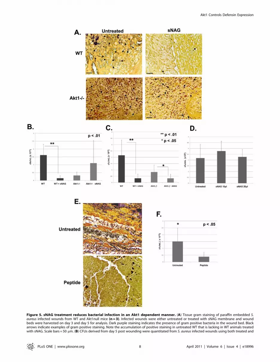
Figure 5. sNAG treatment reduces bacterial infection in an Akt1 dependent manner. (A) Tissue gram staining of paraffin embedded S.aureus infected wounds from WT and Akt1null mice (n = 3). Infected wounds were either untreated or treated with sNAG membrane and woundbeds were harvested on day 3 and day 5 for analysis. Dark purple staining indicates the presence of gram positive bacteria in the wound bed. Blackarrows indicate examples of gram positive staining. Note the accumulation of positive staining in untreated WT that is lacking in WT animals treatedwith sNAG. Scale bars = 50 mm. (B) CFUs derived from day 5 post wounding were quantitated from S. aureus infected wounds using both treated and
Akt1 Controls Defensin Expression
PLoS ONE | www.plosone.org 8 April 2011 | Volume 6 | Issue 4 | e18996

supports their role in cutaneous innate immunity. However, we also
show that sNAG specifically stimulates the expression of three
different a-defensins (1, 4 and 5) in endothelial cells. We show this
by RT-PCR, gene array analysis, immunofluorescence and ELISA
(data not shown). The interaction between endothelial cells and
leukocytes in tissue repair is one of the initial and most important
steps in wound healing. The process of extravasation of leukocytes
from the vasculature is initiated by chemotactic factors, therefore; it
is interesting that a-defensins are induced by sNAG and may
contribute to the necessary neutrophil/endothelial cellular interac-
tions. More recently it has come to light that defensins exhibit
biological activities beyond the inhibition of microbial cells,
including their contribution to the adaptive immune response by
exhibiting chemotactic activity on dendritic [23] and T cells,
monocytes, and macrophages [24] and keratinocytes [25]. Previous
work shows that human beta defensins 1 and 2 have the ability to
chemoattract immature dendritic cells and T cells through the CC-
chemokine receptor 6 (CCR6) [26], and that human beta defensin 2
can chemoattract TNFa treated neutrophils via the CCR6 receptor
[27]. Human b-defensin 2 and 3 have also been shown to induce
chemotaxis by interacting with CCR2, a receptor expressed on
macrophages, monocytes, and neutrophils [28]. Interestingly, we
show that sNAG treatment induces both a and b-defensin
expression in endothelial cells. Taken together, the recent data
suggest that defensins may mediate wound healing not only by their
antimicrobial properties, but also by being chemotactic for other cell
types necessary for proper healing. However, application of b-
defensin 3 alone did not result in an increase in wound closure (data
not shown) implying that topical application of a single defensin
does not sustain the cellular interactions required for increased
chemo attraction, cellular recruitment and wound closure.
It is interesting to note that gram stains from Akt1 null infected
mice show accumulated immune cells, likely neutrophils, which
are staining positive for bacteria. This may suggest that in addition
to their delay in wound healing Akt1 null mice may have immune
trafficking and clearance issues. In a different Akt1 null animal
using an incisional wound assay, others have shown that the
primary phenotype was a lack of vascular maturation (reduced
smooth muscle actin expression), but no changes in wound closure
[29]. However, there are some differences between these studies
and ours. First, we use a full thickness ‘‘punch’’ wound. Secondly,
the Akt1 null animal used here was constructed using an
insertional mutagenesis at the translational start site. In addition,
our model is on a pure C57/Bl6 background rather than on a
C57Bl6/129 background. It is well established that a genetically
mixed background (especially a 129/C57Bl6 mixed background)
increases hybrid vigor which can compensate for the loss of a
single gene, such as Akt1, thus suppressing phenotypes in knockout
animals [30,31]. A full analysis of Akt1 dependent function during
cutaneous wound healing is presently underway in the laboratory.
Our in vivo data using both wild type and Akt1 knockout animals
confirms the requirement for Akt1 in sNAG-induced b-defensin 3
expression. Since mouse leukocytes do not express a-defensins like
most other mammalian leukocytes [32] in vivo a-defensin staining
of infiltrating immune cells was not possible. Treatment of airway
epithelial cells in vitro with alpha defensins 1–3 causes a dose and
time-dependent increased cell migration that requires activation of
PI3K and MAPK pathways [33]. We have previously shown that
sNAG stimulation of endothelial cells results in the activation of
MAPK [17] and in data presented here, pharmacological
inhibition of MEK also inhibits the expression of the defensins in
vitro. These findings suggest that both pathways impinge on the
regulation of defensin expression by sNAG, however, Akt1
ablation results in a marked reduction of its expression both in
vitro and in vivo. In myeloid cells, b-defensin 1 expression is
controlled at the level of transcription, in part, by the Ets-family
member PU.1 [34,35]. PU.1 is a downstream target of Akt1 in the
B-cell lineage [36]. We have shown, in primary endothelial cells
that Akt1 is upstream of Ets1 both in vitro and in vivo during
Drosophila tracheal development [18]. sNAG stimulation of
endothelial cells results in increased expression of Ets1 (probably
through Akt1) which is required for the migration of endothelial
cells [17]. A delineation of the transcriptional regulatory
mechanisms responsible for the regulation of the defensins by
sNAG are currently underway in the laboratory.
Thus far, sNAG treatment has resulted in a series of
downstream activities; hemostasis, cell migration, cell proliferation,
increased wound closure, and as described here, stimulation of the
innate immune response resulting in anti-bacterial functions.
Using a custom gene chip we have also shown that a number of
Toll-like receptors are up-regulated by sNAG treatment of human
endothelial cells (data not shown). Toll-like receptors (TLRs) are
highly conserved receptors that recognize specific molecular
patterns of bacterial components leading to activation of innate
immunity. Interestingly, Drosophila lack an adaptive immune
system but are still resistant to microbial infections [37]. This
host defense is the result of an innate immune system that provides
protection by synthesizing the antimicrobial peptides dToll and
18-wheeler which are induced by TLRs [38,39]. Recent work has
also linked human defensin expression to TLR activation. Human
b-defensin 2 was shown to be induced in airway epithelial cells in a
TLR-2 dependent manner [40]. Toll-like receptor 4 has been
shown to mediate human b-defensin 2 inductions in response to
Chlamydia pneumonia in monocytes [41]. Importantly, the PI3K/Akt
pathway is a key component in TLR signal transduction,
controlling cellular responses to pathogens [42]. Since it is known
that stimulation of TLRs can lead to increased defensin synthesis,
this work suggests the potential for sNAG as a stimulator of innate
immunity and bacterial clearance via the activation of Akt1.
Given the dramatic increase of diabetic patients within the
population who present with chronic wounds and complications
due to wound infection, new clinical treatments are in high
demand. Here, we describe marine derived pGlcNAc nanofibers
that not only increase the kinetics of wound healing but act to
stimulate innate immunity thus providing anti-bacterial activity.
The obvious importance of these observations is the application
untreated WT (n = 3) and Akt1 mice (n = 3). Wild type mice that were sNAG treated show a significant (p,.01) decrease in bacteria load in the woundbeds as compared to Akt1 null animals. All experiments were repeated three independent times and the p values are shown. (C) CFU quantitatedfrom infected wounds at day 3 post wounding in a similar fashion described in (B). sNAG treatment of infected wounds shows a significant decreasein CFU of both WT and Akt1 null animals on day 3, but the WT animals show an approximate 10 fold difference compared to a 2 fold difference inAkt1 animals. (D) Quantitation of CFUs in S. aureus cultures that were either untreated or treated with various amounts of sNAG nanofibers. Eachexperiment was performed three independent times and p values are shown. (E) Tissue gram staining of S. aureus infected wounds harvested on day3 post wound from WT mice (n = 3) that were treated with or without b-defensin 3 peptide. Note the decrease in gram positive staining in infectedwounds that were treated with b-defensin 3 peptide. (F) Quantitation of CFUs from S. aureus infected WT mice (n = 3) treated with or without b-defensin 3 peptide. Infected wounds that were treated with peptide show a significant decrease (p,.05) in CFU. Scale bars = 50 mm. Each experimentwas performed three independent times and p values are shown.doi:10.1371/journal.pone.0018996.g005
Akt1 Controls Defensin Expression
PLoS ONE | www.plosone.org 9 April 2011 | Volume 6 | Issue 4 | e18996

to nosocomial infections. Of the nosocomial infections, surgical
wound infections predominate; with statistics showing up to 8%
of all surgical patients. The direct cost of these types of infections
is approximately 4.5 billion dollars per year. Given that defensins
are part of the innate immune system, activation of these
pathways will preclude the generation of resistant organisms as
well as allow for the antibiotic-independent clearance of bacterial
infection. Use of sNAG in a hospital setting would defray much of
the cost and markedly reduce the production of antibiotic
resistant species. Taken together, these findings suggest that these
marine derived pGlcNAc nanofibers will be highly beneficial in
the clinical arena.
Materials and Methods
Tissue culture, pharmacological inhibition, ELISAHuman umbilical cord vein EC (Lonza) were maintained at 37u
with 5% CO2 in endothelial basal medium 2 (Lonza). Endothelial
basal medium 2 (EBM2) was supplemented with EC growth
medium 2 SingleQuots as described by Lonza procedures and 1%
penicillin/streptomycin (Invitrogen). Serum starvation was per-
formed at 80–90% confluency in EBM2 supplemented with 0.1%
fetal calf serum (Valley Biomedical) for 24 hours followed by
stimulation with highly purified pGlcNAc (50 mg/ml) nanofibers
(sNAG) in sterile water (provided by Marine Polymer Technolo-
Figure 6. Rapid induction of defensin expression by sNAG treatment of S. aureus infected wounds. (A) Paraffin embedded tissue sectionsfrom S. aureus infected wounds, harvested on day 3, were subjected to immunofluorescence using antibodies directed against b-defensin 3 (green),Involucrin (red) to mark the keratinocyte layer, and Topro (blue) from both sNAG treated WT (n = 3) and untreated WT mice (n = 3). Non specificstaining of keratin is indicated by the no primary control which was stained with secondary antibody only. Scale bar = 50 mm. (B) Quantitation of b-defensin 3 expression from paraffin embedded sections using NIH ImageJ software. S. aureus infected wounds that were treated with sNAG show asignificant increase (p,.05) in b-defensin 3 staining. Experiments were repeated three independent times and p values are shown.doi:10.1371/journal.pone.0018996.g006
Akt1 Controls Defensin Expression
PLoS ONE | www.plosone.org 10 April 2011 | Volume 6 | Issue 4 | e18996

Akt1 Controls Defensin Expression
PLoS ONE | www.plosone.org 11 April 2011 | Volume 6 | Issue 4 | e18996

gies, Inc., Danvers, Mass., USA). The pGlcNAc diatom-derived
nanofibers used in this study are short biodegradable fibers derived
from a native, longer form (NAG), and have an average length of
4–7 mm and a polymer molecular weight of approximately
60,000 Da. For inhibition using PD098059 (50 mM) or wortman-
nin (100 nM), cells were pre-treated for 45 minutes prior to 3 hour
stimulation with sNAG (50 mg/ml).
Statistical analysisEach quantitative experiment was performed at least in
triplicate at least three independent times. All statistical analyses
were performed using Microsoft Excell to calculate means,
standard deviations and student t-test
Lentiviral infectionMission shRNA lentiviral constructs directed against Akt1 were
purchased from Sigma/Aldrich. A scrambled pLKO.1 shRNA
vector was purchased from Addgene. Lentiviruses were propagat-
ed in 293T cells, maintained in DMEM supplemented as above.
Lentiviral production was performed using psPAX2 and pMD2.G
packaging vectors purchased from Addgene using the protocol for
producing lentiviral particles from Addgene. For infection of target
cells, 7.56105 cells were plated on 100 mm2 plates and allowed to
incubate overnight. The next day, cells were transduced using a
final concentration of 1 mg/ml polybrene and either scrambled
control or Akt1 shRNA lentiviruses. After transduction, endothe-
lial cells were serum starved overnight and stimulated with sNAG
(50 g/ml) for 3 hours. All infections were monitored for
appropriate knockdown by RT-PCR.
RT-PCRFor semi-quantitative RT-PCR, RNA was extracted with
RNAsol (Teltest, Inc.) following manufacturer’s instructions.
cDNA was synthesized from 2 mg total RNA with a Superscript
First Strand Synthesis Kit (Invitrogen), using Oligo(dT) following
the manufacturer’s instructions. PCR reactions contained equal
amounts of cDNA and 1.25 mM of the appropriate primer pair
(Sigma-Proligo, St. Louis, MO, USA). All primer sequences used
in these analyses are as follows: Akt1: forward 59 GAGGCCGT-
CAGCCACAGTCTG 39, reverse 59 ATGAGCGACGTGGC-
TATTGTG 39; b-Defensin 3:forward 59 GTGGGGTGAAGCC-
TAGCAG 39, reverse 59 GTGGGGTGAAGCCTAGCAG 39; a-
Defensin 1: forward 59 CACTCCAGGCAAGAGCTGAT 39,
reverse 59 TCCCTGGTAGATGCAGGTTC 39; s26: forward 59
CTCCGGTCCGTGCCTCCAAG 39, reverse 59CAGAGAA-
TAGCCTGTCTTCAG 39
Cycling conditions were: 94uC for 5 min; 30–35 cycles of 94uCfor 1 min, 55–65uC (based on primer Tm) for 1 min, 72uC for
1 min; 72uC for 7 min and cooled to 4uC. Cycle number was
empirically determined to be within the linear range of the assay
for each primer pair used. All semi-quantitative RT-PCR was
performed with the ribosomal protein subunit S26 primers as
internal controls. Products were visualized on a BioRad Molecular
Imaging System (Hercules, CA, USA). Real time PCR was
performed using a Brilliant CYBR green QPCR kit in combina-
tion with an Mx3000P Real-Time PCR system both purchased
from Stratagene. Primers detecting the ribosomal subunit S26
were used as internal controls.
Ethics statementAll experiments performed using mice were in accordance with
animal procedure protocols approved by the Medical University of
South Carolina Institutional Animal Care and Use Committee,
approval ID# 2349. Appropriate analgesics were used under all
conditions to insure a lack of pain and suffering.
Excisional wound healing modelWild Type C57Bl/6 and Akt12/2 [43] were used in all
experiments. The Akt1 null animals were created using an
insertional mutagenesis strategy at the translational start site that
blocks expression of the entire protein. Wounding was performed
on anesthetized adult male mice between 8–12 weeks old. Two full
thickness cutaneous wounds were created using a 4 mm biopsy
punch (Miltex), to create two identical wounds on each flank. Mice
were anesthetized using an O2/Isoflurane vaporizing anesthesia
machine (VetEquip, Inc.). Isoflurane was used at 4% for induction;
2% for surgery. Prior to surgery hair was removed by depilation
and the area was washed and sterilized using 70% ethanol.
Wounds were either treated with sNAG membrane moistened
with distilled water or left untreated. On days 3 and 5 animals
were euthanized and entire wounds were harvested including the
surrounding skin using an 8 mm biopsy punch (Miltex). Wounds
were fixed in 4% paraformaldehyde overnight at 4u, embedded in
paraffin, and sectioned for analysis.
Hematoxylin and eosin staining (H&E)All H&E staining was performed in the Histology Core Facility
at the Medical University of South Carolina, Department of
Regenerative Medicine and Cell Biology. Briefly, sections were
cleared in xylene, rehydrated through a series of graded alcohols,
placed in Hematoxylin followed by acid alcohol. Samples were
then placed in ammonia water, rinsed in ethanol and exposed to
Eosin before dehydrating through graded alcohols and clearing in
xylene. Sections were mounted using Cytoseal-XYL (Richard-
Allan Scientific). H&E sections were visualized using an Olympus
BX40 microscope (46objective lens, 0.13) and captured using an
Olympus Camera (Model DP25) and DP2-BSW acquisition
software.
Bacterial inoculation, tissue gram staining, colonyforming unit quantitation
Male mice between 8–12 weeks were wounded as described
above. Single colonies of Staphylococcus aureus (ATCC 25923) were
picked and cultured overnight at 37u and adjusted to an
absorbance of OD600 = 0.53. One mL of S. aureus was spun at
10,000 rpm, re-suspended in sterile PBS, and 15 ml was used to
innoculate each wound. sNAG membranes were applied to the
treated group thirty minutes post inoculation. Mice were
euthanized on day 3 and 5 post wounding and wounds were
harvested using an 8 mm biopsy punch. One wound per animal
was fixed overnight in 4% paraformaldehyde at 4uC and the other
wound was cultured and plated on LB media without antibiotic for
Figure 7. Antibodies against b-defensin 3 impedes antibacterial effects of sNAG treatment. (A) Tissue gram staining of paraffinembedded S. aureus infected wounds treated with sNAG from WT mice (n = 3) that were harvested on Day 3. sNAG treated wounds were treatedwith either b-defensin 3 antibody or isotype control goat IgG antibody prior to sNAG treatment. Representative images show increased accumulationgram positive staining (black arrows) in the wound beds of mice treated with an antibody directed against b-defensin 3. Scale bar = 20 mm. (B)Quantitation of CFUs from S. aureus infected WT mice treated either b-defensin 3 antibody (n = 3) or control IgG antibody (n = 3) prior to sNAGtreatment. b-defensin 3 application significantly increasedd (p,.05) CFU.doi:10.1371/journal.pone.0018996.g007
Akt1 Controls Defensin Expression
PLoS ONE | www.plosone.org 12 April 2011 | Volume 6 | Issue 4 | e18996

bacterial quantitation (see below). Wounds for tissue gram staining
were embedded in paraffin and sectioned. Sections were cleared in
xylene and rehydrated through a series of alcohol and were stained
using a tissue gram stain (Sigma-Aldrich) by procedures described
by the manufacturer.
For culturing, wound sections were placed in 0.5 ml bacterial
media an incubated for 30 min at 37uC while shaking. Colony
forming units (CFU) were quantitated using a dilution series plated
overnight at 37uC. Number of colonies per plate/per dilution were
counted and CFU/ml were calculated. To determine CFU/ml
from sNAG treated bacterial cultures, S. aureus cultures in solution
were treated with varying concentrations of sNAG for three hours.
Cultures were then plated overnight at 37u and CFU/ml were
determined.
b-defensin 3 peptide applicationThree test concentrations (1.0 mM, 2.5 mM, 5.0 mM) of
biologically active human b-defensin 3 peptide (Peptide Institute,
Inc.) were tested for their effect on bacterial growth in the infected
wound healing model described above. Each concentration
negatively affected bacterial growth so the lowest concentration
was chosen for analyses. After each wound was infected with S.
aureus, 10 ul of peptide was applied. After three days, wounds were
harvested, embedded for sectioning and gram staining, or cultured
for CFU/ml quantitation as described above.
b-defensin 3 antibody blockadeWild Type male mice were wounded and infected with 15 ul of
S. aureus as described above. After inoculation, one wound was
treated with 0.2 ug/mL of b-defensin 3 antibody (Santa Cruz)
while the other was treated with 0.2 ug/mL of normal goat IgG
control antibody (Santz Cruz). sNAG membranes were applied to
all mice after antibody treatment on day 0. Antibody was applied
every 24 hours. Mice were euthanized on day 3 and wounds were
harvested using an 8 mm biopsy punch. Wounds were fixed
overnight in 4% paraformaldehyde at 4uC, embedded in paraffin,
sectioned, and analyzed using tissue gram stain. CFU/ml
quantitation was performed from wounds harvested on day 3 as
described above.
Immunofluoresence, microscopyParaffin embedded tissue sections were re-hydrated through
xylene and a series of graded alcohols. Sections were treated with
0.01% Triton-X100 and subjected to antigen retrieval using
antigen unmasking solution (Vector Laboratories) in a pressure
cooker for 5 min and allowed to cool. Skin sections were labeled
with b-defensin 3 goat polyclonal antibody (Santa Cruz),
involucrin rabbit polyclonal antibody (Santa Cruz), and TO-
PRO 3-iodide (Molecular Probes). Sections were incubated in
primary antibody overnight at 4u and appropriate secondary
immunofluorescent antibodies (Invitrogen) for 1 hour at room
temperature. Control sections for each antibody were stained
without primary antibody. Tissue sections were visualized using an
Olympus FluroView laser scanning confocal microscope (Model
IX70) and captured at ambient temperature using an Olympus
camera (Model FV5-ZM) and Fluoview 5.0 acquisition software.
All tissue sections were imaged using 606 oil immersion lens
(Olympus Immersion Oil).
HUVECs were either serum starved or treated with sNAG for
5 hours in culture and stained with antibodies directed against a-
defensin 5 (FITC), b-defensin 3 (Texas Red), or TOPRO 3 (Blue).
Images were taken using immunofluorescent microscopy. Cell
culture defensin expression was visualized using a Zeiss Axiovert
100 M confocal microscope and was captured at ambient
temperature, using water as the medium, using LSM 510 camera
(Zeiss Fluor 63xW/1.2A objective).
Western blot analysisEndothelial cells were serum starved prior to stimulation with
sNAG (50 ml/ml) for a given time course. Cells were then lysed
and subjected to Western blot analysis. The antibodies used for
Western blot analysis are as follows: anti-p85 subunit of PI3K and
phosphospecific Akt antibody (Cell Signaling Technologies).
Acknowledgments
We wish to thank Drs. Amy Bradshaw and Rick Schnellmann for helpful
comments and suggestions.
Author Contributions
Conceived and designed the experiments: HBL AS RM-H. Performed the
experiments: JE HBL RM-H AZ. Analyzed the data: HBL RM-H AS.
Contributed reagents/materials/analysis tools: MD JV PT. Wrote the
paper: HBL RM-H.
References
1. Singer AJ, Clark RA (1999) Cutaneous wound healing. N Engl J Med 341:738–746.
2. Ganz T (2003) Defensins: antimicrobial peptides of innate immunity. Nat RevImmunol 3: 710–720.
3. Huang HW (2000) Action of antimicrobial peptides: two-state model.Biochemistry 39: 8347–8352.
4. Ganz T, Lehrer RI (1994) Defensins. Curr Opin Immunol 6: 584–589.
5. Ganz T (1987) Extracellular release of antimicrobial defensins by human
polymorphonuclear leukocytes. Infect Immun 55: 568–571.
6. Harder J, Bartels J, Christophers E, Schroder JM (1997) A peptide antibiotic
from human skin. Nature 387: 861.
7. Harder J, Bartels J, Christophers E, Schroder JM (2001) Isolation and
characterization of human beta -defensin-3, a novel human inducible peptideantibiotic. J Biol Chem 276: 5707–5713.
8. Mathews M, Jia HP, Guthmiller JM, Losh G, Graham S, et al. (1999)Production of beta-defensin antimicrobial peptides by the oral mucosa and
salivary glands. Infect Immun 67: 2740–2745.
9. Ganz T, Rayner JR, Valore EV, Tumolo A, Talmadge K, et al. (1989) The
structure of the rabbit macrophage defensin genes and their organ-specific
expression. J Immunol 143: 1358–1365.
10. Hirsch JA, Reddy SA, Capasso WE, Linfante I (2003) Non-invasive hemostatic
closure devices: ‘‘patches and pads’’. Tech Vasc Interv Radiol 6: 92–95.
11. Palmer BL, Gantt DS, Lawrence ME, Rajab MH, Dehmer GJ (2004)
Effectiveness and safety of manual hemostasis facilitated by the SyvekPatch
with one hour of bedrest after coronary angiography using six-French catheters.Am J Cardiol 93: 96–97.
12. Thatte HS, Zagarins S, Khuri SF, Fischer TH (2004) Mechanisms of poly-N-
acetyl glucosamine polymer-mediated hemostasis: platelet interactions. J Trauma57: S13–21.
13. Pietramaggiori G, Yang HJ, Scherer SS, Kaipainen A, Chan RK, et al. (2008)Effects of poly-N-acetyl glucosamine (pGlcNAc) patch on wound healing in db/
db mouse. J Trauma 64: 803–808.
14. Scherer SS, Pietramaggiori G, Matthews J, Perry S, Assmann A, et al. (2009)Poly-N-acetyl glucosamine nanofibers: a new bioactive material to enhance
diabetic wound healing by cell migration and angiogenesis. Ann Surg 250:322–330.
15. Fischer TH, Thatte HS, Nichols TC, Bender-Neal DE, Bellinger AD, et al.
(2005) Synergistic platelet integrin signaling and factor XII activation in poly-N-acetyl glucosamine fiber-mediated hemostasis. Biomaterials 26: 5433–5443.
16. Fischer TH, Bode AP, Demcheva M, Vournakis JN (2007) Hemostaticproperties of glucosamine-based materials. J Biomed Mater Res A 80: 167–174.
17. Vournakis JN, Eldridge J, Demcheva M, Muise-Helmericks RC (2008) Poly-N-
acetyl glucosamine nanofibers regulate endothelial cell movement and angiogen-esis: dependency on integrin activation of Ets1. J Vasc Res 45: 222–232.
18. Lavenburg KR, Ivey J, Hsu T, Muise-Helmericks RC (2003) Coordinatedfunctions of Akt/PKB and ETS1 in tubule formation. FASEB J 17: 2278–2280.
19. Watt FM (1983) Involucrin and other markers of keratinocyte terminal
differentiation. J Invest Dermatol 81: 100s–103s.
Akt1 Controls Defensin Expression
PLoS ONE | www.plosone.org 13 April 2011 | Volume 6 | Issue 4 | e18996

20. Selsted ME, Ouellette AJ (2005) Mammalian defensins in the antimicrobial
immune response. Nat Immunol 6: 551–557.21. Salzman NH, Ghosh D, Huttner KM, Paterson Y, Bevins CL (2003) Protection
against enteric salmonellosis in transgenic mice expressing a human intestinal
defensin. Nature 422: 522–526.22. Oren A, Ganz T, Liu L, Meerloo T (2003) In human epidermis, beta-defensin 2
is packaged in lamellar bodies. Exp Mol Pathol 74: 180–182.23. Hubert P, Herman L, Maillard C, Caberg JH, Nikkels A, et al. (2007) Defensins
induce the recruitment of dendritic cells in cervical human papillomavirus-
associated (pre)neoplastic lesions formed in vitro and transplanted in vivo.FASEB J 21: 2765–2775.
24. Garcia JR, Jaumann F, Schulz S, Krause A, Rodriguez-Jimenez J, et al. (2001)Identification of a novel, multifunctional beta-defensin (human beta-defensin 3)
with specific antimicrobial activity. Its interaction with plasma membranes ofXenopus oocytes and the induction of macrophage chemoattraction. Cell Tissue
Res 306: 257–264.
25. Niyonsaba F, Ushio H, Nakano N, Ng W, Sayama K, et al. (2007) Antimicrobialpeptides human beta-defensins stimulate epidermal keratinocyte migration,
proliferation and production of proinflammatory cytokines and chemokines.J Invest Dermatol 127: 594–604.
26. Yang D, Chertov O, Bykovskaia SN, Chen Q, Buffo MJ, et al. (1999) Beta-
defensins: linking innate and adaptive immunity through dendritic and T cellCCR6. Science 286: 525–528.
27. Niyonsaba F, Ogawa H, Nagaoka I (2004) Human beta-defensin-2 functions as achemotactic agent for tumour necrosis factor-alpha-treated human neutrophils.
Immunology 111: 273–281.28. Rohrl J, Yang D, Oppenheim JJ, Hehlgans T (2010) Human {beta}-Defensin 2
and 3 and Their Mouse Orthologs Induce Chemotaxis through Interaction with
CCR2. J Immunol.29. Somanath PR, Chen J, Byzova TV (2008) Akt1 is necessary for the vascular
maturation and angiogenesis during cutaneous wound healing. Angiogenesis 11:277–288.
30. Simpson EM, Linder CC, Sargent EE, Davisson MT, Mobraaten LE, et al.
(1997) Genetic variation among 129 substrains and its importance for targetedmutagenesis in mice. Nat Genet 16: 19–27.
31. Threadgill DW, Yee D, Matin A, Nadeau JH, Magnuson T (1997) Genealogy ofthe 129 inbred strains: 129/SvJ is a contaminated inbred strain. Mamm
Genome 8: 390–393.
32. Ganz T (2004) Defensins: antimicrobial peptides of vertebrates. C R Biol 327:
539–549.
33. Aarbiou J, Verhoosel RM, Van Wetering S, De Boer WI, Van Krieken JH, et al.
(2004) Neutrophil defensins enhance lung epithelial wound closure and mucin
gene expression in vitro. Am J Respir Cell Mol Biol 30: 193–201.
34. Yaneva M, Kippenberger S, Wang N, Su Q, McGarvey M, et al. (2006) PU.1
and a TTTAAA element in the myeloid defensin-1 promoter create an
operational TATA box that can impose cell specificity onto TFIID function.
J Immunol 176: 6906–6917.
35. Ma Y, Su Q, Tempst P (1998) Differentiation-stimulated activity binds an ETS-
like, essential regulatory element in the human promyelocytic defensin-1
promoter. J Biol Chem 273: 8727–8740.
36. Rieske P, Pongubala JM (2001) AKT induces transcriptional activity of PU.1
through phosphorylation-mediated modifications within its transactivation
domain. J Biol Chem 276: 8460–8468.
37. Imler JL, Hoffmann JA (2000) Signaling mechanisms in the antimicrobial host
defense of Drosophila. Curr Opin Microbiol 3: 16–22.
38. Lemaitre B, Nicolas E, Michaut L, Reichhart JM, Hoffmann JA (1996) The
dorsoventral regulatory gene cassette spatzle/Toll/cactus controls the potent
antifungal response in Drosophila adults. Cell 86: 973–983.
39. Williams MJ, Rodriguez A, Kimbrell DA, Eldon ED (1997) The 18-wheeler
mutation reveals complex antibacterial gene regulation in Drosophila host
defense. EMBO J 16: 6120–6130.
40. Hertz CJ, Wu Q, Porter EM, Zhang YJ, Weismuller KH, et al. (2003) Activation
of Toll-like receptor 2 on human tracheobronchial epithelial cells induces the
antimicrobial peptide human beta defensin-2. J Immunol 171: 6820–6826.
41. Romano Carratelli C, Mazzola N, Paolillo R, Sorrentino S, Rizzo A (2009) Toll-
like receptor-4 (TLR4) mediates human beta-defensin-2 (HBD-2) induction in
response to Chlamydia pneumoniae in mononuclear cells. FEMS Immunol Med
Microbiol 57: 116–124.
42. Weichhart T, Saemann MD (2008) The PI3K/Akt/mTOR pathway in innate
immune cells: emerging therapeutic applications. Ann Rheum Dis 67 Suppl 3:
iii70–74.
43. Mao C, Tili EG, Dose M, Haks MC, Bear SE, et al. (2007) Unequal
contribution of Akt isoforms in the double-negative to double-positive thymocyte
transition. J Immunol 178: 5443–5453.
Akt1 Controls Defensin Expression
PLoS ONE | www.plosone.org 14 April 2011 | Volume 6 | Issue 4 | e18996


