Embed Size (px)
Citation preview

ORIGINAL PAPER
Anti-Giardia activity of phenolic-rich essential oils: effectsof Thymbra capitata, Origanum virens, Thymus zygissubsp. sylvestris, and Lippia graveolens on trophozoitesgrowth, viability, adherence, and ultrastructure
Marisa Machado & Augusto M. Dinis & Ligia Salgueiro &
Carlos Cavaleiro & José B. A. Custódio &
Maria do Céu Sousa
Received: 19 January 2010 /Accepted: 29 January 2010 /Published online: 9 March 2010# Springer-Verlag 2010
Abstract The present work evaluates the anti-Giardia activityof phenolic-rich essential oils obtained from Thymbracapitata, Origanum virens, Thymus zygis subsp. sylvestrischemotype thymol, and Lippia graveolens aromatic plants.The effects were evaluated on parasite growth, cell viabilityadherence, and morphology. The tested essential oils inhibitedthe growth of Giardia lamblia. T. capitata essential oil is themost active followed by O. virens, T. zygis subsp. sylvestris,and L. graveolens oils. The tested essential oils at IC50 (71–257) μg/ml inhibited parasite adherence (p<0.001) since thefirst hour of incubation and were able to kill almost 50% of
the parasites population in a time-dependent manner. Themain ultrastructural alterations promoted by essential oilswere deformations in typical trophozoite appearance, oftenroundly shape, irregular dorsal and ventral surface, presenceof membrane blebs, electrodense precipitates in cytoplasmand nuclei, and internalization of flagella and ventral disc. Ourdata suggest that essential oils induced cell death probably byprocesses associated to the loss of osmoregulation caused byplasmatic membrane alterations. Experiments revealed that theessential oils did not present cytotoxic effects in mammaliancells. In conclusion, T. capitata, O. virens, T. zygis subsp.sylvestris chemotype thymol, and L. graveolens essential oilshave antigiardial activity in vitro and seem to have potentialfor the treatment of the parasitic disease caused by theprotozoan G. lamblia.
Introduction
Giardia lamblia (syn. Giardia intestinalis, Giardia duode-nalis) is a binucleated flagellar protozoan that commonlycauses intestinal infection in humans. This parasite isendemic throughout the world, and it is the most commonisolated parasite in Portugal. In developed nations, besidesbeing the most common intestinal protozoan parasite ofhumans, a rising incidence of giardiasis has been noted forchildren who have led to the designation of giardiasis as a “re-emerging” infectious disease (Thompson 2000). Giardiasismay be entirely asymptomatic, may produce a mild self-limiting illness, or may produce chronic diarrhea with orwithout malabsorption (Flanagan 1992). In young, elderly,and immunocompromised patients, giardiasis can lead tomorbidity and even death (Eckmann 2003; Fraser et al.
M. Machado : L. Salgueiro : C. Cavaleiro :M. SousaFaculdade de Farmácia/CEF, Universidade de Coimbra,Azinhaga de Santa Comba,3030-548 Coimbra, Portugal
M. MachadoDepartamento Farmácia, Escola Superior de Saúde do Valedo Ave/Centro de Investigação em Tecnologias da SaúdeIPSN-CESPU,4760 Vila Nova de Famalicão, Portugal
A. M. DinisLaboratório de Microscopia Electrónica,Departamento das Ciências da Vida, Faculdade de Ciênciase Tecnologia da Universidade de Coimbra,Coimbra, Portugal
J. B. A. CustódioCentro de Neurociências da Universidade de Coimbra,3000 Coimbra, Portugal
M. Sousa (*)Faculdade de Farmácia da Universidade de Coimbra,Pólo das Ciências da Saúde, Azinhaga de Santa Comba,3030-548 Coimbra, Portugale-mail: [email protected]
Parasitol Res (2010) 106:1205–1215DOI 10.1007/s00436-010-1800-7

2000; Sawangjaroen et al. 2005). Attachment of G. lambliatrophozoites to enterocytes is essential for colonization of thesmall intestine and is considered a prerequisite for parasite-induced enterocyte dysfunction and clinical disease (Elmendorfet al. 2003). Thus, the adherence process may be a potentialtarget for attack by chemotherapeutic agents that act by eitherprevention of attachment or induction of detachment oforganisms (Holberton 1974; Gillin and Reiner 1982; Sousaet al. 2001; Perez-Arriaga et al. 2006). Giardia’s abundance inhuman and animal populations and environmental sourcessuggests that eradication is not an option; reliance thereforefalls on the treatment of giardiasis (Adam 2001).
The current therapy of giardiasis includes several drugsensuing 5-nitroimidazoles, 5-nitrofurans, and, more recently,benzimidazoles and pyrantel pamoate as drugs of choice.Although these drugs are effective, many problems have beendescribed associated to their use, namely high incidence offailures, frequent relapses, and undesirable side effects(Upcroft and Upcroft 2001; Busatti et al. 2007). Someevidence suggests that drug resistance may be responsible forthe failure (Upcroft and Upcroft 2001). For these reasons, thesearch for therapeutic alternatives is entirely justifiable andimperative. The discovery of potential new pharmaceuticalsproducts from natural sources seems to be emerging. Plantextracts prepared by distillation, named essential oils, are com-plex mixtures of a vast diversity of small hydrophobic mole-cules (<300 Da) which easily diffuse across cell membranesprospecting interactions with intracellular targets (Edris 2007).The potential use of essential oils as antifungal (Gonçalves etal. 2007), antibacterial (Dorman and Deans 2000; Burt 2004;Faleiro et al. 2005), and antiprotozoal activity (Ponce-Macotela et al. 2006; de Almeida et al. 2007; Santoro et al.2007; Machado et al. 2010) has been demonstrated.
The aim of this study was to evaluate the effects ofLippia graveolens, Origanum virens, Thymus zygis subsp.sylvestris, and Thymbra capitata essential oils on growth,adhesion, viability, and morphology of G. lamblia troph-ozoites to assess the antigiardial activity and characteriza-tion of action mechanisms undergoing the activity.
Materials and methods
Chemicals Bile bovine, L-cysteine, L-ascorbic acid, ferricammonium citrate, and dimethyl sulfoxide (DMSO) wereobtained from Sigma Chemical Corp. (St. Louis, MO,USA). Casitone and yeast extract were purchased fromDifco Laboratories and bovine serum and antibioticsolution from Biochrom K.G.
Plant material: essential oils Essential oils were preparedat laboratory by water distillation from the aerial partsof L. graveolens H.B.K., O. virens Hoffmanns. & Link,
T. capitata (L.) Cav., and T. zygis Loefl. Ex L. subsp.sylvestris (Hoffmanns & Link) Brot. ex Coutinho, chemotypethymol. Distillations were performed using Clevenger-typeapparatus, following the procedure described in the EuropeanPharmacopoeia (Council of Europe 1997).
Plant material: essential oil analysis Analyses were carriedout by gas chromatography (GC) and by gas chromatography–mass spectroscopy (GC/MS). Analytical GCwas carried out ina Hewlett-Packard 6890 (Agilent Technologies, Palo Alto, CA,USA) gas chromatograph with a HP GC ChemStation Rev.A.05.04 data handling system, equipped with a single injectorand two flame ionization detection (FID) systems. A graphpakdivider (Agilent Technologies, part no. 5021-7148) wasused for simultaneous sampling to two Supelco (Supelco,Bellefonte, PA, USA) fused silica capillary columns withdifferent stationary phases: SPB-1 (polydimethylsiloxane30 m×0.20 mm i.d., film thickness 0.2 μm) andSupelcoWax-10 (polyethylene glycol 30 m×0.20 mm i.d.,film thickness 0.20 μm). Oven temperature programs were70–220°C (3°C min−1) and 220°C (15 min); injectortemperature was 250°C; carrier gas was helium, adjusted toa linear velocity of 30 cm s−1; splitting ratio was 1:40;detectors temperature was 250°C. GC–MS was carried out ina Hewlett-Packard 6890 gas chromatograph fitted with aHP1 fused silica column (polydimethylsiloxane 30 m×0.25 mm i.d., film thickness 0.25 μm), interfaced with aHewlett-Packard mass selective detector 5973 (AgilentTechnologies) operated by HP Enhanced ChemStationsoftware, version A.03.00. GC parameters were as describedabove; interface temperature was 250°C; MS source temper-ature was 230°C; MS quadrupole temperature was 150°C;ionization energy was 70 eV; ionization current was 60 μA;scan range was 35–350 U; scans s−1 was 4.51. Componentsof each essential oil were identified by their retention indiceson both SPB-1 and SupelcoWax-10 columns and from theirmass spectra. Retention indices, calculated by linear inter-polation relative to retention times of C8–C23 of n-alkanes,were compared with those of authentic samples included inour own laboratory database. Acquired mass spectra werecompared with reference spectra from our own library, WileyNT (Wiley MS database), and literature data (Adams 1995;Joulain and König 1998). Relative amounts of individualcomponents were calculated based on GC peak areas withoutFID response factor correction.
Parasites and cultures G. lamblia (WB strain [ATCC30957] originally from a patient with chronic diarrhea)was obtained from the American Type Culture Collection,Rockville, MD. Trophozoites were maintained in axenicculture at 37°C in 10 ml of Diamond’s TYI-S-33 medium,as modified by Keister (1983) in screw-cap cell culturevials. Penicillin G (250 μg/ml) and streptomycin sulfate
1206 Parasitol Res (2010) 106:1205–1215

(250 μg/ml) were added during routine culture. Log-phasecultures (2 to 3 days) were harvested by cooling culture vials(4°C/15 min) and centrifuged (1,500×g for/5 min). Troph-ozoites were washed three times and were then counted in ahemocytometer (Neubauer cell-counter chamber). These cellswere used to study the effects of essential oils on G. lambliatrophozoites growth, adherence, viability, and morphology.
Growth inhibition assay Susceptibility of G. lambliagrowth to essential oils in vitro was determined as previouslydescribed (Sousa and Poiares-Da-Silva 1999). Essential oilswere diluted in DMSO at 100 mg ml−1 and then in TYI-S-33medium in order to get a range of concentrations from 0.01to 0.4 mg ml−1. Cultures of log-phase trophozoites (5×10⁴)were incubated at 37°C for 48 h as a function of essentialoil concentrations in fresh culture medium using 1.5 mlEppendorf vials. Controls were performed in similar exper-imental conditions with the DMSO solvent and in theabsence of essential oils. After incubation, the detachmentof trophozoites was carried out at 4°C/15 min and the totalnumber of cells was determined by using a hemocytometer,counting under light microscope (Nikon Eclipse E100).
Viability assays The cell viability was determined bymeasuring the total cell number along the time ofincubation and by morphological assay (Hill et al. 1986).Parasites were called viable if they had a characteristic pear-shaped structure, flagellar motility, normal architecture ofventral disc, and refractory quality. An inoculum of 5×10⁴
trophozoites was incubated in Keister medium without serumat 37°C with each tested essential oils at IC50 concentrationsduring 7 h. Control experiments were performed undersimilar experimental conditions with solvent (DMSO) in theabsence of essential oils. The cell number was determinedmicroscopically using a hemocytometer, and the results wereexpressed as percentage of control. The morphologicalalterations were observed in Eclipse E400 microscopycoupled with DN100 Digital Net Camera (Nikon).
Adhesion capacity An inoculum of 5×10⁴ trophozoites wasexposed to essential oils at concentrations that inhibit thegrowth at 50% (IC50) during 7 h at 37°C in culture mediumwithout serum. The number of unattached and attached cellswas determined microscopically using a hemocytometer, andthe results were expressed as percentage of attached troph-ozoites in relation to the total number ofGiardia cells reachedfrom each assay.
Measurement of cell swelling Changes in cell volume weremonitored following the time course of absorbance change at540 nm (Custodio et al. 1998) with a Perkin Elmer, Lambda 6UV/VIS spectrophotometer. The experiments were per-formed with cultures of log-phase trophozoites (1.0×10⁶)
suspended in 2 ml of a reaction medium containing 200 mMsucrose, 10 mM Tris-Mops (pH 7.4), 1 mM KH2PO4, and10 µM EGTA, at 37°C. Essential oils were added to cellsuspensions at IC50 concentrations. Control experiments inthe absence of essential oil components and in the presenceof Triton X-100 (0.1%) were also performed.
Transmission and scanning electron microscopy G. lambliatrophozoites were exposed to essential oils at concentrationsthat inhibit the growth at 50% (IC50) and the morphologicalalterations were investigated by electronic microscopy. Fortransmission electronic microscopy, the samples were treatedas reported previously (Sousa et al. 2001). Briefly, cells werefixed with glutaraldehyde in sodium cacodylate buffer,postfixed in osmium tetroxide and uranyl acetate, dehydratedin ethanol and in propylene oxide, and embedded in Epon812 (TAAB 812 resin). Ultrathin sections were stained withlead citrate and uranyl acetate. For scanning electronicmicroscopy, the samples were fixed and postfixed asdescribed for transmission, dehydrated in ethanol, criticalpoint-dried using CO2, and sputter-coated with gold. Thespecimens were examined in JEOL JEM-100 SX transmis-sion electron microscopy at 80 kV and in JEOL JSM-5400scanning electron microscope at 15 kV.
Cytotoxicity assays For cytotoxicity assays, late log phaseof macrophages (ATCC, RAW 264.7 cell line) and bovineaortic endothelial cells was trypsinized and was incubatedat 37°C in 24-well tissue culture plates under micro-aerophilic condition. When the cells reached the log phaseof growth (2 to 3 days), the medium was removed and thecells were incubated with fresh medium plus essential oilsfor 48 h. The cells viability was evaluated by 3-(4,5-dimethylthiazol-2-yl)-2,5-diphenyl tetrazolium bromide(MTT) test (Denizot and Lang 1986) and by morphologicalobservation by optical microscopy.
Statistical analysis All experiments were performed induplicate and in at least three independent assays (n=6).Values were expressed as mean ± standard error of themean (SEM) and compared by repeated measures analysisof variance, followed by Bonferroni’s multiple comparison.p value<0.05 was considered statistically significant.
Results
Essential oil analysis More than 90% of the global compo-sitions of the essential oils from the four taxa wereelucidated, as summarized in Table 1. They are featuredcompositions characterized by high concentration of mono-terpenic phenols: carvacrol is the dominant compound inthe oils from T. capitata and O. virens (74.6% and 68.2%,
Parasitol Res (2010) 106:1205–1215 1207

Table 1 Composition of the essential oils of T. capitata, O. virens, L. graveolens, and T. zygis subsp. sylvestris
RIa RIb Compound Percent in samples (%)
T. capitata O. virens L. graveolens T. zygis
922 1028 α-Thujene 0.4 1.1 0.6 2.6
930 1028 α-Pinene 0.3 1.0 0.5 1.5
943 1074 Camphene 0.1 0.3 0.3 1.1
959 1447 Oct-1-ene-3-ol 0.3 0.3 0.3 0.6
962 1253 Octan-3-one – 0.2 – 0.4
964 1125 Sabinene t t 0.6 0.2
970 1115 β-Pinene 0.1 0.2 0.3 0.4
977 1385 3-Octanol 0.1 – – –
980 1160 Myrcene 1.0 2.4 3.4 3.0
998 1167 α-Phellandrene 0.1 0.2 – 0.3
1005 1151 ∆-3-Carene 0.1 0.1 4.3 0.2
1009 1183 α-Terpinene 0.7 1.2 – 2.7
1012 1272 p-Cymene 5.5 7.4 16.9 36.6
1019 1212 β-Phellandrene 0.3 0.2 – 0.3
1019 1204 Limonene 0.2 0.3 0.2 0.6
1019 1212 1,8-Cineole – 0.1 6.6 –
1024 1231 Z-β-Ocimene – 0.2 0.2 t
1035 1248 E-β-Ocimene – 0.2 – 0.1
1047 1246 γ-Terpinene 3.6 7.9 0.2 21.0
1050 1457 trans-Sabinene hydrate 0.5 – 2.3 0.6
1073 1438 Cymenene – – – 0.1
1077 1284 Terpinolene 0.1 0.1 0.4 0.2
1082 1540 Linalool 2.8 1.7 5.4 2.9
1105 1555 cis-p-Menth-2-en-1-ol – – 0.4 0.1
1119 1513 Camphor – – – 0.4
1145 1695 Borneol 0.4 – 0.5 0.9
1158 1594 Terpinene-4-ol 1.1 0.8 3.6 0.9
1168 1690 α-Terpineol 0.2 1.1 1.3 0.2
1212 1591 Thymylmethyl oxide – – 0.7
1222 1598 Carvacrylmethyl oxide – 0.6 – –
1271 2183 Thymol 0.2 2.1 19.8 15.2
1277 2206 Carvacrol 74.6 68.2 0.5 0.8
1342 1454 α-Cubebene – – 0.2 –
1368 1488 α-Copaene t – 0.4 –
1381 1584 β-Elemene – – 0.7 –
1409 1590 E-Caryophyllene 3.9 0.9 2.4 1.4
1426 1579 E-α-Bergamotene – – 0.7 –
1440 1659 α-Humulene 0.1 0.1 1.6 t
1447 1635 allo-Aromadendrene – – 0.4 –
1462 1683 γ-Muurolene – – 0.6 –
1470 1701 Germacrene D – – – 0.1
1471 1712 β-Selinene – – 0.8 –
1481 1712 α-Selinene – – 0.8 –
1484 1724 α-Muurolene – – 0.2 –
1494 1724 β-Bisabolene 0.4 0.3 0.4 0.2
1496 1746 γ-Cadinene – – 0.3 –
1500 n.d. cis-Calamelene – – 0.2 –
1208 Parasitol Res (2010) 106:1205–1215

respectively), and thymol is one of the major constituents ofL. graveolens and T. zygis subsp. sylvestris oils (19.8%and 15.2%, respectively). The monoterpenic hydrocarbonsp-cymene and γ-terpinene, metabolic precursors of mono-terpenic phenols, are also important constituents in all theoils, being the major components (36.6% and 21.0%) ofT. zygis subsp. sylvestris oil. Despite the predominance ofthe monoterpenoids (hydrocarbons and oxygenated mono-terpenes), the oil of L. graveolens includes also a relevantamount of sesquiterpenic compounds (21.1%).
Growth inhibition assay The effects of different concen-trations of essential oils were evaluated on cell growth of log-phase trophozoites after 48 h of incubation. As shown inFig. 1, all essential oils decreased the number of G. lambliatrophozoites as a function of concentration when comparedwith nontreated cultures (control cells). However, O. virens(Fig. 1b) and T. capitata (Fig. 1c) essential oils were themost active with significant level of reduction at concen-trations between 70 and 100 μg/ml. At higher concentrations(≥200 μg/ml) were observed a significant reduction onparasite proliferation in the presence of all essential oilstested. The inhibitory concentrations (IC50) of the testedessential oils were determined, and T. capitata essential oilis the most active with an IC50 of 71 µg/ml followed by
O. virens (85 µg/ml), T. zygis subsp. sylvestris (185 µg/ml),and L. graveolens oils (257 µg/ml). Therefore, the resultssuggest that all the tested essential oils inhibited G. lambliaproliferation.
Viability studies The time course of cell viability wasdetermined using IC50 concentrations of essential oils asshown in Fig. 2. After 7 h of incubation, it was observed thatall the essential oils were able to kill almost 50% of theparasites population. T. zygis (p<0.001) revealed that featureafter 30 min of incubation (data not shown), L. graveolens(p<0.0001) from the first hour of incubation, and O. virens(p<0.05) and T. capitata (p<0.05) after the third hour.
Adherence inhibition Giardia attaches to both biologicaland inert substrates, including a wide range of mammaliancell lines, plastic, and glass (Sousa et al. 2001; Hansen et al.2006). Considering that inhibition of G. lamblia tropho-zoites attachment may have therapeutic potential, the abilityof the essential oils to inhibit the G. lamblia attachment wasevaluated. The adherence of Giardia trophozoites wasanalyzed after 1, 3, 5, and 7 h of incubation at 37°C usingan inoculum of 5×10⁴ trophozoites and the IC50 concen-trations determined in the inhibition growth assay. Thetime course of attachment in the presence of L. graveolens,
Table 1 (continued)
RIa RIb Compound Percent in samples (%)
T. capitata O. virens L. graveolens T. zygis
1506 1751 δ-Cadinene t t 0.6 t
1525 2066 Elemol – – 0.3 –
1526 1764 E-α-Bisabolene 0.3 – – –
1542 2031 Nerolidol – – 0.3 –
1557 1966 Caryophyllene oxide 0.7 0.5 5.7 –
1574 n.d. Guaiol – – 1.0 –
1608 2160 γ-Eudesmol – – 0.8 –
1614 2152 T-Cadinol – – 0.4 –
1621 2212 β-Eudesmol – – 1.1 –
1627 2212 α-Eudesmol – – 1.2 –
Monoterpene hydrocarbons 12.5 22.8 27.9 70.9
Phenolic monoterpenes 74.8 70.9 21.0 16.0
Other oxygen containing monoterpenes 5.0 3.7 20.1 6.0
Sesquiterpene hydrocarbons 4.7 1.3 10.3 1.7
Oxygen containing sesquiterpenes 0.7 0.5 10.8 t
Other Compounds 0.4 0.5 0.3 1.0
Total identified 98.1 99.7 90.4 95.6
Compounds listed in order to their elution on the SPB-1 column
t traces (>0.05%), n.d. not determineda Retention indices on the SPB-1 column relative to C8–C22 n-alkanesb Retention indices on the SupelcoWax-10 column relative to C8 to C22 n-alkanes.
Parasitol Res (2010) 106:1205–1215 1209

O. virens, T. zygis subsp. sylvestris, and T. capitata essentialoils is shown in Fig. 3. G. lamblia trophozoite attachmentincreases with incubation time in control assays, but theopposite occurs in the presence of the essential oils. In fact,for all tested essential oils and since the first hour ofincubation, the decline of adhered trophozoites becomessignificant (p<0.001). Moreover, after 7 h of incubation,only about 10% of cells were attached. When comparedwith control assays, the inhibition of attachment from thefirst to seventh hour vary from about 50% to 70% withL. graveolens, 45 to 65% with T. zygis, 50 to 60% withO. virens, and 40 to 60% in the presence of T. capitata.
Effects on cellular volume Light scattering by trophozoitessuspensions in PBS has been used to detect changes in the cell
volume ofGiardia monitored by absorbance between 450 and550 nm (Park et al. 1997). It was demonstrated that Giardiatrophozoites do not swell in response to a slight osmotic shift,occurring changes in cell volume when the cells are exposedto a large osmotic shift conditions or perturbations in the cellmembrane integrity.
In order to clarify themechanisms underlying the inhibitionof cell proliferation and adherence, as well as alterationson morphology and viability induced by essential oils onG. lamblia trophozoites (Figs. 1, 2, and 3), the effects ofthese compounds were evaluated on the integrity of the cellmembrane of G. lamblia by monitoring the decrease inabsorbance at 540 nm (Fig. 4). Under isoosmotic PBS, theA540 nm of giardial suspensions (106 trophozoites) in theabsence of essential oils (control cells) remained constant for
Fig. 1 Effects of essential oils on G. lamblia trophozoites proliferation.Cultures of log-phase trophozoites (5×10⁴) were incubated at 37°C for48 h as a function of essential oil concentrations. a L. graveolens;
b O. virens; c T. capitata; d T. zygis subsp. sylvestris. Values areexpressed as means and SEM (n=6)
Fig. 2 Effects of essential oilson G. lamblia trophozoitesviability. Cell viability ofparasite was calculated as apercentage of control.Determinations were madeat 1, 3, 5, and 7 h afterincubation with essential oilsat IC50 concentrations. Valuesare expressed as means andSEM (n=6). *p<0.05;**p<0.01; ***p<0.001
1210 Parasitol Res (2010) 106:1205–1215

at least 30 min at 37°C (Fig. 4). However, the addition ofTriton X-100, which induces disruption of the cell membraneintegrity, caused a strong decrease in the light scattering ofGiardia’s suspensions reflecting an extensive swelling of cell
(Fig. 4). The pre-incubation of trophozoites suspensions withL. graveolens (Fig. 4a), O. virens (Fig. 4b), T. capitata(Fig. 4c), and T. zygis (Fig. 4d) essential oils, at IC50
concentrations, induced different rates of light-scattering
Fig. 3 Effect of essential oilson adherence of G. lambliatrophozoites. Cells wereincubated in the presenceof essential oil at IC50concentrations for 1, 3, 5,and 7 h and the results wereexpressed as percentage ofattached trophozoites in relationto the total cells reached fromeach assay. Controls were donefrom each essential oil with theincubation of cells with DMSO(vehicle of dissolution). Valuesare the mean (SEM of, at least,three replicates). The controlvalues were encompassed.***p<0.001
Fig. 4 Effects of essential oils on G. lamblia trophozoites swelling. The traces, obtained by following the light scattering at 540 nm, are typical offour separate experiments. a L. graveolens; b O. virens; c T. capitata; d T. zygis
Parasitol Res (2010) 106:1205–1215 1211

decreasing of giardial suspensions. Moreover, the time coursesof the changes in absorbance at 540 nm (ΔA540 nm)promoted by the essential oils are different from theinstantaneous decrease in the ΔA540 nm observed with theaddition of Triton X-100. Furthermore, the plateau at minimalA540 nm reflects the cell swelling driven by water influx dueto cell membrane permeabilization, in contrast to the mem-brane disruption observed with the addition of Triton X-100.
Ultrastructural effects The effects of essential oils on theultrastructural organization of G. lamblia trophozoites wereevaluated using scanning and transmission electron micros-copy. Scanning microscopy of untreated G. lamblia troph-ozoites (Fig. 5a, b) revealed a pear-shaped body with a typicalcytoskeleton with four pairs of flagella and ventral disk andventrolateral flange. Following exposure to the essential oils,trophozoites became completely misshapen. Firstly, the flagellacould not be seen emerging from the body (Fig. 5d, e, g).Secondly, the rigid structure of the ventral disc appeared tohave compromised (Fig. 5f, g). Most cells presenteddeformations in typical trophozoite appearance, often roundlyshape (Fig. 5d, e, g), irregular dorsal and ventral surface(Fig. 5e, f, g), and membrane blebs (Fig. 5c, d, g).
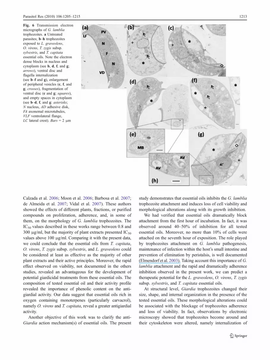
Untreated trophozoites observed in transmission electronmicroscopy presented the main distinguishing features ofG. lamblia trophozoites (Fig. 6a), i.e., the two nuclei, theventral disk composed of numerous microtubules, a largenumber of ribosome and glycogen particles, lateral flange,four pairs of flagella, and a set of peripheral vesicles.Following exposure to essential oils, the cells lost thepyriforme structure and become swollen and misshapen.The main alterations observed included ventral disk andflagella internalization (Fig. 6b, c, f, g), presence of largevacuoles in the cytoplasm (Fig. 6c, f, g), glycogen extractionwith intracellular clearing (Fig. 6b–d, f, g), loss of the nuclearenvelope integrity (Fig. 6d, f), and presence of electrodenseprecipitates in cytoplasm and nuclei (Fig. 6b, d, f, g). Inaddition, the essential oils induced accumulation of multi-lamellar bodies (Fig. 6e) and autophagic vesicles (Fig. 6h).
Cytotoxic assays Essential oils did not cause an significantalteration on the viability, measured by MTT, of treatedmammalian cells (macrophages and epithelial cells) whencompared to control cells and did not induced alterations onthe morphology of the cells when they were observed bylight microscopy (not shown).
Discussion
In the present work, we report the effects of essential oils ofT. capitata, O. virens, T. zygis subsp. sylvestris chemotypethymol, and L. graveolens on G. lamblia trophozoites
growth, viability, adherence, and morphology. In relation tothe effects on growth, the essential oils induced trophozoiteproliferation decline over 48 h incubation and were able tokill 50% of parasites in the earliest hours of contact:T. zygis oil before the first hour, L. graveolens at the firsthour, and O. virens and T. capitata after the third hour.Other works have also demonstrated antigiardial activity ofplant extracts (Harris et al. 2000; Gadelha et al. 2005;
Fig. 5 Scanning electron micrographs of G. lamblia trophozoites. a, bUntreated parasites display pairs of anterior flagella (A), ventral flagella(V), posterior–lateral flagella (PL), caudal flagella (C), and one ventraldisk (VD); c–g trophozoites exposed to O. virens, L. graveolens, T. zygissubsp. sylvestris, and T. capitata essential oils. Note the round shape andthe internalization of flagella (d, e, g), irregular ventral and dorsalsurface (e, f, g: asterisks), and membrane blebs (c, d, g: arrows). a, bBars = 5 μm; c–e bars = 1 μm; f bar = 5 μm; g bar = 1 μm
1212 Parasitol Res (2010) 106:1205–1215
Calzada et al. 2006; Moon et al. 2006; Barbosa et al. 2007;de Almeida et al. 2007; Vidal et al. 2007). These authorsshowed the effects of different plants, fractions, or purifiedcompounds on proliferation, adherence, and, in some ofthem, on the morphology of G. lamblia trophozoites. TheIC50 values described in these works range between 0.8 and300 μg/ml, but the majority of plant extracts presented IC50
values above 100 μg/ml. Comparing it with the present data,we could conclude that the essential oils from T. capitata,O. virens, T. zygis subsp. sylvestris, and L. graveolens couldbe considered at least as effective as the majority of otherplant extracts and their active principles. Moreover, the rapideffect observed on viability, not documented in the othersstudies, revealed an advantageous for the development ofpotential giardicidal treatments from these essential oils. Thecomposition of tested essential oil and their activity profilerevealed the importance of phenolic content on the anti-giardial activity. Our data suggest that essential oils rich inoxygen containing monoterpenes (particularly carvacrol),namely O. virens and T. capitata, reveal a greater antigiardialactivity.
Another objective of this work was to clarify the anti-Giardia action mechanism(s) of essential oils. The present
study demonstrates that essential oils inhibits the G. lambliatrophozoite attachment and induces loss of cell viability andmorphological alterations along with its growth inhibition.
We had verified that essential oils dramatically blockattachment from the first hour of incubation. In fact, it wasobserved around 40–50% of inhibition for all testedessential oils. Moreover, no more than 10% of cells wereattached on the seventh hour of exposition. The role playedby trophozoites attachment on G. lamblia pathogenesis,maintenance of infection within the host’s small intestine andprevention of elimination by peristalsis, is well documented(Elmendorf et al. 2003). Taking account this importance of G.lamblia attachment and the rapid and dramatically adherenceinhibition observed in the present work, we can predict atherapeutic potential for the L. graveolens, O. virens, T. zygissubsp. sylvestris, and T. capitata essential oils.
At structural level, Giardia trophozoites changed theirsize, shape, and internal organization in the presence of thetested essential oils. These morphological alterations couldbe associated with the blockage of trophozoites adherenceand loss of viability. In fact, observations by electronicmicroscopy showed that trophozoites become around andtheir cytoskeleton were altered, namely internalization of
Fig. 6 Transmission electronmicrographs of G. lambliatrophozoites. a Untreatedparasites; b–h trophozoitesexposed to L. graveolens,O. virens, T. zygis subsp.sylvestris, and T. capitataessential oils. Note the electrondense blocks in nucleus andcytoplasm (see b, d, f, and g;arrows), ventral disc andflagella internalization(see b–f and g), enlargementof peripheral vesicles (c, f, andg; crosses), fragmentation ofventral disc (c and g; squares),and empty spaces in cytoplasm(see b–d, f, and g: asterisks;N nucleus, AD adhesive disk,FA axonemal microtubules,VLF ventrolateral flange,LC lateral crest). Bars = 2 μm
Parasitol Res (2010) 106:1205–1215 1213

ventral disc and flagella. These structures play a decisiverole in the attachment of the parasite to inert superficies andto intestinal cells and were a common antigiardial drugtarget explored (Holberton 1973, 1974). Internalization offlagella in response to external stress has been notedpreviously with trichomonads and with Giardia (Abonyi1995; Vidal et al. 2007). Moreover, the effect on Giardiaadherence may be due also an interruption of energyproduction on the parasite exposed to essential oils enablingthe ventral disc to attach (Gardner and Hill 2001). In fact,alteration and decreased on trophozoites mobility, processenergy dependent, is an early event observed by opticalmicroscopy (not shown).
The toxic effects on membrane structure and functionshave been generally used to explain the antimicrobial actionof essential oils and their monoterpenoid components. Infact, as a result of their lipophilic character, monoterpeneswill preferentially partition from an aqueous phase intomembrane structures. Consequently, it occurs membraneexpansion, increase in membrane fluidity and permeability,disturbance membrane-embedded proteins, inhibition ofrespiration, and alteration of ion transport processes(Cristani et al. 2007). Therefore, the tested essential oilsmay also interact with Giardia membranes and inducedchanges in the osmotic balance. Corroborate by these findingswas the trophozoites swelling and the morphological alter-ations observed in the present work. The osmotic disturbcould be the beginning of a cascaded interaction modifyingorganelles structure, cytoplasm constituents, and cytoskeletonand nuclear characteristics. In fact, the size increase ofGiardiacells, the increased volume of the Giardia peripheralvesicles, and the loss of the nuclear envelope integrity wereobserved on trophozoites treated with essential oils. Previousstudies showed that the peripheral vesicles are the essentialcomponent of the endosomal/lysosomal systems of theprotozoan (Lanfredi-Rangel et al. 1998; Abodeely et al2009), and the changes in the nucleic acids distributionsuggest the breaking of the proteins net. Moreover, the denseblocks observed in cytoplasm very likely correspond tocoagulated proteins and/or products of degradation ofribosome. Previous studies with bacteria support the relation-ship between membrane damage, ribosome disappearance,and cell death (Silva et al. 1987; Niven et al. 1999).
Other morphological change was the appearance ofautophagosomal vacuoles on cytoplasm and multilamelarbodies. There are evidences that autophagy represents one ofthe mechanisms used as a cell survival strategy under stressconditions (Edinger and Thompson 2004). The presence ofvacuoles and multilamelar bodies on essential oil-treatedtrophozoites may be due to an abnormal recycling ofmembranes, suggesting that an intense remodeling of cellularorganelles should be happening, and this process may bealtered and/or blocked be essential oils.
Taken together, our findings lead us to propose thatantigiardial effect of tested essential oils may result from adestabilization of plasmic membrane of the protozoan, leadingto alterations on membrane properties and subsequently cellswelling, modifications on cytoplasmic and organelles struc-ture, and, at the end, cell death. Moreover, L. graveolens,O. virens, T. zygis subsp. sylvestris, and T. capitata essentialoils not induced cytotoxicity on mammalian cell lines.Results justify future investigations to validate and improvethe use of these essential oils as therapeutic alternatives forgiardiasis treatment.
Acknowledgments Authors are grateful to Prof. Jorge Paiva for helpin plant taxonomic.
Funding This work was supported by “ProgramaOperacional Ciênciae Inovacão 2010 (POCI)/FEDER” da Fundação para a Ciência eTecnologia.
Transparency declarations None to declare.
References
Abodeely M, DuBois KN, Hehl A et al (2009) A contiguouscompartment functions as endoplasmic reticulum and endosome/lysosome in Giardia lamblia. Eukaryot Cell 8:1665–1676
Abonyi A (1995) Examination of nonflagellate and flagellate roundforms of Trichomonas vaginalis by transmission electronmicroscopy. Appl Parasitol 36:303–310
Adam RD (2001) Biology of Giardia lamblia. Clin Microbiol Rev14:447–475
Adams RP (1995) Identification of essential oils components by gaschromatography/mass spectroscopy. Allured Publishing Corporation,Carol Stream
Barbosa E, Calzada F, Campos R (2007) In vivo antigiardial activityof three flavonoids isolated of some medicinal plants used inMexican traditional medicine for the treatment of diarrhea. JEthnopharmacol 109:552–554
Burt S (2004) Essential oils: their antibacterial properties and potentialapplications in foods—a review. Int J Food Microbiol 94:223–253
Busatti HG, Vieira AE, Viana JC et al (2007) Effect of metronidazoleanalogues on Giardia lamblia cultures. Parasitol Res 102:145–149
Calzada F, Yepez-Mulia L, Aguilar A (2006) In vitro susceptibility ofEntamoeba histolytica and Giardia lamblia to plants used inMexican traditional medicine for the treatment of gastrointestinaldisorders. J Ethnopharmacol 108:367–370
Council of Europe (1997) European Pharmacopoeia. Europe Co.,Strasbourg
Cristani M, D'Arrigo M, Mandalari G et al (2007) Interaction of fourmonoterpenes contained in essential oils with model membranes:implications for their antibacterial activity. J Agric Food Chem55:6300–6308
Custodio JB, Moreno AJ, Wallace KB (1998) Tamoxifen inhibitsinduction of the mitochondrial permeability transition by Ca2+and inorganic phosphate. Toxicol Appl Pharmacol 152:10–17
de Almeida I, Alviano DS, Vieira DP et al (2007) Antigiardial activityof Ocimum basilicum essential oil. Parasitol Res 101:443–452
Denizot F, Lang R (1986) Rapid colorimetric assay for cell growthand survival. Modifications to the tetrazolium dye proceduregiving improved sensitivity and reliability. J Immunol Methods89:271–277
1214 Parasitol Res (2010) 106:1205–1215

Dorman HJ, Deans SG (2000) Antimicrobial agents from plants:antibacterial activity of plant volatile oils. J Appl Microbiol 88:308–316
Eckmann L (2003) Mucosal defences against Giardia. ParasiteImmunol 25:259–270
Edinger AL, Thompson CB (2004) Death by design: apoptosis,necrosis and autophagy. Curr Opin Cell Biol 16:663–669
Edris AE (2007) Pharmaceutical and therapeutic potentials of essentialoils and their individual volatile constituents: a review. PhytotherRes 21:308–323
Elmendorf HG, Dawson SC, McCaffery JM (2003) The cytoskeletonof Giardia lamblia. Int J Parasitol 33:3–28
Faleiro L, Miguel G, Gomes S et al (2005) Antibacterial and antioxidantactivities of essential oils isolated from Thymbra capitata L. (Cav.)and Origanum vulgare L. J Agric Food Chem 53:8162–8168
Flanagan PA (1992) Giardia—diagnosis, clinical course and epidemiology.A review. Epidemiol Infect 109:1–22
Fraser D, Bilenko N, Deckelbaum RJ et al (2000) Giardia lambliacarriage in Israeli Bedouin infants: risk factors and consequences.Clin Infect Dis 30:419–424
Gadelha AP, Vidal F, Castro TM et al (2005) Susceptibility of Giardialamblia to Hovenia dulcis extracts. Parasitol Res 97:399–407
Gardner TB, Hill DR (2001) Treatment of giardiasis. Clin MicrobiolRev 14:114–128
Gillin FD, Reiner DS (1982) Attachment of the flagellate Giardialamblia: role of reducing agents, serum, temperature, and ioniccomposition. Mol Cell Biol 2:369–377
Gonçalves MJ, Vicente AM, Cavaleiro C et al (2007) Compositionand antifungal activity of the essential oil of Mentha cervinafrom Portugal. Nat Prod Res 21:867–871
Hansen WR, Tulyathan O, Dawson SC et al (2006) Giardia lambliaattachment force is insensitive to surface treatments. EukaryotCell 5:781–783
Harris JC, Plummer S, Turner MP et al (2000) The microaerophilicflagellate Giardia intestinalis: Allium sativum (garlic) is aneffective antigiardial. Microbiology 146:3119–3127
Hill DR, Pohl R, Pearson RD (1986) Giardia lamblia: a culturemethod for determining parasite viability. Am J Trop Med Hyg35:1129–1133
Holberton DV (1973) Fine structure of the ventral disk apparatus andthe mechanism of attachment in the flagellate Giardia muris. JCell Sci 13:11–41
Holberton DV (1974) Attachment of Giardia—a hydrodynamic modelbased on flagellar activity. J Exp Biol 60:207–221
Joulain D, König W (1998) The atlas of spectral data of sesquiterpenehydrocarbons. E. B., Hamburg
Keister DB (1983) Axenic culture of Giardia lamblia in TYI-S-33medium supplemented with bile. Trans R Soc Trop Med Hyg77:487–488
Lanfredi-Rangel A, Attias M, de Carvalho TM et al (1998) Theperipheral vesicles of trophozoites of the primitive protozoanGiardia lamblia may correspond to early and late endosomes andto lysosomes. Struct Biol 123:225–235
Machado M, Sousa MC, Salgueiro L et al (2010) Effects of essential oilson the growth of Giardia lamblia trophozoites. Nat Prod Commun5:137–141
Moon T,Wilkinson JM, CavanaghHM (2006) Antiparasitic activity of twoLavandula essential oils against Giardia duodenalis, Trichomonasvaginalis and Hexamita inflata. Parasitol Res 99:722–728
Niven GW, Miles CA, Mackey BM (1999) The effects of hydrostaticpressure on ribosome conformation in Escherichia coli: and invivo study using differential scanning calorimetry. Microbiology145:419–425
Park JH, Schofield PJ, Edwards MR (1997) Giardia intestinalis:volume recovery in response to cell swelling. Exp Parasitol86:19–28
Perez-Arriaga L, Mendoza-Magana ML, Cortes-Zarate R et al (2006)Cytotoxic effect of curcumin on Giardia lamblia trophozoites.Acta Trop 98:152–161
Ponce-Macotela M, Rufino-Gonzalez Y, Gonzalez-Maciel A et al(2006) Oregano (Lippia spp.) kills Giardia intestinalis troph-ozoites in vitro: antigiardiasic activity and ultrastructural damage.Parasitol Res 98:557–560
Santoro GF, das Gracas Cardoso M et al (2007) Effect of oregano(Origanum vulgare L.) and thyme (Thymus vulgaris L.) essentialoils on Trypanosoma cruzi (Protozoa: Kinetoplastida) growth andultrastructure. Parasitol Res 100:783–790
Sawangjaroen N, Subhadhirasakul S, Phongpaichit S et al (2005) Thein vitro anti-giardial activity of extracts from plants that are usedfor self-medication by AIDS patients in southern Thailand.Parasitol Res 95:17–21
Silva MT, Appelberg R, Silva MN et al (1987) In vivo killing anddegradation of Mycobacterium aurum within mouse peritonealmacrophages. Infect Immun 55:2006–2016
Sousa MC, Poiares-Da-Silva J (1999) A new method for assessingmetronidazole susceptibility of Giardia lamblia trophozoites.Antimicrob Agents Chemother 43:2939–2942
Sousa MC, Goncalves CA, Bairos VA et al (2001) Adherence ofGiardia lamblia trophozoites to Int-407 human intestinal cells.Clin Diagn Lab Immunol 8:258–265
Thompson RC (2000) Giardiasis as a re-emerging infectious diseaseand its zoonotic potential. Int J Parasitol 30:1259–1267
Upcroft P, Upcroft JA (2001) Drug targets and mechanisms ofresistance in the anaerobic protozoa. Clin Microbiol Rev14:150–164
Vidal F, Vidal JC, Gadelha AP et al (2007) Giardia lamblia: theeffects of extracts and fractions from Mentha x piperita Lin.(Lamiaceae) on trophozoites. Exp Parasitol 115:25–31
Parasitol Res (2010) 106:1205–1215 1215


