Embed Size (px)
Citation preview

Hindawi Publishing CorporationMediators of InflammationVolume 2011, Article ID 912925, 11 pagesdoi:10.1155/2011/912925
Research Article
Anti-inflammatory and Antinociceptive Activity ofOuabain in Mice
Danielle Ingrid Bezerra de Vasconcelos,1, 2 Jacqueline Alves Leite,1 Luciana Teles Carneiro,1
Marcia Regina Piuvezam,1, 2 Maria Raquel Vitorino de Lima,1 Liana Clebia Lima de Morais,1
Vivian Mary Rumjanek,3 and Sandra Rodrigues-Mascarenhas1, 2
1 Laboratorio de Tecnologia Farmaceutica, Departamento de Fisiologia e Patologia, Centro de Ciencias da Saude,Universidade Federal da Paraıba, Joao Pessoa 58059-900, Brazil
2 Departamento de Biologia Molecular, Centro de Ciencias Exatas e da Natureza, Universidade Federal da Paraıba,Joao Pessoa 58059-900, Brazil
3 Instituto de Bioquımica Medica, Centro de Ciencias da Saude, Universidade Federal do Rio de Janeiro,Rio de Janeiro 21941-902, Brazil
Correspondence should be addressed to Sandra Rodrigues-Mascarenhas, [email protected]
Received 23 November 2010; Revised 17 February 2011; Accepted 26 March 2011
Academic Editor: Muzamil Ahmad
Copyright © 2011 Danielle Ingrid Bezerra de Vasconcelos et al. This is an open access article distributed under the CreativeCommons Attribution License, which permits unrestricted use, distribution, and reproduction in any medium, provided theoriginal work is properly cited.
Ouabain, an inhibitor of the Na+/K+-ATPase pump, was identified as an endogenous substance of human plasma. Ouabain hasbeen studied for its ability to interfere with various regulatory mechanisms. Despite the studies portraying the ability of ouabainto modulate the immune response, little is known about the effect of this substance on the inflammatory process. The aim of thiswork was to study the effects triggered by ouabain on inflammation and nociceptive models. Ouabain produced a reduction inthe mouse paw edema induced by carrageenan, compound 48/80 and zymosan. This anti-inflammatory potential might be relatedto the inhibition of prostaglandin E2, bradykinin, and mast-cell degranulation but not to histamine. Ouabain also modulatedthe inflammation induced by concanavalin A by inhibiting cell migration. Besides that, ouabain presented antinociceptiveactivity. Taken these data together, this work demonstrated, for the first time, that ouabain presented in vivo analgesic and anti-inflammatory effects.
1. Introduction
Ouabain, one of the oldest drugs used for treatment of car-diac insufficiency, is classically known as a specific inhibitorof the plasma membrane Na+/K+-ATPase [1]. Originallyisolated from plants, it was demonstrated that mammalsnaturally produce an endogenous analogue of ouabain, ahormone synthesized in the adrenal glands, hypothalamus,and pituitary and found circulating in the plasma [2–5].Accumulating evidence suggests that circulating levels ofendogenous ouabain are modulated by stress conditions.Elevated plasma levels of ouabain were found in hyperten-sive patients and after physical exercise in different species[6–8].
The steroid ouabain is capable of modulating many as-pects of the immune system, being considered as an im-munomodulatory molecule [9]. Ouabain, in vitro, inhibitsmitogen-induced thymocyte and lymphocyte proliferation[10, 11], inhibits the generation of lymphokine-activatedkiller (LAK) activity induced by IL-2 [12], increases intra-cellular calcium levels [13], increases CD69 expression [14]and acts both in vitro [15] and in vivo [16] synergisticallywith corticoids. In vitro, ouabain increases glucocorticoid-induced plasma membrane depolarization in lymphoid cells[9]. Besides that, ouabain downregulates mCD14 expressionon monocytes, which is related to inflammatory responseagainst several pathogens [17] and induces IL-1 β productionon human mononuclear cells [18].
2 Mediators of Inflammation
Additionally, this molecule is also capable of inducingthe activation of various signal transduction cascades thatare independent of changes in intracellular Na+ and K+ con-centrations, involving the Ras/Raf/mitogen-activated proteinkinase (MAPK) cascade, transactivation of epidermal growthfactor receptor (EGFR), and protein kinase C [17, 19–23]. Inmurine thymocytes, ouabain decreased the levels of phos-phorylated MAPK p38 and nuclear factor of activated T-cells(NFATc1) induced by the mitogen concanavalin A [24].
Besides the effects described in the immune system, littleis known about the role of ouabain in inflammatory pro-cesses. During inflammation, a complex program of intra-cellular signal transduction and transcription events, drivenby multiple proinflammatory mediators and cytokines, isactivated. The acute inflammation is characterized by exu-dation of protein-rich fluid, edema, vasodilatation, and cellmigration, primarily neutrophils, into the site of injury [25].It was reported that ouabain suppressed the production ofthe pro-inflammatory cytokines IL-6 and TNF-α stimulatedwith LPS both in vitro and in vivo [26]. In addition, cardiacglycoside drugs inhibits TNF-α/NF-κB signaling pathway,which is a central common regulator for the process of in-flammation [27].
The aim of the present study was to investigate the role ofouabain in acute peripheral inflammation induced by intra-plantar and intraperitoneal injection of different phlogisticagents and in algesic processes.
2. Material and Methods
2.1. Animals. Female swiss albino mice (2 months old) werehoused in a temperature-controlled room and received waterand food ad libitum. After manipulation, euthanasia wasemployed by cervical dislocation. The study was performedaccording to the Guidelines on Ethical Standards for Investi-gation of Experimental Pain in Animals [28], after approvalof protocol no. 0407/08 by the Institutional Ethics Commit-tee of Laboratorio de Tecnologia Farmaceutica.
2.2. Treatment with Ouabain. In all experiments, 0.56 mg/kgouabain [16] or phosphate buffered saline (PBS) was givenintraperitoneally (i.p.) for three consecutive days. A dose-response curve was also performed using 0.10 mg/kg and0.31 mg/kg ouabain, and 0.56 mg/kg ouabain was given i.p.for one and two consecutive days.
2.3. Inflammatory Paw Edema. To induce inflammation,mice received subcutaneous injections in the plantar surface[29] of carrageenan (2.5%), compound 48/80 (2 μg/paw),zymosan (1%), histamine (100 μg/paw), prostaglandin E2(5 μg/paw), and bradykinin (6 nmol/paw) in 20 μL of phos-phate buffered saline (PBS) in the right (ipsilateral) hindpawand 20 μL of PBS in the left (contralateral) hindpaw. Cap-topril (5 mg/kg) was used 1 h prior to bradykinin challengein order to prevent the action of kininases [30]. Ouabainlast treatment was injected i.p. one hour before intraplantarinjections of phlogistic agents. All the reagents were obtainedfrom Sigma. Hindpaw edema was measured with a digital
micrometer (Instrutemp 070393611) [31] and is expressedas the difference of thickness (mm) between the stimulatedand the saline-injected paw.
Dexamethasone (0.5 mg/kg), indomethacin (10 mg/Kg)and salbutamol (10 mg/kg) were used as antiinflammatorycontrols and injected i.p. or subcutaneous (s.c.) one hourbefore intraplantar injections.
2.4. Peritoneal Inflammation Model. Mice were injectedintraperitoneally with 60 ug Concanavalin A diluted in sterilesaline solution to a final injection volume of 0.1 mL one hourafter the last ouabain administration. After 24 h, animalswere sacrificed and peritoneal lavage was performed with10 mL of sterile PBS [32–34]. The fluid was removed fortotal and differential cell counts and results were reportedas cell number/mL of peritoneal wash. Differentiation ofleukocyte subpopulations in peritoneal lavage fluids wasperformed on cytospin preparations stained with Wright-Giemsa solution (Sigma, St. Louis, Mo, USA). For each slidea minimum of 100 cells was counted microscopically under1000 magnification.
2.5. Acetic Acid Induced Writhing Test. Acetic acid admin-istration causes irritation resulting in painful contortionsfollowed by extension of hind limbs [35]. The animalswere injected i.p. with 0.1 mL/10 g of 0.8% (v/v solution)acetic acid one hour after the last treatment with ouabain(0.56 mg/kg). Morphine (6 mg/Kg) (i.p.) was used for an-algesia. Ten minutes after the administration of acetic acid,mice were placed in separate boxes and the number ofabdominal writhes was counted for 10 minutes. The anti-nociceptive activity was expressed as the reduction in thenumber of abdominal writhes when compared to controlanimals.
2.6. Hot Plate. Animals were placed on a hot plate main-tained at 52 ± 1◦C. The time elapsed between placing theanimal on the hot plate, and the animal either licking its foreor hind paws or jumping on the surface was considered theresponse latency [36]. Mice with baseline latencies of morethan 15 s were excluded from the study. Response latencytesting was measured 30, 60 and 120 minutes after the lasttreatment with Ouabain (0.56 mg/kg), saline or the positivecontrol, morphine (10 mg/Kg). The cutoff time for the hot-plate test latency was set at 30 s to avoid tissue injury. Allanimals were brought to the test room at least 1 h prior tothe experiments and were not tested more than once.
2.7. Possible Involvement of Opioid Receptors. In order to in-vestigate the involvement of the opioidergic system in oua-bain-induced antinociception, separate groups of mice werepretreated with nonselective opioid receptor antagonist,naloxone (5 mg/kg, s.c.), which was injected 15 min beforei.p. administration of ouabain (0.56 mg/kg) and morphine(10 mg/kg, i.p.), and tested using the hot plate test [37].
2.8. Elevated Plus maze. The apparatus is comprised of twoopen arms (35×5 cm) and two closed arms (30×5×15 cm)
Mediators of Inflammation 3
∗∗∗∗∗
∗∗∗∗
∗∗∗
∗∗∗∗∗
∗∗∗∗∗∗
4321
Time after injection (h)
0
0.25
0.5
0.75
1
1.25
1.5
1.75
2
2.25Pa
wed
ema
(mm
)
ZYMOUA 0.1
OUA 0.31OUA0.56
(a)
∗∗∗∗∗∗
∗
54321
Time after injection (h)
−0.5
0
0.5
1
1.5
Paw
edem
a(m
m)
ZYMOUA
DEXAPBS
(b)
∗∗∗∗∗
∗∗∗
∗∗
∗
543210
Time after injection (h)
0
0.5
1
1.5
2
2.5
Paw
edem
a(m
m)
ZYMOUA
DEXAPBS
(c)
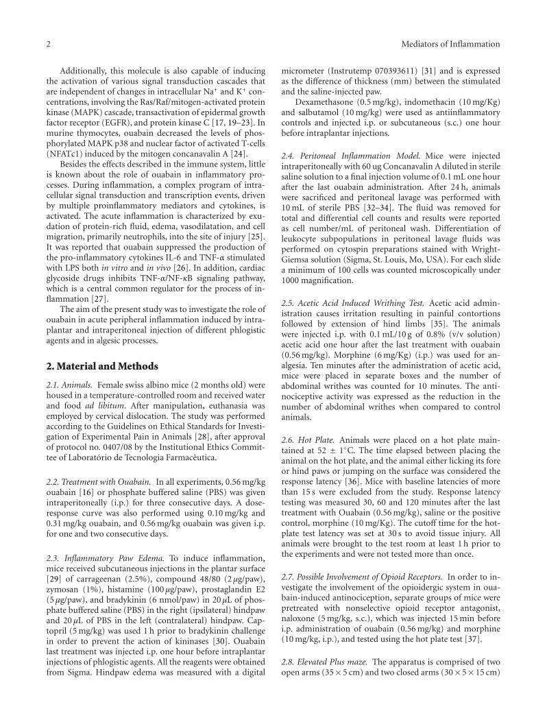
Figure 1: Ouabain dose-response curve and administration for 1 and 2 consecutive days. Mice were pretreated with 0.10 mg/kg, 0.31 mg/kgand 0.56 mg/kg ouabain for 3 consecutive days (a) or with 0.56 mg/kg ouabain for 1 (b) and 2 consecutive days (c); one hour after the lastouabain treatment, mice received intraplantar injections of zymosan in 20 μL phosphate buffered saline (PBS) in the right hindpaw and 20 μLof PBS in the left hindpaw. Each point represents the mean of eight animals. Dexametasone (DEXA, 0.5 mg/kg) was used as antiinflammatorycontrol and injected i.p. one hour before intraplantar challenge. Asterisks denote the significance levels compared with ZYM group. Datawere expressed as mean ± S.E.M. and analyzed by software Graphpad Prism using Student’s t-test followed by unpaired test. ∗P < .05,∗∗P < .01, and ∗∗∗P < .001.
that extended from a common central platform (5 × 5 cm).The entire maze was elevated to a height of 30 cm abovefloor level [38]. One hour after ouabain administration, micewere placed in the central region of the plus-maze apparatus.Diazepan (0.5 mg/Kg) was administred 30 min before the testand used as positive control.
2.9. Statistical Analysis. All data were expressed as mean± S.E.M. and analyzed by software Graphpad Prism usingStudent’s t-test followed by unpaired test or ANOVA fol-lowed Dunnett’s or Mann Whitney test , and the results wereconsidered significant if P < .05.
3. Results
3.1. Effect of Ouabain on Carrageenan, Zymosan, and 48/80-Induced Mice Paw Edema. The paw edema induced bycarrageenan and zymosan involves various mediators suchas histamine, bradykinin, and prostaglandins [39, 40].Although 0.10 mg/kg ouabain was without effect, 0.31 mg/kgand 0.56 mg/kg ouabain prevented zymosan edema forma-tion (Figure 1(a)). On the other hand, ouabain was notable to inhibit zymosan-induced paw inflammation whenadministered only one day prior experiment (Figure 1(b)).Furthermore, when ouabain was given for two days priorexperiment, it did not interfere in the edema present 4 h
4 Mediators of Inflammation
∗∗∗
∗∗∗∗∗∗∗∗∗
2524236543210
Time after injection (h)
0
0.25
0.5
0.75
1
1.25Pa
wed
ema
(mm
)
CAROUA
INDPBS
(a)
∗∗
∗∗∗
∗∗∗
∗∗∗
∗∗∗
543210
Time after injection (h)
0
0.5
1
1.5
2
2.5
Paw
edem
a(m
m)
ZYMOUA
DEXAPBS
(b)
∗∗∗∗∗∗
∗
∗∗∗
1209060300
Time after injection (min)
0
0.25
0.5
0.75
Paw
edem
a(m
m)
48/80OUA
SALB.PBS
(c)
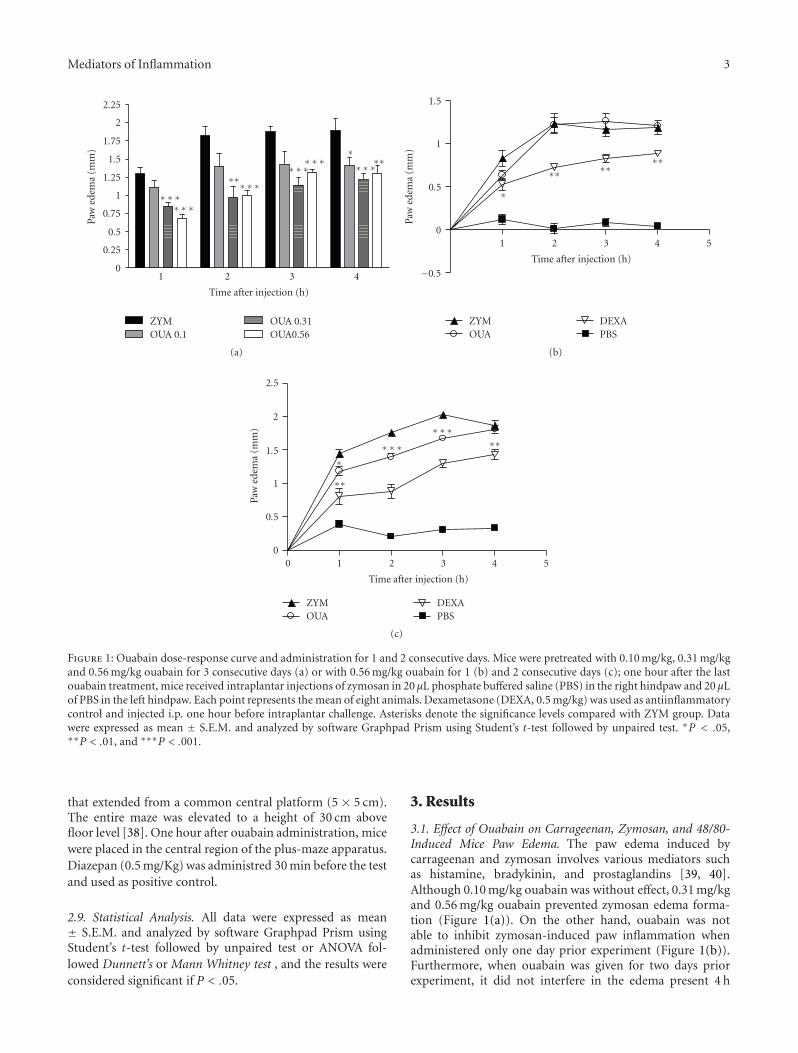
Figure 2: Effect of the pretreatment with Ouabain (OUA, 0.56 mg/kg) administered intraperitoneally on paw edema triggered by carragenan(CAR), zymosan (ZYM), and compound 48/80 in mice. Mice received intraplantar injections of carrageenan (2.5%, (a)), zymosan (1%, (b)),or compound 48/80 (2 μg/paw, (c)) in 20 μL phosphate buffered saline (PBS) in the right hindpaw and 20 μL of PBS in the left hindpaw.Each point represents the mean of eight animals. Indomethacin (IND, 10 mg/Kg), dexametasone (DEXA, 0.5 mg/Kg), and salbutamol (SALB,10 mg/kg) were used as antiinflammatory controls and injected i.p. one hour before intraplantar challenge. Asterisks denote the significancelevels compared with CAR, ZYM, or compound 48/80 group. Data were expressed as mean ± S.E.M. and analyzed by software GraphpadPrism using Student’s t-test followed by unpaired test ∗P < .05, ∗∗P < .01, and ∗∗∗P < .001.
after zymosan (Figure 1(c)). Ouabain 0.56 mg/kg injected forthree consecutive days, prevented zymosan edema formationat the 1st (54.4%), 2nd (47.1%), 3rd (34.7%), and 4th h(26.9%) after treatment, as well as did 0.5 mg/kg dexam-ethasone (Figure 2(b)). Similarly to what was observed withzymosan, carrageenan-induced paw edema was also signifi-cantly reduced in a time-dependent manner by the treatmentof 0.56 mg/Kg ouabain at 30 min (54.9%), 1st (66.4%),2nd (51.0%), and 5 h (51.9%) after carrageenan treatment(Figure 2(a)). On the other hand, ouabain did not interferein the edema present 24 h after carrageenan injection. Asignificant antiinflammatory effect of indomethacin at thedose of 10 mg/Kg was observed at all times studied. Paw
edema induced by compound 48/80 was short lasted, with apeak 30 min after injection. Ouabain significantly inhibitedthe edema, but to a lesser extent than salbutamol, at 30(69.1%), 60 (79.7%), and 120 min (49.7%) after compound48/80 injection (Figure 2(c)).
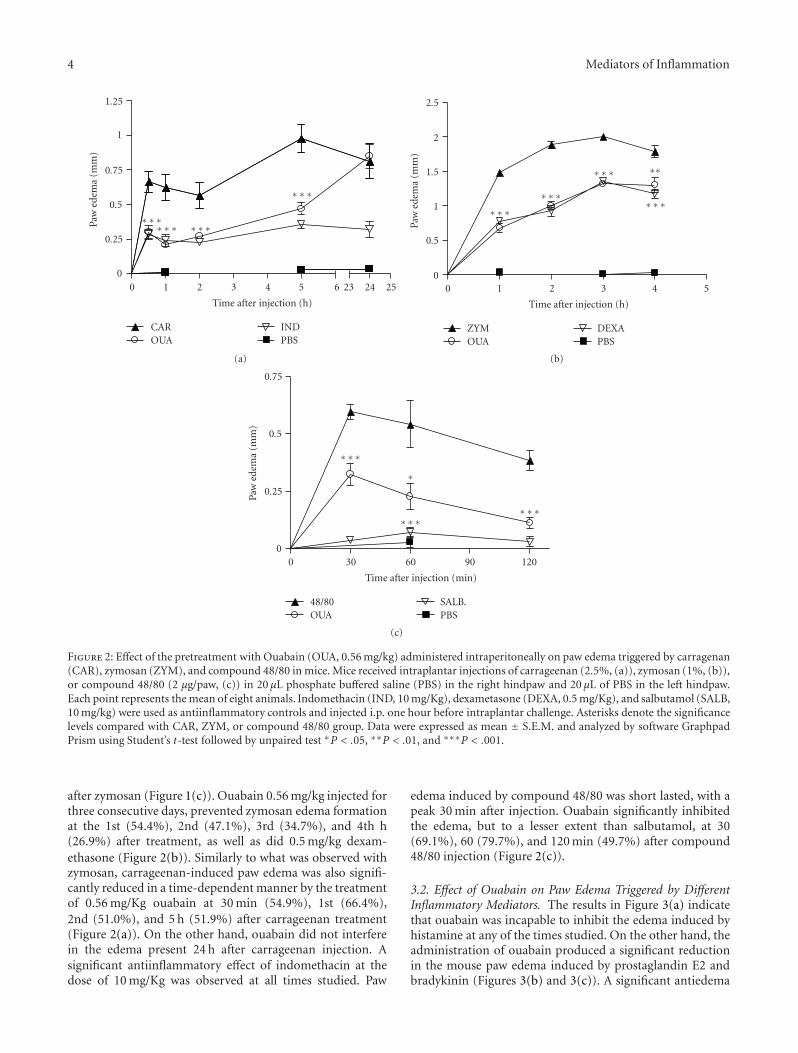
3.2. Effect of Ouabain on Paw Edema Triggered by DifferentInflammatory Mediators. The results in Figure 3(a) indicatethat ouabain was incapable to inhibit the edema induced byhistamine at any of the times studied. On the other hand, theadministration of ouabain produced a significant reductionin the mouse paw edema induced by prostaglandin E2 andbradykinin (Figures 3(b) and 3(c)). A significant antiedema
Mediators of Inflammation 5
∗∗
∗∗∗
∗∗∗
1209060300
Time after injection (min)
0
0.25
0.5
0.75
1
1.25Pa
wed
ema
(mm
)
HISOUA
DEXAPBS
(a)
∗∗∗∗∗∗
∗∗∗
604530150
Time after injection (min)
0
0.25
0.5
0.75
Paw
edem
a(m
m)
PGE2OUA
DEXAPBS
(b)
∗
∗∗
∗∗
30150
Time after injection (min)
0
0.25
0.5
0.75
Paw
edem
a(m
m)
BRADOUA
DEXAPBS
(c)
Figure 3: Effect of the pretreatment with Ouabain (OUA, 0.56 mg/kg) administered intraperitoneally on paw edema triggered by histamine(HIS), prostaglandin E2 (PGE2) and bradykinin (BRAD). Mice received intraplantar injections of histamine (100 μg/paw, (a)), prostaglandinE2 (5 μg/paw, (b)) or bradykinin (6 nmol/paw, (c)) in 20 μL of phosphate buffered saline (PBS) in the right hindpaw and 20 μL of PBS inthe left hindpaw. Each point represents the mean of eight animals. Dexametasone (0.5 mg/kg) was used as antiinflammatory control andinjected i.p. one hour before intraplantar challenge. Data were expressed as mean ± S.E.M. and analyzed by software Graphpad Prism usingStudent’s t-test followed by unpaired test ∗P < .05, ∗∗P < .01, and ∗∗∗P < .001.
effect of ouabain was observed at 15 (79.8%), 30 (82.1%) and60 min (96.0%) after prostaglandin E2 challenge and at 15(34.0%) but not at 30 min after bradykinin challenge.
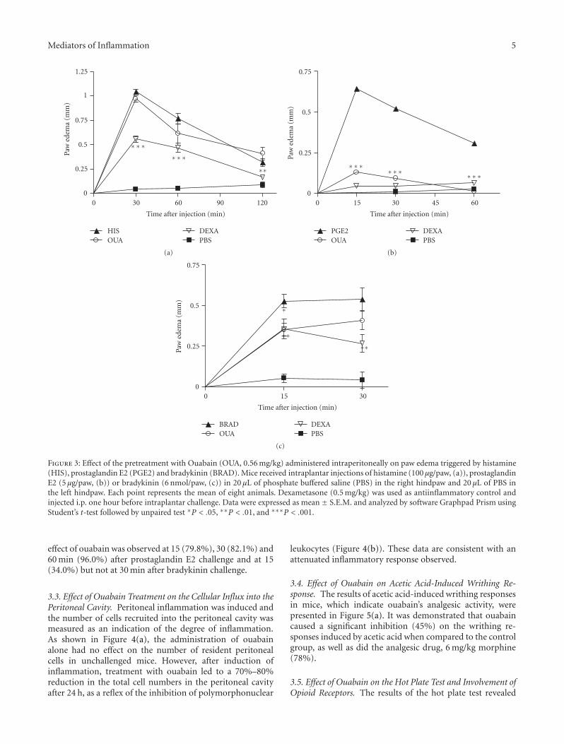
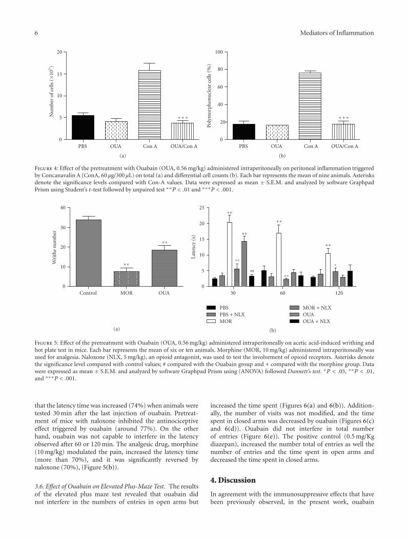
3.3. Effect of Ouabain Treatment on the Cellular Influx into thePeritoneal Cavity. Peritoneal inflammation was induced andthe number of cells recruited into the peritoneal cavity wasmeasured as an indication of the degree of inflammation.As shown in Figure 4(a), the administration of ouabainalone had no effect on the number of resident peritonealcells in unchallenged mice. However, after induction ofinflammation, treatment with ouabain led to a 70%–80%reduction in the total cell numbers in the peritoneal cavityafter 24 h, as a reflex of the inhibition of polymorphonuclear
leukocytes (Figure 4(b)). These data are consistent with anattenuated inflammatory response observed.
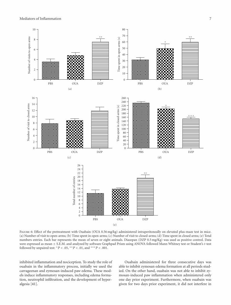
3.4. Effect of Ouabain on Acetic Acid-Induced Writhing Re-sponse. The results of acetic acid-induced writhing responsesin mice, which indicate ouabain’s analgesic activity, werepresented in Figure 5(a). It was demonstrated that ouabaincaused a significant inhibition (45%) on the writhing re-sponses induced by acetic acid when compared to the controlgroup, as well as did the analgesic drug, 6 mg/kg morphine(78%).
3.5. Effect of Ouabain on the Hot Plate Test and Involvement ofOpioid Receptors. The results of the hot plate test revealed
6 Mediators of Inflammation
∗∗∗
OUA/Con ACon AOUAPBS0
5
10
15
20N
um
ber
ofce
lls(×
107)
(a)
∗∗∗
OUA/Con ACon AOUAPBS0
20
40
60
80
100
Poly
mor
phon
ucl
ear
cells
(%)
(b)
Figure 4: Effect of the pretreatment with Ouabain (OUA, 0.56 mg/kg) administered intraperitoneally on peritoneal inflammation triggeredby Concanavalin A (ConA, 60 μg/300 μL) on total (a) and differential cell counts (b). Each bar represents the mean of nine animals. Asterisksdenote the significance levels compared with Con-A values. Data were expressed as mean ± S.E.M. and analyzed by software GraphpadPrism using Student’s t-test followed by unpaired test ∗∗P < .01 and ∗∗∗P < .001.
∗∗
∗∗
OUAMORControl0
10
20
30
40
Wri
the
nu
mbe
r
(a)
+
∗∗
++
∗∗
##
∗∗
++
∗∗
12060300
5
10
15
20
25
Late
ncy
(s)
PBSPBS + NLXMOR
MOR + NLXOUAOUA + NLX
(b)
Figure 5: Effect of the pretreatment with Ouabain (OUA, 0.56 mg/kg) administered intraperitoneally on acetic acid-induced writhing andhot plate test in mice. Each bar represents the mean of six or ten animals. Morphine (MOR, 10 mg/kg) administered intraperitoneally wasused for analgesia. Naloxone (NLX, 5 mg/kg), an opioid antagonist, was used to test the involvement of opioid receptors. Asterisks denotethe significance level compared with control values; # compared with the Ouabain group and + compared with the morphine group. Datawere expressed as mean ± S.E.M. and analyzed by software Graphpad Prism using (ANOVA) followed Dunnett’s test. ∗P < .05, ∗∗P < .01,and ∗∗∗P < .001.
that the latency time was increased (74%) when animals weretested 30 min after the last injection of ouabain. Pretreat-ment of mice with naloxone inhibited the antinociceptiveeffect triggered by ouabain (around 77%). On the otherhand, ouabain was not capable to interfere in the latencyobserved after 60 or 120 min. The analgesic drug, morphine(10 mg/kg) modulated the pain, increased the latency time(more than 70%), and it was significantly reversed bynaloxone (70%), (Figure 5(b)).
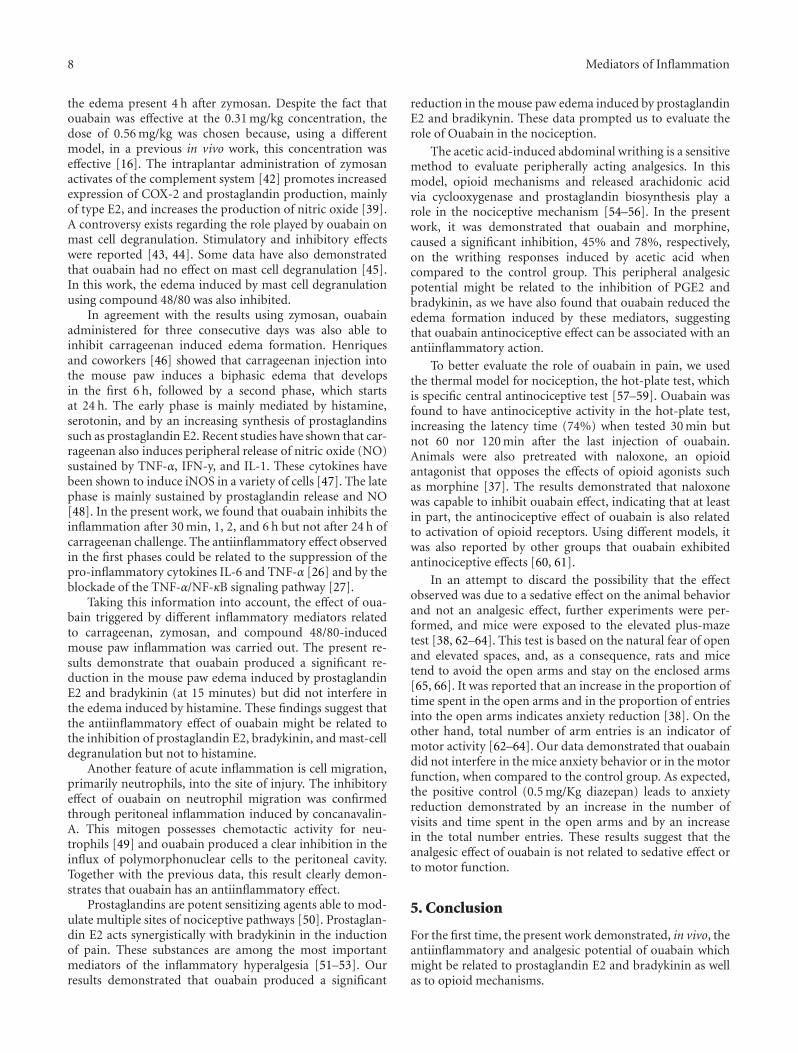
3.6. Effect of Ouabain on Elevated Plus-Maze Test. The resultsof the elevated plus maze test revealed that ouabain didnot interfere in the numbers of entries in open arms but
increased the time spent (Figures 6(a) and 6(b)). Addition-ally, the number of visits was not modified, and the timespent in closed arms was decreased by ouabain (Figures 6(c)and 6(d)). Ouabain did not interfere in total numberof entries (Figure 6(e)). The positive control (0.5 mg/Kgdiazepan), increased the number total of entries as well thenumber of entries and the time spent in open arms anddecreased the time spent in closed arms.
4. Discussion
In agreement with the immunosuppressive effects that havebeen previously observed, in the present work, ouabain
Mediators of Inflammation 7
∗∗
DZPOUAPBS0
2
4
6
8
10N
um
ber
ofvi
sits
toop
enar
ms
(a)
∗∗
∗
DZPOUAPBS0
10
20
30
40
50
60
70
80
Tim
esp
ent
inop
en-a
rms
(s)
(b)
DZPOUAPBS0
2
4
6
8
10
12
14
16
Nu
mbe
rof
visi
tto
clos
edar
ms
(c)
∗∗∗
∗
DZPOUAPBS0
20406080
100120140160180200220240260
Tim
esp
ent
incl
osed
-arm
s(s
)
(d)
∗∗
DZPOUAPBS02468
101214161820222426
Tota
lnu
mbe
rof
entr
ies
(e)
Figure 6: Effect of the pretreatment with Ouabain (OUA 0.56 mg/kg) administered intraperitoneally on elevated plus-maze test in mice.(a) Number of visit to open-arms; (b) Time spent in open-arms; (c) Number of visit to closed-arms; (d) Time spent in closed arms; (e) Totalnumbers entries. Each bar represents the mean of seven or eight animals. Diazepan (DZP 0.5 mg/Kg) was used as positive control. Datawere expressed as mean ± S.E.M. and analyzed by software Graphpad Prism using ANOVA followed Mann Whitney test or Student’s t-testfollowed by unpaired test: ∗P < .05, ∗∗P < .01, and ∗∗∗P < .001.
inhibited inflammation and nociception. To study the role ofouabain in the inflammatory process, initially we used thecarrageenan and zymosan-induced paw edema. These mod-els induce inflammatory responses, including edema forma-tion, neutrophil infiltration, and the development of hyper-algesia [41].
Ouabain administered for three consecutive days wasable to inhibit zymosan edema formation at all periods stud-ied. On the other hand, ouabain was not able to inhibit zy-mosan-induced paw inflammation when administered onlyone day prior experiment. Furthermore, when ouabain wasgiven for two days prior experiment, it did not interfere in
8 Mediators of Inflammation
the edema present 4 h after zymosan. Despite the fact thatouabain was effective at the 0.31 mg/kg concentration, thedose of 0.56 mg/kg was chosen because, using a differentmodel, in a previous in vivo work, this concentration waseffective [16]. The intraplantar administration of zymosanactivates of the complement system [42] promotes increasedexpression of COX-2 and prostaglandin production, mainlyof type E2, and increases the production of nitric oxide [39].A controversy exists regarding the role played by ouabain onmast cell degranulation. Stimulatory and inhibitory effectswere reported [43, 44]. Some data have also demonstratedthat ouabain had no effect on mast cell degranulation [45].In this work, the edema induced by mast cell degranulationusing compound 48/80 was also inhibited.
In agreement with the results using zymosan, ouabainadministered for three consecutive days was also able toinhibit carrageenan induced edema formation. Henriquesand coworkers [46] showed that carrageenan injection intothe mouse paw induces a biphasic edema that developsin the first 6 h, followed by a second phase, which startsat 24 h. The early phase is mainly mediated by histamine,serotonin, and by an increasing synthesis of prostaglandinssuch as prostaglandin E2. Recent studies have shown that car-rageenan also induces peripheral release of nitric oxide (NO)sustained by TNF-α, IFN-y, and IL-1. These cytokines havebeen shown to induce iNOS in a variety of cells [47]. The latephase is mainly sustained by prostaglandin release and NO[48]. In the present work, we found that ouabain inhibits theinflammation after 30 min, 1, 2, and 6 h but not after 24 h ofcarrageenan challenge. The antiinflammatory effect observedin the first phases could be related to the suppression of thepro-inflammatory cytokines IL-6 and TNF-α [26] and by theblockade of the TNF-α/NF-κB signaling pathway [27].
Taking this information into account, the effect of oua-bain triggered by different inflammatory mediators relatedto carrageenan, zymosan, and compound 48/80-inducedmouse paw inflammation was carried out. The present re-sults demonstrate that ouabain produced a significant re-duction in the mouse paw edema induced by prostaglandinE2 and bradykinin (at 15 minutes) but did not interfere inthe edema induced by histamine. These findings suggest thatthe antiinflammatory effect of ouabain might be related tothe inhibition of prostaglandin E2, bradykinin, and mast-celldegranulation but not to histamine.
Another feature of acute inflammation is cell migration,primarily neutrophils, into the site of injury. The inhibitoryeffect of ouabain on neutrophil migration was confirmedthrough peritoneal inflammation induced by concanavalin-A. This mitogen possesses chemotactic activity for neu-trophils [49] and ouabain produced a clear inhibition in theinflux of polymorphonuclear cells to the peritoneal cavity.Together with the previous data, this result clearly demon-strates that ouabain has an antiinflammatory effect.
Prostaglandins are potent sensitizing agents able to mod-ulate multiple sites of nociceptive pathways [50]. Prostaglan-din E2 acts synergistically with bradykinin in the inductionof pain. These substances are among the most importantmediators of the inflammatory hyperalgesia [51–53]. Ourresults demonstrated that ouabain produced a significant
reduction in the mouse paw edema induced by prostaglandinE2 and bradikynin. These data prompted us to evaluate therole of Ouabain in the nociception.
The acetic acid-induced abdominal writhing is a sensitivemethod to evaluate peripherally acting analgesics. In thismodel, opioid mechanisms and released arachidonic acidvia cyclooxygenase and prostaglandin biosynthesis play arole in the nociceptive mechanism [54–56]. In the presentwork, it was demonstrated that ouabain and morphine,caused a significant inhibition, 45% and 78%, respectively,on the writhing responses induced by acetic acid whencompared to the control group. This peripheral analgesicpotential might be related to the inhibition of PGE2 andbradykinin, as we have also found that ouabain reduced theedema formation induced by these mediators, suggestingthat ouabain antinociceptive effect can be associated with anantiinflammatory action.
To better evaluate the role of ouabain in pain, we usedthe thermal model for nociception, the hot-plate test, whichis specific central antinociceptive test [57–59]. Ouabain wasfound to have antinociceptive activity in the hot-plate test,increasing the latency time (74%) when tested 30 min butnot 60 nor 120 min after the last injection of ouabain.Animals were also pretreated with naloxone, an opioidantagonist that opposes the effects of opioid agonists suchas morphine [37]. The results demonstrated that naloxonewas capable to inhibit ouabain effect, indicating that at leastin part, the antinociceptive effect of ouabain is also relatedto activation of opioid receptors. Using different models, itwas also reported by other groups that ouabain exhibitedantinociceptive effects [60, 61].
In an attempt to discard the possibility that the effectobserved was due to a sedative effect on the animal behaviorand not an analgesic effect, further experiments were per-formed, and mice were exposed to the elevated plus-mazetest [38, 62–64]. This test is based on the natural fear of openand elevated spaces, and, as a consequence, rats and micetend to avoid the open arms and stay on the enclosed arms[65, 66]. It was reported that an increase in the proportion oftime spent in the open arms and in the proportion of entriesinto the open arms indicates anxiety reduction [38]. On theother hand, total number of arm entries is an indicator ofmotor activity [62–64]. Our data demonstrated that ouabaindid not interfere in the mice anxiety behavior or in the motorfunction, when compared to the control group. As expected,the positive control (0.5 mg/Kg diazepan) leads to anxietyreduction demonstrated by an increase in the number ofvisits and time spent in the open arms and by an increasein the total number entries. These results suggest that theanalgesic effect of ouabain is not related to sedative effect orto motor function.
5. Conclusion
For the first time, the present work demonstrated, in vivo, theantiinflammatory and analgesic potential of ouabain whichmight be related to prostaglandin E2 and bradykinin as wellas to opioid mechanisms.
Mediators of Inflammation 9
Acknowledgments
This work was supported by Fapesq (Fundacao de Apoio aPesquisa do Estado da Paraıba), CNPq (Conselho Nacionalde Desenvolvimento Cientıfico e Tecnologico), and FAPERJ(Fundacao de Amparo a Pesquisa do Estado do Rio deJaneiro). The authors would like to acknowledge Airlla Laanade Medeiros Cavalcanti, Jessica de Sa Barreto Callou Peixoto,Josenilson Feitosa de Lima, and Juliana da Silva BrandiOliveira for helping in some experiments.
References
[1] G. Scheiner-Bobis and W. Schoner, “A fresh facet for ouabainaction,” Nature Medicine, vol. 7, no. 12, pp. 1288–1289, 2001.
[2] J. M. Hamlyn, M. P. Blaustein, S. Bova et al., “Identi-fication and characterization of a ouabain-like compoundfrom human plasma,” Proceedings of the National Academy ofSciences of the United States of America, vol. 88, no. 14, pp.6259–6263, 1991.
[3] M. Ferrandi, P. Manunta, S. Balzan, J. M. Hamlyn, G. Bianchi,and P. Ferrari, “Ouabain-like factor quantification in mam-malian tissues and plasma: comparison of two independentassays,” Hypertension, vol. 30, no. 4, pp. 886–896, 1997.
[4] J. Laredo, B. P. Hamilton, and J. M. Hamlyn, “Ouabain issecreted by bovine adrenocortical cells,” Endocrinology, vol.135, no. 2, pp. 794–797, 1994.
[5] W. Schoner, “Ouabain, a new steroid hormone of adrenalgland and hypothalamus,” Experimental and Clinical Endocri-nology & Diabetes, vol. 108, no. 7, pp. 449–454, 2000.
[6] A. Goto, K. Yamada, H. Nagoshi, Y. Terano, and M. Omata,“Stress-induced elevation of ouabainlike compound in ratplasma and adrenal,” Hypertension, vol. 26, no. 6, part 2, pp.1173–1176, 1995.
[7] N. Bauer, J. Muller-Ehmsen, U. Kramer et al., “Ouabain-likecompound changes rapidly on physical exercise in humansand dogs: effects of β-blockade and angiotensin-convertingenzyme inhibition,” Hypertension, vol. 45, no. 5, pp. 1024–1028, 2005.
[8] M. P. Blaustein, J. Zhang, L. Chen et al., “The pump, theexchanger, and endogenous ouabain: signaling mechanismsthat link salt retention to hypertension,” Hypertension, vol. 53,no. 2, pp. 291–298, 2009.
[9] S. Rodrigues-Mascarenhas, A. D. S. de Oliveira, N. D. Amoedo,O. R. Affonso-Mitidieri, F. D. Rumjanek, and V. M. Rumjanek,“Modulation of the immune system by ouabain,” Annals of theNew York Academy of Sciences, vol. 1153, pp. 153–163, 2009.
[10] M. Szamel, S. Schneider, and K. Resch, “Functional inter-relationship between (Na+ + K+)-ATPase and lysolecithinacyltransferase in plasma membranes of mitogen-stimulatedrabbit thymocytes,” The Journal of Biological Chemistry, vol.256, no. 17, pp. 9198–9204, 1981.
[11] V. Pires, R. C. Harab, B. Olej, and V. M. Rumjanek, “Ouabaineffects on activated lymphocytes: augmentation of CD25expression on TPA-stimulated cells and of CD69 on PHA- andTPA-stimulated cells,” International Journal of Immunophar-macology, vol. 19, no. 3, pp. 143–148, 1997.
[12] V. L. G. de Moraes, B. Olej, L. de La Rocque, and V. M. Rum-janek, “Lack of sensitivity to ouabain in natural killer activity,”The FASEB Journal, vol. 3, no. 12, pp. 2425–2429, 1989.
[13] J. Echevarria-Lima, E. G. de Araujo, L. de Meis, and V. M.Rumjanek, “Ca2+ mobilization induced by ouabain in thy-mocytes involves intracellular and extracellular Ca2+ pools,”Hypertension, vol. 41, no. 6, pp. 1386–1392, 2003.
[14] S. Rodrigues Mascarenhas, J. Echevarria-Lima, N. F. dosSantos, and V. M. Rumjanek, “CD69 expression induced bythapsigargin, phorbol ester and ouabain on thymocytes isdependent on external Ca2+ entry,” Life Sciences, vol. 73, no.8, pp. 1037–1051, 2003.
[15] C. L. Mann, C. D. Bortner, C. M. Jewell, and J. A. Cidlowski,“Glucocorticoid-induced plasma membrane depolarizationduring thymocyte apoptosis: association with cell shrinkageand degradation of the Na+/K+-adenosine triphosphatase,”Endocrinology, vol. 142, no. 12, pp. 5059–5068, 2001.
[16] S. Rodrigues-Mascarenhas, N. F. dos Santos, and V. M. Rum-janek, “Synergistic effect between ouabain and glucocorticoidsfor the induction of thymic atrophy,” Bioscience Reports, vol.26, no. 2, pp. 159–169, 2006.
[17] R. C. Valente, C. R. Nascimento, E. G. Araujo, and V.M. Rumjanek, “mCD14 expression in human monocytes isdownregulated by ouabain via transactivation of epithelialgrowth factor receptor and activation of p38 mitogen-activated protein kinase,” NeuroImmunomodulation, vol. 16,no. 4, pp. 228–236, 2009.
[18] A. D. Foey, A. Crawford, and N. D. Hall, “Modulation ofcytokine production by human mononuclear cells followingimpairment of Na,K-ATPase activity,” Biochimica et BiophysicaActa, vol. 1355, no. 1, pp. 43–49, 1997.
[19] P. Kometiani, J. Li, L. Gnudi, B. B. Kahn, A. Askari, andZ. Xie, “Multiple signal transduction pathways link Na+/K+-ATPase to growth-related genes in cardiac myocytes: the rolesof Ras and mitogen-activated protein kinases,” The Journal ofBiological Chemistry, vol. 273, no. 24, pp. 15249–15256, 1998.
[20] M. Haas, A. Askari, and Z. Xie, “Involvement of Src andepidermal growth factor receptor in the signal-transducingfunction of Na+/K+-ATPase,” The Journal of Biological Chem-istry, vol. 275, no. 36, pp. 27832–27837, 2000.
[21] K. Mohammadi, P. Kometiani, Z. Xie, and A. Askari, “Roleof protein kinase C in the signal pathways that link Na+/K+-ATPase to ERK1/2,” The Journal of Biological Chemistry, vol.276, no. 45, pp. 42050–42056, 2001.
[22] Z. Xie, “Ouabain interaction with cardiac Na/K-ATPasereveals that the enzyme can act as a pump and as a signaltransducer,” Cellular and Molecular Biology, vol. 47, no. 2, pp.383–390, 2001.
[23] S. Harwood and M. M. Yaqoob, “Ouabain-induced cellsignaling,” Frontiers in Bioscience, vol. 10, 1, pp. 2011–2017,2005.
[24] S. Rodrigues-Mascarenhas, F. F. Bloise, J. Moscat, and V. M.Rumjanek, “Ouabain inhibits p38 activation in thymocytes,”Cell Biology International, vol. 32, no. 10, pp. 1323–1328, 2008.
[25] E. R. Sherwood and T. Toliver-Kinsky, “Mechanisms of theinflammatory response,” Best Practice & Research ClinicalAnaesthesiology, vol. 18, no. 3, pp. 385–405, 2004.
[26] A. Matsumori, K. Ono, R. Nishio et al., “Modulation ofcytokine production and protection against lethal endotox-emia by the cardiac glycoside ouabain,” Circulation, vol. 96,no. 5, pp. 1501–1506, 1997.
[27] Q. Yang, W. Huang, C. Jozwik et al., “Cardiac glycosidesinhibit TNF-α/NF-κB signaling by blocking recruitment ofTNF receptor-associated death domain to the TNF receptor,”Proceedings of the National Academy of Sciences of the UnitedStates of America, vol. 102, no. 27, pp. 9631–9636, 2005.
10 Mediators of Inflammation
[28] M. Zimmermann, “Ethical guidelines for investigations ofexperimental pain in conscious animals,” Pain, vol. 16, no. 2,pp. 109–110, 1983.
[29] D. F. P. Leite, J. Echevarria-Lima, S. C. Ferreira, J. B. Calixto,and V. M. Rumjanek, “ABCC transporter inhibition reduceszymosan-induced peritonitis,” Journal of Leukocyte Biology,vol. 82, no. 3, pp. 630–637, 2007.
[30] C. R. Correa and J. B. Calixto, “Evidence for participation ofB1 and B2 kinin receptors in formalin-induced nociceptiveresponse in the mouse,” British Journal of Pharmacology, vol.110, no. 1, pp. 193–198, 1993.
[31] J. C. Castardo, A. S. Prudente, J. Ferreira et al., “Anti-inflammatory effects of hydroalcoholic extract and twobiflavonoids from Garcinia gardneriana leaves in mouse pawoedema,” Journal of Ethnopharmacology, vol. 118, no. 3, pp.405–411, 2008.
[32] D. Rodriguez, B. S. Cavada, J. T. Abreu-de-Oliveira, R. de-Azevedo-Moreira, and M. Russo, “Differences in macrophagestimulation and leukocyte accumulation in response tointraperitoneal administration of glucose/mannose-bindingplant lectins,” Brazilian Journal of Medical and BiologicalResearch, vol. 25, no. 8, pp. 823–826, 1992.
[33] J. G. Cripps, F. A. Crespo, P. Romanovskis, A. F. Spatola, andR. Fernandez-Botran, “Modulation of acute inflammation bytargeting glycosaminoglycan-cytokine interactions,” Interna-tional Immunopharmacology, vol. 5, no. 11, pp. 1622–1632,2005.
[34] G. V. B. Cruz, P. V. S. Pereira, F. J. Patrıcio et al., “Increase ofcellular recruitment, phagocytosis ability and nitric oxide pro-duction induced by hydroalcoholic extract from Chenopodiumambrosioides leaves,” Journal of Ethnopharmacology, vol. 111,no. 1, pp. 148–154, 2007.
[35] R. Koster, M. Anderson, and E. J. Debber, “Acetic acid foranalgesic screening,” Federation Proceedings, vol. 18, pp. 412–414, 1959.
[36] G. Woolfe and A. D. MacDonald, “The evaluation of the anal-gesic action of pethidine hydrochloride (demerol),” Journal ofPharmacology and Experimental Therapeutics, vol. 80, pp. 300–307, 1944.
[37] F. D. S. Oliveira, D. P. De Sousa, and R. N. de Almeida,“Antinociceptive effect of hydroxydihydrocarvone,” Biological& Pharmaceutical Bulletin, vol. 31, no. 4, pp. 588–591, 2008.
[38] R. G. Lister, “The use of a plus-maze to measure anxiety inthe mouse,” Psychopharmacology, vol. 92, no. 2, pp. 180–185,1987.
[39] S. Cuzzocrea, B. Zingarelli, G. Calapai, F. Nava, and A. P.Caputi, “Zymosan-activated plasma induces paw oedema bynitric oxide and prostaglandin production,” Life Sciences, vol.60, no. 3, pp. 215–220, 1997.
[40] R. Vinegar, J. F. Truax, J. L. Selph, P. R. Johnston, A. L. Vena-ble, and K. K. McKenzie, “Pathway to carrageenan-in-ducedinflammation in the hind limb of the rat,” Federation Proceed-ings, vol. 46, no. 1, pp. 118–126, 1987.
[41] R. L. C. Handy and P. K. Moore, “A comparison of the effects ofL-NAME, 7-NI and L-NIL on caurageenan-induced hindpawoedema and NOS activity,” British Journal of Pharmacology,vol. 123, no. 6, pp. 1119–1126, 1998.
[42] L. Pillemer, L. Blum, I. H. Lepow et al., “The properdin systemand immunity: I. Demonstration and isolation of a new serumprotein, properdin, and its role in immune phenomena,”Science, vol. 120, no. 3112, pp. 279–285, 1954.
[43] J. Lago, A. Alfonso, M. R. Vieytes, and L. M. Botana, “Ou-abain-induced enhancement of rat mast cells response.
Modulation by protein phosphorylation and intracellular pH,”Cellular Signalling, vol. 13, no. 7, pp. 515–524, 2001.
[44] T. Okazaki, V. S. Ilea, A. Okazaki, K. Wicher, R. E. Reisman,and C. E. Arbesman, “Inhibition of antigen induced histaminerelease by ouabain,” Journal of Allergy and Clinical Immunol-ogy, vol. 57, no. 5, pp. 454–462, 1976.
[45] M. Senol, I. H. Ozerol, A. V. Patel, and D. P. Skoner, “The effectof Na+-K+ ATPase inhibition by ouabain on histamine releasefrom human cutaneous mast cells,” Molecular and CellularBiochemistry, vol. 294, no. 1-2, pp. 25–29, 2007.
[46] M. G. Henriques, P. M. Silva, M. A. Martins et al., “Mouse pawedema. A new model for inflammation?” Brazilian Journal ofMedical and Biological Research, vol. 20, no. 2, pp. 243–249,1987.
[47] A. Ianaro, C. A. O’Donnell, M. Di Rosa, and F. Y. Liew, “Anitric oxide synthase inhibitor reduces inflammation, down-regulates inflammatory cytokines and enhances interleukin-10 production in carrageenin-induced oedema in mice,”Immunology, vol. 82, no. 3, pp. 370–375, 1994.
[48] A. R. S. Brito and M. A. Antonio, “Oral anti-inflammatoryand anti-ulcerogenic activities of a hydroalcoholic extractand partitioned fractions of Turnera ulmifolia (Turneraceae),”Journal of Ethnopharmacology, vol. 61, no. 3, pp. 215–228,1998.
[49] J. G. Cripps, F. A. Crespo, P. Romanovskis, A. F. Spatola, andR. Fernandez-Botran, “Modulation of acute inflammation bytargeting glycosaminoglycan-cytokine interactions,” Interna-tional Immunopharmacology, vol. 5, no. 11, pp. 1622–1632,2005.
[50] M. Burian and G. Geisslinger, “COX-dependent mechanismsinvolved in the antinociceptive action of NSAIDs at centraland peripheral sites,” Pharmacology & Therapeutics, vol. 107,no. 2, pp. 139–154, 2005.
[51] A. Dray, “Inflammatory mediators of pain,” British Journal ofAnaesthesia, vol. 75, no. 2, pp. 125–131, 1995.
[52] J. D. Richardson and M. R. Vasko, “Cellular mechanisms ofneurogenic inflammation,” The Journal of Pharmacology andExperimental Therapeutics, vol. 302, no. 3, pp. 839–845, 2002.
[53] H. Vanegas and H.-G. Schaible, “Prostaglandins and cycloxy-genases in the spinal cord,” Progress in Neurobiology, vol. 64,no. 4, pp. 327–363, 2001.
[54] E. M. Franzotti, C. V. F. Santos, H. M. S. Rodrigues, R.H. V. Mourao, M. R. Andrade, and A. R. Antoniolli, “Anti-inflammatory, analgesic activity and acute toxicity of Sidacordifolia L. (Malva-branca),” Journal of Ethnopharmacology,vol. 72, no. 1-2, pp. 273–278, 2000.
[55] R. Deraedt, S. Jouquey, F. Delevallee, and M. Flahaut, “Releaseof prostaglandins E and F in an algogenic reaction and itsinhibition,” European Journal of Pharmacology, vol. 61, no. 1,pp. 17–24, 1980.
[56] H. O. Collier, L. C. Dinneen, C. A. Johnson, and C. Schneider,“The abdominal constriction response and its suppression byanalgesic drugs in the mouse,” British Journal of Pharmacologyand Chemotherapy, vol. 32, no. 2, pp. 295–310, 1968.
[57] R. C. da Silveira e Sa, L. E. G. de Oliveira, F. F. Nobrega, J.Bhattacharyya, and R. N. de Almeida, “Antinociceptive andtoxicological effects of Dioclea grandiflora seed pod in mice,”Journal of Biomedicine and Biotechnology, vol. 2010, Article ID606748, 6 pages, 2010.
[58] D. S. Franca, A. L. Souza, K. R. Almeida, S. S. Dolabella, C.Martinelli, and M. M. Coelho, “B vitamins induce an antinoci-ceptive effect in the acetic acid and formaldehyde models ofnociception in mice,” European Journal of Pharmacology, vol.421, no. 3, pp. 157–164, 2001.
Mediators of Inflammation 11
[59] A. R. Campos, F. A. A. Albuquerque, V. S. N. Rao, M. A. M.Maciel, and A. C. Pinto, “Investigations on the antinociceptiveactivity of crude extracts from Croton cajucara leaves in mice,”Fitoterapia, vol. 73, no. 2, pp. 116–120, 2002.
[60] W. Zeng, S. Dohi, H. Shimonaka, and T. Asano, “Spinalantinociceptive action of Na+-K+ pump inhibitor ouabainand its interaction with morphine and lidocaine in rats,”Anesthesiology, vol. 90, no. 2, pp. 500–508, 1999.
[61] W. Zeng, X. Chen, and S. Dohi, “Antinociceptive synergisticinteraction between clonidine and ouabain on thermal noci-ceptive tests in the rat,” The Journal of Pain, vol. 8, no. 12, pp.983–988, 2007.
[62] J. Borsy, E. Csanyi, and I. Lazar, “A method of assaying tran-quilizing drugs based on the inhibition of orientationalhypermotility,” Archives Internationales de Pharmacodynamieet de Therapie, vol. 124, pp. 180–190, 1960.
[63] S. Pellow, P. Chopin, S. E. File, and M. Briley, “Validation ofopen:closed arm entries in an elevated plus-maze as a measureof anxiety in the rat,” Journal of Neuroscience Methods, vol. 14,no. 3, pp. 149–167, 1985.
[64] R. J. Rodgers and N. J. T. Johnson, “Factor analysis of spa-tiotemporal and ethological measures in the murine elevatedplus-maze test of anxiety,” Pharmacology Biochemistry &Behavior, vol. 52, no. 2, pp. 297–303, 1995.
[65] D. Treit, J. Menard, and C. Royan, “Anxiogenic stimuli in theelevated plus-maze,” Pharmacology Biochemistry & Behavior,vol. 44, no. 2, pp. 463–469, 1993.
[66] K. C. Montgomery, “The relation between fear induced bynovel stimulation and exploratory behavior,” Journal of Com-parative & Physiological Psychology, vol. 48, no. 4, pp. 254–260,1955.


