Embed Size (px)
Citation preview

CLINICAL STUDY
Anti-mullerian hormone is associated with advancedglycosylated end products in lean women with polycysticovary syndromeEvanthia Diamanti-Kandarakis, Athanasia Piouka1, Sarantis Livadas, Christine Piperi2, Ilias Katsikis1,Athanasios G Papavassiliou2 and Demetrios Panidis1
Endocrine Section, First Department of Medicine, University of Athens Medical School, Mikras Asias 75, Goudi 115-27, Athens, Greece, 1Division ofEndocrinology and Human Reproduction, Second Department of Obstetrics and Gynecology, Aristotle University of Thessaloniki, 119, Mitropoleos Street,54622 Thessaloniki, Greece and 2Laboratory of Biological Chemistry, University of Athens Medical School, Mikras Asias 75, Goudi 115-27, Athens,Greece
(Correspondence should be addressed to E Diamanti-Kandarakis; Email: [email protected])
Abstract
Objective: Oocyte maturation process characterizes polycystic ovary syndrome (PCOS). Themechanisms of this abnormality leading to chronic anovulation are under investigation. Advancedglycosylated end products (AGEs), a marker of oxidative stress linked with oocyte maturation arelocalized in granulosa cells and are increased in sera, in women with PCOS. The aim of this studywas to investigate the relationship, whether there is an association between the anti-mullerianhormone (AMH), a hormone produced by granulosa cells and AGEs in ovulatory and anovulatoryPCOS (PCOS-Anov), as well as in non-PCOS anovulatory (Non-PCOS Anov) women.Design: Cross-sectional study.Methods: Data from sixty women with PCOS (37 anovulatory and 23 regularly ovulating) werecompared with eleven Non-PCOS Anov women and 25 normal women. In each subject biochemical,hormonal, and ultrasonographic parameters were studied.Results: AMH values were statistically significantly higher in PCOS-Anov (7.63G3.12) in comparisonwith ovulatory PCOS (PCOS-Ov; 4.92G2.50), Non-PCOS Anov (3.66G1.4), and controls(4.02G1.27 ng/ml). AGEs demonstrated a similar pattern: 8.70G1.65 in PCOS-Anov, 7.43G1.79,PCOS-Ov, 5.21G0.09, Non-PCOS Anov, and 5.85G0.89 U/ml in controls (P!0.005 for all comparisonrespectively). Follicle number was significantly higher in PCOS-Anov in comparison with other groups.A significant positive correlation between AMH and AGEs was observed (r: 0.326, P!0.01), and withthe estimated AMH/AGEs ratio to follicle number (r: 0.42, P: 0.0001) and the presence of anovulation.Conclusions: Thesedata suggest thatanoxidativemarker,AGEs, andAMH,may interact in theanovulatorymechanisms in women with PCOS.
European Journal of Endocrinology 160 847–853
Introduction
Polycystic ovary syndrome (PCOS) is the most commonendocrinopathy in women of reproductive age and ischaracterized by anovulation, hyperandrogenemia, andpolycystic ovary morphology on ultrasound (1, 2) anddifferent phenotypes appear to have different metabolicload (3). The pathophysiological mechanisms leadingto anovulation in PCOS are under investigation (4, 5).The defective selection mechanism seems to lead in anaccumulation of small antral follicles, which contributessignificantly to the production of anti-mullerian hormone(AMH) (6).
AMH is a member of the transforming growth factorß family, which is synthesized by granulosa cells ofprimary follicles and expressed in preantral and small
antral follicles (4 mm) (7). In PCOS, the follicular excessis mainly due to an increase of small antral folliclesranged from 2 to 5 mm in size (8). Therefore, elevatedAMH levels have been demonstrated in PCOS womenfrom several investigators, suggesting that raised AMHlevels could represent an indicator of disturbed ovula-tory process in PCOS (9–13). AMH has an inhibitoryeffect on FSH sensitivity (14), explaining in part themechanism viawhich AMH interferes with anovulation.
Recent data have shown that oxidative stress isinvolved in the pathophysiology of anovulation(15–17). However, clinical studies linking oxidativestress and anovulation in PCOS are lacking, althoughfrom in vitro studies oxidative stress has been shown tobe involved in the pathogenesis of insulin resistance andhyperandrogenism in PCOS (18–20). Oxidative stress
European Journal of Endocrinology (2009) 160 847–853 ISSN 0804-4643
q 2009 European Society of Endocrinology DOI: 10.1530/EJE-08-0510Online version via www.eje-online.org

represented by elevated serum levels of advancedglycosylated end products (AGEs) and increasedexpression of their multiple ligand receptor on macro-phages in PCOS have been demonstrated recently. AGEsthat are products of non-enzymatic glycation andoxidation of proteins and lipids, have been found toinduce oxidative stress and conversely oxidative stressstimulates AGEs formation (21–23). AGEs have beenfound to be elevated in different nosological entitiesincluding insulin and non-insulin resistant women withPCOS (24–26). In fact, it has been shown from ourgroup that AGEs and their receptor (RAGE) have beenlocalized by immunohistochemistry with significantlyhigher intensity on granulosa cells obtained from PCOScompared with controls (27). Moreover, in animals fedwith AGE-enriched diet, significant elevation of serumAGEs and androgens levels, associated with increaseddeposition of AGEs in the ovarian tissues resulted (28).Taking into account the above data, an interaction ofandrogens and AGEs could be linked in the pathophy-siology of ovarian dysfunction in PCOS.
In the present study, the potential relationshipbetween AGEs, as mediators of oxidative stress immu-nonolocalized in the granulosa cells of polycysticovarian tissue was investigated with AMH, a hormonederived from granulosa cells. For this purpose, 96women were studied. Specifically, hormonal andultrasound data from a cohort of 60 women sufferingfrom PCOS (37 anovulatory and 23 regularly ovulating)were compared with 11 anovulatory non-PCOS (Non-PCOS Anov) women, and 25 normal women whoserved as controls. The studied groups were matched forage and body mass index (BMI).
Subjects and methods
Subjects
The study group comprised women who were presentedto the outpatient endocrine clinic due to menstrualirregularities. The diagnosis of PCOS was based onRotterdam Consensus criteria (29). Hyperandrogen-emia was assessed as total testosterone levels above the95th percentile of the levels detected in the group ofnormal menstruating women. Other androgen excessdisorders (congenital adrenal hyperplasia) wereexcluded accordingly. A synacthen test was conductedin each woman with a basal 17-hydroxyprogesterone(17OHP) plasma levelO1.5 ng/ml.
Sixty women with PCOS, further divided into twosubgroups according to their ovulation status werestudied. The anovulatory PCOS (PCOS-Anov) group wascharacterized as 37 women with documented hyperan-drogenemia, anovulation (low progesterone levels onthree consecutive cycles) and polycystic ovarianmorphology on ultrasound, and exclusion of otherreasons of anovulation such as hyperpolactinemia,
hypothyroidism, diabetes mellitus, systemic diseases,and disturbances of hypothalamic–pituitary–adrenalaxis. The ovulating (PCOS-Ov) group was characterizedas 23 women with documented ovulation (progesteronelevels on three consecutive cyclesO5 mg/dl), hyperan-drogenemia and polycystic ovarian morphology onultrasound. Four subjects from each group were oncontraceptive pill therapy until 3 months before thestudy. It must be mentioned that the study of PCOSpatients at least 3 months post pill constitutes auniversally accepted approach. Moreover, since pro-gesterone levels were estimated at three consecutivecycles in all subjects and the classification to ovulatoryand anovulatory was based on these levels, one can bealmost certain that the classification was the best thatcould be done on a clinical basis.
A third group of Non-PCOS Anov women wascomprised from 11 women with anovulation. Namely,these patients suffered from exercise-associated anovu-lation (nZ4), non-classical CAH (nZ4), prolactinoma(nZ2), and eating disorders (nZ2). It is known thatNon-PCOS Anov comprises a very heterogeneous groupand patients with prolactinemia, NC-CAH, or anorexiaare classified as Non-PCOS Anov women (30).
Finally, 25 healthy women with regular periods andno hyperandrogenemia, hirsutism or acne served ascontrol group (C). All patients were euthyroid,normoandrogenemic, normoprolactinemic, and nonehas 17OHPO1.5 ng/ml. Exclusion criteria for the studyincluded age over 40 years, known cardiovasculardisease, neoplasms, current smoking, diabetes mellitus,renal impairment (serum creatinineO120 mmol/l),hypertension (blood pressureO140/90 mmHg). Theenrolled population was clinically healthy and notsuffering from chronic or acute disease. Oral contra-ceptives or other drugs that could interfere with thehormonal and metabolic studies, if administrated, werediscontinued for at least 3 months before the study.
The homeostasis assessment model (HOMA-IR)according to the formula: HOMAZfasting insulin(miU/ml)!fasting glucose (mmol/l)/22.5 has beenused as a marker of insulin resistance, and freeandrogen index (FAI) was calculated by the formula:FAIZ(T in nmol/l/sex hormone-binding globulin(SHBG) in nmol/l)!100.
The protocol was approved by the InstitutionalReview Committee of Hippocratio Hospital and LaikoGeneral Hospital, and written informed consent wasobtained from each subject before entry into the study.
Assays
Blood samples were collected from all patients andhealthy controls between 08:00 and 10:00 h, after anovernight fast. All samples were obtained during theearly follicular phase (day 2–4 from the first day ofspontaneous bleeding episode) or at any time inanovulatory women with progesterone levels!5 ng/ml.
848 E Diamanti-Kandarakis and others EUROPEAN JOURNAL OF ENDOCRINOLOGY (2009) 160
www.eje-online.org
They were centrifuged immediately, and serum wasstored at K80 8C until assayed for glucose, insulin, totaltestosterone, SHBG, androstenedione (D4A), LH, FSH,17-OHP, and DHEA-sulfate (DHEAS) as previouslydescribed (23–25). AGEs were measured by competitiveAGE-ELISA. Just prior to assay, sera were thawed anddiluted 1:5 with dilution buffer (PBS), 0.02% Tween-20and 1 mM sodium azide (NaN3). AGE measurementswere performed at the Chemwell analyzer (Awareness,Palm City, Florida, USA). The competitive AGE-ELISAprocedure was performed as previously described (31).
Ultrasound assessment
Transvaginal ultrasound scans of the ovaries wereperformed during follicular phase for the ovulatorygroup and at any point of time for unovulatory groupconfirmed with progesterone levels!5 ng/ml, byexperienced sonographers in all the subjects whoparticipated in the study (women with PCOS andcontrols). The presence of polycystic ovaries wasdiagnosed by the presence of 12 or more follicles ineach ovary measuring 2–9 mm in diameter, and/orincreased ovarian volume (O10 cm3). Ovarian volumewas calculated by the following formula: VZ(p/6)!(D length!D width!D thickness), where D is dimen-sion. The mean ovarian volume for each participantwas calculated by adding the volume of each ovaryand dividing by 2. If characteristic ultrasound ofPCO morphology, according to Rotterdam criteria, wasfound either on two ovaries or one ovary then thediagnosis of PCO morphology was established. Subjectswith an ovarian cyst O10 mm were excluded from thestudy.
Statistical analysis
All continuous variables showed normal distribution asit was documented by the use of the Kolmogorov–Smirnov test. Data are presented as means and S.D.Comparison of parameters between groups was per-formed using the one-way ANOVA model. Pairwisecomparisons were performed using the Bonferroni test.Multiple regression analysis was used to examine therelationship of AGEs and AMH with all other variables.All tests are two-sided, statistical significance was set atP!0.05. All analyses were carried out using thestatistical package SPSS version 13.00 (SPSS Inc.,Chicago, IL, USA).
Results
Data from 37 PCOS-Anov women were comparedwith data derived from 23 ovulatory PCOS, 11Non-PCOS Anov women and 25 regularly ovulatingwomen (controls). The four groups did not differ inage (24.08G4.5, 25.76G5.56, 27.45G6.72, and
26.02G4.99 years respectively), BMI (22.21G1.82,22.07G1.76, 21.18G2.17, and 22.35G1.50 kg/m2
respectively) and waist-to-hip ratio (WHR; 0.75G0.05,0.73G0.04, 0.77G0.04, and 0.75G0.05 respect-ively). AMH values were statistically significantly higherin PCOS-Anov (7.63G3.12) in comparison withPCOS-Ov (4.92G2.50), Non-PCOS Anov (3.66G1.4),and controls (4.02G1.27 ng/ml).
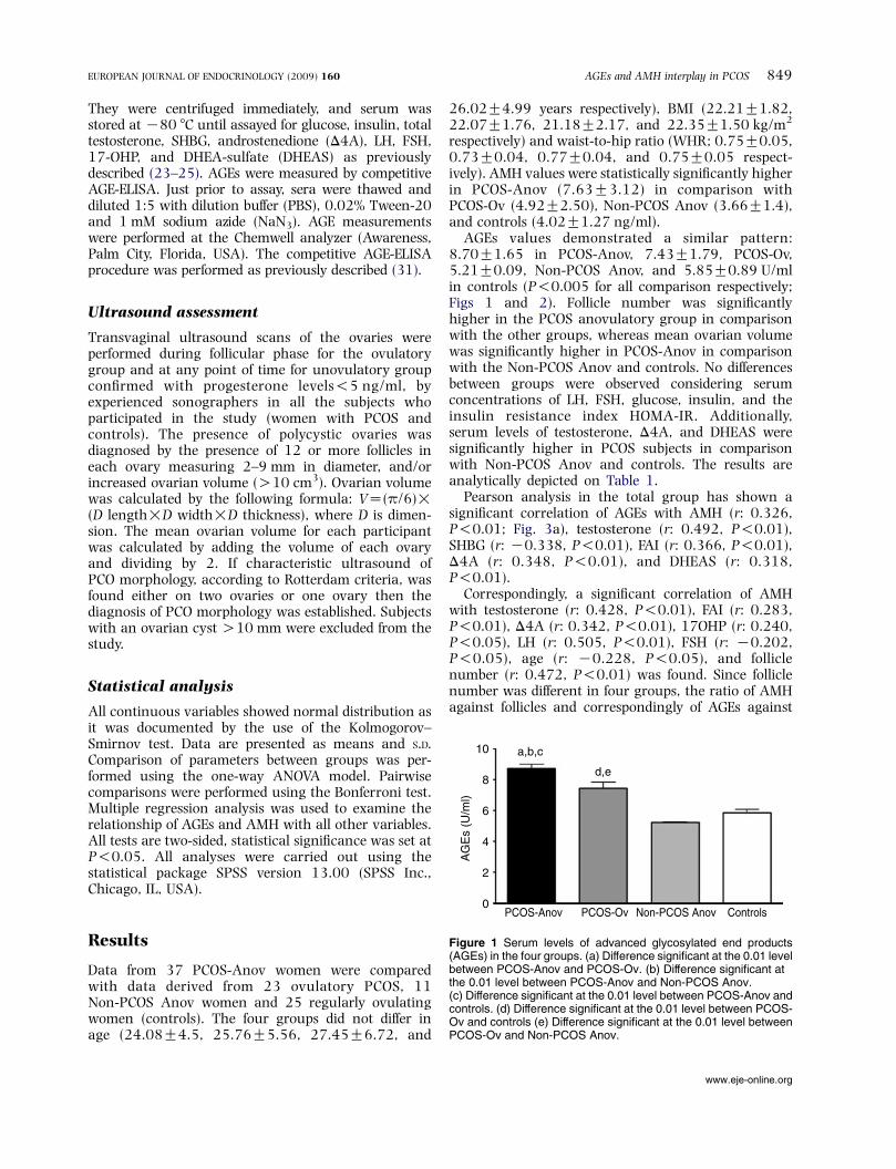
AGEs values demonstrated a similar pattern:8.70G1.65 in PCOS-Anov, 7.43G1.79, PCOS-Ov,5.21G0.09, Non-PCOS Anov, and 5.85G0.89 U/mlin controls (P!0.005 for all comparison respectively;Figs 1 and 2). Follicle number was significantlyhigher in the PCOS anovulatory group in comparisonwith the other groups, whereas mean ovarian volumewas significantly higher in PCOS-Anov in comparisonwith the Non-PCOS Anov and controls. No differencesbetween groups were observed considering serumconcentrations of LH, FSH, glucose, insulin, and theinsulin resistance index HOMA-IR. Additionally,serum levels of testosterone, D4A, and DHEAS weresignificantly higher in PCOS subjects in comparisonwith Non-PCOS Anov and controls. The results areanalytically depicted on Table 1.
Pearson analysis in the total group has shown asignificant correlation of AGEs with AMH (r: 0.326,P!0.01; Fig. 3a), testosterone (r: 0.492, P!0.01),SHBG (r: K0.338, P!0.01), FAI (r: 0.366, P!0.01),D4A (r: 0.348, P!0.01), and DHEAS (r: 0.318,P!0.01).
Correspondingly, a significant correlation of AMHwith testosterone (r: 0.428, P!0.01), FAI (r: 0.283,P!0.01), D4A (r: 0.342, P!0.01), 17OHP (r: 0.240,P!0.05), LH (r: 0.505, P!0.01), FSH (r: K0.202,P!0.05), age (r: K0.228, P!0.05), and folliclenumber (r: 0.472, P!0.01) was found. Since folliclenumber was different in four groups, the ratio of AMHagainst follicles and correspondingly of AGEs against
Figure 1 Serum levels of advanced glycosylated end products(AGEs) in the four groups. (a) Difference significant at the 0.01 levelbetween PCOS-Anov and PCOS-Ov. (b) Difference significant atthe 0.01 level between PCOS-Anov and Non-PCOS Anov.(c) Difference significant at the 0.01 level between PCOS-Anov andcontrols. (d) Difference significant at the 0.01 level between PCOS-Ov and controls (e) Difference significant at the 0.01 level betweenPCOS-Ov and Non-PCOS Anov.
AGEs and AMH interplay in PCOS 849EUROPEAN JOURNAL OF ENDOCRINOLOGY (2009) 160
www.eje-online.org

follicles was estimated and a significant correlationbetween them was observed (r: 0.42, P: 0.0001,Fig. 3b). When multiple regression analysis was carriedout in the total group using AMH as the dependentvariable a significant correlation with gonadotropins(P!0.0005), the existence of PCOS (P: 0.002) andanovulation (P: 0.028) was found. Correspondingly,when AGEs was used as the dependent variable, asignificant correlation with BMI (P: 0.029), theexistence of PCOS (P!0.0005) and anovulation(P: 0.028) was found. Anovulation was highlycorrelated with either AMH (P: 0.032) and AGEs(P: 0.028).
Discussion
In the present study, it is demonstrated that anovulatorywomen with PCOS have higher serum levels of AMH,AGEs, androgens, and follicles number compared withNon-PCOS Anov women. These data support thepresence of different pathophysiological mechanisms,between the two anovulatory groups. However, the factthat anovulatory women with PCOS and PCOS-Ov donot differ in androgens levels but still differ, at astatistically significant level, in AMH and AGEs levelsas well as in follicle level number, suggests thatadditional factors interfere with maturation process inoocytes of PCOS women. The fact that these women donot differ in BMI, WHR, age, and insulin sensitivity,implies that these factors do not account for theobserved differences in the studied groups.
Elevated serum AMH is considered to be linked withanovulation (12, 13, 32, 33). Furthermore, elegantstudies have found that AMH production per granulosacell was raised in PCOS in comparison with controls(11), and also increased expression of AMH and itsreceptor by granulosa cell from PCOS women (34). Inaddition, several research groups have shown thatserum levels of AMH, in women with PCOS are two tothreefold higher than in ovulatory women with normalovaries (10, 35), an observation consistent with thefindings of the present study.
Eldar-Geva et al. reported that AMH values weresignificantly increased in hyperandrogenic PCOS incomparison with normoandrogenic PCOS and controls(36). In the present study, the two anovulatory groups(PCOS & Non-PCOS) that differ in androgen levels differalso in AMH concentrations, a finding in accordancewith these data. Additionally, it has been shown that
Figure 2 Serum levels of AMH in the four groups. (a) Differencesignificant at the 0.01 level between PCOS-Anov and PCOS-Ov.(b) Difference significant at the 0.01 level between PCOS-Anov andNon-PCOS Anov. (c) Difference significant at the 0.01 levelbetween PCOS-Anov and controls. (d) Difference significant at the0.01 level between PCOS-Ov and controls. (e) Differencesignificant at the 0.01 level between PCOS-Ov and Non-PCOSAnov.
Table 1 Pertinent data of the studied subjects.
PCOS-Anov (nZ37) PCOS-Ov (nZ23) Non-PCOS Anov (nZ11) Controls (nZ25)
Age (years) 24.08G4.5 25.76G5.56 27.45G6.72 26.02G4.99BMI (kg/m2) 22.21G1.82 22.07G1.76 21.18G2.17 22.35G1.50WHR 0.75G0.05 0.73G0.04 0.77G0.04 0.75G0.05Testosterone (ng/dl) 84.6G22.13a,b 83.34G22.82c,d 39.20G9.77 40.14G11.51SHBG (nmol/l) 44.69G17.42b 46.6G22.3c,d 57.19G22.23 69.84G21.93D4A (ng/ml) 2.98G1.07a,b 2.99G1.08c,d 1.83G0.43 1.61G0.39FAI 7.82G4.21a,b 7.91G4.62c,d 2.71G1.22 2.23G1.04DHEAS (ng/ml) 289.3G115.2a,b 315.9G147.4c,d 167.7G73.3 190.7G78.517OHP (ng/ml) 1.05G0.5b 1.25G0.61c 1.18G0.64 0.69G0.39LH (IU/l) 8.65G6.02b 6.64G3.24 6.08G3.58 5.94G2.47FSH (IU/l) 6.14G1.57 6.32G2.01 6.61G1.64 6.53G1.77Glucose (mg/dl) 92.91G12 97.56G14 97.09G16.42 93.04G13.13Insulin (mU/ml) 8.5G5.35 8.13G4.93 6.74G2.28 6.90G3.46HOMA-IR 1.98G1.47 1.91G1.09 1.63G0.74 1.59G0.91Mean volume (cm3) 6.73G2.64a,b 6.81G2.23c,d 4.52G1.17 5.02G2.14Follicle number 19.43G7.71a,b,e 12.79G3.95c 12.05G2.98 10.60G5.19
aDifference significant at the 0.01 level between PCOS-Anov and Non-PCOS Anov.bDifference significant at the 0.01 level between PCOS-Anov and controls.cDifference significant at the 0.01 level between PCOS-Ov and controls.dDifference significant at the 0.01 level between PCOS-Ov and Non-PCOS Anov.eDifference significant at the 0.01 level between PCOS-Anov and PCOS-Ov.
850 E Diamanti-Kandarakis and others EUROPEAN JOURNAL OF ENDOCRINOLOGY (2009) 160
www.eje-online.org

AMH by inhibiting aromatase activity leads in turn toincreased local androgen concentrations (37–39).Experiments in rhesus monkeys have shown thatandrogens stimulate the initiation of primordial folliclegrowth and the proliferation of granulosa and thecacells of growing follicles reflected to AMH production(40). This observation is in support of our findingsregarding the statistically significant increased numberof follicles, AMH, and androgen levels in the PCOS-Anovcompared with the Non-PCOS Anov. However, the factthat the two PCOS groups, anovulatory and ovulatorydid differ in AMH levels, but not in the degree ofhyperandrogenemia, implies that other factors in thiscase could also be involved.
It is of interest that the PCOS-Anov group hasstatistically significantly higher serum levels of AGEscompared with PCOS-Ov. This observation, in conjunc-tion with the positive statistically significant correlationbetween AMH and AGEs, suggests an interaction of
these molecules, highlighted after correction of bothparameters for follicle number, which is different in thefour groups and consequently could affect this relation-ship by mass effect of granulosa cells (Fig. 3). Furtherstrengthening of a possible link of these molecules inthe anovulatory process of PCOS, comes from theobservation of higher expression and production ofAMH and its receptor per granulosa cell in PCOS(10, 34), with higher immunohistochemical local-ization of AGEs and RAGE, as well as the nuclearlocalization of NFkB on the granulosa cells from humanpolycystic ovaries (27).
The mechanisms of interaction of AMH and AGEs inthe oocyte maturation process in PCOS can not beelucidated in this study. However, the presented data arein support of the role of oxidative stress in ovulatorydysfunction (41, 42), since AGEs are well recognizedmediators of increased oxidative stress (43, 44). It hasbeen found that apoptosis in follicles is initiated byoxidative stress (45) and also that AGEs have beeninvolved in the impaired folliculogenesis, (46) thereforelinking these parameters with impaired follicle matu-ration. Antioxidant mechanisms have been localized inthe granulosa and thecal cells of the growing follicle andconsequently inadequate protection from oxidativestress could be a potential trigger for follicular atresia(47, 48). It is likely that increased oxidative stress,reflected by elevated serum AGEs in this study, mayinterfere in the ovulatory process, manifested byelevated AMH. From the above, the role of oxidativestress in anovulation appears to be supported, althoughthe mechanisms by which oxidative stress interfereswith oocyte maturation have not been clarified yet.Furthermore, well-designed studies have shown thatoxidative stress modulates androgen production fromtheca cells of women with PCOS, both in vitro and in vivo(18, 42, 49).
On the other hand, it is possible that AGEs, viaincreasing oxidative stress, could be linked withhyperandrogenemia, since oxidative stress has beenshown to induce proliferation and growth of theca-interstitial cells on a dose-dependent manner (50). Thepositive correlation of AMH and AGEs levels betweenthem as well as with androgens, shown in this study,suggests a role of hyperandrogenemia in AMH and/orAGEs levels. This possibility is further supported fromthe notion that the group of PCOS-Anov had signi-ficantly higher concentrations of AMH, AGEs, andandrogens in comparison with Non-PCOS Anov group.Therefore, the interplay between AMH and AGEs couldcontribute to the anovulatory mechanisms via poten-tiation of hyperandrogenemia. Moreover, as it has beenshown in regression analysis, either AMH or AGEsexhibit a strong relationship with anovulation, implyingan interaction between them.
In conclusion, the data of this study demonstrate forthe first time a link between AGEs, an oxidative stressmarker distinctly elevated in women with PCOS, with
Figure 3 (a) Correlation of advanced glycosylated end products(AGEs) with AMH according to ovulation status (r: 0.326, P!0.01).(b) Correlation of advanced glycosylated end products (AGEs) ratiowith follicles with AMH/follicles ratio according to ovulation status(r: 0.42,P: 0.0001). Circles denote PCOS-Anov, Triangles PCOS-Ov,Stars Non-PCOS Anov and Squares control respectively.
AGEs and AMH interplay in PCOS 851EUROPEAN JOURNAL OF ENDOCRINOLOGY (2009) 160
www.eje-online.org

AMH. Although causation cannot be deduced from thepresent study, this association between raised AMH andAGE levels implies an interrelated role to compromisedfollicle maturation process in PCOS. Clearly, furtherstudies are required to explore the mechanisms of thispotentially interactive relationship between oxidativemediators, anovulatory markers, and hyperandro-genemia in the dysfunctional maturation process ofoocytes in patients with PCOS.
Declaration of interest
There is no conflict of interest that could be perceived as prejudicingthe impartiality of the research reported.
Funding
This research did not receive any specific grant from any fundingagency in the public, commercial or not-for-profit sector.
References
1 Dunaif A. Insulin resistance and the polycystic ovary syndrome:mechanism and implications for pathogenesis. Endocrine Reviews1997 18 774–800.
2 Diamanti-Kandarakis E. Polycystic ovarian syndrome: pathophy-siology, molecular aspects and clinical implications. Expert Reviewsin Molecular Medicine 2008 10 e3.
3 Chang WY, Knochenhauer ES, Bartolucci AA & Azziz R.Phenotypic spectrum of polycystic ovary syndrome: clinical andbiochemical characterization of the three major clinical sub-groups. Fertility and Sterility 2005 83 1717–1723.
4 Panidis DK, Rousso DH, Kourtis AI & Papathanasiou KV. Could thetheory of chaos contribute to the interpretation of pathogenesis ofpolycystic ovary syndrome? Clinical and Experimental Obstetrics andGynecology 2003 30 187–189.
5 Franks S, Stark JE & Hardy K. Follicle dynamics and anovulation inpolycystic ovary syndrome. Human Reproduction Update 2008 14367–378.
6 La Marca A, Orvieto R, Giulini S, Jasonni VM, Volpe A & De Leo V.Mullerian inhibiting substance in women with polycystic ovarysyndrome: relationship with hormonal and metabolic charac-teristics. Fertility and Sterility 2004 82 970–972.
7 Weenen C, Laven JS, Von Bergh AR, Cranfield M, Groome NP,Visser JA, Kramer P, Fauser BC & Themmen AP. Anti-Mullerianhormone expression pattern in the human ovary: potentialimplications for initial and cyclic follicle recruitment. MolecularHuman Reproduction 2004 10 77–83.
8 Pigny P, Jonard S, Robert Y & Dewailly D. Serum anti-Mullerianhormone as a surrogate for antral follicle count for definition of thepolycystic ovary syndrome. Journal of Clinical Endocrinology andMetabolism 2006 91 941–945.
9 Fallat ME, Siow Y, Marra M, Cook C & Carrillo A. Mullerianinhibiting substance in follicular fluid and serum: a comparison ofpatients with tubal factor infertility, polycystic ovary syndrome,and endometriosis. Fertility and Sterility 1997 67 962–965.
10 Pigny P, Merlen E, Robert Y, Cortet-Rudelli C, Decanter C,Jonard S & Dewailly D. Elevated serum level of anti-mullerianhormone in patients with polycystic ovary syndrome:relationship to the ovarian follicle excess and to the folliculararrest. Journal of Clinical Endocrinology and Metabolism 2003 885957–5962.
11 Pellatt L, Hanna L, Brincat M, Galea R, Brain H, Whitehead S &Mason H. Granulosa cell production of anti-Mullerian hormone isincreased in polycystic ovaries. Journal of Clinical Endocrinology andMetabolism 2007 92 240–245.
12 Dewailly D, Catteau-Jonard S, Reyss AC, Maunoury-Lefebvre C,Poncelet E & Pigny P. The excess in 2–5 mm follicles seen atovarian ultrasonography is tightly associated to the folliculararrest of the polycystic ovary syndrome.Human Reproduction 200722 1562–1566.
13 Chen MJ, Yang WS, Chen CL, Wu MY, Yang YS & Ho HN. Therelationship between anti-Mullerian hormone, androgen andinsulin resistance on the number of antral follicles in womenwith polycystic ovary syndrome. Human Reproduction 2008 23952–957.
14 Durlinger AL, Gruijters MJ, Kramer P, Karels B, Kumar RT,Matzuk MM, Rose UM, de Jong FH, Uilenbroek JT, Grootegoed AJ &Themmen AP. Anti-Mullerian hormone attenuates the effects ofFSH on follicle development in the mouse ovary. Endocrinology2001 142 4891–4899.
15 Agarwal A, Gupta S & Sharma RK. Role of oxidative stress infemale reproduction. Reproductive Biology and Endocrinology 20053 28.
16 Tatone C, CarboneMC, Falone S, Aimola P, Giardinelli A, Caserta D,Marci R, Pandolfi A, Ragnelli AM & Amicarelli F. Age-dependentchanges in the expressionof superoxide dismutases andcatalase areassociated with ultrastructural modifications in human granulosacells. Molecular Human Reproduction 2006 12 655–660.
17 Fenkci V, Fenkci S, Yilmazer M & Serteser M. Decreasedtotal antioxidant status and increased oxidative stress inwomen with polycystic ovary syndrome may contribute tothe risk of cardiovascular disease. Fertility and Sterility 2003 80123–127.
18 Kwintkiewicz J, Spaczynski RZ, Foyouzi N, Pehlivan T & Duleba AJ.Insulin and oxidative stress modulate proliferation of rat ovariantheca-interstitial cells through diverse signal transductionpathways. Biology of Reproduction 2006 74 1034–1040.
19 Charitidou C, Farmakiotis D, Zournatzi V, Pidonia I, Pegiou T,Karamanis N, Hatzistilianou M, Katsikis I & Panidis D. Theadministration of estrogens, combined with anti-androgens, hasbeneficial effects on the hormonal features and asymmetricdimethylarginine levels, in women with the polycystic ovarysyndrome. Atherosclerosis 2008 196 958–965.
20 Baynes JW. Role of oxidative stress in development of compli-cations in diabetes. Diabetes 1991 40 405–412.
21 McCance DR, Dyer DG, Dunn JA, Bailie KE, Thorpe SR, Baynes JW& Lyons TJ. Maillard reaction products and their relation tocomplications in insulin-dependent diabetes mellitus. Journal ofClinical Investigation 1993 91 2470–2478.
22 Singh R, Barden A, Mori T & Beilin L. Advanced glycation endproducts: a review. Diabetologia 2001 44 129–146.
23 Diamanti-Kandarakis E, Piperi C, Kalofoutis A & Creatsas G.Increased levels of serum advanced glycation end-products inwomen with polycystic ovary syndrome. Clinical Endocrinology2005 62 37–43.
24 Diamanti-Kandarakis E, Katsikis I, Piperi C, Alexandraki K &Panidis D. Effect of long-term orlistat treatment on serum levels ofadvanced glycation end-products in women with polycystic ovarysyndrome. Clinical Endocrinology 2007 66 103–109.
25 Diamanti-Kandarakis E, Katsikis I, Piperi C, Kandaraki E,Piouka A, Papavassiliou AG & Panidis D. Increased serumadvanced glycation end products is a distinct finding in leanwomen with PCOS. Clinical Endocrinology 2008 69 634–641.
26 Peppa M, Uribarri J & Vlassara H. Aging and glycoxidant stress.Hormones 2008 7 123–132.
27 Diamanti-Kandarakis E, Piperi C, Patsouris E, Korkolopoulou P,Panidis D, Pawelczyk L, Papavassiliou AG & Duleba AJ. Immuno-histochemical localization of advanced glycation end-products(AGEs) and their receptor (RAGE) in polycystic and normalovaries. Histochemistry and Cell Biology 2007 127 581–589.
852 E Diamanti-Kandarakis and others EUROPEAN JOURNAL OF ENDOCRINOLOGY (2009) 160
www.eje-online.org

28 Diamanti-Kandarakis E, Piperi C, Korkolopoulou P, Kandaraki E,Levidou G, Papalois A, Patsouris E & Papavassiliou AG. Accumu-lation of dietary glycotoxins in the reproductive system of normalfemale rats. Journal of Molecular Medicine 2007 85 1413–1420.
29 Rotterdam Consensus. Revised 2003 consensus on diagnosticcriteria and long-term health risks related to polycystic ovarysyndrome (PCOS). Human Reproduction 2004 19 41–47.
30 Hamilton-Fairley D & Taylor A. Anovulation. BMJ 2003 327546–549.
31 Diamanti-Kandarakis E, Piperi C, Alexandraki K, Katsilambros N,Kouroupi E, Papailiou J, Lazaridis S, Koulouri E, Kandarakis HA,Douzinas EE, Creatsas G & Kalofoutis A. Short-term effect oforlistat on dietary glycotoxins in healthy women and women withpolycystic ovary syndrome. Metabolism 2006 55 494–500.
32 Visser JA, de Jong FH, Laven JS & Themmen APN. Anti-Mullerianhormone: a new marker for ovarian function. Reproduction 2006131 1–9.
33 Chu MC, Carmina E, Wang J & Lobo RA. Mullerian-inhibitingsubstance reflects ovarian findings in women with polycysticovary syndrome better than does inhibin B. Fertility and Sterility2005 84 1685–1688.
34 Catteau-Jonard S, Soazik PJ, Leclerc A, Gonzales J, Dewailly D & diClemente N. Anti-Mullerian hormone, its receptor, FSH receptorand androgen receptor genes are overexpressed by granulosa cellsfrom stimulated follicles in women with polycystic ovarysyndrome. Journal of Clinical Endocrinology and Metabolism 200893 4456–4461.
35 Laven JS, Mulders AG, Visser JA, Themmen APN, De Jong FH &Fauser BC. Anti-Mullerian hormone serum concentrations innormoovulatory and anovulatory women of reproductiveage. Journal of Clinical Endocrinology and Metabolism 2004 89318–323.
36 Eldar-Geva T, Margalioth EJ, Gal M, Ben-Chetrit A, Algur N,Zylber-Haran E, Brooks B, Huerta M & Spitz IM. Serum anti-Mullerian hormone levels during controlled ovarian hyperstimu-lation in women with polycystic ovaries with and withouthyperandrogenism. Human Reproduction 2005 20 1814–1819.
37 di Clemente N, Goxe B, Remy JJ, Cate RL, Josso N, Vigier B &Salesse R. Inhibitory effect of AMH upon aromatase activity andLH receptors of granulosa cells of rat and porcine immatureovaries. Endocrine 1994 2 553–558.
38 Grossman MP, Nakajima ST, Fallat ME & Siow Y. Mullerian-inhibiting substance inhibits cytochrome P450 aromatase activityin human granulosa lutein cell culture. Fertility and Sterility 200889 1364–1370.
39 Josso N, Racine C, di Clemente N, Rey R & Xavier F. The role of anti-Mullerian hormone in gonadal development.Molecular and CellularEndocrinology 1998 145 3–7.
40 Vendola K, Zhou J, Wang J, Famuyiwa OA, Bievre M & Bondy CA.Androgens promote oocyte insulin-like growth factor I expressionand initiation of follicle development in the primate ovary. Biologyof Reproduction 1999 61 353–357.
41 Agarwal A & Allamaneni SS. Role of free radicals in femalereproductive diseases and assisted reproduction. ReproductiveBiomedicine Online 2004 9 338–347.
42 Gonzalez F, Rote NS, Minium J & Kirwan JP. Reactive oxygenspecies-induced oxidative stress in the development of insulinresistance and hyperandrogenism in polycystic ovarysyndrome. Journal of Clinical Endocrinology and Metabolism2006 91 336–340.
43 Basta G, Lazzerini G, Massaro M, Simoncini T, Tanganelli P, Fu C,Kislinger T, Stern DM, Schmidt AM & De Caterina R. Advancedglycation end products activate endothelium through signal-transduction receptor RAGE: a mechanism for amplification ofinflammatory responses. Circulation 2002 105 816–822.
44 Devangelio E, Santilli F, Formoso G, Ferroni P, Bucciarelli L,Michetti N, Clissa C, Ciabattoni G, Consoli A & Davı G. SolubleRAGE in type 2 diabetes: association with oxidative stress. FreeRadical Biology and Medicine 2007 43 511–518.
45 Tsai-Turton M & Luderer U. Opposing effects of glutathionedepletion and follicle stimulating hormone on reactive oxygenspecies and apoptosis in cultured preovulatory rat follicles.Endocrinology 2006 147 1224–1236.
46 Tatone C, Amicarelli F, Carbone MC, Monteleone P, Caserta D,Marci R, Artini PG, Piomboni P & Focarelli R. Cellular andmolecular aspects of ovarian follicle ageing. Human ReproductionUpdate 2008 14 131–142.
47 Lund SA, Murdoch J, Van Kirk EA & Murdoch WJ. Mitogenic andantioxidant mechanisms of estradiol action in preovulatory ovinefollicles: relevance to luteal function. Biology of Reproduction 199961 388–392.
48 Valdez KE, Cuneo SP & Turzillo AM. Regulation of apoptosis in theatresia of dominant bovine follicles of the first follicular wavefollowing ovulation. Reproduction 2005 130 71–81.
49 Sabuncu T, Vural H, Harma M & Harma M. Oxidative stress inpolycystic ovary syndrome and its contribution to the risk ofcardiovascular disease. Clinical Biochemistry 2001 34 407–413.
50 Duleba AJ, Foyouzi N, Karaca M, Pehlivan T, Kwintkiewicz J &Behrman HR. Proliferation of ovarian theca-interstitial cellsis modulated by antioxidants and oxidative stress. HumanReproduction 2004 19 1519–1524.
Received 4 February 2009Accepted 7 February 2009
AGEs and AMH interplay in PCOS 853EUROPEAN JOURNAL OF ENDOCRINOLOGY (2009) 160
www.eje-online.org


