Embed Size (px)
Citation preview

ORIGINAL ARTICLE
Antidepressants recruit new neurons to improve stressresponse regulationA Surget1,2,3, A Tanti1,2, ED Leonardo4, A Laugeray1,2, Q Rainer1,2, C Touma5, R Palme6, G Griebel7,
Y Ibarguen-Vargas1,2,3, R Hen4 and C Belzung1,2
1U930 Imaging and Brain, Inserm, Tours, France; 2Universite Francois Rabelais, Tours, France; 3Kavli Institute for SystemsNeuroscience & Centre for the Biology of Memory, Norwegian University of Science and Technology (NTNU), Trondheim,Norway; 4Departments of Psychiatry and Neuroscience, Columbia University, New York, NY, USA; 5Research Group ofPsychoneuroendocrinology, Max Planck Institute of Psychiatry, Munich, Germany; 6Department of Biomedical Sciences/Biochemistry, University of Veterinary Medicine, Vienna, Austria and 7Exploratory Unit, Sanofi-Aventis, Chilly-Mazarin, France
Recent research suggests an involvement of hippocampal neurogenesis in behavioral effectsof antidepressants. However, the precise mechanisms through which newborn granuleneurons might influence the antidepressant response remain elusive. Here, we demonstratethat unpredictable chronic mild stress in mice not only reduces hippocampal neurogenesis,but also dampens the relationship between hippocampus and the main stress hormonesystem, the hypothalamo-pituitary-adrenal (HPA) axis. Moreover, this relationship is restoredby treatment with the antidepressant fluoxetine, in a neurogenesis-dependent manner.Specifically, chronic stress severely impairs HPA axis activity, the ability of hippocampus tomodulate downstream brain areas involved in the stress response, the sensitivity of thehippocampal granule cell network to novelty/glucocorticoid effects and the hippocampus-dependent negative feedback of the HPA axis. Remarkably, we revealed that, although ablationof hippocampal neurogenesis alone does not impair HPA axis activity, the ability of fluoxetineto restore hippocampal regulation of the HPA axis under chronic stress conditions, occursonly in the presence of an intact neurogenic niche. These findings provide a mechanisticframework for understanding how adult-generated new neurons influence the response toantidepressants. We suggest that newly generated neurons may facilitate stress integrationand that, during chronic stress or depression, enhancing neurogenesis enables adysfunctional hippocampus to restore the central control on stress response systems, thenallowing recovery.Molecular Psychiatry advance online publication, 3 May 2011; doi:10.1038/mp.2011.48
Keywords: antidepressant; hippocampal neurogenesis; stress; depression; hypothalamo-pituitary-adrenal axis; immediate early gene
Introduction
In mammals, the hippocampus continues to generatenew neurons in the dentate gyrus (DG) throughoutadult life.1,2 Efforts directed at understanding thefunction of adult-generated neurons in the hippocam-pus have focused primarily on their role in cognitiveprocesses such as contextual and spatial memory.There is also evidence linking adult neurogenesis tochronic stress, affective disorders and the responseto antidepressant treatment.3–8 Indeed, stress, a majoretiological factor for anxiety/depressive disorders,decreases the production and the survival of the
new hippocampal neurons,9,10 whereas treatmentswith antidepressants increase them4 and blockinghippocampal neurogenesis prevents the behavioraleffects of antidepressants in several paradigms inrodents.3,11–15 However, although there has been pro-gress in elucidating the mechanisms through whichstress reduces neurogenesis, little is known abouteither the function of these new neurons in triggeringstress-related disorders or in the mechanisms throughwhich they could exert some antidepressant effects.
The hippocampal formation is able to influence theactivity of several cortical and subcortical structuresinvolved in endocrine, motor, affective and cognitivefunctions.16–18 In addition to its role in strictlymnemonic processes, the hippocampus is thereforewell positioned to modulate motivational behaviors,emotional states and stress responses. Particularly,the hippocampus is a regulator of the main neuroen-docrine stress system, the hypothalamo-pituitary-adrenal (HPA) axis.19,20 It can modulate the activity
Received 5 July 2010; revised 28 February 2011; accepted 22March 2011
Correspondence: Dr A Surget, Kavli Institute for SystemsNeuroscience & Centre for the Biology of Memory, NorwegianUniversity of Science and Technology (NTNU), MTFS (3rd Floor,Room 1314), Olav Kyrres Gate 9, Trondheim 7489, Norway.E-mail: [email protected]
Molecular Psychiatry (2011), 1–12& 2011 Macmillan Publishers Limited All rights reserved 1359-4184/11
www.nature.com/mp

of the paraventricular nucleus (PVN) of the hypothala-mus (starter of HPA axis) through two- or three-neuroncircuits. Indeed, hippocampal glutamatergic outputsproject toward PVN-connected GABAergic neuronpopulations of stress-integrative subcortical regionssuch as lateral septum, anteromedial and posteromedialbed nucleus of stria terminalis (amBST and pmBST),medial and ventrolateral preoptic areas, dorsomedialand lateral hypothalamic nuclei (DMH and LH).21,22
Affective disorders have been frequently associatedwith abnormalities in HPA axis activity,23–25 includingglucocorticoid hypersecretion and alterations of itsnegative feedback.26,27 These changes are probablyimportant as it has been shown that the normalizationof HPA axis activity parallels remission and reducesthe risk of relapse in depressed patients with HPAaxis abnormalities.28–31
The DG is exquisitely sensitive to glucocorticoidlevels and may be damaged under conditions of highglucocorticoid levels and chronic stress.15,32,33 Withinthis framework, we hypothesized that the pro-neuro-genic effect of the antidepressant fluoxetine (aselective serotonin reuptake inhibitor—SSRI) pro-vides a fresh source of DG granule neurons highlyresponsive to environmental cues. As such, they arewell positioned to initiate the improvement ofhippocampal function in stress integration andperhaps, as a result, contribute to the antidepressantresponse when stress systems have been disturbed.For this purpose, we exposed mice with inhibitedhippocampal neurogenesis to the unpredictablechronic mild stress (UCMS), an informative modelto study stress-related disorders in animals,34,35 as itmimics the role of socio-environmental stressors inprecipitating anxiety/depressive disorders, induces asyndrome reminiscent of the human neuropathologyand reproduces the timeframe of the therapeuticresponse to antidepressants.33,34,36 Here, we foundthat chronic stress exposure induces severe abnorm-alities in the hippocampus-dependent regulation ofstress systems and that antidepressant treatment withfluoxetine reverses such effects through a neurogen-esis-dependent mechanism.
Materials and methods
A brief description of the materials and methods ispresented in this section. For a full and detailed descri-ption, please refer to the Supplementary Information.
AnimalsMale BALB/c mice were purchased from Taconic(Germantown, NY, USA) or from Centre d’ElevageJanvier (Le Genest Saint Isle, France) and were agedbetween 3 and 4 months at the time of the beginningof the UCMS exposure.
UCMSThe stress regimen used was previously described33
and is a variant of the chronic mild stress proceduresdescribed by Willner in rats.34 UCMS mice were
repeatedly subjected to various socio-environmentalstressors according to an unpredictable schedule fora total period of 6–8 weeks. From the third week ofUCMS onward, mice were administered intraperito-neal (ip), once a day, with either vehicle, fluoxetine(20 mg kg�1 per day) or SSR125543 (20 mg kg�1 perday). Treatment was always maintained until the endof the experiments. Body weight and coat state wereassessed weekly, as markers of the progression of theUCMS-evoked syndrome.33,37
Cookie testThis test required a device containing threealigned compartments with the same dimension(20� 20� 20 cm). Only the colors of the walls and thefloor were different between the compartments (Figure2a). Mice were first familiarized with a chocolatecookie (Pepito, Lu, France) 4.5 weeks before the firsttesting. One hour before testing, the regular food wasremoved from the cage lid. At the time of testing, asmall amount (2±1 g) of chocolate cookie (or regularfood in a control experiment) was placed at the centerof the black compartment. The mouse was initiallyplaced in the white compartment of the apparatus.Each session of the test lasted five minutes. The cookieconsumption (# bites) was recorded within the 5 mintest period. Four sessions of testing were performedwithin 10 days (inter-test interval: 2 days).
X-ray irradiation procedureFive weeks before UCMS, subsets of experimentalanimals were exposed to three targeted X-irradiationsspecifically above the hippocampus sparing the majorpart of the brain according to the procedure pre-viously described.3,11
ImmunohistochemistryImmunohistochemistry for subgranular zone (SGZ)proliferation was performed as previously described.11
Rat anti-BrdU antibody (1:500, Oxford Biotechnology,Oxfordshire, UK) was used as primary antibodyfollowed by rabbit anti-rat biotinylated antibody(1:200, Vector Laboratories, Burlingame, CA, USA).To label Fos, immunohistochemistry was performedwith a rabbit anti-Fos antibody (1:5000, PC38, Calbio-chem, San Diego, CA, USA) followed by a donkey anti-rabbit biotinylated antibody (1:500, Jackson Immuno-research Laboratories, West Grove, PA, USA). Thestaining was amplified with an avidin-biotin complex(Elite ABC kit, Vector Laboratories) and visualizedwith DAB (Sigma-Aldrich, St Louis, MO, USA).
For immunohistochemistry based on fluorescencelabeling, the different primary antibodies used were asfollows: mouse anti-NeuN monoclonal antibody (1:1000,Chemicon International, Billerica, MA, USA), rat anti-BrdU monoclonal antibody (1:500 Oxford Biotechnol-ogy) and a rabbit anti-Fos monoclonal antibody (1:1000Oxford Biotechnology); the secondary antibodies usedwere as follows: Alexa-488 nm anti-mouse, Alexa-546nm anti-rabbit and Alexa-633 nm anti-rat antibodies(1:200, Molecular Probe, Eugene, OR, USA).
New neurons contribute to stress response regulationA Surget et al
2
Molecular Psychiatry

Image analysis and cell quantificationWhen staining was visualized with DAB, the numberof positive cells was counted using � 20 and � 40objective lens with a conventional light microscope.For Fos-labeled cell counting of subcortical regions,the nomenclature and nuclei boundaries used werethose defined by Paxinos and Franklin’s mouse brainatlas.38 Fosþ cells within each region were countedbilaterally in consecutive sections starting from bregma0.62 to bregma �1.58. For each animal and region, thesame number of sections was used and the volume ofeach area was controlled. Regions included andcorresponding coordinates were as follows: lateralseptum (bregma (0.62–0.14)), amBST (0.62–0.02),pmBST (�0.10 to –0.34), anteroventral BST (0.62–0.02), ventrolateral preoptic area (0.26 to �0.10),medial preoptic area (0.14 to �0.46), DMH (�1.34 to�1.58), LH (�0.34 to�0.70) and PVN (�0.58 to �1.22).
When staining was visualized with fluorochrome-labeled secondary antibodies, the number of positivecells was counted using a � 40 objective lens witha confocal or an epifluorescence microscope. Eightsections (16 hippocampus) along the rostro-caudalextent of the hippocampus were examined; Bbregma:�1.46, �1.82, �2.18, �2.54, �2.80, �3.08, �3.28,�3.52.38 We also counted the number of NeuNþ /Fosþ cells in the inner part of the GCL containingthe SGZ (the two deepest rows of the GCL plus cellstouching the last row at the interface with thehilus).39,40 Indeed, granule cells are producedthroughout an outside-in gradient in the GCL, whichresults in a high proportion of young newbornneurons in the deepest rows bordering the hilus.41–44
Quantification of corticosterone levelsFecal samples were analyzed for immunoreactivecorticosterone metabolites using a 5a-pregnane-3b,11b,21-triol-20-one enzyme-immunoassay as pre-viously described.45 Details regarding development,biochemical characteristics and physiological valida-tion of this assay have previously been described.46,47
Plasma corticosterone levels was analyzed using a125I-labeled corticosterone double-antibody radioim-munoassay kit (MP Biomedicals, Solon, OH, USA)according to the manufacturer’s protocol.
Dexamethasone (DEX) administration proceduresDEX suppression test: mice were i.p. injected witheither DEX-P (0.1 mg kg�1, five mice per group) orsaline (0.9% NaCl, five mice per group). All fecaltheboli that were voided between 8 and 10 h after theinjection were collected to measure the level of fecalcorticosterone metabolites.
Sensitivity of granule cells to novelty and gluco-corticoid effects: mice were i.p. injected with eitherDEX-P (0.1 mg kg�1, five mice per group) or saline(0.9% NaCl, five mice per group). Thirty minuteslater, mice were placed in a circular open-field(diameter 35 cm) for 5 min. Then, 90 min later(120 min after injection), mice were anesthetized withketamine/xylazine (120 and 10 mg kg�1, respectively),
transcardially perfused and their brains werecollected for immunohistochemistry.
Intrahippocampal DEX infusion: the infusion pro-cedure was derived from the method previouslydescribed.20 Mice were stereotaxically and bilaterallyimplanted with guide cannulas (6 mm long, 0.6 mmouter diameter, 0.36 mm inner diameter). The follow-ing coordinates were used for the guide cannulaimplantation: bregma =�3.08, lateral = ±2.3,vertical =�1.4.38 After 1-week recovery period fromthe surgery, infusion cannulas (7 mm long, 0.3 mmouter diameter, 0.17 mm inner diameter) were placedand extended 1 mm below the guide cannulas. Theanimals received bilateral infusions during 1.5 minof either vehicle (0.9% NaCl/0.2% ethanol) or DEX(50 ng in 0.6 ml). In one experiment, the brain wascollected 2 h following the infusion to assess DEX-induced Fos changes in downstream brain structuresand the PVN (Figure 3). In another experiment, theanimals were killed by CO2 asphyxiation 4 h follow-ing infusion and trunk blood was collected tomeasure plasma corticosterone levels (Figure 6).
Statistics
Considering that relatively small sample sizes wereused and that assumptions for parametric statisticscould not be ensured (normality and homoscedasti-city), data were analyzed using the non-parametricKruskal–Wallis ‘analysis of variance by ranks’ H-test.Significant effects (that is, P < 0.05) were followed-upwith post-hoc tests (Holm-Bonferroni method) whenappropriate. P-values that are indicated in the‘Results’ section always derived from the between-groups comparisons using the Kruskal–Wallis H-test,whereas P-values resulting from post-hoc compari-sons are indicated in the figures.
Results
Hippocampal neurogenesis is required for chronicstress reversal by fluoxetine
We first assessed the involvement of hippocampalneurogenesis in the emergence and the recoveryof behavioral and physical changes in the UCMS.For this purpose, mice were initially exposed to X-irradiations 5 weeks before UCMS procedure to ablatecell proliferation in the SGZ of the DG withoutaffecting other neurogenic niches (Figure 1a).3,11 Micewere then subjected to a 6- to 8-week UCMSprocedure or maintained under non-stressful condi-tions (control mice). Two weeks after initiatingUCMS, mice were daily treated with either fluoxetine(20 mg kg�1 per day, ip) or a vehicle solution (NaCl0.9%, ip). X-irradiation induced a strong depletion ofSGZ cell proliferation (B92%, Figure 1b) indepen-dently of the environment (control/UCMS) or thetreatment (vehicle/fluoxetine). UCMS exposure re-sulted in significant decreases of SGZ cell prolifera-tion (B27%, Figure 1b) and 4-week-old newbornneurons (B34%, Supplementary Figure S1); these effects
New neurons contribute to stress response regulationA Surget et al
3
Molecular Psychiatry

were counteracted by chronic fluoxetine treatment (SGZproliferation: P<0.0001; neurogenesis, P=0.0082).
Considering that anhedonia is one of the majorsymptoms of depression, we developed a behavioralparadigm based on the motivation for consuming apalatable stimulus (a chocolate cookie): the Cookietest. This test is based on the conflict between thedrive for the stimulus and the neophobic behavior ofthe mouse. A reduction of the cookie consumptionmay therefore be interpreted as anhedonia, a habitua-tion deficit or a combination of both effects. We foundthat session repetition resulted in a progressiveincrease of the cookie consumption in control mice(session 1, P = 0.8149; session 2, P = 0.1387; session 3,P = 0.0013; session 4, P = 0.0001; Figure 2b). UCMSexposure suppressed the consumption increase, aneffect, which was reversed by fluoxetine treatment.Interestingly, although irradiated mice did not displayany behavioral alterations in control or UCMS condi-tions, they were insensitive to the fluoxetine reversal.As a control experiment, the substitution of thecookie by a regular food pellet produced a quasi-nullconsumption during the four sessions for all thegroups (data not shown), underlining the importance
of the ‘hedonic’ feature of the stimulus. Our resultsindicate that the ablation of hippocampal neurogen-esis on its own has no effect in the Cookie test butprevents the fluoxetine reversal in UCMS mice.Strikingly, similar results were also obtained whenexamining body weight change (another main symp-tom in major depression) and the coat state (Supple-mentary Figures S2a,b).
However, we found that ‘antidepressant-like’effects can be obtained even in irradiated micewhen using non-monoaminergic compounds such asthe corticotropin-releasing factor 1 receptor (CRF1)antagonist SSR125543. In a protocol similar to theprevious experiment, SSR125543 (20 mg kg�1 per day,ip) also reversed neurogenesis reduction induced byUCMS (P = 0.018; Supplementary Figure S1c). Thiscompound counteracted UCMS effects in the Cookietest (session 1, P = 0.9998; session 2, P = 0.8644;session 3, P = 0.0714; session 4, P = 0.0108; Figure2c), on body weight and coat state (SupplementaryFigure S2c,d) in non-irradiated mice but also inirradiated mice.
To summarize, hippocampal neurogenesis,although not directly involved in the emergence of a
Figure 1 Focal hippocampal X-irradiation ablates cell proliferation in the subgranular zone (SGZ). (a) Schematicrepresentation of the experimental design. A first cohort of mice was used to assess SGZ cell proliferation, see (b) and (c).Two other cohorts of mice were used for behavioral measures, see Figure 2. (b) Representative images of BrdUþ cells (‘blackcells’) in the SGZ (scale bar, 50mm). (c) The cell proliferation assessed by the number of BrdU-positive cells per mm3 of thegranule cell layer (GCL), n = 5–7 per group.#P < 0.05 UCMS mice vs control-vehicle mice; *P < 0.05 and **P < 0.01 UCMS-fluoxetine mice vs UCMS-vehicle mice, or between line-connected groups. Data represent mean±s.e.m.
Figure 2 Hippocampal neurogenesis is required for the behavioral effects of fluoxetine but not of the corticotropin-releasingfactor 1 antagonist SSR125543. (a) Schematic representation of the apparatus used for the Cookie test (CT). (b, c) Theconsumption of the cookie (number of bites) in the CT, n = 13–14 per group for (b) and n = 10 per group for (c). #P < 0.05 and##P < 0.01 UCMS mice vs control-vehicle mice; *P < 0.05 and **P < 0.01 UCMS-fluoxetine/SSR125543 mice vs UCMS-vehiclemice. Data represent mean±s.e.m.
New neurons contribute to stress response regulationA Surget et al
4
Molecular Psychiatry
depression-like state or the vulnerability to stress,was required for the recovery effects of the mono-aminergic antidepressant fluoxetine. On the otherhand, alternative mechanisms, independent of neu-rogenesis, may be used to induce similar effects by theCRF1 antagonist, which directly target stress systems.
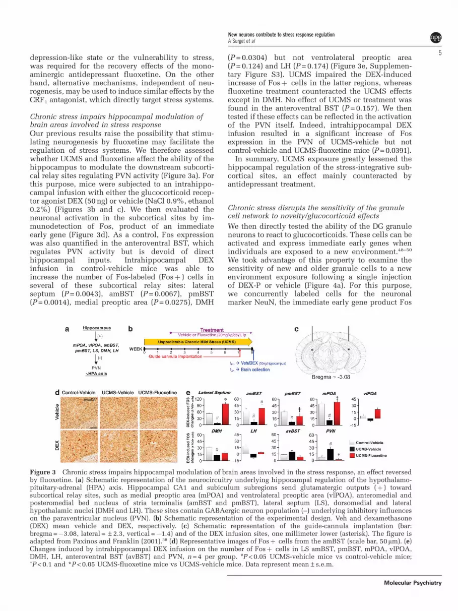
Chronic stress impairs hippocampal modulation ofbrain areas involved in stress responseOur previous results raise the possibility that stimu-lating neurogenesis by fluoxetine may facilitate theregulation of stress systems. We therefore assessedwhether UCMS and fluoxetine affect the ability of thehippocampus to modulate the downstream subcorti-cal relay sites regulating PVN activity (Figure 3a). Forthis purpose, mice were subjected to an intrahippo-campal infusion with either the glucocorticoid recep-tor agonist DEX (50 ng) or vehicle (NaCl 0.9%, ethanol0.2%) (Figures 3b and c). We then evaluated theneuronal activation in the subcortical sites by im-munodetection of Fos, product of an immediateearly gene (Figure 3d). As a control, Fos expressionwas also quantified in the anteroventral BST, whichregulates PVN activity but is devoid of directhippocampal inputs. Intrahippocampal DEXinfusion in control-vehicle mice was able toincrease the number of Fos-labeled (Fosþ ) cells inseveral of these subcortical relay sites: lateralseptum (P = 0.0043), amBST (P = 0.0067), pmBST(P = 0.0014), medial preoptic area (P = 0.0275), DMH
(P = 0.0304) but not ventrolateral preoptic area(P = 0.124) and LH (P = 0.174) (Figure 3e, Supplemen-tary Figure S3). UCMS impaired the DEX-inducedincrease of Fosþ cells in the latter regions, whereasfluoxetine treatment counteracted the UCMS effectsexcept in DMH. No effect of UCMS or treatment wasfound in the anteroventral BST (P = 0.157). We thentested if these effects can be reflected in the activationof the PVN itself. Indeed, intrahippocampal DEXinfusion resulted in a significant increase of Fosexpression in the PVN of UCMS-vehicle but notcontrol-vehicle and UCMS-fluoxetine mice (P = 0.0391).
In summary, UCMS exposure greatly lessened thehippocampal regulation of the stress-integrative sub-cortical sites, an effect mainly counteracted byantidepressant treatment.
Chronic stress disrupts the sensitivity of the granulecell network to novelty/glucocorticoid effects
We then directly tested the ability of the DG granuleneurons to react to glucocorticoids. These cells can beactivated and express immediate early genes whenindividuals are exposed to a new environment.48–50
We took advantage of this property to examine thesensitivity of new and older granule cells to a newenvironment exposure following a single injectionof DEX-P or vehicle (Figure 4a). For this purpose,we concurrently labeled cells for the neuronalmarker NeuN, the immediate early gene product Fos
Figure 3 Chronic stress impairs hippocampal modulation of brain areas involved in the stress response, an effect reversedby fluoxetine. (a) Schematic representation of the neurocircuitry underlying hippocampal regulation of the hypothalamo-pituitary-adrenal (HPA) axis. Hippocampal CA1 and subiculum subregions send glutamatergic outputs (þ ) towardsubcortical relay sites, such as medial preoptic area (mPOA) and ventrolateral preoptic area (vlPOA), anteromedial andposteromedial bed nucleus of stria terminalis (amBST and pmBST), lateral septum (LS), dorsomedial and lateralhypothalamic nuclei (DMH and LH). These sites contain GABAergic neuron population (–) underlying inhibitory influenceson the paraventricular nucleus (PVN). (b) Schematic representation of the experimental design. Veh and dexamethasone(DEX) mean vehicle and DEX, respectively. (c) Schematic representation of the guide-cannula implantation (bar:bregma =�3.08, lateral = ±2.3, vertical =�1.4) and of the DEX infusion sites, one millimeter lower (asterisk). The figure isadapted from Paxinos and Franklin (2001).38 (d) Representative images of Fosþ cells from the amBST (scale bar, 50mm). (e)Changes induced by intrahippocampal DEX infusion on the number of Fosþ cells in LS amBST, pmBST, mPOA, vlPOA,DMH, LH, anteroventral BST (avBST) and PVN, n = 4 per group. #P < 0.05 UCMS-vehicle mice vs control-vehicle mice;wP < 0.1 and *P < 0.05 UCMS-fluoxetine mice vs UCMS-vehicle mice. Data represent mean±s.e.m.
New neurons contribute to stress response regulationA Surget et al
5
Molecular Psychiatry
(‘activated’ granule cells) and the proliferation markerBrdU (4 week-old newborn cells) (Figure 4b).
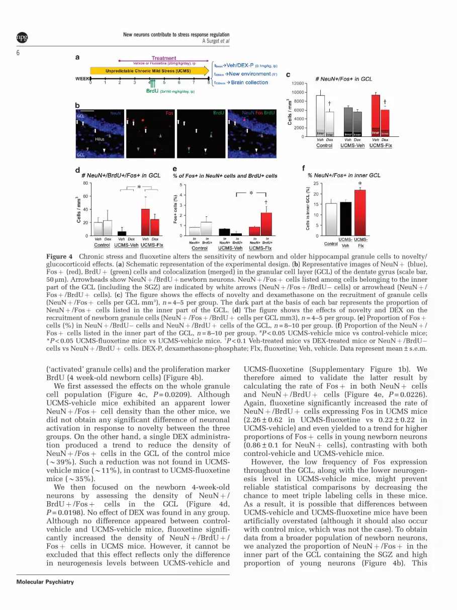
We first assessed the effects on the whole granulecell population (Figure 4c, P = 0.0209). AlthoughUCMS-vehicle mice exhibited an apparent lowerNeuNþ /Fosþ cell density than the other mice, wedid not obtain any significant difference of neuronalactivation in response to novelty between the threegroups. On the other hand, a single DEX administra-tion produced a trend to reduce the density ofNeuNþ /Fosþ cells in the GCL of the control mice(B39%). Such a reduction was not found in UCMS-vehicle mice (B11%), in contrast to UCMS-fluoxetinemice (B35%).
We then focused on the newborn 4-week-oldneurons by assessing the density of NeuNþ /BrdUþ /Fosþ cells in the GCL (Figure 4d,P = 0.0198). No effect of DEX was found in any group.Although no difference appeared between control-vehicle and UCMS-vehicle mice, fluoxetine signifi-cantly increased the density of NeuNþ /BrdUþ /Fosþ cells in UCMS mice. However, it cannot beexcluded that this effect reflects only the differencein neurogenesis levels between UCMS-vehicle and
UCMS-fluoxetine (Supplementary Figure 1b). Wetherefore aimed to validate the latter result bycalculating the rate of Fosþ in both NeuNþ cellsand NeuNþ /BrdUþ cells (Figure 4e, P = 0.0226).Again, fluoxetine significantly increased the rate ofNeuNþ /BrdUþ cells expressing Fos in UCMS mice(2.26±0.62 in UCMS-fluoxetine vs 0.22±0.22 inUCMS-vehicle) and even yielded to a trend for higherproportions of Fosþ cells in young newborn neurons(0.86±0.1 for NeuNþ cells), contrasting with bothcontrol-vehicle and UCMS-vehicle mice.
However, the low frequency of Fos expressionthroughout the GCL, along with the lower neurogen-esis level in UCMS-vehicle mice, might preventreliable statistical comparisons by decreasing thechance to meet triple labeling cells in these mice.As a result, it is possible that differences betweenUCMS-vehicle and UCMS-fluoxetine mice have beenartificially overstated (although it should also occurwith control mice, which was not the case). To obtaindata from a broader population of newborn neurons,we analyzed the proportion of NeuNþ /Fosþ in theinner part of the GCL containing the SGZ and highproportion of young neurons (Figure 4b). This
Figure 4 Chronic stress and fluoxetine alters the sensitivity of newborn and older hippocampal granule cells to novelty/glucocorticoid effects. (a) Schematic representation of the experimental design. (b) Representative images of NeuNþ (blue),Fosþ (red), BrdUþ (green) cells and colocalization (merged) in the granular cell layer (GCL) of the dentate gyrus (scale bar,50 mm). Arrowheads show NeuNþ /BrdUþnewborn neurons. NeuNþ /Fosþ cells listed among cells belonging to the innerpart of the GCL (including the SGZ) are indicated by white arrows (NeuNþ /Fosþ /BrdU� cells) or arrowhead (NeuNþ /Fosþ /BrdUþ cells). (c) The figure shows the effects of novelty and dexamethasone on the recruitment of granule cells(NeuNþ /Fosþ cells per GCL mm3), n = 4–5 per group. The dark part at the basis of each bar represents the proportion ofNeuNþ /Fosþ cells listed in the inner part of the GCL. (d) The figure shows the effects of novelty and DEX on therecruitment of newborn granule cells (NeuNþ /Fosþ /BrdUþ cells per GCL mm3), n = 4–5 per group. (e) Proportion of Fosþcells (%) in NeuNþ /BrdU� cells and NeuNþ /BrdUþ cells of the GCL, n = 8–10 per group. (f) Proportion of the NeuNþ /Fosþ cells listed in the inner part of the GCL, n = 8–10 per group. #P < 0.05 UCMS-vehicle mice vs control-vehicle mice;*P < 0.05 UCMS-fluoxetine mice vs UCMS-vehicle mice. wP < 0.1 Veh-treated mice vs DEX-treated mice or NeuNþ /BrdU�cells vs NeuNþ /BrdUþ cells. DEX-P, dexamethasone-phosphate; Flx, fluoxetine; Veh, vehicle. Data represent mean±s.e.m.
New neurons contribute to stress response regulationA Surget et al
6
Molecular Psychiatry

analysis showed that UCMS-fluoxetine mice holda greater proportion of Fosþ cells (B21%) thancontrol-vehicle (B15%) and UCMS-vehicle mice(B16%) (Figures 4c–f; P = 0.0186), confirming ourprevious results with the BrdU labeling.
In summary, UCMS exposure decreased the sensi-tivity of the GCL network to the combined novelty/glucocorticoid effects. Fluoxetine was able to counter-act this effect by recruiting higher proportions ofnewborn granule cells.
Fluoxetine restores HPA axis negative feedback via aneurogenesis-dependent mechanism
To examine more deeply the link between hippocam-pal neurogenesis and stress response, we investigatedwhether ablation of neurogenesis and UCMS induceabnormalities in the HPA axis similar to those foundin anxiety/depressive disorders. A repeated collectionof fecal samples across 33 h was performed on micepreviously exposed to a 7-week UCMS (Figure 5a andb). Slight but significant effects of UCMS werereported on the circadian activity of the HPA axis,and fluoxetine partially reverse these effects in non-irradiated mice (Supplementary Figure S4). We thenexamined the integrity of the negative feedbackof the HPA axis through the DEX suppression test.In this test, the administration of the glucocorticoidreceptor agonist DEX-P (0.1 mg kg�1, ip) is known tosuppress the subsequent release of endogenousglucocorticoids in the plasma when negative feedbackintegrity is undamaged. UCMS dampened the effec-tiveness of the negative feedback (control B61% vsUCMS B38%) whereas fluoxetine restored DEX-induced suppression to control levels (B66%)(Figure 5c, P = 0.016). However, although ablation ofhippocampal neurogenesis did not significantly affectthe negative feedback integrity in control (B60%)and UCMS (B30%) vehicle-treated mice, it signifi-cantly attenuated the improvement of the HPA axisnegative feedback in UCMS mice by fluoxetinetreatment (B42%).
New neurons contribute to the hippocampal inhibitionof the stress responseOur latter results support the idea that neurogenesismay subserve some stress integration roles of thehippocampus. To precisely determine the role of thenew neurons on hippocampus-dependent regulationof the stress response, we examined the ability of thehippocampus to regulate HPA axis activity underUCMS conditions and assessed how antidepressanttreatment and neurogenesis can affect this function.Following UCMS exposure, mice were bilaterallyimplanted with guide cannulas above the DG (Figures6a and b) and injected with either DEX (50 ng)or vehicle (NaCl 0.9%, ethanol 0.2%). The resultsdemonstrated that the intrahippocampal DEX infu-sion induced 51.2% plasmatic corticosterone sup-pression in non-irradiated control mice (P = 0.0034;Figure 6c, Supplementary Figure S5). The neurogen-esis ablation did not alter on its own the ability of thehippocampus to inhibit the HPA axis, as suggested bythe 50.9% DEX-induced corticosterone suppressionin irradiated control mice. However, UCMS disruptedhippocampal inhibition with low suppression in bothnon-irradiated and irradiated mice (5.1 and 3.7%,respectively). Finally, fluoxetine was able to restorehippocampus-dependent negative feedback on theHPA axis in non-irradiated UCMS mice to controllevels (56.1%), but not in irradiated UCMS mice(12.4%). This latter result indicates that fluoxetinerequires new hippocampal neurons to re-establish thehippocampal inhibition of the HPA axis underchronic stress conditions.
Discussion
Since the first reports linking the behavioral effects ofantidepressants to hippocampal neurogenesis, therehas been little progress in understanding the specificrole of neurogenesis in antidepressant response.Accordingly, it was necessary to identify hippocam-pal functions that could be affected by addingsupplemental new neurons and then contribute to
Figure 5 Fluoxetine-induced improvement of the HPA axis negative feedback under chronic stress conditions involveshippocampal neurogenesis. (a) Schematic representation of the experimental design. (b) Schedule of the fecal samplecollection and of the dexamethasone-phosphate (DEX-P) administration. (c) The DEX suppression test allowed assessing theintegrity of the HPA axis negative feedback. The figure shows the DEX-induced suppression of fecal corticosteronemetabolites (CORT), n = 8–11 per group. #P < 0.05 UCMS-vehicle mice vs control-vehicle mice; *P < 0.05 and **P < 0.01UCMS-fluoxetine mice vs UCMS-vehicle mice, or between line-connected groups. Data represent mean±s.e.m.
New neurons contribute to stress response regulationA Surget et al
7
Molecular Psychiatry

recovery following chronic stress and anxiety/depres-sion-like states. We have identified such functions inthis study. Our findings reveal that chronic stressexposure severely disrupts HPA axis activity,hippocampal regulation of subcortical stress-integra-tive structures, sensitivity of the granule cell networkto combined novelty/glucocorticoid effects andhippocampus-dependent negative feedback of theHPA axis. Furthermore, we demonstrate that theSSRI fluoxetine uses neurogenesis-dependent me-chanisms to restore hippocampal control over stresssystems as well as to reverse the behavioral andphysical effects of UCMS. Taken together, theseresults provide direct evidence for a neurobiologicalprocess through which neurogenesis participates toantidepressant response: new neurons are recruitedby antidepressant drugs to reestablish hippocampalregulation of stress systems, which in turn couldinitiate recovery.
There has been much discussion about a possiblecausative role for adult neurogenesis in anxiety/depression.51–54 Our results do not support such apossibility. First, in line with previous studies, theablation of adult-generated hippocampal neuronsdoes not cause any anxiety/depression-like states(but see ref. 7),3,12–15,55–58 or increases vulnerabilityto chronic stress.11 Second, enhancing neurogenesis isunlikely to be the final process through whichrecovery becomes possible. Indeed, although weconfirmed that hippocampal neurogenesis is requiredfor the reversal of the behavioral and physical effectsof UCMS by monoaminergic antidepressants (fluox-etine and imipramine),11 we also established that asimilar reversal can be elicited even in animals withablated neurogenesis by compounds targeting directlystress response circuits (CRF1 or vasopressin 1bantagonists).11 As a consequence, elevations of hip-pocampal neurogenesis may be a crucial but indirectpathway through which monoaminergic antidepres-
sants exert effects on downstream structures and thenunderlie recovery.
We previously showed that fluoxetine requiredhippocampal neurogenesis to reverse UCMS effectsin standard behavioral tests, such as the Novelty-Suppressed Feeding (NSF) test.11 It is noteworthy thatwe extend here such results to alterations morerelevant for depressive disorders, such as anhedoniaor body weight changes.59 This is critical as it hasrecently been shown that not all tests sensitive to thebehavioral effects of antidepressants might involveneurogenesis-dependent mechanisms.15 Moreover,the neurogenesis-dependence seems to emerge onlyunder chronic stress or corticosterone treatment formouse strains such as C57BL/6 or BALB/c.11,15 Incontrast to chronically stressed mice,11,37 fluoxetinemay induce some behavioral effects independent ofneurogenesis and fail to alter neurogenesis in un-stressed BALB/c mice.58,60 Considering that antide-pressants are mainly devoid of mood-changing effectsin non-depressed healthy humans61–63 and thatfluoxetine effects on behavior and large-scale geneexpression greatly differ depending if the mice areunder control or UCMS conditions,33 paradigmselaborated in ‘non-depressed’ mice may engage differ-ent neurobiological mechanisms that are perhapsirrelevant to clinical remission. Rather, it is likely thatrelevant antidepressant effects are conditional on thepresence of a disorder-related neuropathology.36 In thesame vein, antidepressant effects that arise duringtreatment shorter than 14 days are obviously assumedto be neurogenesis-independent.56,58,64 Indeed, antide-pressants require more than 1 week to significantlyincrease SGZ cell proliferation3,4 and it takes 1–2weeks for new cells to start to integrate into thenetwork.65 This timeframe suggests that at least 2–3weeks should be expected before the neurogenic effectsof antidepressants can significantly impact the DGnetwork if so. It is noteworthy that this delay parallels
Figure 6 Adult-generated granule neurons are required to restore the hippocampal inhibition of the hypothalamo-pituitary-adrenal axis under chronic stress conditions. (a) Schematic representation of the experimental design. (b) Schematicrepresentation of the guide-cannula implantation (bar: bregma =�3.08, lateral = ±2.3, vertical =�1.4) and of thedexamethasone (DEX) infusion sites, one millimeter lower (asterisk). The figure is adapted from Paxinos and Franklin(2001).38 (c) An intrahippocampal DEX suppression test has been used to test the ability of hippocampus to inhibit the HPAaxis. The figure shows the corticosterone (CORT) suppression induced by intrahippocampal DEX infusion, n = 7–8 per group.#P < 0.05 UCMS mice vs control-vehicle mice; *P < 0.05 and **P < 0.01 UCMS-fluoxetine mice vs UCMS-vehicle mice, orbetween line-connected groups. Veh, vehicle; DEX, dexamethasone. Data represent mean±s.e.m.
New neurons contribute to stress response regulationA Surget et al
8
Molecular Psychiatry

the course of the physical and behavioral effects offluoxetine in the UCMS model.11,33,45 This temporalcorrelation strengthens the idea that neurogenesismay be required for fluoxetine to reverse UCMS-induced effects. Interestingly, this timeframe alsoparallels the delay before the first beneficial effectsin patients with major depression,66 suggesting thatneurogenesis could also be involved in antidepres-sant effects in humans. The two first studies investi-gating the relationship between hippocampalneurogenesis and antidepressant drugs in depressedpatients have yielded mixed results: an increase of theSGZ neural progenitors was found after antidepres-sant treatment in adult, but not elderly patients.5,6
One meaningful finding of our study is that,without hippocampal neurogenesis, fluoxetine be-comes unable to restore under chronic stress condi-tions the hippocampus-dependent inhibition of theHPA axis as well as to reverse the behavioral andphysical alterations. This suggests that the improve-ment of stress system regulation is a critical process inthe therapeutic action of antidepressants, at least inthe substantial subset of patients with altered HPAaxis reactivity. This is consistent with clinical datawhich demonstrate that, in most patients with alteredHPA axis negative feedback, remission and decreasedrisk of relapse are associated with the improvementof HPA axis functioning.28–31 Overall, this findingreveals that the hippocampus not only regulates brainsystems involved in the stress response at baseline but isalso critical in mediating the ability of monoaminergicantidepressants to modulate stress response, probablythrough newly generated neurons. This possibility isin agreement with a brief report indicating thatneurogenesis inhibition may yield higher corticoster-one secretion in mice following exposure to a newbright-light condition, suggesting that even underconditions when the negative feedback is not fullydesensitized, newborn neurons may have a moresubtle role in dampening the response to stress.67
As a consequence, we aimed to scrutinize preciselythe relationships between the hippocampus andstress response systems. Specifically a single pre-administration of the synthetic glucocorticoid DEXdecreased Fos expression in the GCL of control-vehicle mice exposed to a new context. Thesedifferences in granule cell activation could poten-tially influence the activity of the next hippocampalcell layer (CA3)68 and then enable the hippocampalnetwork to couple stress and contextual informa-tion.69 Hence, a possible interpretation is to considerthis reduction as a manner for the granule cellpopulation to take into account, at the network level,glucocorticoid releases during new contextual inputs.On the other hand, this Fos-induction produced bynovelty is disrupted under chronic stress with noadditional effect of exogenous glucocorticoids. It istherefore conceivable that the alteration of the GCLsensitivity leads to disturbances in informationprocessing and stress integration. We tested thispossibility by probing the neurocircuitry connecting
the hippocampus to the HPA axis.21,22 Our resultsdemonstrate that chronic stress profoundly moderatesthe hippocampal influence on subcortical structuresinvolved in the stress response such as lateral septum,BST, preoptic area and DMH. Taken together, theseresults suggest that chronic stress, by decreasinginhibitory regulation from the hippocampus, may biasthe net input balance toward greater excitatory influ-ences into the PVN leading to its overactivation andperhaps underlying the impairments of the hippocam-pus-dependent negative feedback on the HPA axis.
Strikingly, the Fos induction in the GCL and theresponse to exogenous glucocorticoids are preservedin fluoxetine-treated UCMS mice. Antidepressanttreatment is therefore able to restore under chronicstress conditions the sensitivity of the granule cellnetwork to novelty/glucocorticoid effects. As adult-generated granule cells are continuously added to thenetwork, they are a potential source of new sensitiveneurons with enhanced plasticity and excitabil-ity.57,70–74 Considering that fluoxetine treatment in-creased neurogenesis levels in UCMS mice, it wasthen conceivable that such compounds compensatethe chronic stress effects on GCL network activity byrecruiting higher proportions of adult newbornneurons among the granule cells expressing Fos.Indeed, this hypothesis was confirmed in UCMS-fluoxetine mice with a greater density of 4-week-oldnewborn cells expressing Fos and a higher rate ofFosþneurons in the inner GCL (containing highlevels of newborn young neurons).41–44 Enhancingneurogenesis may thus be a way for antidepressantdrugs to add a fresh source of new sensitive neuronsthat may affect hippocampal functioning and thenimprove stress integration.
Interestingly, the proportion of Fos expression inthe inner GCL of UCMS-fluoxetine mice are evengreater than in control-vehicle mice, which alsoexhibited high levels of SGZ cell proliferation andneurogenesis. A potential explanation is that fluox-etine does not only enhance neurogenesis but alsoaccelerates maturation and synaptic integration of thenewborn neurons, as already shown.14 This effect maypromote their recruitment and thus contribute tocounteract the effects induced by chronic stress. As amatter of fact, the higher proportion of Fos expressionin the BrdUþ cells, which was found exclusively inUCMS-fluoxetine mice appears to strengthen thisinterpretation. Another important question is whetheror not the effects of newborn neurons during suchprocesses are direct. The fact that Fos expression hasbeen found in BrdUþ cells supports this idea.Moreover, newborn granule cells in mice can beginto express glucocorticoid receptor early: 66% 1 weekafter BrdU integration and almost 100% four weeksafter BrdU integration (as high as older matureneurons).75 These data suggest that newborn neuronsmay potentially be sensitive to glucocorticoid releaseduring their development. However, we did notreport any significant change induced by DEXindicating that newborn neurons could actually be
New neurons contribute to stress response regulationA Surget et al
9
Molecular Psychiatry

less sensitive to glucocorticoids than mature neurons.These results are surprising because taken togetherthey indicate that the reduction of the GCL networkactivation induced by DEX can paradoxically beimproved by increasing a population of young neuronsthough less sensitive to DEX. This apparent discre-pancy can be enlightened by a recent study,76 where theauthors demonstrated that a limited amount of new-born granule cells is able to negatively modulate theactivity in the larger population of mature granule cells,maybe by preferentially activating inhibitory interneur-ons. Accordingly, it is conceivable that, in our context,DEX biases the balance of activity toward younggranule cells (if less sensitive to DEX), then potentiat-ing the DEX-induced reduction of the activation ofolder granule cells. Within this framework, fluoxetine,by enhancing neurogenesis and perhaps improvingmaturation, might re-establish the sensitivity of theGCL network to novelty/glucocorticoid effects.
The hippocampus is also highly interconnectedwith various corticolimbic structures, which controlthe stress response, emotion and motivation such asthe amygdala, cingulate gyrus, prefrontal cortex andventral striatum.16–18 These structures are thoughtto be involved in an integrated stress response andunderlie the main pathological manifestations ofanxiety/depressive disorders.77–79 It is therefore con-ceivable that the UCMS-induced HPA axis abnormal-ities represent only a measurable fraction of largerchanges in the stress integration system and thatelevation of hippocampal neurogenesis might enablemonoaminergic antidepressants to strengthen hippo-campal influences on these brain regions.
Another important finding of our study is thatinhibition of hippocampal neurogenesis does notcause any alteration of the HPA axis functioning orregulation in both control- and UCMS-vehicle mice.The hippocampus is known to contribute to HPA axisregulation: its stimulation can reduce glucocorticoidsecretion80,81 whereas hippocampal damages canincrease stress-induced glucocorticoid release.82,83 Inspite of this, none of our previous and current resultssuggest that neurogenesis disruption alone substan-tially disrupts either the circadian rhythm of HPA axisactivity or the stress response.3 Defects in hippocam-pal functioning should therefore require larger altera-tions in the intrinsic biology of the hippocampus thana neurogenesis decline alone. Accordingly, theseresults reveal that neurogenesis may not be the driverof the hippocampal regulation on the HPA axis, atleast without greater environmental challenges.
Taken together, our data suggest that, while hippo-campal neurogenesis would contribute to the im-provement of stress integration by antidepressantsduring chronic stress exposure, its role could be lessimportant in the absence of previous deficienciesor pathological situations. This result parallelsthe Kempermann’s hypothesis of the neurogenicreserve.84 He suggests that adult newborn hippocam-pal neurons would constitute a reserve, inessential innormal conditions, but which could become decisive
when the organism is confronted with novelty orcomplex situations. In our context, new hippocampalneurons might thus be critical for antidepressanteffects only when the hippocampus-dependent reg-ulation of the stress system is deficient.
In conclusion, this study demonstrates how hippo-campal neurogenesis can contribute to regulate thestress hormone system. This function may explainwhy adult-generated granule cells are required forseveral behavioral effects of monoaminergic anti-depressants. However, not all the patients withanxiety/depression display HPA axis abnormalities,85
as well as not all the mouse strains exhibit HPA axisdisturbances following UCMS exposure.45 It is there-fore likely that the extent of neurogenesis involve-ment in antidepressant effects can depend on thedegree of stress system alterations for each subject ormouse strain. Interestingly, the previous conflictingresults can be enlightened by this hypothesis. Indeed,the utilization of control non-stressed mice orstressed mice without any significant alterations ofstress systems would be less appropriate to highlightthe role of adult-generated hippocampal neurons inantidepressant effects.56,58 In the same vein, not all thepatients respond to antidepressant therapies; accord-ingly, extending this work to the mechanisms of whatis happening in anxious/depressed patients might belimited to some subtypes of stress-related disorders orsubgroups of patients. Future studies will thereforehave to examine whether stress system alterations areclearly a condition for the neurogenesis involvementin antidepressant effects and whether adult-generatedhippocampal neurons can influence stress integrationin other relevant brain regions such as the prefrontalcortex, cingulate cortex or amygdala.
Conflict of interest
AS, AT, AL, QR, CT, RP and YIV, report no biomedicalfinancial interests or potential conflicts of interests.GG is an employee of Sanofi-Aventis. EDL receivescompensation as a consultant to PGxHealth. RHreceives compensation as a consultant for BrainCells,PsychoGenics, Servier, Astra Zeneca and Lundbeck inrelation to the generation of novel antidepressants. CBreceives compensation as a consultant for Takeda.
Acknowledgments
We thank Dr Sylvie Chalon (Inserm), Dr Samuel Leman(Universite F. Rabelais) for help and comments,Dr Ayumu Tashiro (NTNU) for support as well asAnne-Marie Leguisquet, Severine Devers, MarysePingaud, Dominique Le Glaunec, Lucette Garreau,Edith Klobetz-Rassam, Navieta Ramasami and thePPF ‘Analyse des systemes biologiques’ for assistance.AS received research grants from Region Centre, FRMand IBRO, funds from the Bettencourt-SchuellerFoundation and is currently supported by the 7thframework program of the European Commission. EDLis supported by NIH grant K08 MH076083.
New neurons contribute to stress response regulationA Surget et al
10
Molecular Psychiatry

References
1 Eriksson PS, Perfilieva E, Bjork-Eriksson T, Alborn AM, NordborgC, Peterson DA et al. Neurogenesis in the adult human hippo-campus. Nat Med 1998; 4: 1313–1317.
2 Ming GL, Song H. Adult neurogenesis in the mammalian centralnervous system. Annu Rev Neurosci 2005; 28: 223–250.
3 Santarelli L, Saxe M, Gross C, Surget A, Battaglia F, Dulawa S et al.Requirement of hippocampal neurogenesis for the behavioraleffects of antidepressants. Science 2003; 301: 805–809.
4 Malberg JE, Eisch AJ, Nestler EJ, Duman RS. Chronic antidepres-sant treatment increases neurogenesis in adult rat hippocampus.J Neurosci 2000; 20: 9104–9110.
5 Boldrini M, Underwood MD, Hen R, Rosoklija GB, Dwork AJ, JohnMann J et al. Antidepressants increase neural progenitor cells inthe human hippocampus. Neuropsychopharmacology 2009; 34:2376–2389.
6 Lucassen PJ, Stumpel MW, Wang Q, Aronica E. Decreasednumbers of progenitor cells but no response to antidepressantdrugs in the hippocampus of elderly depressed patients. Neuro-pharmacology 2010; 58: 940–949.
7 Revest JM, Dupret D, Koehl M, Funk-Reiter C, Grosjean N, PiazzaPV et al. Adult hippocampal neurogenesis is involved in anxiety-related behaviors. Mol Psychiatry 2009; 14: 959–967.
8 Czeh B, Welt T, Fischer AK, Erhardt A, Schmitt W, Muller MBet al. Chronic psychosocial stress and concomitant repetitivetranscranial magnetic stimulation: effects on stress hormone levelsand adult hippocampal neurogenesis. Biol Psychiatry 2002; 52:1057–1065.
9 Cameron HA, Gould E. Adult neurogenesis is regulated by adrenalsteroids in the dentate gyrus. Neuroscience 1994; 61: 203–209.
10 Gould E, Tanapat P, McEwen BS, Flugge G, Fuchs E. Proliferationof granule cell precursors in the dentate gyrus of adult monkeysis diminished by stress. Proc Natl Acad Sci USA 1998; 95:3168–3171.
11 Surget A, Saxe M, Leman S, Ibarguen-Vargas Y, Chalon S, GriebelG et al. Drug-dependent requirement of hippocampal neurogenesisin a model of depression and of antidepressant reversal.Biol Psychiatry 2008; 64: 293–301.
12 Airan RD, Meltzer LA, Roy M, Gong Y, Chen H, Deisseroth K.High-speed imaging reveals neurophysiological links to behaviorin an animal model of depression. Science 2007; 317: 819–823.
13 Jiang W, Zhang Y, Xiao L, Van Cleemput J, Ji SP, Bai G et al.Cannabinoids promote embryonic and adult hippocampus neuro-genesis and produce anxiolytic- and antidepressant-like effects.J Clin Invest 2005; 115: 3104–3116.
14 Wang JW, David DJ, Monckton JE, Battaglia F, Hen R.Chronic fluoxetine stimulates maturation and synaptic plasticityof adult-born hippocampal granule cells. J Neurosci 2008; 28:1374–1384.
15 David DJ, Samuels BA, Rainer Q, Wang JW, Marsteller D, Mendez Iet al. Neurogenesis-dependent and -independent effects offluoxetine in an animal model of anxiety/depression. Neuron2009; 62: 479–493.
16 Floresco SB, Todd CL, Grace AA. Glutamatergic afferents from thehippocampus to the nucleus accumbens regulate activity ofventral tegmental area dopamine neurons. J Neurosci 2001; 21:4915–4922.
17 Naber PA, Witter MP. Subicular efferents are organized mostly asparallel projections: a double-labeling, retrograde-tracing study inthe rat. J Comp Neurol 1998; 393: 284–297.
18 Amaral DG, Witter MP. The three-dimensional organization ofthe hippocampal formation: a review of anatomical data. Neuro-science 1989; 31: 571–591.
19 Herman JP, Schafer MK, Young EA, Thompson R, Douglass J,Akil H et al. Evidence for hippocampal regulation of neuroendo-crine neurons of the hypothalamo-pituitary-adrenocortical axis.J Neurosci 1989; 9: 3072–3082.
20 Mizoguchi K, Ishige A, Aburada M, Tabira T. Chronic stressattenuates glucocorticoid negative feedback: involvement ofthe prefrontal cortex and hippocampus. Neuroscience 2003; 119:887–897.
21 Herman JP, Ostrander MM, Mueller NK, Figueiredo H. Limbicsystem mechanisms of stress regulation: hypothalamo-pituitary-
adrenocortical axis. Prog Neuropsychopharmacol Biol Psychiatry2005; 29: 1201–1213.
22 Ulrich-Lai YM, Herman JP. Neural regulation of endocrine andautonomic stress responses. Nat Rev Neurosci 2009; 10: 397–409.
23 Rubin RT, Poland RE, Lesser IM, Winston RA, Blodgett AL.Neuroendocrine aspects of primary endogenous depression. I.Cortisol secretory dynamics in patients and matched controls.Arch Gen Psychiatry 1987; 44: 328–336.
24 Nemeroff CB, Widerlov E, Bissette G, Walleus H, Karlsson I,Eklund K et al. Elevated concentrations of CSF corticotropin-releasing factor-like immunoreactivity in depressed patients.Science 1984; 226: 1342–1344.
25 Holsboer F, von Bardeleben U, Wiedemann K, Muller OA, Stalla GK.Serial assessment of corticotropin-releasing hormone response afterdexamethasone in depression. Implications for pathophysiology ofDST nonsuppression. Biol Psychiatry 1987; 22: 228–234.
26 Heuser I, Yassouridis A, Holsboer F. The combined dexametha-sone/CRH test: a refined laboratory test for psychiatric disorders.J Psychiatr Res 1994; 28: 341–356.
27 Lopez-Duran NL, Kovacs M, George CJ. Hypothalamic-pituitary-adrenal axis dysregulation in depressed children andadolescents: a meta-analysis. Psychoneuroendocrinology 2009; 34:1272–1283.
28 Holsboer-Trachsler E, Stohler R, Hatzinger M. Repeated adminis-tration of the combined dexamethasone-human corticotropinreleasing hormone stimulation test during treatment of depres-sion. Psychiatry Res 191; 38: 163–171.
29 Appelhof BC, Huyser J, Verweij M, Brouwer JP, van Dyck R,Fliers E et al. Glucocorticoids and relapse of major depression(dexamethasone/corticotropin-releasing hormone test in relationto relapse of major depression). Biol Psychiatry 2006; 59: 696–701.
30 Kunugi H, Ida I, Owashi T, Kimura M, Inoue Y, Nakagawa S et al.Assessment of the dexamethasone/CRH test as a state-dependentmarker for hypothalamic-pituitary-adrenal (HPA) axis abnormal-ities in major depressive episode: a Multicenter Study. Neuro-psychopharmacology 2006; 31: 212–220.
31 Ising M, Horstmann S, Kloiber S, Lucae S, Binder EB, Kern N et al.Combined dexamethasone/corticotropin releasing hormone testpredicts treatment response in major depression—a potentialbiomarker? Biol Psychiatry 2007; 62: 47–54.
32 Datson NA, Speksnijder N, Mayer JL, Steenbergen PJ, Korobko O,Goeman J et al. The transcriptional response to chronic stress andglucocorticoid receptor blockade in the hippocampal dentate gyrus.Hippocampus; doi: 10.1002/hipo.20905 (e-pub ahead of print).
33 Surget A, Wang Y, Leman S, Ibarguen-Vargas Y, Edgar N, Griebel Get al. Corticolimbic transcriptome changes are state-dependent andregion-specific in a rodent model of depression and of antidepres-sant reversal. Neuropsychopharmacology 2009; 34: 1363–1380.
34 Willner P. Validity, reliability and utility of the chronic mild stressmodel of depression: a 10-year review and evaluation. Psycho-pharmacology (Berl) 1997; 134: 319–329.
35 Cryan JF, Holmes A. The ascent of mouse: advances in modellinghuman depression and anxiety. Nat Rev Drug Discov 2005; 4:775–790.
36 Sibille E, Wang Y, Joeyen-Waldorf J, Gaiteri C, Surget A, Oh S et al.A molecular signature of depression in the amygdala. Am JPsychiatry 2009; 166: 1011–1024.
37 Alonso R, Griebel G, Pavone G, Stemmelin J, Le Fur G, Soubrie P.Blockade of CRF or V(1b) receptors reverses stress-inducedsuppression of neurogenesis in a mouse model of depression.Mol Psychiatry 2004; 9: 278–286, 224.
38 Paxinos G, Franklin KBJ. The mouse brain in stereotaxiccoordinates. Academic Press: San Diego, CA, 2001, 124pp.
39 Snyder JS, Ramchand P, Rabbett S, Radik R, Wojtowicz JM,Cameron HA. Septo-temporal gradients of neurogenesis andactivity in 13-month-old rats. Neurobiol Aging; doi:10.1016/j.neurobiolaging.2009.05.022 (e-pub ahead of print).
40 Snyder JS, Radik R, Wojtowicz JM, Cameron HA. Anatomicalgradients of adult neurogenesis and activity: young neurons in theventral dentate gyrus are activated by water maze training.Hippocampus 2009; 19: 360–370.
41 Seri B, Garcia-Verdugo JM, Collado-Morente L, McEwen BS, Alvarez-Buylla A. Cell types, lineage, and architecture of the germinal zone inthe adult dentate gyrus. J Comp Neurol 2004; 478: 359–378.
New neurons contribute to stress response regulationA Surget et al
11
Molecular Psychiatry

42 Kempermann G, Gast D, Kronenberg G, Yamaguchi M, Gage FH.Early determination and long-term persistence of adult-generatednew neurons in the hippocampus of mice. Development 2003; 130:391–399.
43 Dayer AG, Ford AA, Cleaver KM, Yassaee M, Cameron HA. Short-term and long-term survival of new neurons in the rat dentategyrus. J Comp Neurol 2003; 460: 563–572.
44 Crespo D, Stanfield BB, Cowan WM. Evidence that late-generatedgranule cells do not simply replace earlier formed neurons in therat dentate gyrus. Exp Brain Res 1986; 62: 541–548.
45 Ibarguen-Vargas Y, Surget A, Touma C, Palme R, Belzung C.Multifaceted strain-specific effects in a mouse model of depres-sion and of antidepressant reversal. Psychoneuroendocrinology2008; 33: 1357–1368.
46 Touma C, Palme R, Sachser N. Analyzing corticosterone metabo-lites in fecal samples of mice: a noninvasive technique to monitorstress hormones. Horm Behav 2004; 45: 10–22.
47 Touma C, Sachser N, Mostl E, Palme R. Effects of sex and time ofday on metabolism and excretion of corticosterone in urine andfeces of mice. Gen Comp Endocrinol 2003; 130: 267–278.
48 Tashiro A, Makino H, Gage FH. Experience-specific functionalmodification of the dentate gyrus through adult neurogenesis: acritical period during an immature stage. J Neurosci 2007; 27:3252–3259.
49 Kee N, Teixeira CM, Wang AH, Frankland PW. Preferentialincorporation of adult-generated granule cells into spatial memorynetworks in the dentate gyrus. Nat Neurosci 2007; 10: 355–362.
50 Ramirez-Amaya V, Marrone DF, Gage FH, Worley PF, Barnes CA.Integration of new neurons into functional neural networks.J Neurosci 2006; 26: 12237–12241.
51 Henn FA, Vollmayr B. Neurogenesis and depression: etiology orepiphenomenon? Biol Psychiatry 2004; 56: 146–150.
52 Sapolsky RM. Is impaired neurogenesis relevant to the affectivesymptoms of depression? Biol Psychiatry 2004; 56: 137–139.
53 Eisch AJ, Cameron HA, Encinas JM, Meltzer LA, Ming GL,Overstreet-Wadiche LS. Adult neurogenesis, mental health, andmental illness: hope or hype? J Neurosci 2008; 28: 11785–11791.
54 Duman RS. Depression: a case of neuronal life and death? BiolPsychiatry 2004; 56: 140–145.
55 Meshi D, Drew MR, Saxe M, Ansorge MS, David D, Santarelli Let al. Hippocampal neurogenesis is not required for behavioraleffects of environmental enrichment. Nat Neurosci 2006; 9:729–731.
56 Bessa JM, Ferreira D, Melo I, Marques F, Cerqueira JJ, Palha JAet al. The mood-improving actions of antidepressants do notdepend on neurogenesis but are associated with neuronalremodeling. Mol Psychiatry 2009; 14: 764–773, 739.
57 Saxe MD, Battaglia F, Wang JW, Malleret G, David DJ, Monckton JEet al. Ablation of hippocampal neurogenesis impairs contextualfear conditioning and synaptic plasticity in the dentate gyrus.Proc Natl Acad Sci USA 2006; 103: 17501–17506.
58 Holick KA, Lee DC, Hen R, Dulawa SC. Behavioral effects ofchronic fluoxetine in BALB/cJ mice do not require adulthippocampal neurogenesis or the serotonin 1A receptor. Neuro-psychopharmacology 2008; 33: 406–417.
59 American Psychiatric Association. Diagnostic and StatisticalManual of Mental Disorders, 4th edn American Psychiatric Press:Washington DC, 1994.
60 Huang GJ, Bannerman D, Flint J. Chronic fluoxetine treatmentalters behavior, but not adult hippocampal neurogenesis, inBALB/cJ mice. Mol Psychiatry 2008; 13: 119–121.
61 Yeragani VK, Pohl R, Mallavarapu M, Balon R. Approximateentropy of symptoms of mood: an effective technique to quantifyregularity of mood. Bipolar Disord 2003; 5: 279–286.
62 Gelfin Y, Gorfine M, Lerer B. Effect of clinical doses of fluoxetineon psychological variables in healthy volunteers. Am J Psychiatry1998; 155: 290–292.
63 Barr LC, Heninger GR, Goodman W, Charney DS, Price LH.Effects of fluoxetine administration on mood response totryptophan depletion in healthy subjects. Biol Psychiatry 1997;41: 949–954.
64 Vollmayr B, Simonis C, Weber S, Gass P, Henn F. Reduced cellproliferation in the dentate gyrus is not correlated with thedevelopment of learned helplessness. Biol Psychiatry 2003; 54:1035–1040.
65 Zhao C, Deng W, Gage FH. Mechanisms and functional implica-tions of adult neurogenesis. Cell 2008; 132: 645–660.
66 Wong ML, Licinio J. Research and treatment approaches todepression. Nat Rev Neurosci 2001; 2: 343–351.
67 Schloesser RJ, Manji HK, Martinowich K. Suppression of adultneurogenesis leads to an increased hypothalamo-pituitary-adrenalaxis response. Neuroreport 2009; 20: 553–557.
68 Treves A, Tashiro A, Witter ME, Moser EI. What is the mammaliandentate gyrus good for? Neuroscience 2008; 154: 1155–1172.
69 Becker S, Wojtowicz JM. A model of hippocampal neurogenesis inmemory and mood disorders. Trends Cogn Sci 2007; 11: 70–76.
70 Wang S, Scott BW, Wojtowicz JM. Heterogenous properties ofdentate granule neurons in the adult rat. J Neurobiol 2000; 42:248–257.
71 Snyder JS, Kee N, Wojtowicz JM. Effects of adult neurogenesis onsynaptic plasticity in the rat dentate gyrus. J Neurophysiol 2001;85: 2423–2431.
72 Ge S, Yang CH, Hsu KS, Ming GL, Song H. A critical period forenhanced synaptic plasticity in newly generated neurons of theadult brain. Neuron 2007; 54: 559–566.
73 Schmidt-Hieber C, Jonas P, Bischofberger J. Enhanced synapticplasticity in newly generated granule cells of the adult hippo-campus. Nature 2004; 429: 184–187.
74 Laplagne DA, Esposito MS, Piatti VC, Morgenstern NA, Zhao C,van Praag H et al. Functional convergence of neurons generated inthe developing and adult hippocampus. PLoS Biol 2006; 4: e409.
75 Garcia A, Steiner B, Kronenberg G, Bick-Sander A, KempermannG. Age-dependent expression of glucocorticoid- and mineralocor-ticoid receptors on neural precursor cell populations in the adultmurine hippocampus. Aging Cell 2004; 3: 363–371.
76 Lacefield CO, Itskov V, Reardon T, Hen R, Gordon JA. Effects of adult-generated granule cells on coordinated network activity in the dentategyrus. Hippocampus; doi: 10.1002/hipo.20860 (e-pub ahead of print).
77 Seminowicz DA, Mayberg HS, McIntosh AR, Goldapple K, KennedyS, Segal Z et al. Limbic-frontal circuitry in major depression: a pathmodeling metanalysis. Neuroimage 2004; 22: 409–418.
78 Drevets WC, Price JL, Furey ML. Brain structural and functionalabnormalities in mood disorders: implications for neurocircuitrymodels of depression. Brain Struct Funct 2008; 213: 93–118.
79 Carlson PJ, Singh JB, Zarate Jr CA, Drevets WC, Manji HK. Neuralcircuitry and neuroplasticity in mood disorders: insights for noveltherapeutic targets. NeuroRx 2006; 3: 22–41.
80 Rubin RT, Mandell AJ, Crandall PH. Corticosteroid responses tolimbic stimulation in man: localization of stimulus sites. Science1966; 153: 767–768.
81 Dunn JD, Orr SE. Differential plasma corticosterone responses tohippocampal stimulation. Exp Brain Res 1984; 54: 1–6.
82 Herman JP, Cullinan WE, Morano MI, Akil H, Watson SJ.Contribution of the ventral subiculum to inhibitory regulation ofthe hypothalamo-pituitary-adrenocortical axis. J Neuroendocrinol1995; 7: 475–482.
83 Sapolsky RM, Krey LC, McEwen BS. Glucocorticoid-sensitivehippocampal neurons are involved in terminating the adrenocor-tical stress response. Proc Natl Acad Sci USA 1984; 81: 6174–6177.
84 Kempermann G. The neurogenic reserve hypothesis: what is adulthippocampal neurogenesis good for? Trends Neurosci 2008; 31:163–169.
85 Holsboer F. The corticosteroid receptor hypothesis of depression.Neuropsychopharmacology 2000; 23: 477–501.
This work is licensed under the CreativeCommons Attribution-NonCommercial-
Share Alike 3.0 Unported License. To view a copyof this license, visit http://creativecommons.org/licenses/by-nc-sa/3.0/
Supplementary Information accompanies the paper on the Molecular Psychiatry website (http://www.nature.com/mp)
New neurons contribute to stress response regulationA Surget et al
12
Molecular Psychiatry


