Embed Size (px)
Citation preview

This article appeared in a journal published by Elsevier. The attachedcopy is furnished to the author for internal non-commercial researchand education use, including for instruction at the authors institution
and sharing with colleagues.
Other uses, including reproduction and distribution, or selling orlicensing copies, or posting to personal, institutional or third party
websites are prohibited.
In most cases authors are permitted to post their version of thearticle (e.g. in Word or Tex form) to their personal website orinstitutional repository. Authors requiring further information
regarding Elsevier’s archiving and manuscript policies areencouraged to visit:
http://www.elsevier.com/copyright

Author's personal copy
Antifouling activity of commercial biocides vs. natural and natural-derivedproducts assessed by marine bacteria adhesion bioassay
Mercedes Camps a, Jean-François Briand a,⇑, Linda Guentas-Dombrowsky a, Gérald Culioli a,Alexis Bazire b, Yves Blache a
a Laboratoire MAPIEM, EA 4323, Biofouling et Substances Naturelles Marines, Université du Sud Toulon-Var, 83162 La Valette-du-Var, Franceb Laboratoire de Biotechnologie et Chimie Marine, EA 3883, Université de Bretagne Sud, 56321 Lorient, France
a r t i c l e i n f o
Keywords:BiofoulingBiofilmMicrotiter plateFluorescenceNatural productsCommercial biocides
a b s t r a c t
Biofilm formation is a key step during marine biofouling, the natural colonization of immersed substrata,leading to major economic and ecological consequences. Consequently, bacteria have been used for thescreening of new non-toxic antifoulants: the adhesion of five strains isolated on three French locationswas monitored using a fluorescence-based assay and toxicity was also evaluated. Nine biocides includingcommercial, natural and natural-derived products were tested. The commercial antifoulants, TBTO andSea Nine showed low EC50 but high toxicity. The non-commercial products TFA-Z showed significantanti-adhesion activities and appeared to be non-toxic, suggesting a specific anti-adhesion mechanism.In addition, the strains could be classified depending on their sensitivity to the molecules used even ifstrain sensitivity also depended on the molecules tested. In conclusion, TFA-Z would be a promising can-didate as non-toxic antifoulant and our results strengthen the need to perform antifouling bioassays witha panel of strains showing different response profiles.
� 2011 Elsevier Ltd. All rights reserved.
1. Introduction
Biofouling refers to the natural colonization of submerged sur-faces, involving a wide range of organisms from bacteria to inver-tebrates. The first stage of this process corresponds to theformation of a biofilm, initially constituted of pioneer bacteria.Then the adhesion of macrofoulers such as algae or invertebratessignificantly increased the attached biomass (e.g. Wahl, 1989;Railkin, 2004). Biofouling has a major economic impact, especiallywhen it occurs on ship hulls or aquaculture facilities. In addition,invasive species can be spread inadvertently due to inadequateprotection against fouling (Piola et al., 2009), leading also to eco-logical consequences. In this context, since several commercial bio-cides commonly used in antifouling (AF) coatings have beenrecently banned, the screening for alternative eco-friendly biocidesappears to be urgent.
Marine biofouling is the consequence of a wide range of physi-cal, chemical and biological processes, and reproducing such acomplex phenomenon in a laboratory bioassay is considered upto now quite difficult to achieve. Nevertheless, in vitro assays arerequired, as the direct evaluation of AF coatings in situ is expensive(panels and paints, access to secure raft(s) and raft(s) care, peopleto perform regular macrofouling inspection of immersed paintedpanels, . . .) and time consuming (several months to years of expo-
sure). For ecological purposes, AF bioassays have been carried outin order to elucidate the mechanism of chemical defences of mar-ine organisms in epibiosis control (e.g. Nylund et al., 2007; Qianet al., 2007). Other bioassays have also been developed to deter-mine whether novel natural products inhibit the growth or adhe-sion of colonizing organisms (e.g. DeNys and Steinberg, 2002;Fusetani, 2004). Finally, biocidal AF paints (e.g. Burgess et al.,2003; Watermann et al., 2005) and fouling-release coatings (e.g.Cassé et al., 2007; Stafslien et al., 2007b; D’Souza et al., 2010) havebeen assessed through specific AF assays. There is a general agree-ment that, due to the biodiversity in marine ecosystems, no singlesubstance or substrate could inhibit the settlement and/or growthof all the organisms implicated in the marine biofouling process.Consequently, several different target organisms are required(Qian et al., 2010) for the evaluation of the AF potential of biocidesand/or coatings. A wide range of model organisms has been used inAF bioassays. Predominant biofouling species such as marine bac-teria, diatoms, algae, mussels and barnacles, are generally chosen(see in Briand, 2009). Relative to the bacteria, a wide diversity oftaxa was used. Bacteria involved in the adhesion process onartificial hard substrata are generally selected when AF compoundsfor man-made devices are screened (Wilsanand et al., 1999;Ramasamy and Murugan, 2007). Cell adhesion and biofilm forma-tion have been increasingly studied in bioassays as it appeared tobe more relevant for AF purposes than growth inhibition (Stafslienet al., 2006 and related; Leroy et al., 2007). Microtiter plate bioas-says for the assessment of bacterial biofilm formation have been
0025-326X/$ - see front matter � 2011 Elsevier Ltd. All rights reserved.doi:10.1016/j.marpolbul.2011.02.031
⇑ Corresponding author. Tel.: +33 (0)4 94 14 25 79.E-mail address: [email protected] (J.-F. Briand).
Marine Pollution Bulletin 62 (2011) 1032–1040
Contents lists available at ScienceDirect
Marine Pollution Bulletin
journal homepage: www.elsevier .com/locate /marpolbul

Author's personal copy
also increasingly used for AF screening of new biocides. With suchplates, several compounds could be simultaneously tested, includ-ing controls, and an easy evaluation of the repeatability of the as-say could be carried out. Moreover, only small amounts of testedsubstances are required and the use of a multiwell plate reader isfaster and more reliable than with a direct microscopic counting.Adhesion of bacteria, biofilm formation (Stafslien et al., 2006;Leroy et al., 2007; Shakeri et al., 2007; Stafslien et al., 2007a,b; Ler-oy et al., 2008; Melander et al., 2009; D’Souza et al., 2010) and bio-film removal (Leroy et al., 2007; Stafslien et al., 2007a; D’Souzaet al., 2010) could be assessed with such bioassays. The most com-monly used stains are crystal violet (Stafslien et al., 2006, 2007b,c;Shakeri et al., 2007; Melander et al., 2009) and nucleic acid fluoro-chromes such as DAPI (40,6-diamidino-2-phenylindole dihydro-chloride) (Leroy et al., 2007; Leroy et al., 2008) or Syto 13(D’Souza et al., 2010). The latters theoretically provide greaterspecificity and higher sensitivity but no direct comparison withcolorimetric assays has been clearly reported for microtiter plateanalyses. Only few studies combined the assessment of marinebacteria adhesion using microtiter plates and fluorescence stainingdespite its previous development in health sciences (e.g. Rudneyand Staikov, 2002; Vesterlund et al., 2005; Burton et al., 2007).
The first aim of this work was to verify the reliability (repeat-ability and reproducibility) of the bioassay. The second aim wasto assess and compare the efficacy of five common biocides usedin commercial AF systems and four natural or natural-derivedproducts already known to show various AF activities (bacterialgrowth or barnacles adhesion assays). A bioassay based on theadhesion of ‘‘pioneer’’ (i.e. corresponding to a short adhesion timein situ) bacteria in microtiter plates with fluorescence (DAPI) stain-ing was used. The inhibition of bacterial strain growth and theirviability were also determined in order to discuss the actual poten-tial of these compounds as environmentally friendly antifoulants.The last aim of this study was to investigate the variability in theresponse of five different bacterial strains isolated from three dis-tinct areas along Atlantic and Mediterranean French coasts.
2. Materials and methods
2.1. Chemicals
All solvents (methanol (MeOH), ethanol (EtOH) or dimethylsulfoxide (DMSO)) were of HPLC grade and were purchased fromSigma–Aldrich (St. Quentin, France).
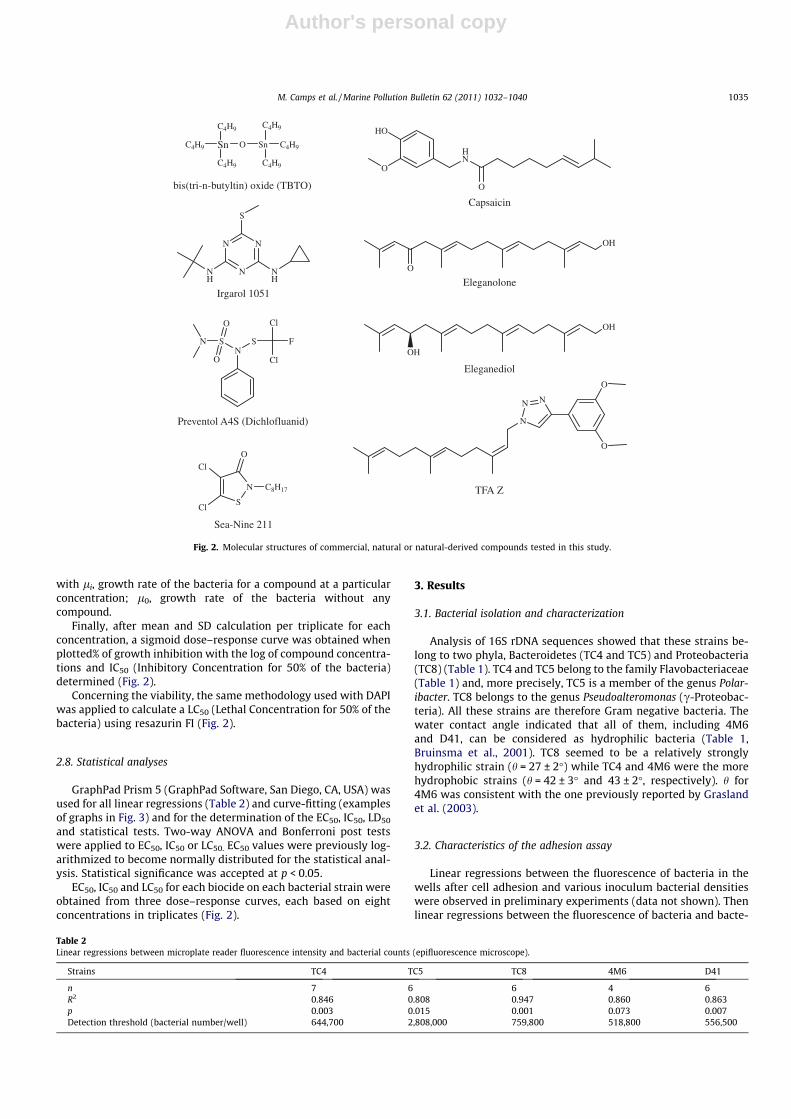
Sea-Nine 211� (SN) was used as received from the manufac-turer (Rohm and Haas SAS, Paris, France), bis(tri-n-butyltin) oxide(TBTO) came from Acros (Fisher Scientific, Illkirch, France), Irgarol1051� was purchased from CIBA (Lyon, France) and Preventol A4S�
(Dichlofluanid or DCF) from Lanxess (Courbevoie, France).Capsaicin, copper sulphate and resazurin were purchased from
Sigma–Aldrich. Eleganolone and eleganediol were extracted andpurified from the brown alga Bifurcaria bifurcata (Ortalo-Magnéet al., 2005). TFA-Z is a synthetic analogue of farnesol (Praud-Tabaries et al., 2009). These three last molecules have been synthe-sized and/or isolated in our laboratory.
2.2. Bacterial strains isolation and cultures
The D41 strain was provided by the IFREMER (Table 1). It wasisolated from a natural biofilm on Teflon coupons after 24 h ofimmersion in November 1998 in the Brest bay (St. Anne du Portzic,48�2103200N-4�3205800W, Atlantic Ocean) (Leroy et al., 2007). Thestrain 4M6 (Table 1) was provided by the LBCM (Université de Bre-tagne Sud). It was isolated on glass slides immersed during 6 h at1 m depth in March 2000 in the Morbihan Gulf (Bailleron Island,47�3403700N-2�4405400W, Atlantic Ocean) (Grasland et al., 2003)(Fig. 1).
TC4 (Strains from the Toulon Collection were denominated TCi),TC5 and TC8 (Table 1) were isolated from natural biofilms on 6 himmersed (1 m depth) polystyrene (PS) (TC4) and silicon coupons(TC5 and TC8) in February 2008 in the Toulon bay (43�0602300N-5�5701700E, Mediterranean sea, Fig. 1). Briefly, once in the labora-tory, these coupons were washed with sterile artificial sea water(ASW, Sigma–Aldrich) and scraped. Bacteria were isolated by suc-cessive subcultures on marine agar Petri dishes (Difco™, Sigma–Aldrich).
All the strains were transferred in the Vaatanen nine-salt solu-tion (VNSS, Mårdén et al., 1985) for liquid planktonic culture(20 �C, 120 rpm). Unlike Marine Broth for example, the VNSS is anon-turbid medium that allows minimizing the error usingOD600 nm to assess bacterial density.
2.3. Phylogenetic analyses
Bacterial DNA was extracted from 1.5 ml of overnight cultureusing QIAmp DNA Mini Kit (Qiagen GmbH, Hilden, Germany).
Table 1Strains characterization.
Strains TC4 TC5 TC8 4M6 D41
Taxonomic identification(16S rDNA)
Flavo-bacteriaceae(Flavobacteria)
Polaribacter sp.(Flavobacteria)
Pseudo-alteromonas sp.(c-Proteobacteria)
Paracoccus sp.(a-Proteobacteria)
Pseudo-alteromonas sp.(c-Proteobacteria)
Substrata of isolation PS Silicon Silicon Glass Teflon�
Water contact angle(h, �, n = 3)
42 ± 3 37 ± 3 27 ± 2 43 ± 2 35 ± 2
Fig. 1. Localization of the three French sites of bacterial isolation (Brest, Lorient andToulon).
M. Camps et al. / Marine Pollution Bulletin 62 (2011) 1032–1040 1033

Author's personal copy
The amplification of 16s rDNA was performed by PCR using TaqDNA polymerase from Invitrogen (Carlsbad, CA, USA) and the fol-lowing primers 16sF (50-AGAGTTTGATYMTGGCTCAG-30) and 16sR(50-CGGYTACCTTGTTACGAC-30) (Grasland et al., 2003). PCR prod-ucts (approximately 1500 bp) were cloned in TOPO TA CloningKit (Invitrogen). Plasmid DNA was extracted from five positivesclones with Qiagen Plasmid Mini Kit. Two plasmids were se-quenced for each strain (TC4, TC5 and TC8) by Genome Express(Cogenics, UK) using T7 Pro primers. Results were aligned to bacte-rial genome data bank using tblastn with default parameters fromNCBI website (http://blast.ncbi.nlm.nih.gov).
2.4. Water contact angle measurement
The hydrophobicity of the bacterial surface was determined bythe measurement of water contact angles (h) according to Bruinsmaet al. (2001) using a Digidrop apparatus (GBX Instruments, Bourg dePéage, France). Filters (0.22 lm) were saturated with bacterial cul-tures and five measurements were performed per filter. This proce-dure was applied on three different cultures for each strain.
2.5. Anti-adhesion assay
Bacterial strains were grown on VNSS at 20 �C under shakingconditions (120 rpm) and collected at the stationary phase. Aftercentrifugation, cells were suspended in sterile ASW (adapted fromLeroy et al., 2007). Bacterial density for the inocula was optimizedfor each strain to reach the highest fluorescence response butremaining in the range where fluorescence was proportional tothe bacterial density inoculated (between 0.2 and 0.4 of OD600 nm
in the wells depending on the strains). Microtiter plates (sterileblack PS; Nunc, Fisher Scientific, Illkirch, France) were filled as fol-lows: 100 lL at eight different concentrations in four replicates foreach tested compounds (standard biocides, natural or natural-derived products) were added. After a preliminary test at500 lM, the eight concentrations were chosen to range from100% to 0% of adhesion. All the concentrations were tested in trip-licate and the fourth well was filled for non-specific stainingcontrol (with the stain but without bacteria). The maximum per-centage of solvent(s) used for the dilution of biocides was alsotested as additional control. For the bacterial adhesion control,100 lL of ASW were added in six wells. Then 100 lL of the bacte-rial suspension were inoculated on all the wells except the border-row wells. The latters were filled to 200 lL with ASW and consti-tuted the non-specific staining control.
After an optimized time for adhesion (between 15 and 24 hdepending on the strain following kinetic studies; a shorter time(6–12 h) could have been chosen but involving automation or unu-sual working hours), the non-adhered bacteria were eliminated bythree successive washes (36 gL�1 sterile NaCl solution). The ad-hered bacteria were fixed for 90 min at 4 �C with 2% formalde-hyde-NaCl 36 g L�1 sterile solution. Staining was performed byadding 200 lL of a DAPI (Sigma–Aldrich) solution (4 lg ml�1 in a36 g L�1 NaCl sterile solution). After 20 min, the excess stain wasremoved by three washes (36 gL�1 NaCl solution). The DAPI wasthen solubilized with 200 lL of a 95% EtOH hydroalcoholic solution(15 min under shaking conditions). Fluorescence intensity (FI) wasmeasured (kexc = 380 nm, kem = 495 nm) using an Infinit 200 micro-plate fluorescence reader (Tecan, Lyon, France).
For molecules with a low solubility (SN and Preventol A4S�), theprotocol was slightly modified. Compounds were solubilized in100% MeOH and added in the wells (in triplicates at eight concen-trations). Evaporation was then performed under sterile conditionsleading to a coating of the well bottom with the molecule to betested. The above described protocol for the microtiter plate assay
was next carried out. A percent of adhesion was calculated perwell:
ðFIi� nsCiÞ=ðMean FIc�Mean BÞ � 100
with FIi, fluorescence intensity in a treated well (tested com-pound + bacteria + DAPI); FIc, fluorescence intensity in a controlwell (bacteria + DAPI); nsCi, non-specific control (tested compoundwithout bacteria + DAPI); B, blank, i.e. stain control (only DAPI).
After mean and standard deviations (SD) calculation per tripli-cate for each concentration, a sigmoid dose–response curve wasobtained when plotted% of adhesion with the log of compoundconcentrations and EC50 calculated (Fig. 2).
2.6. Bacterial count
To assess the bacterial density in the microtiter plate wells, acorrelation curve was achieved for each strain. Several densitiesof bacteria were seeded in 96 wells black plates with a clear PSbottom (Corning incorporated, Fisher Scientific). Once the bacte-ria have attached, the above protocol was performed exceptedfor the extraction with EtOH. Then, the microplates were ob-served with an inverted epifluorescence microscope (x400, LeicaDM1 4000B). Pictures (five per well and two wells per bacterialdensity) for at least five different bacterial densities were cap-tured with a camera (Leica DFC 420C). DAPI stained bacteriawere counted and expressed in bacteria per well. Extraction withEtOH was then carried out and FI determined with the multiwellplate reader as described above to correlate counts and FI afterextraction per well. We are aware that the presence of attachedbacteria on the side of the wells could have underestimated thenumber of bacteria related to fluorescence intensity. But thealternative would have been to correlate planktonic stained bac-teria to FI after extraction which overestimated the number ofbacteria related to FI because 100% of planktonic bacteria wouldnot attach on the wells. So we assume that it was more relevantto perform the count for the correlation in the real condition ofthe assay, i.e. after adhesion on the wells.
2.7. Bacterial growth inhibition and viability assays
Bacterial strains were grown on VNSS at 20 �C under shakingconditions (120 rpm) and collected during the exponential phase.After centrifugation, cells were suspended in sterile VNSS(OD600 nm = 0.1). 180 lL at eight concentrations for each testedcompounds (standard biocides, natural or natural-derived prod-ucts) were added in four wells of the microtiter plates (steriletransparent PS; Nunc, Fisher Scientific). All the concentrationswere tested in triplicate and the fourth well was filled for con-trol. The maximum percentage of solvent(s) used for the dilutionof biocides was also tested in triplicate as additional control. Forthe growth inhibition control, 180 lL of VNSS was added in sixwells. Then 20 lL of the bacterial suspension was inoculatedon all the wells except the border-row wells and all the wellswere filled out to 200 lL with VNSS. Turbidity (OD600 nm) wasmeasured every hour during 8 h. When the stationary phasewas reached, resazurin (20 lM) was added on all the wells andfluorescence was measured after two hours (kexc = 535 nm,kem = 595 nm) using the microplate fluorescence reader (follow-ing Duarte et al., 2009).
The growth rate l (h�1) was calculated during the exponentialphase for each strain, at each concentration: B = B0 elt, where Bis the bacterial density at time t, expressed as the OD, and B0 isdensity of the inoculum. A percent of growth inhibition was calcu-lated as follow:
ðli � l0=l0Þ � 100
1034 M. Camps et al. / Marine Pollution Bulletin 62 (2011) 1032–1040
Author's personal copy
with li, growth rate of the bacteria for a compound at a particularconcentration; l0, growth rate of the bacteria without anycompound.
Finally, after mean and SD calculation per triplicate for eachconcentration, a sigmoid dose–response curve was obtained whenplotted% of growth inhibition with the log of compound concentra-tions and IC50 (Inhibitory Concentration for 50% of the bacteria)determined (Fig. 2).
Concerning the viability, the same methodology used with DAPIwas applied to calculate a LC50 (Lethal Concentration for 50% of thebacteria) using resazurin FI (Fig. 2).
2.8. Statistical analyses
GraphPad Prism 5 (GraphPad Software, San Diego, CA, USA) wasused for all linear regressions (Table 2) and curve-fitting (examplesof graphs in Fig. 3) and for the determination of the EC50, IC50, LD50
and statistical tests. Two-way ANOVA and Bonferroni post testswere applied to EC50, IC50 or LC50. EC50 values were previously log-arithmized to become normally distributed for the statistical anal-ysis. Statistical significance was accepted at p < 0.05.
EC50, IC50 and LC50 for each biocide on each bacterial strain wereobtained from three dose–response curves, each based on eightconcentrations in triplicates (Fig. 2).
3. Results
3.1. Bacterial isolation and characterization
Analysis of 16S rDNA sequences showed that these strains be-long to two phyla, Bacteroidetes (TC4 and TC5) and Proteobacteria(TC8) (Table 1). TC4 and TC5 belong to the family Flavobacteriaceae(Table 1) and, more precisely, TC5 is a member of the genus Polar-ibacter. TC8 belongs to the genus Pseudoalteromonas (c-Proteobac-teria). All these strains are therefore Gram negative bacteria. Thewater contact angle indicated that all of them, including 4M6and D41, can be considered as hydrophilic bacteria (Table 1,Bruinsma et al., 2001). TC8 seemed to be a relatively stronglyhydrophilic strain (h = 27 ± 2�) while TC4 and 4M6 were the morehydrophobic strains (h = 42 ± 3� and 43 ± 2�, respectively). h for4M6 was consistent with the one previously reported by Graslandet al. (2003).
3.2. Characteristics of the adhesion assay
Linear regressions between the fluorescence of bacteria in thewells after cell adhesion and various inoculum bacterial densitieswere observed in preliminary experiments (data not shown). Thenlinear regressions between the fluorescence of bacteria and bacte-
HO
O
HN
O
NS S
Cl
Cl
FN
O
O
Eleganolone
Eleganediol
Capsaicin
Preventol A4S (Dichlofluanid)
S
N
O
Cl
Cl
C8H17
Sea-Nine 211
N
NN
NH
S
NH
Irgarol 1051
TFA Z
Sn O Sn C4H9
C4H9
C4H9C4H9
C4H9
C4H9
bis(tri-n-butyltin) oxide (TBTO)
N
NN
O
O
O
OH
OH
OH
Fig. 2. Molecular structures of commercial, natural or natural-derived compounds tested in this study.
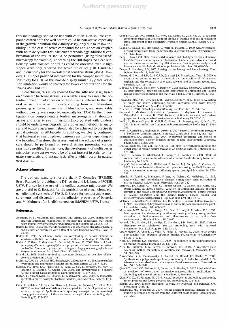
Table 2Linear regressions between microplate reader fluorescence intensity and bacterial counts (epifluorescence microscope).
Strains TC4 TC5 TC8 4M6 D41
n 7 6 6 4 6R2 0.846 0.808 0.947 0.860 0.863p 0.003 0.015 0.001 0.073 0.007Detection threshold (bacterial number/well) 644,700 2,808,000 759,800 518,800 556,500
M. Camps et al. / Marine Pollution Bulletin 62 (2011) 1032–1040 1035

Author's personal copy
rial count under fluorescence microscopy – both from the samewells – gave R2 values higher than 0.8 (Table 2). The quantitativethreshold for the fluorescence microplate reader in our protocolcan be deduced from these curves (x = 0, Table 2). The sensitivityof the method ranged from approximately 518 800 (4M6) to 2808 000 (TC5) bacteria per well depending on the strains.
The reproducibility of in vitro adhesion in PS microplates rangedbetween 99% and 53% depending on the strain: D41 (99%,n = 55) > TC8 (91%, n = 38) > 4M6 (71%, n = 34) and TC5 (70%,n = 33) > TC4 (53%, n = 35).
Validation conditions for the assay were the following ones: (i)ratio between the fluorescence adhesion controls and fluorescenceof non-specific staining controls (biocide and stain without bacte-ria) should be higher than 2, (ii) goodness of dose–response curvefit (R2) should be higher than 0.7.
SD of percent of adhesion for each concentration in a samemicrotiter plate (repeatability, data not shown except examples
in Fig. 3) were low. SD for EC50 (reproducibility, Table 3) were alsorather low, especially for EC50 lower than 200 lM. However, itshould be noticed that high SD were often observed for Hill slopes(data not shown). Consequently, but also in order to make theirinterpretation easier, Hill slope values were gathered into 3 maingroups: high when the value was lower than �5, low when thevalue was higher than �2 and median between these two values(Table 3). Taken together, these data showed that this antifoulingbioassay was reliable and allowed the evaluation and the compar-ison of the different adhesion responses for the five bacterialstrains tested.
3.3. Adhesion assay results
Responses to the adhesion assay appeared to be dependant onboth the strains and the tested compounds (Table 3, Fig. 4, two-way ANOVA p < 0.0001). The EC50 were distributed in a wide range,
Fig. 3. Examples of sigmoid dose–response curves obtained with GraphPad Prism� and used for the calculation of EC50, IC50 and LC50. (a) Adhesion assay TBTO_TC5[EC50 = 12.9 lM, Hill slope = �5.4 (high), R2 = 0.96, n = 8], TFA-Z_TC8 [EC50 = 64.3 lM, Hill slope = �0.8 (low), R2 = 0.98, n = 10]. (b) Growth inhibition assay TBTO_D41[IC50 = 4.4 lM, R2 = 0.99, n = 8], TBTO_TC5 [IC50 = 0.1 lM, R2 = 0.99, n = 8]. (c) Viability assay TBTO_D41 [LC50 = 5.0 lM, R2 = 0.99, n = 8], TBTO_TC5 [LC50 = 0.3 lM, R2 = 0.99, n = 8].
Table 3Effect of biocides and natural or natural-derived products on the bacterial adhesion n = 4 for TBTO and n = 3 for the other molecules tested R2 corresponds to the fitting quality forthe dose–response curve (>0.7). For the Hill slopes, ‘‘high’’ corresponded to a value lower than �5, ‘‘low’’ a value higher than �2 and median an intermediate value.
TC4 TC5 TC8 4M6 D41
TBTO EC50 (lM) 17.1 ± 6.7 9.6 ± 5.5 12.1 ± 3.1 15.4 ± 7.5 13.9 ± 4.0Hill slope low low high high lowR2 0.88 ± 0.04 0.90 ± 0.12 0.95 ± 0.01 0.85 ± 0.13 0.72 ± 0.17
Sea Nine� (SN) EC50 (lM) >500* 3.5 ± 3.7* 13.2 ± 5.2* 0.9 ± 0.5* 69.6 ± 31.2*
Hill slope low low high lowR2 0.89 ± 0.16 0.87 ± 0.15 0.97 ± 0.04 0.90 ± 0.07
Preventol� (A4S) EC50 (lM) >500 >500 >500 45.4 ± 13.1* >500Hill slope lowR2 0.82 ± 0.19
CuSO4 EC50 (lM) >500 299.8 ± 100.2 >500 14.8 ± 9.3 >500Hill slope low highR2 0.89 ± 0.11 0.76 ± 0.04
Capsaicin EC50 (lM) >500 >500 >500 >500 >500
Irgarol� EC50 (lM) >500 >500 >500 >500 >500
Eleganolone EC50 (lM) >500 58.4 ± 1.7 427.7 ± 75.0 40.3 ± 13.5 174.0 ± 56.1Hill slope median median median medianR2 0.99 ± 0.00 0.77 ± 0.13 0.98 ± 0.02 0.96 ± 0.02
Eleganediol EC50 (lM) >500 63.5 ± 8.3 482.0 ± 62.4 103.9 ± 12.8 224.0 ± 65.6Hill slope low median median medianR2 0.93 ± 0.07 0.95 ± 0.02 0.96 ± 0.03 0.82 ± 0.05
TFA-Z EC50 (lM) 79.0 ± 14.9 63.5 ± 5.6 139.0 ± 69.3 71.3 ± 1.7 63.4 ± 19.1Hill slope low low low low lowR2 0.90 ± 0.1 0.98 ± 0.02 0.90 ± 0.05 0.75 ± 0.14 0.84 ± 0.10
* Molecules tested using the ‘‘coating’’ protocol.
1036 M. Camps et al. / Marine Pollution Bulletin 62 (2011) 1032–1040

Author's personal copy
with values from 0.9 ± 0.5 (for SN against the 4M6 strain of Para-coccus sp.) to higher than 500 lM, which were considered asnon-active concentrations.
When looking at biocide effect one by one, TBTO showed no sig-nificant differences with high activities (EC50 < 17.1 ± 6.7 lM) andTFA-Z no significant differences with good activities (EC50 in therange between 63.4 ± 19.1 lM and 79.0 ± 14.9 lM except forTC8) on all strains (p > 0.05). SN was highly active(EC50 < 13.2 ± 5.2) but only on three strains (TC5, TC8 and 4M6).Eleganolone and Eleganediol showed a good activity on Paracoccussp. (4M6) and Polaribacter sp. (TC5) (EC50 in the range between40.3 ± 13.5 lM and 103.9 ± 12.8 lM) but a low efficacy againstthe three other strains. These two biocides only exhibited signifi-cant difference for the 4M6 (p < 0.05). Irgarol and capsaicin hadno effect while Preventol and copper sulphate showed significantactivities only on one strain (4M6).
Considering all the biocides tested, each strain displayed highdifferences of sensitivity. When Irgarol and capsaicin (non-activebiocides for all the strains) were excluded (Fig. 4), the inhibitionof the adhesion could be observed with all other biocides onlyfor the 4M6 strain. For this strain, SN, TBTO and copper sulphatecould be considered as very active (EC50 < 20 lM). In contrast,the TC4 strain belonging to the Flavobacteriaceae family was onlysensitive to TBTO (17.1 ± 6.7 lM) and TFA-Z (79.0 ± 14.9 lM). The
other strains exhibited intermediate profiles. The TC5 strain, aPolaribacter sp., was sensitive to all the biocides except Preventolwith a high activity for SN and TBTO. Pseudoalteromonas spp.(D41 and TC8) were both sensitive to all the biocides except Pre-ventol and copper sulphate but with different intensities. EC50 dis-played extremely significant difference for SN and significantdifferences for eleganolone (respectively p < 0.001 and p < 0.05).
Considering the hill slopes, we could mostly observed low val-ues except for the 4M6 strain (66% of high values). No general ten-dency could be highlighted when we studied the strainindividually. Taking biocides individually, SN (except for 4M6),TFA-Z, copper sulphate and Preventol showed always low Hillslopes whereas eleganediol and eleganolone displayed medianones (except in one case for eleganediol with the TC5). Finally,the three types of Hill slopes (high, median and low) were ob-served for the TBTO in quite equal proportions.
3.4. Growth inhibition and viability assay results
Assays were achieved with compounds that displayed a positiveanti-adhesion effect (TBTO, SN, eleganolone, eleganediol and TFA-Z) on three strains. These strains appeared as the most sensitiveones after the adhesion assays: 4M6, TC5 and D41. IC50 and LC50
obtained were shown in Table 4. Eleganolone, eleganediol andTFA-Z exhibited no growth inhibition or toxicity. In contrast, TBTOand SN have strong growth inhibitory effects associated with ahigh toxicity on the three bacterial strains (IC50 andLC50 < 5.1 ± 0.5 lM for TBTO and between 5.2 ± 1.9 and10.3 ± 1.8 lM for SN). For both IC50 and LC50, no significant differ-ence could be observed between the three strains (p > 0.05, Two-way ANOVA) but significant differences were observed betweenTBTO and SN (p < 0.001, Two-way ANOVA). Hill slopes were allrather high (data not shown).
4. Discussion
The aims of this study were to evaluate the reliability of thisadapted bioassay developed in our laboratory. Then, we used it to as-sess commercial vs. natural and natural-derived products anti-adhesion activities and to determine the variability of the responsefor five ‘‘pioneer’’ marine bacterial strains which were isolated fromthree different locations, four substrata and which belonged to fourdistinct genera. As one of the main objective for our laboratory is theidentification of non-toxic compounds with anti-adhesion proper-ties against marine biofilms, the choice of a specific adhesion assayusing ASW to prevent the growth of bacteria (Leroy et al., 2007) ap-peared relevant. However, the inhibition of adhesion may resultfrom two mechanisms, i.e. a specific inhibition of the adhesion pro-cess or a consequence of toxicity towards the strain(s). The discrim-ination between these two modes of action was realized usinggrowth inhibition and viability assays for the molecules whichshowed an anti-adhesion potential. TBTO, SN, Irgarol, copper sul-phate and Preventol are commonly used as antifoulants in AF coat-ings (e.g. Konstantinou and Albanis, 2004) but some of them arenow totally (TBTO) or partially banned (Irgarol). Concerning naturalcompounds, eleganolone and eleganediol, two acyclic diterpenesbiosynthesized abundantly by a common Atlantic brown alga(Culioli et al., 1999; Ortalo-Magné et al., 2005), were selected dueto their AF potential (bacterial growth inhibition and barnacleanti-adhesion assays, Hellio et al., 2001; Maréchal et al., 2004).TFA-Z has been previously selected within a library of synthetic ana-logues in our laboratory (Praud-Tabaries et al., 2009).
Overall, the results obtained with the adhesion assay wererepetitive and reproducible. Organisms (i.e. clonal populations)based assays displayed of course a significant variability. Therefore,
Preventol
Copper sulphate
Eleganediol
Eleganolone
TFA ZSea Nine
TBTO
TC4
TC8
D41
TC5
4M6
0
50
100
150
200
250
300
350
400
450
500
EC
50
(µM
)
Fig. 4. Adhesion response (EC50) for the five strains with all the biocides.
Table 4Growth inhibition (IC50) and viability (LC50) on 3 bacterial strains for biocides withanti-adhesion activities.
TC5 4M6 D41
TBTO (n = 3) IC50 (lM) 0.2 ± 0.1 2.1 ± 1.5 4.2 ± 0.3R2 0.95 ± 0.06 0.97 ± 0.01 0.96 ± 0.03LC50 (lM) 0.2 ± 0.1 2.1 ± 0.8 5.1 ± 0.5R2 0.99 ± 0.00 0.99 ± 0.01 0.99 ± 0.00
Sea Nine� (n = 3) IC50 (lM) 10.3 ± 1.8 7.9 ± 1.1 5.9 ± 1.5R2 0.97 ± 0.02 0.97 ± 0.02 0.97 ± 0.03LC50 (lM) 6.9 ± 3.4 7.2 ± 0.8 5.2 ± 1.9R2 0.98 ± 0.01 0.99 ± 0.00 0.98 ± 0.02
Eleganolone (n = 2) IC50 (lM) >400 >400 >400LC50 (lM) >400 >400 >400
Eleganediol (n = 2) IC50 (lM) >300 >300 >300LC50 (lM) >300 >300 >300
TFA-Z (n = 2) IC50 (lM) >300 >300 >300LC50 (lM) >300 >300 >300
M. Camps et al. / Marine Pollution Bulletin 62 (2011) 1032–1040 1037

Author's personal copy
positive adhesion and anti-adhesion controls were required. Thecalculation of EC50 allowed an easy discrimination between bio-cides. Considering its wide toxicity spectra and history of favouredantifoulant for hull ship (Yebra et al., 2004), TBTO was foundunsurprisingly to be the most powerful biocide against all thestrains. Our study also showed that SN, which displays a broad-spectrum activity on marine foulers including bacteria, was anactive biocide. But these two biocides showed toxic effects on bac-teria. Non-commercial TFA-Z, and in a lesser extent eleganoloneand eleganediol, displayed remarkable results with quite goodanti-adhesion activities associated with no toxic effects. Thesecould be illustrated calculating the ‘‘therapeutic ratio’’ (=LC50/EC50, Rittschof, 1999). TBTO and SN exhibited a therapeutic ratioaround 1 (toxic compounds, data not shown) depending on thestrains. Unfortunately, as precise LC50 could not be determinedfor eleganolone, eleganediol and TFA-Z (>300 lM), their therapeu-tic ratio could not be calculated. But as no toxicity could be ob-served at 300 or 400 lM, we can assume a minimal LC50 of500 lM. Then therapeutic ratio would be around 10 for TFA-Z(non-toxic compounds). For eleganolone and eleganediol, the ratiowould be more difficult to interpret as wide variations dependingon the strains could be observed (data not shown). If AF bioassaytargeted on other fouling species (especially macroorganisms) isrequired to complete the TFA-Z AF profile, algal extracts includingeleganolone and eleganediol were already reported as active in theprevention of barnacle adhesion (Maréchal et al., 2004). In con-trast, the herbicide Irgarol and the fungicide Preventol unsurpris-ingly exhibited no toxicity against bacteria (Fernández-Albaet al., 2002; Konstantinou and Albanis, 2004; Zhou et al., 2006). De-spite its broad toxicity, no actual activity of copper sulphate wasobserved except for one strain (4M6). As cupric ion is the maintoxic form, theses results could be due to copper speciation thatwas not controlled during the assays. 4M6 already exhibited great-er sensitivity to other biocides which could be related to specificcell surface interaction or membrane permeability and could alsoexplain the higher sensitivity to copper sulphate.
Finally, capsaicin, a capsinoid isolated from chili peppers (Cap-sicum spp.), was reported to show in situ AF properties in freshwa-ter systems against bacteria as well as mussels (Xu et al., 2005;Angarano et al., 2007). These activities were not in agreement withour findings. This result could be explained as a putative highersensitivity of freshwater bacteria or an expression of the diversityin the sensitivity among bacteria.
Few bacterial strains have been used in marine adhesion assays:Vibrio harveyi (Mittelman et al., 1993), Pseudomonas sp. (Becker,1996, 1998), Pseudoalteromonas sp. (D41, Leroy et al., 2007), whichwas also used in this study, Cobetia marina, Cytophaga lytica,Halomonas pacifica, Pseudoalteromonas atlantica (Stafslien et al.,2006 and related papers), and Halomonas pacifica (Melanderet al., 2009). Except C. lytica (Bacteroidetes), all these bacteria be-long to the c-Proteobacteria. Concerning the strains used in thiswork, TC4 and TC5 belonged to the same family in the Bacteroide-tes (Flavobacteriaceae) which is distinct from C. lytica family (Cyto-phagaceae). The three other strains belong to the Rhodobactergroup (a-Proteobacteria, 4M6) or the c-Proteobacteria (TC8 andD41, same genus Pseudoalteromonas). Phylogenetic affiliations ofstrains used for anti-adhesion assays, including ours, were coher-ent with recent in situ descriptions of marine pioneer bacterialcommunities on artificial surfaces by culture-independent tech-niques (fingerprinting and cloning-sequencing methods). Theyshowed that the predominance by c-Proteobacteria during earlystage of biofilm formation was followed by secondary stages dom-inated by the Rhodobacter group (a-Proteobacteria). Strains fromthe Bacteroidetes group were also reported during pioneer stagesbut always in weak proportions (Dang and Lovell, 2002; Joneset al., 2007; Lee et al., 2008; Chung et al., 2010).
Overall, sensitivity of the strains seemed to vary. The 4M6exhibited low EC50 for almost all the biocides whereas the adhe-sion of TC4 was only inhibited by two of them (TBTO and TFA-Z).Not surprisingly, the sensitivity to a biocide was strain-dependant.The only differences between 4M6 and TC5 were related to SN, Pre-ventol (p < 0.001) and copper sulphate (p < 0.001). The last twocompounds showed no activities for TC5 on the contrary to 4M6.To discriminate between the two Pseudoalteromonas strains D41and TC8, the activities of SN (p < 0.001) and eleganolone(p < 0.05) could be compared. Finally, SN was the only compoundwhich showed a marked dissimilarity of activity between TC8and TC4 (p < 0.001). Furthermore, TBTO and TFA-Z showed thesame activity for all the strains (p > 0.05), TBTO being more activethan TFA-Z. Furthermore, no clear relationship could be observedbetween EC50 and geographical origins, hydrophilicity or adhesionreproducibility in vitro. In the literature, only one study had in-cluded a strain sensitivity comparison in the description of a mar-ine adhesion bioassay. Regarding the different bacteria used toevaluate coatings containing SN, C. marina consistently demon-strated a higher tolerance to all coating compositions evaluatedwhen compared to H. pacifica and P. atlantica (Stafslien et al.,2007b). Differences in strain sensitivity for biocides were also re-ported for oral pathogen biofilms (e.g. Bodet et al., 2008) or biofilmdisinfection (e.g. Pitts et al., 2003). In addition to possible differ-ences in the protocol (e.g. growth and adhesion or only adhesion,strains used), marine adhesion assays were only applied to coat-ings (Stafslien et al., 2006 and related articles), industrial cleaningagents (Leroy et al., 2007), enzymes (Leroy et al., 2008) or non-commercial natural products (Melander et al., 2009).
Concerning technical aspect of the assay, the reproducibility ofin vitro adhesion in the wells, i.e. not to mention biocides activities,was another expression of the diversity of the response of thestrains. Neither the hydrophobicity of substrata of isolation or ofbacteria (h), nor the phylogenetic affiliation could explain this re-sult. In the same way, the hydrophilicity of the strains was neitherrelated to their substrata of isolation nor to their phylogeneticcharacterization. The two strains that belong to the same genus(TC8 and D41), as well as the two strains isolated from the samesurface (TC5 and TC8 from silicon coupons), displayed dissimilarhydrophilicity and adhesion properties. Furthermore, no informa-tion on the strain sensitivity to molecules could be correlated withtheir intrinsic characteristics including adhesion.
Significant linear relationships between fluorescence and directbacterial count were reported for all the strains (Table 2) indicatingthat the method can be applied for the estimation of bacterial den-sity. The sensitivity for the detection of the strains varied in therange, which appeared to us not too wide considering the precisionof microscope counts (between 417 500 (4M6) and 2 808 000 (TC5)bacteria per well). The detection threshold for the D41 strain wasfirst reported in microtiter plate by Leroy et al. (2007) and wasaround 3.75 � 107 attached cells per well. Nevertheless, theirmethod was slightly different from ours (one part of the DAPI-stained bacterial suspension was filtered onto a 0.22 lm black fil-ter and counted under microscope, the other part was settled in themicrotiter plate to give the fluorescence intensity per well). As thenumber of attached cells should be lower than the inoculated onein wells, our lower sensitivity threshold did not appear to be incon-sistent. Notwithstanding this validation point, the choice of alter-native stain (e.g. SYTO 13, D’Souza et al., 2010 or SYTO 61, datanot shown) to eliminate the DAPI extraction with EtOH and to al-low measuring only the fluorescence on the bottom of the well willbe of great interest, especially for coatings assessment in multiwellplates.
It should be noticed that lower EC50 occurred when biocideswere pre-coated in the wells (the comparison between the twoprocedures was done for the TBTO, data not shown). In addition,
1038 M. Camps et al. / Marine Pollution Bulletin 62 (2011) 1032–1040
Author's personal copy
this methodology should be use with caution. Non-soluble com-pound coated onto the well bottom could be non-active, especiallyin the growth inhibition and viability assay, only due to its low sol-ubility. In the case of active compound for anti-adhesion coupledwith no toxicity with this particular methodology, additional con-firmation of the results should be performed (using ‘‘Live/Dead’’microscopy for example). Concerning the Hill slopes, no clear rela-tionship with biocides or strains could be observed even if highslopes were only reported for active molecules (TBTO and SN)and in our study for the overall most sensitive strain (4M6). How-ever, Hill slopes provided information for the comparison of strainsensitivity for TBTO as this biocide display similar EC50: total adhe-sion inhibition would be reached for lower concentrations for thestrains 4M6 and TC8.
In conclusion, this study showed that the adhesion assay basedon ‘‘pioneer’’ bacterial strains is a reliable assay to assess the po-tential prevention of adhesion of these strains. Relative to the nat-ural or natural-derived products coming from our laboratory,promising activities on marine biofilm bacteria, and remarkablywithout toxicity, were reported especially for TFA-Z. Further inves-tigations on complementary fouling macroorganisms laboratoryassays and after in situ immersions (incorporated with binders)should be undertaken. Degradation studies and metabolites analy-ses and toxicity assessment should also be achieved to precise itsactual potential as AF biocide. In addition, we clearly confirmedthat bacterial strains displayed various sensitivities depending onthe biocides. Therefore, the assessment of the efficacy of a mole-cule should be performed on several strains presenting varioussensitivity profiles. Furthermore, the development of multispeciesmicrotiter plate assays would be of great interest in order to inte-grate synergistic and antagonistic effects which occur in naturalecosystems.
Acknowledgments
The authors want to sincerely thank C. Compère (IFREMER,Brest, France) for providing the D41 strain and J.-L. Jamet (PROTEE,USTV, France) for the use of the epifluorescence microscope. Weare grateful to O. Bottzeck for the purification of eleganolone, ele-ganediol and synthesis of TFA-Z, C. Bressy for contact angle mea-surements and discussion on the adhesion properties of bacteriaand M. Molmeret for English correction (MAPIEM, USTV, France).
References
Angarano, M.-B., McMahon, R.F., Hawkins, D.L., Schetz, J.A., 2007. Exploration ofstructure-antifouling relationships of capsaicin-like compounds that inhibitzebra mussel (Dreissena polymorpha) macrofouling. Biofouling 23, 295–305.
Becker, K., 1996. Exopolysaccharide production and attachment strength of bacteriaand diatoms on substrates with different surface tensions. Microbial. Ecol. 32,23–33.
Becker, K., 1998. Detachment studies on microfouling in natural biofilms onsubstrata with different surface tensions. Int. Biodeter. Biodegr. 41, 93–100.
Bodet, C., Epifano, F., Genovese, S., Curini, M., Grenier, D., 2008. Effects of 3-(40-geranyloxy-30-methoxyphenyl)-2-trans propenoic acid and its ester derivativeson biofilm formation by two oral pathogens, Porphyromonas gingivalis andStreptococcus mutans. Eur. J. Med. Chem. 43, 1612–1620.
Briand, J.F., 2009. Marine antifouling laboratory bioassays, an overview of theirdiversity. Biofouling 25, 297–311.
Bruinsma, G.M., van der Mei, H.C., Busscher, H.J., 2001. Bacterial adhesion to surfacehydrophilic and hydrophobic contact lenses. Biomaterials 22, 3217–3224.
Burgess, J.G., Boyd, K.G., Armstrong, E., Jiang, Z., Yan, L., Berggren, M., May, U.,Pisacane, T., Granmo, Å., Adams, D.R., 2003. The development of a marinenatural product-based antifouling paint. Biofouling 19, 197–205.
Burton, E., Yakandawala, N., LoVetri, K., Madhyastha, M.S., 2007. A microplatespectrofluorometric assay for bacterial biofilms. J. Ind. Microbiol. Biotech. 34, 1–4.
Cassé, F., Stafslien, S.J., Bahr, J.A., Daniels, J., Finlay, J.A., Callow, J.A., Callow, M.E.,2007. Combinatorial materials research applied to the development of newsurface coatings V. Application of a spinning water-jet for the semi-highthroughput assessment of the attachment strength of marine fouling algae.Biofouling 23, 121–130.
Chung, H.C., Lee, O.O., Huang, Y.L., Mok, S.Y., Kolter, R., Qian, P.Y., 2010. Bacterialcommunity succession and chemical profiles of subtidal biofilms in relation tolarval settlement of the polychaete Hydroides elegans. I.S.M.E. Journal 4, 817–828.
Culioli, G., Daoudi, M., Mesguiche, V., Valls, R., Piovetti, L., 1999. Geranylgeraniol-derived diterpenoids from the brown alga Bifurcaria bifurcata. Phytochemistry52, 1447–1454.
Dang, H., Lovell, C.R., 2002. Numerical dominance and phylotype diversity of marineRhodobacter species during early colonization of submerged surfaces in coastalmarine waters as determined by 16S ribosomal DNA sequence analysis andfluorescence in situ hybridization. Appl. Environ. Microbiol. 68, 496–504.
DeNys, R., Steinberg, P.D., 2002. Linking marine biology and biotechnology. Curr.Opinion Biotech. 13, 244–248.
Duarte, M., Giordani, R.B., Carli, G.A.D., Zuanazzi, J.A., Macedo, A.J., Tasca, T., 2009. Aquantitative resazurin assay to determinate the viability of Trichomonasvaginalis and the cytotoxicity of organic solvents and surfactant agents. Exp.Parasitol. 123, 195–198.
D’Souza, F., Bruin, A., Biersteker, R., Donnelly, G., Klijnstra, J., Rentrop, C., Willemsen,P., 2010. Bacterial assay for the rapid assessment of antifouling and foulingrelease properties of coatings and materials. J. Ind. Microbiol. Biotech. 37, 363–370.
Fernández-Alba, A.R., Hernando, M.D., Piedra, L., Chisti, Y., 2002. Toxicity evaluationof single and mixed antifouling biocides measured with acute toxicitybioassays. Anal. Chim. Acta 456, 303–312.
Fusetani, N., 2004. Biofouling and antifouling. Nat. Prod. Rep. 21, 94–104.Grasland, B., Mitalane, J., Briandet, R., Quemener, E., Meylheuc, T., Linossier, I.,
Vallee-Rehel, K., Haras, D., 2003. Bacterial biofilm in seawater, cell surfaceproperties of early-attached marine bacteria. Biofouling 19, 307–313.
Hellio, C., Thomas-Guyon, H., Culioli, G., Piovetti, L., Bourgougnon, N., Le Gal, Y.,2001. Marine antifoulants from marine brown macroalgae. Biofouling 17, 189–201.
Jones, P., Cottrell, M., Kirchman, D., Dexter, S., 2007. Bacterial community structureof biofilms on artificial surfaces in an estuary. Microbial. Ecol. 53, 153–162.
Konstantinou, I.K., Albanis, T.A., 2004. Worldwide occurrence and effects ofantifouling paint booster biocides in the aquatic environment: a review.Environ. Int. 30, 235–248.
Lee, J.W., Nam, J.H., Kim, Y.H., Lee, K.H., Lee, D.H., 2008. Bacterial communities in theinitial stage of marine biofilm formation on artificial surfaces. J. Microbiol. 46,174–182.
Leroy, C., Delbarre, C., Ghillebaert, F., Compere, C., Combes, D., 2008. Effects ofcommercial enzymes on the adhesion of a marine biofilm-forming bacterium.Biofouling 24, 11–22.
Leroy, C., Delbarre-Ladrat, C., Ghillebaert, F., Rochet, M.J., Compère, C., Combes, D.,2007. A marine bacterial adhesion microplate test using the DAPI fluorescentdye, a new method to screen antifouling agents. Lett. Appl. Microbiol. 44, 372–378.
Mårdén, P., Tunlid, A., Malmcrona-Friberg, K., Odham, G., Kjelleberg, S., 1985.Physiological and morphological changes during short term starvation ofmarine bacterial isolates. Arch. Microbiol. 142, 326–332.
Maréchal, J.P., Culioli, G., Hellio, C., Thomas-Guyon, H., Callow, M.E., Clare, A.S.,Ortalo-Magné, A., 2004. Seasonal variation in antifouling activity of crudeextracts of the brown alga Bifurcaria bifurcata (Cystoseiraceae) against cypridsof Balanus amphitrite and the marine bacteria Cobetia marina andPseudoalteromonas haloplanktis. J. Exp. Mar. Biol. Ecol. 313, 47–62.
Melander, C., Moeller, P.D.R., Ballard, T.E., Richards, J.J., Huigens III, R.W., Cavanagh,J., 2009. Evaluation of dihydrooroidin as an antifouling additive in marine paint.Int. Biodeter. Biodegr. 63, 529–532.
Mittelman, M.W., Packard, J., Arrage, A.A., Bean, S.L., Angell, P., White, D.C., 1993.Test systems for determining antifouling coating efficacy using on-linedetection of bioluminescence and fluorescence in a laminar-flowenvironment. J. Microbiol. Meth. 18, 51–60.
Nylund, G.M., Gribben, P.E., de Nys, R., Steinberg, P.D., Pavia, H., 2007. Surfacechemistry versus whole-cell extracts, antifouling tests with seaweedmetabolites. Mar. Ecol. Prog. Ser. 329, 73–84.
Ortalo-Magné, A., Culioli, G., Valls, R., Pucci, B., Piovetti, L., 2005. Polar acyclicditerpenoids from Bifurcaria bifurcata (Fucales, Phaeophyta). Phytochemistry66, 2316–2323.
Piola, R.F., Dafforn, K.A., Johnston, E.L., 2009. The influence of antifouling practiceson marine invasions. Biofouling 25, 633–644.
Pitts, B., Hamilton, M.A., Zelver, N., Stewart, P.S., 2003. A microtiter-platescreening method for biofilm disinfection and removal. J. Microbiol. Meth.54, 269–276.
Praud-Tabaries, A., Dombrowsky, L., Bottzek, O., Briand, J.F., Blache, Y., 2009.Synthesis of a polyprenyl-type library containing 1, 4-disubstituted-1, 2, 3-triazoles with anti-biofilm activities against Pseudoalteromonas sp. TetrahedronLett. 50, 1645–1648.
Qian, P.Y., Lau, S.C.K., Dahms, H.U., Dobretsov, S., Harder, T., 2007. Marine biofilmsas mediators of colonization by marine macroorganisms, implications forantifouling and aquaculture. Mar. Biotechnol. 9, 399–410.
Qian, P.Y., Xu, Y., Fusetani, N., 2010. Natural products as antifouling compounds,recent progress and future perspectives. Biofouling 26, 223–234.
Railkin, A.I., 2004. Marine Biofouling: Colonization Processes and Defenses. CRCPress, Boca Raton, FL.
Ramasamy, M.S., Murugan, A., 2007. Fouling deterrent chemical defence in threemuricid gastropod egg masses from the Southeast coast of India. Biofouling 23,259–265.
M. Camps et al. / Marine Pollution Bulletin 62 (2011) 1032–1040 1039

Author's personal copy
Rittschof, D., 1999. Fouling and natural products as antifoulants. In: Fingerman, M.,Nagabhushanam, R., Thompson, M.-F. (Eds.), Recent Advances in MarineBiotechnology. Oxford and IBH Publishing Co. Pvt. Ltd., New Delhi andCalcutta, pp. 245–257.
Rudney, J.D., Staikov, R.K., 2002. Simultaneous measurement of the viability, aggregation,and live and dead adherence of Streptococcus crista, Streptococcus mutans andActinobacillus actinomycetemcomitans in human saliva in relation to indices of caries,dental plaque and periodontal disease. Arch. Oral Biol. 47, 347–359.
Shakeri, S., Kermanshahi, R.K., Moghaddam, M.M., Emtiazi, G., 2007. Assessment ofbiofilm cell removal and killing and biocide efficacy using the microtiter platetest. Biofouling 23, 79–86.
Stafslien, S., Bah, J., Daniels, J., Wal, L., Nevins, J., Smith, J., Schiele, K., Chisholm, B., 2007a.Combinatorial materials research applied to the development of new surface coatingsVI: an automated spinning water jet apparatus for the high-throughputcharacterization of fouling-release marine coatings. Rev. Sci. Instrum. 78, 1–6.
Stafslien, S., Daniels, J., Chisholm, B., Christianson, D., 2007b. Combinatorialmaterials research applied to the development of new surface coatings III.Utilisation of a high-throughput multiwell plate screening method to rapidlyassess bacterial biofilm retention on antifouling surfaces. Biofouling 23, 37–44.
Stafslien, S., Daniels, J., Mayo, B., Christianson, D., Chisholm, B., Ekin, A., Webster, D.,Swain, G., 2007c. Combinatorial materials research applied to the developmentof new surface coatings IV. A high-throughput bacterial biofilm retention andretraction assay for screening fouling-release performance of coatings.Biofouling 23, 45–54.
Stafslien, S.J., Bahr, J.A., Feser, J.M., Weisz, J.C., Chisholm, B.J., Ready, T.E., Boudjouk,P., 2006. Combinatorial materials research applied to the development of newsurface coatings I, a multiwell plate screening method for the high-throughputassessment of bacterial biofilm retention on surfaces. J. Comb. Chem. 8, 156–162.
Vesterlund, S., Paltta, J., Karp, M., Ouwehand, A.C., 2005. Measurement of bacterialadhesion – in vitro evaluation of different methods. J. Microbiol. Meth. 60, 225–233.
Wahl, M., 1989. Marine epibiosis I. Fouling and antifouling some basic aspects. Mar.Ecol. Prog. Ser. 58, 175–189.
Watermann, B.T., Daehne, B., Sievers, S., Dannenberg, R., Overbeke, J.C., Klijnstra,J.W., Heemkend, O., 2005. Bioassays and selected chemical analysis of biocide-free antifouling coatings. Chemosphere 60, 1530–1541.
Wilsanand, V., Wagh, A.B., Bapuji, M., 1999. Antibacterial activities of anthozoancorals on some marine microfoulers. Microbios 99, 137–145.
Xu, Q., Barrios, C., Cutright, T., Newby, B.Z., 2005. Assessment of antifoulingeffectiveness of two natural product antifoulants by attachment study withfreshwater bacteria. Environ. Sci. Pollut. R 12, 28–284.
Yebra, D.M., Kiil, S., Dam-Johansen, K., 2004. Antifouling technology – past, presentand future steps towards efficient and environmentally friendly antifoulingcoatings. Prog. Org. Coat. 50, 75–104.
Zhou, X.J., Okamura, H., Nagata, S., 2006. Remarkable synergistic effects inantifouling chemicals against Vibrio fischeri in a bioluminescent assay. J.Health Sci. 52, 243–251.
1040 M. Camps et al. / Marine Pollution Bulletin 62 (2011) 1032–1040


