Embed Size (px)
Citation preview

Carbohydrate Polymers 102 (2014) 708– 716
Contents lists available at ScienceDirect
Carbohydrate Polymers
j ourna l ho me p age: www.elsev ier .com/ locate /carbpol
Antimicrobial and physical-mechanical properties of agar-based filmsincorporated with grapefruit seed extract
Paulraj Kanmani, Jong-Whan Rhim !
Department of Food Engineering and Bionanocomposite Research Institute, Mokpo National University, 61 Dorimri, Chungkyemyon, Muangun, 534-729Jeonnam, Republic of Korea
a r t i c l e i n f o
Article history:Received 22 August 2013Received in revised form 30 October 2013Accepted 31 October 2013Available online 8 November 2013
Keywords:AgarGrapefruit seed extractAntimicrobialActive packagingFood packaging
a b s t r a c t
The use of synthetic petroleum based packaging films caused serious environmental problems due totheir difficulty in recycling and poor biodegradability. Therefore, present study was aimed to developnatural biopolymer-based antimicrobial packaging films as an alternative for the synthetic packagingfilms. As a natural antimicrobial agent, grapefruit seed extract (GSE) has been incorporated into agar toprepare antimicrobial packaging film. The films with different concentrations of GSE were prepared by asolvent casting method and the resulting composite films were examined physically and mechanically. Inaddition, the films were characterized by FE-SEM, XRD, FT-IR and TGA. The incorporation of GSE causedincrease in color, UV barrier, moisture content, water solubility and water vapor permeability, whiledecrease in surface hydrophobicity, tensile strength and elastic modulus of the films. As the concentrationof GSE increased from 0.6 to 13.3 !g/mL, the physical and mechanical properties of the films were affectedsignificantly. The addition of GSE changed film microstructure of the film, but did not influence thecrystallinity of agar and thermal stability of the agar-based films. The agar/GSE films exhibited distinctiveantimicrobial activity against three test food pathogens, such as Listeria monocytogenes, Bacillus cereusand Escherichia coli. These results suggest that agar/GSE films have potential to be used in an active foodpackaging systems for maintaining food safety and extending the shelf-life of the packaged food.
© 2013 Elsevier Ltd. All rights reserved.
1. Introduction
Concerns on environmental waste problems and depletion ofnatural resources caused by non-biodegradable petroleum-basedplastic packaging materials as well as consumer’s demand forsafe and high quality foods triggered an increased interest in thedevelopment of innovative food packaging materials using biopoly-mers (Duncan, 2011; Rhim & Ng, 2007; Sorrentino, Gorrasi, &Vittoria, 2007; Tang, Kumar, Alavi, & Sandeep, 2012). Biopolymersfrom various natural resources have been considered as attractivealternatives for non-biodegradable petroleum-based plastic pack-aging materials, since they are abundant, renewable, inexpensive,environmentally friendly, as well as biodegradable and biocompat-ible (Sorrentino et al., 2007; Tang et al., 2012). Biopolymer-basedpackaging materials have some beneficial properties, when incor-porated with active compounds, such as improving food quality,securing food safety, and extending shelf-life of food (Rhim, Wang,& Hong, 2013; Yu et al., 2013). Since food quality and safety aremajor concerns in the food industry, biopolymer-based antimicro-bial packaging has been considered as an emerging technology that
! Corresponding author. Tel.: +82 61 450 2423; fax: +82 61 454 1521.E-mail addresses: [email protected], [email protected] (J.-W. Rhim).
have a significant impact on maintaining food quality and extend-ing shelf-life of packaged foods.
Packaging materials with antimicrobial function have long beenrecognized as one of the most promising active packaging systemsfor extending shelf-life of food, maintaining food safety, quality andimproving storage stability by destroying or inhibiting spoilage andpathogenic microorganisms that contaminate foods (Han, 2000;Falguera, Quintero, Jiménez, Munoz, & Ibarz, 2011). A number ofnaturally derived polymers such as polysaccharides, proteins andlipids have been widely used to develop biodegradable packag-ing films. Among them, polysaccharide based packaging films areparticularly attractive due to their better film forming property,moderate oxygen and moisture permeability, and unique colloidalnature. Polysaccharides such as cellulose, cellulose derivatives, car-rageenan, agar, chitosan, pectin, starch and alginate have beenfrequently used for making biodegradable antimicrobial packag-ing films (García, Pinotti, Martino, & Zaritzky, 2004; Rhim, Hong,Park, & Ng, 2006; Rhim, 2011; Rhim & Ng, 2007). Agar is one ofthe most promising polysaccharide for developing biodegradableantimicrobial packaging films (Gimenez, Lopez de Lacey, Perez-Santín, Lopez-Caballero, & Montero, 2013; Wu, Geng, Chang, Yu,& Ma, 2009). Agar is a gelatinous polysaccharide which is extractedfrom marine red algae, such as Gelidium and Gracilaria spp. It hasbeen widely used for the preparation of packaging films due to its
0144-8617/$ – see front matter © 2013 Elsevier Ltd. All rights reserved.http://dx.doi.org/10.1016/j.carbpol.2013.10.099

P. Kanmani, J.-W. Rhim / Carbohydrate Polymers 102 (2014) 708– 716 709
high mechanical strength and moderate water resistant proper-ties (Gimenez et al., 2013) and also been used for blend with someother biopolymeric materials such as starch (Phan, Debeaufort, Luu,& Voilley, 2005), protein (Letendre, D’Aprano, Lacroix, Salmieri, &Sr-Gelais, 2002), carrageenan (Rhim, 2013), and gelatin (Gimenezet al., 2013), to improve physical and mechanical properties of thepackaging films. In addition, agar has been used to carry antimicro-bials such as silver nanoparticles, nanoclays and green tea extract(Rhim, 2011; Rhim et al., 2013; Gimenez et al., 2013).
Usually, antimicrobial packaging films are produced by incor-porating antimicrobial compounds into the polymeric materials ormixing them during polymer processing step. The substances suchas organic acids, bacteriocins, spice extracts or essential oils, fattyacids, plant seed extract, enzymes, nano-sized metal and metallicoxides have been used as effective antimicrobials to produce activepackaging films (Balasubramnian, Rogenberg, Yam, & Chikindas,2009; Han, 2000). Among them, natural antimicrobials such asessential oils, spice extracts, and fruit seed extracts are widelyused particularly in the food packaging sector due to their potentantimicrobial activity and compatibility with biopolymer matri-ces (Han, 2000). As one of such natural substances grapefruit seedextract (GSE) is interesting since GSE is known to have power-ful antimicrobial activity (Cvetnic & Vladmir-Knezevic, 2004). TheGSE is usually extracted from the seed and pulp of grapefruit (Cit-rus paradisi Macf., Rutaceace), and it contains large quantities ofpolyphenolic compounds, flavonoids (mainly naringin), citric acid,ascorbic acid, tocopherol, limonoid and some other trace com-pounds (Cho, Seo, Choi, & Joo, 1990). The beneficial actions ofGSE have partly been attributed to the antioxidant activity of cit-rus flavonoids. However, GSE has become a subject of controversysince some commercially available products are not completelynatural. Artificial preservatives, such as benzethonium chloride,triclosan, and methylparabens, were identified in some commer-cially available products (Ganzera, Aberham, & Stuppner, 2006;Takeoka, Dao, Wong, Lundin, & Mahoney, 2001). It has been claimedthat antimicrobial activity of GSE has been attributed to the syn-thetic preservative agents added in the GSE (von Woedtke, Schlüter,Pflegel, Lindequist, & Jülich, 1999). On the other hand, Cvetnicand Vladmir-Knezevic (2004) demonstrated that pure ethanolicextract of grapefruit seed and pulp exhibited strong antimicro-bial activity against Salmonella enteritidis and other pathogenicor non-pathogenic microorganisms. GSE has been applied for thepreservation and extension of shelf-life of food such as fish products(Cho et al., 1990), fruits (Cho et al., 1991), and minimally processedvegetables (Xu et al., 2007). However, only a few works on the appli-cation of GSE for the preparation of antimicrobial food packagingfilms are available in the literature (Lim, Jang, & Song, 2010; Song,Shin, & Song, 2012).
Therefore, this research aimed to develop a biodegradableantimicrobial packaging film by incorporating GSE into agar films.The effect of GSE incorporation was assessed on the antimicrobialactivity and physical-mechanical properties of agar films includ-ing X-ray diffraction, Fourier transform infrared spectroscopy,mechanical resistance, water vapor permeability, affinity to water(solubility, moisture content, contact angle), color and optical prop-erties, microstructural analysis, and thermal stability.
2. Materials and methods
2.1. Materials
Agar was purchased from Fine Agar-Agar Co., Ltd. (Damyang,Jeonnam, Korea). Grapefruit seed extract (GSE, DF-100) waspurchased from Komipharm International Co., Ltd. (Seoul, Korea).Microbiological media such as brain heart infusion broth (BHI),tryptic soy broth (TSB), and agar powder were obtained from
Duksan Pure Chemicals Co., Ltd (Gyeonggi-do, South Korea).Glycerol was procured from Daejung Chemicals & Metals Co., Ltd(Siheung, Gyonggido, South Korea). All solutions were preparedusing ultra-filtered high purity deionized water.
2.2. Preparation of agar and agar/GSE films
Antimicrobial agar films were prepared using a solution cast-ing method. First, GSE solution was prepared by dissolving 10 g ofGSE in 10 mL of distilled water under stirring at room tempera-ture for 10 min. Different concentration of GSE solutions (0.6, 3.3,6.6, 10 and 13.3 !g/mL) were mixed with 150 mL of water, then3 g of agar and 0.9 g of glycerol were added into the solution anddissolved the agar by vigorous mixing using a magnetic stirrer at90 "C for 30 min. The film solutions were cast evenly onto a leveledTeflon film (Cole-Parmer Instrument Co., Chicago, IL, USA) coatedglass plate (24 cm # 30 cm), and allowed to dry at room tempera-ture (22–25 "C) for 2 days. Dried films were peeled off from the glassplate and preconditioned at 25 "C and 50% RH for 48 h to normalizethe moisture content prior to further analysis. The control agar filmwas prepared with the same method without GSE.
2.3. Microorganisms and antimicrobial activity assay
The antimicrobial activity of the films was tested using a disk dif-fusion method using Listeria monocytogenes (ATCC 15313), Bacilluscereus (ATCC 21366) and Escherichia coli O157:H7 (ATCC 43895)as target microorganisms. All the strains were purchased fromKorean Collection for Type Culture (KCTC, Seoul, Korea) and asep-tically inoculated into brain heart infusion (BHI) and tryptic soybroth (TSB) broths and incubated at 37 "C. After 16 h of incubation,100 !L of cultured broth was serially diluted into two folds usingsterile water (900 !L). 100 !L of the diluted culture broth werespread on TSA and BHI agar media. Then, various film discs (4.0 mmdiameter) were placed on the surface of the agar media and sub-sequently incubated at 37 "C for 24 h. Bacterial growth inhibitionzones around the discs were measured.
2.4. XRD and FT-IR analysis
X-ray diffraction (XRD) pattern of the agar and agar/GSE filmswas analyzed using a X-ray diffractometer (PANanalytical X-pertpro MRD diffractometer, Amsterdam, Netherlands). Film sampleswere cut into rectangular shape and mounted on a glass slide andthe XRD spectra were recorded using Cu-K" radiation and a nickelmonochromator filtering wave at a voltage and current of 40 kVand 30 mA, respectively. The diffraction patterns were obtained atdiffraction angles between 10" and 90".
Fourier transform infrared (FT-IR) spectra of the agar andagar/GSE films were analyzed using FT-IR spectroscopy (TENSOR37 spectrophotometer with OPUS 6.0 software, Billerica, MA, USA)operated at a resolution of 4 cm$1. Film sample was placed on theray exposing stage and the spectrum was recorded between thewave number ranges of 500–4000 cm$1.
2.5. Measurement of thickness
Thickness of the agar and agar/GSE films was measured usinga hand-held micrometer (Dial Thickness gauge 7301, MitutoyoCorporation, Kanagawa, Japan) with an accuracy of 0.01 mm. Themeasurements were made at least six random locations on eachfilm sample and the average values were calculated.
2.6. Mechanical properties
Tensile properties of the agar and agar/GSE films were measuredaccording to the standard test method ASTMD-882-88 (ASTM,

710 P. Kanmani, J.-W. Rhim / Carbohydrate Polymers 102 (2014) 708– 716
2012a). Tensile strength (TS), percent elongation at break (EAB),and elastic modulus (EM) of the films were determined usingInstron Universal Testing Machine (Model 5565, Instron Engi-neering Corporation, Canton, Mass, USA). The rectangular sample(2.54 cm # 15 cm) of each film was stretched with an initial gripseparation and crosshead speed of 50 mm and 50 mm/min, respec-tively. Ten replicates were tested for each film and the averagevalues were reported.
2.7. Water vapor permeability
Water vapor permeability (WVP) of the agar and agar/GSEfilms was determined gravimetrically using standard ASTM E96-95 method with slight modification (ASTM, 2012b). First, watervapor transmission rate (WVTR) was determined as follows: Filmsamples (7.5 cm # 7.5 cm) were mounted horizontally on the topof polymethacrylate cups (2.5 cm in depth and 6.8 cm of diameter)containing 18 mL of water. The cup with film sample was coveredby the cup lid and crewed for sealing. Weight of the entire cup wasmeasured and placed in an environmental chamber set at 25 "C witha constant RH of 50%. The weight loss of the cup was measured atevery 1 h interval for 8 h. The WVTR was determined from the slopeof the weight change of the cup vs. time curve. Then the WVP of thefilm was calculated using the following equation:
WVP = WVTR # L!p
(1)
where WVTR was the measured water vapor transmission rate(g/(m2 s)), L was the mean film thickness (m), and !p was the par-tial water vapor pressure difference (Pa) across both sides of thefilm.
2.8. Affinity to water
2.8.1. Moisture content and water solubilityMoisture content (MC) of the agar and agar/GSE films was
determined according to the method of Soradech, Nunthanid,Limmatvapirat, and Luangtana-anan (2012). Each film sample wascut into square of 3 cm # 3 cm. The initial weight (W0) of samplewas determined and dried at 105 "C for 24 h using a hot air oven.The final weight (Wl) of the film samples was measured and percentMC of the films were calculated as follows:
MC(%) = W0 $ Wl
W0# 100 (2)
Water solubility (WS) of the agar and agar/GSE films was deter-mined by measuring the weight of film samples before and afterimmersion into water. For this, a piece of film sample (3 cm # 3 cm)was cut and dried at 100 "C for 24 h. Initial weight of the film(W0) was measured and directly immersed into the water (30 mL)in 50 mL of beaker with gentle stirring. After 8 h of immersion,the remaining piece of sample was taken from the beaker andremaining water on surface of the films were removed by filterpaper. Then, the film was dried in hot air oven for 24 h to deter-mine the final weight (Wf). The WS of the sample was calculated asfollows:
WS(%) =W0 $ Wf
W0# 100 (3)
2.8.2. Water contact angleContact angle of water on the film surface was measured to esti-
mate surface hydrophobicity of the agar and agar/GSE films usinga water contact angle (WCA) analyzer (Model Phoenix 150, Sur-face Electro Optics Co., Ltd., Kunpo, Korea). Film sample was cutinto rectangular piece (3 cm # 10 cm) and placed on the horizontal
movable stage (black Teflon coated steel, 7 cm # 11 cm) fitted withthe WCA analyzer. Then, 10 !L of water was dropped on the surfaceof film sample using a micro-syringe (Rhim et al., 2006). The CA onboth sides of the water drop was measured to assume symmetryand horizontal level.
2.9. Color and optical properties
The surface color of the agar and agar/GSE films was evalu-ated using a Chroma meter (Minolta, CR-200, Tokyo, Japan). Awhite color plate (L = 97.75, a = $0.49 and b = 1.96) was used as astandard background color. Color parameters such as L, a, and b(L = lightness, a = red-green, and b = yellow-blue) values were deter-mined by average of five readings from each film sample. The totalcolor difference of the film (!E) was calculated as follows:
"E = [(!L)2 + (!a)2 + (!b)2]0.5
(4)
where !L, !a, and !b are the difference between the color plateand samples.
Optical properties of the agar and agar/GSE composite filmswere measured by UV–vis absorption and transmission spectraof the films. For this, a rectangular piece of film (4 cm # 4 cm)was cut from each film sample and absorbance was measuredat wavelength ranged from 300 to 800 nm using a UV–vis spec-trophotometer (Model 8451A, Hewlett-Packard Co., Santa Alara,CA, USA). Transparency of the films was determined by measur-ing the percent of transmittance at 280 and 660 nm using a UV–visspectrophotometer.
2.10. Film microstructure analysis
Scanning electron microscopy (SEM) analysis was used toobserve microstructure of the cross sectional area of the agar andagar/GSE films. Film sample was cut into small pieces and directlymounted on a specimen holder for the analysis. The cross sectionalarea of the films was analyzed using a Field Emission Scanning Elec-tron Microscopy (FE-SEM, S-4800, Hitachi Co., Ltd., Matsuda, Japan)with an accelerating voltage of 5.0 kV.
2.11. Thermal stability
Thermal properties of the film samples were measured by ther-mogravimetric analysis (TGA) using a TGA analyzer (Hi-Res TGA2950 Thermo Gravimetric Analyzer, TA Instrument, New Castle, DE,USA). About 5 mg of a sample was added into a standard aluminumpan and heated from 30 to 600 "C at heating rate of 10 "C/min undera nitrogen flow of 50 cm3/min.
2.12. Statistical analysis
Antimicrobial activity and physical-mechanical properties ofthe films were measured in triplicate with individually preparedfilm samples. One-way analysis of variance (ANOVA) was con-ducted and significant difference (p < 0.05) between each meanproperty value was determined with the Duncan’s multiple rangetests using a statistical software package (SPSS 12.0, SPSS Inc.,Chicago, IL, USA).
3. Results and discussion
3.1. Antimicrobial activity
The antimicrobial activity of agar and agar/GSE films was testedusing disk diffusion method against food pathogens such as E. coli,L. monocytogenes, and B. cereus and the results were shown in

P. Kanmani, J.-W. Rhim / Carbohydrate Polymers 102 (2014) 708– 716 711
Table 1Antimicrobial activity of the agar and agar/GSE composite films against commonthree foodborne pathogens.
Films Zone of inhibition (mm)
L. monocytogenes E. coli B. cereus
Control ND ND ND0.6 !g/mL 13.03 ± 1.2b ND ND3.3 !g/mL 15.43 ± 1.2b ND ND6.6 !g/mL 17.66 ± 0.5c ND 5.33 ± 0.5b
10.0 !g/mL 21.33 ± 1.5b 5.23 ± 0.5a 6.33 ± 0.5a
13.3 !g/mL 23.66 ± 0.5b 5.66 ± 0.5a 8.66 ± 1.2a
Bacterial growth inhibitory zone (mm) observed after 24 h of incubation at 37 "C.The values are represented as mean ± S.D. Values with the same superscript letterin the same column indicate that are statistically not different (p > 0.05). ND indicatethe absence of growth inhibitory zone.
Table 1. As expected, the control film did not exhibit any antibacte-rial activity against E. coli, L. monocytogenes, and B. cereus. But, filmsincorporated with GSE showed antimicrobial activity against all thetested bacterial strains. Even the film contained the least concentra-tion of GSE (0.6 !g/mL) showed very clear zone of growth inhibition(13.6 mm) against Gram-positive pathogen, L. monocytogenes. Thezone of inhibition increased significantly as the concentration ofGSE increased from 0.6 to 13.3 !g/mL (Table 1). The maximum zoneof growth inhibition (23.6 mm) was observed with film contain-ing 13.3 !g/mL of GSE. At the same time, Gram-negative pathogen,E. coli was found to be less susceptible to the agar/GSE films.Even though, the film containing the highest concentration of GSE(13.3 !g/mL) showed less inhibitory effect against E. coli comparedwith the Gram-positive bacteria. On the contrary, clear growth inhi-bition zones were observed in the agar/GSE composite films withhigher concentration of GSE (10 and 13.3 !g/mL) against E. coli. Thisresult indicates that Gram-positive L. monocytogenes was more sus-ceptible to GSE than the Gram-negative bacterium, E. coli. This isprobably due to the surrounding of additional external membraneon the cell wall of Gram-negative bacteria prevents the diffusionof hydrophobic or hydrophilic compounds through its lipopolysac-charide layer. Jang, Shin, and Song (2011) also reported that GSEincorporated rapeseed protein/gelatin films showed antimicrobialactivity against Gram-positive and Gram-negative bacteria. Theyobserved that Gram-positive L. monocytogenes was more highlyinhibited by GSE compared with Gram-negative E. coli O157:H7.Song et al. (2012) also found that barley bran protein/gelatin/GSEfilms effectively inhibited the growth of E. coli and L. monocytogenes.The film containing GSE showed the maximum inhibition activity
Fig. 1. XRD patterns of the agar and agar/GSE (13.3 !g/mL) composite films.
Fig. 2. FT-IR spectra of the agar and its different composite films.
against L. monocytogenes, while somewhat lower inhibition activ-ity was observed against E. coil. Reagor, Gusman, McCoy, Carino,and Heggers (2002) reported that the GSE had strong antimicrobialeffect against both E. coli and L. monocytogenes, but it had higherinhibitory activity against Gram-positive bacterium than Gram-negative one. Lim et al. (2010) prepared antimicrobial compositefilms by incorporation of GSE into Gelidium corneum (GC)/clay filmsand their antimicrobial activity against Gram-positive and Gram-negative bacteria. The GSE incorporated composite film showedbetter antimicrobial effect against Gram-positive L. monocytogenesthan the Gram-negative E. coli O157:H7. Hong, Lim, and Song(2009) reported that GSE inhibited the activity of bacterial enzymesand weakened the cell wall and membrane of bacterium. More-over, Corrales, Han, and Tauscher (2009) investigated antimicrobialeffect of GSE incorporated pea starch films in vitro with pork loinsinfected by Gram-positive Brochothrix thermosphacta. The greatreduction in bacterial cell viability (1.3 Log CFU/mL) was observedwith GSE containing pea starch film.
3.2. X-ray diffraction and Fourier transform infraredspectroscopy analysis
X-ray diffraction (XRD) patterns of the agar and agar/GSE(13.3 !g/mL) composite films were shown in Fig. 1. The controlfilm showed a peak at 19.0" and slight shoulder at 13.9" whichindicated high degree of crystallinity of agar (Freile-Pelegrin et al.,2007; Wu et al., 2009). Though the major peaks of agar/GSE filmsshifted slightly to higher angle, the incorporation of GSE, as a whole,did not affect the degree of crystallinity of agar film.
Fourier transform infrared spectroscopy (FT-IR) spectra of theagar and agar/GSE composite films were shown in Fig. 2. The spec-trum of agar exhibited various characteristic peaks in the rangefrom 3354 cm$1 to 771 cm$1. The characteristic broad absorp-tion band at about 3354 cm$1 indicated stretching of hydroxyl(O-H) groups (Wu et al., 2009). The intense peak at 2925 cm$1 isattributed to stretching of C H which is associated with the ring ofmethine hydrogen atoms. The peak appeared at 1642 cm$1 is dueto the stretching vibration of the conjugated peptide bond forma-tion by amine (NH) and acetone groups. The absorption band at1371 cm$1 is assigned to ester sulfate. The characteristic absorp-tion peaks at 1074, 1039 and 931 cm$1 indicates C O stretchinggroup of 3,6-anhydro-galactose. The peak at 886 cm$1 is due to theC H stretching residual carbon of #-galactose (Wu et al., 2009).Peaks observed in the FT-IR spectrum of the control agar film are

712 P. Kanmani, J.-W. Rhim / Carbohydrate Polymers 102 (2014) 708– 716
Table 2Thickness and mechanical properties of the agar and its various composite films.
Film Thickness (!m) TS (MPa) EAB (%) EM (MPa)
Control 36.7 ± 5.2a 34.6 ± 2.7c 25.6 ± 2.9a 1004.9 ± 88.4d
0.6 !g/mL 36.7 ± 5.2a 34.1 ± 2.9c 28.1 ± 1.7ab 979.9 ± 43.8c
3.3 !g/mL 38.3 ± 4.1a 27.8 ± 4.5b 29.0 ± 1.6abc 684.9 ± 83.3c
6.6 !g/mL 43.3 ± 5.2ab 26.6 ± 3.5b 32.1 ± 2.0cd 591.2 ± 27.5b
10.0 !g/mL 51.7 ± 7.5bc 7.8 ± 0.6a 33.0 ± 2.2d 102.6 ± 10.7a
13.3 !g/mL 53.3 ± 5.2c 5.5 ± 0.4a 29.5 ± 3.1bc 73.7 ± 13.3a
Results are represented as mean ± standard deviation and any two means in the same column followed by the same letter were not significantly different (p > 0.05) byDuncan’s multiple range test.
relatively similar to those of agar/GSE film. However, some of thepeaks were shifted to lower and higher frequency with increasingconcentration of GSE. For example, peaks at 2925, 1371, 1074 and931 cm$1 were shifted to 2931, 1370, 1071 and 928 cm$1. Theseshifts are presumably attributed to the less interactions of polymerwith GSE. Moreover, two more additional peaks were observed at2870 and 1325 cm$1 in the FI-IR spectrum of agar/GSE compositefilms with higher concentration of GSE (10 and 13.3 !g/mL), whichis probably due to the presence of phenolic compounds in GSE. As awhole, the FT-IR peaks of the agar/GSE composite films were sim-ilar to those of the control film which suggests that there were nosignificant chemical bond formed between the polymer matrix andGSE.
3.3. Thickness measurement
Table 2 shows thickness of the agar and agar/GSE compos-ite films. Thickness of the films increased from 36.7 ± 8.2 to43.3 ± 5.2 !m as the concentration of GSE increased from 0.6 to13.3 !g/mL. Thickness of the agar films was strongly dependent onthe concentration of GSE added. Rubilar et al. (2013) and Song et al.(2012) also reported similar trend of thickness for chitosan/GSE andbarley bran protein/gelatin/GSE composite films.
3.4. Mechanical properties
The mechanical properties of the agar and agar/GSE compositefilms determined by means of tensile strength (TS), elastic mod-ulus (EM) and elongation at break (EAB), and the results wereshown in Table 2. The control and agar/GSE (0.6 !g/mL) filmsexhibited maximum tensile strength with values of 34.6 ± 2.7and 34.1 ± 2.9 MPa, respectively. But, incorporation of GSE withmore concentration decreased tensile strength of the agar filmssignificantly (p < 0.05). Similar phenomenon has been observed byGimenez et al. (2013) in the test of agar and agar/green tea extract(GTE) composite films. The incorporation of GTE into the agarfilm significantly decreased the tensile strength of the agar filmsfrom 18.4 ± 1.3 to 7.9 ± 1.0 MPa which was probably due to thereduction in the molecular interaction between the agar polymerstrands and GTE. Similar results have been reported that the tensilestrength of biopolymer films decreased with addition of GSE into
the agar/nanoclay and chitosan films (Lim et al., 2010; Moradiet al., 2012). These results indicate that GSE is not so compatiblewith those biopolymers to make strong interaction between them.In general, the mechanical properties of the packaging films areclosely associated with the distribution and density of inter- andintra-molecular interactions between the polymer chains in thefilm matrix (Chambi & Grosso, 2006).
Elastic modulus (EM) and elongation at break (EAB) indicatesthe intrinsic stiffness and flexibility of a film. As shown in Table 2,the control film showed higher values of EM (1004.9 ± 88.4 MPa)compared with that of agar/GSE composite films. However, theincorporation of GSE decreased EM of the agar films significantly(p < 0.05). As the concentration of GSE increased from 0.6 to13.3 !g/mL, the EM of the films decreased substantially. This resultis consistent with that of the TS measurement. On the contrary, theEAB of the agar films increased significantly (p < 0.05) by the addi-tion of GSE. The maximum value (33.0 ± 2.2%) was observed foragar film containing 10 !g/mL concentration of GSE. Overall, theaddition of GSE into the agar films greatly affected the mechanicalproperties of the films.
3.5. Water vapor permeability
The water vapor permeability (WVP) of the agar and agar/GSEcomposite films were shown Table 3. Though the control filmshowed lower WVP (1.33 ± 0.11 g m/(m2 s Pa)) compared withthose of agar/GSE composite films, the difference was notsignificantly different (p < 0.05). This result indicates that the incor-poration GSE did not affect water vapor barrier property of theagar films. However, some controversial results on this have beenreported. Hong et al. (2009) reported that the WVP of the agar richred algae films decreased by addition of high concentration of greentea extract or GSE which was explained by the compact interactionbetween them forming less permeable polymer network. On thecontrary, Song et al. (2012) and Lim et al. (2010) found that incorpo-ration of GSE increased WVP of the barley bran protein/gelatin andG. corneum (GC)/clay composite films, probably due to the reduc-tion in intermolecular interactions between the components andchanging pore size of the films. Increase in WVP was also reportedby Jang et al. (2011) for rapeseed protein/gelatin/GSE compositefilms.
Table 3Moisture content, water solubility, water contact angle and water vapor permeability of agar and various composite films.
Film WVP (#10$9 g m/(m2 Pa s)) MC (%) WS (%) WCA (")
Control 1.32 ± 0.11a 18.73 ± 2.31a 10.42 ± 2.10a 66.54 ± 1.41e
0.6 !g/mL 1.34 ± 0.11a 22.45 ± 0.59b 12.81 ± 1.03ab 58.56 ± 0.72d
3.3 !g/mL 1.41 ± 0.06a 25.28 ± 0.98b 23.73 ± 1.72c 52.44 ± 1.56c
6.6 !g/mL 1.49 ± 0.06a 30.80 ± 1.18c 41.12 ± 0.56d 47.83 ± 0.60b
10.0 !g/mL 1.74 ± 0.26a 37.35 ± 0.76d 52.40 ± 1.23e 43.86 ± 2.68b
13.3 !g/mL 1.48 ± 0.18a 41.56 ± 0.07e 61.56 ± 0.88f 37.14 ± 1.19a
Results are represented as mean ± standard deviation and any two means in the same column followed by the same letter were not significantly different (p > 0.05) byDuncan’s multiple range test.

P. Kanmani, J.-W. Rhim / Carbohydrate Polymers 102 (2014) 708– 716 713
3.6. Moisture content, water solubility and water contact angle
Table 3 shows moisture content (MC) of the agar and agar/GSEcomposite films. MC is an important film property which is closelyassociated with the total water molecules occupied in the net-work microstructures of the composite films (Jiang, Li, Chai, & Leng,2010). Neat agar film exhibited lower MC (18.7 ± 2.3%) comparedwith those of agar/GSE composite films. The incorporation of GSEinto the agar film increased the MC of the composite films. Rubilaret al. (2013) also found that the addition of high concentration ofGSE into the chitosan films increased the MC of the chitosan films,which was probably attributed to the hydrophilic nature of the GSE.
Water solubility (WS) of the agar and agar/GSE composite filmswas determined and their results were also shown in Table 3. WSof the films were greatly affected by the addition of GSE. The WS ofthe neat agar film was 10.4 ± 2.1%, but it increased significantly byblending with GSE. The WS of agar/GSE composite films increasedlinearly up to 61.6 ± 0.9% with increasing concentration of GSE. Thelinear regression equation between the WS vs. the GSE content wasexpressed as:
y = 25.56x + 11.1 (R2 = 0.99)
where x and y are GSE content (!g) and the WS (%), respectively.As the concentration of GSE increased, the water solubility of
agar films increased remarkably which is probably due to the incor-poration of hydrophilic GSE. This result suggests that the additionof GSE could decrease water resistant of the agar films. The high WSof agar/GSE composite films may be due to the hydrophilic natureof GSE. Similarly, Gimenez et al. (2013) reported that the incorpo-ration of green tea extract (GTE) into the agar films increased WSof the agar film from 24% up to 58%.
Surface hydrophobicity and wettability of the agar and agar/GSEfilms was also evaluated by measuring the water contact angle(WCA) of the films. In general, higher WCA indicates lesshydrophilic nature of the film surface. Hydrophobicity of the agarand agar/GSE composite films was greatly affected by addition ofGSE (Table 3). The incorporation of GSE had profound effect on thehydrophilic nature of the resulting composite films, which couldbe due to the hydrophilic nature of GSE. The control film exhibitedhigher water contact angle (66.5 ± 1.4") compared with those ofcomposite films. The significant decrease in the WCA was observedwith addition of GSE with increasing concentrations into the agarfilms (Table 3). When water droplet is placed on the surface of afilm, water contact angle is usually decreased over time due to theevaporation and absorption of water (Salarbashi et al., 2013). Theevaporation is caused by the difference of water vapor pressurebetween the droplet and surrounding environment. The absorptionis due to the sucking of water inside the polymer matrix. Conse-quently, hydrophobic character of the agar films decreased withincorporation of GSE. These results are in agreement with those ofMoradi et al. (2012) who found that the surface hydrophilicity ofchitosan film increased with addition of GSE.
3.7. Surface color and optical properties
The control and agar/GSE composite films were free-standingand flexible with smooth-surface. The control film was transpar-ent without any color, while the film incorporated with GSE wasless transparent with a slight yellow tint, which is clearly shown inthe result of surface color properties and transparency of the films(Table 4). As shown in Table 4, Hunter L-values decreased, whileHunter a- and b-values increased after blending with GSE. Decreasein L values with increase in b values indicates that a yellowishtint appeared in the film. This result is in agreement with visibleobservation. The incorporation of GSE at increasing level resulted
Fig. 3. UV–vis absorption spectra of the agar and its composite films.
remarkable increase in total color difference (!E) of the agar filmfrom 2.15 ± 0.23 to 7.85 ± 0.75. Similar results were observed withaddition of GSE into chitosan (Moradi et al., 2012) and pea starchfilm (Corrales et al., 2009). Changes in color were more pronouncedwith increasing concentration of GSE. Du et al. (2009) reported thattype and concentration of the essential oil played an important rolein the enhancement of color edible apple films.
The light absorption properties and transparency of the agar andagar/GSE composite films was determined by measuring absorp-tion spectra (200–800 nm) and percent transmittance (280 and660 nm) of the films. UV–vis absorption spectra of the compositefilms are shown in Fig. 3. The control film did not absorb radiationat scanned wavelength, but GSE incorporated agar films absorbedradiations at UV region (270–280 nm), probably due to the pres-ence of phenolic compounds in GSE. Shojaee-Aliabadi et al. (2013)reported that the presence of the phenolic compounds in the essen-tial oil might be absorbed light at lower wave length. The intensityof the absorption peak increased as the concentration of the GSEincreased from 0.6 to 13.3 !g/mL.
Percent transmittance of the agar and agar/GSE composite filmswas measured at UV and visible regions at the wavelength of 280and 660 nm, respectively, and the results are also presented inTable 4. The control film is clear and transparent as indicated inthe high transmittance values of 60.3 and 87.8% observed at bothUV (T280 nm) and visible (T660 nm) ranges, respectively. Transmit-tance of agar films decreased significantly after incorporation ofGSE and the decrease in transmittance of both UV and visible lightwas more pronounced with increase in the concentration of GSE.Overall, transparency of the composite films was reduced by theaddition of GSE. These results are in good agreement with thoseof Shojaee-Aliabadi et al. (2013) who reported that the additionof Satureja hortensis essential oil (SEO) greatly reduced the trans-parency of carrageenan films, which could be due to the opaquenessof SEO. The dispersion or distribution of the oil droplets in the wholepolymer network influenced the light scattering, resulting in lesstransparent than the carrageenan control film. Pereda, Aranguren,and Marcovich (2010) also reported that the increased formationof opaqueness of the film can reduce transmission of light of thefilm. It is interesting to note that the differences in transmittancebetween both control and agar/GSE composite films were not sohigh at visible region, but remarkable differences were observedwith dramatic decrease in transmittance at UV region. These resultsindicate that the GSE incorporated agar films could prevent pene-tration of UV efficiently without sacrificing see-through propertyof the packaging film. This great UV barrier property is a desirable
714 P. Kanmani, J.-W. Rhim / Carbohydrate Polymers 102 (2014) 708– 716
Table 4Color and percent light transmittance of agar and various agar/GSE composite films.
Films L a b !E T280 nm T660 nm
Control 92.82 ± 0.26d $0.32 ± 0.01a 4.35 ± 0.02a 2.15 ± 0.23a 60.26 ± 0.27f 87.77 ± 0.13d
0.6 !g/mL 92.49 ± 0.15cd $0.43 ± 0.38a 4.83 ± 0.05b 2.70 ± 0.07ab 44.46 ± 0.30e 87.27 ± 0.23d
3.3 !g/mL 91.59 ± 0.32c $0.11 ± 0.29b 5.32 ± 0.07c 3.72 ± 0.25b 31.38 ± 0.36d 87.30 ± 0.31d
6.6 !g/mL 89.81 ± 0.14b 0.32 ± 0.23c 5.97 ± 0.01d 5.59 ± 0.19c 4.76 ± 0.07b 86.31 ± 0.17c
10.0 !g/mL 87.81 ± 0.41a 1.09 ± 0.26d 5.72 ± 0.08d 6.69 ± 0.59d 3.88 ± 0.49c 84.33 ± 0.37b
13.3 !g/mL 87.27 ± 0.65a 1.08 ± 0.26d 5.70 ± 0.08d 7.85 ± 0.75cd 1.76 ± 0.22a 81.35 ± 0.26a
Results are represented as mean ± standard deviation and any two means in the same column followed by the same letter were not significantly different (p > 0.05) byDuncan’s multiple range test.
functional property for food packaging films in order to preventphotochemical reactions caused by the UV light.
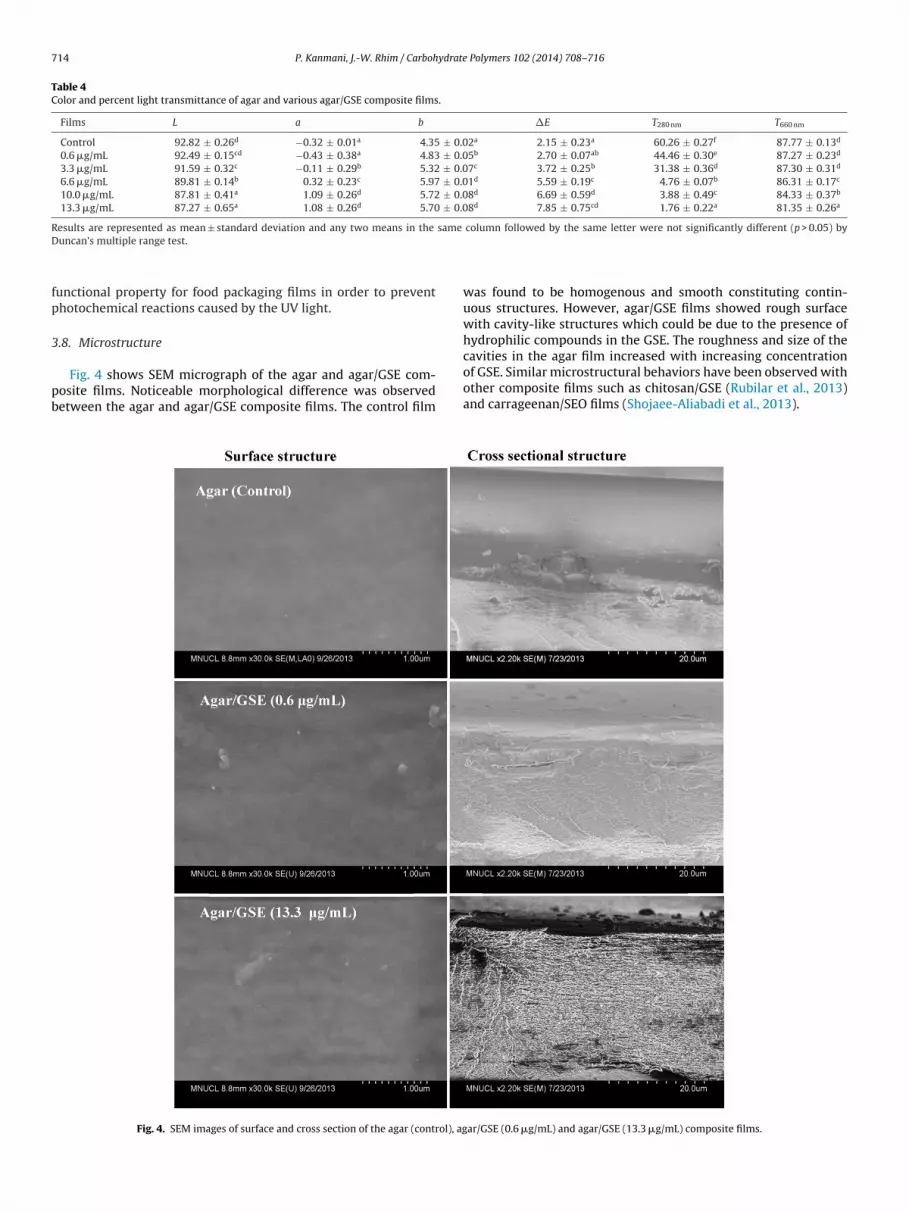
3.8. Microstructure
Fig. 4 shows SEM micrograph of the agar and agar/GSE com-posite films. Noticeable morphological difference was observedbetween the agar and agar/GSE composite films. The control film
was found to be homogenous and smooth constituting contin-uous structures. However, agar/GSE films showed rough surfacewith cavity-like structures which could be due to the presence ofhydrophilic compounds in the GSE. The roughness and size of thecavities in the agar film increased with increasing concentrationof GSE. Similar microstructural behaviors have been observed withother composite films such as chitosan/GSE (Rubilar et al., 2013)and carrageenan/SEO films (Shojaee-Aliabadi et al., 2013).
Fig. 4. SEM images of surface and cross section of the agar (control), agar/GSE (0.6 !g/mL) and agar/GSE (13.3 !g/mL) composite films.

P. Kanmani, J.-W. Rhim / Carbohydrate Polymers 102 (2014) 708– 716 715
Fig. 5. TGA thermograms and DTGA curves of the agar and agar/GSE (13.3 !g/mL)composite films.
3.9. Thermgarvimetric analysis
Thermal property of the agar and agar/GSE (13.3 !g/mL) com-posite films was measured using a thermogravimetric analysis(TGA) and the results were shown in Fig. 5. As shown in the TGAthermograms, three distinctive steps of thermal degradation wereobserved in thermal destruction of agar and agar/GSE compos-ite films. A series of thermal degradation of films were observedaround 90 "C, 250 "C, and 325 "C for agar and 90 "C, 220 "C, and320 "C for agar/GSE composite films, respectively. The first ther-mal degradation was mainly attributed to the evaporation of waterremained in both the agar and agar/GSE films. The second and thirdstep of degradation could be due to the decomposition of GSE, glyc-erol and agar polymer, respectively (Rhim et al., 2006). As shownin Fig. 5, the onset thermal degradation temperature of agar/GSE(13.3 !g/mL) film was little different from those of control film. Thisresult indicates that the addition of GSE did not improve thermalstability of the agar films.
4. Conclusion
Antimicrobial films were prepared by incorporating GSE as nat-ural antimicrobial agent with various concentrations into agarfilm for the use of active food packaging. Remarkable changesin color, transmittance, moisture content, water solubility, waterbarrier, hydrophobicity and mechanical properties were recorded.The agar/GSE composite film exhibited high UV barrier propertywhich implies that the composite films have high potential for
being used to protect food stuffs from UV light induced photo-chemical reactions. FE-SEM, FT-IR, XRD, and TGA were performed tocharacterize morphological properties of the resulting agar basedcomposite films. Changes in film microstructures were observed inthe agar/GSE films. XRD and TGA analysis indicated that the pres-ence of GSE did not change crystallinity and thermal stability ofagar film. The agar/GSE composite films exhibited strong antimi-crobial activity against various Gram-positive and Gram-negativefood-borne pathogens. The antimicrobial activity was dependenton the concentration of GSE and bacterial strains. The antimicrobialactivity of the film is due to the migration of phenolic compoundsin the GSE. The GSE entrapped by the polymer matrix might bereleased by replaced with water molecules to exert antimicrobialactivity. The agar served as effective vehicle to carry antimicrobialagents. Conclusively, the agar/GSE films with remarkable functionalproperties can be used as an environmentally friendly active foodpackaging film as well as a promising alternative to synthetic poly-mer based packaging films. However, further studies are needed toinvestigate potential performance improvement for industrializeduse of the film.
Acknowledgement
This research was supported by the Agriculture Research Center(ARC 710003) program of the Ministry for Agriculture, Food andRural Affairs, Korea.
References
ASTM. (2012a). Standard test method for tensile properties of thin plastic sheeting,Standard D882. Annual book of ASTM. Philadelphia, PA: American Society forTesting and Materials.
ASTM. (2012b). Standard test methods for water vapor transmission of materialE96/E96M. Annual book of ASTM. Philadelphia, PA: American Society for Testingand Materials.
Balasubramnian, A., Rogenberg, L. E., Yam, K., & Chikindas, M. (2009). Antimicrobialpackaging: Potential vs. reality – A review. Journal of Applied Packaging Research,3, 193–221.
Chambi, H., & Grosso, C. (2006). Edible films produced with gelatin and casein cross-linked with transglutaminase. Food Research International, 39, 458–466.
Cho, S., Seo, I., Choi, J., & Joo, I. (1990). Antimicrobial and antioxidant activity ofgrapefruit and seed extract on fishery product. Bulletin of the Korean FisheriesSociety, 23, 289–295.
Cho, S. H., Lee, H. C., Seo, I. W., Kim, Z. U., Chang, Y. S., & Shin, Z. Y. (1991). Efficiency ofgrapefruit seed extract in the preservation of Satsuma mandarin. Korean Journalof Food Science and Technology, 23, 614–618.
Corrales, M., Han, J. H., & Tauscher, B. (2009). Antimicrobial properties of grapeseed extracts and their effectiveness after incorporation into pea starch films.International Journal of Food Science and Technology, 44, 425–433.
Cvetnic, Z., & Vladmir-Knezevic, S. (2004). Antimicrobial activity of grapefruit seedand pulp ethanolic extract. Acta Pharmaceutica, 54, 243–250.
Du, W. X., Olsen, C. W., Avena-Bustilos, R. J., McHugh, T. H., Levin, C. E., & Friedman,M. (2009). Effects of allspice, cinnamon, and clove bud essential oils in edibleapple films on physical properties and antimicrobial activities. Journal of FoodScience, 74, M372–M378.
Duncan, T. V. (2011). Application of nanotechnology in food packaging and foodsafety: Barrier materials, antimicrobials and sensors. Journal of Colloid and Inter-face Science, 363, 1–24.
Falguera, V., Quintero, J. P., Jiménez, A., Munoz, J. A., & Ibarz, A. (2011). Edible filmsand coatings: Structures, active functions and trends in their use. Trends in FoodScience and Technology, 22, 292–303.
Freile-Pelegrin, Y., Madera-Santana, T., Robledo, D., Veleva, L., Quintana, P., & Aza-mar, J. A. (2007). Degradation of agar films in a humid tropical climate: Thermal,mechanical, morphological and structural changes. Polymer Degradation andStability, 92, 244–252.
Ganzera, M., Aberham, A., & Stuppner, H. (2006). Development and validation ofan HPLC/UV/MS method for simultaneous determination of 18 preservatives ingrapefruit seed extract. Journal of Agricultural and Food Chemistry, 54, 3768–3772.
García, M. A., Pinotti, A., Martino, M. N., & Zaritzky, N. E. (2004). Characterization ofcomposite hydrocolloid films. Carbohydrate Polymers, 56, 339–345.
Gimenez, B., Lopez de Lacey, A., Perez-Santín, E., Lopez-Caballero, M. E., & Montero,P. (2013). Release of active compounds from agar and agar-gelatin films withgreen tea extract. Food Hydrocolloids, 30, 264–271.
Han, J. H. (2000). Antimicrobial food packaging. Food Technology, 54(3), 56–65.Hong, Y. H., Lim, G. O., & Song, K. B. (2009). Physical properties of Gelidium corneum-
gelatin blends films containing grapefruit seed extract or green tea extract and

716 P. Kanmani, J.-W. Rhim / Carbohydrate Polymers 102 (2014) 708– 716
its application in the packaging of pork loins. Journal of Food Science, 74(1),C6–C10.
Jang, S. A., Shin, Y. J., & Song, K. B. (2011). Effects of rapeseed protein-gelatin film con-taining grapefruit seed extract on ‘Maehyang’ strawberry quality. InternationalJournal of Food Science and Technology, 46, 620–625.
Jiang, Y. F., Li, Y. X., Chai, Z., & Leng, X. J. (2010). Study of the physical propertiesof whey protein isolate and gelatin composite films. Journal of Agricultural andFood Chemistry, 58, 5100–5108.
Letendre, M., D’Aprano, G., Lacroix, M., Salmieri, S., & Sr-Gelais, D. (2002). Physi-cochemical properties and bacterial resistance of biodegradable milk proteinfilms containing agar and pectin. Journal of Agricultural and Food Chemistry, 50,6017–6022.
Lim, G. O., Jang, S. A., & Song, K. B. (2010). Physical and antimicrobial properties ofGelidium corneum/nano-clay composite film containing grapefruit seed extractand thymol. Journal of Food Engineering, 98, 415–420.
Moradi, M., Tajik, H., Razavi Rohani, S. M., Oromiehie, A. R., Malekinejad, H., Aliak-barlu, J., et al. (2012). Characterization of antioxidant chitosan film incorporatedwith Zataria multiflora Boiss essential oil and grape seed extract. LWT-Food Sci-ence and Technology, 46, 477–484.
Pereda, M., Aranguren, M. I., & Marcovich, N. E. (2010). Caseinate films modified withtung oil. Food Hydrocolloids, 24, 800–808.
Phan, D., Debeaufort, F., Luu, D., & Voilley, A. (2005). Functional properties of edi-ble agar-based and starch-based films for food quality preservation. Journal ofAgricultural and Food Chemistry, 53, 973–981.
Reagor, L., Gusman, J., McCoy, L., Carino, E., & Heggers, J. P. (2002). The effectivenessof processed grapefruit-seed extract as an antibacterial agent: I. An in vitro agarassay. Journal of Alternative and Complementary Medicine, 8, 325–332.
Rhim, J. W. (2011). Effect of clay contents on mechanical and water vapor bar-rier properties of agar-based nanocomposite films. Carbohydrate Polymers, 86,691–699.
Rhim, J. W. (2013). Effect of PLA lamination on performance characteristics ofagar/$-carrageenan/clay bio-nanocomposite film. Food Research International,51, 714–722.
Rhim, J. W., Hong, S. I., Park, H. M., & Ng, P. K. W. (2006). Preparation and charac-terization of chitosan-based nanocomposite films with antimicrobial activity.Journal of Agricultural and Food Chemistry, 54, 5814–5822.
Rhim, J. W., & Ng, P. K. W. (2007). Natural biopolymer-based nanocomposite filmsfor packaging applications. Critical Reviews in Food Science and Nutrition, 47,411–433.
Rhim, J. W., Wang, L. F., & Hong, S. I. (2013). Preparation and characterizationof agar/silver nanoparticles composite films with antimicrobial activity. FoodHydrocolloids, 33, 327–335.
Rubilar, J. F., Cruz, R. M. S., Silva, H. D., Vicente, A. A., Khmelinskii, I., & Vieira, M.C. (2013). Physico-mechanical properties of chitosan films with carvacrol andgrape seed extract. Journal of Food Engineering, 115(4), 466–474.
Salarbashi, D., Tajik, S., Shojaee-Aliabadi, S., Ghasemlou, M., Moayyed, H.,Khaksar, R., et al. (2013). Development of new active packaging filmmade from a soluble soybean polysaccharide incorporated Zatariamultiflora Boiss and Mentha pulegium essential oils. Food Chemistry,http://dx.doi.org/10.1016/j.foodchem.2013.09.014
Shojaee-Aliabadi, S., Hosseini, H., Mohammadifar, M. A., Mohammadi, A., Ghasem-lou, M., Ojagh, S. M., et al. (2013). Characterization of antioxidant antimicrobial$-carrageenan films containing Satureja hortensis essential oil. InternationalJournal of Biological Macromolecules, 52(1), 116–124.
Song, H. Y., Shin, Y. J., & Song, K. B. (2012). Preparation of a barley bran protein–gelatincomposite film containing grapefruit seed extract and its application in salmonpackaging. Journal of Food Engineering, 113, 541–547.
Soradech, S., Nunthanid, J., Limmatvapirat, S., & Luangtana-anan, M. (2012). Anapproach for the enhancement of the mechanical properties and film coatingefficiency of shellac by the formation of composite films based on shellac andgelatin. Journal of Food Engineering, 108, 94–102.
Sorrentino, A., Gorrasi, G., & Vittoria, V. (2007). Potentilal perspectives of bio-nanocomposites for food packaging applications. Trends in Food Science andTechnology, 18, 84–95.
Takeoka, G., Dao, L., Wong, R. Y., Lundin, R., & Mahoney, N. (2001). Identificationof benzethonium chloride in commercial grapefruit seed extracts. Journal ofAgricultural and Food Chemistry, 49, 3316–3320.
Tang, X. G., Kumar, P., Alavi, S., & Sandeep, K. P. (2012). Recent advances in biopoly-mers and biopolymer-based nanocomposites for food packaging materials.Critical Reviews in Food Science and Nutrition, 52, 426–442.
von Woedtke, T. H., Schlüter, B., Pflegel, P., Lindequist, U., & Jülich, W. D. (1999).Aspect of the antimicrobial efficacy of grapefruit seed extract and its relation topreserve substances contained. Pharmazie, 54, 452–456.
Wu, Y., Geng, F., Chang, P. R., Yu, J., & Ma, X. (2009). Effect of agar on the microstruc-ture and performance of potato starch film. Carbohydrate Polymers, 76,299–304.
Xu, W., Qu, W., Huang, K., Guo, F., Yang, J., Zhao, H., et al. (2007). Antibacterialeffect of grapefruit seed extract on food-borne pathogens and its applicationin the preservation of minimally processed vegetables. Postharvest Biology andTechnology, 45, 126–133.
Yu, S. H., Hsieh, H. Y., Pang, J. C., Tang, D. W., Shih, C. M., Tsai, M. L., et al. (2013).Active films from water-soluble chitosan/cellulose composites incorporatingreleasable caffeic acid for inhibition of lipid oxidation in fish oil emulsions. FoodHydrocolloids, 32, 9–19.


