Embed Size (px)
Citation preview

131
Journal of Environmental Pathology, Toxicology and Oncology, 33(2):131–143 (2014)
0731-8898/14/$35.00 © 2014 by Begell House, Inc.
Antioxidant and Hepatoprotective Effects of Crataegus songarica Methanol Extract
Showkat Ahmad Ganie,a Tanveer Ali Dar,a Bilal Zargar,b Rabia Hamid,c Ovais Zargar,a Parvaiz Ahmad Dar,a Shayaq Ul Abeer,d Akbar Masood,c Shajrul Amin,c,*
& Mohammad Afzal Zargara,*
aDepartment of Clinical Biochemistry, University of Kashmir, Srinagar, IndiabDepartment of Pharmacy, University of Kashmir, Srinagar, IndiacDepartment of Biochemistry, University of Kashmir, Srinagar, IndiadDepartment of Biotechnology, University of Kashmir, Srinagar, India
*Address all correspondence to Dr. Shajrul Amin and Professor Mohammad Afzal Zargar, Department of Biochemistry, University of Kashmir Srinagar, India; Tel.: +91- 9419016466, +91-9419018174; [email protected] and [email protected]
ABSTRACT: The protective activity of the methanolic extract of the Crataegus songarica leaves was investigated against CCl4- and paracetamol-induced liver damage. On folklore levels, this plant is popularly used to treat var-ious toxicological diseases. We evaluated both in vitro and ex vivo antioxidant activity of C. songarica. At higher concentration of plant extract (700 µg/ml), 88.106% inhibition on DPPH radical scavenging activity was observed and reducing power of extract was increased in a concentration-dependent manner. We also observed its inhibition on Fe2+/ascorbic acid-induced lipid peroxidation on rat liver microsomes in vitro. In addition, C. songarica extract exhibited antioxidant effects on calf thymus DNA damage induced by Fenton reaction. Hepatotoxicity was induced by challenging the animals with CCl4 (1 ml/kg body weight, i.p.) and paracetamol (500 mg/kg body weight) and the extract was administered at three concentrations (100, 200, and 300 mg/kg body weight). Hepatoprotection was evaluated by determining the activities of liver function marker enzymes and antioxidant status of liver. Administration of CCl4 elevated the levels of liver function enzymes, SGOT, SGPT, and LDH. We also observed a dramatic increase in ALT, AST, bilirubin, and alkaline phosphatase levels in rats administered 500 mg/kg body weight of paracetamol. Decreased antioxidant defense system as glutathione (GSH), catalase (CAT), glutathione peroxidase (GPX), glutathi-one reductase (GR), glutathione-S-transferase (GST), and superoxide dismutase (SOD) were observed in rats treated with CCl4 and paracetamol. Pretreatment with the extract decreased the elevated serum GOT, GPT, LDH, bilirubin, and alkaline phosphatase activities and increased the antioxidant enzymes in a dose-dependent manner. Therefore, C. songarica methanol extract may be an effective hepatic protective agent and viable candidate for treating hepatic disorders and other oxidative stress-related diseases.
KEY WORDS: hepatoprotective effect, Crataegus songarica extract, liver injury, chronic CCl4, paracetamol, ox-idative stress
I. INTRODUCTION
Liver is an important organ for detoxification. How-ever, liver diseases are becoming a serious health problem worldwide. Most of the hepatotoxins induce tissue injury after having been metabolized to free radicals and cause subsequent cell damage through mechanism of covalent binding and lipid peroxida-tion.1 Most of the chemicals must undergo metabolic activation by phase I enzymes to form electrophilic
reactants, which can interact with nucleophilic groups in macromolecules including DNA.2 Reactive oxygen species are demonstrated to play a significant role in the inflammation process after intoxication with hep-atotoxins.3,4 Normally, host cells are protected from oxygen-derived radical injury by naturally occurring free radical scavengers and antioxidants.5 When these natural defenses are overwhelmed by excessive generation of pro-oxidants, a situation of oxidative stress evolves and cellular macromolecules suffer oxidative damage.6 Carbon tetrachloride (CCl4), a

Journal of Environmental Pathology, Toxicology and Oncology
Ganie et al.132
selective hepatotoxic chemical agent, is one of the most widely used toxins for the experimental induc-tion of liver and other tissue damage in laboratory animals.1 The principle causes of CCl4-induced organ damage is lipid peroxidation and decreased activi-ties of antioxidant enzymes and generation of free radicals.1,7 Paracetamol (acetaminophen) is a widely used antipyretic/analgesic and produces acute liver damage if overdoses are consumed and is mainly metabolized in liver to excretable glucuronide and sulphate conjugates.8 However, the hepatotoxicity of paracetamol has been attributed to the formation of toxic metabolites when a part of the paracetamol is activated by hepatic cytochrome P-450,9 to a highly reactive metabolite N-acetyl-P-benzoquinoneimine (NAPQI).10 NAPQI is initially detoxified by con-jugation with reduced glutathione (GSH) to form mercapturic acid.11 However, when the rate of NAPQI formation exceeds the rate of detoxification by GSH, it oxidizes tissue macromolecules such as lipids or -SH groups of protein and alters the homeostasis of calcium after depleting GSH. Paracetamol, an analgesic and antipyretic, is assumed to be safe in recommended doses; overdoses, however, produce hepatic necrosis. Extensive liver damage by parac-etamol itself decreases its rate of metabolism and other substrates for hepatic microsomal enzymes. Induction of cytochrome P450 or depletion of hepatic glutathione is a prerequisite for paracetamol-induced toxicity.
Unfortunately, many synthetic drugs used in the treatment of liver diseases are inadequate and also cause serious side effects.12 In view of severe undesirable side effects of synthetic agents, there is growing interest in evaluating traditional herbal medicines that are claimed to possess hepato- protective activity. A single drug cannot be effective for all types of severe liver diseases. Therefore, an effective formulation using indigenous medicinal plants has to be developed with proper pharma- cological experiments and clinical trials.
13 Consid-ering the above limitations, methanolic extract of leaves of Crataegus songarica (Kashmiri name Barr) was subjected to various in vitro and in vivo assays.
C. songarica belongs to the family Rosaceace. It is an Asian species of hawthorn with black fruit
that is sometimes used medicinally. C. songarica is used for cardiovascular conditions such as congestive heart failure, coronary circulation problems, and arrhythmias. It is also used to increase cardiac output reduced by hypertension or pulmonary disease, to support hypotension and hypertension, atheroscle-rosis, hyperlipidemia, and Buerger’s disease. The barriers of the plant possess antihypertensive and cardio ionic potential. In addition, the plant was shown to improve cardiac activity in patients with congestive heart failure.14 C. songarica is also used as a sedative, antispasmodic, astringent, and diuretic, as well as for gastrointestinal conditions such as indigestion, enteritis, epigastric distension, diarrhea, and abdominal pain. It relaxes the uterus and intestine smooth vessel; however, it constricts the bronchi and coronary vessels.15 C. songarica fruit may also be effective orally for tapeworm infections, acute bacillary dysentery, and amenorrhea. C. songarica fruit liquid preparations are used as washes for sores, itching, and frostbite. Fruits are also used as a popular remedy for diarrhea or slight phlegmasia.16
Therefore, the current study was undertaken to evaluate the in vitro antioxidant potential and protective effect of C. songarica methanolic extract on CCl4- and paracetamol-induced liver toxicity in comparison with known antihepatotoxic agents.
II. MATERIALS AND METHODS
A. Plant Material Collection and Extraction
The fresh leaves of C. songarica were collected from Vanaspati nursery of Ganderbal Srinagar, Jammu and Kashmir, India, in the months of September and October 2012, identified by the Centre of Plant Taxonomy, Department of Botany, University of Kashmir, and authenticated by Dr. Irshad Ahmad Nawchoo (Department of Botany) and Akhter Hus-sain Malik (Curator, Centre for Plant Taxonomy, University of Kashmir). A reference specimen has been retained in the herbarium of the Department of Botany at the University of Kashmir under reference number KASH-bot/Ku/CS-702-SAG.
The leaves were dried in the shade at 30 ± 2°C. The dried material was ground into a powder using a

Volume 33, Number 2, 2014
Antioxidant and Hepatoprotective Effects of Crataegus songarica Methanol Extract 133
mortar and pestle and passed through a sieve of 0.3 mm mesh size. The powder obtained was extracted with methanol for 48 h using a Soxhlet extractor (60–80°C). The extract was then concentrated with the help of a rotary evaporator under reduced pressure and the solid extract was stored in a refrigerator for further use.
B. DPPH Radical Scavenging Activity
The DPPH method (2, 2-diphenyl-1- picrylhydrazyl, Sigma-Aldrich, Germany) was carried out according to the method modified by Kim et al.17 An aliquot of the radical formed from DPPH was left to react with 100–700 µg/ml of the extract for 30 min. The spectrophotometric absorbance measurements were made at 517 nm. Catechin was used as the standard (10 mg/10 ml). The percentage of radical inhibition was calculated by the following:
where A0 is the absorbance without sample and Ae is absorbance with sample.
C. Reducing Power
The reducing power was based on Fe (III) to Fe (II) transformation in the presence of the solvent frac-tions.18 The Fe (II) can be monitored by measuring the formation of Perl’s Prussian blue at 700 nm. Various concentrations of the sample (2 ml) were mixed with 2 ml of phosphate buffer (0.2 M, pH 6.6) and 2 ml of potassium ferricyanide (10 mg/ml). The mixture was incubated at 50°C for 20 min followed by addi-tion of 2 ml of trichloroacetic acid (100 mg/l). The mixture was centrifuged at 3000 rpm for 10 min to collect the upper layer of the solution. A volume of 2 ml from each of the mixtures mentioned previously was mixed with 2 ml of distilled water and 0.4 ml of 0.1% (w/v) fresh ferric chloride. After 10-min reaction, the absorbance was measured at 700 nm. Higher absorbance of the reaction mixture indicates a higher reducing power.
D. Microsomal Lipid Peroxidation
Rat liver microsomal lipid peroxidation was carried out according to the method of Urate et al.19 with little modification. The test sample (20–100 µg/ml) was added to 1 ml of liver microsomes. Lipid peroxidation was induced by adding 100 µl of ferric nitrate (20 mM) and 100 µl of ascorbic acid (100 mM). After incubation for 1 h at 37°C, the reaction was stopped by the addition of 1 ml of TCA (10%), then 1 ml of (1.67%) TBA was added and the reac-tion mixture was boiled for 15 min, cooled, and centrifuged, and the absorbance of the supernatant was measured at 532 nm.
E. Hydroxyl Radical Scavenging Assay
Hydroxyl radical scavenging activity was measured by the ability of the different concentrations of C. songarica extract to scavenge the hydroxyl radicals generated by the Fe3+-ascorbate-H2O2 system (Fenton reaction).20 The reaction mixture contained 500 μl of 2-deoxyribose (2.8 mM) in phosphate buffer (50 mM, pH 7.4), 200 μl of premixed ferric chloride (100 mM), 100 μl of H2O2 (200 mM) with or without the extract solution (100–500 μg/ml). The reaction was triggered by adding 100 μl of 300 mM ascorbate and incubated for 1 h at 37°C. 0.5 ml of the reaction mixture was added to 1 ml of TCA (10%), then 1 ml of 1% TBA was added to the reaction mixture. The mixture was heated for 15 min in a boiling water bath. After the mixture was cooled, the absorbance at 532 nm was noted against a blank (the same solution but without reagent). The scavenging activity on hydroxyl radical was calculated as follows:
Scavenging activity (%) = (1 – absorbance of sample/absorbance of control) × 100
F. Antioxidant Activity against Oxidative Damage to DNA
Hydroxyl radical generated by Fenton reaction was used to induce oxidative damage to DNA.21 The reaction mixture (15 μl) contained 25 mg of calf thymus DNA in 20.0 mM phosphate buffer saline (pH 7.4) and different concentrations of plant extract (10,

Journal of Environmental Pathology, Toxicology and Oncology
Ganie et al.134
30, 50, and 80 μg) were added and incubated with DNA for 15 min at room temperature. The oxidation was induced by treating DNA with 20 mM ferric nitrate and 100 mM ascorbic acid and incubated them for 1 h at 37°C. The reaction was terminated by the addition of loading buffer bromophenol blue (0.25%) and glycerol (30%) and the mixture was subjected to gel electrophoresis in 0.7% agarose/TAE buffer run at 100 V. DNA was visualized and photographed by gel doc.
G. Animals (In Vivo Assay)
All experiments were performed in compliance with the Indian legislation on the use and care of labora-tory animals and were approved by the Institutional Animal Ethics Committee (Reg. No 801/03/CA/CPCSEA/2012), University of Kashmir. Male Wistar rats (200 ± 20 g) were grouped (six rats per cage) with free access to food and water, and kept in a regulated environment at 25 ± 1°C under a 12/12-h (light/dark) cycle. After 1 week of acclimation to the laboratory, rats were selected randomly into nine groups (six animals in each group). The extract was suspended in normal saline at dose levels of 100, 200, and 300 mg/kg body weight. The final volume of extract at each dose was 1 ml, which was fed to rats by gavage. Group I received water only (normal con-trol), group II received CCl4 (1 ml/kg body weight), group III was administered with plant extract (100 mg/kg body weight) and CCl4, group IV received CCl4 and plant extract (200 mg/kg body weight), group V received 1 ml of methanolic extract of C. songarica at the concentration of 300 mg/kg body weight along with CCl4, group VI was administered with vitamin E (50 mg/kg body weight), group VII received paracetamol (500 mg/kg body weight), group VIII received 1 ml of methanolic extract of C. songarica at the concentration of 300 mg/kg body weight along with paracetamol, and group IX received 1 ml of silymarin at the concentration of 50 mg/kg body weight along with paracetamol. The experiment continued for a period of 3 weeks. The animals of all groups were sacrificed at the end of the experiment and groups I–VI were administered with CCl4 48 h prior to sacrifice. Liver tissue was
collected and post-mitochondrial supernatant was prepared as written under preparation of PMS in the Materials and Methods.
H. Preparation of Tissue and Biochemical Assays
Before sacrificing the experimental animals, blood was collected from retro-orbital plexus without the use of anticoagulant. The blood was allowed to stand for 10 min before being centrifuged at 5000 × g for 10 min to obtain serum. The liver was quickly removed, weighed, and homogenized in ice-cold 50 mM phosphate buffer, pH 7. After centrifuging of the homogenates (3000 rpm, 15 min), the supernatants were used for biochemical assays related to CCl4- and paracetamol-induced liver function damage, described below.
Plasma enzymes serum alanine aminotransami-nase (ALT) and serum aspartate aminotransaminase (AST) were estimated by the method of Reitman and Frankel.22 Alkaline phosphatase activity was estimated by the method of King and Kind.23
For the direct bilirubin and total protein assay, Direct bilirubin was estimated by the method of Jendrassik and Grof24 and the total serum proteins were estimated with the method of Gornall et al.25
Antioxidant assays were conducted as follows. Hepatic GSH content was determined after the reaction with 5,5-dithio-bis-(2-nitrobenzoic acid) and the yellow color developed was read at 412 nm according to Moren et al.26 The content of GSH in tissue samples were expressed as nm/g protein. Glutathione peroxidase (GPx) and glutathione reduc-tase (GR) activity was assayed using the method of Sharma et al.27 Glutathione-S-transferase (GST) activity was assayed using the method of Haque et al.28 Catalase (CAT) was assayed by the method of Claiborne.29 Change in absorbance was recorded at 240 nm. CAT activity was calculated in terms of nanomoles of H2O2 consumed/min/mg of protein.
I. Statistical Analysis
Data were analyzed by a one-way ANOVA and the values are expressed as mean ± standard deviation

Volume 33, Number 2, 2014
Antioxidant and Hepatoprotective Effects of Crataegus songarica Methanol Extract 135
(SD). The results were evaluated by using SPSS (version 12.0) and Origen 6 software and each experiment was performed at least thrice.
III. RESULTS
A. DPPH Radical Scavenging Activity
The percentage of DPPH radical scavenging activity of methanol extract of C. songarica is presented in Fig. 1. The extract exhibited dose-dependent inhi-bition on DPPH, and the scavenging activities of the extract and known antioxidant increased with increasing concentration. A maximum of 88.106% DPPH radical scavenging activity was found at 700 μg/ml of plant extract used, whereas for catechin the scavenging activity was 88.71%. Our results indicated that C. songarica extract has a noticeable effect on scavenging of DPPH free radicals.
B. Reducing Power Assay
As illustrated in Fig. 2, Fe3+ was transformed to Fe2+ in the presence of C. songarica extract and the ref-erence compound catechin to measure the reductive capability. The reducing power of the extract and catechin increased with increasing in concentration. At 100 µg/ml, the absorbance of the plant extract and catechin were 0.0183 and 0.152, while at the higher concentration 700 µg/ml, the absorbance of both extract and catechin were increased to 0.190 and 0.249, respectively.
C. Microsomal Lipid Peroxidation
This model system contained Fe++/ascorbic acid as oxidizing agent to initiate lipid peroxidation in rat liver microsomes. The lipids in membrane are continuously subjected to oxidant challenges leading to the formation of thiobarbituric acid reac-tive species (TBARS) like malonaldehyde (MDA) via lipid peroxidation (LPO). MDA forms a pink chromogen with TBA that absorbs at 535 nm. The C. songarica extract dose dependently inhibited the
FIG. 1: The effect of methanol extract and known antioxidant on DPPH radical scavenging activity. The results represent mean ± SD of three separate experiments. Absorbance at 517 nm.
FIG. 2: The effect of methanol extract and known antioxidant catechin on reducing power method. The results represent mean ± SD of three separate experiments.

Journal of Environmental Pathology, Toxicology and Oncology
Ganie et al.136
MDA formation, and thus the lipid peroxidation in liver microsomes. At a concentration of 100 µg/ml, around 78.59% inhibition in LPO was observed. In the presence of known antioxidant catechin at 250 µg/ml concentration, LPO was inhibited by 84.48% (Fig. 3).
D. Hydroxyl Radical Scavenging Activity
This assay shows the abilities of the extract and standard catechin to inhibit hydroxyl radical-me-diated deoxyribose degradation in a Fe3+- ascorbic acid and H2O2 reaction mixture. The results are shown in Fig. 4. In our experiment, we observed that methanol extract of C. songarica was more efficient to scavenge hydroxyl radicals than that of the known antioxidant catechin at all concentrations. At a concentration of 500 µg/ml, the extract shows
the maximum inhibitory effect of ~89.05%; catechin at the same concentration shows only ~62.47% inhibition on hydroxyl radical.
E. Antioxidant Activity against Oxidative Damage to DNA
The protective effect of C. songarica methanol extract on calf thymus DNA is shown in Fig. 5. Hydroxyl radicals generated by Fenton reaction were found to induce DNA strand breaks in calf thymus DNA. H2O2 alone did not cause DNA strand cleavage (lane 8). However, H2O2 in the presence of ferric nitrate and ascorbic acid induce DNA strand breaks and help the DNA molecule to run fast (lane 2). C. songarica extract at 10–80 μg offered complete protection to DNA damage induced by hydroxyl radicals in calf thymus DNA (lanes 3–7). Thus, the hydroxyl radical quenching ability of polyphenolic
FIG. 3: The effect of Crataegus songarica methanol extract and catechin on rat liver homogenate lipid peroxidation. The results represent mean ± SD and evaluated by one-way ANOVA followed by the Bonferroni t-test.
FIG. 4: The effect of methanol extract and known antioxidant on hydroxyl radical scavenging activity. The results represent mean ± SD of three separate experiments. Results are reported as the percentage of the maximum formation of OH● radical (100% deoxyribose oxidized). In absorbency, 100% is 0.543 ± 0.007 (control). Absorbance at 532 nm.

Volume 33, Number 2, 2014
Antioxidant and Hepatoprotective Effects of Crataegus songarica Methanol Extract 137
compounds of C. songarica could be responsible for the protection against oxidative damage to DNA.
F. Plasma AST, ALT, ALP, Direct Bilirubin, and Total Proteins
The levels of serum AST, ALT, ALP, and direct bilirubin were elevated significantly after the admin-istration of CCl4 and paracetamol. The AST, ALT, ALP activities and direct bilirubin were 89.199 ± 3.32 IU/L, 206.17 ± 3.16 IU/L, 146.68 ± 5.62 IU/L, and 2.030 ± 0.02, respectively, in the CCl4 alone injected animals and 53.32 ± 3.12 IU/L, 165.19 ± 10.30 IU/L, 129.29 ± 6.68 IU/L, and 1.782 ± 0.019, respectively, in the paracetamol alone treated animals. However, the treatments with C. songarica extract (100, 200, and 300 mg/kg) reduced the elevated enzyme activi-
ties and bilirubin levels in a dose-dependent manner. Treatment with 300 mg/kg body weight of the extract significantly reduced the level of serum AST, ALT, ALP, and direct bilirubin level to 53.26 ± 2.31 IU/L, 132.06 ± 4.29 IU/L, 82.22 ± 2.62 IU/L, and 0.5883 ± 0.060 in CCl4 injected animals and 21.05 ± 2.89 IU/L, 73.95 ± 14.67 IU/L, 75.25 ± 5.84 IU/L, and 0.407 ± 0.010 in paracetamol-treated animals when compared to the control group. Total serum proteins were decreased in CCl4- and paracetamol-induced rats, which were restored in a dose-dependent manner after treatment with the different concentrations of the plant extract (Tables 1 and 2).
G. Effect of C. songarica on GSH and Hepatic Antioxidant Enzyme Activities
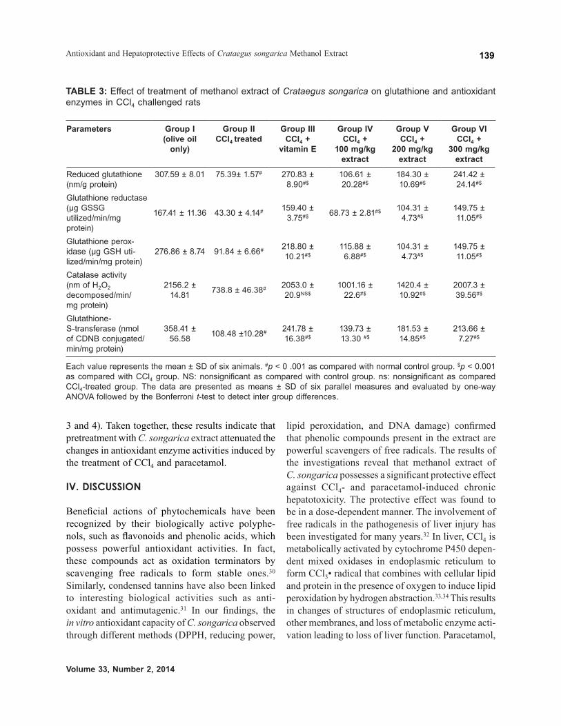
The activities of tissue antioxidants CAT, SOD, GPx, and GST and level of GSH were decreased significantly in animals consequent to CCl4- or paracetamol-induced hepatic damage as compared to the normal animals. In the extract-treated group of animals, a marked increase in the antioxidant status of the liver was evident (Tables 3 and 4, p < 0.01, p < 0.05). As shown in the tables, the administration of CCl4 and paracetamol alone markedly depleted GSH levels (75.39 ± 1.57, 95.73 ± 5.21) as compared to control group rats (307.59 ± 8.01). Rats pretreated with C. songarica extract were significantly protected from the GSH decrease produced by CCl4 and parac-etamol (Tables 3 and 4), and this protective effect occurred dose dependently. It has been suggested that GST, GPx, GR, and CAT serve as the detoxifying system for preventing damage caused by ROS and play a pivotal role in the scavenging of free radicals. To provide insight into the relationship between the hepatoprotective effect and the antioxidant effect, the activities of these antioxidant enzymes were analyzed. As shown in Tables 3 and 4, CCl4 and paracetamol induced substantial modifications to the hepatic antioxidative enzymes and the decreased hepatic GPx, GST, GR, and CAT activities. In the extract and known antioxidant treated groups of animals, a marked increase in the antioxidant status of the liver was evident, and this effect was strengthened with the increase of C. songarica extract concentration (Tables
FIG. 5: Protective effect of methanol extract of Crataegus songarica on oxidative damage to calf thymus DNA. Lane 1: Native calf thymus DNA. Lane 2: DNA + 20 mM ferric nitrate + 100 mM ascorbic acid + 30 mM H2O2. Lane 3: DNA + 20 mM ferric nitrate + 100 mM ascorbic acid + 30 mM H2O2 + 10 µg of methanol extract. Lane 4: DNA + 20 mM ferric nitrate + 100 mM ascorbic acid + 30 mM H2O2 + 20 µg of methanol extract. Lane 5: DNA + 20 mM ferric nitrate + 100 mM ascorbic acid + 30 mM H2O2+ 30 µg of methanol extract. Lane 6: DNA + 20 mM ferric nitrate + 100 mM ascorbic acid +30 mM H2O2+ 50 µg of methanol extract. Lane 7: DNA + 20 mM ferric nitrate + 100 mM ascorbic acid + 30 mM H2O2+ 80 µg of methanol extract. Lane 8: DNA + 20 mM ferric nitrate + 100 mM ascorbic acid + 30 mM H2O2 + 10 µg of catechin.

Journal of Environmental Pathology, Toxicology and Oncology
Ganie et al.138
TABLE 1: Effect of Crataegus songarica methanol extract on biochemical parameters in CCl4-induced hepatotoxicity in albino rats
Treatment Dose mg/kg
AST U/L ALT U/L Direct bilirubin
Total proteins
Alkaline phosphatase
Control group (Olive oil only)
5 20.74 0.41 43.76 ± 2.35 0.282 ± 0.012 3.52 ± 0.517 50.12 ± 0.11
CCl4-treated group
1 89.199 ± 3.32# 206.17 ± 3.16# 2.030 ± 0.02# 0.501 ± 0.086# 146.68 ± 5.62#
CCl4 treated + V.E
50 46.57 ± 2.54#$ 125.70 ± 2.09#$ 0.553 ± 0.0234#$ 0.974 ± 0.045#$ 79.25 ± 2.84#$
CCl4-treated + C.S extract
100 79.28 ± 2.18#$ 151.105 ± 13.13#$ 1.617 ± 0.019#$ 0.6618 ± 0.033#$ 104.78 ± 4.85#$
CCl4-treated + C.S extract
200 64.28 ± 2.64#$ 135.74 ± 0.91#$ 0.763 ± 0.066#$ 0.801 ± 0.038#$ 89.91 ± 6.86#$
CCl4 treated + C.S extract
300 53.26 ± 2.31#$ 132.06 ± 4.29#$ 0.5883 ± 0.060#$ 0.998 ± 0.014#$ 82.22 ± 2.62#$
Each value represents the mean ± SD of six animals. #p < 0 .001 as compared with the normal control group. $p < 0.001 as compared with the CCl4 group. The data are presented as means ± SD of six parallel mea-sures and evaluated by one-way ANOVA followed by the Bonferroni t-test to detect intergroup differences.
TABLE 2: Effect of Crataegus songarica methanol extract on biochemical parameters in paracetamol-induced hepatotoxicity in albino rats
Treatment Dose mg/kg
AST U/L ALT U/L Direct bilirubin
Total proteins
Alkaline phosphatase
Control group – 25.76 ± 4.35 40.76 ± 4.35 0.275 ± 0.032 2.68 ± 0.019 52.72 ± 2.11
Paracetamol-treated group
1 53.32 ± 3.12# 165.19 ± 10.30# 1.782 ± 0.019# 0.675 ± 0.022# 129.29 ± 6.68#
Paracetamol + silymarin
50 21.37 ± 3.42NS$ 72.35 ± 4.23#$ 0.367 ± 0.0189#$ 1.431 ± 0.021#$ 75.25 ± 5.84#$
Paracetamol + C.S extract
100 36.47 ± 7.21#$ 105.70 ± 1.09#$ 1.433 ± 0.259#ns 0.920 ± 0.055#$ 99.52 ± 9.44#$
Paracetamol + C.S extract
200 30.33 ± 5.14NS$ 93.69 ± 4.02#$ 0.486 ± 0.29#$ 1.113 ± 0.110#$ 86.47 ± 5.46#$
Paracetamol + C.S extract
300 21.05 ± 2.89NS$ 73.95 ± 14.67#$ 0.407 ± 0.010#$ 1.423 ± 0.030#$ 75.25 ± 5.84#$
Each value represents the mean ± SD of six animals. #p < 0 .001 as compared with normal control group. $p < 0.001 as compared with paracetamol-treated group. NS: nonsignificant as compared with normal group. ns: nonsignificant as compared with paracetamol-treated group. The data are presented as means ± SD of six parallel measures and evaluated by one-way ANOVA followed by the Bonferroni t-test to detect inter group differences.
Volume 33, Number 2, 2014
Antioxidant and Hepatoprotective Effects of Crataegus songarica Methanol Extract 139
3 and 4). Taken together, these results indicate that pretreatment with C. songarica extract attenuated the changes in antioxidant enzyme activities induced by the treatment of CCl4 and paracetamol.
IV. DISCUSSION
Beneficial actions of phytochemicals have been recognized by their biologically active polyphe-nols, such as flavonoids and phenolic acids, which possess powerful antioxidant activities. In fact, these compounds act as oxidation terminators by scavenging free radicals to form stable ones.30 Similarly, condensed tannins have also been linked to interesting biological activities such as anti- oxidant and antimutagenic.31 In our findings, the in vitro antioxidant capacity of C. songarica observed through different methods (DPPH, reducing power,
lipid peroxidation, and DNA damage) confirmed that phenolic compounds present in the extract are powerful scavengers of free radicals. The results of the investigations reveal that methanol extract of C. songarica possesses a significant protective effect against CCl4- and paracetamol-induced chronic hepatotoxicity. The protective effect was found to be in a dose-dependent manner. The involvement of free radicals in the pathogenesis of liver injury has been investigated for many years.32 In liver, CCl4 is metabolically activated by cytochrome P450 depen-dent mixed oxidases in endoplasmic reticulum to form CCl3• radical that combines with cellular lipid and protein in the presence of oxygen to induce lipid peroxidation by hydrogen abstraction.33,34 This results in changes of structures of endoplasmic reticulum, other membranes, and loss of metabolic enzyme acti-vation leading to loss of liver function. Paracetamol,
TABLE 3: Effect of treatment of methanol extract of Crataegus songarica on glutathione and antioxidant enzymes in CCl4 challenged rats
Parameters Group I(olive oil
only)
Group IICCl4 treated
Group IIICCl4 +
vitamin E
Group IVCCl4 +
100 mg/kg extract
Group VCCl4 +
200 mg/kg extract
Group VICCl4 +
300 mg/kg extract
Reduced glutathione (nm/g protein)
307.59 ± 8.01 75.39± 1.57# 270.83 ± 8.90#$
106.61 ± 20.28#$
184.30 ± 10.69#$
241.42 ± 24.14#$
Glutathione reductase (µg GSSG utilized/min/mg protein)
167.41 ± 11.36 43.30 ± 4.14# 159.40 ± 3.75#$ 68.73 ± 2.81#$ 104.31 ±
4.73#$149.75 ± 11.05#$
Glutathione perox-idase (µg GSH uti-lized/min/mg protein)
276.86 ± 8.74 91.84 ± 6.66# 218.80 ± 10.21#$
115.88 ± 6.88#$
104.31 ± 4.73#$
149.75 ± 11.05#$
Catalase activity (nm of H2O2 decomposed/min/ mg protein)
2156.2 ± 14.81 738.8 ± 46.38# 2053.0 ±
20.9NS$1001.16 ±
22.6#$1420.4 ± 10.92#$
2007.3 ± 39.56#$
Glutathione- S-transferase (nmol of CDNB conjugated/min/mg protein)
358.41 ± 56.58 108.48 ±10.28# 241.78 ±
16.38#$139.73 ± 13.30 #$
181.53 ± 14.85#$
213.66 ± 7.27#$
Each value represents the mean ± SD of six animals. #p < 0 .001 as compared with normal control group. $p < 0.001 as compared with CCl4 group. NS: nonsignificant as compared with control group. ns: nonsignificant as compared CCl4-treated group. The data are presented as means ± SD of six parallel measures and evaluated by one-way ANOVA followed by the Bonferroni t-test to detect inter group differences.

Journal of Environmental Pathology, Toxicology and Oncology
Ganie et al.140
a widely used antipyretic-analgesic drug, produces acute hepatic damage on accidental overdosage. It is established that a fraction of paracetamol is converted via the cytochrome P450 pathway to a highly toxic metabolite, NAPQI,35 which is normally conjugated with glutathione and excreted in urine. Overdose of paracetamol depletes glutathione stores, leading to accumulation of NAPQI, mitochondrial dysfunction,36 and development of acute hepatic necrosis. Several P450 enzymes are known to play an important role in N-acetyl-p-aminophenol (APAP) bioactivation to NAPQI. P450 2E1 (CYP2E1) have been suggested to be primary enzymes for parac-etamol bioactivation in liver microsomes.37 Studies demonstrated that paracetamol-induced hepatotox-icity can be modulated by substances that influence P450 activity.38
The protective action of antioxidants is usually due to the inhibition of free radical induced chain reaction and the resultant prevention of peroxidative deterioration of structural lipids in membranous organelles. Our results showed that methanolic extract of C. songarica possessed significant in vitro antioxidant activity. These results support our findings on the significant role of antioxidants in hepatoprotective activity of the extract.
Liver cells participate in a variety of metabolic activities and thus contain a host of enzymes. In severe liver damage, serum transaminases activity parallel those of the organs indicating that both cellular and mitochondrial membranes have been damaged.39 In both CCl4- and paracetamol-treated rats, a manyfold increase in the activities of GOT, GPT, and ALP were observed. This can be attributed to the altered structural integrity of hepatic cells. The enzyme ALP, located in the cytoplasm, is also
TABLE 4: The effect of treatment of methanol extract of Crataegus songarica on glutathione and antioxidant enzymes in paracetamol-challenged rats
Parameters Group I(normal)
Group IIparacetamol
treated
Group IIIparacetamol+ silymarin
Group IVparacetamol + 100 mg/kg
extract
Group Vparacetamol + 200 mg/kg
extract
Group VIparacetamol + 300 mg/kg
extract
Reduced glutathione (nm/g protein)
317.10 ± 12.69 95.73± 5.21# 282.15 ± 6.35#$ 152.90 ±8.05#$ 222.48 ± 7.31#$ 292.60 ± 3.1NS$
Glutathione reduc-tase (µg GSSG uti-lized/min/mg protein)
190.42 ± 5.51 58.57 ± 2.66# 170.58 ± 6.97NS$ 77.74± 4.47#ns 113.74 ± 1.98#$ 178.04 ± 6.97#$
Glutathione perox-idase (µg GSH uti-lized/ min/mg protein)
315.15 ±4.96 118.44 ± 2.47# 224.96 ± 14.12#$ 133.42 ± 2.54#ns 189.28 ± 6.43#$ 227.78 ± 3.13#$
Catalase activity (nm of H2O2 decomposed /min/mg protein)
2483.75 ± 38.25 1045.95 ± 55.0# 2255.37 ±48.12#$
1145.59 ± 10.46#$
1498.45 ±9.32#$ 2186.15 ± 49.55#$
Glutathione- S- transferase (nmoles of CDNB conjugated/min/mg protein)
347.85 ± 2.66 128.96±6.24# 280.71 ± 9.01#$ 166.80 ± 4.91#$ 208.17 ± 6.99#$ 252.18 ± 7.52#$
Each value represents the mean ± SD of six animals. #p < 0 .001 as compared with normal control group. $p < 0.001 as compared with CCl4 group. NS: nonsignificant as compared with control group. ns: nonsignificant as compared CCl4-treated group. The data are presented as means ± SD of six parallel measures and evaluated by one-way ANOVA followed by the Bonferroni t-test to detect inter group differences.

Volume 33, Number 2, 2014
Antioxidant and Hepatoprotective Effects of Crataegus songarica Methanol Extract 141
released to the circulation after cellular damage.40 On administering the extract to CCl4- or parac-etamol-treated rats, serum GOT, GPT, and ALP activities and direct bilirubin were found to be decreased. This indicates that the extract, to a greater extent, preserves the structural integrity of the liver from the adverse effects of CCl4 and paracetamol.
Free radicals like hydroxyl radical are known to cause marked injuries to the surrounding tissues and organs. Therefore, removing free radicals is probably one of the most effective defense mechanisms to a variety of diseases. Activities of antioxidant enzymes that help to scavenge free radicals were significantly lowered in both CCl4- and paracetamol-treated rats, compared to normal rats. Declined activities of antioxidant enzymes will result in the generation of highly reactive free radicals leading to deleterious effects such as loss of cell membrane integrity and membrane function.41
Glutathione (GSH) is a major nonprotein thiol in living organisms, which plays a central role in coordinating the antioxidant defense processes in the body.42 In states of oxidative stress, GSH is converted to GSSG, leading to lipid peroxidation. Therefore, the role of GSH as a reasonable marker for evaluation of oxidative stress is important since it acts as an antioxidant both extracellularly and intracellularly.43 Rats on treatment with CCl4 and paracetamol expe-rienced reduced levels of tissue GSH as compared to the extract-treated groups. Administration of CCl4 and paracetamol induced a loss of glutathione from the liver and a decrease in its hepatic content. These lowered levels may be due to the increased utilization of GSH for the detoxification process.41 The gluta-thione-depleted state, particularly during oxidative stress of hepatocytes, causes a rise in the cytosolic Ca2+ concentration and in addition leads to protein thiol oxidation. Administering C. songarica extract to CCl4- and paracetamol-treated rats appeared to help restore the GSH levels to normal level.
The activity of GPx and GR were also found to be decreased in the CCl4- or paracetamol-treated rats. In the extract-treated rats, the activity of Se-dependent GPx and GR were found to be enhanced and this could partially explain the protection of biomembrane from oxidative stress.44 Total GST activity, a phase
II enzyme in the hepatocyte, was enhanced in the extract-treated group of animals, compared to the negative control groups. Such increased activity of GST could protect the liver against damage induced by electrophilic metabolites.2
Catalase (CAT) is an enzymatic antioxidant widely distributed in all animal tissues, and the highest activity is found in the red cells and liver. CAT decomposes hydrogen peroxide and protects the tissues from highly reactive hydroxyl radicals. Therefore, reduction in the activity of CAT in CCl4- and paracetamol-treated animals may result in a number of deleterious effects due to the assimilation of superoxide radical and hydrogen peroxide. In our study, pretreated groups with C. songarica showed significant increase in CAT activity, and this could be responsible for the increased resistance to oxi-dative stress.
V. CONCLUSION
In conclusion, the present findings reveal that the methanolic extract of C. songarica possesses profound in vitro antioxidant and hepatoprotective activity. The hepatoprotective activity elicited by the extract might be due to its free radical scavenging activity and ability to activate antioxidant enzymes.45 These findings indicate that the use of C. songarica might be considered as an adjunct therapeutic strat-egy to combat hepatic disorders and other oxidative stress-related diseases. In addition, further studies to characterize the active principles of C. songarica and to elucidate the mechanism are in progress.
ACKNOWLEDGMENTS
This study was funded, in part, by National Medicinal Plants Board, Department of AYUSH, Ministry of Health and Family Welfare, GOI, to M. A. Zargar with Grant No. Z18017-187/PR/GO/JK/04/2005-06/NMPB; the assistance is greatly acknowledged.
REFERENCES
1. Kanter M, Coskun O, Budancamnak M. Hepatoprotective effect of Nigella sativa L and Utrica dioica L on lipid peroxidation, antioxidant enzyme systems and liver

Journal of Environmental Pathology, Toxicology and Oncology
Ganie et al.142
enzymes in carbon tetrachloride treated rats. World J Gastroenterol. 2005;11:6684–8.
2. Ajith TA, Sheena N, Janardhanan KK. Phellinus rimosus protects carbon tetrachloride induced chronic hepatotox-icity in rats: antioxidant defense mechanism. Pharmaceut Biol. 2006;44:467–74.
3. Yoshikawa T, Tanaka H, Yoshida N, Seto O, Sugino N, Kondo N. Adjuvant arthritis and lipid peroxide pro-tection by superoxide dismutase. Lipid Peroxide Res. 1983;7:108–10.
4. Yuda Y, Tanaka J, Hirano F, Igarani K, Satch T. Participation of lipid peroxidation in rat pertussis vaccine pleurisy. Chem Pharm Bull. 1991;39:505–6.
5. Coskun O, Yakan B, Oztas E, Sezen S, Gunaydin A. Antioxidant and hepatoprotective activity of Vitamin E and Egb 761 in experimental endotoxemic rats. Int J Med Sci. 2000;30:427–32.
6. Weisman H, Halliwell B. Damage to DNA by reactive oxygen and nitrogen species: role in inflammatory diseases and progression to cancer. Biochem J. 1996;313:17–29.
7. Lin CC, Yen MH, Lo TS, Lin JN. Evaluation of hepato-protective and antioxidant activity of Boehmeria nivea var. nivea and B. nivea var. tenacissima. J Ethnopharmacol. 1997;60:9–17.
8. Wong LT, Whitehouse LW, Solemonraj G, Paul CG. Pathways of disposition of acetaminophen conjugate in the mouse. Toxicity Lett. 1981;9(2):145–51.
9. Savides MC, Oehme FW. Acetaminophen and its toxicity. J Appl Toxicol. 1983;3:95–111.
10. Vermeulen NPE, Bessems JGM, Van de Streat R. Molecular aspects of paracetamol-induced hepatotoxicity and it mechanism based prevention. Drug Metab Rev. 1992;24:367–407.
11. Moore M, Thor H, Moore G, Nelson S, Moldeus P, Orrenius S. The toxicity of acetaminophen and N-acetyl P-benzoquinoneimine in isolated hepatocytes is associated with thio depletion and increased cystosolic Ca2+. J Biol Chem. 1985;260:13035–40.
12. Guntupalli M, Chandana V, Palpu P, Annie, Shirwaikar IJ. Hepatoprotective effects of rubiadin, a major con-stituent of Rubia cordifolia Linn. J Ethnopharmacol. 2006;103:484–90.
13. Shahani, S. Evaluation of hepatoprotective efficacy of APCL-A polyherbal formulation in-vivo in rats. Indian Drugs. 1999;36:628–31.
14. Kiritikar KR, Basu BD. Indian medicinal plants, part-I. Allahabad (India): Indian Press; 1918. 111: p. 1630–2.
15. Holubarsch CJF, Colucci WS, Meinertz T, Ganus W, Tendera M. Survival and prognosis: Investigation of Crataegus extract WS 1442 in congestive heart failure (SPICE)- rationale, study design and study protocol. Eur J Heart Fail. 2000;2:431–7.
16. Khan AA, Ashfaq M, Ali MN. Pharmacognostic studies of selected indigenous plants of Pakistan. Peshawar Pakistan: Pakistan Forest Institute; 1979. 6: p. 7–.
17. Kim DO, Lee LW, Lee HJ, Lee CY. Vitamin C equivalent antioxidant capacity (VCEAC) of phenolics phytochem-icals. J Agric Food Chem. 2002;50:3713–7.
18. Fejes S, Blazovics A, Lugasi A, Lemberkovics A, Petri G, Kery A. In vitro antioxidant activity of Anthriscus cerefolium L. (Hoffm.) extracts. J Ethnopharmacol. 2000;69:259–65.
19. Urata Y, Yoshida S, Irie Y, Tanigawa T, Amayasu H, Nakabayashi M, Akahori K.. Treatment of asthma patients with herbal medicine Tj-96: a randomized controlled trial. Respir Med. 2002;96(6):469–74.
20. Ilavarasan R, Mallika M,Venkataraman K. Anti-inflammation and antioxidant activities of Cassia fistula Linn. bark extracts. Afr J Trad Compl Altern Med. 2005;2:70–85.
21. Ani V, Varadaraj MC, Akhilender K. Antioxidant and antibacterial activities of polyphenolic compounds from bitter cumin (Cuminum nigrum L.). Eur Food Res Technol. 2006;224:109–15.
22. Reitman S, Frankel S. A calorimetric method for the deter-mination of serum glutamate oxaloacetate and glutamate pyruvate transaminase. Am J Clin Pathol. 1957;28:53–6.
23. King EJ, Kind RPN. Alkaline phosphatase activity assay. Clin Path. 1954;7:332.
24. Jendrassik L, Grof P. Estimation of direct and total Bilirubin. Biochem. 2000;297: 81–9.
25. Gornall AG, Bardawill CS, David MM. Determination of serum proteins by means of Biuret reaction. J Biol Chem. 1984;177–751.
26. Moren MA, Depierre JW, Mannervick B. Levels of gluta-thione, glutathione reductase and glutathione S- transferase activities in rat lung and liver. Biochim Biophys Acta. 1985;582,67–78.
27. Sharma N, Trikha PA, Raisuddin SM. Inhibition of ben-zo[a]pyrene- and cyclophosphamide-induced mutagen-icity by cinnamomum cassia. Mutat Res. 2001;480-481: 179–88.
28. Haque RH, Parvez B, Pandey S, Sayeed, S, Ali I, Raisuddin SM. Aqueous extract of walnut protects mice against cyclophosphamide induced biochemical toxicity. Hum Exp Toxicol. 2003;22:473–80.
29. Claiborne A. Catalase activity in Greenwald RA. In: CRC handbook of methods of oxygen radicals research. Boca Raton (FL): CRC Press; 1985. p. 283–4.
30. Rice-Evans CA, Miller NJ, Paganga G. Antioxidant properties of phenolic compounds. Trends Plant Sci. 1997;2(4):152–9.

Volume 33, Number 2, 2014
Antioxidant and Hepatoprotective Effects of Crataegus songarica Methanol Extract 143
31. Grimmer HR, Parbhoo V, McGrath RM. Antimutagenicity of polyphenol-rich fractions from sorghum-bicolor grain. J Sci Food Agric. 1992;59:251–6.
32. Poli G. Liver damage due to free radicals. Br Med Bull. 1993;49:604–20.
33. Kadiiska MB, Gladen BC, Baird DD, Dikalova AE, Sohal RS, Hatch GE, Jones DP, Mason RP, Barrett JC. Biomarkers of oxidative stress study are plasma antioxi-dants markers of CCl4 poisoning. Free Radical Biol Med. 2000;28:838–45.
34. Lim HK, Kim HS, Choi HS, Oh S, Choi J. Hepatoprotective effects of bergenen, a major constituent of Mallotus Japonicus, on carbon-tetrachloride-intoxicated rats. J Ethnopharmacol. 2000;72:469–74.
35. Galigher AE, Kozloff EN. Essentials of Practical Micro technique. 2nd ed. Philadelphia: Lea and Febiger; 1961. p. 77.
36. Dahlin DC, Miwa GT, Lu AY, Nelson SD. N-acetyl-p-benzoquinone imine: a cytochrome P-450-mediated oxidation product of acetaminophen. Proc Nat Acad Sci USA. 1984;81:1327–31.
37. Parmar DV, Ahmed G, Khandkar MA, Katyare SS. Mitochondrial ATPase: a target for paracetamol-induced hepatotoxicity. Eur J Pharmacol. 1995;293:225–9.
38. Raucy JL, Lasker JM, Lieber CS, Black M. Acetaminophen activation by human liver cytochromes P450IIE1 and
P450IA2. Arch Biochem Biophys. 1989;271:270–83.39. Bhadauria M, Jadon A, Sharma A, Shukla S. Effect
of propriety herbal formulation against chronic carbon tetrachloride induced hepatotoxicity. Indian J Exp Biol. 2002;40:1254–9.
40. Sallie R, Tredger JM, William R. Drugs and the liver. Biopharm Drug Dispos. 1999;12:251–9.
41. Rajagopal SK, Manickam P, Periyasami V, Namasivayam N. Activity of Cassia auriculata leaf extract in rats with alcoholic liver injury. J Nutr Biochem. 2003;14:452–8.
42. Gueeri H. Influence on prolonged ethanol intake on the level and turnover of alcohol and aldehyde dehydroge-nase and glutathione. Adv Exp Med Biol. 1995;23:12–4.
43. Recknagel RO, Glende JEA, Waller RL, Loery K. Lipid peroxidation. Biochemistry measurement and significance in liver cell injury. In: Plaa GL, Hewitt WR, editors. Toxicology of the liver. New York: Raven Press; 1982; p. 213–41.
44. Rana SVS, Kumar S. Antioxidant enzymes in the liver of rat treated with carbon tetrachloride after parathyroid-ectomy. Physiol Chem Phys. 1993;25:41–6.
45. Nitha B, Strayo D, Adhikari SK, Devasagayam TPA, Janardhanan KK. Evaluation of free radical scavenging activity of morel mushroom, Morchella esculenta mycelia: a potential source of therapeutically useful antioxidants. Pharmaceut Biol. 2010;48(4):453–60.


