Embed Size (px)
Citation preview

Talanta 67 (2005) 286–303
Applications of synchrotron radiation in forensictrace evidence analysis
Ivan M. Kempsona,∗, K. Paul Kirkbrideb, William M. Skinnera, John Coumbarosb
a Ian Wark Research Institute, University of South Australia, Mawson Lakes, SA 5095, Australiab Forensic Science, SA, 21 Divett Place, Adelaide, SA 5000, Australia
Available online 11 July 2005
Abstract
Synchrotron radiation sources have proven to be highly beneficial in many fields of research for the characterization of materials. However,only a very limited proportion of studies have been conducted by the forensic science community. This is an area in which the analyticalbenefits provided by synchrotron sources could prove to be very important. This review summarises the applications found for synchrotronradiation in a forensic trace evidence context as well as other areas of research that strive for similar analytical scrutiny and/or are applied tosimilar sample materials. The benefits of synchrotron radiation are discussed in relation to common infrared, X-ray fluorescence, tomographica ES andE plicationso©
K R
1
ahawtbma
magtse
n ismen-tionsheresuchoren-tronthe
enti-nd
ilitypeci-ddi-theiew,clu-aryoint
ive ofeity.
0d
nd briefly, X-ray diffraction and scattering techniques. In addition, X-ray absorption fine structure analysis (incorporating XANXAFS) is highlighted as an area in which significant contributions into the characterization of materials can be obtained. The imf increased spatial resolution on microheterogeneity are also considered and discussed.2005 Elsevier B.V. All rights reserved.
eywords:Synchrotron radiation; Infrared; XAFS; XANES; EXAFS; Microheterogeneity; X-ray tomography; Trace evidence; Impurity profiling; GS
. Introduction
This review describes the production of synchrotron radi-tion, summarises the impact that synchrotron light sourcesave made upon forensic science, and indicates potentialpplications through citation of relevant examples from theider analytical chemistry field. The articles featured suggest
hat forensic science could benefit from the unique capa-ilities synchrotron facilities promise for analysis of traceaterial with greater spatial resolution, more rapid analysisnd higher sensitivity.
Synchrotron radiation offers a superior light source forany conventional ‘photon in’ techniques that could playmajor role in trace evidence analysis. This would allow
reater sensitivity and analysis of much lower concentra-ions and smaller samples than typically possible. As such,ynchrotron sources are relevant to forensic science tracevidence analysis when: conventional techniques fail due
∗ Corresponding author. Tel.: +61 8 8302 3495; fax: +61 8 8302 3683.E-mail address:[email protected] (I.M. Kempson).
to limits of detection; added scrutiny and discriminatiorequired such as in high profile cases; to perform fundatal research into ‘evidence material science’. Demonstraof superior sensitivity and resolving power are presentedthat portray the benefits of synchrotron sources in fieldsas materials research. In addition, areas of current fsic research are identified where application of synchroradiation could greatly assist interpretation, for examplecomposition and structure of gunshot residues and idfying other discriminatory vibrational bands in paints afibres.
Increasing analytical sensitivity gives rise to an abto study smaller samples or volumes/areas within a smen. Following from this however, is the emergence of ational instrumental and analytical complexities. Althoughinstrumental implications are beyond the focus of this revsome of the considerations for deriving analytical consions will be discussed. For example, one area of primconcern is the limit of heterogeneity in a sample and the pat which microscopic samples cease to be representatthe original source, i.e. the impact of microheterogen
039-9140/$ – see front matter © 2005 Elsevier B.V. All rights reserved.oi:10.1016/j.talanta.2005.05.026

I.M. Kempson et al. / Talanta 67 (2005) 286–303 287
This is an area of active research which has particular rel-evance to forensic analysis and for which synchrotrons areideally suited.
2. Synchrotron radiation
Synchrotron radiation is emitted by charged particles trav-elling in curved trajectories at relativistic speeds (near thespeed of light). This process is exploited in a synchrotronfacility, a purpose-built machine that hosts an energetic beamof electrons or positrons in a closed loop (the remainder of thissection will refer only to electron beams). The heart of a syn-chrotron is the storage ring that confines the beam in vacuumaround a polygonal course with magnets bending the beamaround each apex of the ring (seeFig. 1). Radiation is emittedin a direction tangential to the electron trajectory with a highdegree of collimation. The ring is “injected” with electronseither periodically or continuously from a booster ring at theappropriate energy, which is in turn fed by a linear accelera-tor. The electromagnetic radiation that is emitted at each bendcovers a broad range of the spectrum from the infrared regionthrough visible, ultraviolet, X-rays to gamma rays. The radia-tion is accessed via beamlines that include optics for focusingand monochromation (wavelength selection), and the exper-i risesd len-i
dia-t es”i t sec-t e non ve as pro-c ducep ncingt s ist pec-t their
F urce,( linea rotronP
strength are chosen correctly, constructive interference maybe exploited and a pseudo-monochromatic radiation beam ofeven higher brightness and laser-like properties may be pro-duced. Insertion devices of this type are termed “undulators”.
The combination of storage ring energy, insertion devicesand beamline optics allows the characteristics and propertiesof synchrotron radiation to be varied and controlled. Theseproperties include:
• Brightness (i.e. flux per unit area). The brightness of thephoton beam can be several orders of magnitude greaterthan any conventional laboratory source (Fig. 2). Thisunparalleled intensity enables extremely rapid measure-ments to be made. Furthermore, with use of appropriatebeamline optics, samples and features as small as a fewtens of nanometers may be probed efficiently in experi-ments utilising short-wavelength radiation such as X-rayfluorescence, photoemission and absorption spectroscopictechniques, imaging and X-ray diffraction.
• Tunability. Discrete wavelengths at extremely high resolv-ing power (E/�E) can be selected using appropriatemonochromators (gratings, crystals) on the experimentalbeamline. This is extremely powerful as it allows a wholeunique class of measurements to be performed; namely X-
Fig. 2. Comparative on-axis brilliance and spectral range of synchrotroninsertion device and bending magnet sources compared to conventionalsources. Provided courtesy of R. Fenner, Argonne National Laboratories.
mental end-station. The rest of the storage ring compevices for maintaining beam confinement and for rep
shing the energy lost by the beam upon radiation.The intensity and wavelength range of the emitted ra
ion may be modified by the inclusion of “insertion devicn the straight sections of the storage ring. Each straighion can have one or more insertion devices, which havet affect on the direction of the electron beam but involeries bends that give rise to an accumulative radiativeess. These involve a series of magnetic fields that ineriodic accelerations in the electron beam, thus enha
he emitted light intensity. The simplest of these deviceermed a “Wiggler” which produces a semi-continuous srum. If the period of the alternating magnet poles and
ig. 1. Layout of the Australian Synchrotron showing: (1) electron so2) linear accelerator (LINAC), (3) booster ring, (4) storage ring, (5) beamnd (6) experimental endstation. Figure courtesy Australian Synchroject.

288 I.M. Kempson et al. / Talanta 67 (2005) 286–303
ray absorption spectroscopy (XAS) in which the excitingphoton energy is scanned across an element absorptionedge (see Section4). 2D and 3D (tomographic) X-rayimaging techniques can also exploit tunability to enhancecontrast by comparing data collected at wavelengths aboveand below an adsorption edge. Spectroscopic interferencesmay also be minimized in this way.
• Polarization. Synchrotron radiation is normally linearlypolarized in the orbital plane and elliptically polarizedabove and below the orbital plane. Certain insertiondevices may also be used to alter the polarization asdesired. This characteristic of synchrotron light can beexploited to provide orientation information by alteringeither the polarization (via the insertion device) or thebeam-sample geometry.
• Time-resolved. The energy of the electron beam, whichis lost during synchrotron radiation emission, is replen-ished by an oscillating voltage in radio frequency (rf)cavities situated within the storage ring. The result is aperiodic “bunching” of electrons with a variable frequencydepending on the cavity frequency and circumference ofthe storage ring. Time-resolved measurements are thenpossible using the resultant pulses of synchrotron radia-tion, which may be shorter than a ns with�s−1 to ns−1
repletion rates[1].• Coherence. Laser-like properties result from the construc-
ribed
• ecten-glassins),nce
alysis
rop-e ques.C rac-t ),p y bep oiser , theh l res-o mst -raya opy.I ion-r s ofm pos-s iquei thet ilityt
de.O the
Fig. 3. An aerial view of the Advanced Photon Source synchrotron atArgonne National Laboratories, IL, USA. Provided courtesy of R. Fenner,Argonne National Laboratories.
1960s. These rings were originally designed as acceleratorsfor high-energy particle physics, but when it was realised thatthe detrimental loss of energy as electromagnetic radiationwas a novel, bright source for analytical probes, ‘parasitic’beamlines emerged. Second generation facilities emerged inthe late 1970s and early 1980s, which were built as dedi-cated synchrotron radiation sources. Third generation sourceshave been appearing since the early to mid 1990s. SPring-8(Japan), the Advanced Photon Source (APS, USA,Fig. 3)and the European Synchrotron Radiation Facility (ESRF,Grenoble, France) are currently the premier examples of thirdgeneration sources that have been designed to optimise theuse of insertion devices to produce the brightest beams cur-rently available. Each of these operates at energies of 8, 7 and6 GeV, respectively.
Numerous techniques that utilise incident electromagneticradiation are available at synchrotron sources. A selection ofreferences has been included throughout this review, whererelevant, that deals explicitly with specific beam lines and areuseful for reference to individual line capabilities, applica-tions and instrumentation. These references give an overviewof typical beamlines available at many synchrotrons but areby no means exhaustive.
Access to a synchrotron facility has been somewhat lim-ited and would not necessarily be available for routine anal-ysis. This is due to the limited numbers of synchrotronf avail-a iatedf r, asm d andh ccessm . thes eed,n ivelys p ofa ource( ples.
tive interference produced by undulators (as descabove)[1].Non-destructive. This is possibly the most important aspfor forensic investigations. For the majority of trace forsic evidence (e.g. contact traces such as paint flakes,fragments, hair and fibres, metal fragments, soils, stalittle, if any, sample preparation is required. The evidemay thus be analysed by other techniques or the anrepeated if the results are contested.
The combination of unique and controllable source prties enables a broad range of experimental technionventional laboratory techniques such as X-ray diff
ion (XRD) and X-ray fluorescence (XRF), infrared (IRhotoemission and ultraviolet (UV) spectroscopies, maerformed much more rapidly with enhanced signal-to-natios. Coupled with high performance monochromationigh brightness results in great improvements in spectralution. Efficient focusing also allows bright photon bea
o be concentrated on spot sizes down to tens of nm in Xpplications and diffraction limited spots for IR spectrosc
n the context of forensic science, reliable and informatich data may be obtained from extremely small sampleaterial. Most importantly, new measurements are made
ible by the use of synchrotron radiation. One such techns X-ray absorption spectroscopy (XAS), which utilisesunability of synchrotron sources. XAS and its applicabo forensic analysis will be discussed in Section4.
Over 50 synchrotron radiation facilities exist worldwif these, first generation rings were initially utilised in
acilities, and as a consequence, user demand for theble facilities is high and there are high costs assoc
or external agencies that ‘purchase’ access. Howeveany of the major synchrotron techniques have matureardware for unattended analysis developed, rapid aechanisms have been increasingly implemented, e.g
ynchrotron radiation source (SRS, Daresbury, UK). Indewer facilities under construction at present, have actought industry and public agency advice in the setting uccess management structures. The Canadian Light SCLS) and the Australian Synchrotron Project are exam

I.M. Kempson et al. / Talanta 67 (2005) 286–303 289
3. Synchrotron infrared (IR)
In keeping with the general characteristics of synchrotronradiation, IR beams exhibit extreme brightness. Additionally,the output profile of the source is quite “flat”, decaying to onlyone-half of peak output over the 1–1000�m range. A Globarsource, by comparison, decays by four orders of magnitudeover the same range. As a consequence, if a spectrometerequipped with a synchrotron source has an appropriate rangeof beamsplitters and detectors, near IR, mid IR and far IRexperiments can be conducted. However, as the beam hasextremely low divergence and very small diameter, the inter-action between the beam and the specimen occurs over a verysmall domain. As a consequence, spectrometers equippedwith a synchrotron source do not offer increased perfor-mance compared to conventional spectrometers for analysisof large specimens, except in the far IR range where theextreme brightness of the source does afford a performanceadvantage despite the small beam diameter. Compared to con-ventional systems, however, a synchrotron source equippedwith a microspectrometer does offer unrivalled performancein the mid IR region for very small specimens or small fea-tures within specimens (e.g. smaller than about 50�m). Asinfrared microspectroscopy in this spectral region is of greatrelevance to the trace evidence examiner, this section will belimited to that subject.
ughs tioncf bout2 ffnera tos tesf sub-j iring4 trons ) on ac lentn veryl highd ignaltt itudegc e of as illiont con-c ith as mentw
o-s eterii spec-i ctiono roge-
nous specimen, diaphragms (“apertures”) must be establishedat one or more of the focal planes in the microscope totrim the beam dimensions. In trace evidence examination,where thin fibres, tiny mineral inclusions within a paintbinder matrix, or paint cross-sections comprising thin filmsare encountered, it is necessary to delineate specimens a fewor few tens of microns across. Under these circumstancesthe radiation throughput is extremely low. Moreover, thenarrow slit through which the beam passes is of approx-imately the same size as the wavelength of the probingradiation (10�m corresponds to 1000 cm−1, which is withinthe mid-IR region), therefore the beam suffers considerablediffraction. This causes a significant proportion of the beamto impact outside the region delineated by the diaphragms.In the case that a homogeneous but microscopic specimen isunder examination, such as a thin fibre, the diffracted radi-ation passes though the air surrounding the specimen andis therefore recorded as stray radiation with some negativeimpact upon the photometric accuracy of the spectral data.However, when heterogeneous specimens such as paint cross-sections are examined, the diffracted radiation will impactoutside the desired analytical area with the result of “spectralleakage” from the adjacent areas, contaminating the spectraldata of the target region. Synchrotron beams are extremelynarrow and suffer from extremely low divergence (approx-imately 80 mrad horizontally by 40 mrad vertically[5]); ineti eamps ag-n icro-s ns th aG witha oughd thel on-vW me-t at theds ofa urcet ter-r tionalm rfor-m h it isl
om-e rr eta pti-m angei llip-s e ofa tors
High performance for IR microspectroscopy arises thouperior signal-to-noise ratio capability and better diffracharacteristics. In relation to the former, Carr et al.[2] hasound that the noise applicable to synchrotron sources is a0 times smaller than noise from a Globar source. Rend Martoglio[3] indicates that the low noise inherentynchrotron IR radiation is due to the fact that it originarom a source that is not thermal and is not thereforeect to Boltzmann noise. Therefore, an acquisition requmin with a microspectrometer equipped with a synchroource would take about 400 times longer (at least a dayonventional microspectrometer in order to attain equivaoise. While the inherent noise of a synchrotron source is
ow, the signal received by the detector can be extremelyue to the brightness of the source. As a consequence s
o-noise ratios can be extremely high. Carr et al.[2] indicateshat the level of brightness can be three orders of magnreater than that for a Globar. Dumas and Williams[4] hasalculated that as a result of source brightness, the usynchrotron as an IR source offers an advantage of 1-mo one over a Globar source as far as signal-to-noise iserned. From Dumas’ calculations, a 1 s measurement wynchrotron source is equivalent to an 11 day measureith a conventional source.In relation to diffraction, with conventional IR micr
copes, a relatively dull beam tens of millimeters in diams brought into focus in a spot approximately 100–200�mn diameter at the specimen plane. In order to analysemens smaller than the beam diameter, or avoid collef unwanted spectral data from certain regions of a hete
-
ssence the effective source size is about 100�m comparedo many millimeters for a conventional source[6]. Once its passed through the optics of an IR microscope, the broduces an illuminated area of about 15�m× 20�m at thetage of the microscope. This is about two orders of mitude smaller that the illuminated area in the same mcope equipped with a Globar source[2]. With a synchrotroource, the impact of diffraction is much less than wilobar source, allowing microspectrometers equippedsynchrotron source to record good spectral data thr
iaphragms set to 10�m2 or even less. For comparison,imit of performance for microscopes equipped with centional sources is approximately 30�m [6]. Reffner andilliams has estimated that equipping IR microspectro
ers with a synchrotron source increases performanceiffraction limit by about 40–100 times[3]. Obviously if apecimen or a feature of about 100�m is placed at the focusn IR microspectrometer equipped with a synchrotron so
hen a substantial fraction of the specimen will not be inogated by the beam. For specimens of this size convenicrospectrometers offer approximately equivalent peance because the beam is so much larger, even thoug
ess bright.Further performance developments in IR microspectr
ters equipped with synchrotron sources are likely. Cal. [2] indicates that current IR microscopes are not oized for synchrotron beam characteristics and that a ch
n the shape of reflecting optics from spherical to eoidal would allow focussing of the beam to a spot sizbout 2–10�m at the stage. Conventional MCT detec

290 I.M. Kempson et al. / Talanta 67 (2005) 286–303
have detector elements of approximately 250�m× 250�msize. As a consequence the narrow beam arising from a syn-chrotron “under-fills” such a detector. Greater signal-to-noiseperformance using synchrotrons can be achieved if the newer,smaller detector elements are utilized.
As well as transmission IR microspectroscopy in the mid-IR range, the extreme brightness of synchrotron sourcesshould offer performance advantages in other experiments.For example, IR microspectroscopy in reflectance mode formicroscopic specimens (i.e. approximately 50�m or smaller)could prove a valuable technique. The high brightness ofthe source should allow infrared microspectroscopy to beconducted down to perhaps 100 cm−1, assuming the micro-scope is equipped with a bolometer. Obviously, the impactsof diffraction are far worse in this region than in the mid IR,therefore the spatial resolution will be of the order of 100�mor greater.
Although synchrotron hardware is extremely expensiveand sophisticated, the IR microscopes, optical benches,detectors, and associated software are of the kind commonlyfound in forensic labs. Specimen preparation and handlingrequirements are therefore not unusual. In order to achievethe absolute best results from a microscope equipped with asynchrotron, some attention to detail regarding focusing andconfiguration of apertures is required; Carr has published anexcellent article on this matter[7].
f IRm tera-t thati ncea pabil-i ighs rac-t ncet nI n ofc nalsw (asa Them nedh giono ear-l obicd tendt uteiw idingi thenu sames cium( -e alsoi( ofo hilet cted
Fig. 4. Comparative IR absorbance spectra obtained from the same individ-ual cell utilising a conventional and synchrotron sources. Reprinted, withpermission, from[9], copyright by EDP Sciences, Les Ulis.
by Dumas employed 3�m× 3�m diaphragms to record thedata, a feat that is not possible with conventional instruments.Therefore, whereas conventional IR microspectroscopy workdealing with necrotic tissue on a scale covering several cellsindicated that oxidation was taking place somewhere in thespecimen, synchrotron IR microspectroscopy allowed res-olution of oxidation processes within a single cell.Fig. 4,after Dumas[9], demonstrates the ability to collect data ofhigh signal-to-noise ratio using small diaphragms and a syn-chrotron source. The spectrum recorded on a conventionalinstrument required a sampling area four times bigger andthree times the number of scans.
An article by Wilkinson et al.[11] indicates the potentialfor extremely energy-limited experiments of direct foren-sic interest: synchrotron specular reflectance IR microspec-troscopy of inks on paper. The spatial resolution allows analy-sis of a single ink dot, and the high signal-to-noise ratio allowsthe identification of individual vibrational bands. Most signif-icantly, the technique allows for non-destructive analysis ofthe ink specimen in situ, compared to other methods that callfor extraction of ink components with the attendant risk thatsome chemical information might be discriminated against.Wilkinson et al., in another article[12], describes the useof synchrotron reflectance IR microspectroscopy to study,
What follows is a discussion of specific examples oicrospectroscopy using synchrotron sources in the li
ure, either forensic applications or those in allied fieldsndicate a potential application in forensic trace evidenalysis. Several workers have demonstrated the ca
ty offered by synchrotron IR microspectrometers for hignal-to-noise operation at or below the conventional diffion limit; this utility is possibly the one of greatest relevao the trace evidence analyst. Kalasinsky[8] used synchrotroR microspectroscopy to detect and map the distributioocaine and 6-acetylmorphine in hair. Although the sigere subtle, cocaine at a concentration of 150 ng/mgscertained by GC–MS) was detected by Kalasinsky.apping experiments conducted on longitudinally sectioairs indicated that cocaine was concentrated in the ref the medulla and depleted in the cortex, confirming
ier findings by the same author suggesting that hydrophrugs accumulate in the medulla while hydrophilic drugs
o be distributed in the cortex. The ability to identify minnclusions in a matrix has also been illustrated by Dumas[9],ho also examined human hair. Microscopic nodules res
n the medulla were found to be rich in fatty acids. Dumassed synchrotron X-ray fluorescence microscopy on thepecimens to indicate that the nodules were rich in calalso observed by Kempson et al.[10]), leading to the hypothsis that the nodules were calcium “soaps”. Dumas
maged the CO band (1740 cm−1) from individual, necroticdying) cells [9] and was able to identify the presencexidized proteins in the cytoplasm around the nucleus w
he nucleus itself was virtually unaffected. Studies condu

I.M. Kempson et al. / Talanta 67 (2005) 286–303 291
characterize, and discriminate 25-year-old inks on Cana-dian stamps. By extension, it would appear that synchrotronreflectance IR microspectroscopy would have application forthe analysis of not just inks on paper but other coatings suchas photocopier toner and printer toners, especially those filledwith carbon black that absorb IR very strongly. Synchrotronsallow the rapid collection of high resolution IR images at highsignal-to-noise, this technique might have application in themapping of inks or coatings on documents for the purpose ofpictorially indicating where alterations have taken place.
Wilkinson et al.[13] conducted other experiments thathighlighted the signal-to-noise advantage offered by syn-chrotron microspectroscopy for the analysis of microscopicfeatures, he characterised components found in human fin-gerprints. In the same article Wilkinson describes analy-sis of fingerprints with X-ray fluorescence in order to mapelectrolytes. The goal of Wilkinson’s work was to developadvanced methods for visualization of complex, partial prints,including those of pre-pubescent children.
Suzuki[14] has indicated that the extended mid-IR region(220–700 cm−1) contains useful information for the forensicdiscrimination of paints. Minerals such as silicon dioxide,titanium dioxide and ferric oxide yield characteristic bandsin this region allowing their positive identification and clas-sification of crystalline form. Merill and Bartick[15] havealso indicated that extended range IR is useful for the dis-c uponi andc , thee tionsa iderfi oly-m oft ucha worko ausec thee cyt copyo ngeI rebyo mainl dero rys sedt -p se int , sul-p enti-fi pec-t thec
ronI manm npre-d ack-
ground fluorescence. On the other hand, the probe beam usedin Raman microspectroscopy can be brought into focus ona spot only a few microns in size. Scattering from a spotof this size can provide vibrational spectral data down toabout 50 cm−1. Infrared spectroscopy cannot match this spa-tial resolution performance, even with a synchrotron source,because of the wavelength of probe radiation lengthens intothe near-IR, the diffraction limit applicable also increases.Raman therefore might be the technique of choice for thecollection of extended range vibrational data at high spatialresolution for trace evidence. Raman microspectroscopy andIR microspectroscopy are best described as complementarytechniques: each has something to offer the trace evidenceanalyst and can be used effectively in conjunction.
Synchrotron radiation is highly polarized; this has twoimplications for trace evidence examination: one a poten-tial benefit; the other a caution. In relation to the former, IRdichroism offers an extension to IR spectroscopy as a meansof within-class discrimination of those polymers that conformto a preferred molecular orientation. As a result of polymerchain orientation along the axis of the fibre, certain peaksin the IR spectrum of polyethyleneterephthalate (PET), forexample, are strong or weak depending upon whether thepolarization of the IR beam is oriented along the axis of thefibre or perpendicular to it. The ratios of the two extremes ofpeak areas are referred to as dichroic ratios. Of relevance tof ten-s re tos cture.D tingfi l.[ dis-c sify3 heird ofos ona ec-t dis-c ios.F eaka hint llows lm-f s ofd dd tronI ven-t rizet n IRm here-f ouldb oneda con-s romd
rimination of pressure-sensitive adhesive tapes basednorganic material content (e.g. fillers, cross-linkers,olour enhancers). With the exception of acrylate resinsxtended mid-IR region does not contain useful absorprising from paint binders. This carries through to the weld of trace evidence examination in general; common pers do not yield much information, with the exception
hose containing vinyl chloride, silicone, heavy atoms ss bromine and sulphur, and certain aromatic rings. Thef Suzuki was carried out on the macroscopic scale beconventional IR microspectrometers cannot operate inxtended region. A 700 cm−1 is usually the lowest frequenhat can be measured. Synchrotron IR microspectrosn the other hand offers the possibility for extended-ra
R microspectroscopy of paints and other materials, thepening up new horizons in trace evidence analysis. The
imitation is spatial resolution, which would be of the orf 30�m at 300 cm−1, therefore the examination of vemall flakes of paint or thin fibres would be compromio some extent. Stich et al.[16], using a Raman microrobe, has indicated that vibrational spectroscopy is of u
he characterization of primer-derived GSR; carbonateshates, and oxides of lead and barium were tentatively ided. Extended range synchrotron reflectance IR microsroscopy might contribute more information regardingomposition of primer-derived residues.
The literature is silent as to whether synchrotR microspectroscopy offers any advantages over Raicrospectroscopy. Raman spectroscopy is subject to uictable, and sometimes intractable, problems due to b
orensic science is that dichroic ratios depend upon theion applied to the fibres as they are spun, and therefoome extent are dependant upon the method of manufaichroic ratios therefore can be a means of discriminabres. As indicated by Cho et al.[17] and Tungol et a18], IR dichroic ratio analysis is a useful means ofriminating between PET fibres. Cho was able to clas2 distinct PET fibres into 22 groups on the basis of tichroic ratios, IR analysis alone allowed discriminationnly 13 groups. In another study, Cho et al.[19] demon-trated the forensic utility of IR dichroic measurementscrylic and nylon textile fibres. Synchrotron IR microsp
roscopy might have something to offer in relation torimination of materials on the basis of their dichroic ratirstly, the high signal-to-noise capability will enhance prea precision; this might allow better discrimination wit
he PET class. Secondly, better data reliability might aubtle dichroic ratios to be measured in other fibre- or fiorming polymers of forensic interest, offering new meaniscrimination. For example, Ellis et al.[20] has measureichroic ratios in isotactic polypropylene using synchro
R microspectroscopy. As a cautionary note, unlike conional IR microscopy, where one must intentionally polahe beam in order to measure dichroic ratios, synchrotroicrospectroscopy always involves a polarized beam. T
ore, at least in the case when fibres are examined, it we good practice to ensure that the orientation of questind control fibres with respect to the beam remainstant in order to guard against type 1 errors arising fichroism.

292 I.M. Kempson et al. / Talanta 67 (2005) 286–303
In summary, microspectrometers equipped with syn-chrotron sources offer greatly superior analytical capabili-ties in the mid-IR for specimens smaller than about 50�m.Enhancements can be expected in relation to signal-to-noiseratio, which allows more reliable measurement of weak sig-nals, and as a result of better diffraction characteristics, whichallows better photometric accuracy and rejection of spectralleakage. Synchrotrons also offer the capability for extremelyhigh resolution experiments, and the recording of spectraldetails of rapidly evolving chemical systems such as livingcells. However, potential or real examples of applications ofthese capabilities to trace evidence examination are not evi-dent. The ability to collect data rapidly at high signal-to-noiseratio makes synchrotron IR microspectroscopy the techniqueof choice for IR mapping experiments. The advent of focalplane array detectors coupled to synchrotron sources will alsoassist in this area. Although such a capability is not oftenrequired for case work, it might be of great use for researchpurposes.
4. X-ray absorption and fluorescence techniques
In this section we have included any technique that relieson the absorption of incident X-ray radiation to infer chem-ical information. However, absorption is often not measuredd soci-a e tot ugere
thatr be ine mayb tront ionalm e sur-f
yn-c s fora re areo cat-t xhibita re-f orager usinga con-v tionc hences
andt phy( pro-v ss-s , ah videsd s on
Fig. 5. An X-ray tomography ‘slice’ obtained from a glass containing gun-shot residue particle. Bar = 100�m.
the order of 100 nm[21]. This technique can yield struc-tural information of materials while being non-destructive.One area of application to forensic science is fundamentalstudies into the formation and/or structure of trace evidence.For example, fired Winchester 0.22 calibre ammunition gun-shot residues (GSR) (Fig. 5), have been “cross-sectioned”by us to provide insight into their physical composition. Inthe example shown, the particle appears to be a conglom-erate of low-density regions separated by a denser phase.While some of these low-density regions appear to containmaterial, others appear as though they are simply voids. Thehighly absorbing inclusions that appear white are condensedmetallic spheroids. Scattering artefacts are observed in thisexample but can be reduced under optimised conditions.This preliminary example was generated at the 2-BM lineat the X-ray Operations and Research Collaborative AccessTeam (XOR-CAT), APS, with a 16 keV incident beam andeach pixel represents 1.3�m× 1.3�m. While analysis forthe complete three-dimensional reconstruction of this par-ticle required 1.5 h, under optimal conditions would haveonly required∼15 min. The cross-sections produced by X-ray micro-tomography closely resemble those produced byNiewohner and Wenz[22], who used ion beams to physi-cally cross-section particles. The intensity impinged upon asample by conventional micro-CT instruments is extremelylow and the associated bremsstrahlung radiation reducesm urcer cea suit-a lld athsf po-s ben-e frac-t zS a-p
evi-d , for
irectly. X-rays are impinged upon a sample and the asted absorption and interaction with the matter gives ris
he emission of photoelectrons, secondary electrons, Alectrons and fluorescent X-rays.
X-ray absorption techniques offer many applicationselate to the characterization of materials. These canither solid, liquid or gas forms and hence experimentse run in situ. Analyses can be performed with synchro
echniques with sensitivities unsurpassed by conventethods and can include bulk analysis or arranged to b
ace sensitive.While the increased brightness and tunability of s
hrotron sources are offered as significant advantagebsorption, transmission and fluorescent techniques, thether factors that additionally improve signal-to-noise. S
er cross-sections are dependant on polarisation and eminimum in the plane of the synchrotron ring. The
ore, a detector placed in the same plane as the sting experiences less scatter then would be detected
non-polarised beam (such as that generated fromentional sources). It is worthwhile to note that absorpross-sections are not dependant on polarisation andensitivity is not affected.
Perhaps the simplest concept utilising absorptionransmission of X-rays is micro-computed tomogramicro-CT). Beamlines have been developed that canide incredible resolving power to produce “virtual croections” in any plane of minute artefacts. At the APSigh-throughput system has been described that proata acquisition within minutes at anticipated resolution
onochromacy. However, the use of a synchrotron soesults in high X-ray flux with very small beam divergennd a wide energy spectrum for selection of the mostble energy for experiments[23–25]. Furthermore, the smaivergence of synchrotron X-rays allows longer optical p
or imaging, thus reducing scattered radiation superimition. Other imaging capabilities are also an area tofit from the advantages of synchrotrons such as dif
ion enhanced imaging at the Laboratorio Nacional de Luıncrotron (LNLS, Brazil)[26], and radiography, tomogrhy and diffraction topography at ESRF[27].
Rather than purely physical properties, however, traceence analysis largely depends on chemical information

I.M. Kempson et al. / Talanta 67 (2005) 286–303 293
which synchrotrons offer a number of advantageous tech-niques that are the primary focus of this review.
There is a good general awareness and understanding ofthe mechanisms which common techniques such as IR spec-troscopy, X-ray diffraction and fluorescence utilize. X-rayabsorption fine structure (XAFS) techniques are less wellknown and are worthy of a brief description to increase famil-iarity. As X-rays pass through a material, a proportion of themwill be absorbed. Measurements of the amount of absorptionas a function of X-ray energy reveals edge structures wherethe level of absorption abruptly increases. The edge occurswhen, with increasing energy, an incident photon has suffi-cient energy to cause the transition of an electron to an unfilledbound state, e.g. from an s-character to a p-character state.
A typical X-ray absorption spectrum is reproduced inFig. 6. The absorption edge, which is essentially the X-ray absorption coefficient as a function of incident X-rayenergy, is element specific and comprises electron transi-tion and configuration information together with features thatare modified by electron scattering with nearest neighbouratoms. An advantage of this technique is its usefulness inanalysing a wide variety of solids and liquids, including dis-solved species. High energy-resolution detectors also allowhigher scrutiny of the XAFS features giving rise to techniquessuch as X-ray absorption near edge structure (XANES) andextended X-ray absorption fine structure (EXAFS) (Fig. 6).E t theE ory(s fines ch-n , i.e.a ansi-t ientt andh ectrac turesa the-
F fromC withp te.L
ory to derive the structure of a material. Detection limitsare usually in the low-ppm range and hence, in a foren-sic context, can be used to speciate trace elements within asample.
The XANES region, comprising the absorption edge itselfand the features immediately beyond the edge (to∼50 eVafter the edge)[31], are strongly sensitive to the oxidationstate and co-ordination chemistry of the absorbing atom ofinterest. XANES spectra are commonly compared to stan-dards to determine which species are present in an unknownsample. Once species are identified, their relative abundanceis quantified using linear-combination fitting (or other curve-fitting algorithms) using XANES spectra of the standardsto reconstruct the experimental data. It is important to notethat XANES is sensitive to bonding environment as well asoxidation state. Consequently, XANES is capable of discrim-inating species of similar formal oxidation state but differentco-ordination. For example, with the use of appropriate stan-dards, the proportions of Fe(II) and Fe(III) in a material maybe determined and whether Fe is octahedrally or tetrahedrallyco-ordinated with sulphur or oxygen.
The EXAFS region comprises information on nearestneighbour distances and the particular ligand atoms. EXAFScomprise periodic undulations in the absorption spectrumthat decay in intensity as the incident energy increases wellpast (∼1000 eV) the absorption edge. These undulations arisef ricalw pro-c ingly.A ck-g thepT diald nceoi CuK
F inF mpu-t dingac
xamples of XAFS beamlines have been described aSRF[28], the National Synchrotron Radiation Laborat
NSRL, China) [29] and the APS[30]. XANES is alsoometimes referred to as near edge X-ray absorptiontructure (NEXAFS) by soft X-ray specialists. These teiques utilize fine features in the absorption spectrumbsorption versus energy. The absorption edge of a tr
ion is the region at which the incident energy is suffico start interacting with the element under considerationence, is absorbed. Interpretation of X-ray absorption spommonly uses standard compounds with known strucnd oxidation states, but can also be compared with
ig. 6. An example of an XAFS spectrum obtained of the Cu K-edgeu metal highlighting the XANES and EXAFS regions. Reprinted,ermission, from[137], copyright by World Scientific Publishing Co. Ptd.
rom the scattering of the emitted photoelectron spheave with the electrons of surrounding atoms. Thisess acts to modulate the absorption coefficient accordnalysis of this part of the spectrum firstly involves baround subtraction and normalization, and convertinghoton energy scale tok-space units (wavenumbers,A−1).he Fourier transform of the normalized data yields a raistribution series of maxima corresponding to the distaf successive shells of nearest neighbour atoms[1]. Fig. 7
llustrates the final treatment of the EXAFS portion of the-edge spectrum from Cu metal shown inFig. 6. Alterna-
ig. 7. AR (A) plot for Cu metal derived from the Cu K-edge spectrumig. 6reveals radial distributions of the surrounding atomic shells. Co
ational modelling (dotted line) confirms the four atomic shells surrouncentral Cu atom in its metal form. Reprinted, with permission, from[137],opyright by World Scientific Publishing Co. Pte. Ltd.

294 I.M. Kempson et al. / Talanta 67 (2005) 286–303
tively, an EXAFS may be calculated from a proposed modelstructure using theoretical scattering models. The theoreticalspectrum can then be matched to the experimental spectrum,confirming the identity, number and distance of neighbouringatoms[1,31].
Acquisition of data may be performed by a number ofmechanisms. Transmission or absorption simply requires adetector positioned in line with the incident X-rays behind thesample. In the soft and intermediate energy X-ray regime (lessthan ∼3–4 keV), scanning transmission X-ray microscopy(STXM) lines such as those at the Advanced Light Source(ALS, USA)[32] and the National Synchrotron Light Source(NSLS, USA)[33] offer microscopic and spectroscopic anal-ysis of low energy transitions including C-, N- and O-edgeswith NEXAFS capabilities[34]. These lines can often alsoprovide analysis of the K-edges of the group 3 metals, and alsothe L- and M-edges of higher atomic number elements such asthe 100–1500 eV line at the Laboratoire pour l’Utilization duRayonnement Electromagnetique (LURE)[35]. Other soft X-ray spectroscopy experiments have been described at NSLS[36], and has also been combined with tomography[37].However, soft energy experiments are complicated by a num-ber of factors including severe absorption in air (particularlyby Ar). Hence, such analysis should be conducted in a low-Zatmosphere such as He[38], or in vacuum. If organic compo-nents are not important, higher energies can be utilised withl
raya entso withl M,e ver,h f theb s then XASo
bem fromd ores-c re-m y, FYu itor-i rentm n ont lumei -raya -raysc ectrumo iningF andA for-m of thee cur-r ichi elec-t
4.1. Synchrotron X-ray fluorescence (SXRF)
Several conventional techniques are available to the traceevidence examiner for the purpose of trace element profilingand bulk analysis; this section compares the performance ofthese techniques with SXRF. Perhaps the most commonlyavailable technique is scanning electron microscopy-energydispersive X-ray microanalysis (SEM-EDX). Although thistechnique can be use for the analysis of bulk materials (e.g.window glass[48]), its detection limits preclude its utilizationas a tool for trace element profiling. Conventional X-ray flu-orescence finds some use in trace evidence examination. Forexample, Howden et al.[39] used energy dispersive X-ray flu-orescence for distinguishing sheet glass from container glass.A similar approach was taken by Ryland[40] who employedenergy dispersive X-ray fluorescence (ED-XRF) to classifyglass types (i.e. sheet or container) by elemental composition.Koons et al.[41] performed a comparison between refrac-tive index (RI), ED-XRF and inductively coupled plasmaatomic emission spectrometry (ICP-AES) in their applicationto forensic characterization of sheet glass fragments. Morerecently, Kunicki-Goldfinger et al.[42] employed radioiso-tope ED-XRF in provenance studies of glass originating from18th century glasshouses in Central Europe. Becker et al.[43]and Hicks et al.[44] also utilised energy dispersive X-raymicrofluorescence for the classification and discriminationo hicha -of-fl lasera (LA-I acee evi-d r andS lassf e todT talca s andc pliedt s inS olda fe ck toa gin.A ysist anda lev-e everd ala ately5 spa-tf tinga e tog ion
ess instrumental and acquisition problems.Hard X-ray (>3–4 keV) beamlines are employed for X-
bsorption spectroscopy (XAS) measurements for elemf atomic number above potassium as those elements
ower atomic number do not have electrons (i.e. K, L,tc. transitions) with binding energies in this range. Howeard XAS may be performed in air, outside the vacuum oeamline, via an X-ray transparent window. This reduceeed for vacuum-safe sample preparation and enablesf liquids and suspensions.
Alternatively fluorescence yield or electron yield mayeasured. Fluorescence and electron emission resulte-excitation processes occurring after absorption. Fluence yield (FY) and total electron yield (TEY) measuents, as they are called, can be made simultaneouslsing a semiconductor X-ray detector and TEY by mon
ng the sample drain current with a high precision cureter. X-ray fluorescence can be measured in a positio
he incident beam side of the sample. The excited vos represented by a depth approximately equal to the Xttenuation length. The re-emission of the fluorescent Xan be detected and used to produce a fluorescence spr analysed to give an absorption-based analysis. CombY and TEY measurements yields both bulk (few thousngstroms) and near-surface (few tens of Angstroms) ination, respectively, due to the shallow escape depthsmitted secondary electrons contributing to the drainent. A further variant is partial electron yield (PEY), whnvolves the energy-selective detection of emitted photorons or Auger electrons[31].
f glass. Other highly sensitive analytical techniques, wlso find application within forensic science, include timeight secondary ion mass spectrometry (ToF-SIMS) andblation inductively couples plasma mass spectrometry
CP-MS). Both techniques offer the ability to obtain trlement and isotopic ratio data for a wide variety of traceence, including glass, gunshot residues and inks. Becketoecklein[45] demonstrated that elemental analysis of g
ragments by LA-ICP-MS was a rapid screening techniquifferentiate between different glass types. Almirall[46] andrejos et al.[47] have applied LA-ICP-MS for the elemenharacterization of glass while Watling[48,49] additionallynalysed metallic residues resulting from safes, firearmonstruction material. The technique has also been apo the fingerprinting of gold deposits and gold productouth Africa[50] and the provenance establishment of gnd diamonds in Western Australia[51]. By comparison olemental composition, such minerals can be related baspecific mineralizing event, mine, and country of oridvantages offered by LA-ICP-MS include short anal
imes, little sample preparation, high precision (<10%)ccuracy, and excellent detection limits (sub picogramls)[47]. The technique is said to be non-destructive, howata presented by Trejos et al.[47] indicate that under typiccquisition parameters, an ablation crater of approxim0�m results. Furthermore, despite the capability for
ial resolution down to 10�m, Almirall and Furton[52]ound that better reproducibility is obtained when operat 50�m. This therefore limits the allowable sample sizreater than 50�m and may result in significant destruct

I.M. Kempson et al. / Talanta 67 (2005) 286–303 295
of small samples. ToF-SIMS on the other hand, is a highlysurface sensitive (first one to two monolayers) technique thatcan provide chemical as well as elemental information withmuch less destruction of the sample. Compared to LA-ICP-MS and ToF-SIMS, SXRF offers equivalent or better limitsof detection together with no sample destruction.
Conventional X-ray tubes for XRF experiments are lim-ited by the ability of the X-ray source material to dissipateheat generated from the impacting electron beam used to gen-erate the X-rays. Collimation and focusing of conventionalsources for microanalytical techniques also leads to a severereduction in transmitted flux due to the lack of directionalityin their emission. Synchrotron X-rays are generated with anaturally low divergence and hence the production of a micro-analytical beam does not suffer from the same degradation.An early assessment of synchrotron micro-XRF comparedto conventional XRF has been made from the mid- to late-1990s[53]. While this publication highlights the advantagesof synchrotron XRF over conventional methods, it is also ofinterest to compare how far the synchrotron techniques havedeveloped in recent years. The sensitivity of synchrotron X-ray fluorescence offers a technique that is non-destructiveand can measure trace quantities with unsurpassed sensitiv-ity. This enables profiling material based on trace elementalcomposition that can reflect origins or identify evidence asoriginating from the same, or different sources. “Impurityp plest andw
s isl eom-e usa cases lassa tionss cal-i buts syn-c
c-t ngleoo lene-d r tod ture.D andcr ula-t nalT n. Itw men-t n.T ith-o g tot , i.e.m tion.
Fig. 8. Impurity profiling. Synchrotron TXRF spectra from a controlmethamphetamine HCl (a), a seized methamphetamine HCl (b), and a con-ventional TXRF spectrum from the same seized sample (c). Reprinted, withpermission, from J. Forensic Sci. 47 (5), copyright ASTM International,West Conshohocken, PA 19428.
An additional advantage of using a synchrotron source isthe tunability of the incident X-ray energy. In some cases,a specimen may contain elements that produce overlappingX-ray fluorescence peaks, for example the As-K and Pb-L�emission lines. Since for Pb, the fluorescence is generatedfrom an L transition, more energy is required to remove theinner shell electron than what is required for the As inner
rofiling” can be done more sensitively on smaller samhan alternative techniques while being non-destructiveith no, or very little, sample preparation.Synchrotron XRF applied to forensic or similar sample
imited, but active, and has been applied in art and archatry contexts[54]. The latter, studied and classified varioncient glasses as well as presenting other interestingtudies involving ink analysis and weathering effects on gnd coins. These types of cases typically explore quesimilar to forensic investigations such as aging effects, lozing origins of artefacts and identifying composition,imilarly have not appeared in a significant number ofhrotron orientated publications.
Muratsu et al.[55] used synchrotron radiation total refleion X-ray fluorescence (TXRF) spectroscopy (incident af 0.005◦) to detect trace elements in small amounts (10�g)f drugs (methamphetamine, amphetamine, 3,4-methyioxymethamphetamine, cocaine, and heroin) in ordeiscriminate sources and synthetic routes of manufacata acquired from methamphetamine from synchrotrononventional sources has been reproduced inFig. 8. Theyeport on measuring quantities down to 10 pg with accumion times of 500 s. Accumulations of 1000 s by conventioXRF could not detect iodine or iron in the example showould therefore appear possible to use SXRF for the ele
al profiling of single crystals of drug within a “cut” specimehis would allow comparison of seizures to take place wut the confusing issue of the cutting agent contributin
he elemental profile. In addition, trace botanical samplesarijuana and opium, were also analysed for discrimina

296 I.M. Kempson et al. / Talanta 67 (2005) 286–303
shell electron. Therefore, the incident energy can be tunedto a point above that required to remove the As electron,but insufficient for the Pb-L� transition to occur. There-fore, the presence of both of these elements can be con-firmed. Note that Pb-L� can still be utilised for a unique Pbsignal.
Anjos et al.[56] employed total reflection X-ray fluores-cence using a polychromatic synchrotron radiation sourcefor quantitative trace elemental determination in red andwhite wines, indicating application for quality control oper-ations. The same technique was applied by Costa et al.[57]for the elemental analysis of mineral water. Detection lim-its were quoted in the low-ppm range and the accuracy ofthe technique was determined to be 4%[57]. This work alsodemonstrated the ability to determine the origin of the min-eral water and wines based on elemental composition.
A relatively high profile investigation that has utilisedsynchrotron radiation was that in which four people died afterconsuming poisoned curry dosed with arsenic at a festival inJapan. Suzuki et al.[58] used SXRF in an attempt to discrim-inate 13 samples of arseneous acid based on heavy metalcontent due to origin and manufacturing method. By thisanalysis they were able to profile arseneous acid from a selec-tion of sources including samples obtained from a suspect’shome. An exciting energy of 115 keV was used to analyse forK-� lines to avoid interferences of lighter elements. Nakai eta tion( pmc glasss ppmf umd nts)w syn-c ousm
iono cantd tracee plesw sedb rs.I g thei monv es-c h thatt peci-m re bea uallyr DXo rp tf lay-e XRFd llowst th anys
It is not unusual for synchrotron XRF experiments toquote fluxes much greater than 1010 photons/s focused intomicron sized spots. When discussing such large impingingfluxes, concern arises regarding any detrimental effects ofthe analysis on the sample[62–64]. Radiation damage hasbeen shown to be a problem, especially with, but not limitedto, organic samples[65–67]. While damage can readily alteror even destroy a sample, precautionary approaches in samplepreparation or sample conditions during analysis can mini-mize any risk of sample degradation or alteration. While flux,beam size, or analysis time can be reduced (to the detrimentof the experiment) to help prolong sample integrity, otheroptions are also available which do not inhibit sensitivity,such as minimizing water content and using streams of cryo-genic gases[68]. However, under appropriate conditions formore robust samples, analysis is truly non-destructive. Kem-penaers et al.[69] conducted 300 replicate XRF analysesupon a single 5�m× 8�m region within a 1 mm thick waferof glass (NIST SRM 613) and concluded that there was nosign of material degradation or loss of analyte throughout theexperiment.
Quantification is an important issue especially when itis desirable to compare results or develop methodologiesagainst other well-established techniques that provide quanti-fied data. Quantification is available from synchrotron anal-ysis and becomes easier when considering monochromaticb ever,t ari-o till beau ro-s
rs ateds es offl sedfe int ands h par-t ogoust peri-e iningv beena lighta eth-oc ivedG ea lua-t wasp n-t ei yaa race
l. [59] also showed the use of high energy exciting radia116 keV) for the detection of rare-earth (all at 50 poncentrations) and heavy metals in standard rock andamples. The best detection limits obtained were 0.1or W after 500 s acquisitions. The estimated minimetection limit for Lu (as an example of rare-earth elemeas 16 pg. This data demonstrates the potential ofhrotron XRF analysis to be used for the profiling of variaterials.Suzuki et al.[60] has used SXRF for the determinat
f the elemental content in car headlight glasses. Signifiifferences were measured non-destructively in severallemental concentrations to discriminate between samith less than 0.5 mg of material. Confocal SXRF was uy Smit et al.[61] for the elemental analysis of paint laye
n confocal SXRF a two-lens system is used for focussinrradiating beam and the fluorescence detection to a comolume. Only elements within this volume yield a fluorence signal that is detected. The beam control is suche excitation volume can be arranged to be within the sen at a user-defined depth. Depth profiling can therefochieved without the specimen being sectioned, as is usequired with conventional techniques such as SEM-Er XRF. The analysis by Smit et al.[61] on multilayer caaints achieved a depth resolution of 30�m, quite sufficien
or determining composition and thickness of the paintrs. Apart from the obvious advantage that confocal Soes not require the sectioning of specimens, it also a
he operator to analyse the specimen in a region beneaurface contamination.
eams since matrix effects are easier to describe. Howo apply synchrotron XRF to more novel applications vus analytical complications are encountered, but can sccounted for, for instance in analyses of inclusions[70],ncommon materials, soft X-ray transitions or on miccales[71].
Kawai et al.[72] studied fly-ash particles with TEY fourface sensitivity and FY for bulk data. The results indicurface concentration differences in Pb between sourcy-ash particles from incinerators. Soft X-rays were uor excitation of the S K-edge and the Pb M4- and Pb M5-dges in vacuum at 10−6 Torr. They propose that the Pb
he higher temperature incinerator (1600 K) vaporizesubsequently condenses onto the surface of the fly-asicles when cooled. These samples are somewhat analo GSR particles, in that they are small particles that exnce high temperatures in oxidizing environments contaaporized metals. Conventional techniques have alsopplied to the characterization of GSR and again, highn area in which fundamental research by synchrotron mds could be applied. Charpentier and Desrochers[73] usedonventional micro-XRF to analyse lead free primer-derSR while Brazeau and Wong[74] used micro-XRF for thnalysis of GSR on human tissue and clothing. An eva
ion of micro-XRF on the elemental analysis of GSRerformed by Flynn et al.[75]. It was found that a disadva
age of the technique using a 100�m beam collimator was thnability to analyse particles smaller that 10�m, a feat easilchievable with a synchrotron source. Vincze[76] developedMonte Carlo simulation based approach to quantify t
I.M. Kempson et al. / Talanta 67 (2005) 286–303 297
elemental levels in individual micrometer sized fly-ash par-ticles. SXRF analysis was performed with 300 s live timecounting and improved detection limits up to roughly twoorders of magnitude better than conventional systems.
Rindby et al.[77] also used micro-XRF to analyse largefly-ash particles with a 13 keV 2.3�m full beam with1010 photons/�m2/s and an energy bandwidth of 10−4�E/E.In their arrangement, concentrations were measured downto ∼200 ppb for Zn and Cu in a few seconds correspondingto about 10−16 g of material for thin samples. This enabledanalysis of particles between 20 and 40�m in diameter andeven composition and homogeneity of sub-particles with sub-micron diameters within the larger particles.
The use of synchrotron XRF for environmental monitor-ing was demonstrated by Ide-Ektessabi et al.[78]. In theirstudy the elemental composition of teeth was measured toindicate environmental exposure such as heavy metal con-tamination. Similarly, Anjos et al.[79] performed elemen-tal mapping of teeth using synchrotron micro-XRF. Teethare excellent indicators of environmental exposure as ele-ments are sequestered by the mineral phase of teeth duringtheir formation. Advantages of SXRF for such applicationsinclude its non-destructive nature and simple sample prepara-tion. The beam size in this study was 7�m× 6�m allowinghigh spatial resolution, and the high-collimation of the syn-chrotron source permits high efficiency for trace elementd lica-b one race-e is ofmw theres talst o bed nal-y eali rib-a e ah ands hingc -s RFt e ofa hesee oni-t suret ari-a nts.B esw edr ams al-y ealedc ctiono ouldb hair
treatment products for comparison of individual samples.Soft biological matrices can also be analysed, for exam-ple breast tissue Geraki et al.[85] and brain sections Liuet al.[86].
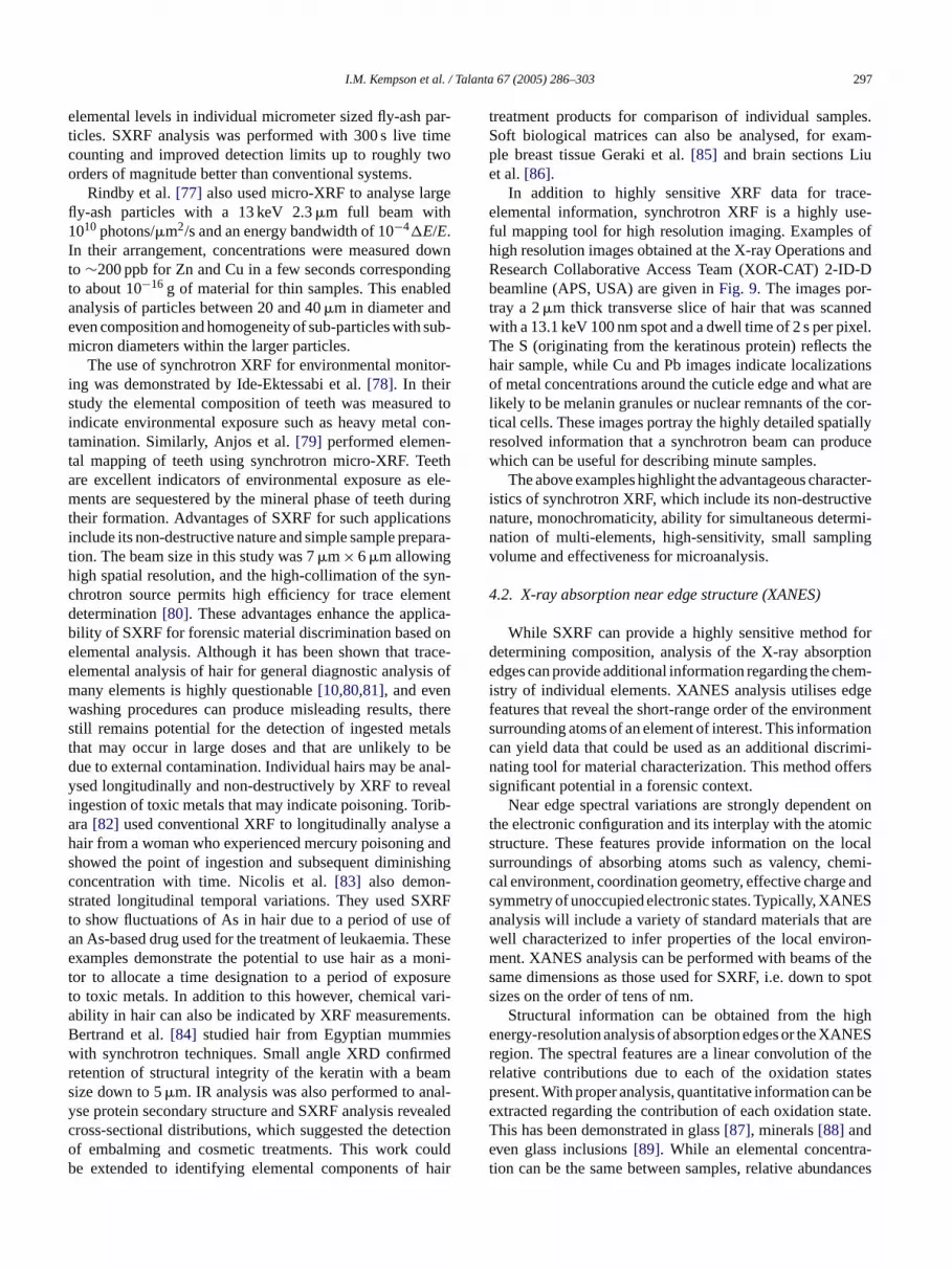
In addition to highly sensitive XRF data for trace-elemental information, synchrotron XRF is a highly use-ful mapping tool for high resolution imaging. Examples ofhigh resolution images obtained at the X-ray Operations andResearch Collaborative Access Team (XOR-CAT) 2-ID-Dbeamline (APS, USA) are given inFig. 9. The images por-tray a 2�m thick transverse slice of hair that was scannedwith a 13.1 keV 100 nm spot and a dwell time of 2 s per pixel.The S (originating from the keratinous protein) reflects thehair sample, while Cu and Pb images indicate localizationsof metal concentrations around the cuticle edge and what arelikely to be melanin granules or nuclear remnants of the cor-tical cells. These images portray the highly detailed spatiallyresolved information that a synchrotron beam can producewhich can be useful for describing minute samples.
The above examples highlight the advantageous character-istics of synchrotron XRF, which include its non-destructivenature, monochromaticity, ability for simultaneous determi-nation of multi-elements, high-sensitivity, small samplingvolume and effectiveness for microanalysis.
4.2. X-ray absorption near edge structure (XANES)
ford tione em-i dgef ments tionc rimi-n ferss
nt ont mics locals emi-c ands ESa arew on-m f thes spots
ighe NESr f ther atesp n bee tate.Te a-t ances
etermination[80]. These advantages enhance the appility of SXRF for forensic material discrimination basedlemental analysis. Although it has been shown that tlemental analysis of hair for general diagnostic analysany elements is highly questionable[10,80,81], and evenashing procedures can produce misleading results,till remains potential for the detection of ingested mehat may occur in large doses and that are unlikely tue to external contamination. Individual hairs may be ased longitudinally and non-destructively by XRF to revngestion of toxic metals that may indicate poisoning. Tora[82] used conventional XRF to longitudinally analysair from a woman who experienced mercury poisoninghowed the point of ingestion and subsequent diminisoncentration with time. Nicolis et al.[83] also demontrated longitudinal temporal variations. They used SXo show fluctuations of As in hair due to a period of usn As-based drug used for the treatment of leukaemia. Txamples demonstrate the potential to use hair as a mor to allocate a time designation to a period of expoo toxic metals. In addition to this however, chemical vbility in hair can also be indicated by XRF measuremeertrand et al.[84] studied hair from Egyptian mummiith synchrotron techniques. Small angle XRD confirm
etention of structural integrity of the keratin with a beize down to 5�m. IR analysis was also performed to anse protein secondary structure and SXRF analysis revross-sectional distributions, which suggested the detef embalming and cosmetic treatments. This work ce extended to identifying elemental components of
While SXRF can provide a highly sensitive methodetermining composition, analysis of the X-ray absorpdges can provide additional information regarding the ch
stry of individual elements. XANES analysis utilises eeatures that reveal the short-range order of the environurrounding atoms of an element of interest. This informaan yield data that could be used as an additional discating tool for material characterization. This method ofignificant potential in a forensic context.
Near edge spectral variations are strongly dependehe electronic configuration and its interplay with the atotructure. These features provide information on theurroundings of absorbing atoms such as valency, chal environment, coordination geometry, effective chargeymmetry of unoccupied electronic states. Typically, XANnalysis will include a variety of standard materials thatell characterized to infer properties of the local envirent. XANES analysis can be performed with beams o
ame dimensions as those used for SXRF, i.e. down toizes on the order of tens of nm.
Structural information can be obtained from the hnergy-resolution analysis of absorption edges or the XAegion. The spectral features are a linear convolution oelative contributions due to each of the oxidation stresent. With proper analysis, quantitative information caxtracted regarding the contribution of each oxidation shis has been demonstrated in glass[87], minerals[88] andven glass inclusions[89]. While an elemental concentrion can be the same between samples, relative abund

298 I.M. Kempson et al. / Talanta 67 (2005) 286–303
Fig. 9. Synchrotron XRF mapping of a 2�m thick hair section. The optical micrograph indicates the areas over which elemental maps were produced with a100 nm spot size and 2 s dwell time per pixel. The bar = 10 and 2�m for the larger and smaller images, respectively.
in oxidation states can be different providing an additionaldegree of discrimination. The data can reflect environmentalconditions during the formation of a substance, for instancethe formation of volcanic glasses, from which the oxidationstates present are used to infer redox conditions at formation.This information has obvious potential to be extended intoforensic work to identify or discriminate manufacture meth-ods of materials or the conditions under which evidence hasbeen generated.
Typical XANES analysis of particulate matter may beextended to an additional discriminating factor for particlesobtained as trace evidence such as soil, glass and GSR. In thecase of GSR there is application in studying the content andchemistry of the resulting particles. Understanding the effectof discharge on trace-elemental concentrations and the oxida-tion state and coordination of those species can help to relateor discriminate fired and unfired ammunition and differencesbetween ammunition or firearms. In addition, particles mayconclusively be discriminated from environmental particles.SEM-EDX is the most common technique for GSR anal-ysis, which has a depth resolution of approximately 1�m.Hellmiss et al.[90] applied Auger electron spectroscopy(AES) to the analysis of GSR with a greater surface sen-sitivity, generating data that originates from the uppermostatomic layers (0.3–3 nm). AES is not limited to surface anal-ysis however, and can be used simultaneously with ion sputtere ed
with windowed X-ray detectors, AES has the ability to detectelements of low atomic number. The study by Hellmiss pro-vided some insight into the complex internal composition ofGSR particles. AES was also applied by Zeichner et al.[91] tostudy the surface concentrations of antimony (0.5–3%, w/w)in hardened lead projectiles. ToF-SIMS has also been appliedto gunshot residue analysis[92,93]where it was determinedthat the elements present in GSR are not simply in elementalform but form compounds such as oxides and hydroxides.Furthermore, the presence of glass in several types of 0.22calibre ammunition was ascertained. Elemental analysis byToF-SIMS allowed characterization of the glass present andthe linking of residues with putative source ammunition.Synchrotron analysis (XRF, XANES, AES, etc.) however,can detect all elemental constituents in a completely non-destructive manner, with or without surface sensitivity, withthe added benefit of deducing elemental oxidation states andcoordinations. Examples of Fe K-edge XANES spectra areprovided inFig. 10 which were acquired at the AustralianNational Beamline Facility at the Photon Factory (Japan).Several spectra from standard materials are included alongwith data recorded from a group of glass-containing GSR.The XANES spectrum from the GSR appears to be a convo-lution of different chemical forms. A significant proportionresembles the spectrum of iron metal, while comparison withFe compounds presented by Westre et al.[94], indicates thep r
tching to allow depth profiling. Unlike SEM-EDX equipp ossibility of an octahedral FeIII complex. Other oxides o
I.M. Kempson et al. / Talanta 67 (2005) 286–303 299
Fig. 10. Fe-K XANES spectra from a variety of iron containing compounds.The spectrum of the iron content in the GSR particles indicates similaritiesto a combination of iron metal and a higher coordination phase.
iron sulphate do not appear to have contributed in any signif-icance.
The low energy photoelectrons produce analysis of a sur-face sensitive nature, which can also be utilised for theXANES technique. Kawai[95] have shown that S (0.1–0.3%)exists differently in the bulk and surface of fly-ash powders(70% of particles had diameters less than 5�m) with simulta-neous total electron yield and XRF measurements. The bulkand surface S composition were characterized. The outersurface exhibited S6+ while the core was a mixture of S2−and S6+. They also indicated that the ratio of the oxidationstates varied depending on the sources of coal, and hencethis information has potential in discriminating such samples[96].
Fe oxidation state ratios have received the focus of manyXANES studies[94], especially in glasses[97]. It follows thatXANES analysis could extend itself to forensic discrimina-tion of glass. The glass industry has demonstrated applicationin analysis of the depth dependence on the Sn, Fe and S oxi-dation states in float glass[98]. In addition to analysis ofFe2+ and Fe3+ ratios in glass, they have also been examinedin historic inks[99]. Work of Coumbaros using ToF-SIMSdemonstrates the presence of various inorganic elements aswell as the organic dyes[100]. It follows that there is scope toenhance discrimination using XANES for these components.Here, XANES analysis could be applied to identifying thes anica
tiliseX iquef nts.A ysesa suchm cteri-z siso tingm testu ointi eenp
4.3. Extended X-ray absorption fine structure (EXAFS)
High resolution analysis of absorption effects can addi-tionally produce EXAFS spectra which extends approxi-mately 1000 eV above the absorption edge for an elementaltransition. The information extracted from such spectra pro-vides medium range order, giving type of ligating atomsand very accurate metal–ligand distances, but little informa-tion regarding coordination and active site geometry. Samplepreparation is minimal and does not require complicatingextraction, dissolution or concentration procedures.
Huggins et al.[101] performed XAFS analysis of S, Cl,V, Cr, Cu, Zn, As, Br, Cd and Pb as well as Mossbauer anal-ysis for Fe on NIST particulate matter SRMs 1648 and 1650.This work demonstrates the ability to identify the specia-tion of each element and produce a highly detailed descrip-tion of the composition of a material. A major conclusionindicated that differences in EXAFS data could be used ascharacteristic signatures to source particulate matter. This israther more intensive analysis than XANES but is somewhatmore rewarding in characterizing samples, in particular whenanalysed in parallel. Relative changes in coordination num-ber between samples of borate glasses with equal elemental(samarium) concentrations has been shown from XAFS spec-tra [102]. Other highly detailed studies have been reportedwhere the metal contents of contaminated soils have beenc anr ichm therc ichm etalsi har-at
ofX ameb gen-e lyd thati pre-h log-ia cter-i
5
GSRph sci-e m-p ing.S ddi-t r-m ques
ource of inks as well as studying alteration of both orgnd inorganic components over time.
The amount of materials research available that uANES analysis demonstrates this as a powerful techn
or understanding the chemistry of compositional elemelthough there are not many examples of XANES analpplied to forensic samples, there is the potential ofeasurements providing an additional degree of chara
ation. It is unlikely that XANES will add value to analyf those types of evidence that yield highly discriminaulti-trace element profiles, it would appear to be of greatility where only a few elements are present. A case in p
s GSR, where elemental ratios are highly variable betwarticles from the same source.
haracterized[103,104]. While elemental compositions ceflect a site of origin of soil material, the manner in whetals are incorporated is highly variable and reflect o
hemical information regarding the origins, e.g. pH, whay not be able to be measured from trace evidence. M
n soils are bound in different forms depending on the ccteristics of the bulk soil properties such as pH[105] and
he amount of organic matter[106].To make EXAFS analysis more powerful, acquisition
-ray diffraction data can often be acquired on the seamlines and hence from identical positions used forrating EXAFS spectra[107]. This can provide a highetailed reconstruction of the composition of materials
s not available from any other instrumental setup. A comensive review of synchrotron techniques applied to geo
cal samples has been given elsewhere[108] including X-raybsorption, diffraction and fluorescence studies to chara
ze geological samples.
. X-ray diffraction (XRD) and scattering
Conventional XRD has been used to characterizearticles [109] and, soils and rocks[110]. Rendle [111]as recently reviewed conventional XRD in forensicnce. XRD is useful in the identification of mineral coosition; however, data collection can be time consumynchrotron XRD alleviates this issue, and has the a
ional ability of providing 2D diffraction patterns. Furtheore, the application of synchrotron micro-beam techni

300 I.M. Kempson et al. / Talanta 67 (2005) 286–303
allows for localised analysis. Broekmans et al.[112] inves-tigated the application of synchrotron radiation XRD for theanalysis of ancient (third millennium BC) cooking pottery.Data regarding the pottery’s mineralogy was used to makeinferences concerning pottery technology (i.e. clay prepa-ration and firing techniques). Synchrotron XRD was alsoused by Dooryhee et al.[113] for the analysis of art andarchaeological material in order to associate them with apossible source, their provenance and means of manufac-ture. Hard X-ray microprobe beamlines often have capabil-ities to perform combined XRF mapping and spectroscopywith micro-XRD[114]. Powder diffraction experiments per-formed with synchrotron sources are also very rapid andsensitive[115,116]. Other interesting reports include highresolution experiments at SPring-8[117] and time-resolvedexperiments at the ESRF[118]. Others have described micro-diffraction of protein crystals at the ESRF[119] and sin-gle crystals at the APS[120]. diffraction anomalous fine-structure (DAFS)[121]and protein crystallography by singleand multiple anomalous diffraction (SAD/MAD)[122,123]are also beneficiary techniques. Systems designed for diffrac-tion experiments are becoming highly automated[124,125]with high throughput[126]. Some beamlines have becomevery versatile such as at the SRS where in addition to offer-ing combined XRD and XAS, small- and wide-angle X-rayscattering (SAXS/WAXS) are also offered on the one line[ uralf ar-a otherm chp RDO ludeh -t yH ctso hae-o sefuli
notb robef eend dingi tionw useo abil-i tialr
6
eityn rtiesb ighlyl vol-u lyses
from other volumes returns the same result within the errorof the instrument, i.e. the measurement is representative ofthe bulk property. As the analysis volume is decreased, itbecomes more probable that fluctuations in the property willarise from inhomogeneity. At some point, while the instru-ment is still capable of an accurate measurement, the errorbetween alternative analysis volumes has increased beyondthe instrument error. To understand what the minimum sizeof a useful piece of evidence is, it is necessary to find thelimit at which the real error between different volumes ofthe same sample exceeds, by an unacceptable amount, theinstrumental error. What is of interest is to find the ultimatelimit of distinguishability, i.e. what is the smallest usefulpiece of trace evidence that can be used for the purpose ofreliable comparison with its putative source or other relatedpieces of evidence? Although statistics can be used to averageover many sample volumes, in microscopic pieces of mate-rial there might not be sufficient amount of sample to obtainsuch statistics.
Microheterogeneity is likely to be dependant on manyfactors and vary between samples, materials and also onthe actual property being measured. Although a sample canbe broken down into smaller pieces until the limit due toheterogeneity is deduced, it is impossible to say that onefragment from another sample is also limited by the samevolume requirement for homogeneity. However, understand-i dingc entsf pleso ely,i rti-c tativeo
rorso con-c ers eta usa mea-s iticalm ampleb RFa ISTS s ref-e oig theX thisw ativeot Nb,MS on, aM tero-g f thee t least1 at of
127]. SAX can be applied to the analysis of microstructeatures in the 10–1000A domain and is often applied to chcterising size, shape and orientation of polymers andaterials. WAX is typically performed in conjunction, whirovides information on a much smaller scale similar to Xther examples of scattering experiment beamlines incigh energy diffuse scattering at the APS[128] and inelas
ic X-ray scattering at LNLS[129]. An interesting study biller et al. [130] used SAXS to investigate heating effen bone mineralisation, recognising the forensic and arclogical applications. These techniques would also be u
n characterising the composition of GSR.While capabilities regarding spatial resolution have
een exhibited, the combined analysis of a micropor XRF, XAFS analysis and XRD experiments have bemonstrated to be a compilation of techniques provi
ncredibly detailed compositional and structural informaith sensitivity and selectivity not possible without thef a synchrotron source. Such papers demonstrate the
ty to characterize materials in high detail with high spaesolution.
. Microheterogeneity studies
With the increase in spatial resolution, microheterogeneeds to be considered when trying to infer bulk propeased on a technique which, although sensitive, is also h
ocalised. If a property is measured in a randomly chosenme of a sample it is desirable that any subsequent ana
ng typical limitations can offer valuable assistance in adonfidence to the value placed on individual measuremrom low sample numbers of minute evidence if other samf similar origin have been well characterized. Alternativ
t might give some indication to the number of minute pales required for an average to be derived that is represenf the bulk material.
It is therefore necessary to clearly define the erbtained due to the experiment and real fluctuations inentration. This has been demonstrated by Kempenal. [69,131,132]. The former publication discusses variopproaches, causes and descriptions of heterogenousurements, and specifically discusses obtaining the crass required for analysis to represent a homogenous sy SXRF. Of most interest is the work that used micro-Xs a means of describing the microheterogeneity of NRM 613 (trace-elements in 1 mm glass wafers), a glasrence material. An X-ray beam of 5�m× 8�m was used t
nterrogate the NIST glass wafer at 441 positions (a 21× 21rid pattern) and intra- and inter-regional variances inRF intensities for many elements were measured. Inay the minimum mass of glass analytically representf the entire specimen was estimated to be 0.1�g for cer-
ain trace elements (Ni, Cu, Zn, Ga, Ge, Se, Rb, Sr, Y,o and Pb). At a later point[69], similar analysis of NISTRM 613 was presented. In this subsequent publicationte Carlo model was used to assist in the study heeneity. For a particle to appear homogenous for all olements measured in that study, a sample mass of a00 ng was suggested. Assuming a density equal to th

I.M. Kempson et al. / Talanta 67 (2005) 286–303 301
SiO2 (2.6 g cm−3), then this would relate to a spherical parti-cle with a radius of∼21�m. However, other elements suchas Se only require 1 ng (or roughly a sphere of 4.5�m radius).NIST SRM 610–617 glasses are commonly used for ele-mental analysis. LA-ICP-MS analysis of these standards byEggins and Shelley[133] revealed that all the standards hadregions where at least 25 elements were either depleted orenriched. Additional standards that are being employed forLA-ICP-MS are the Schott manufactured float glass stan-dards FGS1 and FGS2[46], which are also currently beingassessed by us using ToF-SIMS. Ongoing work by us hasdemonstrated that tin penetrates into both the float and non-float surfaces with concomitant depletion of certain elementswithin the glass.
In addition to variability in concentration, microhetero-geneity may also influence chemical features. Cho et al.[17]observed variability between fibres from the same sourcerelating to microheterogeneity using IR analysis. EXAFS canalso be used to study heterogeneity of speciation of metals.Sulphur species have been found to be inhomogenous in soils.Sulphur compounds were quantified in bulk samples as wellas individual particles at hundreds of nm resolution[134].
Understanding microheterogeniety in trace evidence isvital to enable confident discrimination and characterizationof samples and determining adequate sample size. Micro-p of ah thate nablet
7
rcesp ns.H stedi ibil-i basisw icatet ar-r tiont om-m racee art oc ec-i tweens haveb s, thee allyw ed tos ivedG an isc omG ed byTa d a
potential for linking GSR with putative source ammunition;synchrotron-based techniques have the capability to providevaluable information. Substantial work has been devoted tothe exploration of elemental profiling as a means of compar-ing glass specimens, but at what size does a microscopic par-ticle no longer represent the bulk glass composition, and howclose to the float surface does the composition represent thebulk?
Compared to conventional techniques, synchrotron tech-niques are capable of providing the highest level of charac-terization in a non-destructive manner. Due to a relativelyrecent advent of the most powerful techniques, the potentialcharacterizing power achievable in the near future promisesto be an exciting time for taking trace evidence analysis tolimits not seen before.
Acknowledgements
This work was supported by the Australian SynchrotronResearch Program, which is funded by the Commonwealthof Australia under the Major National Research FacilitiesProgram. Part of this work was performed at the AustralianNational Beamline Facility, also supported by the AustralianSynchrotron Research Program. Use of the Advanced Pho-t ergy,O t No.W atedb .S.D alsow arryF ther tt andG
R
ncesn of
95)
95)
pli-UV
, pp.
ey,a, S.002)
s.
robe synchrotron techniques offer the characteristicsighly sensitive, localised and non-destructive analysisnable detailed studies in this area of research and will e
he determination of such limits.
. Summary
Obviously the extremely high-cost of synchrotron soureclude their utilization in routine forensic investigatioowever, a substantial number of countries have inve
n national synchrotron facilities, opening up the possty that synchrotrons could be used on an as neededhen conventional analysis and case significance ind
hat further discrimination power is both possible and wanted. Another major use of synchrotrons is in relao fundamental research into the nature of materials conly encountered in forensic casework, particularly t
vidence. Such research underpins and advances theasework by indicating new points of discrimination of spmens, and new means for establishing associations bepecimens. For example, although gunshot residueseen analysed by forensic scientists for many decadexact composition of primer-derived residues is not reell described. Sophisticated new techniques are requirhed more light onto the exact chemistry of primer-derSR in order to identify whether there is more scope thurrently thought for the discrimination of primer GSR frSR-like environmental particles, such as those describorre et al.[135]. As described by Romolo and Margot[136]nd Coumbaros et al.[92], respectively, there is a need an
f
on Source was supported by the U.S. Department of Enffice of Science, Basic Energy Sciences, under Contrac-31-109-Eng-38. Argonne National Laboratory is oper
y The University of Chicago under contract with the Uepartment of Energy, Office of Science. The authorsish to thank Francesco De Carlo, Cathy Harland, Goran and Barry Lai for their assistance in acquiringesults presented here. The assistance of Richard Garreerry Roe was also appreciated.
eferences
[1] Synchrotron Radiation: Earth, Environmental and Material ScieApplications. Short Course Series 30, Mineralogical AssociatioCanada, 2002.
[2] G.L. Carr, J.A. Reffner, G.P. Williams, Rev. Sci. Instrum. 66 (191490–1492.
[3] J.A. Reffner, P.A. Martoglio, Rev. Sci. Instrum. 66 (191298–1302.
[4] P. Dumas, G.P. Williams, in: T.-K. Sham (Ed.), Chemical Apcations of Synchrotron Radiation, Part I: Dynamcis and VSpectroscopy, World Scientific Publishing, Singapore, 2002356–386.
[5] N.S. Marinkovic, R. Huang, P. Bromberg, M. Sullivan, J. ToomL.M. Miller, E. Sperber, S. Moshe, K.W. Jones, E. ChouparovLappi, S. Franzen, M.R. Chance, J. Synchrotron Radiat. 9 (2189–197.
[6] G.L. Carr, Vib. Spectrosc. 19 (1999) 53–60.[7] G.L. Carr, Rev. Sci. Instrum. 72 (2001) 1613–1619.[8] K.S. Kalasinsky, Cell. Mol. Biol. 44 (1998) 81–87.[9] P. Dumas, J. Phys. IV 104 (2003) 359–364.
[10] I.M. Kempson, W.M. Skinner, P.K. Kirkbride, Biochim. BiophyActa 1624 (2003) 1–5.

302 I.M. Kempson et al. / Talanta 67 (2005) 286–303
[11] T.J. Wilkinson, D.L. Perry, M.C. Martin, W.R. McKinney, A.A.Cantu, Appl. Spectrosc. 56 (2002) 800–803.
[12] T.J. Wilkinson, D.L. Perry, M.C. Martin, W.R. McKinney, Proceed-ings of the 225th American Chemical Society National Meeting,U975–U976 157–IEC Part 1, New Orleans, USA, 2003.
[13] T.J. Wilkinson, D.L. Perry, M.C. Martin, W.R. McKinney, Proceed-ings of the 225th American Chemical Society National Meeting,U976–U976 158–IEC Part 1, New Orleans, USA, 2003.
[14] E.M. Suzuki, J. Forensic Sci. 44 (1999) 1151–1175.[15] R.A. Merrill, E.G. Bartick, J. Forensic Sci. 45 (2000) 93–98.[16] S. Stich, D. Bard, L. Gros, H.W. Wenz, J. Yarwood, K. Williams,
J. Raman Spectrosc. 29 (1998) 787–790.[17] L. Cho, J.A. Reffner, D.L. Wetzel, J. Forensic Sci. 44 (1999)
283–291.[18] M.W. Tungol, E.G. Bartick, A. Montaser, in: H.J. Humecki (Ed.),
Practical Guide to Infrared Microspectroscopy, Marcel Dekker Inc.,New York, 1995, pp. 245–286.
[19] L. Cho, J.A. Reffner, B.M. Gatewood, D.L. Wetzel, J. Forensic Sci.44 (1999) 275–282.
[20] G. Ellis, M.A. Gomez, C. Marco, J. Macromol. Sci. Phys. B43(2004) 191–206.
[21] Y. Wang, F. DeCarlo, D.C. Mancini, I. McNulty, B. Tieman, J.Bresnahan, I. Foster, J. Insley, P. Lane, G. von Laszewski, C.Kesselman, M.-H. Su, M. Thiebaux, Rev. Sci. Instrum. 72 (2001)2062–2068.
[22] L. Niewohner, H.W. Wenz, J. Forensic Sci. 44 (1999) 105–109.[23] D.V. Rao, T. Takeda, T. Kawakami, K. Uesugi, Y. Tsuchiya, J. Wu,
T.T. Lwin, Y. Itai, T. Zeniya, T. Yuasa, T. Akatsuka, Nucl. Instrum.Meth. A 523 (2004) 206–216.
[24] M. Kocsis, A. Snigirev, Nucl. Instrum. Meth. A 525 (2004) 79–84.[25] S.K. Sinha, Radiat. Phys. Chem. 70 (2004) 633–640.
lves,421–
, P.01.on
ao,
g, J.
96.ch-
.E.n-
rt,er, I.ng,4.k,tron
yer,
000)
or, B.
37
Soc.
trom.
[42] J. Kunicki-Goldfinger, J. Kierzek, A. Kasprzak, B. Malozewska-Bucko, X-ray Spectrom. 29 (2000) 310–316.
[43] S. Becker, L. Gunaratnum, T. Hicks, W. Stoecklein, G. Warman,Probl. Forensic Sci. XLVII (2001) 80–92.
[44] T. Hicks, F. Monard Sermier, T. Goldman, A. Brunelle, C. Cham-pod, P. Margot, Forensic Sci. Int. 137 (2003) 107–118.
[45] S. Becker, W. Stoecklein, Trace Session of the Second EuropeanAcademy of Forensic Sciences Meeting, Cracow, Poland, 2000.
[46] J.R. Almirall, NITECRIME Workshop, Wellington, New Zealand,2004.
[47] T. Trejos, S. Montero, J.R. Almirall, Anal. Bioanal. Chem. 376(2003) 1255–1264.
[48] R.J. Watling, Proceedings of the Second European Academy ofForensic Sciences Meeting, Cracow, Poland, 2000.
[49] R.J. Watling, B.F. Lynch, D. Herring, J. Anal. Atom. Spectrom. 12(1997) 195–203.
[50] R.D. Dixon, R.K.W. Merkle, A. Kijko, NITECRIME Workshop,Wellington, New Zealand, 2004.
[51] R.J. Watling, NITECRIME Workshop, Wellington, New Zealand,2004.
[52] J.R. Almirall, K.G. Furton, Proceedings of the 16th ANZFSS Sym-posium on the Forensic Sciences, Canberra, Australia, 2002.
[53] F. Adams, K. Janssens, A. Snigirev, J. Anal. Atom. Spectrom. 13(1998) 319–331.
[54] K. Janssens, G. Vittiglio, I. Deraedt, A. Aerts, B. Vekemans, L.Vincze, F. Wei, I. Deryck, O. Schalm, F. Adams, A. Rindby, A.Knochel, A. Simionovici, A. Snigirev, X-ray Spectrom. 29 (2000)73–91.
[55] S. Muratsu, T. Ninomiya, Y. Kagoshima, J. Matsui, J. Forensic Sci.47 (2002) 944–949.
[56] M.J. Anjos, R.T. Lopes, E.F.O. de Jesus, S. Moreira, R.C. Barroso,
sus,
. 17
t. 8
, Y.
eth.
t. 9
t. 9
tron
8.Syn-
itt,t. 9
ogyi,02)
, P.
ec-
–452.
sic
[26] C. Giles, M.G. Honnicke, R.T. Lopes, H.S. Rocha, O.D. ConcaI. Mazzaro, C. Cusatis, J. Synchrotron Radiat. 10 (2003)423.
[27] J. Baruchel, P. Cloetens, J. Hartwig, W. Ludwig, L. ManciniPernot, M. Schlenker, J. Synchrotron Radiat. 7 (2000) 196–2
[28] F. D’Acapito, I. Davoli, P. Ghigna, S. Mobilio, J. SynchrotrRadiat. 10 (2003) 260–264.
[29] F. Xu, W. Liu, S. Wei, C. Xu, G. Pan, X. Zhang, J. Sun, W. ZhH. Cui, W. Ye, J. Synchrotron Radiat. 8 (2001) 348–350.
[30] S. Heald, E. Stern, D. Brewe, R. Gordon, D. Crozier, D. JianCross, J. Synchrotron Radiat. 8 (2001) 342–344.
[31] J. Stoehr, NEXAFS Spectroscopy, Springer Verlag, Berlin, 19[32] A.L.D. Kilcoyne, T. Tylisczcak, W.F. Steele, S. Fakra, P. Hit
cock, K. Franck, E. Anderson, B. Harteneck, E.G. Rightor, GMitchell, A.P. Hitchcock, L. Yang, T. Warwick, H. Ade, J. Sychrotron Radiat. 10 (2003) 125–136.
[33] B. Winn, H. Abe, C. Buckley, M. Feser, M. Howells, S. HulbeC. Jacobsen, K. Kaznacheyev, J. Kirz, A. Osanna, J. MasMcNulty, J. Miao, T. Oversluizen, S. Spector, B. Sullivan, Y. WaS. Wirick, H. Zhang, J. Synchrotron Radiat. 7 (2000) 395–40
[34] T. Warwick, H. Ade, D. Kilcoyne, M. Kritscher, T. TylisczcaS. Fakra, A. Hitchcock, P. Hitchcock, H. Padmore, J. SynchroRadiat. 9 (2002) 254–257.
[35] F. Sirotti, F. Polack, J.L. Cantin, M. Sacchi, R. Delaunay, M. MeM. Liberati, J. Synchrotron Radiat. 7 (2000) 5–11.
[36] C. Jacobsen, S. Wirick, G. Flynn, C. Zimba, J. Microsc. 197 (2173–184.
[37] J. Maser, A. Osanna, Y. Wang, C. Jacobsen, J. Kirz, S. SpectWinn, D. Tennant, J. Microsc. 197 (2000) 68–79.
[38] M. Polentarutti, R. Glazer, K.D. Carugo, J. Appl. Crystallogr.(2004) 319–324.
[39] C.R. Howden, R.J. Dudley, K.W. Smalldon, J. Forensic Sci.18 (1978).
[40] S. Ryland, J. Forensic Sci. 31 (1986) 1314–1329.[41] R.D. Koons, C.A. Peters, P.S. Rebbert, J. Anal. Atom. Spec
6 (1991) 451–456.
C.R.F. Castro, Spectrochim. Acta B 58 (2003) 2227–2232.[57] A.C.M. Costa, M.J. Anjos, S. Moreira, R.T. Lopes, E.F.O. de Je
Spectrochim. Acta B 58 (2003) 2199–2204.[58] S. Suzuki, Y. Suzuki, M. Kasamatsu, Jpn. Soc. Anal. Chem
(Suppl.) (2001) i163–i164.[59] I. Nakai, Y. Terada, M. Itou, Y. Sakurai, J. Synchrotron Radia
(2001) 1078–1081.[60] Y. Suzuki, M. Kasamatsu, R. Sugita, H. Ohta, S. Suzuki
Marumo, Bunseki Kagaku 52 (2003) 469–474.[61] Z. Smit, K. Janssens, K. Proost, I. Langus, Nucl. Instrum. M
B 219–220 (2004) 35–40.[62] V. Cherezov, K.M. Riedi, M. Caffrey, J. Synchrotron Radia
(2002) 333–341.[63] P. O’Neill, D.L. Stevens, E.F. Garman, J. Synchrotron Radia
(2002) 329–332.[64] T. Teng, K. Moffat, J. Synchrotron Radiat. 7 (2000) 313–317.[65] R. Muller, E. Weckert, J. Zellner, M. Drakopoulos, J. Synchro
Radiat. 9 (2002) 368–374.[66] E. Garman, C. Nave, J. Synchrotron Radiat. 9 (2002) 327–32[67] R.B.G. Ravelli, P. Theveneau, S. McSweeney, M. Caffrey, J.
chrotron Radiat. 9 (2002) 355–360.[68] B.L. Hanson, J.M. Harp, K. Kirschbaum, C.A. Schall, A. DeW
A. Howard, A.A. Pinkerton, G.J. Bunick, J. Synchrotron Radia(2002) 375–381.
[69] L. Kempenaers, K. Janssens, L. Vincze, B. Vekemans, A. SomM. Drakopoulos, A. Simionovici, F. Adams, Anal. Chem. 74 (205017–5026.
[70] P. Philippot, B. Menez, P. Chevallier, F. Gibert, F. LegrandPopulus, Chem. Geol. 144 (1998) 121–136.
[71] B. Kanngiesser, Spectrochim. Acta B 58 (2003) 609–614.[72] J. Kawai, S. Tohno, Y. Kitajima, O.E. Raola, M. Takaoka, Sp
trochim. Acta B 58 (2003) 635–639.[73] B. Charpentier, C. Desrochers, J. Forensic Sci. 45 (2000) 447[74] J. Brazeau, R.K. Wong, J. Forensic Sci. 42 (1997) 424–428.[75] J. Flynn, M. Stoilovic, C. Lennard, I. Prior, H. Kobus, Foren
Sci. Int. 97 (1998) 21–36.

I.M. Kempson et al. / Talanta 67 (2005) 286–303 303
[76] L. Vincze, A. Somogyi, J. Osan, B. Vekemans, S. Torok, K.Janssens, F. Adams, Anal. Chem. 74 (2002) 1128.
[77] A. Rindby, K. Janssens, J. Osan, X-ray Spectrom. 32 (2003)248–257.
[78] A. Ide-Ektessabi, K. Shirasawa, A. Koizumi, M. Azechi, Nucl.Instrum. Meth. B 213 (2004) 761–765.
[79] M.J. Anjos, R.C. Barroso, C.A. Perez, D. Braz, S. Moreira,K.R.H.C. Dias, R.T. Lopes, Nucl. Instrum. Meth. B 213 (2004)569–573.
[80] I.M. Kempson, W.M. Skinner, P.K. Kirkbride, A.J. Nelson, A.M.Martin, Eur. J. Mass Spectrom. 9 (2003) 589–597.
[81] I.M. Kempson, W.M. Skinner, K.P. Kirkbride, Sci. Total Environ.9 (2003) 589–597.
[82] T.Y. Toribara, Instrum. Sci. Technol. 23 (1995) 217–226.[83] I. Nicolis, P. Deschamps, E. Curis, O. Corriol, V. Acar, N. Zerrouk,
J.C. Chaumeil, F. Guyon, S. Benazeth, J. Synchrotron Radiat. 8(2001) 984–986.
[84] L. Bertrand, J. Doucet, P. Dumas, A. Simionovici, G. Tsoucaris, P.Walter, J. Synchrotron Radiat. 10 (2003) 387–392.
[85] K. Geraki, M.J. Farquharson, D.A. Bradley, R.P. Hugtenburg, Nucl.Instrum. Meth. B 213 (2004) 564–568.
[86] N.Q. Liu, F. Zhang, X.F. Wang, Z.Y. Zhang, Z.F. Chai, Y.Y. Huang,W. He, X.Q. Zhao, A.J. Zuo, R. Yang, Spectrochim. Acta B 59(2004) 255–260.
[87] A.J. Berry, H.S.C. O’Neill, K.D. Jayasuriya, S.J. Campbell, G.J.Foran, Am. Mineral. 88 (2003) 967–977.
[88] L. Galoisy, G. Calas, M.A. Arrio, Chem. Geol. 174 (2001)307–319.
[89] M. Bonnin-Mosbah, A.S. Simionovici, N. Metrich, J.P. Duraud,D. Massare, P. Dillmann, J. Non-Cryst. Solids 288 (2001) 103–113.
987)
998)
Sci.
W.
dg-4.Y.
., K.95)
.S.
99)
Nucl.
ity of
r. 74
222
nal.
97)
in,1.156., B.
[108] C.M.B. Henderson, G. Cressey, S.A.T. Redfern, Radiat. Phys.Chem. 45 (1995) 459–481.
[109] M. Tassa, Y. Leist, M. Steinberg, J. Forensic Sci. 27 (1982)677–683.
[110] A. Ruffell, P. Wiltshire, Forensic Sci. Int. 145 (2004) 13–23.[111] D.F. Rendle, Rigaku J. 19–20 (2003) 11–22.[112] T. Broekmans, A. Adriaens, E. Pantos, Nucl. Instrum. Meth. B 226
(2004) 92–97.[113] E. Dooryhee, P. Martinetto, P. Walter, M. Anne, Radiat. Phys.
Chem. 71 (2004) 863–868.[114] M.A. Marcus, A.A. MacDowell, R. Celestre, A. Manceau, T.
Miller, H.A. Padmore, R.E. Sublett, J. Synchrotron Radiat. 11(2004) 239–247.
[115] M. Knapp, C. Baehtz, H. Ehrenberg, H. Fuess, J. SynchrotronRadiat. 11 (2004) 328–334.
[116] C. Meneghini, G. Artioli, A. Balerna, A.F. Gualtieri, P. Norby, S.Mobilio, J. Synchrotron Radiat. 8 (2001) 1162–1166.
[117] T. Ikeda, A. Nisawa, M. Okui, N. Yagi, H. Yoshikawa, S.Fukushima, J. Synchrotron Radiat. 10 (2003) 424–429.
[118] A. Martorana, G. Deganello, A. Longo, F. Deganello, L. Liotta, A.Macaluso, G. Pantaleo, A. Balerna, C. Meneghini, S. Mobilio, J.Synchrotron Radiat. 10 (2003) 177–182.
[119] C. Riekel, J. Synchrotron Radiat. 11 (2004) 4–6.[120] W.R. Wikoff, W. Schildkamp, R.E. Johnson, Acta Crystallogr. D
56 (2000) 890–893.[121] H. Renevier, S. Grenier, S. Arnaud, J.F. Berar, B. Caillot, J.L.
Hodeau, A. Letoublon, M.G. Proietti, B. Ravel, J. SynchrotronRadiat. 10 (2003) 435–444.
[122] E. Pohl, A. Gonzalez, C. Hermes, R.G. van Silfhout, J. SynchrotronRadiat. 8 (2001) 1113–1120.
[123] M. Roth, P. Carpentier, O. Kaikati, J. Joly, P. Charrault, M. Piroc-toni,05–
chel,.-L.
l, M.llogr.
Hel-
kun,.M.rrill,
lmer,
004)
T.J.
B 55
mans,om.
002)
bner,
. 47
s oftific
[90] G. Hellmiss, W. Lichtenberg, M. Weiss, J. Forensic Sci. 32 (1747–760.
[91] A. Zeichner, B. Schecter, R. Brener, J. Forensic Sci. 43 (1493–501.
[92] J. Coumbaros, K.P. Kirkbride, G. Klass, W. Skinner, ForensicInt. 119 (2001) 72–81.
[93] P. Collins, J. Coumbaros, G. Horsley, B. Lynch, P.K. Kirkbride,Skinner, G. Klass, J. Forensic Sci. 48 (2003).
[94] T.E. Westre, P. Kennepohl, J.G. DeWitt, B. Hedman, K.O. Hoson, E.I. Solomon, J. Am. Chem. Soc. 119 (1997) 6297–631
[95] J. Kawai, K. Hayakawa, S.Y. Zheng, Y. Kitajima, H. Adachi,Gohshi, F. Esaka, K. Furuya, Physica B 209 (1995) 237–238
[96] J. Kawai, S. Hayakawa, F. Esaka, S.Y. Zheng, Y. KitajimaMaeda, H. Adachi, Y. Gohshi, K. Furuya, Anal. Chem. 67 (191526–1529.
[97] Z.Y. Wu, M. Bonnin-Mosbah, J.P. Duraud, N. Metrich, JDelaney, J. Synchrotron Radiat. 6 (1999) 344–346.
[98] X. Li, J.A. Monroe Snider, D. Schiferl, PPG Technol. J. 5 (1931–41.
[99] K. Proost, K. Janssens, B. Wagner, E. Bulska, M. Schreiner,Instrum. Meth. B 213 (2004) 723–728.
[100] J. Coumbaros, Thesis, Ian Wark Research Institute, UniversSouth Australia, Adelaide, 2002.
[101] F.E. Huggins, G.P. Huffman, J.D. Robertson, J. Hazard. Mate(2000) 1–23.
[102] Y. Shimizugawa, J.R. Qui, K. Hirao, J. Non-Cryst. Solids(1997) 310–315.
[103] E. Welter, W. Calmano, S. Mangold, L. Troger, Fresenius J. AChem. 364 (1999) 238–244.
[104] M.L. Peterson, G.E. Brown, G.A. Parks, J. Phys. IV 7 (19781–783.
[105] M.I. Boyanov, S.D. Kelly, K.M. Kemner, B.A. Bunker, J.B. FeD.A. Fowle, Geochim. Cosmochim. Acta 67 (2003) 3299–331
[106] D.G. Strawn, D.L. Sparks, Soil Sci. Soc. Am. J. 64 (2000) 144–[107] A. Manceau, M.A. Marcus, N. Tamura, O. Proux, N. Geoffroy
Lanson, Geochim. Cosmochim. Acta 68 (2004) 2467–2483.
chi, R. Kahn, E. Fanchon, L. Jacquamet, F. Borel, A. BerP. Israel-Gouy, J.-L. Ferrer, Acta Crystallogr. D58 (2002) 8814.
[124] J. Ohana, L. Jacquamet, J. Joly, A. Bertoni, P. Taunier, L. MiP. Charrault, M. Pirocchi, P. Carpentier, F. Borel, R. Kahn, JFerrer, J. Appl. Crystallogr. 37 (2004) 72–77.
[125] L. Jacquamet, J. Ohana, J. Joly, P. Legrand, R. Kahn, F. BorePirocchi, P. Charrault, P. Carpentier, J.-L. Ferrer, Acta CrystaD60 (2004) 888–894.
[126] A. Olczak, M. Cianci, Q. Hao, P.J. Rizkallah, J. Raftery, J.R.liwell, Acta Crystallogr. A59 (2003) 327–334.
[127] R.J. Cernik, P. Barnes, G. Bushnell-Wye, A.J. Dent, G.P. DiaJ.V. Flaherty, G.N. Greaves, E.L. Heeley, W. Helsby, S.DJacques, J. Kay, T. Rayment, A. Ryan, C.C. Tang, N.J. TeJ. Synchrotron Radiat. 11 (2004) 163–170.
[128] T.R. Welberry, D.J. Goossens, D.R. Haeffner, P.L. Lee, J. AJ. Synchrotron Radiat. 10 (2003) 284–286.
[129] G. Tirao, G. Stutz, C. Cusatis, J. Synchrotron Radiat. 11 (2335–342.
[130] J.C. Hiller, T.J.U. Thompson, M.P. Evison, A.T. Chamberlain,Wess, Biomaterials 24 (2003) 5091–5097.
[131] L. Kempenaers, L. Vincze, K. Janssens, Spectrochim. Acta(2000) 651–669.
[132] L. Kempenaers, K. Janssens, K.P. Jochum, L. Vincze, B. VekeA. Somogyi, M. Drakopoulos, F. Adams, J. Anal. Atom. Spectr18 (2003) 350–357.
[133] S.M. Eggins, J.M.G. Shelley, Geostandard. Newslett. 26 (2269–286.
[134] J. Prietzel, J. Thieme, U. Neuhausler, J. Susini, I. Kogel-KnaEur. J. Soil Sci. 54 (2003) 423–433.
[135] C. Torre, G. Mattutino, V. Vasino, C. Robino, J. Forensic Sci(2002) 494–594.
[136] F.S. Romolo, P. Margot, Forensic Sci. Int. 119 (2001).[137] S. Heald, D. Jiang, in: T.K. Sham (Ed.), Chemical Application
Synchrotron Radiation. Part II: X-ray Applications, World ScienPublishing Co. Pty. Ltd., Singapore, 2002, pp. 761–798.


