Embed Size (px)
Citation preview
ARFGAP1 Is Dynamically Associated with Lipid Dropletsin HepatocytesJoan Gannon1.¤, Julia Fernandez-Rodriguez2., Hussam Alamri1, Shi Bo Feng1, Fariba Kalantari1,
Sarita Negi1, Amy H. Y. Wong1, Alexander Mazur1, Lennart Asp3, Ali Fazel1, Ayat Salman1,
Anthoula Lazaris1, Peter Metrakos1, John J. M. Bergeron1, Tommy Nilsson1*
1 The Research Institute of the McGill University Health Centre, McGill, Royal Victoria Hospital, 687 Pine Avenue West, Montreal, Quebec, Canada, 2 The Centre for Cellular
Imaging at the University of Gothenburg, Gothenburg, Sweden, 3 The Department of Medical and Clinical Genetics, Institute of Biomedicine, University of Gothenburg,
Gothenburg, Sweden
Abstract
The ARF GTPase Activating Protein 1 (ARFGAP1) associates mainly with the cytosolic side of Golgi cisternal membraneswhere it participates in the formation of both COPI and clathrin-coated vesicles. In this study, we show that ARFGAP1associates transiently with lipid droplets upon addition of oleate in cultured cells. Also, that addition of cyclic AMP shiftsARFGAP1 from lipid droplets to the Golgi apparatus and that overexpression and knockdown of ARFGAP1 affect lipiddroplet formation. Examination of human liver tissue reveals that ARFGAP1 is found associated with lipid droplets at steadystate in some but not all hepatocytes.
Citation: Gannon J, Fernandez-Rodriguez J, Alamri H, Feng SB, Kalantari F, et al. (2014) ARFGAP1 Is Dynamically Associated with Lipid Droplets inHepatocytes. PLoS ONE 9(11): e111309. doi:10.1371/journal.pone.0111309
Editor: Paul A. Randazzo, National Cancer Institute, United States of America
Received September 2, 2013; Accepted September 30, 2014; Published November 14, 2014
Copyright: � 2014 Gannon et al. This is an open-access article distributed under the terms of the Creative Commons Attribution License, which permitsunrestricted use, distribution, and reproduction in any medium, provided the original author and source are credited.
Funding: This study was supported by Fonds de Recherche Quebec-Sante (www.frqs.gouv.qc.ca); Canadian Institute of Health Research (www.cihr-irsc.gc.ca),#00287-003 and MOP 5605; Canadian Foundation for Innovation (www.innovation.ca), #5200 and #11476; Canada Research Chairs (www.chairs-chaires.gc.ca),#950-212342; Research Institute of the McGill University Health Centre (www.muhc.ca/research); McGill University (www.mcgill.ca); McGill University HealthCentre (www.muhc.ca). The funders had no role in study design, data collection and analysis, decision to publish, or preparation of the manuscript.
Competing Interests: The authors have declared that no competing interests exist.
* Email: [email protected]
. These authors contributed equally to this work.
¤ Current address: Chemistry Research Laboratory, University of Oxford, 12 Mansfield Road, Oxford, OX1 3TA, United Kingdom
Introduction
Both genetics- and cell biology-based experiments link COPI
coat components including the small GTPase, ARF1, coatomer
and the exchange factor, GBF1 to lipid droplet biogenesis [1–5].
ARF1 is one of 5 ARFs associated with the Golgi apparatus (for
review, see [6]) and through GTP hydrolysis catalyzed by an ARF
GTP Activating Protein (ARFGAP), regulates COPI coat forma-
tion [7–9] and sorting of proteins into COPI vesicles [10–12].
ARF1 is also linked to recruitment of lipid modifying enzymes
such as phospholipase D (PLD1) [13,14] and phosphoinositol
kinases [15] to Golgi membranes as well as GGA proteins involved
in clathrin- coated vesicle formation from Golgi membranes
[16,17]. In addition, ARF1 regulates association of adipocyte
differentiation-related protein (ADRP, PLIN2) and PLD1 on the
cytosolic leaflet of lipid droplets [18,19]. ARF1 function is
regulated by the exchange of bound GDP for GTP via an
exchange factor (e.g. GBF1) transitioning ARF1 from an inactive
to an active conformation. This is then followed by hydrolysis of
bound GTP to GDP transitioning ARF1 back to an inactive
conformation. Due to its intrinsically low GTP hydrolysis rate,
ARF1 requires an activating protein for GTP hydrolysis. In
mammalian cells, three related activating proteins termed
ARFGAP1-3 are linked to COPI vesicle formation through ARF
of which ARFGAP1 is the prototypical protein. Of the three,
ARFGAP1 appears sensitive to both membrane lipid composition
[20,21] and membrane curvature [22,23] and has also been
proposed to act as a structural component alongside coatomer to
form the COPI coat [24,25]. In addition, ARFGAP1 has been
shown to regulate AP-2-dependent endocytosis through the
binding of adaptor proteins [26] as well as LRRK2 linked to
sporadic and autosomal dominant forms of Parkinson’s disease
[27,28]. In this study, we show that ARFGAP1 is present on lipid
droplets and that it appears important for lipid droplet formation.
Lipid droplet association of ARFGAP1 is observed transiently in
cultured cells and at steady state in some but not all hepatocytes of
human liver tissue.
Results
Overexpression or knockdown of ARFGAP1 affect lipiddroplet size and numbers
In previous work examining the role of ARFGAP in vesicle
budding and scission in the Golgi apparatus of HeLa cells [20], we
noted that upon high over-expression, ARFGAP1 fused to
enhanced yellow fluorescence protein (ARFGAP1YFP) localized
to ring-like structures suggestive of lipid droplets (Fig. 1A, upper
right panel) rather than the Golgi apparatus (Fig. 1A, upper left
panel). Some over-expressing cells also revealed an intermediate
pattern with some ring-like structures positive for ARFGAP1YFP in
addition to the juxta-nuclear Golgi apparatus (Fig. 1A, upper
middle panel). When examined at the ultra structural level, lipid
PLOS ONE | www.plosone.org 1 November 2014 | Volume 9 | Issue 11 | e111309

droplet-like structures were observed surrounded by extensive
vesicular/tubular profiles (Fig. 1A, lower panels). Overexpression
of ARFGAP1 is known to affect the integrity of the Golgi
apparatus resulting in a brefeldin A-like phenotype [29] and it is
possible that observed vesicular/tubular profiles correspond to
Golgi remnants. Another possibility is that such vesicular/tubular
profiles represent smaller lipid droplets and that ARFGAP1 is
engaged in trafficking between such smaller lipid droplets and
larger ones. ARFGAP1YFP fluorescence was observed both
adjacent to and around lipid droplet-like structures (see also
Figure S1 detailing the movement of ARFGAP1YFP fluorescent
structures over the course of 40 minutes). To confirm that
observed structures were lipid-based, we transfected plasmid
DNA encoding ARFGAP1YFP into cultured HepG2 human
hepatoma cells and counter-stained with Bodipy 558/568 C12 to
stain neutral lipids. As can be seen in the upper and lower middle
panels of Figure 1B and in the enlarged region of interest (ROI),
ARFGAP1YFP localized to and near ring-like structures surround-
ing neutral lipid material when expressed at high levels as
compared to cells expressing low levels of ARFGAP1YFP (Fig. 1B
upper and lower left panels). The average size of Bodipy-stained
lipid droplets was measured in 250 cells/experiment in 3
independent experiments (see Materials and Methods). Quantifi-
cation (Fig. 1B-lower right) revealed a two-fold increase in total
lipid droplet area in cells with high (H) expression compared to
mock (M) transfected cells. Even cells with low to moderate (L–M)
expression revealed an increase in the area of lipid droplet-like
structures staining positive for Bodipy 558/568 C12. This suggests
that over-expression of ARFGAP1YFP promotes lipid droplet
formation in both HeLa and HepG2 cells. Note that in contrast to
HeLa cells, HepG2 cells have a basal level of lipid droplets.
We next examined whether knockdown of endogenous
ARFGAP1 affects lipid droplet formation in HepG2 cells. siRNA
specific for endogenous ARFGAP1 causing knockdown (KD) or
siRNA composed of a scrambled sequence serving as a mock (M)
control were transfected into HepG2 cells. At 24 hours post
transfection, cells were incubated with oleate (final concentration
at 0.5 mM) for either 1 or 4 hours. After exposure to oleate, cells
were fixed and processed for indirect immunofluorescence using
an antibody specific to ARFGAP1 [30]. In mock transfected cells
incubated with oleate for 1 hour, this antibody gave rise to a juxta
nuclear ribbon-like structure consistent with the Golgi apparatus
(Fig. 2A, upper left panel) whilst in cells transfected with siRNA
specific for ARFGAP1, only weak staining was observed consistent
with knockdown of ARFGAP1. Under these conditions, lipid
structures staining positive for Bodipy 493/503 decreased both in
area per cell and number per cell (Fig. 2 C). Total area of Bodipy-
staining of lipid droplets in each cell as well as the number of
stained lipid droplets per cell was quantified in 150 cells per
experiment in five independent experiments using ImageJ 1.45 s
(see Image Analysis in Materials and Methods). About a 60% and
45% reduction in the area and number of Bodipy positive
structures were observed upon knockdown of ARFGAP1,
respectively (Fig. 2C). The difference between mock transfected
cells and cells transfected with siRNA specific for ARFGAP1 was
less pronounced after 4 hours of incubation with oleate with
respect to number of observed droplet-like structures staining
positive for Bodipy (Figure 2B). Instead, we observed a significant
decrease in size of lipid droplets produced in cells where
ARFGAP1 expression had been diminished (compare the two
lower panels of Fig. 2B). In addition, we noted in mock transfected
cells after 4 hours of incubation of oleate, that there was an
apparent increase in cytoplasmic staining of endogenous ARF-
GAP1 (Fig. 2B upper left panel) in close proximity to structures
staining positive for Bodipy. That siRNA specific for ARFGAP1
caused knockdown of endogenous ARFGAP1 was further
examined by western blotting. Figure 2D shows that cells
transfected with siRNA specific for ARFGAP1 to cause knock-
down (KD) have a marked decrease in the antibody-generated
reaction product specific for ARFGAP1 compared to that of mock
(M) transfected cells. The ER-resident chaperone calnexin was
monitored as a control using an antibody specific for its
cytoplasmic domain [31]. Thus, decreased staining of ARFGAP1
as deduced by indirect immunofluorescence and western blotting
suggest that the majority of ARFGAP1 had been knocked down.
We conclude from this that lowering expression of endogenous
ARFGAP1 affects lipid droplet formation. Also, that exposure to
oleate for 4 hours affects localization of ARFGAP1.
ARFGAP1 is transiently recruited to lipid droplets inHepG2 cells upon addition of oleate
To investigate the localization of ARFGAP1 upon oleate
addition more precisely; we fixed and processed HepG2 cells for
indirect immunofluorescence both before and 4 hours after
addition of 0.5 mM oleate (final concentration). As shown in
Figure 3 (upper panels), there was no apparent co-localization
between endogenous ARFGAP1 and structures staining positive
for Bodipy before addition of oleate. In contrast, after 4 hours of
incubation with oleate, most of the ARFGAP1 staining was seen
surrounding Bodipy-positive lipid droplets (Fig. 3 lower panels).
The staining for ARFGAP1 was compared with a known lipid
droplet marker, PLIN3 (Perilipin 3/TIP47) which has also been
implicated in Golgi/endosomal trafficking [32]. As shown in
Figure 4 and in the enlarged region of interest (ROI), there was
considerable co-localization (yellow) between ARFGAP1 (red) and
PLIN3 (green). This shows that ARFGAP1 localizes to lipid
droplet structures 4 hours after oleate addition partly overlapping
with PLIN3. As ARFGAP1 was first identified as an activator
protein for the small GTPase ARF1 in the context of coat
regulation of COPI coatomer responsible for the generation of
COPI transport vesicles and retrograde transport in the Golgi
apparatus (for review, see [32]), it was of further interest to test
whether these and other Golgi markers also localized to lipid
droplet structures upon addition of oleate. Figure 5 row of upper
panels show the distribution of COPB1 (bCOP), a component of
the 7 subunit coatomer complex as revealed by the mouse
monoclonal antibody CM1A10 specific for one of its components
[33] before (-OA) and 4 hours after addition of oleate (4 hr OA).
Most staining forCOPB1 appeared as juxta nuclear Golgi staining
with additional punctate staining throughout the cytoplasm, a
staining pattern typical of coatomer. No apparent co-localization
with lipid droplets was observed under these conditions. ARF1 was
probed for in the hepatoma cell line, McA-RH7777 using a rabbit
polyclonal (Fig. 5, aARF1) [30] resulting in a predominant juxta
nuclear Golgi staining with occasional small punctate structures
throughout the cytoplasm of cells before addition of oleate. At
4 hours after addition of oleate, staining remained predominantly
juxta nuclear with an increase in staining of cytoplasmic structures.
A minor portion of such cytoplasmic structures appeared close to
lipid droplets as revealed by Bodipy stain. It is likely that the
antibody used here recognizes additional ARFs due to their close
homology. From above experiments, it appears that ARFGAP1
distributes to lipid droplets upon addition of oleate whereas
coatomer and most of ARF1 do not. At 4 hours after addition of
oleate, the Golgi apparatus appears intact. This was confirmed in
HepG2 cells using an antibody to TMED7 (gp27) (Fig. 5,
aTMED7), a member of the gp25L/emp24 family of small
transmembrane proteins of the early secretory pathway [34].
ARFGAP1 Is Associated with Lipid Droplets
PLOS ONE | www.plosone.org 2 November 2014 | Volume 9 | Issue 11 | e111309

Exclusive juxta nuclear Golgi staining was observed before and
4 hours after addition of oleate. An antibody to the cytoplasmic
domain of CANX (calnexin) was used to test for changes of the ER
in HepG2 cells 4 hours after addition of oleate. No discernable
difference was observed (Fig 5, aCANX). This shows that the
gross architecture of the Golgi apparatus or the ER is not affected
at 4 hours after addition of oleate despite loss of ARFGAP1 to
lipid droplets.
Figure 1. Overexpression of ARFGAP1 promotes lipid droplet formation. In A, HeLa cells were transfected with plasmid DNA encodingARFGAP1 fused to EYFP (ARFGAP1YFP) [47], fixed and imaged at 24 hours post transfection. Representative fields were captured with cells expressingrelatively low (upper left panel) or high (upper middle and right panels) levels of the fusion protein. Bars in upper panels = 5mm. Epon-embeddedtransfected cells were also examined at the ultrastructural level through transmission electron microscopy (see Materials and Methods). Bars in lowerleft and right panels from electron micrographs correspond to 1mm and 0.5mm, respectively. In B, HepG2 cells were transfected with plasmid DNAencoding ARFGAP1YFP as above and counterstained with Bodipy 558/568C12 to reveal lipid droplets. Representative images are shown of cells withlow (left two panels) and high expression levels (middle two panels). The upper right panel shows an enlarged region of interest (ROI). Bars = 5mm.The average size of Bodipy-stained structures was quantified in cells with low to moderate (L–M) and high expression levels of ARFGAP1YFP as well asin Mock (M) transfected cells. P-values were obtained using the Mann–Whitney rank sum test comparing the average size of Bodipy-stained lipiddroplets in Mock transfected cells with those in cells of low to moderate (P,0.01) or high expression (P,0.001) of ARFGAP1YFP.doi:10.1371/journal.pone.0111309.g001
ARFGAP1 Is Associated with Lipid Droplets
PLOS ONE | www.plosone.org 3 November 2014 | Volume 9 | Issue 11 | e111309

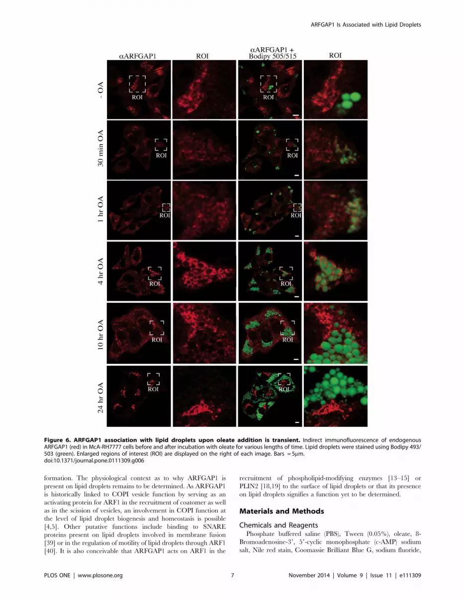
To investigate when ARFGAP1 redistributes to lipid droplet
structures, McA-RH7777 cells were incubated with oleate for
various lengths of time (Fig. 6). This revealed that though most of
ARFGAP1 localized to lipid droplets 4 hours after addition of
oleate, much less was observed at later time points suggesting a
transient association. At 10 hours after oleate addition, over half of
ARFGAP1 still remained associated with lipid droplet structures
whereas after 24 hours, ARFGAP1 was exclusively located at the
juxta nuclear Golgi area. The shift in distribution of endogenous
ARFGAP1 from the Golgi apparatus to lipid droplets was
quantified 4 hours after addition of oleate in 350 cells per
experiment in 5 independent experiments using ImageJ 1.45 s
software (see Materials and Methods). In HepG2 and McA-
RH7777 cells, the percentage of cells that had ARFGAP1
associated with lipid droplets increased from 0% to 80% and
0% to 75% at 4 hours after addition of oleate, respectively (Fig. 7-
left). About 65% and 50% of Bodipy-stained structures were
associated with ARFGAP1 per cell at 4 hours after oleate addition
in HepG2 and McA-RH7777 cells, respectively (Fig. 7, middle).
Addition of oleate caused a marked increase in the area of Bodipy-
positive lipid droplets corresponding to a 10 and 5 fold increase in
HepG2 and McA-RH7777 cells, respectively (Fig. 7, right). To
confirm that ARFGAP1 indeed localizes to lipid droplets upon
addition of oleate, HepG2 cells were fixed and processed for
immuno-gold transmission electron microscopy (see Materials and
Methods). Figure 8 shows single antigen gold-labeled thin frozen
sections after 4 hours incubation with oleate incubated with a
control antibody (MOCK), a goat polyclonal antibody to ADRP
(PLIN2), a lipid droplet marker, or a rabbit polyclonal antibody to
ARFGAP1 [30]. Arrowheads in black and white point to gold
particles detected revealing staining at the periphery of lipid
droplets of both ADRP and ARFGAP1. On occasion, gold
particles for ADRP were detected also within the lipid droplet
structure.
Figure 2. Knockdown of ARFGAP1 affects lipid droplet formation. Shown in A and B are HepG2 cells transfected with siRNA specific forARFGAP1 or with a scrambled sequence (Mock) and 24 hours post transfection, incubated with 0.5 mM oleate to promote lipid droplet formation.Cells were then fixed 1 hour or 4 hours after addition of oleate and stained to reveal endogenous ARFGAP1 (red) and lipid droplets (green) using arabbit polyclonal to ARFGAP1 and Bodipy 493/503, respectively. Bars = 5mm. In C, left, lipid droplets per cell were quantified as total area ofBodipy493/503 stained droplets per cell expressed as a percentage of the total area of Bodipy493/503 stained droplets in cells transfected with siRNAspecific to ARFGAP1 compared to Mock transfected cells. In C, right, the number of Bodipy493/503 stained lipid droplets per cell was comparedbetween cells transfected with siRNA specific to ARFGAP1 and Mock-transfected cells. P-values were obtained using the Mann–Whitney rank sum testcomparing the number (P,0.001) or average size (P,0.001) of Bodipy-stained lipid droplets in Mock transfected cells with those of transfected cells.In D, western blots of ARFGAP1 and calnexin. Extracts from cells transfected either with scrambled siRNA (Mock, M) or siRNA specific to ARFGAP1 forknockdown (KD) were subjected to SDS-PAGE, transfer to PVDF membranes and blotted against ARFGAP1 and Calnexin using specific antibodies.doi:10.1371/journal.pone.0111309.g002
ARFGAP1 Is Associated with Lipid Droplets
PLOS ONE | www.plosone.org 4 November 2014 | Volume 9 | Issue 11 | e111309

Lipid droplet association of ARFGAP1 is reversed uponaddition of cAMP
Based on above data, it appears that most if not all ARFGAP1
associates transiently with lipid droplets peaking at 4 hours after
addition of oleate. Such transient association suggests a dynamic
recruitment of ARFGAP1 to lipid droplets upon oleate uptake,
possibly regulated by cAMP effector proteins such as cAMP-
dependent protein kinase (PKA) shown to affect lipid homeostasis
[35]. To test this, McA-RH7777 cells were incubated in the
presence of oleate for 4 hours (Fig. 9) followed by addition of
cAMP. Before addition of cAMP, most if not all of endogenous
ARFGAP1 localized to peri-lipid droplet structures positive for
Bodipy (Fig. 9 upper panels). As above, no discernable juxta
nuclear Golgi staining was observed. At 10 minutes after cAMP
addition, staining for endogenous ARFGAP1 was detected in the
juxta nuclear Golgi region as well as around lipid droplet
structures (Fig. 9-middle panels). At 30 minutes after cAMP
addition, staining for endogenous ARFGAP1 was mainly detected
in the juxta nuclear Golgi apparatus with some staining remaining
around lipid droplet structures. At these time points (10 and
30 minutes), no discernable decrease in the overall amount of lipid
droplets was observed (Fig. 9).
ARFGAP1 associates with lipid droplets in human liverThough it appears that ARFGAP1 localizes transiently to lipid
droplets structures at 4 hours after addition of oleate in both McA-
RH7777 and HepG2, it was important to rule out that such a shift
in steady state distribution reflects an unphysiological condition
induced by a sudden influx of oleate. We therefore examined
ARFGAP1 in human hepatic tissue. Thin frozen sections were
generated from liver tissue resected from patients undergoing
surgery for removal of metastases of the liver as well as from donor
livers obtained through Transplant Quebec (http://
transplantquebec.ca/en) (see Materials and Methods) and subject-
ed to indirect immunofluorescence. Figure 10A shows a frozen
section stained for endogenous ARFGAP1 (aARFGAP1, left
panel), DAPI to reveal nuclear chromatin (middle panel) and
Bodipy 493/503 to stain for neutral lipids (right panel). Some of
the staining for ARFGAP1 decorates what appear to be large lipid
droplet structures often comparable or larger in size than the
nucleus. In this section, ARFGAP1 is seen both as a compact
Golgi-like staining as well as decorating lipid droplet-like structures
(arrowheads). The staining of endogenous ARFGAP1 was also
compared with the lipid droplet marker protein PLIN3 and
Bodipy 493/503 to stain for neutral lipids (Fig. 10B-right panel).
Note that staining of endogenous ARFGAP1 (Fig. 10B-left panel)
mostly but not fully overlap with that of PLIN3 (Fig. 10B-right
panel). In liver sections examined, overlapping staining between
ARFGAP1 and PLIN3 approached 60% based on 138 lipid
droplets. However, the appearance of ARFGAP1 on lipid droplet
structures in a given liver section is not uniform nor is the overall
frequency of ARFGAP1 positive lipid droplet structures when
compared between livers of different patients/donors. Neverthe-
less, the appearance of ARFGAP1 on lipid droplet structures
suggests a physiological relevance prompting further studies.
Discussion
Previous proteomics data has included ARFGAP1 as a potential
lipid droplet–associated protein under conditions where GTP
hydrolysis was inhibited through addition of the non-hydrolyzable
GTP analogue, GTPcS [36]. More recent data based on
interference RNA-based experiments suggests that ARFGAPs 1-
3 promote lipid droplet formation in HeLa cells [37]. In that study,
similar effects were also observed upon knockdown of COPB1,
GBF1 and class II ARFs (ARF4 and ARF5). The authors further
Figure 3. ARFGAP1 relocates from the Golgi apparatus to lipiddroplets upon addition of oleate. HepG2 cells were fixed andprocessed for indirect immunofluorescence before (-OA) and 4 hoursafter addition of 0.5 mM oleate and stained to reveal endogenousARFGAP1 (green) and lipid droplets (red) using a rabbit polyclonal toARFGAP1 and Bodipy 558/568 C12, respectively. Bars = 5mm.doi:10.1371/journal.pone.0111309.g003
Figure 4. ARFGAP1 co-localizes with PLIN3 on lipid droplets.Endogenous ARFGAP1 staining (red) was compared with antibodystaining of endogenous PLIN3 (green). An enlarged region of interest(ROI) is shown in the bottom right panel of the merged image(ARFGAP1/PLIN3). Bar = 5mm.doi:10.1371/journal.pone.0111309.g004
ARFGAP1 Is Associated with Lipid Droplets
PLOS ONE | www.plosone.org 5 November 2014 | Volume 9 | Issue 11 | e111309

concluded, based on indirect immunofluorescence data, that
COPB1, GBF1, ARF4 and ARFGAP1 do not associate with lipid
droplets, at steady state [37]. This is in contrast to recent studies
suggesting a role for COPI components in establishing/maintain-
ing interconnections between lipid droplets and the ER as well as
in the formation of vesicles budding of the surface of the lipid
droplet [4,5]. Our study shows that ARFGAP1 associates
transiently with lipid droplets in cultured hepatoma cells (HepG2
and McA-RH7777) upon addition of oleate and at steady state on
lipid droplets of some but not all hepatocytes of human liver.
Indirect immunofluorescence staining of endogenous ARFGAP1
or fluorescence from expressed ARFGAP1YFP is clearly visible as
peri-droplet staining but also on adjacent juxta-posed structures.
Though immuno-gold labeling of ARFGAP1 show gold particles
decorating the perimeter of the lipid droplet, we cannot formally
rule out that such gold particles label ARFGAP1 on structures that
reside very close to the surface of the lipid droplet (the combined
size of primary and secondary antibodies gives rise to an
uncertainty in distance of up to 30nm between the antigen and
the gold particle).
Based on the evidence at hand, we suggest that ARFGAP1
association with lipid droplets of hepatocytes of human liver
indicates a physiological and functional context between ARF-
GAP1 and lipid droplets. Indeed, over expression of ARFGAP1
promotes lipid droplet formation in both HeLa and HepG2 cells
whereas knockdown experiments of ARFAGP1 impair lipid
droplet formation in both HepG2 and McA-RH7777 cells. It is
possible that over-expression of ARFGAP1 causes cellular stress
and that stress in turn promotes lipid droplet formation [38]
independent from ARFGAP1 function. Such stress could either be
through its catalytic activity or lipid-binding properties. As such,
we interpret our over-expression results with caution. It is further
possible that knockdown experiments cause cellular stress.
However, the phenotype is here the opposite in that knockdown
of ARFGAP1 impairs lipid droplet formation. As components of
the COPI machinery have been linked to lipid droplet biogenesis,
the phenotype observed may be explained through an impaired
Golgi-derived delivery of components needed for lipid droplet
formation [1–3], maintenance of LD-ER connections and/or
formation of lipid-droplet derived vesicles [4,5]. It is further
possible that addition of oleate results in cellular stress and that this
prompts a transient relocation of ARFGAP1 to lipid droplets.
However, even though relocation is transient, addition of cyclic
AMP reverses ARFGAP1 back to the Golgi apparatus indicating
and underlying physiological process. This is supported by the
observation that ARFGAP1 localizes to lipid droplets at steady
state in some but not all hepatocytes of human livers. As these
livers all have varying degrees of steatosis, it will be of interest in
the future to determine whether ARFGAP1 localization to lipid
droplets correlates with certain pathophysiological states. In
conclusion, we have demonstrated that ARFGAP1 associates
transiently or at steady state with lipid droplets and to structures in
close juxtaposition to lipid droplets in cell culture and human liver
tissue, respectively, and that ARFGAP1 affects lipid droplet
Figure 5. Lipid droplet localization of ARFGAP1 is distinct from other endogenous Golgi and ER markers. Indirect immunofluorescenceof COPB1 in HepG2 cells, ARF1 in McA-RH7777 cells, TMED7 and CANX in HepG2 cells before (4 right panels) and 4 hours after addition of 0.5 mMoleate. Enlarged regions of interest are shown in the 4 left panels. Bars = 7mm.doi:10.1371/journal.pone.0111309.g005
ARFGAP1 Is Associated with Lipid Droplets
PLOS ONE | www.plosone.org 6 November 2014 | Volume 9 | Issue 11 | e111309
formation. The physiological context as to why ARFGAP1 is
present on lipid droplets remains to be determined. As ARFGAP1
is historically linked to COPI vesicle function by serving as an
activating protein for ARF1 in the recruitment of coatomer as well
as in the scission of vesicles, an involvement in COPI function at
the level of lipid droplet biogenesis and homeostasis is possible
[4,5]. Other putative functions include binding to SNARE
proteins present on lipid droplets involved in membrane fusion
[39] or in the regulation of motility of lipid droplets through ARF1
[40]. It is also conceivable that ARFGAP1 acts on ARF1 in the
recruitment of phospholipid-modifying enzymes [13–15] or
PLIN2 [18,19] to the surface of lipid droplets or that its presence
on lipid droplets signifies a function yet to be determined.
Materials and Methods
Chemicals and ReagentsPhosphate buffered saline (PBS), Tween (0.05%), oleate, 8-
Bromoadenosine-39, 59-cyclic monophosphate (c-AMP) sodium
salt, Nile red stain, Coomassie Brilliant Blue G, sodium fluoride,
Figure 6. ARFGAP1 association with lipid droplets upon oleate addition is transient. Indirect immunofluorescence of endogenousARFGAP1 (red) in McA-RH7777 cells before and after incubation with oleate for various lengths of time. Lipid droplets were stained using Bodipy 493/503 (green). Enlarged regions of interest (ROI) are displayed on the right of each image. Bars = 5mm.doi:10.1371/journal.pone.0111309.g006
ARFGAP1 Is Associated with Lipid Droplets
PLOS ONE | www.plosone.org 7 November 2014 | Volume 9 | Issue 11 | e111309

Figure 7. Quantification of ARFGAP1 associated with lipid droplets. Endogenous ARFGAP1 as revealed by indirect immunofluorescence wasquantified before (-OA) and 4 hours after addition of oleate (+OA) in HepG2 and McA-RH7777 cells expressed as % cells with ARFGAP1 staining (asrevealed through indirect immunofluorescence) or % Bodipy-stained droplets associated with ARFGAP1 per cell. On the right, total area of Bodipy-stained lipid droplets per mm2 per cell.doi:10.1371/journal.pone.0111309.g007
Figure 8. ARFGAP1 localizes to lipid droplets as revealed by immuno-gold transmission electron microscopy. Thin frozen sections ofHepG2 cells fixed 4 hours after addition of 0.5 mM oleate were incubated individually with antibodies to either PLIN2 (white arrowheads) or ARFGAP1(black arrowheads) followed by gold-conjugated secondary antibodies to reveal endogenous proteins at the ultra structural level. Bars = 100 nm.doi:10.1371/journal.pone.0111309.g008
ARFGAP1 Is Associated with Lipid Droplets
PLOS ONE | www.plosone.org 8 November 2014 | Volume 9 | Issue 11 | e111309

EDTA, Tris-HCl, sucrose, tricine, NaCl, Na2HPO4, KH2PO4,
CaCl2, MgCL2, Tween 20, Fish skin gelatin, Saponin, PFA,
Mowiol, ammonium choride, lead citrate, bromophenol blue,
Biomax X-Omat XAR or MR films from Eastman Kodak, 10 nm
colloidal gold-affinity purified rabbit anti-Goat, ACN, gelatin,
formic acid, and sodium citrate were all from Sigma-Aldrich
(Oakville, Ontario, Canada). Acetone, PVDF, KCl, water (LCMS
grade), DMSO and trypsin were from Fisher Scientific (Quebec,
Canada). FuGENE HD Transfection Reagent, SDS and Tris base
were from Roche Ltd (Laval, Qc, Canada). Sodium cacodylate
and Epon 812 were from Mecalab Ltd (Quebec, Canada). DTT
was from Biomol (Hamburg, Germany). ECL detection kit was
from GE Healthcare (Baie d’Urfe, Quebec, Canada). Thirty
percent (wt/vol) acrylamide/0.8% (wt/vol) bis-acrylamide solution
was from Bio-Rad (Mississauga, On. Canada). Uranyl acetate,
glutaraldehyde, and glycerol were from Merck (Kirkland, Qc,
Canada). Osmium tetroxide was from Agar Scientific (Essex,
United Kingdom). Laemmli 2 X from Bio-Rad (Ontario, Canada).
ProLong Gold Antifade, MEM, GIBCO DMEM, FBS, penicillin
streptomyocin, Bodipy 493/503, Bodipy 558/568 C12, glutamine,
Lipofectamine siRNA was purchased from Invitrogen (Carlsbad,
CA). 12 nm colloidal gold-affiniPure Goat anti-Rabbit IgG (H+L)
was purchased from Cedar Lane laboratories (Ontario, Canada).
ARFGAP1-EYFP was kindly supplied by Dr. J. Lippincott-
Schwartz (National Institutes of Health, Bethesda, MD). The
mouse monoclonal antibody CMIA10 against COPB1 and rabbit
polyclonal antibodies to ARFGAP1 ARF1, TMED7 and Calnexin
have all been described previously [10,30,33,34,41]. Goat
polyclonal antibodies to PLIN3 (Tip47) and PLIN2 (ADRP) were
purchased from Santa Cruz Biotechnologies Inc (Santa Cruz, Ca,
USA). AlexaFluor 488 and AlexaFluor 568 secondary antibodies
were purchased from Invitrogen (Carlsbad, CA). HRP-labeled
polyclonal antibodies to rabbit IgG were purchased from Dianova
(Hamburg, Germany). Protein A-HRP conjugate was purchased
from Bio-Rad (Ontario, Canada).
Indirect immunofluorescence and confocal microscopyHeLa, McA-RH7777 and HepG2 cells were grown in DMEM
supplemented with 10% FBS, penicillin (100 U/ml) and strepto-
mycin (100 mg/ml). Lipid droplet formation was induced by
adding 0.5 mM oleate (from 100 mM stock in ethanol) directly to
media. In selected experiments, Bodipy 558/568 C12 was added at
the same time as oleate to a final concentration of 3 mM in order
to stain newly forming lipid droplets. cAMP was added cells to a
final concentration of 37.5 mg/ml. Cells were processed for
indirect immunofluorescence as described previously [34]. Post
staining of neutral lipid droplets using 1 mg/ml Bodipy 493/503
was performed as described in [42]. All images were acquired
Figure 9. Cyclic AMP reverses lipid droplet association of ARFGAP1. McA-RH7777 cells were incubated with 0.5 mM oleate for 4 hours afterwhich cyclic AMP (cAMP) was added to a final concentration of 37.5mg/ml. The cells were simultaneously stained for endogenous ARFGAP1 (red) andneutral lipid with Bodipy 493/503 (green). Bars = 5mm.doi:10.1371/journal.pone.0111309.g009
ARFGAP1 Is Associated with Lipid Droplets
PLOS ONE | www.plosone.org 9 November 2014 | Volume 9 | Issue 11 | e111309

using a LSM 700 series microscopy system (Carl Zeiss) fitted with a
Plan-Apochromat 63x/1.40 Oil DIC objective in sequential
scanning mode with the pinhole set to obtain an optical section
of about 0.8 mm in both channels (approximately 1 Airy unit). For
Alexa 488 and Bodipy 493/503, a 488 nm Argon ion laser was
used and emitted fluorescence was filtered through a 505 to
530 nm band-pass. Bodipy 558/568 C12 and Alexa 568 was
excited with a 561 nm DPSS laser and emitted fluorescence was
filtered through a 585 to 690 nm band-pass. Tissue cryosections
(see below) were cut from isopentane frozen tissue cubes at a
thickness of approximately 6 microns and air dried for an hour
then fixed in 4% paraformaldehyde solution for 30 minutes.
Subsequent indirect immunofluorescence was performed as above.
For live cell imaging, cells were grown and imaged as above in
MatTek dishes (MatTek Corporation, Ashland, MA, USA) and
imaged as above under normal tissue culture conditions (i.e. CO2,
humidity and temperature). Prior to live cell imaging, cells were
transfected with FuGENE HD according to manufacturers
instructions (Roche Ltd, Laval, QC, Canada). EYFP was excited
using a 512 nm laser line from an Argon ion laser and emission
was filtered through an LP535 band-pass.
siRNA transfection, lipid droplet staining and westernblotting
RNA oligos were transfected with the siRNAMAX transfection
reagent according to manufacturer instructions (Invitrogen,
Carlsbad, CA). ARFGAP1 was knocked down using a sequence
previously published [20,43]. A scrambled sequence was used as a
negative control. At 24 hours post transfection, cells were
incubated with 0.5 mM oleate for 1 or 4 hours or left untreated.
Cells were then fixed, imaged and processed as above. Lipid
droplets were stained with Bodipy 493/503 (1 mg/ml) for
30 minutes. Western blotting was carried out as described
previously [30].
Image AnalysisFluorescence levels were coded with a gray scale representing
pixel intensity of 25-255. For semi-quantitation, in all cases
background (pixel values outside the cells) was relatively low (,
10% of the signal). Therefore, to minimize data manipulation,
background subtraction was not performed. All images were
processed and analyzed with ImageJ 1.45 s (National Institutes of
Health, USA), where the mean intensities of the Golgi region and
lipid images were measured to quantify the lipid droplets and the
ARFGAP1 associated with the lipid droplets in each cell in the
field of view. Quantitation of fluorescence was made from 150 to
350 cells for each experiment (10 sites/slide). Each experiment was
performed at least three times. For quantification, images of HeLa,
McA-RH7777 or HepG2 cells were processed with a binary
segmentation algorithm. Briefly, after a sharpening, brightness/
contrast adjustment (equal values for all images) and find edges, an
Otsu’s method thresholding step was run to automatically perform
a reduction of a gray level image to a binary image. Watershed
processing to identify solitary particles followed. Finally, lipid
droplets (Bodipy 493/503 or 558/568) or Golgi region (TMED7
or ARFGAP1) were identified with the generic ‘‘analyze particles’’
function of the ImageJ software with the following settings: for the
Golgi apparatus size from 100 to 10,000 pixels; and for the lipid
droplets size from 1 to 2500 pixels and circularity from zero to one
(final values were expressed in physical size square units); outlines
as well as measurement results displayed and measurements on the
edges excluded. The summary of the results was done through the
‘ROI’ (Region Of Interest) Manager that is a tool for working with
multiple selections where areas to be evaluated can be specified
editing out unwanted elements. For each image, the average
particle size and cumulative measured area for all particles
(‘‘area’’) were reported. The obtained information was used to
calculate the ratio of lipid droplets per cells as a measure of lipid
storage (‘‘number of Bodipy-stained droplet/cell’’ or ‘‘of total area
Bodipy-stained droplet/cell’’) or ARFGAP1 associated with lipid
Figure 10. ARFGAP1 associates with lipid droplets at steady state in human livers. In A and B, thin frozen sections of two human liverswere fixed and processed for indirect immunofluorescence as described in Materials and Methods. Endogenous ARFGAP1 was revealed as in Figure 4.Nuclear DNA was in A stained with DAPI and lipid droplets in A and B with Bodipy 493/503. Bar = 10mm. In B, endogenous ARFGAP1 and PLIN3 wererevealed as in Figure 4. Pseudo colors were assigned to each panel. Bar = 15mm.doi:10.1371/journal.pone.0111309.g010
ARFGAP1 Is Associated with Lipid Droplets
PLOS ONE | www.plosone.org 10 November 2014 | Volume 9 | Issue 11 | e111309

droplets. All data were imported into Sigma Plot (SPSS Inc.). The
Mann–Whitney rank sum test was used for statistical analysis. A Pvalue of less than 0.05 was considered to be significant.
Transmission electron microscopyHeLa cells were fixed using a double fixation protocol with
osmium and tannic acid [44]. Samples were dehydrated in graded
ethanol series, and embedded in Epon 812 (Serva). After 48 h at
60uC, ultra-thin sections (60 nm) were cut and mounted on grids.
Samples were examined on a LEO 912 OMEGA (Energy Filter
Transmission Electron Microscope, Zeiss) at 120 kV accelerating
voltage. Digital images were obtained through a side-mounted
MegaView III TEM CCD camera.
Immuno-gold transmission electron microscopyCryosections were prepared using an adaptation of the standard
protocol developed by Tokuyasu [45]. Cells were fixed with a mix
of 4% Formaldehyde and 0.1% Glutaraldehyde for 2 hours at 4uCand washed twice with PBS. A small volume of 1% gelatin in PBS
was then added after which cells were removed from the dish using
a rubber policeman cell scraper and transferred to a 1.5 ml
Eppendorf tube and centrifuged for 30–60 sec at 1500 rpm. The
1% gelatin was then replaced with a 10% gelatin solution at 37uC.
Cells were then resuspended and centrifuged as above after which
excess gelatin was removed. Cells in gelatin were then solidified on
ice for 30 minutes. At 4uC, the bottom of the Eppendorf tube was
then cut off and 1-mm gelatin blocks were prepared from the pellet
and incubated end over end in phosphate buffered 2.3 M sucrose
overnight at 4uC. After trimming, ultrathin sections (75 nm) were
prepared at 2120uC. Sections were immediately transferred to a
1:1 mixture of 2% methylcellulose and 2.3 M sucrose [46] and
2 ml of 4% uranyle acetate, and transferred to copper grids bearing
a carbon-coated Formvar supporting film. Labeling was carried
out at room temperature using specific antibodies and gold
conjugates diluted in 0.5% fish skin gelatin in PBS. 10 nm
colloidal gold-affiniPure Rabbit anti-Goat and 12 nm colloidal
gold-affiniPure Goat anti-Rabbit IgG (H+L) were used to detect
primary polyclonal antibodies. Sections were then embedded in a
thin layer of 2% methylcellulose with 0.4% uranyl acetate, pH 4.0,
air-dried, examined and photographed using a Philips/FEI
Tecnai12 120 kV Transmission Electron Microscope equipped
with an AMT XR80C CCD camera system.
Processing of human liver tissueDonor material was obtained through Transplant Quebec
(http://transplantquebec.ca/en) and healthy resected liver mate-
rial from patients undergoing liver surgery to remove liver
metastasis. For resected material, removed tissue was immediately
perfused with ice-cold Wisconsin solution, and within 30 minutes
on ice, inspected by an on site pathologist, then processed for
biobanking. For donor material, liver slices from discarded livers
were perfused using ice-cold Wisconsin solution then processed for
biobanking. For biobanking, liver tissue was diced into smaller
pieces and placed for 5 minutes in isopentane precooled to 245uCin order to preserve morphology. Samples were then wrapped in
tinfoil and kept frozen at 245uC until sectioned.
Ethics statementAll handling and use of human patient and donor material was
performed under IRB ethics approved protocols: "Liver Disease –
Biobank" # 11-066-SDR and ‘‘Human Hepatic Tissue Organel-
lar Isolation and Characterization" # 11-196-SDR filed at the
McGill University Health Centre, Montreal, Canada in the name
of Dr. Peter Metrakos. Patient and donor consent was obtained in
written form for all human samples collected.
Supporting Information
Figure S1 Localization of ARFGAP1YFP in HeLa cellsover the course of 40 minutes. HeLa cells expressing
ARFGAP1YFP were imaged for 40 minutes to monitor the
movement of ARFGAP1YFP-positive structures. The arrow shows
the saltatory movement of a ring formed and lipid droplet-like
ARFGAP1YFP-positive structure that moves in and out of the juxta
nuclear Golgi region. Time of events is expressed in seconds.
Images were recorded using a Zeiss LSM 510 META equipped
with a 37uC climate chamber. Bar = 10mm.
(TIF)
Acknowledgments
We wish to thank Transplant Quebec with donors and their families and
patients consenting to liver resection. We thank the FEMR team (http://
www.medicine.mcgill.ca/femr/) and Jeannie Mui, for use of the transmis-
sion electron microscope and preparation of thin frozen sections,
respectively.
Author Contributions
Conceived and designed the experiments: TN JFR JG AS. Performed the
experiments: JFR JG HA FK AHYW AM LA AF AL SBF SN. Analyzed
the data: TN. Contributed reagents/materials/analysis tools: TN PM
JJMB. Wrote the paper: TN JFR JG. Initial observation of ARFGAP1
association with lipid droplets: JFR LA TN. Human patient and donor
material, biobanking, resection and characterization: AS AL PM.
References
1. Guo Y, Walther TC, Rao M, Stuurman N, Goshima G, et al. (2008) Functional
genomic screen reveals genes involved in lipid-droplet formation and utilization.
Nature 453: 657–661.
2. Beller M, Sztalryd C, Southall N, Bell M, Jackle H, et al. (2008) COPI complex
is a regulator of lipid homeostasis. PLoS Biol 6: e292.
3. Soni KG, Mardones GA, Sougrat R, Smirnova E, Jackson CL, et al. (2009)
Coatomer-dependent protein delivery to lipid droplets. J Cell Sci 122: 1834–
1841.
4. Thiam AR, Antonny B, Wang J, Delacotte J, Wilfling F, et al. (2013) COPI buds
60-nm lipid droplets from reconstituted water-phospholipid-triacylglyceride
interfaces, suggesting a tension clamp function. Proc Natl Acad Sci U S A 110:
13244–13249.
5. Wilfling F, Thiam AR, Olarte MJ, Wang J, Beck R, et al. (2014) Arf1/COPI
machinery acts directly on lipid droplets and enables their connection to the ER
for protein targeting. Elife 3: e01607.
6. Kahn RA (2009) Toward a model for Arf GTPases as regulators of traffic at the
Golgi. FEBS Lett 583: 3872–3879.
7. Kahn RA, Randazzo P, Serafini T, Weiss O, Rulka C, et al. (1992) The amino
terminus of ADP-ribosylation factor (ARF) is a critical determinant of ARF
activities and is a potent and specific inhibitor of protein transport. J Biol Chem
267: 13039–13046.
8. Balch WE, Kahn RA, Schwaninger R (1992) ADP-ribosylation factor is required
for vesicular trafficking between the endoplasmic reticulum and the cis-Golgi
compartment. J Biol Chem 267: 13053–13061.
9. Donaldson JG, Kahn RA, Lippincott-Schwartz J, Klausner RD (1991) Binding
of ARF and beta-COP to Golgi membranes: possible regulation by a trimeric G
protein. Science 254: 1197–1199.
10. Lanoix J, Ouwendijk J, Lin CC, Stark A, Love HD, et al. (1999) GTP hydrolysis
by arf-1 mediates sorting and concentration of Golgi resident enzymes into
functional COP I vesicles. EMBO J 18: 4935–4948.
11. Malsam J, Gommel D, Wieland FT, Nickel W (1999) A role for ADP
ribosylation factor in the control of cargo uptake during COPI-coated vesicle
biogenesis. FEBS Lett 462: 267–272.
ARFGAP1 Is Associated with Lipid Droplets
PLOS ONE | www.plosone.org 11 November 2014 | Volume 9 | Issue 11 | e111309

12. Pepperkok R, Whitney JA, Gomez M, Kreis TE (2000) COPI vesicles
accumulating in the presence of a GTP restricted arf1 mutant are depleted of
anterograde and retrograde cargo. J Cell Sci 113 (Pt 1): 135–144.
13. Cockcroft S, Thomas GM, Fensome A, Geny B, Cunningham E, et al. (1994)
Phospholipase D: a downstream effector of ARF in granulocytes. Science 263:
523–526.
14. Brown HA, Gutowski S, Moomaw CR, Slaughter C, Sternweis PC (1993) ADP-
ribosylation factor, a small GTP-dependent regulatory protein, stimulates
phospholipase D activity. Cell 75: 1137–1144.
15. Godi A, Pertile P, Meyers R, Marra P, Di Tullio G, et al. (1999) ARF mediates
recruitment of PtdIns-4-OH kinase-beta and stimulates synthesis of
PtdIns(4,5)P2 on the Golgi complex. Nat Cell Biol 1: 280–287.
16. Boman AL, Zhang C, Zhu X, Kahn RA (2000) A family of ADP-ribosylation
factor effectors that can alter membrane transport through the trans-Golgi. Mol
Biol Cell 11: 1241–1255.
17. Dell’Angelica EC, Puertollano R, Mullins C, Aguilar RC, Vargas JD, et al.
(2000) GGAs: a family of ADP ribosylation factor-binding proteins related to
adaptors and associated with the Golgi complex. J Cell Biol 149: 81–94.
18. Nakamura N, Akashi T, Taneda T, Kogo H, Kikuchi A, et al. (2004) ADRP is
dissociated from lipid droplets by ARF1-dependent mechanism. Biochem
Biophys Res Commun 322: 957–965.
19. Nakamura N, Banno Y, Tamiya-Koizumi K (2005) Arf1-dependent PLD1 is
localized to oleic acid-induced lipid droplets in NIH3T3 cells. Biochem Biophys
Res Commun 335: 117–123.
20. Asp L, Kartberg F, Fernandez-Rodriguez J, Smedh M, Elsner M, et al. (2009)
Early stages of Golgi vesicle and tubule formation require diacylglycerol. Mol
Biol Cell 20: 780–790.
21. Fernandez-Ulibarri I, Vilella M, Lazaro-Dieguez F, Sarri E, Martinez SE, et al.
(2007) Diacylglycerol is required for the formation of COPI vesicles in the Golgi-
to-ER transport pathway. Mol Biol Cell 18: 3250–3263.
22. Antonny B, Huber I, Paris S, Chabre M, Cassel D (1997) Activation of ADP-
ribosylation factor 1 GTPase-activating protein by phosphatidylcholine-derived
diacylglycerols. J Biol Chem 272: 30848–30851.
23. Bigay J, Gounon P, Robineau S, Antonny B (2003) Lipid packing sensed by
ArfGAP1 couples COPI coat disassembly to membrane bilayer curvature.
Nature 426: 563–566.
24. Cukierman E, Huber I, Rotman M, Cassel D (1995) The ARF1 GTPase-
activating protein: zinc finger motif and Golgi complex localization. Science 270:
1999–2002.
25. Lee SY, Yang JS, Hong W, Premont RT, Hsu VW (2005) ARFGAP1 plays a
central role in coupling COPI cargo sorting with vesicle formation. J Cell Biol
168: 281–290.
26. Bai M, Gad H, Turacchio G, Cocucci E, Yang JS, et al. (2011) ARFGAP1
promotes AP-2-dependent endocytosis. Nat Cell Biol 13: 559–567.
27. Stafa K, Trancikova A, Webber PJ, Glauser L, West AB, et al. (2012) GTPase
activity and neuronal toxicity of Parkinson’s disease-associated LRRK2 is
regulated by ArfGAP1. PLoS Genet 8: e1002526.
28. Xiong Y, Yuan C, Chen R, Dawson TM, Dawson VL (2012) ArfGAP1 is a
GTPase activating protein for LRRK2: reciprocal regulation of ArfGAP1 by
LRRK2. J Neurosci 32: 3877–3886.
29. Aoe T, Cukierman E, Lee A, Cassel D, Peters PJ, et al. (1997) The KDEL
receptor, ERD2, regulates intracellular traffic by recruiting a GTPase-activatingprotein for ARF1. EMBO J 16: 7305–7316.
30. Lanoix J, Ouwendijk J, Stark A, Szafer E, Cassel D, et al. (2001) Sorting of Golgi
resident proteins into different subpopulations of COPI vesicles: a role forArfGAP1. J Cell Biol 155: 1199–1212.
31. Ou WJ, Cameron PH, Thomas DY, Bergeron JJ (1993) Association of foldingintermediates of glycoproteins with calnexin during protein maturation. Nature
364: 771–776.
32. Diaz E, Pfeffer SR (1998) TIP47: a cargo selection device for mannose 6-phosphate receptor trafficking. Cell 93: 433–443.
33. Duden R, Griffiths G, Frank R, Argos P, Kreis TE (1991) Beta-COP, a 110 kdprotein associated with non-clathrin-coated vesicles and the Golgi complex,
shows homology to beta-adaptin. Cell 64: 649–665.34. Fullekrug J, Suganuma T, Tang BL, Hong W, Storrie B, et al. (1999)
Localization and recycling of gp27 (hp24gamma3): complex formation with
other p24 family members. Mol Biol Cell 10: 1939–1955.35. Jungas RL (1966) Role of cyclic-3’,5’-amp in the response of adipose tissue to
insulin. Proc Natl Acad Sci U S A 56: 757–763.36. Bartz R, Zehmer JK, Zhu M, Chen Y, Serrero G, et al. (2007) Dynamic activity
of lipid droplets: protein phosphorylation and GTP-mediated protein translo-
cation. J Proteome Res 6: 3256–3265.37. Takashima K, Saitoh A, Hirose S, Nakai W, Kondo Y, et al. (2011) GBF1-Arf-
COPI-ArfGAP-mediated Golgi-to-ER transport involved in regulation of lipidhomeostasis. Cell Struct Funct 36: 223–235.
38. Gubern A, Barcelo-Torns M, Casas J, Barneda D, Masgrau R, et al. (2009) Lipiddroplet biogenesis induced by stress involves triacylglycerol synthesis that
depends on group VIA phospholipase A2. J Biol Chem 284: 5697–5708.
39. Bostrom P, Andersson L, Rutberg M, Perman J, Lidberg U, et al. (2007)SNARE proteins mediate fusion between cytosolic lipid droplets and are
implicated in insulin sensitivity. Nat Cell Biol 9: 1286–1293.40. Chen JL, Xu W, Stamnes M (2005) In vitro reconstitution of ARF-regulated
cytoskeletal dynamics on Golgi membranes. Methods Enzymol 404: 345–358.
41. Wada I, Rindress D, Cameron PH, Ou WJ, Doherty JJ 2nd, et al. (1991) SSRalpha and associated calnexin are major calcium binding proteins of the
endoplasmic reticulum membrane. J Biol Chem 266: 19599–19610.42. Gocze PM, Vahrson HW, Freeman DA (1994) Serum levels of squamous cell
carcinoma antigen and ovarian carcinoma antigen (CA 125) in patients withbenign and malignant diseases of the uterine cervix. Oncology 51: 430–434.
43. Frigerio G, Grimsey N, Dale M, Majoul I, Duden R (2007) Two human
ARFGAPs associated with COP-I-coated vesicles. Traffic 8: 1644–1655.44. Simionescu N, Simionescu M (1976) Galloylglucoses of low molecular weight as
mordant in electron microscopy. I. Procedure, and evidence for mordantingeffect. J Cell Biol 70: 608–621.
45. Tokuyasu KT (1973) A technique for ultracryotomy of cell suspensions and
tissues. J Cell Biol 57: 551–565.46. Liou W, Geuze HJ, Slot JW (1996) Improving structural integrity of cryosections
for immunogold labeling. Histochem Cell Biol 106: 41–58.47. Liu W, Duden R, Phair RD, Lippincott-Schwartz J (2005) ArfGAP1 dynamics
and its role in COPI coat assembly on Golgi membranes of living cells. J CellBiol 168: 1053–1063.
ARFGAP1 Is Associated with Lipid Droplets
PLOS ONE | www.plosone.org 12 November 2014 | Volume 9 | Issue 11 | e111309


